the sphingosine 1-phosphate receptor s1p₂ maintains the homeostasis of germinal center b cells and...
TRANSCRIPT

The sphingosine 1-phosphate receptor S1P2 maintains germinalcenter B cell homeostasis and promotes niche confinement
Jesse A. Green1, Kazuhiro Suzuki1,*,‡, Bryan Cho1,2,*, L. David Willison3,*,‡, Daniel Palmer3,Christopher D.C. Allen1,‡, Timothy H. Schmidt1, Ying Xu1, Richard L. Proia4, Shaun R.Coughlin3, and Jason G. Cyster1
1Howard Hughes Medical Institute and Department of Microbiology and Immunology, Universityof California San Francisco, 513 Parnassus Ave., San Francisco, CA 94143-0414, USA2Department of Dermatology, University of California San Francisco, 513 Parnassus Ave., SanFrancisco, CA 94143-0414, USA3Cardiovascular Research Institute, University of California San Francisco, 600 16th StreetS472D, San Francisco, CA 94143-2240, USA4Genetics of Development and Disease Branch, National Institute of Diabetes and Digestive andKidney Diseases, NIH, Bethesda, MD 20892, USA
AbstractSphingosine-1-phosphate receptor-2 (S1P2)-deficient mice develop diffuse large B celllymphoma. However, the role of S1P2 in normal germinal center (GC) physiology is unknown.Here we show that S1P2-deficient GC B cells outgrow their wild-type counterparts in chronically-established GCs. We find that S1P2-, G12–G13- and p115RhoGEF-mediated antagonism of Aktregulates cell viability and is required for growth control in chronically proliferating GCs. We alsofind that S1P2 inhibits GC B cell responses to follicular chemoattractants and helps confine cellsto the GC. Moreover, S1P2 overexpression promotes centering of activated B cells within thefollicle. We suggest that by inhibiting Akt activation and migration, S1P2 helps restrict GC B cellsurvival and localization to an S1P-low niche at the follicle center.
Germinal centers (GCs) are induced in response to T-dependent antigens and support eventsnecessary for antibody affinity maturation1,2. They arise in the center of B cell follicles andgrow from small numbers of starting B cells to a size of ~10,000 cells in a matter of days.Despite the rapid growth rate, GC size is tightly regulated, even in mesenteric lymph nodes(mLNs) and Peyer's Patches (PPs) where the responses are chronically stimulated by gutflora3. Apoptosis is an integral part of GC growth control as GC B cells are highly prone to
Address correspondence to: J.G.C. ([email protected]).*These authors contributed equally to this study‡Current addresses:K. Suzuki: WPI Immunology Frontier Research Center, Osaka University, 3-1 Yamada-oka, Suita, Osaka 565-0871, Japan.L. D. Willison: Semel Institute for Neuroscience and Human Behavior, University of California Los Angeles, 760 Westwood Plaza,Los Angeles, CA 90024, USA.C.D.C. Allen: Sandler-Newmann Foundation, Department of Microbiology and Immunology, University of California San Francisco,505 Parnassus Ave., San Francisco, CA 94143-0414, USA.Please provide author contributions statement.JAG, BC and JGC designed the experiments. JAG performed the bulk of the experiments and BC performed some of the earlyexperiments. KS and JAG performed the imaging experiments. CDCA and YX performed the initial gene expression analysis. THSand JAG performed the PTEN inhibitor experiments. DW and SRC generated the G12-deficient mice and DP and SRC generated theGna12−/−Gna13f/fMx1cre BM. RLP generated the S1P2-deficient mice. JAG, KS, BC, and JGC analyzed the data and JAG and JGCwrote the paper.
NIH Public AccessAuthor ManuscriptNat Immunol. Author manuscript; available in PMC 2012 January 1.
Published in final edited form as:Nat Immunol. ; 12(7): 672–680. doi:10.1038/ni.2047.
NIH
-PA Author Manuscript
NIH
-PA Author Manuscript
NIH
-PA Author Manuscript

apoptotic cell death and are strongly dependent on CD40L and other trophic factors4. Thesefactors act at least in part by maintaining expression of anti-apoptotic Bcl2-family proteins,including Mcl-1, that are critical for GC formation5. However, despite knowledge of keyrequirements for maintaining GC cell viability, the environmental cues involved inregulating GC size are not fully understood.
GCs are organized into dark and light zones by CXCL12 and CXCL13, respectively6.CXCL13 is present throughout the follicle and in the GC light zone; CXCL12 is presentwithin the dark zone6. Despite the important roles of these chemokines, combineddeficiency in the function of their receptors does not cause a complete loss of GCformation6. Another chemoattractant receptor, EBI2, is up-regulated in early-activated (pre-GC) B cells and functions in guiding these cells to the outer follicle7,8. EBI2 is down-regulated in GC B cells, a change that is important for GC B cells to access the folliclecenter7. However, in the absence of EBI2, GCs form in their normal location indicating thatadditional cues must act to promote clustering of GC-precursors at the follicle center.
Sphingosine-1-phosphate (S1P) is a metabolic intermediate made by all eukaryotic cell typesduring sphingolipid metabolism through the action of sphingosine kinase-1 (sphk-1) andsphk-29. S1P is secreted by some cell types. The extracellular lipid acts as a ligand for anyof five G-protein coupled receptors, S1P1–S1P59. Extracellular S1P is abundant (high nM toµM) in blood and lymph but has a half-life shorter than 15 minutes10 and although no directmeasurements of interstitial concentrations have been reported, indirect measurementsindicate they are very low11,12. Red blood cells and endothelial cells are important sourcesof circulatory S1P10,12,13. Two S1P phosphatases, three lipid phosphate phosphatases(LPPs) and S1P lyase can degrade S1P and catabolism plays a critical role in maintainingthe low interstitial concentrations14. S1P promotes egress of lymphocytes from lymphoidtissue into circulatory fluids. Whether sufficient amounts of interstitial S1P exist in thelymphoid parenchyma to regulate cell behavior has been unclear.
In this study we set out to define molecular cues involved in regulating GC size and GC Bcell clustering. We found that S1P2 was expressed by GC B cells and was necessary tomaintain control over the size of chronically-stimulated GCs. S1P2 and its downstreammediators G12, G13 and p115RhoGEF antagonized Akt signaling and cell viability. S1P2also inhibited GC B cell chemotaxis to follicular chemoattractants and helped to promoteconfinement of GC B cells to the GC. In addition, S1P2 overexpression in non-GC B cellspromoted their localization to the follicle center. Based on these studies, we propose a modelin which S1P signals through S1P2 to regulate GC B cell survival and positioning, thuspromoting GC homeostasis through dual roles.
ResultsUncontrolled growth of S1P2-deficient GCs
Genome-wide comparison of gene expression between follicular and GC B cells identifiedS1P2 as one of the most strongly induced genes in GC B cells15 (and data not shown), andthis differential expression was confirmed by qRT-PCR (Fig. 1a). When 8–12 week oldS1P2-deficient mice16 were immunized with T-dependent antigens, they appeared to mountGC responses of normal magnitude. However, analysis of one year-old S1P2-deficient micerevealed expansion of GC B cell numbers in mLNs (Fig. 1b) as well as an increase in total Band T cell numbers. In about half the animals GC B cell numbers reached as much as 100×normal, and the architecture of the LN was effaced (Fig. 1b, c). We speculate that thebimodality of GC expansion in these mice is due to cooperation between S1P2-deficiencyand secondary genetic events, resulting in a loss of GC homeostasis and development ofGC-type lymphoma. Similar observations were made in another S1P2-deficient mouse line
Green et al. Page 2
Nat Immunol. Author manuscript; available in PMC 2012 January 1.
NIH
-PA Author Manuscript
NIH
-PA Author Manuscript
NIH
-PA Author Manuscript

and the outgrowths were classified as diffuse large B cell lymphoma (DLBCL)17. Theseresults demonstrate a requirement for S1P2 in maintaining GC B cell homeostasis.
Growth advantage of S1pr2−/− cells in chronic GCsTo determine if the S1P2-requirement for GC B cell homeostasis was cell intrinsic westudied mixed bone marrow (BM) chimeras. In mice reconstituted with ~1:2 mixtures ofS1pr2−/− and wild-type BM, S1P2-deficient GC B cells over-accumulated relative to theirwild-type counterparts in mLNs, whereas follicular B cells were represented in proportion tothe BM chimerism (Fig. 1d, e and Supplementary Fig. 1). The outgrowth was even moreevident in mice reconstituted with ~1:9 mixtures of S1pr2−/− and wild-type BM, and wasevident in both mLNs and PPs (Fig. 1f). The outgrowth was not observed in chimeric miceimmunized intra-peritoneally with sheep red blood cells (SRBCs) to induce splenic GCs atday 8 post-immunization (Fig. 1e, f) or in splenic or mLN GCs induced 14 days after NP-CGG immunization (Supplementary Fig. 1). However, a gradual outgrowth of S1P2-deficient GC B cells was noted in the endogenous splenic response occurring inunimmunized mixed BM chimeras (Supplementary Fig. 1). This suggests that the outgrowthmay depend on the long-term persistence of the GCs rather than on unique properties ofmucosal tissues. The advantage for S1P2-deficient cells was dependent on S1P derived fromradiation-resistant cells, because it was reduced in polyinosine polycytidylic acid (polyI:C)treated Mx1-cre+Sphk1f/−Sphk2−/− hosts, animals that lack Sphk2 and are broadly deficientfor Sphk1 in IFNα/β-responsive cells (Fig. 1g).
S1P2 can couple to G12 and G1318. To test the role of these G-proteins in GC B cells, wegenerated Gna12-deficient mice and intercrossed them with Gna13f/f mice19 carrying theMx1-cre transgene. Animals treated to induce Cre expression were used as a source of G12–G13-double deficient BM cells. In mice reconstituted with a mixture of wild-type and G12–G13-deficient BM, follicular B cells reflected the BM chimerism20, while there wasoutgrowth of G12–G13-deficient B cells in mucosal GCs (Fig. 1h). These observationssuggest a selective role for G12 and G13 within GC B cells in mediating S1P2 signals. Thesmall GC B cell accumulation seen at day 8 in splenic SRBC responses was similar in thecontrol (G12-deficient) and G12–G13-double deficient groups and may reflect an effect ofG12 single-deficiency or influences of genes from the 129 background. A major effectorpathway of G12–G13 in lymphocytes is activation of p115RhoGEF (encoded by Arhgef1),leading to Rho activation21–23. 8–12 weeks after reconstitution with wild-type andArhgef1−/− BM, p115RhoGEF-deficient GC B cells outgrew wild-type cells, although inthis case the advantage was not focused in mucosal sites (Fig. 1i and Supplementary Fig. 1).These observations suggest that S1P2 signals through G12–G13 and Rho to maintain B cellhomeostasis in chronically stimulated GCs.
S1P2 regulates cell survival in GC B cellsThe rate of GC B cell proliferation, determined by measuring bromodeoxyuridine (BrdU)incorporation 0.5 and 6 hr after a single BrdU injection, was similar in S1P2-deficient andcontrol GC B cells (Supplementary Fig. 2). By contrast, the rate of apoptotic cell death,examined ex vivo by measuring active caspase-3 and DNA fragmentation was reduced inS1P2-deficient cells (Fig. 2a, b and Supplementary Fig. 2) and in G12–G13-deficient cells(Supplemental Fig. 2). Because S1P2 and Rho were suggested to negatively regulate Akt insome contexts24–27, we asked whether the activity of this pro-survival kinase was altered inthe absence of S1P2. Intracellular flow cytometry for the threonine 308 phosphorylated formof Akt (pAkt T308) showed that S1P2-deficient GC B cells had higher amounts of activekinase than control GC B cells isolated from the same mixed BM chimeras (Fig. 2c). Asimilar elevation in pAkt T308 was observed after 30 min incubation of wild-type GC Bcells with the S1P2 antagonist, JTE-013 (Fig. 2d) or when JTE-013 was added to the media
Green et al. Page 3
Nat Immunol. Author manuscript; available in PMC 2012 January 1.
NIH
-PA Author Manuscript
NIH
-PA Author Manuscript
NIH
-PA Author Manuscript

used to prepare the cell suspensions (Supplementary Fig. 2). The cell preparation andincubation was performed in the absence of added S1P, indicating that cells had boundsufficient ligand in vivo or during isolation to promote S1P2 signaling. Co-incubation withthe PI3K inhibitor, wortmannin, prevented the increase in pAkt (Fig. 2d). The S1P2-mediated inhibition of Akt activation was dependent on endogenous S1P, because pAkt waselevated in GC B cells from Sphk-deficient mice (Fig. 2e). GC B cells isolated from G12–G13 double-deficient and p115RhoGEF-deficient mice showed similar elevations of pAkt(Fig. 2f). The propensity of wild-type GC B cells to undergo prompt apoptosis in vitro limitsthe biochemical measurements that can be performed in these cells. To assess the S1P2downstream signals that regulate Akt we used the human GC B cell line, Ramos, whichexpresses S1P2 (data not shown). Ramos cells had high constitutive pAkt levels, which isnot unusual for transformed cell lines (Fig. 2g). Exposure of these cells to S1P led to areduction in pAkt, which was prevented by co-incubation with the S1P2 antagonist (Fig.2g). Treatment with Y27632, an inhibitor of the Rho downstream effector ROCK, preventedS1P from causing the full reduction in pAkt levels (Fig. 2h, i). ROCK can inhibit Akt byactivating PTEN, a phosphatidylinositol-3,4,5-triphosphate 3-phosphatase28. However,incubation in the presence of the PTEN inhibitors bpV(pic) (Fig. 2h), bpV(phen) or VO-OHpic (not shown) did not prevent the S1P-mediated down-modulation of pAkt (Fig. 2h).These observations suggest that upon GC B cell encounter with S1P, S1P2 acts via G12–G13 and p115RhoGEF triggered activation of Rho and ROCK to antagonize Akt activation.
Akt activation confers an advantage in mucosal GCsTo address if elevated Akt activity was sufficient to give GC B cells a growth advantage,BM cells were transduced with a constitutively active myr-Akt29 or control retrovirus andused to reconstitute wild-type mice. Stem cells harboring the myr-Akt construct wereinefficient at generating follicular B cells, but there was enrichment for myr-Akt expressingcells within the mLN GC compartment (Supplementary Fig. 3). In animals receiving BMcells from Cr2-cre transgenic mice transduced with a flox-stop-flox modified version of themyr-Akt construct (containing a Thy1.1 reporter) resulting in mature B cell-restrictedexpression, follicular B cell reconstitution was more successful. The myr-Akt expressingcells were enriched within the GCs of mucosal lymphoid tissues (Fig. 3a) and GC B cellsshowed a reduced rate of apoptotic cell death (Fig. 3b). These results suggest that increasedAkt activation is sufficient to confer a growth advantage on GC B cells.
Akt can promote cell viability by a number of pathways, including phosphorylation andinhibition of the pro-apoptotic molecule Bad, inhibition of Foxo transcription factors thatinduce pro-apoptotic molecules such as Bim and phosphorylation and inhibition of thetranslation initiation inhibitor, 4E-BP1, which lead to small increases in cap-dependenttranslation of many proteins including some pro-survival molecules30. Analysis of micereconstituted with a mixture of wild-type and Bad-deficient BM did not reveal a growthadvantage for Bad-deficient GC B cells (Supplementary Fig. 3) and gene expression analysisof sorted wild-type and S1P2-deficient GC B cells failed to reveal differences in expressionof Foxo target genes such as Bim (Supplementary Fig. 3). Intracellular staining revealedelevated amounts of phosphorylated 4E-BP1 in S1P2-deficient GC B cells (Fig. 3c). Todirectly test whether loss of S1P2 signaling promoted translation in GC-type B cells, wemeasured 35S-cys/met incorporation in Ramos GC B cells. In the absence of S1P, or in thepresence of the S1P2 antagonist, 35S-cys/met incorporation by Ramos cells was increased(Fig. 3d). The translation inhibitory effect of S1P2 signaling was similar in magnitude tothat achieved by rapamycin, an mTOR inhibitor (Fig. 3d). These findings suggest that S1P2signaling in GC B cells normally suppresses Akt and thus mTOR activity, leading toreduced 4E-BP1 phosphorylation and a reduction in cap-dependent translation, which mayaffect the translation of a number of pro-survival molecules30.
Green et al. Page 4
Nat Immunol. Author manuscript; available in PMC 2012 January 1.
NIH
-PA Author Manuscript
NIH
-PA Author Manuscript
NIH
-PA Author Manuscript

S1P2 regulates GC B cell migration and positioningS1P2 coupling to Rho activation is associated with inhibition of migration in a number ofcell types31,32. To test the impact of S1P2 on GC B cell migration to follicular chemo-attractants, we intercrossed S1P2-deficient mice with Bcl2 transgenic mice to overcome therapid in vitro death of GC B cells6. nM concentrations of S1P inhibited GC, but notfollicular B cell migration to CXCL13 and CXCL12 in a manner that could be reversed byco-incubation with S1P2 antagonist (Fig. 4a). The selective action of S1P via S1P2 ininhibiting GC B cell migration was confirmed using S1P2-deficient GC B cells (Fig. 4b).Although GCs continued to form in their normal location in the absence of S1P2, theboundary between the GC and mantle zone was often less defined (Fig. 4c andSupplementary Fig 4). In mixed BM chimeras, S1P2-deficient cells were segregated fromwild-type cells and enriched at or beyond the GC perimeter (Supplementary Fig. 5). Thesegregation was largely reversed in Sphk-deficient hosts (Supplementary Fig. 5). G12–G13double-deficient GC B cells showed a similar tendency to segregate from wild-type GC Bcells (Supplementary Fig. 5). To enable a dynamic analysis of S1P2-deficient GC B cellbehavior in the context of wild-type GCs, S1P2-deficient hen egg lysozyme (HEL)-specificHy10 B cells and ovalbumin (OVA)-specific OTII T cells were transferred to wild-typehosts that were then immunized with the low affinity duck egg lysozyme (DEL)-OVAantigen complex to induce GC responses33. In frozen sections, S1P2-deficient Hy10 B cellswere concentrated at the GC perimeter, often appearing to intermingle with IgDhi follicularmantle cells (Fig. 4d). Two-photon microscopy of explanted LNs containing GFP+ S1P2-deficient Hy10 B cells and CFP+ wild-type Hy10 B cells revealed many more S1P2-deficient than wild-type GC B cells moving beyond the confines of the GC, as identified bythe location of immune-complex laden follicular dendritic cells (FDCs) (SupplementaryMovies 1–3). The S1P2-deficient GC B cells moved at higher velocities and with lessturning than the wild-type GC B cells (Fig. 4e and Supplementary Fig. 6). As a result, theytraveled in straighter paths (Fig. 4f and Supplementary Fig. 6). When the GC surface wasdefined based on the distribution of naive B cells (Fig. 4g, Supplementary Fig. 6 and Movies4–6) it was apparent that S1P2-deficient GC B cells had similar velocities and turning angleswhether located outside or inside the GC surface (Fig. 4g, h and Supplementary Fig. 6). Incontrast, wild-type GC B cells showed a reduced velocity and increased turning whenoutside the GC surface (Fig. 4g, h and Supplementary Fig. 6). These observations suggestthat as GC B cells move beyond the confines of the GC they receive S1P2-mediated signalsthat inhibit their migration and prompt their turning.
Despite the marked alteration in distribution of S1P2-deficient cells in the context of apredominantly wild-type GC, we did not observe effects on affinity maturation(Supplementary Fig. 7). This suggests that under the immunization conditions used here,S1P2-deficient cells continued to gain adequate access to antigen and helper T cells. Thisdoes not exclude the possibility that during the response to some antigen types, S1P2-deficient cells will exhibit defects in affinity maturation.
S1P2 promotes GC cell clusteringGCs continue to form in S1P2-deficient mice and although S1P2-deficient cells extendbeyond the normal GC boundary, they do not disperse freely throughout the follicle,suggesting that additional cues promote their GC association. While the nature of these cuesremains undefined, we considered the possibility that removal of general follicularorganizing factors might produce a hypomorphic state that could better reveal the confiningactivity of S1P2. CXCL13 is abundant in the follicle and GC light zone and immunizedCXCL13-deficient mice have smaller, less organized GCs than wild-type mice but do retainthese structures6 (Fig. 5a). S1P2-deficient GC B cells failed to form GC clusters inimmunized CXCL13-deficient hosts (Fig. 5a). Instead, the GL7hi GC B cells were dispersed
Green et al. Page 5
Nat Immunol. Author manuscript; available in PMC 2012 January 1.
NIH
-PA Author Manuscript
NIH
-PA Author Manuscript
NIH
-PA Author Manuscript

throughout the lymphoid areas (Fig. 5a). In another approach, mice were pretreated with anantagonist of lymphotoxin (LT)-α1β2 for 3 weeks to disrupt follicular stromal cellnetworks34 prior to SRBC immunization. This treatment also led to a marked dispersal ofS1P2-deficient compared to wild-type GC B cells (Fig. 5b). These data suggest that whenGC organization is compromised by removing CXCL13 or altering follicular stromal cells,S1P2 plays a non-redundant role in promoting GC B cell clustering. The data also establishthat S1P2 promotes GC B cell clustering by a mechanism beyond inhibition of the CXCL13response.
B cells are capable of regulating S1P levelsThe interstitial S1P concentrations in the lymphoid tissues are low compared to the highamounts in circulatory fluids but are not precisely defined and may vary between differentparts of the tissue9,35,36. Although it is not possible to directly measure interstitial S1P, theextent of S1P1 down-modulation from the surface of cells has provided an indirectmeasure11,12. According to the staining levels of an anti-mouse S1P1 monoclonal antibody,wild-type follicular B cells express S1P1 at levels intermediate between blood B cells (thatare exposed to µM S1P9) and B cells from S1P-deficient mice (Fig. 6a). By comparison withthe amounts of S1P needed to cause partial down-modulation of S1P1 in lymphocytes invitro11, it can be approximated that the follicle contains interstitial S1P in the low nM range.These amounts are sufficient to reduce migration of S1P2-expressing cells to chemokines(Fig. 4a).
Analysis of S1P-degrading enzyme transcript abundance revealed that B cells expressedmost of the enzymes that degrade extracellular S1P and, in comparison to T cells, they hadespecially high amounts of Lpp2 and Lpp3 (Fig. 6b), which have their active site in theextracellular region14. Incubation of S1P with purified B cells or T cells led to rapiddegradation of the lipid by B cells (Fig. 6c). Because the source of extracellular S1P seemedto be radiation-resistant and thus likely stromal cells (Fig. 1g), we next asked whether FDCs,the main radiation resistant cell type present in the center of follicles and within GCs, were anecessary source of S1P. Deletion of Sphk1 (in Sphk2-deficient mice) in FDCs using Cr2-cre did not prevent the growth advantage of S1P2-deficient mLN GC B cells and S1P2-deficient cells remained segregated from wild-type GC B cells (Supplementary Fig. 5). Thisindicated that FDCs are not a required source of S1P for mediating S1P2 functions in GC Bcells. These combined findings suggest that extracellular S1P derives from stromal cellsoutside the follicle center and that it is rapidly degraded as it travels through the follicle,leading us to propose that interstitial S1P concentrations will be lowest in the follicle center.
S1P2 directs B cells to the follicle centerIf our hypothesis regarding S1P distribution were correct, cells expressing S1P2 might beantagonized in their migration to chemoattractants in the outer follicle. In consequence,S1P2 expression should favor migration toward the center of the follicle (SupplementaryFig. 8). To test this possibility, we examined the distribution of transferred S1P2-expressingB cells in GC-containing follicles (Fig. 7a). Transduction of B cells with S1P2 led to theirpreferential clustering around GCs compared to cells transduced with a control vector (Fig.7a). S1P2 overexpression was also sufficient to cause occasional clustering of the transferredcells in the center of primary follicles (that lack GCs), but this result was more variable. Bcell activation, which is required for B cells retroviral transduction, results in upregulation ofEBI2 (encoded by Gpr183), a receptor that guides B cells to the outer follicle8. Weconsidered the possibility that increased EBI2 function might favor movement of thetransduced B cells to the outer follicle and thus counteract the effect of S1P2 expression.Gpr183+/− B cells express about half as much Gpr183 as wild-type B cells (SupplementaryFig. 5). Transferred S1pr2-transduced Gpr183+/− B cells showed a strong bias for migration
Green et al. Page 6
Nat Immunol. Author manuscript; available in PMC 2012 January 1.
NIH
-PA Author Manuscript
NIH
-PA Author Manuscript
NIH
-PA Author Manuscript

to the follicle center (Fig. 7b). Importantly, vector-transduced Gpr183+/− cells did not showany bias in their distribution, demonstrating that EBI2 heterozygosity was not sufficient tocause a centering effect (Fig. 7b). Transfer of S1P2 expressing cells to hosts broadlydeficient in Sphks did not result in a distribution bias (Fig. 7c), indicating that S1P was thesignal required. However, the bias was still observed in hosts selectively lacking Sphks inFDCs (Supplementary Fig. 5). These observations suggest that S1P2 upregulation favors cellmovement to the follicle center, likely as a consequence of this being a low point in the S1Pfield (Supplementary Fig. 8). While considered less likely, the results do not exclude thepossibility that S1P2-overexpressing cells preferentially die in the outer follicle compared tothe center follicle as a result of differential S1P exposure.
DiscussionHere we demonstrate that S1P2 has a dual role in GC B cells, regulating survival andpromoting clustering at the follicle center. We provide evidence that S1P2 acts through asignaling module involving G12–G13, p115RhoGEF, and most likely Rho and ROCK, todampen Akt activation in GC B cells. When S1P2 is lacking, elevated Akt activity can leadto increased phosphorylation of 4E-BP1, a modification that releases this inhibitor fromeIF4E, allowing for small increases in cap-dependent translation of a range of transripts37.4E-BP1 regulates translation of transcripts with complex 5’UTRs, including those for anumber of pro-survival molecules38–40. Our data do not exclude the possibility that Aktpromotes GC B cell survival through additional pathways, such as regulation of GSK3 orGlut141. S1P2 can signal via Akt-independent pathways 31,32 and it is possible that theirreduction also contributes to the growth advantage observed in S1P2-deficient cells.
Although the ex vivo analysis suggests a strong prosurvival effect of S1P2-deficiency, the invivo advantage only becomes evident over periods of weeks in chronically stimulated GCs.This suggests that S1P2’s contribution is relatively small during acute antigen-driven GCresponses, while it is more strongly revealed under the conditions associated with cellisolation. This may be a consequence of GC B cell exposure to the higher amounts of S1Ppresent outside the GC during tissue preparation as well as the stresses of in vitro culture.We suggest that S1P2’s contribution in vivo may become most evident under conditions ofelevated genotoxic stress arising due to chronic GC stimulation at mucosal sites. Thesurvival advantage may increase the likelihood of cells acquiring and surviving secondaryoncogenic hits, thereby setting the stage for progression to DLBCL. Gq negatively regulatesAkt activation in naïve B cells42, indicating that multiple GPCRs exert a controllinginfluence over Akt activation in B cells. B cells with elevated Akt activity due to PTENdeficiency or Akt over-expression were reported to have reduced isotype switching as aresult of decreased activation induced cytidine-deaminase (AID) function43,44. We did notobserve defects in isotype switching or affinity maturation in S1P2-deficient Hy10 B cells,suggesting AID function is intact. These differences may reflect a lesser increase in Aktactivity in S1P2-deficient compared to PTEN-deficient B cells, or they may indicatedifferences in the way distinct pools of activated Akt are integrated into downstreamsignaling networks45.
S1P has a well-established role in promoting lymphocyte egress from lymphoid tissues9,12.The present findings establish that S1P also has a role in regulating cell behavior withinlymphoid tissue. Follicular S1P is derived from radiation resistant cells other than FDCs andis rapidly degraded by B cells. S1P has a half-life in plasma shorter than 15 minutes10.Given that the most frequent cells in blood (red blood cells) do not express S1P degradingenzymes9 whereas B cells do, it seems likely that the S1P half-life in densely packed B cellfollicles will be at least this short.
Green et al. Page 7
Nat Immunol. Author manuscript; available in PMC 2012 January 1.
NIH
-PA Author Manuscript
NIH
-PA Author Manuscript
NIH
-PA Author Manuscript

We propose a model where S1P concentrations are relatively high in the outer follicle anddecay to a low point over the FDC network at the follicle center. We suggest that S1P2induction in GC-precursor cells, by inhibiting their propensity to migrate towards CXCL13and other attractants in the S1Phigh outer follicle, helps to focus the cells to the folliclecenter. S1P2 may also act by promoting chemorepulsion46, though we have not found GC Bcells to move away from S1P in transwell migration assays. EBI2 down-regulation is alsoimportant in allowing GC cells to move away from the outer follicle7. Once a GC forms,S1P within this microenvironment may be kept at low levels by local degradation and byminimal carriage of S1P into the structure by newly arriving cells. When an S1P2-expressing GC B cell reaches the GC perimeter it likely encounters higher amounts of S1P,causing activation of Rho at the leading edge, prompting retraction of cellular processes47
and cell turning. Because S1P2 signaling also causes Akt inhibition, this growth-regulatoryeffect may be strongest in GC B cells migrating near the GC perimeter. Thus, through dualgrowth regulatory and migration inhibitory activities, S1P2 may act as one of multiplefactors that helps link GC size to the volume of the supportive niche at the follicle center. Infuture studies it will be important to develop techniques to measure interstitial S1Pconcentrations in order to have a better understanding of how S1P2 could be controllingboth processes. Nevertheless, we feel it reasonable to suggest, based on the existing data,that a general property of G12–G13-coupled receptors may be to help coordinate nicheconfinement and cell survival.
MethodsMice and immunizations
C57BL/6 and B6-CD45.1 mice were from the National Cancer Institute or JacksonLaboratories. S1pr2−/− mice16 were either seven generations to B6 or on a B6/129background. BM chimeras7 were with B6 donors while experiments in S1pr2−/− mice wereon the mixed background. To generate the Ga12-lacZ knockin allele, Gna12 gene fragmentswere from BAC-4922.1. The 5' arm contained 2.8 kb of 5' sequence and the first coding51bp of exon 1 joined in-frame to a lacZ-Sv40polyA cassette followed by a floxed neocassette (neor). A 2.2 kb 3' arm comprised of exon 1 (26 bp) and intron 1 was insertedbetween the neo and thymidine kinase (tk) cassette. Mice were generated from a clone oftargeted RF8 ES cells. The neor cassette was excised by crossing to β-actin-Cre mice. TheG12–G13-deficient mice were Gna12−/−, Gna13fl/fl 19, and Mx1Cre-tg+/o (on a 129SV/B6mixed background, MGI 2176073), and were treated neonatally with poly(I:C). The Rosa26-YFP locus was introduced as a genetic marker of Cre activity. Sphk1/2-deficient mice12
were Sphk2−/−Sphk1f/f or f/− Mx1-cre-tg+/o mice that had been treated with poly(I:C) at day3–5 after birth. Arhgef1−/− 48, Gpr183+/− 7, HEL-specific MD4 tg (MGI 2384162) andHy10 mice (MGI 3702699), and CXCL13−/− (MGI 2384500) mice were from an internalcolony. Cr2-cre tg mice (MGI 3047571) were from Klaus Rajewsky. BAD-deficient (MGI2675966) BM was provided by Nika Danial (Dana-Farber). LTβR-Fc (provided by J.Browning, Biogen Idec) was injected i.v. once per week for 4 weeks in doses of 100 µg. NP-CGG (Solid Phase Sciences) immunizations were i.p. (50µg) in Alhydrogel (AccurateChemical and Scientific Corp.). DEL-OVA immunizations were as previously33 in SigmaAdjuvant System. SRBC immunizations were i.p. injections of 2.5 × 10⁸ SRBC in Hank'sBSS. Animals were housed in specific pathogen-free environment in the Laboratory AnimalResearch Center at UCSF and all experiments conformed to ethical principles and guidelinesapproved by the UCSF Institutional Animal Care and Use Committee.
Cell isolation and flow cytometryB cells from spleen, LNs and PPs were isolated and stained as previously33. For detection ofsurface S1P1, we used a rat monoclonal anti-mouse S1P1 antibody that is under
Green et al. Page 8
Nat Immunol. Author manuscript; available in PMC 2012 January 1.
NIH
-PA Author Manuscript
NIH
-PA Author Manuscript
NIH
-PA Author Manuscript

development at R&D Systems. For detection of active caspase-3, 3×10⁶ cells were stainedfor GC markers in 5mL polystyrene tubes, washed, and incubated for 40min at 37°C withFITC-DEVD-FMK (Biovision) in staining buffer (PBS 2% fetal bovine serum (FBS), 1mMEDTA, 0.1% sodium azide) then washed in Caspase Wash Buffer (Biovision). For TUNELassays, 5×10⁶ cells were incubated in RPMI1640 2% FBS for 3 hr at 37°C in 5mLpolystyrene tubes. Cells were then fixed with 2% paraformaldehyde (Electron MicroscopySciences) for 10min at 37°C, washed once in PBS, permeabilized in ice-cold methanol for30min on ice, then washed and stained for DNA fragmentation as per the manufacturer'sprotocol (APO-BRDU Apoptosis Detection Kit; BD Biociences) and for GC markers. 3–5×10⁶ cells prepared in RPMI1640 containing 0.5% fatty acid free (FAF) BSA (EMDBiosciences) were fixed and permeabilized as above, washed twice in staining buffer, andstained for pAkt T308 (9275 Cell Signaling Technology) or p4E-BP1 T37/46 (2855 CellSignaling Technology) on ice for 45min followed by biotinylated goat anti-rabbit IgG (BDBiosciences) and streptavidin-Qdot605 (Invitrogen) as well as GC markers. Ramos cellswere stained similarly, sometimes using streptavidin-APC (Invitrogen). For intracellularanalysis of cells expressing Thy1.1, cells were stained with biotinylated anti-Thy1.1 (HIS51;eBioscience) prior to fixation.
Cell culture, inhibitor treatments, metabolic labeling, and chemotaxisFor Akt activation, 3×10⁶ spleen or mLN cells in 5mL polystyrene tubes were resuspendedin 200µL RPMI1640 containing 0.5% FAF BSA. 10 µM JTE-013 (Tocris Biosciences) and200 nM wortmannin (Sigma) were added, and cells were incubated for 30min at 37°C beforebeing fixed and stained as above. Ramos cells were washed twice and sensitized for 1 hr in0.5% FAF BSA media. Cells were pretreated in 10µM JTE-013 or 10µM Y-27632 (Sigma)for 15 min and then with S1P (Sigma) for 5 min. For Western blots, cells were lysed inprewarmed 2X SDS lysis buffer (100 mM Tris pH 6.8, 4% SDS, 10% Glycerol, 2%betamercaptoethanol, and protease inhibitor cocktail (Roche)), ultracentrifuged, separated bySDS-PAGE, and probed with rabbit anti-phospho-Akt S473 (Cell Signaling Technology)and rabbit anti-actin (Sigma). For metabolic labeling, Ramos cells were washed twice,cultured for 90min in RPMI containing 0.5% FAF BSA with 10nM S1P and 10µM JTE-013or 200nM Rapamycin (MBL International), washed once and plated in 24-well plates in thesame conditions. After 45min, 35S-met/cys labeling mix (EasyTag Express35S LabelingMix; Perkin Elmer) was added. After 30min, cells were washed, lysed, and soluble lysatewas loaded into scintillation fluid (EcoLume; MP Biomedicals) and counts measured with ascintillation counter. Transwell assays were as described6 with cells from Eµ-Bcl2-22crossed mice (MGI 3052827). For measurement of S1P degradation, B cells were enrichedfrom spleens using anti-CD43 or T cells using anti-B220 microbeads (Miltenyi Biotec).5×106 cells were incubated in 250µl RPMI containing 0.5% FAF BSA with 1µM S1P, spundown and 100µl of supernatants were tested for S1P concentrations by measuringdownmodulation of Flag-S1P1 in WEHI231 cells11.
Gene expression profilingTotal RNA was isolated from ~45,000 sorted wild-type CD45.1 and S1pr2−/− GC B cellsfrom mixed chimeric mLNs with the RNEasy Micro Kit (Qiagen). RNA was amplifiedusing the Ovation RNA Amplication System V2 (NuGEN) and cDNA fragments werelabeled with the Encore Biotin Module (NuGEN). Samples from 3 experiments were thenhybridized to the Affymetrix Mouse Gene ST array, stained, scanned, and normalized by theUCSF Gladstone Genomics Core facility. Normalized data were analyzed using dChipsoftware (http://biosun1.harvard.edu/complab/dchip/). Foxo target genes were selectedbased on49.
Green et al. Page 9
Nat Immunol. Author manuscript; available in PMC 2012 January 1.
NIH
-PA Author Manuscript
NIH
-PA Author Manuscript
NIH
-PA Author Manuscript

Immunohistochemistry7µm cryosections were cut and prepared as described6. For human CD4 staining, a tyramidekit (TSA Biotin System; Perkin Elmer) was used according. Images were captured with aZeiss AxioOberver Z1 inverted microscrope.
Retroviral constructs and transductionsMouse S1pr2 or the control truncated Ngfr were inserted in a retroviral vector containing acytoplasmic-domain truncated human CD4 reporter50. An insert containing a HA tag and the14-amino acid myristoylation site of human v-src at the N-terminus of Akt1 was insertedinto the MSCV2.2 retroviral vector containing GFP downstream of the IRES. This constructwas also inserted into a Thy1.1 reporter vector containing a loxP-eGFP-stop-loxP cassetteupstream of the insertion site, and BM from Cr2-cre mice was transduced. MD4 B cellswere transduced as previously8. For S1pr2, due to effects on cell viability or homing, ~3-fold more cells were activated and transduced. For BM transductions, donor mice wereinjected i.p. with 3 mg 5-Fluorouracil (Sigma). BM was harvested after 4 days and culturedin DMEM containing 15% FBS, antibiotics (50 IU/ml penicillin, 50 µg/ml streptomycin;Cellgro) and 10 mM HEPES (Cellgro), supplemented with IL-3, IL-6, and stem cell factor(SCF) (each at 0.1 µg ml−1, Peprotech). Cells were spin-infected twice at day 1 and 2 andtransferred to irradiated recipients on day 3.
Two-photon microscopyTwo similar experimental setups were used. In the first, 3000 S1pr2+/+ or S1pr2−/− GFP+
Hy10 B cells were transferred with 3000 CFP+ Hy10 B cells, 100,000 unlabeled Hy10 Bcells, and 50,000 OT-II T cells. In the second, S1pr2+/+ or S1pr2−/− Hy10 BM wasretrovirally transduced with a GFP-containing vector. After reconstitution of irradiatedrecipients, 3000 GFP+ and 15,000 GFP− Hy10 S1pr2−/− B cells were transferred along with3000 CFP+ Hy10 B cells and 85,000 unlabeled Hy10 B cells. At day 6 after DEL-OVAimmunization, 2 mg of rabbit anti-PE (Rockland) was injected i.v. and 2 hours later 10µg PEwas injected subcutaneously to label FDC networks with PE immune complexes.Alternatively, the mice received 107 CMTMR labeled naïve B cells. On day 7, explantedLNs were prepared33, and imaged with a custom two-photon microscope equipped with aMaiTai Ti-Saphire laser (Spectra-Physics) or Zeiss LSM 7MP equipped with a Chameleonlaser (Coherent). Images were acquired with Video Savant (IO industries) or Zen (Zeiss).Videos were processed with a median noise filter. Excitation wavelength was 910 nm.Emission filters were 570–640 nm for PE, 500–550 nm for GFP and 455–485 nm for CFP inthe custom system, or 570–640 nm for PE, 500–550 nm for GFP, and 450–490 nm for CFPin the Zeiss system. Cell tracks were made with Imaris 5.7.2. ×64 (BitPlane) and correctedmanually. The velocities and turning angles of cells between each imaging frame werecalculated with MatLab (MathWorks). Annotation and final compilation of videos were withAdobe After Effects 7.0. Video files were converted to MPEG format with AVI-MPEGConverter for Windows 1.5 (FlyDragon Software). The boundary of the GC was traced foreach x–y slice according to the positioning of naïve follicular B cells, and then the GCsurface was calculated by the ‘Contour surface’ function of Imaris software.
Statistical analysisAll statistical analysis was with Prism software (GraphPad Software, Inc.). For comparisonof two nonparametric data sets (Fig. 4f, g, and i), the Mann-Whitney U-test was used.Otherwise, means of two groups were compared with the two-tailed non-paired Student’s t-test.
Green et al. Page 10
Nat Immunol. Author manuscript; available in PMC 2012 January 1.
NIH
-PA Author Manuscript
NIH
-PA Author Manuscript
NIH
-PA Author Manuscript

Supplementary MaterialRefer to Web version on PubMed Central for supplementary material.
AcknowledgmentsThe authors thank N. Danial (Dana-Farber) for BAD-deficient bone marrow, J. An for help with the mouse colony,and S. Schwab for critical reading of the manuscript. J.A.G. was supported by an NSF fellowship and J.G.C. is anInvestigator of the Howard Hughes Medical Institute. This work was supported in part by the Intramural ResearchProgram of the NIH, National Institute of Diabetes and Digestive and Kidney Diseases (RP) and NIH grantsHL65590 and HL44907 to SRC and AI45073 to JGC.
References1. MacLennan ICM. Germinal Centers. Annu. Rev. Immunol. 1994; 12:117–139. [PubMed: 8011279]2. Allen CD, Okada T, Cyster JG. Germinal-center organization and cellular dynamics. Immunity.
2007; 27:190–202. [PubMed: 17723214]3. Fagarasan S, Kawamoto S, Kanagawa O, Suzuki K. Adaptive immune regulation in the gut: T cell-
dependent and T cell-independent IgA synthesis. Annu. Rev. Immunol. 2010; 28:243–273.[PubMed: 20192805]
4. Goodnow CC, Vinuesa CG, Randall KL, Mackay F, Brink R. Control systems and decision makingfor antibody production. Nat. Immunol. 2010; 11:681–688. [PubMed: 20644574]
5. Vikstrom I, et al. Mcl-1 is essential for germinal center formation and B cell memory. Science.2010; 330:1095–1099. [PubMed: 20929728]
6. Allen CD, et al. Germinal center dark and light zone organization is mediated by CXCR4 andCXCR5. Nat. Immunol. 2004; 5:943–952. [PubMed: 15300245]
7. Pereira JP, Kelly LM, Xu Y, Cyster JG. EBI2 mediates B cell segregation between the outer andcentre follicle. Nature. 2009; 460:1122–1126. [PubMed: 19597478]
8. Pereira JP, Kelly LM, Cyster JG. Finding the right niche: B cell migration in the early phases of T-dependent antibody responses. Int. Immunol. 2010; 22:413–419. [PubMed: 20508253]
9. Schwab SR, Cyster JG. Finding a way out: lymphocyte egress from lymphoid organs. Nat.Immunol. 2007; 8:1295–1301. [PubMed: 18026082]
10. Venkataraman K, et al. Vascular endothelium as a contributor of plasma sphingosine 1-phosphate.Circ. Res. 2008; 102:669–676. [PubMed: 18258856]
11. Schwab SR, et al. Lymphocyte sequestration through S1P lyase inhibition and disruption of S1Pgradients. Science. 2005; 309:1735–1739. [PubMed: 16151014]
12. Pappu R, et al. Promotion of Lymphocyte Egress into Blood and Lymph by Distinct Sources ofSphingosine-1-Phosphate. Science. 2007; 316:295–298. [PubMed: 17363629]
13. Pham TH, et al. Lymphatic endothelial cell sphingosine kinase activity is required for lymphocyteegress and lymphatic patterning. J. Exp. Med. 2010; 207:17–27. [PubMed: 20026661]
14. Spiegel S, Milstien S. Sphingosine-1-phosphate: an enigmatic signalling lipid. Nat. Rev. Mol. CellBiol. 2003; 4:397–407. [PubMed: 12728273]
15. Luckey CJ, et al. Memory T and memory B cells share a transcriptional program of self-renewalwith long-term hematopoietic stem cells. Proc. Natl. Acad. Sci. U. S. A. 2006; 103:3304–3309.[PubMed: 16492737]
16. Kono M, et al. The sphingosine-1-phosphate receptors S1P1, S1P2, and S1P3 functioncoordinately during embryonic angiogenesis. J. Biol. Chem. 2004; 279:29367–29373. [PubMed:15138255]
17. Cattoretti G, et al. Targeted disruption of the S1P2 sphingosine 1-phosphate receptor gene leads todiffuse large B-cell lymphoma formation. Cancer Res. 2009; 69:8686–8692. [PubMed: 19903857]
18. Ishii I, Fukushima N, Ye X, Chun J. LYSOPHOSPHOLIPID RECEPTORS: Signaling andBiology. Annu. Rev. Biochem. 2004; 73:321–354. [PubMed: 15189145]
19. Ruppel KM, et al. Essential role for Galpha13 in endothelial cells during embryonic development.Proc. Natl. Acad. Sci. U. S. A. 2005; 102:8281–8286. [PubMed: 15919816]
Green et al. Page 11
Nat Immunol. Author manuscript; available in PMC 2012 January 1.
NIH
-PA Author Manuscript
NIH
-PA Author Manuscript
NIH
-PA Author Manuscript

20. Rieken S, et al. G12/G13 family G proteins regulate marginal zone B cell maturation, migration,and polarization. J. Immunol. 2006; 177:2985–2993. [PubMed: 16920934]
21. Girkontaite I, et al. Lsc is required for marginal zone B cells, regulation of lymphocyte motilityand immune responses. Nat. Immunol. 2001; 2:855–862. [PubMed: 11526402]
22. Rubtsov A, et al. Lsc regulates marginal-zone B cell migration and adhesion and is required for theIgM T-dependent antibody response. Immunity. 2005; 23:527–538. [PubMed: 16286020]
23. Siehler S. Regulation of RhoGEF proteins by G12/13-coupled receptors. Br. J. Pharmacol. 2009;158:41–49. [PubMed: 19226283]
24. Ming XF, et al. Rho GTPase/Rho kinase negatively regulates endothelial nitric oxide synthasephosphorylation through the inhibition of protein kinase B/Akt in human endothelial cells. Mol.Cell. Biol. 2002; 22:8467–8477. [PubMed: 12446767]
25. Wolfrum S, et al. Inhibition of Rho-kinase leads to rapid activation of phosphatidylinositol 3-kinase/protein kinase Akt and cardiovascular protection. Arterioscler. Thromb. Vasc. Biol. 2004;24:1842–1847. [PubMed: 15319269]
26. Sanchez T, et al. PTEN as an effector in the signaling of antimigratory G protein-coupled receptor.Proc. Natl. Acad. Sci. U. S. A. 2005; 102:4312–4317. [PubMed: 15764699]
27. Schuppel M, Kurschner U, Kleuser U, Schafer-Korting M, Kleuser B. Sphingosine 1-phosphaterestrains insulin-mediated keratinocyte proliferation via inhibition of Akt through the S1P2receptor subtype. J. Invest. Dermatol. 2008; 128:1747–1756. [PubMed: 18219276]
28. Li Z, et al. Regulation of PTEN by Rho small GTPases. Nat. Cell Biol. 2005; 7:399–404.[PubMed: 15793569]
29. Ahmed NN, Grimes HL, Bellacosa A, Chan TO, Tsichlis PN. Transduction of interleukin-2antiapoptotic and proliferative signals via Akt protein kinase. Proc. Natl. Acad. Sci. U. S. A. 1997;94:3627–3632. [PubMed: 9108028]
30. Engelman JA. Targeting PI3K signalling in cancer: opportunities, challenges and limitations. Nat.Rev. Cancer. 2009; 9:550–562. [PubMed: 19629070]
31. Skoura A, Hla T. Regulation of vascular physiology and pathology by the S1P2 receptor subtype.Cardiovasc. Res. 2009; 82:221–228. [PubMed: 19287048]
32. Michaud J, Im DS, Hla T. Inhibitory role of sphingosine 1-phosphate receptor 2 in macrophagerecruitment during inflammation. J. Immunol. 2010; 184:1475–1483. [PubMed: 20042570]
33. Allen CD, Okada T, Tang HL, Cyster JG. Imaging of germinal center selection events duringaffinity maturation. Science. 2007; 315:528–531. [PubMed: 17185562]
34. Browning JL. Inhibition of the lymphotoxin pathway as a therapy for autoimmune disease.Immunol. Rev. 2008; 223:202–220. [PubMed: 18613838]
35. Pham TH, Okada T, Matloubian M, Lo CG, Cyster JG. S1P1 receptor signaling overrides retentionmediated by G alpha i-coupled receptors to promote T cell egress. Immunity. 2008; 28:122–133.[PubMed: 18164221]
36. Sinha RK, Park C, Hwang IY, Davis MD, Kehrl JH. B lymphocytes exit lymph nodes throughcortical lymphatic sinusoids by a mechanism independent of sphingosine-1-phosphate-mediatedchemotaxis. Immunity. 2009; 30:434–446. [PubMed: 19230723]
37. Ma XM, Blenis J. Molecular mechanisms of mTOR-mediated translational control. Nat. Rev. Mol.Cell Biol. 2009; 10:307–318. [PubMed: 19339977]
38. Mamane Y, et al. Epigenetic activation of a subset of mRNAs by eIF4E explains its effects on cellproliferation. PLoS One. 2007; 2:e242. [PubMed: 17311107]
39. Wendel HG, et al. Dissecting eIF4E action in tumorigenesis. Genes Dev. 2007; 21:3232–3237.[PubMed: 18055695]
40. Hsieh AC, et al. Genetic dissection of the oncogenic mTOR pathway reveals druggable addictionto translational control via 4EBP-eIF4E. Cancer Cell. 2010; 17:249–261. [PubMed: 20227039]
41. Wieman HL, Wofford JA, Rathmell JC. Cytokine stimulation promotes glucose uptake viaphosphatidylinositol-3 kinase/Akt regulation of Glut1 activity and trafficking. Mol. Biol. Cell.2007; 18:1437–1446. [PubMed: 17301289]
Green et al. Page 12
Nat Immunol. Author manuscript; available in PMC 2012 January 1.
NIH
-PA Author Manuscript
NIH
-PA Author Manuscript
NIH
-PA Author Manuscript

42. Misra RS, et al. G alpha q-containing G proteins regulate B cell selection and survival and arerequired to prevent B cell-dependent autoimmunity. J. Exp. Med. 2010; 207:1775–1789. [PubMed:20624888]
43. Omori SA, et al. Regulation of class-switch recombination and plasma cell differentiation byphosphatidylinositol 3-kinase signaling. Immunity. 2006; 25:545–557. [PubMed: 17000121]
44. Suzuki A, et al. Critical roles of Pten in B cell homeostasis and immunoglobulin class switchrecombination. J. Exp. Med. 2003; 197:657–667. [PubMed: 12615906]
45. Schenck A, et al. The endosomal protein Appl1 mediates Akt substrate specificity and cell survivalin vertebrate development. Cell. 2008; 133:486–497. [PubMed: 18455989]
46. Ishii M, Kikuta J, Shimazu Y, Meier-Schellersheim M, Germain RN. Chemorepulsion by bloodS1P regulates osteoclast precursor mobilization and bone remodeling in vivo. J. Exp. Med. 2010
47. Carmona-Fontaine C, et al. Contact inhibition of locomotion in vivo controls neural crestdirectional migration. Nature. 2008; 456:957–961. [PubMed: 19078960]
48. Francis SA, Shen X, Young JB, Kaul P, Lerner DJ. Rho GEF Lsc is required for normalpolarization, migration, and adhesion of formyl-peptide-stimulated neutrophils. Blood. 2006;107:1627–1635. [PubMed: 16263795]
49. Dengler HS, et al. Distinct functions for the transcription factor Foxo1 at various stages of B celldifferentiation. Nat. Immunol. 2008; 9:1388–1398. [PubMed: 18978794]
50. Reif K, et al. Balanced responsiveness to chemoattractants from adjacent zones determines B-cellposition. Nature. 2002; 416:94–99. [PubMed: 11882900]
Green et al. Page 13
Nat Immunol. Author manuscript; available in PMC 2012 January 1.
NIH
-PA Author Manuscript
NIH
-PA Author Manuscript
NIH
-PA Author Manuscript
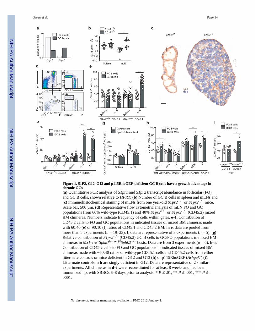
Figure 1. S1P2, G12–G13 and p115RhoGEF-deficient GC B cells have a growth advantage inchronic GCs(a) Quantitative PCR analysis of S1pr1 and S1pr2 transcript abundance in follicular (FO)and GC B cells, shown relative to HPRT. (b) Number of GC B cells in spleen and mLNs and(c) immunohistochemical staining of mLNs from one year-old S1pr2+/− or S1pr2−/− mice.Scale bar, 500 µm. (d) Representative flow cytometric analysis of mLN FO and GCpopulations from 60% wild-type (CD45.1) and 40% S1pr2+/+ or S1pr2−/− (CD45.2) mixedBM chimeras. Numbers indicate frequency of cells within gates. e–f, Contribution ofCD45.2 cells to FO and GC populations in indicated tissues of mixed BM chimeras madewith 60:40 (e) or 90:10 (f) ratios of CD45.1 and CD45.2 BM. In e, data are pooled frommore than 5 experiments (n = 19–23); f, data are representative of 3 experiments (n = 5). (g)Relative contribution of S1pr2−/− (CD45.2) GC B cells to GC/FO populations in mixed BMchimeras in Mx1-cre+Sphk1f/− or f/fSphk2−/− hosts. Data are from 3 experiments (n = 6). h–i,Contribution of CD45.2 cells to FO and GC populations in indicated tissues of mixed BMchimeras made with ~60:40 ratios of wild-type CD45.1 cells and CD45.2 cells from eitherlittermate controls or mice deficient in G12 and G13 (h) or p115RhoGEF (Arhgef1) (i).Littermate controls in h are singly deficient in G12. Data are representative of 2 similarexperiments. All chimeras in d–i were reconstituted for at least 8 weeks and had beenimmunized i.p. with SRBCs 6–8 days prior to analysis. * P ≤ .01, ** P ≤ .001, *** P ≤ .0001.
Green et al. Page 14
Nat Immunol. Author manuscript; available in PMC 2012 January 1.
NIH
-PA Author Manuscript
NIH
-PA Author Manuscript
NIH
-PA Author Manuscript

Figure 2. Apoptosis resistance and increased Akt activation in S1P2, G12–G13, andp115RhoGEF-deficient GC B cellsFrequency of GC B cells with activated caspase-3 (a) or fragmented DNA detected byTUNEL assay (b) in chimeras reconstituted with mixtures of S1pr2+/+ or S1pr2−/−
(CD45.2) and wild-type (CD45.1) BM. The mice were immunized with SRBCs andanalyzed after 6–8 days. Data are from 3 experiments (n = 8–9). (c) Flow cytometricanalysis of P-Akt T308 in follicular and GC B cells from mixed chimeras. Right panelshows mean fluorescence index (MFI) of P-Akt T308 in the indicated GC B cell populations(n = 7). (d) P-Akt T308 analysis in wild-type GC B cells from spleen suspensions treatedwith JTE-013 or JTE-013 and wortmannin (WMN) for 30 min immediately ex vivo (n = 3).(e) Graph showing P-Akt T308 MFI of mLN GC B cells from Mx1-cre+Sphk1f/− or f/fSphk2−/− (S1P-deficient) mice, relative to the average of the controls (n =12–13, pooled from 6 experiments). (f) P-Akt T308 analysis in GC B cells deficient inp115RhoGEF or both G12 and G13, compared to littermate control cells and wild-type(CD45.1) cells, from mixed BM chimeras. The G12–G13 DKO littermate control was G12-single deficient. Right panels show a summary of P-Akt T308 MFIs in GC B cells frommixed chimeras (data are representative of 2 experiments). (g) Western blot for P-Akt S473in Ramos cells treated for 5 min with S1P in the presence or absence of JTE-013(representative of 3 experiments). (h) P-Akt T308 analysis in Ramos cells treated for 5 minwith S1P (10nm) alone or in the presence of the indicated inhibitors. Y27632, 10µm;JTE-013, 10µm; bpV(pic), 500nm. (i) Relative P-Akt T308 MFIs in Ramos cells treatedwith the indicated conditions, compared to untreated (dashed line) (n = 8, pooled from 8experiments). # P ≤ .05, * P ≤ .01, ** P ≤ .001, *** P ≤ .0001.
Green et al. Page 15
Nat Immunol. Author manuscript; available in PMC 2012 January 1.
NIH
-PA Author Manuscript
NIH
-PA Author Manuscript
NIH
-PA Author Manuscript

Figure 3. Akt activation confers an advantage in mucosal GCs and S1P2 regulates translation inGC cells(a) BM from Cr2-cre transgenic mice was transduced with loxP-stop-loxP Thy-1.1 controlretrovirus (Vector) or myristoylated Akt (myr-Akt) Thy-1.1 retrovirus and used toreconstitute irradiated recipient mice. Representative flow cytometric analysis showingrepresentation of myr-AKT–Thy-1.1-expressing cells in IgDlo Fas+ GC B cells relative toIgDhi FO B cells of mLNs. Right panel shows summary of data as percent contribution ofThy-1.1+ cells to FO and GC B cell populations (n = 7, pooled from 2 experiments). (b)TUNEL assay of mLN GC B cells from mice of the type in a. (c) Flow cytometric analysisof P-4E-BP1 in FO and GC B cells from mixed BM chimeras containing wild-type(CD45.1) cells and either S1pr2+/+ or S1pr2−/− (CD45.2) cells. Graph on right shows MFIof P-4E-BP1 in the indicated GC populations (n = 4, from 4 experiments). (d) Relativeincorporation of 35S-labeled cysteine and methionine in 30 min by Ramos cells removedfrom S1P (No S1P) or treated with JTE-013, relative to cells cultured with S1P. Controlsamples were removed from S1P and treated with rapamycin. Triplicate measurements wereobtained in each experiment and all data points were divided by the mean of the untreatedgroup. # P ≤ .05, * P ≤ .01, ** P ≤ .001, *** P ≤ .0001.
Green et al. Page 16
Nat Immunol. Author manuscript; available in PMC 2012 January 1.
NIH
-PA Author Manuscript
NIH
-PA Author Manuscript
NIH
-PA Author Manuscript
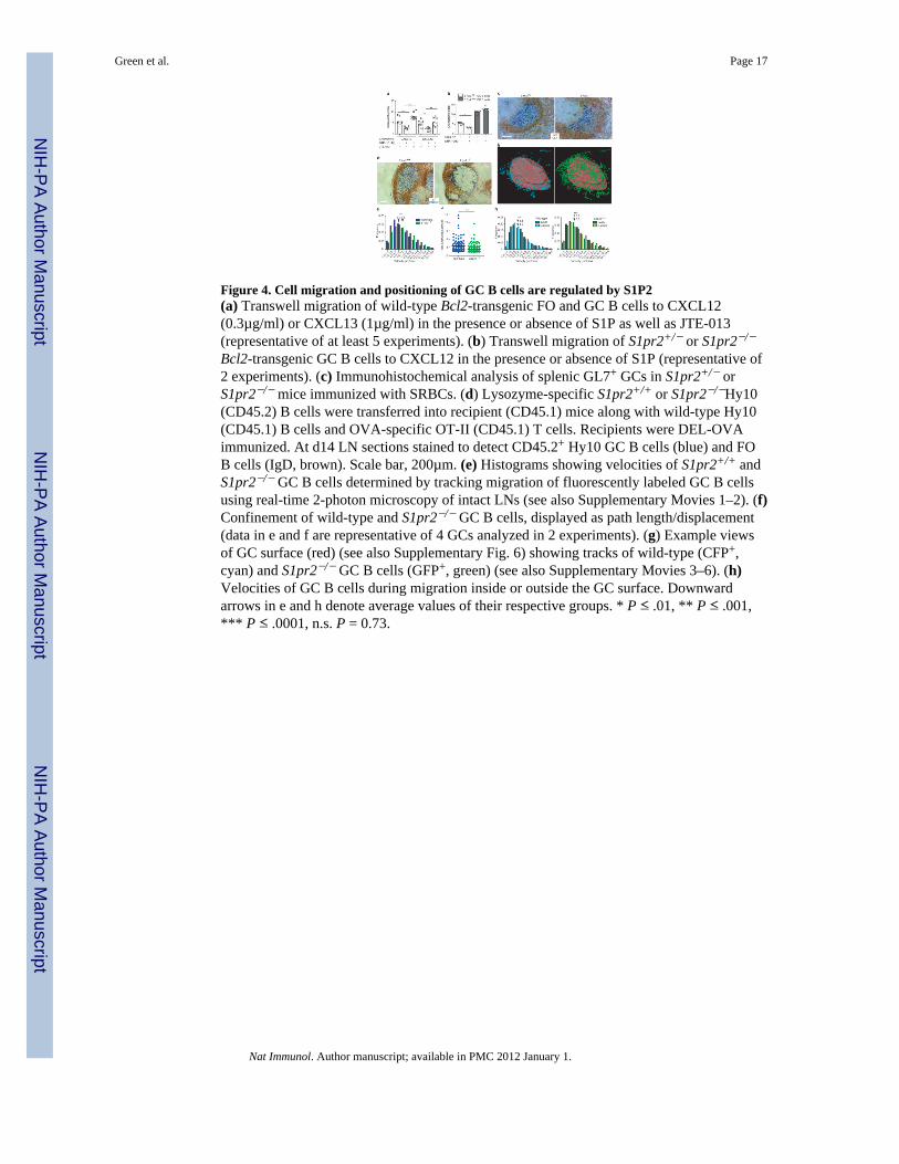
Figure 4. Cell migration and positioning of GC B cells are regulated by S1P2(a) Transwell migration of wild-type Bcl2-transgenic FO and GC B cells to CXCL12(0.3µg/ml) or CXCL13 (1µg/ml) in the presence or absence of S1P as well as JTE-013(representative of at least 5 experiments). (b) Transwell migration of S1pr2+/− or S1pr2−/−
Bcl2-transgenic GC B cells to CXCL12 in the presence or absence of S1P (representative of2 experiments). (c) Immunohistochemical analysis of splenic GL7+ GCs in S1pr2+/− orS1pr2−/− mice immunized with SRBCs. (d) Lysozyme-specific S1pr2+/+ or S1pr2−/−Hy10(CD45.2) B cells were transferred into recipient (CD45.1) mice along with wild-type Hy10(CD45.1) B cells and OVA-specific OT-II (CD45.1) T cells. Recipients were DEL-OVAimmunized. At d14 LN sections stained to detect CD45.2+ Hy10 GC B cells (blue) and FOB cells (IgD, brown). Scale bar, 200µm. (e) Histograms showing velocities of S1pr2+/+ andS1pr2−/− GC B cells determined by tracking migration of fluorescently labeled GC B cellsusing real-time 2-photon microscopy of intact LNs (see also Supplementary Movies 1–2). (f)Confinement of wild-type and S1pr2−/− GC B cells, displayed as path length/displacement(data in e and f are representative of 4 GCs analyzed in 2 experiments). (g) Example viewsof GC surface (red) (see also Supplementary Fig. 6) showing tracks of wild-type (CFP+,cyan) and S1pr2−/− GC B cells (GFP+, green) (see also Supplementary Movies 3–6). (h)Velocities of GC B cells during migration inside or outside the GC surface. Downwardarrows in e and h denote average values of their respective groups. * P ≤ .01, ** P ≤ .001,*** P ≤ .0001, n.s. P = 0.73.
Green et al. Page 17
Nat Immunol. Author manuscript; available in PMC 2012 January 1.
NIH
-PA Author Manuscript
NIH
-PA Author Manuscript
NIH
-PA Author Manuscript
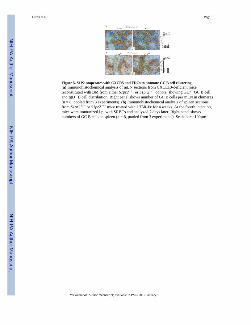
Figure 5. S1P2 cooperates with CXCR5 and FDCs to promote GC B cell clustering(a) Immunohistochemical analysis of mLN sections from CXCL13-deficient micereconstituted with BM from either S1pr2+/− or S1pr2−/− donors, showing GL7+ GC B celland IgD+ B cell distribution. Right panel shows number of GC B cells per mLN in chimeras(n = 8, pooled from 3 experiments). (b) Immunohistochemical analysis of spleen sectionsfrom S1pr2+/− or S1pr2−/− mice treated with LTβR-Fc for 4 weeks. At the fourth injection,mice were immunized i.p. with SRBCs and analyzed 7 days later. Right panel showsnumbers of GC B cells in spleen (n = 8, pooled from 3 experiments). Scale bars, 100µm.
Green et al. Page 18
Nat Immunol. Author manuscript; available in PMC 2012 January 1.
NIH
-PA Author Manuscript
NIH
-PA Author Manuscript
NIH
-PA Author Manuscript

Figure 6. B cells are capable of degrading S1P(a) S1P1 surface abundance on follicular B cells from the indicated tissues of control (CTL),S1pr1-deficient (S1P1 KO), or Mx1-cre+Sphk1f/− or f/fSphk2−/− (S1P-deficient) mice. Dataare representative of more than 3 mice of each type. (b) Transcript abundance of S1P lyase(Sgpl), sphingosine-1-phosphate phosphatase-1 (Sgpp1), and lipid phosphate phosphatases(Lpp) 1–3 in B cells and T cells, shown relative to HPRT. (c) Relative amount of S1Premaining in culture supernatants after incubation with B or T cells for the indicated numberof minutes, determined by the extent of down-modulation of Flag-S1P1 on a reporter cellline. Data are plotted as MFI of Flag-S1P1 staining relative to reporter cells not exposed toS1P (data are from 3 experiments).
Green et al. Page 19
Nat Immunol. Author manuscript; available in PMC 2012 January 1.
NIH
-PA Author Manuscript
NIH
-PA Author Manuscript
NIH
-PA Author Manuscript

Figure 7. S1P2 directs activated B cells to the GC and the center of the follicle(a) MD4 B cells retrovirally transduced with vectors encoding either S1P2 or control surfacereceptor (truncated Ngfr) as well as the hCD4 reporter were transferred into day 6 SRBCimmunized recipient mice for 24 hours. Immunohistochemical staining of splenic sectionsshows localization of hCD4+ transduced cells in GC-containing follicles. (b) S1P2 or controlvector transduced Gpr183+/− MD4 B cells were transferred into unimmunized recipients.Immunohistochemical staining shows localization of transduced cells in primary follicles.(c) S1P2 transduced Gpr183+/− MD4 B cells were transferred into either littermate control(Sphk2−/−) or Mx1-cre+Sphk1f/− or f/fSphk2−/− (S1P-deficient) recipient mice.Immunohistochemical staining shows localization of transduced cells in primary follicles.Data in a–c are all representative of 3 independent experiments. Scale bars, 200 µm.
Green et al. Page 20
Nat Immunol. Author manuscript; available in PMC 2012 January 1.
NIH
-PA Author Manuscript
NIH
-PA Author Manuscript
NIH
-PA Author Manuscript