tkt maintains intestinal atp production and inhibits ... - nature
TRANSCRIPT
ARTICLE OPEN
TKT maintains intestinal ATP production and inhibitsapoptosis-induced colitisNa Tian 1,2,5✉, Lei Hu2,5, Ying Lu 2, Lingfeng Tong2, Ming Feng3, Qi Liu2, Yakui Li2, Yemin Zhu2, Lifang Wu2, Yingning Ji2,Ping Zhang2, Tianle Xu4 and Xuemei Tong 2✉
© The Author(s) 2021
Inflammatory bowel disease (IBD) has a close association with transketolase (TKT) that links glycolysis and the pentose phosphatepathway (PPP). However, how TKT functions in the intestinal epithelium remains to be elucidated. To address this question, wespecifically delete TKT in intestinal epithelial cells (IECs). IEC TKT-deficient mice are growth retarded and suffer from spontaneouscolitis. TKT ablation brings about striking alterations of the intestine, including extensive mucosal erosion, aberrant tight junctions,impaired barrier function, and increased inflammatory cell infiltration. Mechanistically, TKT deficiency significantly accumulates PPPmetabolites and decreases glycolytic metabolites, thereby reducing ATP production, which results in excessive apoptosis anddefective intestinal barrier. Therefore, our data demonstrate that TKT serves as an essential guardian of intestinal integrity andbarrier function as well as a potential therapeutic target for intestinal disorders.
Cell Death and Disease (2021) 12:853 ; https://doi.org/10.1038/s41419-021-04142-4
INTRODUCTIONThe mammalian intestinal epithelium is composed of a monolayerof columnar epithelial cells organized into crypts and villi thatrenews every 4–5 days [1]. It is an important metabolic tissue,serving a variety of physiological functions including digestion andabsorption of nutrients as well as forming a physical andbiochemical barrier against enteric pathogens to maintain tissuehomeostasis [1–3]. Once the intestinal barrier function is disturbedby various risk factors such as deregulated apoptosis, the intestinalmucosa directly contacts with the luminal invading pathogens andingested toxins to promote inflammatory responses, leading tointestinal disorders including inflammatory bowel disease (IBD) [4].Recent studies have intensively suggested a strong link
between intestinal disorders and metabolites or metabolic genes.Some products from carbon metabolism, such as pyruvate, play ananti-inflammatory role in colitis [5, 6]. In addition, lipids, short fattyacids and amino acids also have a close association with intestinalbarrier function and IBD [7–10]. Moreover, metabolic genesincluding pyruvate kinase (PKM2), NADPH oxidase (NOX) andTIGAR have been shown to involve in the progression of IBD[11–13]. Thus, focus on the regulation of metabolism in IBD mayprovide new insights into clinical therapies.Transketolase (TKT), linking the pentose phosphate pathway (PPP)
and glycolysis, plays a critical role in the non-oxidative PPP [14, 15].TKT catalyzes two reversible reactions, determining the direction ofmetabolites in PPP according to metabolic demands [15]. Onereaction catalyzed by TKT is the conversion of xylulose-5-phosphate
(Xu5P) and ribose-5-phosphate (R5P) into glyceraldehyde-3-phosphate (G3P) and sedoheptulose-7-phosphate (S7P). The otheris to produce G3P and fructose-6-phosphate (F6P) from Xu5P anderythrose-4-phosphate (E4P) [15]. Our previous data have shownthat TKT serves as a critical regulator of carbohydrate and lipidcatabolism [16]. Cellular TKT deficiency accumulates non-oxidativePPP metabolites and decreases glycolytic products [16, 17], whichare associated with intestinal barrier function [5, 6, 18]. The TKTactivity is dependent on thiamine, and thiamine deficiency has astrong association with IBD [19]. Moreover, erythrocyte TKTactivity is declined in patients suffering from IBD [20]. Further-more, lack of transketolase-like 1 (TKTL1), which belongs to theTKT gene family, aggravates murine experimental colitis [21].However, the functions of TKT gene family in regulating intestinalhomeostasis remain unknown.In mice, inactivation of both TKT alleles is lethal whereas
disruption one of TKT alleles causes growth retardation andpreferential reduction of adipose tissues [22]. Nevertheless, ourprevious data have indicated that TKT ablation in the liver andadipose tissues has no effect on the growth and development inmice fed with normal chow diet [16, 17]. Therefore, TKTdownregulation in other tissues such as the intestine possiblyaccounts for the growth retardation phenotypes.To investigate the possible role of TKT in intestinal physiology,
we generate an intestinal epithelium-specific TKT knockout mousemodel. In the present study, we show that TKT deficiency in theintestinal epithelium results in reduction of glycolytic metabolites
Received: 31 March 2021 Accepted: 26 August 2021
1Department of Neurology, Shandong Provincial Hospital Affiliated to Shandong First Medical University, Jinan, Shandong, China. 2Department of Biochemistry and MolecularCell Biology, Shanghai Key Laboratory for Tumor Microenvironment and Inflammation, Key Laboratory of Cell Differentiation and Apoptosis of Chinese Ministry of Education,Shanghai Jiao Tong University School of Medicine, Shanghai, China. 3Department of Physiology, Weifang Medical University, Weifang, Shandong, China. 4Department of Anatomyand Physiology, Center for Brain Science of Shanghai Children’s Medical Center, Shanghai Jiao Tong University School of Medicine, Shanghai, China. 5These authors contributedequally: Na Tian, Lei Hu. ✉email: [email protected]; [email protected] by Hans-Uwe Simon
www.nature.com/cddis
Official journal of CDDpress
and insufficient ATP production, which can lead to epithelial cellapoptosis, intestinal barrier defects, spontaneous colitis andmouse growth retardation. Therefore, TKT maintains intestinalhomeostasis through protecting epithelial cells from energyinsufficiency and apoptosis.
RESULTSTKT deletion in intestinal epithelium leads to growthretardation in miceTo directly investigate the physiological functions of TKT in theintestine, we generated IEC-specific TKT knockout (abbreviated
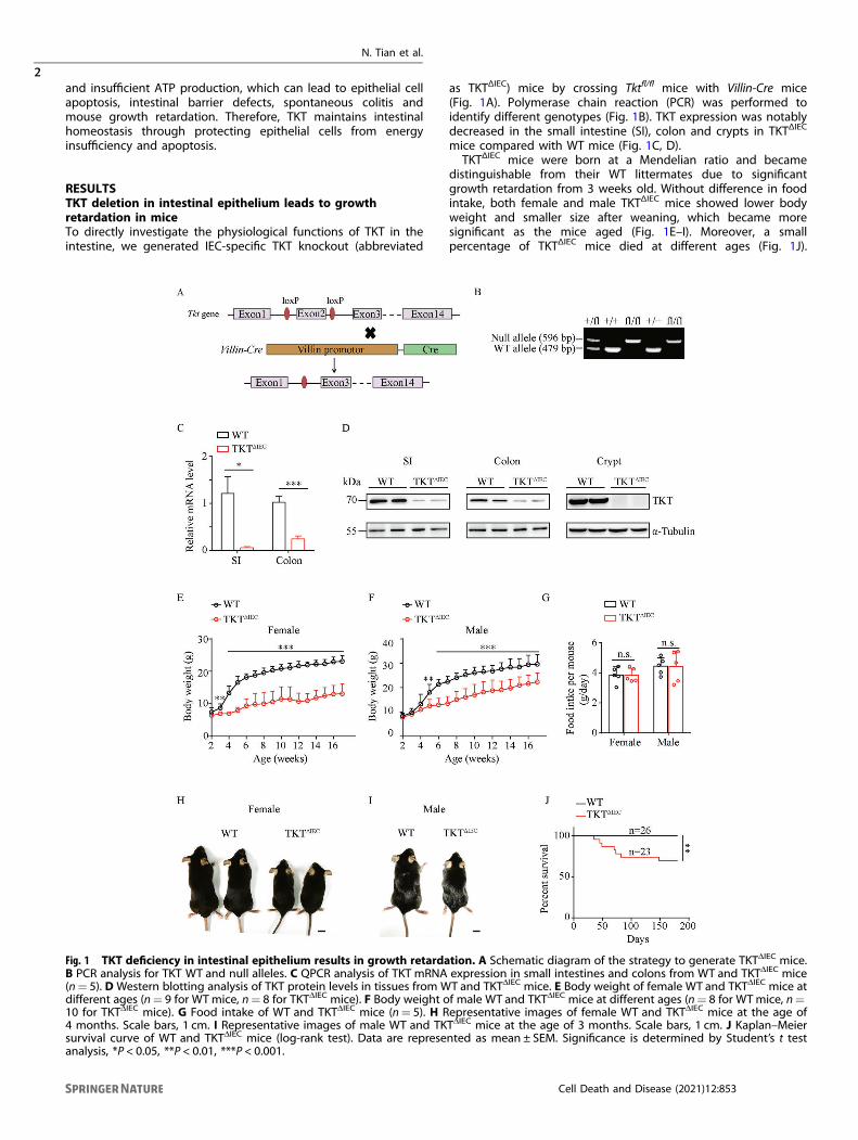
as TKTΔIEC) mice by crossing Tktfl/fl mice with Villin-Cre mice(Fig. 1A). Polymerase chain reaction (PCR) was performed toidentify different genotypes (Fig. 1B). TKT expression was notablydecreased in the small intestine (SI), colon and crypts in TKTΔIEC
mice compared with WT mice (Fig. 1C, D).TKTΔIEC mice were born at a Mendelian ratio and became
distinguishable from their WT littermates due to significantgrowth retardation from 3 weeks old. Without difference in foodintake, both female and male TKTΔIEC mice showed lower bodyweight and smaller size after weaning, which became moresignificant as the mice aged (Fig. 1E–I). Moreover, a smallpercentage of TKTΔIEC mice died at different ages (Fig. 1J).
Fig. 1 TKT deficiency in intestinal epithelium results in growth retardation. A Schematic diagram of the strategy to generate TKTΔIEC mice.B PCR analysis for TKT WT and null alleles. C QPCR analysis of TKT mRNA expression in small intestines and colons from WT and TKTΔIEC mice(n= 5). D Western blotting analysis of TKT protein levels in tissues from WT and TKTΔIEC mice. E Body weight of female WT and TKTΔIEC mice atdifferent ages (n= 9 for WTmice, n= 8 for TKTΔIEC mice). F Body weight of male WT and TKTΔIEC mice at different ages (n= 8 for WTmice, n=10 for TKTΔIEC mice). G Food intake of WT and TKTΔIEC mice (n= 5). H Representative images of female WT and TKTΔIEC mice at the age of4 months. Scale bars, 1 cm. I Representative images of male WT and TKTΔIEC mice at the age of 3 months. Scale bars, 1 cm. J Kaplan–Meiersurvival curve of WT and TKTΔIEC mice (log-rank test). Data are represented as mean ± SEM. Significance is determined by Student’s t testanalysis, *P < 0.05, **P < 0.01, ***P < 0.001.
N. Tian et al.
2
Cell Death and Disease (2021) 12:853
1234567890();,:
Collectively, these data suggest that sufficient TKT in IECs is criticalfor murine normal growth.
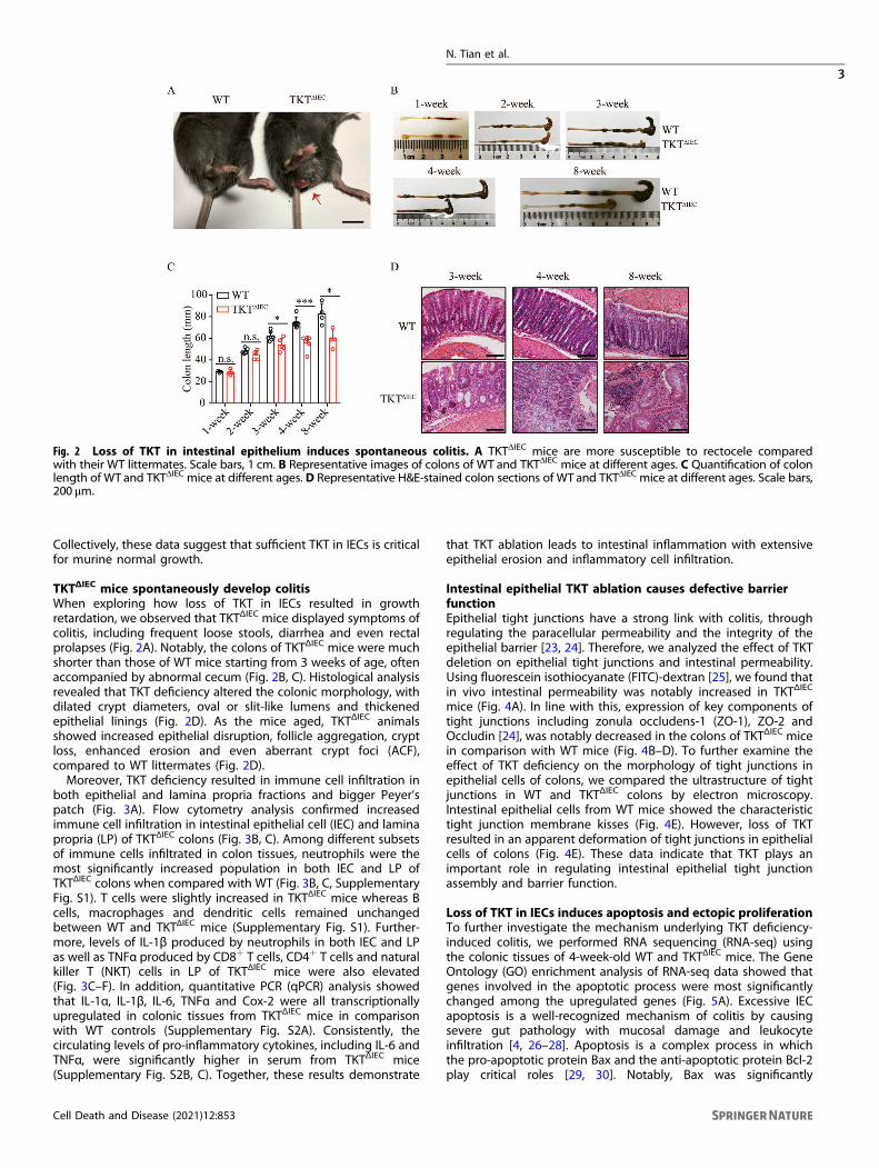
TKTΔIEC mice spontaneously develop colitisWhen exploring how loss of TKT in IECs resulted in growthretardation, we observed that TKTΔIEC mice displayed symptoms ofcolitis, including frequent loose stools, diarrhea and even rectalprolapses (Fig. 2A). Notably, the colons of TKTΔIEC mice were muchshorter than those of WT mice starting from 3 weeks of age, oftenaccompanied by abnormal cecum (Fig. 2B, C). Histological analysisrevealed that TKT deficiency altered the colonic morphology, withdilated crypt diameters, oval or slit-like lumens and thickenedepithelial linings (Fig. 2D). As the mice aged, TKTΔIEC animalsshowed increased epithelial disruption, follicle aggregation, cryptloss, enhanced erosion and even aberrant crypt foci (ACF),compared to WT littermates (Fig. 2D).Moreover, TKT deficiency resulted in immune cell infiltration in
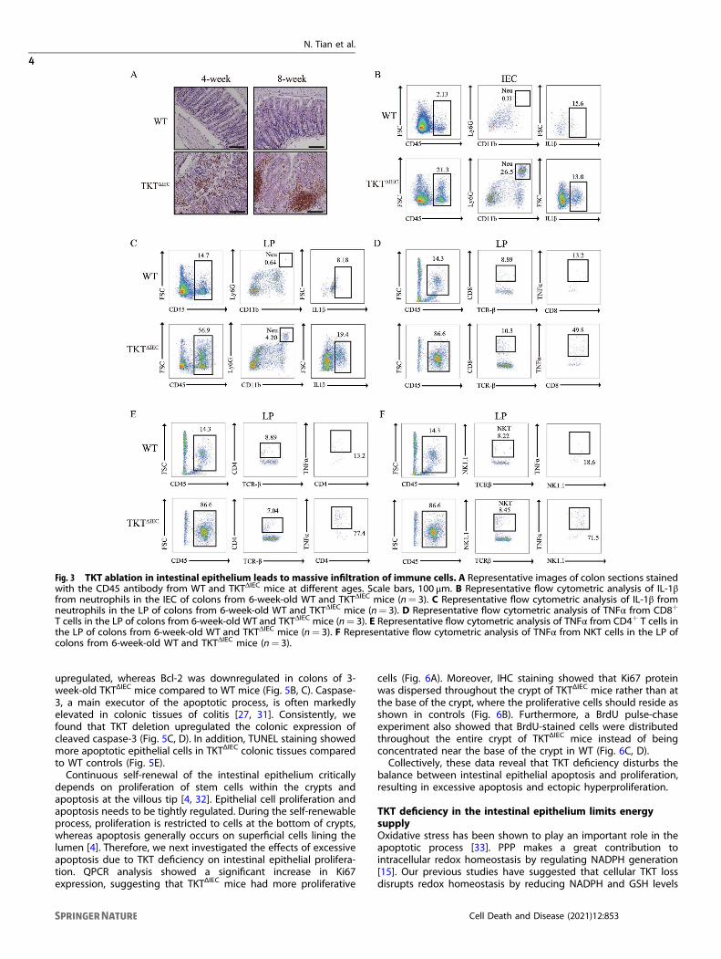
both epithelial and lamina propria fractions and bigger Peyer’spatch (Fig. 3A). Flow cytometry analysis confirmed increasedimmune cell infiltration in intestinal epithelial cell (IEC) and laminapropria (LP) of TKTΔIEC colons (Fig. 3B, C). Among different subsetsof immune cells infiltrated in colon tissues, neutrophils were themost significantly increased population in both IEC and LP ofTKTΔIEC colons when compared with WT (Fig. 3B, C, SupplementaryFig. S1). T cells were slightly increased in TKTΔIEC mice whereas Bcells, macrophages and dendritic cells remained unchangedbetween WT and TKTΔIEC mice (Supplementary Fig. S1). Further-more, levels of IL-1β produced by neutrophils in both IEC and LPas well as TNFα produced by CD8+ T cells, CD4+ T cells and naturalkiller T (NKT) cells in LP of TKTΔIEC mice were also elevated(Fig. 3C–F). In addition, quantitative PCR (qPCR) analysis showedthat IL-1α, IL-1β, IL-6, TNFα and Cox-2 were all transcriptionallyupregulated in colonic tissues from TKTΔIEC mice in comparisonwith WT controls (Supplementary Fig. S2A). Consistently, thecirculating levels of pro-inflammatory cytokines, including IL-6 andTNFα, were significantly higher in serum from TKTΔIEC mice(Supplementary Fig. S2B, C). Together, these results demonstrate
that TKT ablation leads to intestinal inflammation with extensiveepithelial erosion and inflammatory cell infiltration.
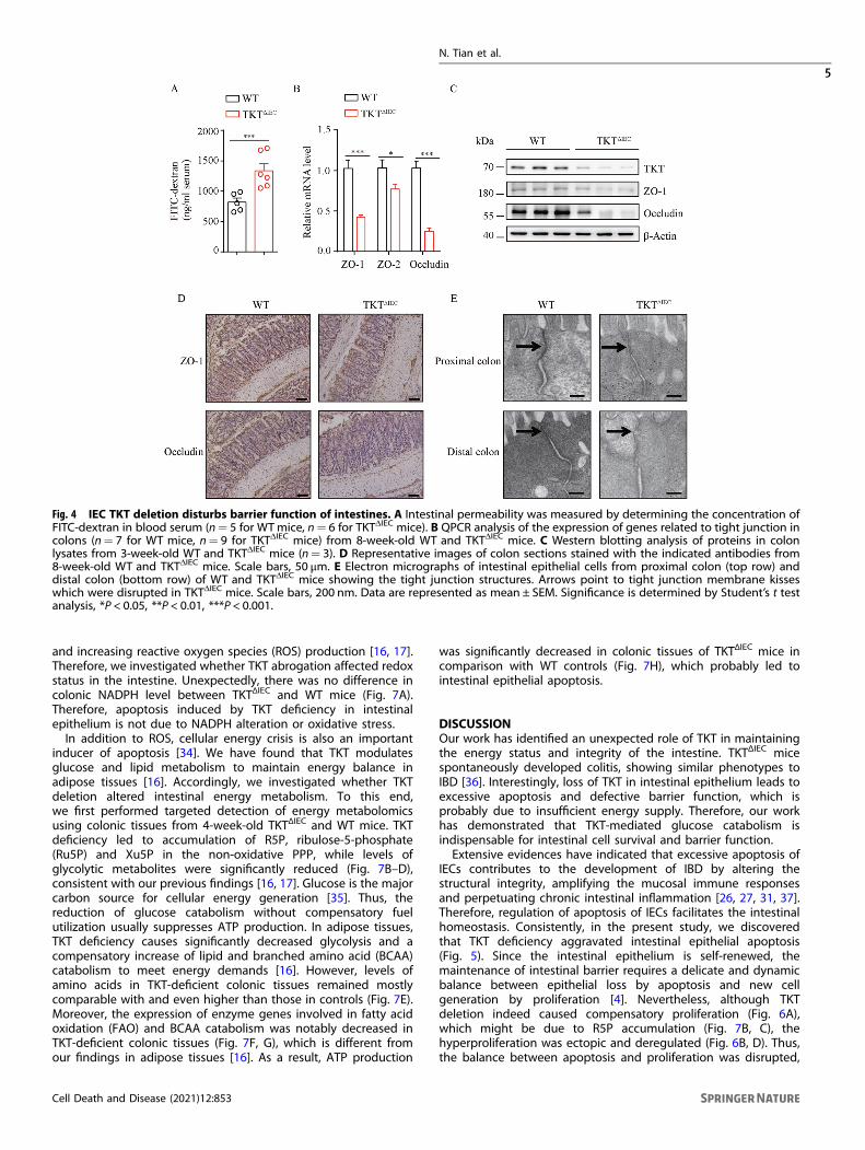
Intestinal epithelial TKT ablation causes defective barrierfunctionEpithelial tight junctions have a strong link with colitis, throughregulating the paracellular permeability and the integrity of theepithelial barrier [23, 24]. Therefore, we analyzed the effect of TKTdeletion on epithelial tight junctions and intestinal permeability.Using fluorescein isothiocyanate (FITC)-dextran [25], we found thatin vivo intestinal permeability was notably increased in TKTΔIEC
mice (Fig. 4A). In line with this, expression of key components oftight junctions including zonula occludens-1 (ZO-1), ZO-2 andOccludin [24], was notably decreased in the colons of TKTΔIEC micein comparison with WT mice (Fig. 4B–D). To further examine theeffect of TKT deficiency on the morphology of tight junctions inepithelial cells of colons, we compared the ultrastructure of tightjunctions in WT and TKTΔIEC colons by electron microscopy.Intestinal epithelial cells from WT mice showed the characteristictight junction membrane kisses (Fig. 4E). However, loss of TKTresulted in an apparent deformation of tight junctions in epithelialcells of colons (Fig. 4E). These data indicate that TKT plays animportant role in regulating intestinal epithelial tight junctionassembly and barrier function.
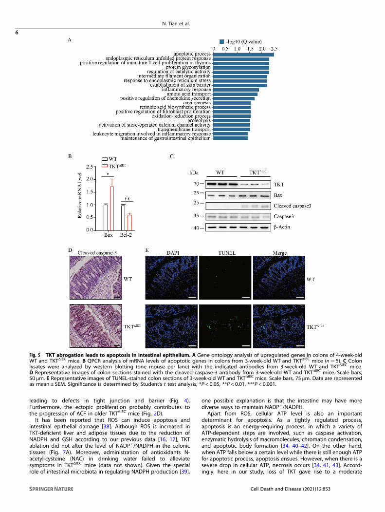
Loss of TKT in IECs induces apoptosis and ectopic proliferationTo further investigate the mechanism underlying TKT deficiency-induced colitis, we performed RNA sequencing (RNA-seq) usingthe colonic tissues of 4-week-old WT and TKTΔIEC mice. The GeneOntology (GO) enrichment analysis of RNA-seq data showed thatgenes involved in the apoptotic process were most significantlychanged among the upregulated genes (Fig. 5A). Excessive IECapoptosis is a well-recognized mechanism of colitis by causingsevere gut pathology with mucosal damage and leukocyteinfiltration [4, 26–28]. Apoptosis is a complex process in whichthe pro-apoptotic protein Bax and the anti-apoptotic protein Bcl-2play critical roles [29, 30]. Notably, Bax was significantly
Fig. 2 Loss of TKT in intestinal epithelium induces spontaneous colitis. A TKTΔIEC mice are more susceptible to rectocele comparedwith their WT littermates. Scale bars, 1 cm. B Representative images of colons of WT and TKTΔIEC mice at different ages. C Quantification of colonlength of WT and TKTΔIEC mice at different ages. D Representative H&E-stained colon sections of WT and TKTΔIEC mice at different ages. Scale bars,200 μm.
N. Tian et al.
3
Cell Death and Disease (2021) 12:853
upregulated, whereas Bcl-2 was downregulated in colons of 3-week-old TKTΔIEC mice compared to WT mice (Fig. 5B, C). Caspase-3, a main executor of the apoptotic process, is often markedlyelevated in colonic tissues of colitis [27, 31]. Consistently, wefound that TKT deletion upregulated the colonic expression ofcleaved caspase-3 (Fig. 5C, D). In addition, TUNEL staining showedmore apoptotic epithelial cells in TKTΔIEC colonic tissues comparedto WT controls (Fig. 5E).Continuous self-renewal of the intestinal epithelium critically
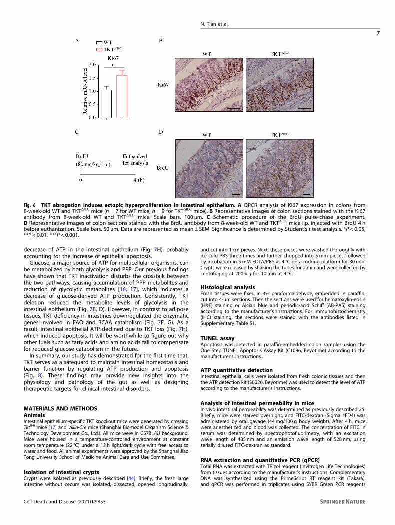
depends on proliferation of stem cells within the crypts andapoptosis at the villous tip [4, 32]. Epithelial cell proliferation andapoptosis needs to be tightly regulated. During the self-renewableprocess, proliferation is restricted to cells at the bottom of crypts,whereas apoptosis generally occurs on superficial cells lining thelumen [4]. Therefore, we next investigated the effects of excessiveapoptosis due to TKT deficiency on intestinal epithelial prolifera-tion. QPCR analysis showed a significant increase in Ki67expression, suggesting that TKTΔIEC mice had more proliferative
cells (Fig. 6A). Moreover, IHC staining showed that Ki67 proteinwas dispersed throughout the crypt of TKTΔIEC mice rather than atthe base of the crypt, where the proliferative cells should reside asshown in controls (Fig. 6B). Furthermore, a BrdU pulse-chaseexperiment also showed that BrdU-stained cells were distributedthroughout the entire crypt of TKTΔIEC mice instead of beingconcentrated near the base of the crypt in WT (Fig. 6C, D).Collectively, these data reveal that TKT deficiency disturbs the
balance between intestinal epithelial apoptosis and proliferation,resulting in excessive apoptosis and ectopic hyperproliferation.
TKT deficiency in the intestinal epithelium limits energysupplyOxidative stress has been shown to play an important role in theapoptotic process [33]. PPP makes a great contribution tointracellular redox homeostasis by regulating NADPH generation[15]. Our previous studies have suggested that cellular TKT lossdisrupts redox homeostasis by reducing NADPH and GSH levels
Fig. 3 TKT ablation in intestinal epithelium leads to massive infiltration of immune cells. A Representative images of colon sections stainedwith the CD45 antibody from WT and TKTΔIEC mice at different ages. Scale bars, 100 μm. B Representative flow cytometric analysis of IL-1βfrom neutrophils in the IEC of colons from 6-week-old WT and TKTΔIEC mice (n= 3). C Representative flow cytometric analysis of IL-1β fromneutrophils in the LP of colons from 6-week-old WT and TKTΔIEC mice (n= 3). D Representative flow cytometric analysis of TNFα from CD8+
T cells in the LP of colons from 6-week-old WT and TKTΔIEC mice (n= 3). E Representative flow cytometric analysis of TNFα from CD4+ T cells inthe LP of colons from 6-week-old WT and TKTΔIEC mice (n= 3). F Representative flow cytometric analysis of TNFα from NKT cells in the LP ofcolons from 6-week-old WT and TKTΔIEC mice (n= 3).
N. Tian et al.
4
Cell Death and Disease (2021) 12:853
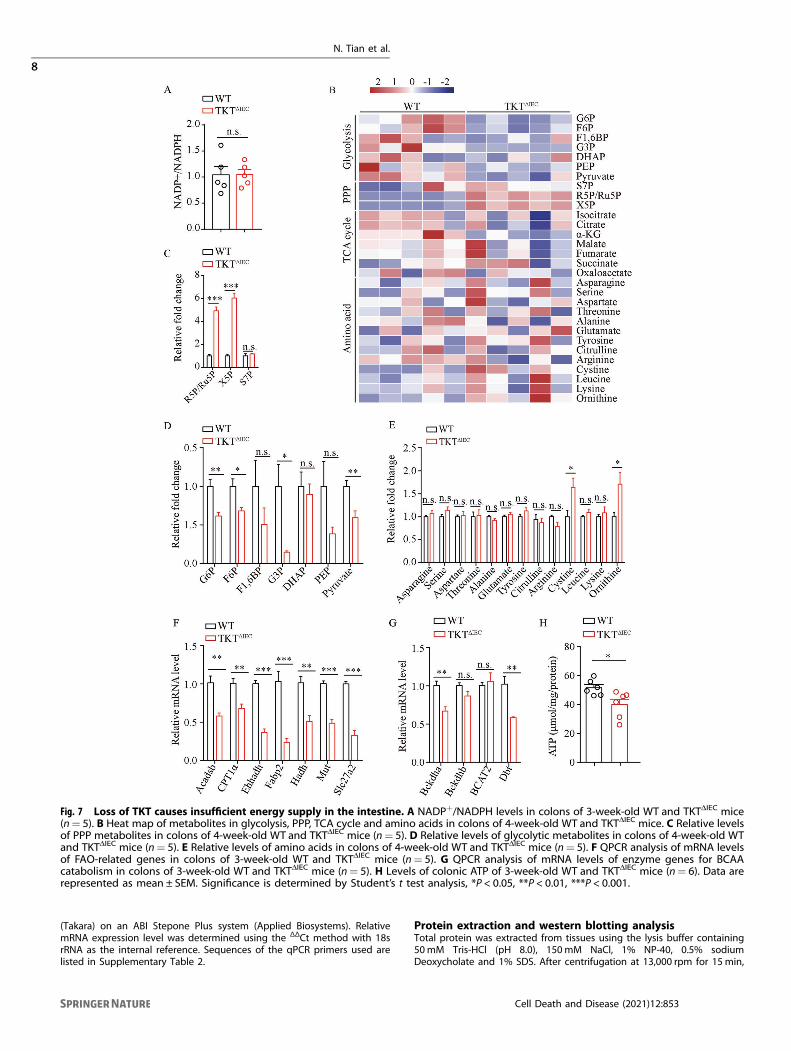
and increasing reactive oxygen species (ROS) production [16, 17].Therefore, we investigated whether TKT abrogation affected redoxstatus in the intestine. Unexpectedly, there was no difference incolonic NADPH level between TKTΔIEC and WT mice (Fig. 7A).Therefore, apoptosis induced by TKT deficiency in intestinalepithelium is not due to NADPH alteration or oxidative stress.In addition to ROS, cellular energy crisis is also an important
inducer of apoptosis [34]. We have found that TKT modulatesglucose and lipid metabolism to maintain energy balance inadipose tissues [16]. Accordingly, we investigated whether TKTdeletion altered intestinal energy metabolism. To this end,we first performed targeted detection of energy metabolomicsusing colonic tissues from 4-week-old TKTΔIEC and WT mice. TKTdeficiency led to accumulation of R5P, ribulose-5-phosphate(Ru5P) and Xu5P in the non-oxidative PPP, while levels ofglycolytic metabolites were significantly reduced (Fig. 7B–D),consistent with our previous findings [16, 17]. Glucose is the majorcarbon source for cellular energy generation [35]. Thus, thereduction of glucose catabolism without compensatory fuelutilization usually suppresses ATP production. In adipose tissues,TKT deficiency causes significantly decreased glycolysis and acompensatory increase of lipid and branched amino acid (BCAA)catabolism to meet energy demands [16]. However, levels ofamino acids in TKT-deficient colonic tissues remained mostlycomparable with and even higher than those in controls (Fig. 7E).Moreover, the expression of enzyme genes involved in fatty acidoxidation (FAO) and BCAA catabolism was notably decreased inTKT-deficient colonic tissues (Fig. 7F, G), which is different fromour findings in adipose tissues [16]. As a result, ATP production
was significantly decreased in colonic tissues of TKTΔIEC mice incomparison with WT controls (Fig. 7H), which probably led tointestinal epithelial apoptosis.
DISCUSSIONOur work has identified an unexpected role of TKT in maintainingthe energy status and integrity of the intestine. TKTΔIEC micespontaneously developed colitis, showing similar phenotypes toIBD [36]. Interestingly, loss of TKT in intestinal epithelium leads toexcessive apoptosis and defective barrier function, which isprobably due to insufficient energy supply. Therefore, our workhas demonstrated that TKT-mediated glucose catabolism isindispensable for intestinal cell survival and barrier function.Extensive evidences have indicated that excessive apoptosis of
IECs contributes to the development of IBD by altering thestructural integrity, amplifying the mucosal immune responsesand perpetuating chronic intestinal inflammation [26, 27, 31, 37].Therefore, regulation of apoptosis of IECs facilitates the intestinalhomeostasis. Consistently, in the present study, we discoveredthat TKT deficiency aggravated intestinal epithelial apoptosis(Fig. 5). Since the intestinal epithelium is self-renewed, themaintenance of intestinal barrier requires a delicate and dynamicbalance between epithelial loss by apoptosis and new cellgeneration by proliferation [4]. Nevertheless, although TKTdeletion indeed caused compensatory proliferation (Fig. 6A),which might be due to R5P accumulation (Fig. 7B, C), thehyperproliferation was ectopic and deregulated (Fig. 6B, D). Thus,the balance between apoptosis and proliferation was disrupted,
Fig. 4 IEC TKT deletion disturbs barrier function of intestines. A Intestinal permeability was measured by determining the concentration ofFITC-dextran in blood serum (n= 5 for WTmice, n= 6 for TKTΔIEC mice). B QPCR analysis of the expression of genes related to tight junction incolons (n= 7 for WT mice, n= 9 for TKTΔIEC mice) from 8-week-old WT and TKTΔIEC mice. C Western blotting analysis of proteins in colonlysates from 3-week-old WT and TKTΔIEC mice (n= 3). D Representative images of colon sections stained with the indicated antibodies from8-week-old WT and TKTΔIEC mice. Scale bars, 50 μm. E Electron micrographs of intestinal epithelial cells from proximal colon (top row) anddistal colon (bottom row) of WT and TKTΔIEC mice showing the tight junction structures. Arrows point to tight junction membrane kisseswhich were disrupted in TKTΔIEC mice. Scale bars, 200 nm. Data are represented as mean ± SEM. Significance is determined by Student’s t testanalysis, *P < 0.05, **P < 0.01, ***P < 0.001.
N. Tian et al.
5
Cell Death and Disease (2021) 12:853
leading to defects in tight junction and barrier (Fig. 4).Furthermore, the ectopic proliferation probably contributes tothe progression of ACF in older TKTΔIEC mice (Fig. 2D).It has been reported that ROS can induce apoptosis and
intestinal epithelial damage [38]. Although ROS is increased inTKT-deficient liver and adipose tissues due to the reduction ofNADPH and GSH according to our previous data [16, 17], TKTablation did not alter the level of NADP+/NADPH in the colonictissues (Fig. 7A). Moreover, administration of antioxidants N-acetyl-cysteine (NAC) in drinking water failed to alleviatesymptoms in TKTΔIEC mice (data not shown). Given the specialrole of intestinal microbiota in regulating NADPH production [39],
one possible explanation is that the intestine may have morediverse ways to maintain NADP+/NADPH.Apart from ROS, cellular ATP level is also an important
determinant for apoptosis. As a tightly regulated process,apoptosis is an energy-requiring process, in which a variety ofATP-dependent steps are involved, such as caspase activation,enzymatic hydrolysis of macromolecules, chromatin condensation,and apoptotic body formation [34, 40–42]. On the other hand,when ATP falls below a certain level while there is still enough ATPfor apoptotic process, apoptosis ensues. However, when there is asevere drop in cellular ATP, necrosis occurs [34, 41, 43]. Accord-ingly, here in our study, loss of TKT gave rise to a moderate
Fig. 5 TKT abrogation leads to apoptosis in intestinal epithelium. A Gene ontology analysis of upregulated genes in colons of 4-week-oldWT and TKTΔIEC mice. B QPCR analysis of mRNA levels of apoptotic genes in colons from 3-week-old WT and TKTΔIEC mice (n= 5). C Colonlysates were analyzed by western blotting (one mouse per lane) with the indicated antibodies from 3-week-old WT and TKTΔIEC mice.D Representative images of colon sections stained with the cleaved caspase-3 antibody from 3-week-old WT and TKTΔIEC mice. Scale bars,50 μm. E Representative images of TUNEL-stained colon sections of 3-week-old WT and TKTΔIEC mice. Scale bars, 75 μm. Data are representedas mean ± SEM. Significance is determined by Student’s t test analysis, *P < 0.05, **P < 0.01, ***P < 0.001.
N. Tian et al.
6
Cell Death and Disease (2021) 12:853
decrease of ATP in the intestinal epithelium (Fig. 7H), probablyaccounting for the increase of epithelial apoptosis.Glucose, a major source of ATP for multicellular organisms, can
be metabolized by both glycolysis and PPP. Our previous findingshave shown that TKT inactivation disturbs the crosstalk betweenthe two pathways, causing accumulation of PPP metabolites andreduction of glycolytic metabolites [16, 17], which indicates adecrease of glucose-derived ATP production. Consistently, TKTdeletion reduced the metabolite levels of glycolysis in theintestinal epithelium (Fig. 7B, D). However, in contrast to adiposetissues, TKT deficiency in intestines downregulated the enzymaticgenes involved in FAO and BCAA catabolism (Fig. 7F, G). As aresult, intestinal epithelial ATP declined due to TKT loss (Fig. 7H),which induced apoptosis. It will be worthwhile to figure out whyother fuels such as fatty acids and amino acids fail to compensatefor reduced glucose catabolism in the future.In summary, our study has demonstrated for the first time that,
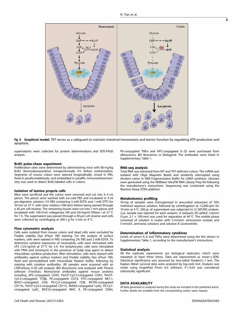
TKT serves as a safeguard to maintain intestinal homeostasis andbarrier function by regulating ATP production and apoptosis(Fig. 8). These findings may provide new insights into thephysiology and pathology of the gut as well as designingtherapeutic targets for clinical intestinal disorders.
MATERIALS AND METHODSAnimalsIntestinal epithelium-specific TKT knockout mice were generated by crossingTktfl/fl mice [17] and Villin-Cre mice (Shanghai Biomodel Organism Science &Technology Development Co., Ltd.). All mice were in C57BL/6J background.Mice were housed in a temperature-controlled environment at constantroom temperature (22 °C) under a 12 h light/dark cycle with free access towater and food. All animal experiments were approved by the Shanghai JiaoTong University School of Medicine Animal Care and Use Committee.
Isolation of intestinal cryptsCrypts were isolated as previously described [44]. Briefly, the fresh largeintestine without cecum was isolated, dissected, opened longitudinally,
and cut into 1 cm pieces. Next, these pieces were washed thoroughly withice-cold PBS three times and further chopped into 5mm pieces, followedby incubation in 5mM EDTA/PBS at 4 °C on a rocking platform for 30min.Crypts were released by shaking the tubes for 2 min and were collected bycentrifuging at 200 × g for 10 min at 4 °C.
Histological analysisFresh tissues were fixed in 4% paraformaldehyde, embedded in paraffin,cut into 4-μm sections. Then the sections were used for hematoxylin-eosin(H&E) staining or Alcian blue and periodic-acid Schiff (AB-PAS) stainingaccording to the manufacturer’s instructions. For immunohistochemistry(IHC) staining, the sections were stained with the antibodies listed inSupplementary Table S1.
TUNEL assayApoptosis was detected in paraffin-embedded colon samples using theOne Step TUNEL Apoptosis Assay Kit (C1086, Beyotime) according to themanufacturer’s instructions.
ATP quantitative detectionIntestinal epithelial cells were isolated from fresh colonic tissues and thenthe ATP detection kit (S0026, Beyotime) was used to detect the level of ATPaccording to the manufacturer’s instructions.
Analysis of intestinal permeability in miceIn vivo intestinal permeability was determined as previously described 25.Briefly, mice were starved overnight, and FITC-dextran (Sigma #FD4) wasadministered by oral gavage (44mg/100 g body weight). After 4 h, micewere anesthetized and blood was collected. The concentration of FITC inserum was determined by spectrophotofluorimetry, with an excitationwave length of 485 nm and an emission wave length of 528 nm, usingserially diluted FITC-dextran as standard.
RNA extraction and quantitative PCR (qPCR)Total RNA was extracted with TRIzol reagent (Invitrogen Life Technologies)from tissues according to the manufacturer’s instructions. ComplementaryDNA was synthesized using the PrimeScript RT reagent kit (Takara),and qPCR was performed in triplicates using SYBR Green PCR reagents
Fig. 6 TKT abrogation induces ectopic hyperproliferation in intestinal epithelium. A QPCR analysis of Ki67 expression in colons from8-week-old WT and TKTΔIEC mice (n= 7 for WT mice, n= 9 for TKTΔIEC mice). B Representative images of colon sections stained with the Ki67antibody from 8-week-old WT and TKTΔIEC mice. Scale bars, 100 μm. C Schematic procedure of the BrdU pulse-chase experiment.D Representative images of colon sections stained with the BrdU antibody from 8-week-old WT and TKTΔIEC mice i.p. injected with BrdU 4 hbefore euthanization. Scale bars, 50 μm. Data are represented as mean ± SEM. Significance is determined by Student’s t test analysis, *P < 0.05,**P < 0.01, ***P < 0.001.
N. Tian et al.
7
Cell Death and Disease (2021) 12:853
(Takara) on an ABI Stepone Plus system (Applied Biosystems). RelativemRNA expression level was determined using the ΔΔCt method with 18srRNA as the internal reference. Sequences of the qPCR primers used arelisted in Supplementary Table 2.
Protein extraction and western blotting analysisTotal protein was extracted from tissues using the lysis buffer containing50mM Tris-HCl (pH 8.0), 150mM NaCl, 1% NP-40, 0.5% sodiumDeoxycholate and 1% SDS. After centrifugation at 13,000 rpm for 15min,
Fig. 7 Loss of TKT causes insufficient energy supply in the intestine. A NADP+/NADPH levels in colons of 3-week-old WT and TKTΔIEC mice(n= 5). B Heat map of metabolites in glycolysis, PPP, TCA cycle and amino acids in colons of 4-week-old WT and TKTΔIEC mice. C Relative levelsof PPP metabolites in colons of 4-week-old WT and TKTΔIEC mice (n= 5). D Relative levels of glycolytic metabolites in colons of 4-week-old WTand TKTΔIEC mice (n= 5). E Relative levels of amino acids in colons of 4-week-old WT and TKTΔIEC mice (n= 5). F QPCR analysis of mRNA levelsof FAO-related genes in colons of 3-week-old WT and TKTΔIEC mice (n= 5). G QPCR analysis of mRNA levels of enzyme genes for BCAAcatabolism in colons of 3-week-old WT and TKTΔIEC mice (n= 5). H Levels of colonic ATP of 3-week-old WT and TKTΔIEC mice (n= 6). Data arerepresented as mean ± SEM. Significance is determined by Student’s t test analysis, *P < 0.05, **P < 0.01, ***P < 0.001.
N. Tian et al.
8
Cell Death and Disease (2021) 12:853
supernatants were collected for protein determinations and SDS-PAGEanalysis.
BrdU pulse-chase experimentProliferation rates were determined by administering mice with 80mg/kgBrdU (bromodeoxyuridine) intraperitoneally 4 h before euthanization.Segments of mouse colons were opened longitudinally, rinsed in PBS,fixed in paraformaldehyde, and embedded in paraffin. Immunohistochem-istry was used to detect BrdU-labeled cells in colons.
Isolation of lamina propria cellsMice were sacrificed and the colons were removed and cut into 4–5 cmpieces. The pieces were washed with ice-cold PBS and incubated in 5mlpre-digestion solution (1X PBS containing 5 mM EDTA and 1mM DTT) for30min at 37 °C with slow rotation (180 rpm) before being passed througha 40 μm cell strainer. The remaining tissues were cut into 1mm pieces andincubated with 100 U/ml collagenase VIII and 0.8 mg/ml DNase I at 37 °Cfor 1 h. The supernatant was passed through a 40 μm cell strainer and cellswere collected by centrifuging at 300 × g for 5 min at 4 °C.
Flow cytometric analysisCells were isolated from mouse colons and dead cells were excluded byFixable viability Dye eFluor 780 staining. For the analysis of surfacemarkers, cells were stained in PBS containing 2% FBS and 2mM EDTA. Todetermine cytokine expression of neutrophils, cells were stimulated withLPS (125 ng/ml) at 37 °C for 2 h. For lymphocytes, cells were stimulatedwith PMA and ionomycin in the presence of Golgi stop agent to detectintracellular cytokine production. After stimulation, cells were stained withantibodies against surface markers and Fixable viability Dye eFluor 780,fixed and permeabilized with Intracellular fixation buffer, following bystaining with cytokine antibodies. All samples were acquired with anLSRFortessa X-20 cell analyzer (BD Bioscience) and analyzed with FlowJosoftware (TreeStar). Monoclonal antibodies against mouse proteinsincluding APC-conjugated CD45, PerCP-Cy5.5-conjugated CD45, PerCP-Cy5.5-conjugated TCRβ, PE-conjugated CD19, FITC-conjugated NK1.1,BV421-conjugated CD4, PE-Cy7-conjugated CD8, BV785-conjugatedCD11b, PerCP-Cy5.5-conjugated CD11c, BV650-conjugated Ly6G, PE-Cy7-conjugated Ly6C, BV510-conjugated MHC II, PE-conjugated CD64,
PE-conjugated TNFα and APC-conjugated IL-1β were purchased fromeBioscience, BD Bioscience or Biolegend. The antibodies were listed inSupplementary Table 1.
RNA-seq analysisTotal RNA was extracted from WT and TKT-deficient colons. The mRNA wasisolated with Oligo Magnetic Beads and randomly interrupted usingdivalent cation in NEB Fragmentation Buffer for cDNA synthesis. Librarieswere generated using the NEBNext UltraTM RNA Library Prep Kit followingthe manufacturer’s instructions. Sequencing was conducted using theIllumina Hiseq XTEN platform.
Metabolomics profiling50mg of samples were homogenized in precooled extractant of 70%methanol aqueous solution, followed by centrifugation at 12,000 rpm for10min at 4 °C. 200 μL of supernatant was subjected to LC-MS/MS analysis.2 μL sample was injected for each analysis. A SeQuant ZIC-pHILIC column(5 µm, 2.1 × 100mm) was used for separation at 40 °C. The mobile phaseconsisted of solution A (water with 5mmol/L ammonium acetate and5mmol/L ammonia solution) and solution B (acetonitrile).
Determination of inflammatory cytokinesLevels of serum IL-6 and TNFα were determined using the kits shown inSupplementary Table 1, according to the manufacturer’s instructions.
Statistical analysisAll the replicate experiments are biological replicates, which wererepeated at least three times. Data are represented as mean ± SEM.Statistical significance was assessed by two-tailed Student’s t test. TheKaplan–Meier survival data were analyzed by log-rank test. Analysis wasmade using GraphPad Prism 6.0 software. P < 0.05 was consideredstatistically significant.
DATA AVAILABILITYAll data generated or analyzed during this study are included in this published article.Further details are available from the corresponding author upon request.
Fig. 8 Graphical model. TKT serves as a safeguard to maintain intestinal homeostasis and barrier function by regulating ATP production andapoptosis.
N. Tian et al.
9
Cell Death and Disease (2021) 12:853
REFERENCES1. van der Flier LG, Clevers H. Stem cells, self-renewal, and differentiation in the
intestinal epithelium. Annu Rev Physiol. 2009;71:241–260.2. Barker N. Adult intestinal stem cells: critical drivers of epithelial homeostasis and
regeneration. Nat Rev Mol Cell Biol. 2014;15:19–33.3. Peterson LW, Artis D. Intestinal epithelial cells: regulators of barrier function and
immune homeostasis. Nat Rev Immunol. 2014;14:141–153.4. Edelblum KL, Yan F, Yamaoka T, Polk DB. Regulation of apoptosis during
homeostasis and disease in the intestinal epithelium. Inflamm Bowel Dis.2006;12:413–424.
5. Algieri F, Rodriguez-Nogales A, Garrido-Mesa J, Camuesco D, Vezza T, Garrido-Mesa N, et al. Intestinal anti-inflammatory activity of calcium pyruvate in theTNBS model of rat colitis: comparison with ethyl pyruvate. Biochem Pharm.2016;103:53–63.
6. Davé SH, Tilstra JS, Matsuoka K, Li F, DeMarco RA, Beer-Stolz D, et al. Ethylpyruvate decreases HMGB1 release and ameliorates murine colitis. J Leukoc Biol.2009;86:633–643.
7. Nielsen OH, Li Y, Johansson-Lindbom B, Coskun M. Sphingosine-1-phosphatesignaling in inflammatory bowel disease. Trends Mol Med. 2017;23:362–374.
8. Machiels K, Joossens M, Sabino J, De Preter V, Arijs I, Eeckhaut V, et al. Adecrease of the butyrate-producing species Roseburia hominis and Faecali-bacterium prausnitzii defines dysbiosis in patients with ulcerative colitis. Gut.2014;63:1275–1283.
9. Nikolaus S, Schulte B, Al-Massad N, Thieme F, Schulte DM, Bethge J, et al.Increased tryptophan metabolism is associated with activity of inflammatorybowel diseases. Gastroenterology. 2017;153:1504–1516 e1502.
10. Couto MR, Goncalves P, Magro F, Martel F. Microbiota-derived butyrate regulatesintestinal inflammation: focus on inflammatory bowel disease. Pharm Res.2020;159:104947.
11. Sun X, Yao L, Liang H, Wang D, He Y, Wei Y, et al. Intestinal epithelial PKM2 servesas a safeguard against experimental colitis via activating beta-catenin signaling.Mucosal Immunol. 2019;12:1280–1290.
12. Iatsenko I, Boquete JP, Lemaitre B. Microbiota-derived lactate activates produc-tion of reactive oxygen species by the intestinal NADPH oxidase Nox andshortens Drosophila lifespan. Immunity. 2018;49:929–942 e925.
13. Cheung EC, Athineos D, Lee P, Ridgway RA, Lambie W, Nixon C, et al. TIGAR isrequired for efficient intestinal regeneration and tumorigenesis. Dev Cell.2013;25:463–477.
14. Ge T, Yang J, Zhou S, Wang Y, Li Y, Tong X. The role of the pentose phosphatepathway in diabetes and cancer. Front Endocrinol. 2020;11:365.
15. Patra KC, Hay N. The pentose phosphate pathway and cancer. Trends BiochemSci. 2014;39:347–354.
16. Tian N, Liu Q, Li Y, Tong L, Lu Y, Zhu Y, et al. Transketolase deficiency in adiposetissues protects mice from diet-induced obesity by promoting lipolysis. Diabetes.2020;69:1355–1367.
17. Li M, Lu Y, Li Y, Tong L, Gu XC, Meng J, et al. Transketolase deficiency protects theliver from DNA damage by increasing levels of ribose 5-phosphate and nucleo-tides. Cancer Res. 2019;79:3689–3701.
18. Iraporda C, Romanin DE, Bengoa AA, Errea AJ, Cayet D, Foligné B, et al. Localtreatment with lactate prevents intestinal inflammation in the TNBS-inducedColitis model. Front Immunol. 2016;7:651.
19. Ghishan FK, Kiela PR. Vitamins and minerals in inflammatory bowel disease.Gastroenterol Clin North Am. 2017;46:797–808.
20. Mańkowska-Wierzbicka D, Michalak S, Karczewski J, Dobrowolska A, Wierzbicka A,Stelmach-Mardas M. Erythrocyte transketolase deficiency in patients sufferingfrom Crohn’s disease. Eur Rev Med Pharm Sci. 2019;23:8501–8505.
21. Bentz S, Pesch T, Wolfram L, de Vallière C, Leucht K, Fried M, et al. Lack oftransketolase-like (TKTL) 1 aggravates murine experimental colitis. Am J PhysiolGastrointest Liver Physiol. 2011;300:G598–607.
22. Xu ZP, Wawrousek EF, Piatigorsky J. Transketolase haploinsufficiency reducesadipose tissue and female fertility in mice. Mol Cell Biol. 2002;22:6142–6147.
23. Tsukita S, Furuse M, Itoh M. Multifunctional strands in tight junctions. Nat Rev MolCell Biol. 2001;2:285–293.
24. Slifer ZM, Blikslager AT. The integral role of tight junction proteins in the repair ofinjured intestinal epithelium. Int J Mol Sci. 2020;21:972.
25. Gupta J, del Barco Barrantes I, Igea A, Sakellariou S, Pateras IS, Gorgoulis VG, et al.Dual function of p38alpha MAPK in colon cancer: suppression of colitis-associated tumor initiation but requirement for cancer cell survival. Cancer Cell.2014;25:484–500.
26. Dirisina R, Katzman RB, Goretsky T, Managlia E, Mittal N, Williams DB, et al. p53and PUMA independently regulate apoptosis of intestinal epithelial cells inpatients and mice with Colitis. Gastroenterology. 2011;141:1036–1045.
27. Kuo WT, Shen L, Zuo L, Shashikanth N, Ong M, Wu L, et al. Inflammation-inducedOccludin downregulation limits epithelial apoptosis by suppressing caspase-3expression. Gastroenterology. 2019;157:1323–1337.
28. Nenci A, Becker C, Wullaert A, Gareus R, van Loo G, Danese S, et al. EpithelialNEMO links innate immunity to chronic intestinal inflammation. Nature.2007;446:557–561.
29. Cosentino K, Garcia-Saez AJ. Bax and Bak pores: are we closing the circle? TrendsCell Biol. 2017;27:266–275.
30. Warren CFA, Wong-Brown MW, Bowden NA. BCL-2 family isoforms in apoptosisand cancer. Cell Death Dis. 2019;10:177.
31. Eissa N, Hussein H, Diarra A, Elgazzar O, Gounni AS, Bernstein CN, et al. Sema-phorin 3E regulates apoptosis in the intestinal epithelium during the develop-ment of colitis. Biochem Pharm. 2019;166:264–273.
32. Gunther C, Buchen B, Neurath MF, Becker C. Regulation and pathophysiologicalrole of epithelial turnover in the gut. Semin Cell Dev Biol. 2014;35:40–50.
33. Nita M, Grzybowski A. The role of the reactive oxygen species and oxidative stressin the pathomechanism of the age-related ocular diseases and other pathologiesof the anterior and posterior eye segments in adults. Oxid Med Cell Longev.2016;2016:3164734–23.
34. Richter C, Schweizer M, Cossarizza A, Franceschi C. Control of apoptosis by thecellular ATP level. FEBS Lett. 1996;378:107–110.
35. Mulukutla BC, Yongky A, Le T, Mashek DG, Hu WS. Regulation of glucosemetabolism - a perspective from cell bioprocessing. Trends Biotechnol.2016;34:638–651.
36. Xavier RJ, Podolsky DK. Unravelling the pathogenesis of inflammatory boweldisease. Nature. 2007;448:427–434.
37. Qiu W, Wu B, Wang X, Buchanan ME, Regueiro MD, Hartman DJ, et al. PUMA-mediated intestinal epithelial apoptosis contributes to ulcerative colitis inhumans and mice. J Clin Invest. 2011;121:1722–1732.
38. Circu ML, Aw TY. Reactive oxygen species, cellular redox systems, and apoptosis.Free Radic Biol Med. 2010;48:749–762.
39. Aviello G, Knaus UG. NADPH oxidases and ROS signaling in the gastrointestinaltract. Mucosal Immunol. 2018;11:1011–1023.
40. Eguchi Y, Shimizu S, Tsujimoto Y. Intracellular ATP levels determine cell death fateby apoptosis or necrosis. Cancer Res. 1997;57:1835–1840.
41. Skulachev VP. Bioenergetic aspects of apoptosis, necrosis and mitoptosis.Apoptosis. 2006;11:473–485.
42. Leist M, Single B, Castoldi AF, Kühnle S, Nicotera P. Intracellular adenosine tri-phosphate (ATP) concentration: a switch in the decision between apoptosis andnecrosis. J Exp Med. 1997;185:1481–1486.
43. Moley KH, Mueckler MM. Glucose transport and apoptosis. Apoptosis.2000;5:99–105.
44. Liu X, Lu J, Liu Z, Zhao J, Sun H, Wu N, et al. Intestinal epithelial cell-derivedLKB1 suppresses colitogenic microbiota. J Immunol. 2018;200:1889–1900.
ACKNOWLEDGEMENTSWe thank the members from Core Facility of Basic Medical Sciences in Shanghai JiaoTong University School of Medicine for their excellent technical supports.
AUTHOR CONTRIBUTIONSNT and XT designed the research; NT, LH, YL, LF, QL and YJ performed theexperiments; MF, YL, YZ and PZ provided guidance on experimental technology; TXprovided Tktfl/fl mice; NT, LH, and XT analyzed the data and wrote the manuscript. Allauthors contributed to refinement of the study protocol and approved the finalmanuscript.
FUNDING INFORMATIONThis work was supported by grants from the National Natural Science Foundation ofChina (92057117, 81972210, 82003012, 82173002, 31900562); the ShanghaiMunicipal Science and Technology Major Project (19JC1410200); the National KeyResearch and Development Program of China (2019YFA09006100); and the ResearchIncubation Funding of Shandong Provincial Hospital (2020FY033).
COMPETING INTERESTSThe authors declare no competing interests.
ETHICS APPROVALAll animal experiments were approved by the Shanghai Jiao Tong University Schoolof Medicine Animal Care and Use Committee.
N. Tian et al.
10
Cell Death and Disease (2021) 12:853
ADDITIONAL INFORMATIONSupplementary information The online version contains supplementary materialavailable at https://doi.org/10.1038/s41419-021-04142-4.
Correspondence and requests for materials should be addressed to Na Tian orXuemei Tong.
Reprints and permission information is available at http://www.nature.com/reprints
Publisher’s note Springer Nature remains neutral with regard to jurisdictional claimsin published maps and institutional affiliations.
Open Access This article is licensed under a Creative CommonsAttribution 4.0 International License, which permits use, sharing,
adaptation, distribution and reproduction in anymedium or format, as long as you giveappropriate credit to the original author(s) and the source, provide a link to the CreativeCommons license, and indicate if changes were made. The images or other third partymaterial in this article are included in the article’s Creative Commons license, unlessindicated otherwise in a credit line to the material. If material is not included in thearticle’s Creative Commons license and your intended use is not permitted by statutoryregulation or exceeds the permitted use, you will need to obtain permission directlyfrom the copyright holder. To view a copy of this license, visit http://creativecommons.org/licenses/by/4.0/.
© The Author(s) 2021
N. Tian et al.
11
Cell Death and Disease (2021) 12:853