atp-diphosphophydrolase activity in rat heart tissue
TRANSCRIPT
Vol. 39, No. 5, August 1996 BIOCHEMISTRY and MOLECULAR BIOLOGY INTERNATIONAL Pages 905-915
ATP-DIPHOSPHOPHYDROLASE ACTIVITY IN RAT HEART TISSUE
Victoria Espinosa, Marco Galleguillos, Marta Mancilla, Jorge Garrido, Aria M. Kettlun, Lucia Collados, Liliana Chayet, Lorena Garcia, Aida Traverso-Cod and M. Antonieta Valenzuela*
Departamento de Bioquimica y Biologia Molecular, Facultad de Ciencias Quimicas y Farmaceuticas, Universidad de Chile, Casilla 174, correo 22, Santiago, Chile. Fax: 562- 2227900.
Received November 2, 1995 Received after revision May 31, 1996
SUMMARY
Extracellular nucleotides interact with specific receptors on the cell surface and are locally metabolized by ecto-nucleotidases. Biochemical characterization of the ATPase and ADPase activities detected in rat heart sarcolemma, under conditions where mitochondrial ATPase and adenylate kinase were blocked, supports our proposal that both activities correspond to a single enzyme, known as ATP-diphosphohydrolase or apyrase. The physiological function of this enzyme could be dephosphorylation of the nucleotides present in the interstitial heart compartment acting together with 5'-nucleotidase. Both hydrolytic activities have similarities in: sarcolemma localization, bivalent metal ion dependence, optimum pH, effect of several amino acid residue modifiers, competitive inhibition of nucleotide analogs, and broad nucleoside di- and triphosphate specificity. The ATPase activity could not be separated from the ADPase either through isoelectrofocusing or electrophoresis under acid conditions.
INTRODUCTION
The function of intracellular ATPase in myocardial cells has been well characterized,
however the role of membrane-bound extracellular ATPase is less clear. Extracetlular ATP
affects coronary flow and cardiac function through at least three potential sites of action:
coronary smooth muscle cells, endothelial cells and cardiac myocytes (1). a) Vascular tone is
regulated by vascular smooth muscle, resulting in vasoconstriction or vasodilation depending
on which purinoceptors the ATP binds to. b) Hypotensive and vasodilation actions of ATP on
coronary arteries are mediated by the release of nitric oxide (which also has inotropic effects on
the heart through a direct action on cardiac myocytes) and prostacyclin from endothelial cells.
c) The effects of ATP on cardiac function include induction of Ca 2+ transients in cardiac
myocytes potentiated by norepinephrine (2) which might account in part, for its inotropic effects
on heart and similar effects in Na + channels which affect cardiac excitability (3).
* To whom correspondence should be addressed
905
1039-9712/96/050905-11 $05.00/0 Copyright © 1996 by Academic Press Australia. All rights of reproduction in any form reserved.
Vol. 39, No. 5, 1996 BIOCHEMISTRYand MOLECULAR BIOLOGY INTERNATIONAL
Also adenosine is known to be a potent extracellular messenger in the heart (4,5).
Adenosine: a) causes coronary smooth muscle relaxation; b) inhibits the hemodynamic and
metabolic effects of 13-adrenergic stimulation by presynaptic and postsynaptic mechanisms. This
action reduces the energy expenditure of the heart when oxygen supply becomes limited; c)
reduces heart rate by inhibition of impulse generation and conduction in the sinus and
atrioventricular nodes, respectively, accounting for its negative inotropic and chronotropic
effects; d) inhibits superoxide anion generation by human neutrophils, which during cardiac
ischemia may limit infarct size; e) inhibits platelet aggregation preventing the embolization of
coronary vessels. A similar role is played by ADP (6).
The presence of ATP, ADP and adenosine in the extracellular fluid can result from cell lysis
(after damage of vessel walls and cardiac myocytes) in response to hypoxia, exocytosis of
secretory granules from platelets and from purinergic nerve terminals (1,5,6,7,8). Mechanisms
for the inactivation of the circulating adenine nucleotides are therefore important in the control of
their actions. A possible extracellular route of catabolism could be the sequential
dephosphorylation of ATP-->ADP-->AMP-->adenosine, catalysed by ectonucleotidases (4,6).
The activity of ecto-5'-nucleotidase in ventricle myocytes is sufficient to account for adenosine
formation, supporting the existence this catabolic pathway (9).
A divalent cation-dependent ATPase (Ca 2+ or Mg2+-ATPase), activated in the presence of
millimolar concentrations of Ca 2+ or Mg 2+, is present in cardiac plasma membrane (10,11,12).
This enzyme is different from the (Ca 2+ + Mg2+)-ATPase responsible for Ca 2+ transport, which
is activated by micromolar concentrations of Ca 2+ and requires MgATP as substrate. There has
been one published report on adenosine diphosphatase in the rat heart (13). The real function
of the Ca 2+ or Mg2+-ATPase and the ADPase activities in the heart is not clear at the present,
but these activities do not appear to be involved in any ion transport processes (12).
We propose that ATPase and ADPase activities not related to ion transport, which have
been described in cardiac sarcolemma, could be part of a single enzyme, the ATP-
diphosphohydrolase (EC 3.6.1.5) or apyrase. This enzyme hydrolyses pyrophosphate bonds of
ATP and ADP to AMP releasing orthophosphate in the presence of a divalent cation. Apyrase
has an ectonucleotidase localization in endothelial and smooth muscle cells (14), erythrocytes
(15), cerebral cortex synaptosomes (16) and platelets (17). A reasonable hypothesis is that the
extracellular hydrolysis of ATP and ADP to AMP is catalysed by this enzyme, and the 5'-
nucleotidase finally produces the adenosine (18). Mammalian apyrases have the following
general properties: low specificity towards nucleoside di- and triphosphates, no
phosphomonoesterase activity, bivalent metal activation with Ca 2+ being the most effective,
insensitivity towards ouabain, oligomycin and AP5A [adenylyl (3',5')-adenosine pentaphosphate]
and the lack of essentiality of sulfhydryl and aliphatic hydroxyl groups for activity
(16,17,19,20,21, 22). We have investigated the presence of a putative apyrase in cardiac
plasma membrane.
906
Vol. 39, No. 5, 1996 BIOCHEMISTRYand MOLECULAR BIOLOGY INTERNATIONAL
METHODS
Preparation of sarcolemmal vesicles from rat cardiac muscle. Ventricular tissue isolated from female Sprague-Dawley rats (250-300 g) was enriched in saroolemmal vesicles as described by Pitts (23), except that the homogenization buffer contained additionally 0.1 mM PMSF (phenylmethylsulfonyl fluoride, protease inhibitor). On the basis of enzymatic characterization, this membrane fraction has been shown to be of saroolemmal origin, and to have minimal contamination by other subcellular organelles. Enzyme assays. ATPase-ADPase activities were followed with ATP (ATPase activity) or ADP (ADPase activity) as substrates (final concentration 2 mM) in the presence of 5 mM CaCI 2 in 100 mM Tds-HCI, pH 8.0. The Pi released was determined by the methods of Fiske and SubbaRow (24) or Ernster eta/. (25), according to the sensitivity required. For optimum pH determination buffers used at 100 mM final concentration were: sodium acetate between pH 5.0 and 5.5; 2-N-morpholinoethanesulphonic acid between pH 6.0 and 7.0; Tris-HCI between pH 7.5 and 8.5 and Tds-glycine between pH 9.0 and 10.0. The effect of 0.5 mM AMP-PcP (13,7- methylene adenosine 5'-triphosphate) and AMPcP (cq~-methylene adenosine 5'-diphosphate) was tested on ATPase and ADPase activities using either ATP or ADP at concentrations ranging from 0.2 to 2 mM. The Pi liberated was measured according to the Emster method (25). using the same buffer and calcium concentration described above.
5'-nucleotidase and glucose 6-phosphatase (26), glutamate dehydrogenase (27) were assayed as described elsewhere. One unit of activity (U) of ATPase-ADPase and 5'- nucleotidase is equivalent to 1 ~mol of Pi liberated per rain at 30°C. Immunocytochemical detection of endothelium. The presence of endothelium was followed immunochemically using antibodies (diluted 1:100) against human von Willebrand factor (28), developed in rabbits, employing as second antibody (diluted 1:250), anti-rabbit IgG conjugated to FITC (fiuorescein isothiocyanate). As positive control an aorta microsomal fraction was used. Protein determination. Protein concentrations were measured by the method of Lowry (29), with bovine serum albumin as standard. Chemical modification of amino acid residues. The modification reactions were done following the specifications of Means and Feeney (30). The modification time, temperature and reagent concentration employed were tested for each modifier as shown in Table 3. Reactions were stopped by dilution in the assay medium, and portions of these samples were later assayed for ATPase-ADPase activities. Control experiments were performed without specific reagents but with the respective modifier solvent. The modification was prevented with 5 mM ATP or ADP. The buffer conditions were as follows: 0.1 M Tris-HCI pH 8.0 for bis- dithionitrobenzoic acid (DTNB), PMSF, tetranitromethane (TNM) and methylmethane thiosulfonate modifications; 0.1 M sodium bicarbonate pH 8.0 for the reaction with phenylglyoxal; 0.05 M MES pH 6.0 for modification with Woodward K; 0.08 M sodium acetate pH 5.0 for reactions with diethylpyrecarbonate (DEP) and Koshland reagent; 0.05 M Tricine pH 9.0 for modification with maleic anhydride. Solubilization of proteins associated with membranes. The solubilization of membrane- bound proteins was done by incubation of membrane fractions with n-octylglucoside (10 rag/rag protein) at 30°C for 10 or 30 min. Solubilized proteins were separated by centrifugation at 100,000 g for 1 hour. Gel isoelectrofocusing, PAGE under native and denaturating conditions. Protein separation was achieved by isoelectrofocusing according to Sanchez et a/(31) in the presence of 1% n-octylglucoside. The solubilized protein sample with n-octylglucoside was added before polymerization to get a homogeneous distribution of the sample along the gel. After measuring the pH gradient, the cylindrical gel was sliced at 2 mm intervals and proteins were eluted in the assay medium, without the substrate but in the presence of 0.2% n-octylglucoside. After at least 6 hours, portions of the eluted proteins were assayed for ATPase and ADPase activities,
907
Vol. 39, No. 5, 1996 BIOCHEMISTRYGnd MOLECULAR BIOLOGY INTERNATIONAL
measuring the Pi by the Ernster method (25). PAGE under native acidic conditions (pH 4.3) was done as described in ref. (32) in the presence of 1% n-octylglucoside and 2 M urea. Elution and activity detection was done in similar way as electrofocusing experiments. Samples with ATPase and ADPase activities separated by isoelectrofocusing and native PAGE were electroeluted, electrodialyzed (33), concentrated by lyophilization, resuspended in Laemmli sample buffer and applied to SDS/PAGE (34). Protein bands were detected with Coomassie brilliant blue.
RESULTS
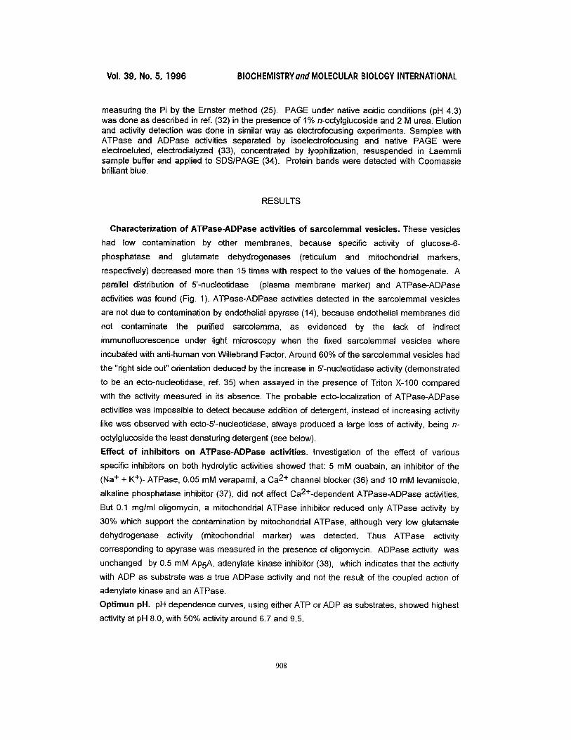
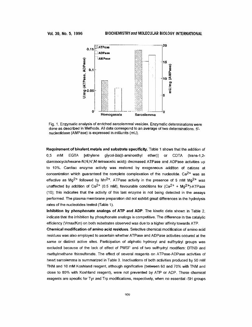
Characterization of ATPase-ADPase activities of sarcolemmal vesicles. These vesicles
had low contamination by other membranes, because specific activity of glucose-6-
phosphatase and glutamate dehydrogenases (reticulum and mitochonddal markers,
respectively) decreased more than 15 times with respect to the values of the homogenate. A
parallel distribution of 5'-nucleotidase (plasma membrane marker) and ATPase-ADPase
activities was found (Fig. 1). ATPase-ADPase activities detected in the sarcolemmal vesicles
are not due to contamination by endothelial apyrase (14), because endothelial membranes did
not contaminate the purified samolemma, as evidenced by the lack of indirect
immunofluorescence under light microscopy when the fixed sarcolemmal vesicles where
incubated with anti-human von Willebrand Factor. Around 60% of the sarcolemmal vesicles had
the "right side out" orientation deduced by the increase in 5'-nudeotidase activity (demonstrated
to be an ecto-nucleotidase, ref. 35) when assayed in the presence of Triton X-100 compared
with the activity measured in its absence. The probable ecto-localization of ATPase-ADPase
activities was impossible to detect because addition of detergent, instead of increasing activity
like was observed with ecto-5'-nucleotidase, always produced a large loss of activity, being n-
octylglucoside the least denaturing detergent (see below).
Effect of inhibitors on ATPase-ADPase activities. Investigation of the effect of various
specific inhibitors on both hydrolytic activities showed that: 5 mM ouabain, an inhibitor of the
(Na + + K+) - ATPase, 0.05 mM verapamil, a Ca 2+ channel blocker (36) and 10 mM levamisole,
alkaline phosphatase inhibitor (37), did not affect Ca2+-dependent ATPase-ADPase activities.
But 0.1 mg/ml oligomycin, a mitochonddal ATPase inhibitor reduced only ATPase activity by
30% which support the contamination by mitochondrial ATPase, although very low glutamate
dehydrogenase activity (mitochondrial marker) was detected. Thus ATPase activity
corresponding to apyrase was measured in the presence of oligomycin. ADPase activity was
unchanged by 0.5 mM AP5A, adenylate kinase inhibitor (38), which indicates that the activity
with ADP as substrate was a true ADPase activity and not the result of the coupled action of
adenylate kinase and an ATPase.
Optimun pH. pH dependence curves, using either ATP or ADP as substrates, showed highest
activity at pH 8.0, with 50% activity around 6.7 and 9.5.
908
V01. 39, No. 5, 1996 BIOCHEMISTRYandMOLECULAR BIOLOGY INTERNATIONAL
• ADPase
i1.
0"1 I ~ 0 . 0 . . . .
Homogenate
\ \ \ \ ' ,
\ \ \ \ . \ \ \ \ , - - - - \ \ \ \ \
N \ \ \ x
\ - . . \ \ x
, , , , \ \ ' - ~
, \ \ \ \ ~ \ \ \ , , \ \ \ \ , \ \ \ x \ \ \ ' \ \ \ \ \ \ \ ~ \ \ - , \
. \ \ x ,
\ \ \ \ " I " \ ~ \ ' 1 \ \ \ \ .
Sarcolemm~
20
15
¢= 13.
10~,~ 01
E 5
Fig. 1. Enzymatic analysis of enriched sarcolemmal vesicles. Enzymatic determinations were done as described in Methods. All data correspond to an average of two determinations. 5'- nucleotidase (AMPase) is expressed in milliunits (mU).
Requirement of bivalent metals and substrate specificity. Table 1 shows that the addition of
0,5 mM EGTA [ethylene glycol-bis(~-aminoethyl ether)] or CDTA (trans-1,2-
diaminocyclohexane-N,N,N',N'-tetraacetic acid)) decreased ATPase and ADPase activities up
to 10%. Cardiac enzyme activity was restored by exogeneous addition of cations at
concentration which guaranteed the complete complexation of the nucleotide. Ca 2+ was as
effective as Mg 2+ followed by Mn 2+. ATPase activity in the presence of 5 mM Mg 2+ was
unaffected by addition of Ca 2+ (0.5 raM), favourable conditions for (Ca 2+ + Mg2+)-ATPase
(10); this indicates that the activity of this last enzyme is not being detected in the assays
performed. The plasma membrane preparation did not exhibit great differences in the hydrolysis
rates of the nucleotides tested (Table 1).
Inhibition by phosphonate analogs of ATP and ADP. The kinetic data shown in Table 2,
indicate that the inhibition by phosphonate analogs is competitive. The difference in the catalytic
efficiency (Vmax/Km) on both substrates observed was due to a higher affinity towards ATP.
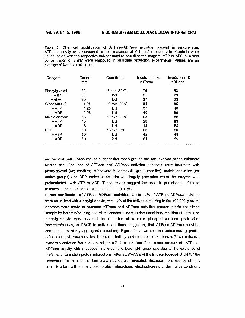
Chemical modification of amino acid residues. Selective chemical modification of amino acid
residues was also employed to ascertain whether ATPase and ADPase activities occured at the
same or distinct active sites. Participation of aliphatic hydroxyl and sulfhydryl groups was
excluded because of the lack of effect of PMSF and of two sulfhydryl modifiers: DTNB and
methylmethane thiosulfonate. The effect of several reagents on ATPase-ADPase activities of
heart sarcolemma is summarized in Table 3. Inactivations of both activities produced by 50 mM
TNM and 10 mM Koshland reagent, although significative (between 60 and 70% with TNM and
close to 80% with Koshland reagent), were not prevented by ATP or ADP. These chemical
reagents are specific for Tyr and Trp modifications, respectively, when no essential -SH groups
909
Vol. 39, No. 5, 1996 BIOCHEMISTRY and MOLECULAR BIOLOGY INTERNATIONAL
Table 1. Bivalent metal ion dependence and nucleotide specificity of the ATPase-ADPase activities of sarcolemmal vesicles. Nucleoside triphosphatase activity was determined in the presence of 0.1 mg/ml oligomycin. Assay conditions are described under Methods using 2 mM nucleotide. For bivalent metal studies, cations were added in the presence of EGTA and CDTA (0.5 mM each one). For substrate specificity, 5 mM CaCI 2 was used. Values are an average of two determinations. 100% activity is defined as ATPase and ADPase activities in the presence of 5 mM CaCI 2.
Bivalent metal ion dependence
Chelator and cation Concn (mM) ATPase % ADPase %
EGTA + CDTA 0.5 each one 11 13 CaCI2 5.0 100 100 MgCI2 5.0 96 95 MnCI2 5.0 67 55 MgCI2 + CaCI2 5.0 and 0.25 96 -* No addition 40 45
Nucleotide specificity
Nucleotide U/mg Nucleotide U/mg
ATP 0.053 ADP 0.069 GTP 0.066 GDP 0.077 CTP 0.105 CDP 0.070 UTP 0.071 UDP 0.062
dTTP 0.075 dTDP 0.062
*Not tested.
Table 2. Kinetic parameters of ATPase-ADPase activities of the sarcolemmal fraction in the presence and absence of nucleotide analogs. The enzyme activity was determined as described in Methods. The activity using ATP as substrate was measured in the presence of 0.1 mg/ml oligomycin. Values are an average of two determinations.
Substrate Vmax (pmol/min ml) Km (mM) Vmax/Km
ATP 0.112 0.13 0.862 ATP + AMP-PcP 0.113 0.31 0.365 ATP + AMPcP 0.112 0.32 0.350
ADP 0.145 0.57 0.250 ADP + AMP-PcP 0.141 0.73 0.192 ADP + AMPcP 0.147 0.92 0.160
910
Vol. 39, No. 5, 1996 BIOCHEMISTRYand MOLECULAR BIOLOGY INTERNATIONAL
Table 3. Chemical modification of ATPase-ADPase activities present in sarcolemma. ATPase activity was measured in the presence of 0.1 mg/ml oligomycin. Controls were preincubated with the respective solvent used to solubilize the reagent. ATP or ADP at a final concentration of 5 mM were employed in substrate protection experiments. Values are an average of two determinations.
Reagent Concn Conditions Inactivation % Inactivation % mM ATPase ADPase
Phenylglyoxal 30 5 min, 30°C 79 63 + ATP 30 ibid 21 29 + ADP 30 ibid 37 23
Woodward K 1.25 10 min, 30°C 84 85 + ATP 1.25 ibid 67 48 + ADP 1.25 ibid 40 55
Maieic anhydr 16 10 min, 30°C 63 89 + ATP 16 ibid 28 63 + ADP 16 ibid 13 54
DEP 50 10 min, 0°C 88 86 + ATP 50 ibid 42 49 + ADP 50 ibid 61 59
i
are present (30). These results suggest that these groups are not involved at the substrate
binding site. The loss of ATPase and ADPase activities observed after treatment with
phenylglyoxal (Arg modifier), Woodward K (carboxylic group modifier), maleic anhydride (for
amino groups) and DEP (selective for His) was largely prevented when the enzyme was
preincubated with ATP or ADP. These results suggest the possible participation of these
residues in the substrate binding and/or in the catalysis.
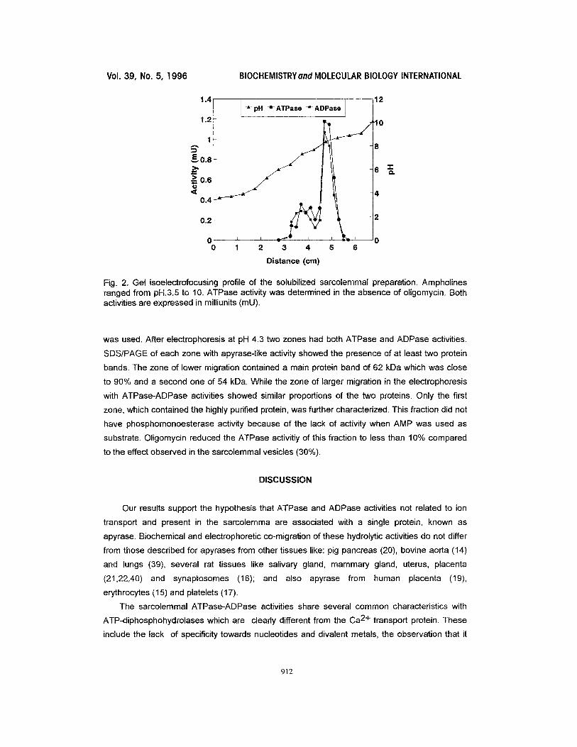
Partial purification of ATPase-ADPase activities. Up to 40% of ATPase-ADPase activities
were solubilized with n-octylglucoside, with 10% of the activity remaining in the 100,000 g pellet.
Attempts were made to separate ATPase and ADPase activities present in this solubilized
sample by isolectrofocusing and electrophoresis under native conditions. Addition of urea and
n-octylglucoside was essential for detection of a main phosphohydrolase peak after
isoelectrofocusing or PAGE in native conditions, suggesting that ATPase-ADPase activities
correspond to highly aggregable protein(s). Figure 2 shows the isoelectrofocusing profile;
ATPase and ADPase activities distributed similarly, and the main peak (close to 70%) of the two
hydrolytic activities focused around pH 8.7. It is not clear if the minor amount of ATPase-
ADPase activity which focused in a wider and louver pH range was due to the existence of
isoforms or to protein-protein interactions. After SDS/PAGE of the fraction focused at pH 8.7 the
presence of a minimum of four protein bands was revealed. Because the presence of salts
could interfere with some protein-protein interactions, electrophoresis under native conditions
911
Vol. 39, No. 5, 1996 BIOCHEMISTRYand MOLECULAR BIOLOGY INTERNATIONAL
1.4
1.2
1
E~0,8
> 0.6 O
0.4
0,2
0 0
-~ pH -e- ATPase -=- ADPase
1 2 3 4 5 6
Distance (cm)
12
'10
8
6 ~ e~
4
2
0
Fig. 2. Gel isoelectrofocusing profile of the solubilized sarcolemmal preparation. Ampholines ranged from pH.3.5 to 10. ATPase activity was determined in the absence of oligomycin. Both activitiesare expressed in milliunits (mU).
was used. After electrophoresis at pH 4.3 two zones had both ATPase and ADPase activities.
SDS/PAGE of each zone with apyrase-like activity showed the presence of at least two protein
bands. The zone of lower migration contained a main protein band of 62 kDa which was close
to 90% and a second one of 54 kDa. While the zone of larger migration in the electrophoresis
with ATPase-ADPase activities showed similar proportions of the two proteins. Only the first
zone, which contained the highly purified protein, was further characterized. This fraction did not
have phosphomonoesterase activity because of the lack of activity when AMP was used as
substrate. Oligomycin reduced the ATPase activitiy of this fraction to less than 10% compared
to the effect observed in the sarcolemmal vesicles (30%).
DISCUSSION
Our results support the hypothesis that ATPase and ADPase activities not related to ion
transport and present in the sarcolemma are associated with a single protein, known as
apyrase. Biochemical and electrophoretic co-migration of these hydrolytic activities do not differ
from those described for apyrases from other tissues like: pig pancreas (20), bovine aorta (14)
and lungs (39), several rat tissues like salivary gland, mammary gland, uterus, placenta
(21,22,40) and synaptosomes (16); and also apyrase from human placenta (19),
erythrocytes (15) and platelets (17).
The sarcolemmal ATPase-ADPase activities share several common characteristics with
ATP-diphosphohydrolases which are clearly different from the Ca 2+ transport protein. These
include the lack of specificity towards nucleotides and divalent metals, the observation that it
912
Vol. 39, No. 5, 1996 BIOCHEMISTRYand MOLECULAR BIOLOGY INTERNATIONAL
does not require Ca 2+ and Mg 2+ simultaneously, and its insensitivity to some specific ATPase
inhibitors and sulfhydryl group reagents.
A common active site for ATPase and ADPase activity is suggested by the similarities in the
pH profile and in the effect of all of the amino acid modifying agents. These last
experiments also allowed a preliminary study of the possible residues involved at the active site.
A comparison of these results with previous data on several rat tissues and human
placental apyrase, indicates an active site homology between this enzyme and the human
placental apyrase (19). These similarities include the possible essentiality of Arg and His
residues, the lack of requirement of sulfhydryl and aliphatic hydroxyl groups, Tyr and Trp.
However, a difference was observed in the probable participation of some Lys and carboxylic
groups in cardiac apyrase compared to the human placental enzyme.
The competitive inhibitory effects of AMP-PcP and AMPcP (nucleotide analogues) on
both ATPase and ADPase activities also support the existence of a single active site for both
activities. This finding agrees with observations on the effect of these compounds on substrate
hydrolysis by human placental apyrase (19).
The potential role of apyrase in the metabolism of nucleotides present in the interstitial
space, regulating their levels and facilitating their reincorporation to the cell, should be
supported by a localization as an ectoenzyme. This localization has been reported in
erythrocytes (15), vascular endothelial and smooth muscle cells (14), synaptosomes (16) and
platelets (17). Circulating nucleotides in blood can bind to the purinoceptors which exist in heart
tissue. Their concentration regulated by ectonucleotidases, in turn, permits regulation of
intracellular processes (1). The existence of a single enzyme with both ATPase and ADPase
activities in the cell surface represents a more efficient system for the modulation of the
extracellular levels of ATP, ADP and adenosine, compounds that have a direct effect in
modulating cardiac function. Finally, the clearance of adenosine can be explained by the
existence of transmembranes adenosine transport (4,41).
Changes in Ca 2+ or Mg 2+ ATPase in some cardiac pathologies has been reported. In
hearts infected with Streptococcus vifidans, which induces endocarditis and myocardial
dysfunction, activities of sarcolemmal Mg2+-ATPase, Ca2+-ATPase and (Na +, K+)-ATPase
were lower in early stages of the infection in comparison to controls (42). These authors suggest
that there is a relation between heart failure and changes in the function of cellular components
during bacterial infective cardiomyopathy. A decrease in Ca2+-ATPase after infection with
Trypanosoma cruzi has also been detected (43). And sarcolemmal Mg2+-dependent ATPase
activity is elevated in rats with chronic diabetes (44). It would be very interesting to study if these
altered bivalent metal dependent ATPases correspond to apyrase activity or to transport
enzymes.
Acknowledgements. This work was supported by Grant Fondecyt No.193-1003 We gratefully acknowledge Dr Phyllis Stein for her critical reading of the manuscript, and Dr Juan Carlos Saez for his valuable suggestions.
913
Vol. 39, NO. 5, ] 996 BIOCHEMISTRYand MOLECULAR BIOLOGY INTERNATIONAL
REFERENCES
1. EI-Moatassim, C., Dornand, J. and Mani, J. C. (1992) Biochim Biophys. Acta 1134, 31-45 2. De Young, M. B. and Scarpa, A. (1987) FEBS Lett. 223, 53-58. 3. Scamps, F. and Vassort, G. (1994) Cir. Res. 74, 710-717. 4. Olsson, R. A. and Pearson, J. D. (1990) Physiol. Rev. 70, 761-845. 5. Schrader, J. (1990) Circulation 81,389-391. 6. Gordon, J. L. (1986) Biochem. J. 233, 309-319. 7. Forrester, T. and Williams, C. A. (1977) J. Physiol. 268, 371-390. 8. Clemens, M. G. and Forrester, T. (1981)J. Physiol. 312, 143-158. 9. Meghji, P., Middleton, K. M. and Newby, A. C. (1988) Biochem. J. 249,695-703. 10. Tuana, B. S. and Dhalla, N. S. (1988) Mol. Cell. Biochem. 81, 75-88. 11. Zhao, D. and Dhalla, N. S. (1991) Mol. Cell. Biochem. 107, 135-149. 12. Zhao, D., Elimban, V. and Dhalla, N. S. (1991) Mol. Cell. Biochem. 107, 151-160. 13. De Vente, J., Velema, J. and Zaagsma, J. (1984) Arch. Biochim. Biophys. 233,180-187. 14. Yagi, K., Shinbo, M., Hashizume, M., Shimba, L. S., Kurimura, S. and Miura, Y. (1991)
Biochem. Biophys. Res. Commun. 180, 1200-1206. 15. Lethje, J., Schomburg, A. and Ogilvie, A. (1988) Eur. J. Biochem. 175, 285-289. 16. Battastini, A. M. O., Rocha, J. B. T., Barcellos, C. K., Dias, R. D. and Sarkis, J. J. F. (1991)
Neurochem. Res. 16, 1303-1310. 17. Frassetto, S. S., Dias, R. D. and Sarkis, J. J. F. (1993) Mol. Cell. Biochem. 129, 47-55. 18. Stanley, K. K., Newby, A. C. and Luzio, J. P. (1982) TIBS, 4, 145-147. 19. Kettlun, A. M., Alvarez, A., Quintar, R., Valenzuela, M. A., Collados, L., Aranda, E., Banda,
A., Chayet, L., Mancilla, M. and Traverso-Cori, A. (1994) Int. J. Biochem. 26,437-448. 20. LeBel, D., Poirier, G. G., Phaneuf, S., St-Jean, P., Laliberte, J. F. and Beaudoin, A. R.
(1980) J. Biol. Chem. 255, 1227-1233. 21. Pieber, M., Valenzuela, M. A., KetUun, A. M., Mancilla, M., Aranda, E., Collados, L. and
Traverso-Cori. A. (1991) Comp. Biochem. Physiol. 100B, 281-285. 22. Valenzuela, M. A., L6pez, J., Depix, M., MancUla, M, Kettlun, A. M., Catal&n, L., Chiong,
M., Garrido, J. and Traverso-Cori, A. (1989) Comp. Biochem. Physiol. 93B, 911-919. 23. Pitts, B. J. R. (1979) J. Biol. Chem. 254, 6232-6235. 24. Fiske, C. H. and SubbaRow, Y. (1925) J. Biol. Chem. 66, 375-400. 25. Ernster, L., Zetterstr5m, R. C. and Lindberg, O. (1950) Acta Chem. Scand. 4, 942-947. 26. Beaufay, H., Amar-Costesec, A., Feytmans, E., Thines-Sempoux, D., Wibo, M, Robbi, M.
and Berthat, J. (1974) J. Cell Biol. 61,188-200. 27. Leighton, F., Poda, B., Beaufay, H., Baudhuin, P., Coffey, J. W., Fowler, S. and De Duve,
C. (1968) J. Cell Biol. 37,482-513. 28. Abbott, N. J., Hughes, C. C. W., Revest, P. A. and Greenwood, J. (1992) J. Cell Sci. 103,
23-37.. 29. Lowry, O. H., Rosebrough, N. J., Farr, A. L. and Randall, R. J. (1951) J. Biol. Chem. 193,
265-275. 30. Means, G. E. and Feeney, R.E. (1971)in Chemical Modification of proteins. Holden-Day.
California. 31. S~nchez, G., Gajardo, M and De Ioannes, A. (1980) Dev. Comp. Immunol. 4, 667-678. 32 Reisfeld, R.A., Lewis, U. J. and Williams, D. E. (1962) Nature 195, 281-283. 33.. Dumbar, B. (1987) in Two-dimensional electrophoresis and immunological techniques.
pp 347-351. Plenum Publishing Co., New York. 34. Laemmli, U. K. (1970) Nature 227, 680-685 35. Frick, G. P. and Lowenstein, J. M. (1976) J. Biol. Chem. 251,6372-6378. 36. Atlas, A. and Adler, M. (1981) Proc. Natl. Acad. Sci. USA 78, 1237-1241. 37. Van Belle, H. (1972) Biochim. Biophys. Acta 289, 158-168.
914
Vol. 39, No. 5, 1996 BIOCHEMISTRYond MOLECULAR BIOLOGY INTERNATIONAL
38. Feldhaus, P., FrShlich, T., Goody, R. S., Isakov, M. and Schirmer. H. (1975) Eur. J. Biochem. 57, 197-204.
39. Picher, M., C5t6, Y. P., B61iveau, R., Poitier, M. and Beaudoin, A. R. (1993) J. Biol. Chem. 268, 4699-4703.
40. Valenzuela, M. A., Collados, L., Kettlun, A. M., Mancilla, M, Lara, H., Puente, J., Aranda, E., Chayet, L., Alvarez, A. and Traverso-Cori, A. (1992) Comp. Biochem. Physiol. 103B, 113-118.
41. Rovetto, M. J. (1985) Annu. Rev. Physiol. 47,605-616. 42. Tomlinson, C. W., Lee, S. L. and Dhalla, N. S. (1976) Cir. Res. 39, 82-92. 43. Nazar~, M. and LeaI-Meirelles, M. N. (1990) J. Histochem. Cytochem. 38, p. 1070. 44. Pierce, G. N. and Dhalla, N. S. (1983)Am. J. Physiol. 245, C241-C247.
915