levels of heat shock protein transcripts in normal follicles and ovarian follicular cysts
TRANSCRIPT
Vol. 11, No. 3
276
SHORT COMMUNICATION
Levels of heat shock protein transcripts in normal follicles and ovarian follicular cysts
Melisa M.L. Velázquez2,3, Natalia S. Alfaro 2,3, Natalia R. Salvetti2,3, Matías
L. Stangaferro2, Florencia Rey2,3, Carolina G. Panzani2, Hugo H. Ortega1,2,3 2Morphological Sciences Department, Faculty of Veterinary Sciences,
National University of Litoral (FCV-UNL), Esperanza, Santa Fe, Argentina; 3Argentine National Research Council (CONICET)
Received: 25 Jannary 2011; accepted: 10 September 2011
SUMMARY In the study, the gene expression of several heat shock proteins (HSPs) was determined in normal follicles and cystic follicles from cattle. A lower expression of HSP10 and HSP40 was observed in granulosa and theca cells of cysts compared to normal follicles. HSP27 was significantly less expressed in granulosa cells in cystic and large antral follicles than in other follicular categories. HSP60 and HSP90a expressions were highest in theca cells of cysts. However, HSP70 and HSP90b exhibited a lower expression in cysts than in healthy follicles. Reproductive Biology 2011 11 3:276-283. Key words: ovarian cyst, cow, cystic ovarian disease, anovulation, heat shock proteins, folliculogenesis INTRODUCTION Heat shock proteins (HSPs) form a diverse group of proteins that are classified according to their molecular weight. Most members of the HSP family perform a chaperone or chaperonin-like function inside the cell [3]. Previous studies demonstrated that several HSPs are
1Corresponding author: Morphological Sciences Department, Faculty of Veterinary Sciences, National University of Litoral (FCV-UNL); R.P. Kreder 2805; (3080) Esperanza, Santa Fe, Argentina; e-mail: [email protected]

277 HSP expression in follicular cysts
expressed under physiological conditions and play an important role in normal cell functions [14]. It is well known that a 90 kDa HSP (HSP90) modulates the function of sex steroid receptor proteins. A 72 kDa HSP (HSP70) has also been associated with sex steroid receptors [8]. These biological processes are very important in ovarian physiology, particularly in follicular development, and they have been implicated in the pathogenesis of cystic ovarian disease (COD; [5, 10]). COD is a common affliction that is characterized by the presence of big ovarian follicular structures (diameter >20 mm), in absence of corpus luteum and ovarian cyclicity. This disease causes financial losses for cattle farmers by delaying conception, and is a primary reason for reproductive culling [13]. In previous studies, protein expressions of HSP27, HSP60, HSP70 and HSP90 were determined to evaluate their associations with healthy follicles or cysts [14]. The aim of this study was to examine the mRNA expressions of HSPs in relation to COD using reverse transcription-polymerase chain reaction (RT-PCR). MATERIALS AND METHODS Bovine ovaries with normal follicles and spontaneous cystic follicles were collected at a local abattoir and were transported immediately to the laboratory. Antral follicles were removed using scissors and scalpel dissection. Follicles were classified as small (<5 mm, n=10), medium (5-10 mm, n=10), large (10-20 mm, n=10) or cystic follicles (>20 mm, n=10; [1]). Granulosa cells (GCs) were obtained from follicular fluid by centrifugation at 1000×g for 10 min. The GC pellets were resuspended in Trizol LS reagent (Invitrogen, CA, USA). The remaining tissue was washed three times with PBS to remove GCs, and subsequently used as thecal tissue samples [1]. The samples were immediately frozen until total RNA extraction.
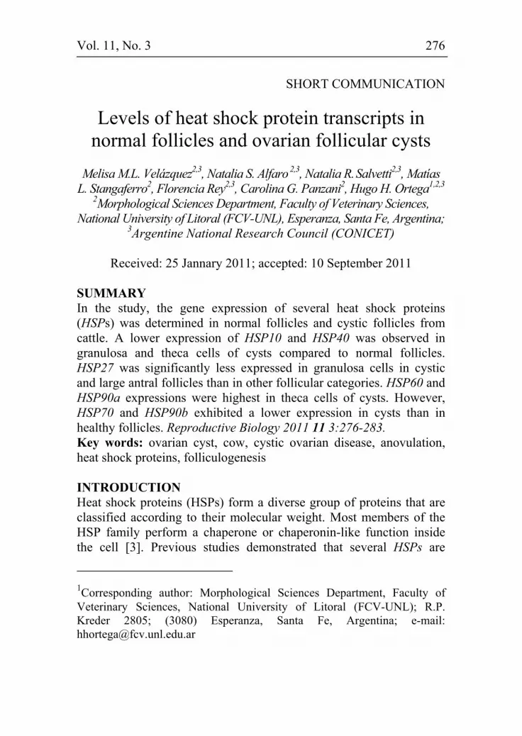
Total RNA was isolated from the samples after treatment with the Trizol LS reagent (Invitrogen, CA, USA) using a homogenizer Ultra Turrax® T10 Basic (IKA WERKE GMBH & CO. KG, Staufen, Germany). Total RNA was obtained with the chloroform/isopropanol method, and the RNA integrity and purity was tested using a fluoroscopy (Qubit, Invitrogen, CA, USA). Reverse transcription (RT) and treatment with DNAse I (Invitrogen) were carried out as reported previously [1]. The used primers and its PCR conditions are presented in Table 1. Glyceraldehyde-3-phosphate dehydrogenase (GAPDH)
278 Velázquez et al
279 HSP expression in follicular cysts
was used as a housekeeping gene [12]. The PCR assay was carried out using reactants in a master mix (Invitrogen, CA, USA) and specific forward/reverse primers for HSP10, HSP27, HSP40, HSP60, HSP70, HSP90a and HSP90b. Prior to HSPs amplification, the cross-contamination of theca and granulosa cells was verified in each sample by PCR detection of the mRNAs that encoded CYP19A1 and CYP17A1.
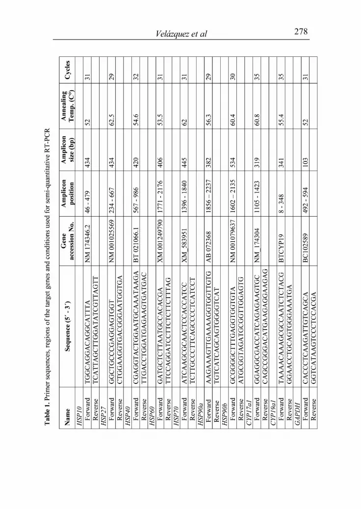
The correct amplified sequences were verified by a direct sequencing service (Macrogen Inc., Seoul, Korea). The PCR products were resolved on a 2% agarose gel containing GelRed Nucleic Acid Gel Stain (Biotium, CA, USA). The image of each gel was visualized under UV illumination, digitized and recorded using a digital camera (Olympus, Tokyo, Japan). The intensity of each band was assessed by densitometry using the Image Pro-Plus 3.1 program. The relative amount of the mRNA of interest was normalized to the GAPDH amplified band for the corresponding sample. GAPDH expression remained constant in all studied samples [9]. Statistic analysis was performed using the SPSS software package (SPSS 11.0 for Windows, SPSS Inc, Chicago, IL, USA). The relative amount of each HSP was analyzed by one-way ANOVA followed by Duncan’s multiple range tests. A p<0.05 value was considered significant. The results are expressed as mean±SEM. RESULTS AND DISCUSSION This is the first report to describe the expressions of HSPs genes in healthy bovine ovarian follicles and cystic follicles. We found lower expression of HSP10 mRNA in granulosa and theca layers of cystic follicles compared to those of healthy follicles (fig. 1). HSP10 protein may be involved in the regulation of several biological functions including protection of healthy follicular cells against apoptosis. Moreover, the decreased level of HSP10 expression observed in cysts might be associated with follicular persistence [11]. The HSP27 protein is involved in steroidogenesis, protein chaperoning and the protection of cells against apoptosis. HSP27 specifically interacts with cytochrome c in the cytosol, and this interaction has functional consequences since it prevents the formation of the apoptosome [4]. In the current study, HSP27 mRNA expression in granulosa cells was significantly lower in cystic and large follicles than in small and medium follicles (fig. 1). In theca cells, the highest expression of
280 Velázquez et al
HSP27 mRNA was detected in healthy medium follicles; no difference was found between large follicles and cysts. Previously, we found that the expression of HSP27 protein in theca cells of tertiary follicles was similar to that present in cysts [14].
Figure 1. Relative mRNA expression (mean±SEM) of HSP10, HSP27, HSP40, HSP60 in granulosa and theca cells of small antral follicles (SF; n=10); medium antral follicles (MF; n=10); large antral follicles (LF; n=10) and cystic follicles (CF, n=10). AU: arbitrary units; different superscripts depict significant differences (p<0.05).
In the current study, we found a lower level of mRNA HSP40 expression in granulosa and theca cell layers of cysts compared to normal follicles (fig. 1). Similar results were observed in the theca cell layer with reference to HSP70 mRNA (fig. 2). It should be emphasized that the HSP40 protein functions as a co-chaperone of mammalian HSP70 [2]. Both, HSP60 and HSP70 are involved in the modulation of steroid receptor function and are related to fertility [6]. In the present study, we noted higher HSP60 mRNA expression in granulosa cells of medium follicles compared to other healthy follicles and cysts, whereas in theca cells the highest expression was observed in cystic follicles. Similarly, more intense immunohistochemical staining of HSP60 was detected in the theca cells of cystic follicles than healthy follicles [14]. Mitochondrial and nuclear HSPs may play a role in the maintenance of metabolic activity and survival of the

281 HSP expression in follicular cysts
oocyte [7], and this may be a reason for higher level of HSP60 observed in the granulosa cell layer of healthy medium follicles.
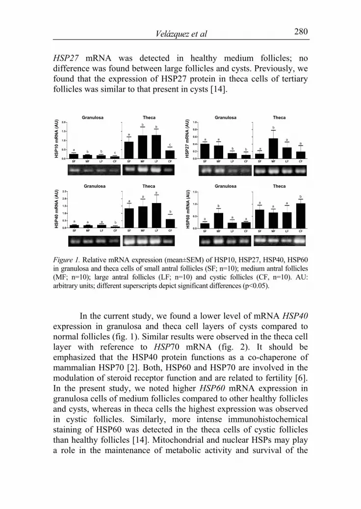
Figure 2. Relative mRNA expression (mean±SEM) of HSP70, HSP90a and HSP90b in granulosa and theca cells of small antral follicles (SF; n=10); medium antral follicles (MF; n=10); large antral follicles (LF; n=10) and cystic follicles (CF, n=10). AU: arbitrary units; different superscripts depict significant differences (p<0.05).
Similar levels of HSP70 transcript were detected in theca cells of all healthy follicles, while its lower expression was observed in cystic follicles (p<0.05; fig. 2). However, granulosa cells of small and medium follicles showed higher expression levels of HSP70 than large and cystic follicles (p<0.05). The ability to inhibit apoptosis, which is widely recognized feature of HSP70 may contribute to the protein protective effect against cell death in healthy ovaries. The expression in HSP70 is probably associated with aberrant expression of steroid hormone receptors in follicles of COD cows [10, 14]. We found some differences in the expression of HSP90b compared to HSP90a (fig. 2). A lower HSP90b mRNA expression was found in theca and granulosa cells of cystic follicles in comparison to healthy follicles. In contrast, there was a higher expression of HSP90 protein in cysts in comparison to remaining follicles [14]. The high expression of HSP90a mRNA found in follicular cysts in the current study was
282 Velázquez et al
corresponding to the protein level [14]. It is possible that antibodies used in our immunohistochemistry-based experiment [14] were able to recognize only the HSP90a isoform. The imbalance between mRNA and protein expression of HSP90 may be a part of cellular deregulation that occurred during the development of COD. Generally, the relative levels of HSP transcripts observed in this study are consistent with the levels of HSP protein reported previously [14]. Our results demonstrated differences in the expression of some HSPs transcripts between normal follicles and ovarian follicular cysts. Therefore, it remains to be determined if any of these HSPs plays a role in the formation of cysts. ACKNOWLEDGEMENTS This study was supported by a grant from the Argentine National Agency for the Promotion of Science and Technology (ANPCyT) (PICT 2005-38101 & 2007-01193). N.R.S., F.R. & H.H.O., are Research Career Members and M.M.L.V. and N.S.A. are Fellows of the National Scientific Research Council (CONICET). REFERENCES 1. Alfaro NS, Salvetti NR, Velazquez MM, Stangaferro ML, Rey F,
Ortega HH 2011 Steroid receptor mRNA expression in the ovarian follicles of cows with cystic ovarian disease. Research in Veterinary Science doi:10.1016/j.rvsc.2011.04.009.
2. Edwards MJ 1998 Apoptosis, the heat shock response, hyperthermia, birth defects, disease and cancer. Where are the common links? Cell Stress Chaperones 3 213-220.
3. Ellis RJ 1993 The general concept of molecular chaperones. Philosophical Transactions of the Royal Society of London Series B-Biologic 339 257-261.
4. Garrido C, Gurbuxani S, Ravagnan L, Kroemer G 2001 Heat shock proteins: endogenous modulators of apoptotic cell death. Biochemical and Biophysical Research Communications 286 433-442.
5. Isobe N, Yoshimura Y 2007 Deficient proliferation and apoptosis in the granulosa and theca interna cells of the bovine cystic follicle. Journal of Reproduction and Development 53 1119-1124.
6. Neuer A, Spandorfer SD, Giraldo P, Dieterle S, Rosenwaks Z, Witkin SS 2000 The role of heat shock proteins in reproduction. Human Reproduction Update 6 149-159.

283 HSP expression in follicular cysts
7. Ohsako S, Bunick D, Hayashi Y 1995 Immunocytochemical observation of the 90 kD heat shock protein (HSP90): high expression in primordial and pre-meiotic germ cells of male and female rat gonads. Journal of Histochemistry & Cytochemistry 43 67-76.
8. Pratt WB, Toft DO 1997 Steroid receptor interactions with heat shock protein and immunophilin chaperones. Endocrine Reviews 18 306-360.
9. Robert C, McGraw S, Massicotte L, Pravetoni M, Gandolfi F, Sirard MA 2002 Quantification of housekeeping transcript levels during the development of bovine preimplantation embryos. Biology of Reproduction 67 1465-1472.
10. Salvetti NR, Muller LA, Acosta JC, Gimeno JE, Ortega HH 2007 Estrogen receptors alpha and beta and progesterone receptors in normal bovine ovarian follicles and cystic ovarian disease. Veterinary Pathology 44 373-378.
11. Salvetti NR, Stangaferro ML, Palomar MM, Alfaro NS, Rey F, Gimeno EJ, Ortega HH 2010 Cell proliferation and survival mechanisms underlying the abnormal persistence of follicular cysts in bovines with cystic ovarian disease induced by ACTH. Animal Reproduction Science 122 98-110.
12. Shibaya M, Matsuda A, Hojo T, Acosta TJ, Okuda K 2007 Expressions of estrogen receptors in the bovine corpus luteum: cyclic changes and effects of prostaglandin F2alpha and cytokines. Journal of Reproduction and Development 53 1059-1068.
13. Silvia WJ, Halter TB, Nugent AM, Laranja da Fonseca LF 2002 Ovarian follicular cysts in dairy cows: an abnormality in folliculogenesis. Domestic Animal Endocrinology 23 167-177.
14. Velázquez MM, Alfaro NS, Dupuy CR, Salvetti NR, Rey F, Ortega HH 2010 Heat shock protein patterns in the bovine ovary and relation with cystic ovarian disease. Animal Reproduction Science 118 201-209.