regulation of neuronal survival and morphology by the e3
TRANSCRIPT
Regulation of neuronal survival and morphology by theE3 ubiquitin ligase RNF157
A Matz1,5, S-J Lee1,5, N Schwedhelm-Domeyer1,5, D Zanini2, A Holubowska1,3, M Kannan1, M Farnworth1, O Jahn3,4, MC Göpfert2
and J Stegmüller*,1,3
Neuronal health is essential for the long-term integrity of the brain. In this study, we characterized the novel E3 ubiquitin ligase ringfinger protein 157 (RNF157), which displays a brain-dominant expression in mouse. RNF157 is a homolog of the E3 ligasemahogunin ring finger-1, which has been previously implicated in spongiform neurodegeneration. We identified RNF157 as aregulator of survival in cultured neurons and established that the ligase activity of RNF157 is crucial for this process. We alsouncovered that independently of its ligase activity, RNF157 regulates dendrite growth and maintenance. We further identified theadaptor protein APBB1 (amyloid beta precursor protein-binding, family B, member 1 or Fe65) as an interactor and proteolyticsubstrate of RNF157 in the control of neuronal survival. Here, the nuclear localization of Fe65 together with its interaction partnerRNA-binding protein SART3 (squamous cell carcinoma antigen recognized by T cells 3 or Tip110) is crucial to trigger apoptosis.In summary, we described that the E3 ligase RNF157 regulates important aspects of neuronal development.Cell Death and Differentiation (2015) 22, 626–642; doi:10.1038/cdd.2014.163; published online 24 October 2014
Neurodegeneration leads to loss of neurons and thus tosevere and irreparable damage of the brain. A commonhistopathological feature in postmortem brains of patients withneurodegenerative diseases such as Parkinson’s or Alzhei-mer’s disease is the presence of ubiquitin-laden proteindeposits.1–3 These deposits implicate the ubiquitin protea-some system (UPS) in neurodegeneration. In addition tohistopathological clues, genetic evidence demonstrates thaterroneous UPS components have detrimental effects on thedeveloping and adult brain resulting in neurodegenerativedisorders.4,5
The UPS is responsible for the posttranslational modifica-tion of proteins by ubiquitin, which requires an enzymaticcascade.6 The E3 ubiquitin ligases specifically recognize thesubstrate proteins and mediate their ubiquitination, which canresult in their degradation that ensures the homeostasis incells or in non-proteolytic signaling events.7,8 The largestgroup of E3 ligases constitutes the RING (really interestingnew gene) ligases, which serve as scaffold proteins to recruitboth the substrate and the E2 ubiquitin-conjugating enzymethat binds to the RING domain,9 facilitating the transfer ofubiquitin from the E2 to the substrate.Although there are several hundred E3 ligases,10 only
a few have been studied so far in the context of neuronalsurvival or neurodegeneration.11–15 Among those, mahoguninring finger-1 (MGRN1) has been implicated in anage-dependent spongiform encephalopathy characterized ina mouse model.15
In this study, we characterized the novel E3 ubiquitin ligasering finger protein 157 (RNF157), the homolog of MGRN1. Wedescribed that RNF157, which is predominantly expressed inthe brain, regulates neuronal survival and morphology incultured neurons. We further identified the adaptor proteinAPBB1 (amyloid beta precursor protein-binding, family B,member 1 or Fe65) as a substrate and a downstreamcomponent in RNF157-regulated neuronal survival. Also, wedemonstrated that nuclear Fe65 together with the RNA-binding protein SART3 (squamous cell carcinoma antigenrecognized by T cells 3 or Tip110) triggers apoptosis. Takentogether, we described that the E3 ligase RNF157 acts indifferent aspects of neuronal development.
Results
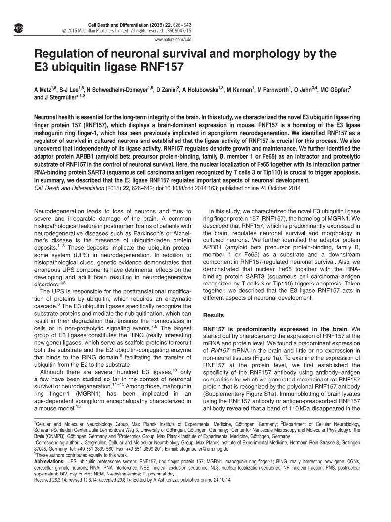
RNF157 is predominantly expressed in the brain. Westarted out by characterizing the expression of RNF157 at themRNA and protein level. We found a predominant expressionof Rnf157 mRNA in the brain and little or no expression innon-neural tissues (Figure 1a). To examine the expression ofRNF157 at the protein level, we first established thespecificity of the RNF157 antibody using antibody–antigencompetition for which we generated recombinant rat RNF157protein that is recognized by the polyclonal RNF157 antibody(Supplementary Figure S1a). Immunoblotting of brain lysatesusing the RNF157 antibody or antigen-preabsorbed RNF157antibody revealed that a band of 110 kDa disappeared in the
1Cellular and Molecular Neurobiology Group, Max Planck Institute of Experimental Medicine, Göttingen, Germany; 2Department of Cellular Neurobiology,Schwann-Schleiden Center, Julia Lermontowa Weg 3, University of Göttingen, Göttingen, Germany; 3Center for Nanoscale Microscopy and Molecular Physiology of theBrain (CNMPB), Göttingen, Germany and 4Proteomics Group, Max Planck Institute of Experimental Medicine, Göttingen, Germany*Corresponding author: J Stegmüller, Cellular and Molecular Neurobiology Group, Max Planck Institute of Experimental Medicine, Hermann Rein Strasse 3, Göttingen37075, Germany. Tel: +49 551 3899 560; Fax: +49 551 3899 201; E-mail: [email protected] authors contributed equally to this work.
Received 26.3.14; revised 19.8.14; accepted 29.8.14; Edited by A Ashkenazi; published online 24.10.14
Abbreviations: UPS, ubiquitin proteasome system; RNF157, ring finger protein 157; MGRN1, mahogunin ring finger-1; RING, really interesting new gene; CGNs,cerebellar granule neurons; RNAi, RNA interference; NES, nuclear exclusion sequence; NLS, nuclear localization sequence; NF, nuclear fraction; PNS, postnuclearsupernatant; DIV, day in vitro; NEM, N-ethylmaleimide; P, postnatal day
Cell Death and Differentiation (2015) 22, 626–642& 2015 Macmillan Publishers Limited All rights reserved 1350-9047/15
www.nature.com/cdd

Figure 1 For caption see next page
RNF157 controls neuronal survivalA Matz et al
627
Cell Death and Differentiation
latter condition (Figure 1b). Also, immunoblotting of lysatesfrom cultured neurons with RNF157 antibody and antigen-preabsorbed RNF157 antibody abolished the 110 kDa band(Figure 1c). The calculated size of rat RNF157 (644 aa;70 kDa), however, did not match the apparent size of RNF157in the gel. We then generated a larger recombinant ratRNF157 protein that lacks the RING domain and whosecalculated size also did not match the size in the gel(Supplementary Figure S1b). Mass spectrometric analysis,however, revealed the correct size and identity of therecombinant RNF157 (Supplementary Figure S1c). Thediscrepancy of theoretical and apparent size could beattributed to the relatively high content of acidic amino acids.Although acidic proteins have been reported to migrate moreslowly in SDS-PAGE,16,17 we cannot exclude that theposttranslational modifications contribute to the anomalousrunning behavior of RNF157. Taken together, these experi-ments established the specificity of the RNF157 antibody andidentified RNF157 as an acidic, 110 kDa protein.Consistent with the RT-PCR results, immunoblotting
demonstrated that RNF157 was predominantly expressed innervous tissue at postnatal day (P) 12, whereas no expressionwas detectable in non-neural tissues (Figure 1d). To determinethe temporal expression of RNF157 in hippocampus, cortexand cerebellum, we analyzed tissues from embryonic day 18to P60 and found robust RNF157 expression in hippocampusand cortex and lower expression in the cerebellum (Figure 1e).We also found this expression pattern in a sagittal brainsection of an RNF157 knockout mouse (Figure 1f,Supplementary Figures S2a–c). Also, we found robustexpression of RNF157 in cultured neurons (Figure 1g).Interestingly, while RNF157 and the homologous E3
ligase MGRN-1 are encoded by two independent genes inmammals, Drosophila has one RNF157-like gene (CG9941).To assess its expression pattern, we generated transgenicflies and found that CG9941 is restricted to the nervoussystem both in larvae and adult flies overlapping with the pan-neuronal marker nc82 (bruchpilot) (Supplementary FiguresS2d and e). These analyses revealed that RNF157 isevolutionarily conserved and predominantly expressed inthe brain.We then determined the subcellular localization of RNF157
and subjected cerebellar granule neurons (CGNs) to fractio-nation analysis and found RNF157 in the postnuclear super-natant (PNS) (Figure 1h). With further fractionation analyses
using postnatal cortices, we subjected the PNS to ultracen-trifugation steps and found that RNF157 was present in thepurified cytoplasmic fraction (Figure 1i). To confirm theseresults, we transfected hippocampal neurons with the GFP-RNF157 plasmid because the RNF157 antibody fails to workin immunocytochemistry. Here, we foundGFP-RNF157 evenlydistributed in the somatic cytoplasm and processes but absentfrom the nucleus (Figure 1j). These results identify RNF157 asa cytoplasmic protein.
The E3 ligase RNF157 promotes neuronal survival.Having generated the RNF157 knockout mouse, we did notfind any obvious phenotypic differences in young mice (5-day/6-week-old mice). The full analysis of the RNF157 KO micehas only recently been started and is still work in progress.Before generating this mouse, we took an RNA interference(RNAi) approach to determine the function of RNF157 incultured neurons. To acutely knockdown RNF157, wedesigned several vector-based small hairpins targetingRNF157 and determined that RNF157 RNAi #3 was mostefficient in doing so (Figure 2a). Neither of the RNF157 RNAiconstructs induced knockdown of MGRN1 (SupplementaryFigure S3a). We also verified the endogenous knockdown ofRNF157 in neurons (Figure 2b). When we examined CGNs,transfected at day in vitro (DIV) 2 with control vector (U6promoter in pBluescript) or the RNF157 RNAi #2 and #3plasmids, we found that acute knockdown of RNF157 withboth functional RNF157 RNAi plasmids induced a significantincrease in apoptosis, with RNF157 RNAi #3 being morepotent (Figure 2c). Also, RNF157 knockdown neuronsdisplayed characteristic signs of apoptosis including pyknoticnuclei, axonal fragmentation and cleaved caspase-3 immuno-reactivity (Figure 2d). In contrast to RNF157 RNAi inmammalian neurons, knockdown of the Drosophila ortholo-gue CG9941 in the fly brain did not produce any obvioussigns of neuronal apoptosis in the brain of young and olderflies (Supplementary Figures S3b and c). We then examinedif RNF157 supports neuronal survival in other mammalianneuronal cell types. We analyzed hippocampal neuronstransfected with control vector or RNF157 RNAi #3 plasmidand found a similar apoptotic response in RNF157knockdown neurons (Figure 2e). We next inducedRNF157 knockdown in neurons of organotypic corticalslices and found that RNF157 RNAi #3 significantlyinduced apoptosis as compared with control conditions
Figure 1 RNF157 is predominantly expressed in the brain. (a) cDNA of indicated tissues isolated from P12 rats were subjected to PCR analysis with RNF157- and GAPDH-specific primers. The latter served as loading control. (b) Cerebellar and hippocampal lysates from P12 rats were subjected to immunoblotting with the RNF157 antibody, theRNF157 antibody preabsorbed with RNF157 antigen or the γ-tubulin antibody. The latter served as loading control. Arrowhead indicates specific RNF157 band. Asterisks indicatenonspecific bands. (c) Lysates of cultured cortical (CTN), hippocampal (HPN) and CGNs were immunoblotted with the RNF157 antibody, the RNF157 antibody preabsorbed withRNF157 antigen or the γ-tubulin antibody. The latter served as loading control. (d) Lysates of indicated neural and non-neural rat tissues were immunoblotted with the RNF157 orthe 14-3-3β antibody. The latter served as loading control. (e) Lysates of rat cortex, cerebellum and hippocampus isolated at embryonic day (E) 18 to P60, were immunoblottedwith the RNF157 or the 14-3-3β antibodies. The latter served as loading control. (f) Sagittal vibratome sections from P28 wild-type and RNF157− /− mice, in which the Rnf157gene was interrupted by a gene trap cassette encoding β-Geomycin, were subjected to X-Gal staining. Scale bar equals 1 mm. (g) Lysates of CGNs and cortical neurons, culturedfor indicated days (DIV), were immunoblotted with the RNF157 or the 14-3-3β antibodies. The latter served as loading control. (h) Lysates of CGNs were subjected to subcellularfractionation and immunoblotted with the RNF157, 14-3-3β and SP1 antibodies. The latter two served as quality control for PNS and NF, respectively. (i) P5 cortex was lysed andfractionated into NF and PNS. The PNS was further processed by ultracentrifugation to remove plasma membrane and heavy membrane components, resulting in PNS I, PNS II,pellet I and II and the cytoplasmic fraction. These fractions were immunoblotted with the RNF157, SnoN (NF), pan14-3-3 (cytoplasmic fraction) and N-Cadherin (plasmamembrane marker) antibodies. Lysates of wild-type and RNF157− /− brains were used as positive control for the specific RNF157 band indicated by arrowhead.(j) Hippocampal neurons were transfected with the GFP-RNF157 expression plasmid, subjected to immunostaining with the GFP and the neuronal marker TuJ1 antibodies (classIII β-tubulin) and analyzed with confocal microscopy. Scale bar= 20 μm
RNF157 controls neuronal survivalA Matz et al
628
Cell Death and Differentiation

Figure 2 For caption see next page
RNF157 controls neuronal survivalA Matz et al
629
Cell Death and Differentiation

(Figure 2f, Supplementary Figure S3d). These results indicatethat RNF157 promotes survival in mammalian neurons.To ensure the specific phenotype of RNF157 knockdown
and to rule out off-target effects, we constructed and validatedan RNF157 RNAi-resistant RNF157-Rescue (Res) plasmid(Supplementary Figures S3e and f). Subsequent rescueexperiments in CGNs, in which the RNF157-Res plasmidwas expressed together with RNF157 RNAi, demonstrated asignificantly lower apoptotic rate as compared to RNF157knockdown neurons (Figure 2g). In a similar experiment, weconfirmed the full rescue of RNF157 knockdown-induced celldeath in hippocampal neurons (Figure 2h). To underscore thatRNF157 promotes neuronal survival, we investigated ifoverexpression of RNF157 protects CGNs from apoptosis.We exposed control-transfected (pCMVmyc) or RNF157-overexpressing CGNs to serum/KCl withdrawal to induceapoptosis. Remarkably, we found that RNF157-expressingneurons displayed significant resistance to serum/KCl with-drawal (Figure 2i). We also determined whether neuronalsurvival is dependent on the E3 ligase activity of RNF157 andtransfected the neurons with the RNF157 ΔRING mutant, inwhich the RING domain is deleted to abolish ligase activity.Here we found that unlikewild-type RNF157, expression of theRNF157 ΔRING mutant did not result in a decrease ofapoptosis (Figure 2i). These data support the notion thatRNF157 is promoting survival in an E3 ligase-dependentmanner in cultured neurons.
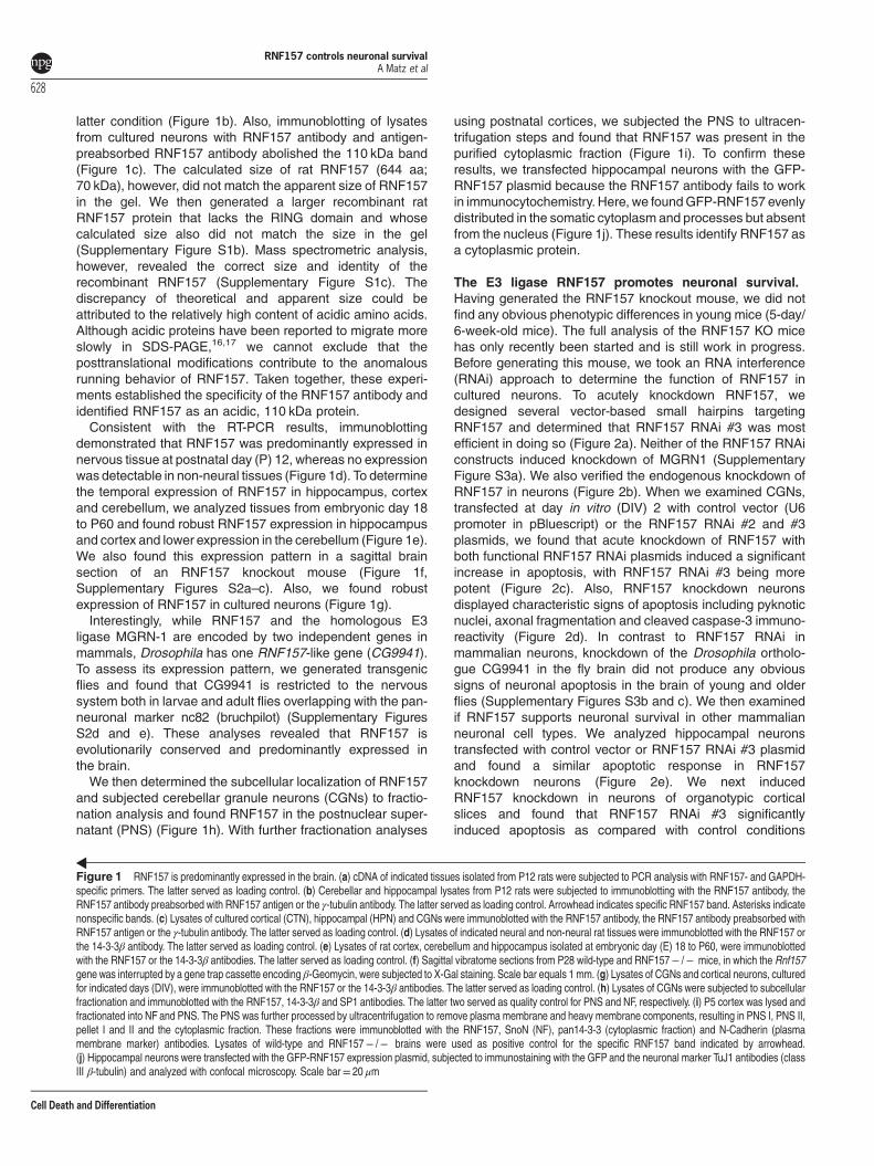
RNF157 supports dendrite growth and maintenanceindependent of its E3 ligase activity. To investigate furtherRNF157 functions in neurons, we transfected CGNs withcontrol vector or RNF157 RNAi #2 and #3 plasmids at a timewhen neuronal polarity is fully established. To preventmorphological effects because of compromised cellularhealth upon RNF157 knockdown, we co-transfected aplasmid encoding Bcl-XL, which promotes survival withoutaffecting neuronal morphology.18 We then subjected trans-fected CGNs to morphological analyses, which revealed thatRNF157 knockdown led to reduced total dendritic length ascompared to control neurons (Figures 3a and b). The lengthof the axon (longest process), however, remained unaffected(Figure 3c). As a consequence of reduced total dendriticlength, RNF157 RNAi neurons harbored much simpler
dendritic arbors (Figure 3d). When we monitored dendritedevelopment and stability for several days, we found that lossof RNF157 led to a destabilization and shrinkage ofpreviously established dendrites (Figure 3e). To ensure thespecific dendritic phenotype of RNF157 knockdown, wecarried out rescue experiments, which revealed that expres-sion of the RNF157 rescue plasmid upon RNF157 knock-down restored dendrites (Figure 3f). In gain-of-functionexperiments, we addressed whether overexpression ofRNF157 has the opposite effect on dendrite length. Wetransfected neurons with the control vector or the RNF157expression plasmid and found that RNF157 overexpressionresulted in exuberant dendritic growth (Figures 3g and h).Interestingly, expression of RNF157 ΔRING induced anequally strong increase in dendritic growth (Figures 3g andh). Accordingly, RNF157 overexpression resulted a morecomplex dendritic arborization (Figure 3i). To examinewhether the dendrite-stabilizing function of RNF157 holdstrue in other types of neurons, we induced RNF157 knock-down in hippocampal neurons and found very small dendritictrees, while axonal lengths were unaffected (Figures 3j–l).Collectively, these data indicate that RNF157 supportsgrowth and maintenance of dendrites independent of its E3ligase activity in cultured neurons.
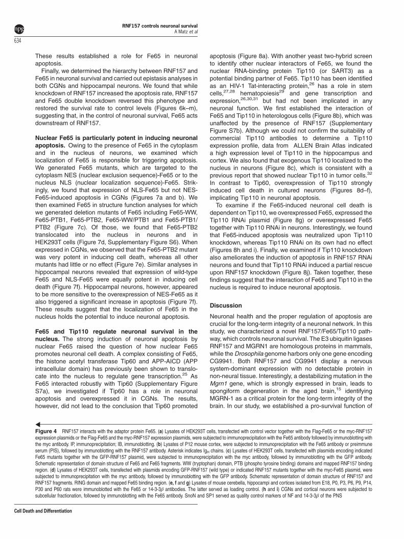
The adaptor protein Fe65 interacts with RNF157. Todelineate a mechanism by which the E3 ligase RNF157operates in neurons, we carried out a yeast two-hybridanalysis using a fetal human brain library. Here, we identifiedthe adaptor protein Fe65 (or APBB1) as an interactor ofRNF157. Fe65, Fe65-L1 and Fe65-L2 constitutes the Fe65adaptor protein family,19,20 of which only Fe65 displays abrain-dominant expression.20
To validate the interaction of Fe65 and RNF157, we carriedout immunoprecipitation experiments and found that Fe65robustly associated with RNF157 (Figure 4a). In a reciprocalco-immunoprecipitation, we confirmed this interaction(Supplementary Figure S4a). To establish their interaction inthe brain, we first demonstrated that the Fe65 antibodyimmunoprecipitated Fe65 from brain tissue (SupplementaryFigure S4b). In subsequent co-immunoprecipitation analyses,we found that RNF157 and Fe65 interacted endogenouslyin the brain (Figure 4b,Supplementary Figure S4c).
Figure 2 RNF157 promotes neuronal survival. (a) Lysates of HEK293T cells, transfected with the GFP-RNF157 plasmid together with control vector or plasmids encodingRNF157 RNAi #1, #2 or #3, were immunoblotted with the GFP or γ-tubulin antibodies. The latter served as loading control. (b) Cortical neurons were mixed with control vector orRNF157 RNAi #3 plasmid and subjected to electroporation using the Amaxa nucleofector. After 5 days in culture, lysates of neurons were immunoblotted with the RNF157 or theγ-tubulin antibodies. The latter served as loading control. (c) CGNs, transfected at DIV2 with control vector or plasmids encoding RNF157 RNAi #2 or #3 together with the β-Galexpression plasmid, were subjected to immunocytochemistry at DIV6 with the β-Gal antibody and the nuclear dye bisbenzimide Hoechst 33258. A total of 1225 neurons fromthree independent experiments were included in the analysis (ANOVA, *Po0.05, ***Po0.001, mean +S.E.M.). (d) Representative neurons from 2c, which were co-stained withβ-Gal and cleaved caspase-3 antibodies together with the DNA dye bisbenzimide (Hoechst). Arrowheads indicate transfected neurons. Insets show higher magnification ofHoechst-stained nuclei. Asterisks indicate apoptotic bodies, arrowheads indicate transfected neurons and arrow indicates pyknotic nucleus. Scale bar equals 20 μm.(e) Hippocampal neurons, transfected at DIV2 with control vector or a plasmid encoding RNF157 RNAi #3 together with the β-Gal expression plasmid, were analyzed as describedin 2c. A total of 655 neurons from three independent experiments were included in the analysis (Student’s t-test, ***Po0.001, mean +S.E.M.). (f) Organotypic slice cultures from P0cortex were transfected at DIV2 with control vector, RNF157 RNAi #1 or #3 and analyzed at DIV6. A total of 217 neurons from three independent experiments were included in theanalysis (ANOVA, ***Po0.001, mean +S.E.M.). (g) CGNs, transfected with control vectors, RNF157 RNAi or RNF157 RNAi and the RNF157-Res plasmid together with the β-Galexpression plasmid, were analyzed as in 2c. A total of 2171 neurons from four independent experiments were included in the analysis (ANOVA, ***Po0.001, mean +S.E.M.).(h) Hippocampal neurons, transfected at DIV2 with control vector, the RNF157 RNAi #3, the RNF157-Res plasmid, or both the RNF157 RNAi #3 and RNF157-Res plasmids, wereanalyzed at DIV7. A total of 1099 neurons from four independent experiments were included in the analysis (ANOVA, ***Po0.001, mean +S.E.M.). (i) CGNs, transfected withcontrol vector, the RNF157 or RNF157 ΔRING expression plasmids together with the β-Gal plasmid, were exposed to serum and KCl withdrawal for 24 h before analysis asdescribed in 2c. A total 1383 neurons from four independent experiments were included in the analysis (ANOVA, *Po0.05 ***Po0.001, mean +S.E.M.)
RNF157 controls neuronal survivalA Matz et al
630
Cell Death and Differentiation

Figure 3 For caption see next page
RNF157 controls neuronal survivalA Matz et al
631
Cell Death and Differentiation
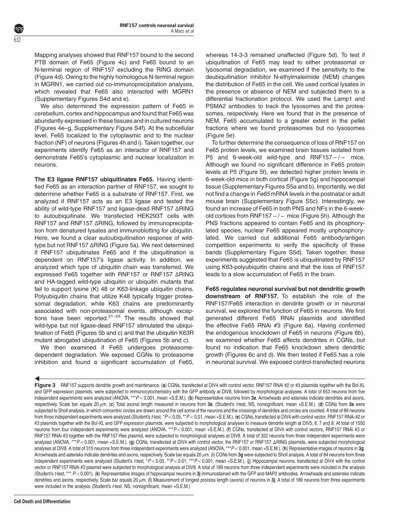
Mapping analyses showed that RNF157 bound to the secondPTB domain of Fe65 (Figure 4c) and Fe65 bound to anN-terminal region of RNF157 excluding the RING domain(Figure 4d). Owing to the highly homologousN-terminal regionin MGRN1, we carried out co-immunoprecipitation analyses,which revealed that Fe65 also interacted with MGRN1(Supplementary Figures S4d and e).We also determined the expression pattern of Fe65 in
cerebellum, cortex and hippocampus and found that Fe65 wasabundantly expressed in these tissues and in cultured neurons(Figures 4e–g, Supplementary Figure S4f). At the subcellularlevel, Fe65 localized to the cytoplasmic and to the nuclearfraction (NF) of neurons (Figures 4h and i). Taken together, ourexperiments identify Fe65 as an interactor of RNF157 anddemonstrate Fe65’s cytoplasmic and nuclear localization inneurons.
The E3 ligase RNF157 ubiquitinates Fe65. Having identi-fied Fe65 as an interaction partner of RNF157, we sought todetermine whether Fe65 is a substrate of RNF157. First, weanalyzed if RNF157 acts as an E3 ligase and tested theability of wild-type RNF157 and ligase-dead RNF157 ΔRINGto autoubiquitinate. We transfected HEK293T cells withRNF157 and RNF157 ΔRING, followed by immunoprecipita-tion from denatured lysates and immunoblotting for ubiquitin.Here, we found a clear autoubiquitination response of wild-type but not RNF157 ΔRING (Figure 5a). We next determinedif RNF157 ubiquitinates Fe65 and if the ubiquitination isdependent on RNF157’s ligase activity. In addition, weanalyzed which type of ubiquitin chain was transferred. Weexpressed Fe65 together with RNF157 or RNF157 ΔRINGand HA-tagged wild-type ubiquitin or ubiquitin mutants thatfail to support lysine (K) 48 or K63-linkage ubiquitin chains.Polyubiquitin chains that utilize K48 typically trigger protea-somal degradation, while K63 chains are predominantlyassociated with non-proteasomal events, although excep-tions have been reported.21–24 The results showed thatwild-type but not ligase-dead RNF157 stimulated the ubiqui-tination of Fe65 (Figures 5b and c) and that the ubiquitin K63Rmutant abrogated ubiquitination of Fe65 (Figures 5b and c).We then examined if Fe65 undergoes proteasome-
dependent degradation. We exposed CGNs to proteasomeinhibition and found a significant accumulation of Fe65,
whereas 14-3-3 remained unaffected (Figure 5d). To test ifubiquitination of Fe65 may lead to either proteasomal orlysosomal degradation, we examined if the sensitivity to thedeubiquitination inhibitor N-ethylmaleimide (NEM) changesthe distribution of Fe65 in the cell. We used cortical lysates inthe presence or absence of NEM and subjected them to adifferential fractionation protocol. We used the Lamp1 andPSMA2 antibodies to track the lysosomes and the protea-somes, respectively. Here we found that in the presence ofNEM, Fe65 accumulated to a greater extent in the pelletfractions where we found proteasomes but no lysosomes(Figure 5e).To further determine the consequence of loss of RNF157 on
Fe65 protein levels, we examined brain tissues isolated fromP5 and 6-week-old wild-type and RNF157− /− mice.Although we found no significant difference in Fe65 proteinlevels at P5 (Figure 5f), we detected higher protein levels in6-week-old mice in both cortical (Figure 5g) and hippocampaltissue (Supplementary Figures S5a and b). Importantly, we didnot find a change in Fe65mRNA levels in the postnatal or adultmouse brain (Supplementary Figure S5c). Interestingly, wefound an increase of Fe65 in both PNS and NFs in the 6-week-old cortices from RNF157− /− mice (Figure 5h). Although thePNS fractions appeared to contain Fe65 and its phosphory-lated species, nuclear Fe65 appeared mostly unphosphory-lated. We carried out additional Fe65 antibody/antigencompetition experiments to verify the specificity of thesebands (Supplementary Figure S5d). Taken together, theseexperiments suggested that Fe65 is ubiquitinated by RNF157using K63-polyubiquitin chains and that the loss of RNF157leads to a slow accumulation of Fe65 in the brain.
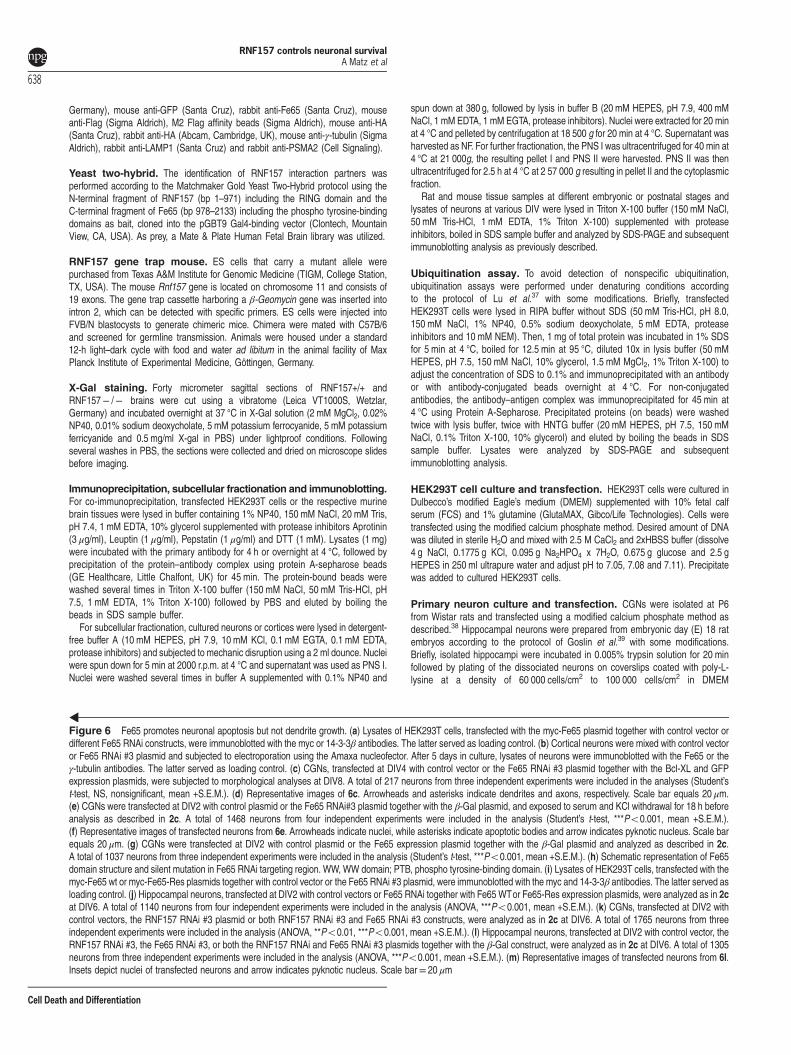
Fe65 regulates neuronal survival but not dendritic growthdownstream of RNF157. To establish the role of theRNF157/Fe65 interaction in dendrite growth or in neuronalsurvival, we explored the function of Fe65 in neurons. We firstgenerated different Fe65 RNAi plasmids and identifiedthe effective Fe65 RNAi #3 (Figure 6a). Having confirmedthe endogenous knockdown of Fe65 in neurons (Figure 6b),we examined whether Fe65 affects dendrites in CGNs, butfound no indication that Fe65 knockdown alters dendriticgrowth (Figures 6c and d). We then tested if Fe65 has a rolein neuronal survival. We exposed control-transfected neurons
Figure 3 RNF157 supports dendrite growth and maintenance. (a) CGNs, transfected at DIV4 with control vector, RNF157 RNAi #2 or #3 plasmids together with the Bcl-XLand GFP expression plasmids, were subjected to immunocytochemistry with the GFP antibody at DIV8, followed by morphological analyses. A total of 653 neurons from fiveindependent experiments were analyzed (ANOVA, ***Po0.001, mean +S.E.M.). (b) Representative neurons from 3a. Arrowheads and asterisks indicate dendrites and axons,respectively. Scale bar equals 20 μm. (c) Total axonal length measured in neurons from 3a. (Student’s t-test, NS, nonsignificant, mean +S.E.M.). (d) CGNs from 3a weresubjected to Sholl analysis, in which concentric circles are drawn around the cell soma of the neurons and the crossings of dendrites and circles are counted. A total of 89 neuronsfrom three independent experiments were analyzed (Student’s t-test, *Po0.05, **Po0.01, mean +S.E.M.). (e) CGNs, transfected at DIV4 with control vector, RNF157 RNAi #2 or#3 plasmids together with the Bcl-XL and GFP expression plasmids, were subjected to morphological analyses to measure dendrite length at DIV5, 6, 7 and 8. At total of 1550neurons from four independent experiments were analyzed (ANOVA, ***Po0.001, mean +S.E.M.). (f) CGNs, transfected at DIV4 with control vectors, RNF157 RNAi #3 orRNF157 RNAi #3 together with the RNF157-Res plasmid, were subjected to morphological analyses at DIV8. A total of 322 neurons from three independent experiments wereanalyzed (ANOVA, ***Po0.001, mean +S.E.M.). (g) CGNs, transfected at DIV4 with control vector, the RNF157 or RNF157 ΔRING plasmids, were subjected morphologicalanalyses at DIV8. A total of 315 neurons from three independent experiments were analyzed (ANOVA, ***Po0.001, mean +S.E.M.). (h) Representative images of neurons in 3g.Arrowheads and asterisks indicate dendrites and axons, respectively. Scale bar equals 20 μm. (i) CGNs from 3g were subjected to Sholl analysis. A total of 84 neurons from threeindependent experiments were analyzed (Student’s t-test, *Po0.05, **Po0.01, ***Po0.001, mean +S.E.M.). (j) Hippocampal neurons, transfected at DIV4 with the controlvector or RNF157 RNAi #3 plasmid were subjected to morphological analysis at DIV8. A total of 189 neurons from three independent experiments were included in the analysis(Student’s t-test, *** Po0.001). (k) Representative images of hippocampal neurons in 3j immunostained with the GFP and MAP2 antibodies. Arrowheads and asterisks indicatedendrites and axons, respectively. Scale bar equals 20 μm. (l) Measurement of longest process length (axons) of neurons in 3j. A total of 189 neurons from three experimentswere included in the analysis (Student’s t-test, NS, nonsignificant, mean +S.E.M.)
RNF157 controls neuronal survivalA Matz et al
632
Cell Death and Differentiation
and Fe65 RNAi neurons, without co-transfection of Bcl-XL, toserum/KCl withdrawal and determined the apoptotic rateinduced by stress. Interestingly, we found that knockdown ofFe65 had a rescue-like effect in neurons (Figures 6e and f).To support this finding, we overexpressed Fe65 and foundthat this led to an increase in apoptosis (Figure 6g). Using a
validated Fe65-Rescue (Res) plasmid (Figures 6h and i), wecarried out a rescue experiment, in which we overexpressedwild-type Fe65 or Fe65-Res in the background of Fe65 RNAi.Although expression of wild-type Fe65/Fe65 RNAi had noeffect on neuronal survival, expression of Fe65-Res/Fe65RNAi led to a significant increase in cell death (Figure 6j).
Figure 4 For caption see next page
RNF157 controls neuronal survivalA Matz et al
633
Cell Death and Differentiation
These results established a role for Fe65 in neuronalapoptosis.Finally, we determined the hierarchy between RNF157 and
Fe65 in neuronal survival and carried out epistasis analyses inboth CGNs and hippocampal neurons. We found that whileknockdown of RNF157 increased the apoptosis rate, RNF157and Fe65 double knockdown reversed this phenotype andrestored the survival rate to control levels (Figures 6k–m),suggesting that, in the control of neuronal survival, Fe65 actsdownstream of RNF157.
Nuclear Fe65 is particularly potent in inducing neuronalapoptosis. Owing to the presence of Fe65 in the cytoplasmand in the nucleus of neurons, we examined whichlocalization of Fe65 is responsible for triggering apoptosis.We generated Fe65 mutants, which are targeted to thecytoplasm NES (nuclear exclusion sequence)-Fe65 or to thenucleus NLS (nuclear localization sequence)-Fe65. Strik-ingly, we found that expression of NLS-Fe65 but not NES-Fe65-induced apoptosis in CGNs (Figures 7a and b). Wethen examined Fe65 in structure function analyses for whichwe generated deletion mutants of Fe65 including Fe65-WW,Fe65-PTB1, Fe65-PTB2, Fe65-WW/PTB1 and Fe65-PTB1/PTB2 (Figure 7c). Of those, we found that Fe65-PTB2translocated into the nucleus in neurons and inHEK293T cells (Figure 7d, Supplementary Figure S6). Whenexpressed in CGNs, we observed that the Fe65-PTB2 mutantwas very potent in inducing cell death, whereas all othermutants had little or no effect (Figure 7e). Similar analyses inhippocampal neurons revealed that expression of wild-typeFe65 and NLS-Fe65 were equally potent in inducing celldeath (Figure 7f). Hippocampal neurons, however, appearedto be more sensitive to the overexpression of NES-Fe65 as italso triggered a significant increase in apoptosis (Figure 7f).These results suggest that the localization of Fe65 in thenucleus holds the potential to induce neuronal apoptosis.
Fe65 and Tip110 regulate neuronal survival in thenucleus. The strong induction of neuronal apoptosis bynuclear Fe65 raised the question of how nuclear Fe65promotes neuronal cell death. A complex consisting of Fe65,the histone acetyl transferase Tip60 and APP-AICD (APPintracellular domain) has previously been shown to translo-cate into the nucleus to regulate gene transcription.25 AsFe65 interacted robustly with Tip60 (Supplementary FigureS7a), we investigated if Tip60 has a role in neuronalapoptosis and overexpressed it in CGNs. The results,however, did not lead to the conclusion that Tip60 promoted
apoptosis (Figure 8a). With another yeast two-hybrid screento identify other nuclear interactors of Fe65, we found thenuclear RNA-binding protein Tip110 (or SART3) as apotential binding partner of Fe65. Tip110 has been identifiedas an HIV-1 Tat-interacting protein,26 has a role in stemcells,27,28 hematopoiesis29 and gene transcription andexpression,26,30,31 but had not been implicated in anyneuronal function. We first established the interaction ofFe65 and Tip110 in heterologous cells (Figure 8b), which wasunaffected by the presence of RNF157 (SupplementaryFigure S7b). Although we could not confirm the suitability ofcommercial Tip110 antibodies to determine a Tip110expression profile, data from ALLEN Brain Atlas indicateda high expression level of Tip110 in the hippocampus andcortex. We also found that exogenous Tip110 localized to thenucleus in neurons (Figure 8c), which is consistent with aprevious report that showed nuclear Tip110 in tumor cells.32
In contrast to Tip60, overexpression of Tip110 stronglyinduced cell death in cultured neurons (Figures 8d–f),implicating Tip110 in neuronal apoptosis.To examine if the Fe65-induced neuronal cell death is
dependent on Tip110, we overexpressed Fe65, expressed theTip110 RNAi plasmid (Figure 8g) or overexpressed Fe65together with Tip110 RNAi in neurons. Interestingly, we foundthat Fe65-induced apoptosis was neutralized upon Tip110knockdown, whereas Tip110 RNAi on its own had no effect(Figures 8h and i). Finally, we examined if Tip110 knockdownalso ameliorates the induction of apoptosis in RNF157 RNAineurons and found that Tip110 RNAi induced a partial rescueupon RNF157 knockdown (Figure 8j). Taken together, thesefindings suggest that the interaction of Fe65 and Tip110 in thenucleus is required to induce neuronal apoptosis.
Discussion
Neuronal health and the proper regulation of apoptosis arecrucial for the long-term integrity of a neuronal network. In thisstudy, we characterized a novel RNF157/Fe65/Tip110 path-way, which controls neuronal survival. The E3 ubiquitin ligasesRNF157 and MGRN1 are homologous proteins in mammals,while theDrosophila genome harbors only one gene encodingCG9941. Both RNF157 and CG9941 display a nervoussystem-dominant expression with no detectable protein innon-neural tissue. Interestingly, a destabilizing mutation in theMgrn1 gene, which is strongly expressed in brain, leads tospongiform degeneration in the aged brain,15 identifyingMGRN-1 as a critical protein for the long-term integrity of thebrain. In our study, we established a pro-survival function of
Figure 4 RNF157 interacts with the adaptor protein Fe65. (a) Lysates of HEK293T cells, transfected with control vector together with the Flag-Fe65 or the myc-RNF157expression plasmids or the Flag-Fe65 and the myc-RNF157 expression plasmids, were subjected to immunoprecipitation with the Fe65 antibody followed by immunoblotting withthe myc antibody. IP, immunoprecipitation; IB, immunoblotting. (b) Lysates of P12 mouse cortex, were subjected to immunoprecipitation with the Fe65 antibody or preimmuneserum (PIS), followed by immunoblotting with the RNF157 antibody. Asterisk indicates IgH chains. (c) Lysates of HEK293T cells, transfected with plasmids encoding indicatedFe65 mutants together with the GFP-RNF157 plasmid, were subjected to immunoprecipitation with the myc antibody, followed by immunoblotting with the GFP antibody.Schematic representation of domain structure of Fe65 and Fe65 fragments. WW (tryptophan) domain, PTB (phospho tyrosine binding) domains and mapped RNF157 bindingregion. (d) Lysates of HEK293T cells, transfected with plasmids encoding GFP-RNF157 (wild type) or indicated RNF157 mutants together with the myc-Fe65 plasmid, weresubjected to immunoprecipitation with the myc antibody, followed by immunoblotting with the GFP antibody. Schematic representation of domain structure of RNF157 andRNF157 fragments. RING domain and mapped Fe65 binding region. (e, f and g) Lysates of mouse cerebella, hippocampi and cortices isolated from E18, P0, P3, P6, P9, P14,P30 and P60 rats were immunoblotted with the Fe65 or 14-3-3β antibodies. The latter served as loading control. (h and i) CGNs and cortical neurons were subjected tosubcellular fractionation, followed by immunoblotting with the Fe65 antibody. SnoN and SP1 served as quality control markers of NF and 14-3-3β of the PNS
RNF157 controls neuronal survivalA Matz et al
634
Cell Death and Differentiation

RNF157 in neurons because acute RNF157 knockdowninduced an apoptosis in different neuronal cell types. Takentogether, our data and the study by He et al.15 suggest that therelated E3 ligases RNF157 and MGRN1 ensure neuronalhealth and integrity.
Our results show that RNF157 has a dual role in neurons asit is also required for dendrite growth and maintenance forwhich its ligase activity is not critical. We can only speculatethat RNF157 may act as a scaffold molecule to regulate thisprocess. RNF157’s ligase activity, however, is required to
Figure 5 For caption see next page
RNF157 controls neuronal survivalA Matz et al
635
Cell Death and Differentiation

support neuronal survival. With the adaptor protein Fe65,which has gained prominence owing to its interaction withamyloid precursor protein (APP) and its effect on APPprocessing,19,20,33,34 we identified an interactor and substrateof RNF157 that has a pro-apoptotic gain-of-function. Despitethe exclusive expression of Fe65 in the brain, deletion of theFe65 gene has no further consequences,35 which is consis-tent with our finding that Fe65 RNAi has no effect on neuronalsurvival.Our Fe65 loss-of-function analysis also did not reveal any
effect on dendrite growth. A recent study, however, reportedthat expression of Fe65 leads to an increase in neurite length,the accompanying loss-of-function experiments consequentlyshowed a decrease.36 Although there appears to be anobvious discrepancy as to whether or not Fe65 controlsneuronal morphology, it can be attributed to the differences inthe experimental design. Hence, it would require furtherexperiments if Fe65 loss-of-function critically affects theinitiation of neurites but not so much already establishedaxons and dendrites.Our results support the view that ubiquitination of Fe65 by
RNF157 leads to formation of K63-polyubiquitin chains ratherthan K48-linked chains. The consequence of this type ofubiquitination is typically associated with functional modifica-tion or stabilization,21–23 but degradation by the proteasomehas also been reported.24 In the light of our findings that Fe65is epistatic to RNF157 in axon growth control, proteasomalinhibition leads to accumulation of Fe65 and that Fe65 co-migrates with proteasomal components, together with theaccumulation of Fe65 protein in the brain of the RNF157− /−mouse, ubiquitination of Fe65 likely leads to proteasomaldegradation.AlthoughRNF157 andMGRN1 share sequence similarities,
it seems unlikely that there is complete functional redundancyas the MGRN1 mutant mouse shows age-dependentneurodegeneration,15 for which RNF157 probably cannotcompensate. We found that the RNF157 knockout mouse isalso viable, suggesting that these ligases fully compensate foreach other at least during development. Aswe found that Fe65also associates with MGRN1, both RNF157 and MGRN1 mayact on Fe65 in a compensatory manner. Our cell culturesystem, however, which induces acute loss of RNF157 asopposed to systemic loss of RNF157 in the mouse, revealed arole in neuronal survival, which may become relevant in theaged brain.
Our experiments showed that in particular nuclear Fe65acts as a regulator of neuronal apoptosis. A previous reportidentified the nuclear complex Fe65/Tip60/APP.25 Althoughour experiments do not support a role for Tip60 in apoptosis,our results suggest that Fe65 acts together with the RNA-binding protein Tip110 in the control of apoptosis. Accordingto the ALLEN Brain Atlas, Tip110 appears to be highlyexpressed in the nervous system, and with this study, weintroduce Tip110 as a novel pro-apoptotic protein inneurons.Although RNF157 lies upstream of Fe65 and Tip110 in the
control of neuronal survival, RNF157 appears to have adifferent role in Drosophila. In contrast to CG9941, Fe65 is notconserved in the fly but an invention of higher ordered speciesof the animal kingdom. This may also explain why we do notobserve neuronal cell death in the fly brain. Collectively, ourdata indicate that while evolutionarily conserved, the E3 ligaseRNF157 may have a different function in the fly, whereas inmammalian neurons it regulates dendrite growth and neuronalsurvival.
Materials and MethodsPlasmids. Rat Rnf157 was amplified from cerebellar granule neuron cDNA andcloned into pCMVmyc or pEGFP-C2 to produce a myc- or GFP-tagged RNF157protein. RNF157 ΔRING was created by subcloning the fusion PCR product ofRNF157, which lacks the RING domain, into the pCMVmyc plasmid. pcDNA3.1-myc-Fe65 was a gift from Uwe Konitzko (University of Zurich, Zurich, Switzerland),Flag-Fe65 was a gift from Toshiharu Suzuki (University of Hokkaido, Hokkaido,Japan). In addition, Fe65 was subcloned into the pCMVmyc plasmid. pBICEP-cmv-Tip60 was a gift from Gregor Eichele (MPI of Biophysical Chemistry, Göttingen,Germany). pCS2-HA-hTip110 and pCS2-myc-hTIP110 were gifts from MichaelRape (University of California, Berkeley, CA, USA). pEGFP-C1-mMGRN1 was giftfrom Ramanujan Hedge (Cambridge Biomedical Campus, Cambridge, UK).The shRNA plasmids were generated by cloning of the following RNAi targeting
regions into a modified pBluescript-U6 plasmid: RNF157 RNAi #2: 5′-AGAGGACATGCGCATTTCTA-3′; RNF157 RNAi #3: 5′-GGACAATAAGCTGTGCTCTG-3′; Fe65RNAi #3: 5′-AAGCTGACCCAGATGCTCAA-3′; Tip110 RNAi 5′-AGTCAGTACCTAGATCGACA-3′.Site-directed mutagenesis of the RNF157 and Fe65 targeting regions was
performed using standard protocols and confirmed by sequencing. The myc-NES-Fe65 and myc-NLS-Fe65 plasmids were constructed by appending the Fe65sequence with the HIV-1 Rev NES and SV40 large T-antigen NLS, respectively.
Antibodies. The antibodies used in this study include rabbit anti-RNF157(Sigma Aldrich, Hamburg, Germany), mouse anti-pan14-3-3 or anti-14-3-3β (SantaCruz, Heidelberg, Germany), rabbit anti-SP1 (Santa Cruz), mouse anti-N-Cadherin(Becton Dickinson, Heidelberg, Germany), mouse anti-myc (Santa Cruz), mouseanti-β-galactosidase (Santa Cruz), rabbit anti-cleaved caspase-3 (Cell Signaling,Danvers, MA, USA), rabbit anti-GFP (Invitrogen/Life Technologies, Darmstadt,
Figure 5 RNF157 ubiquitinates the adaptor protein Fe65. (a) Lysates of HEK293T cells, transfected with the myc-RNF157 or the myc-RNF157ΔRING plasmid together withthe HA-ubiquitin expression plasmid, were subjected to a denaturing protocol followed by immunoprecipitation with the myc antibody and immunoblotting with the HA-antibody.(b) Lysates of HEK293T cells, transfected with the myc-RNF157 or the myc-RNF157 ΔRING plasmid and the Flag-Fe65 plasmid together with plasmids encoding HA-ubiquitinWTor indicated ubiquitin mutants, were subjected to a denaturing protocol followed by immunoprecipitation with Flag-sepharose beads and immunoblotting with the HA-antibody.(c) Quantification of three independent experiments of which one is depicted in 4b (ANOVA, *Po0.05, **Po0.01, ***o0.001, mean +S.E.M.). (d) CGNs (DIV2) were exposed tolactacystin for 5 h. Lysates were then analyzed using immunoblotting with the Fe65 or 14-3-3β antibodies. The latter served as loading control. (e) Lysates from mouse cortices(8 weeks), in the presence and absence of NEM, were subjected to differential centrifugation procedure, in which the lysate was first centrifuged at 2000 g, and then divided intosupernatant 1 and pellet 1. Supernatant 1 was then subjected to a run at 4000 g and again divided into supernatant 2 and pellet 2. This procedure was repeated at 10 000 g,100 000 g and 335 000 g. Indicated pellets 1–5 and supernatant fractions 1–5 were immunoblotted with the lysosomal marker Lamp1, the proteasomal marker PSMA2 and Fe65.Arrowheads indicate enrichment of Fe65. (f and g) Cortical lysates from P5 (f) and 6 weeks old (g) wild-type and RNF157− /− littermates (three pairs) were immunoblotted withthe Fe65 and γ–tubulin antibodies. The latter served as loading control. Graphs show quantification of blots. Three independent littermates were included in the analyses(Student’s t-test, **Po0.01, NS, nonsignificant). (h) Week 6 wild-type and RNF157− /− cortices were subjected to subcellular fractionation followed by immunoblotting with theFe65, SnoN and pan14-3-3 antibodies. The latter two served as quality control for NF and PNS, respectively. Graphs show quantification of relative expression of Fe65 in the NFand PNS fractions from three independent littermates
RNF157 controls neuronal survivalA Matz et al
636
Cell Death and Differentiation

Figure 6 For caption see next page
RNF157 controls neuronal survivalA Matz et al
637
Cell Death and Differentiation
Germany), mouse anti-GFP (Santa Cruz), rabbit anti-Fe65 (Santa Cruz), mouseanti-Flag (Sigma Aldrich), M2 Flag affinity beads (Sigma Aldrich), mouse anti-HA(Santa Cruz), rabbit anti-HA (Abcam, Cambridge, UK), mouse anti-γ-tubulin (SigmaAldrich), rabbit anti-LAMP1 (Santa Cruz) and rabbit anti-PSMA2 (Cell Signaling).
Yeast two-hybrid. The identification of RNF157 interaction partners wasperformed according to the Matchmaker Gold Yeast Two-Hybrid protocol using theN-terminal fragment of RNF157 (bp 1–971) including the RING domain and theC-terminal fragment of Fe65 (bp 978–2133) including the phospho tyrosine-bindingdomains as bait, cloned into the pGBT9 Gal4-binding vector (Clontech, MountainView, CA, USA). As prey, a Mate & Plate Human Fetal Brain library was utilized.
RNF157 gene trap mouse. ES cells that carry a mutant allele werepurchased from Texas A&M Institute for Genomic Medicine (TIGM, College Station,TX, USA). The mouse Rnf157 gene is located on chromosome 11 and consists of19 exons. The gene trap cassette harboring a β-Geomycin gene was inserted intointron 2, which can be detected with specific primers. ES cells were injected intoFVB/N blastocysts to generate chimeric mice. Chimera were mated with C57B/6and screened for germline transmission. Animals were housed under a standard12-h light–dark cycle with food and water ad libitum in the animal facility of MaxPlanck Institute of Experimental Medicine, Göttingen, Germany.
X-Gal staining. Forty micrometer sagittal sections of RNF157+/+ andRNF157− /− brains were cut using a vibratome (Leica VT1000S, Wetzlar,Germany) and incubated overnight at 37 °C in X-Gal solution (2 mM MgCl2, 0.02%NP40, 0.01% sodium deoxycholate, 5 mM potassium ferrocyanide, 5 mM potassiumferricyanide and 0.5 mg/ml X-gal in PBS) under lightproof conditions. Followingseveral washes in PBS, the sections were collected and dried on microscope slidesbefore imaging.
Immunoprecipitation, subcellular fractionation and immunoblotting.For co-immunoprecipitation, transfected HEK293T cells or the respective murinebrain tissues were lysed in buffer containing 1% NP40, 150 mM NaCl, 20 mM Tris,pH 7.4, 1 mM EDTA, 10% glycerol supplemented with protease inhibitors Aprotinin(3 μg/ml), Leuptin (1 μg/ml), Pepstatin (1 μg/ml) and DTT (1 mM). Lysates (1 mg)were incubated with the primary antibody for 4 h or overnight at 4 °C, followed byprecipitation of the protein–antibody complex using protein A-sepharose beads(GE Healthcare, Little Chalfont, UK) for 45 min. The protein-bound beads werewashed several times in Triton X-100 buffer (150 mM NaCl, 50 mM Tris-HCl, pH7.5, 1 mM EDTA, 1% Triton X-100) followed by PBS and eluted by boiling thebeads in SDS sample buffer.For subcellular fractionation, cultured neurons or cortices were lysed in detergent-
free buffer A (10 mM HEPES, pH 7.9, 10 mM KCl, 0.1 mM EGTA, 0.1 mM EDTA,protease inhibitors) and subjected to mechanic disruption using a 2 ml dounce. Nucleiwere spun down for 5 min at 2000 r.p.m. at 4 °C and supernatant was used as PNS I.Nuclei were washed several times in buffer A supplemented with 0.1% NP40 and
spun down at 380 g, followed by lysis in buffer B (20 mM HEPES, pH 7.9, 400 mMNaCl, 1 mM EDTA, 1 mM EGTA, protease inhibitors). Nuclei were extracted for 20 minat 4 °C and pelleted by centrifugation at 18 500 g for 20 min at 4 °C. Supernatant washarvested as NF. For further fractionation, the PNS I was ultracentrifuged for 40 min at4 °C at 21 000g, the resulting pellet I and PNS II were harvested. PNS II was thenultracentrifuged for 2.5 h at 4 °C at 2 57 000 g resulting in pellet II and the cytoplasmicfraction.Rat and mouse tissue samples at different embryonic or postnatal stages and
lysates of neurons at various DIV were lysed in Triton X-100 buffer (150 mM NaCl,50 mM Tris-HCl, 1 mM EDTA, 1% Triton X-100) supplemented with proteaseinhibitors, boiled in SDS sample buffer and analyzed by SDS-PAGE and subsequentimmunoblotting analysis as previously described.
Ubiquitination assay. To avoid detection of nonspecific ubiquitination,ubiquitination assays were performed under denaturing conditions accordingto the protocol of Lu et al.37 with some modifications. Briefly, transfectedHEK293T cells were lysed in RIPA buffer without SDS (50 mM Tris-HCl, pH 8.0,150 mM NaCl, 1% NP40, 0.5% sodium deoxycholate, 5 mM EDTA, proteaseinhibitors and 10 mM NEM). Then, 1 mg of total protein was incubated in 1% SDSfor 5 min at 4 °C, boiled for 12.5 min at 95 °C, diluted 10x in lysis buffer (50 mMHEPES, pH 7.5, 150 mM NaCl, 10% glycerol, 1.5 mM MgCl2, 1% Triton X-100) toadjust the concentration of SDS to 0.1% and immunoprecipitated with an antibodyor with antibody-conjugated beads overnight at 4 °C. For non-conjugatedantibodies, the antibody–antigen complex was immunoprecipitated for 45 min at4 °C using Protein A-Sepharose. Precipitated proteins (on beads) were washedtwice with lysis buffer, twice with HNTG buffer (20 mM HEPES, pH 7.5, 150 mMNaCl, 0.1% Triton X-100, 10% glycerol) and eluted by boiling the beads in SDSsample buffer. Lysates were analyzed by SDS-PAGE and subsequentimmunoblotting analysis.
HEK293T cell culture and transfection. HEK293T cells were cultured inDulbecco’s modified Eagle’s medium (DMEM) supplemented with 10% fetal calfserum (FCS) and 1% glutamine (GlutaMAX, Gibco/Life Technologies). Cells weretransfected using the modified calcium phosphate method. Desired amount of DNAwas diluted in sterile H2O and mixed with 2.5 M CaCl2 and 2xHBSS buffer (dissolve4 g NaCl, 0.1775 g KCl, 0.095 g Na2HPO4 x 7H2O, 0.675 g glucose and 2.5 gHEPES in 250 ml ultrapure water and adjust pH to 7.05, 7.08 and 7.11). Precipitatewas added to cultured HEK293T cells.
Primary neuron culture and transfection. CGNs were isolated at P6from Wistar rats and transfected using a modified calcium phosphate method asdescribed.38 Hippocampal neurons were prepared from embryonic day (E) 18 ratembryos according to the protocol of Goslin et al.39 with some modifications.Briefly, isolated hippocampi were incubated in 0.005% trypsin solution for 20 minfollowed by plating of the dissociated neurons on coverslips coated with poly-L-lysine at a density of 60 000 cells/cm2 to 100 000 cells/cm2 in DMEM
Figure 6 Fe65 promotes neuronal apoptosis but not dendrite growth. (a) Lysates of HEK293T cells, transfected with the myc-Fe65 plasmid together with control vector ordifferent Fe65 RNAi constructs, were immunoblotted with the myc or 14-3-3β antibodies. The latter served as loading control. (b) Cortical neurons were mixed with control vectoror Fe65 RNAi #3 plasmid and subjected to electroporation using the Amaxa nucleofector. After 5 days in culture, lysates of neurons were immunoblotted with the Fe65 or theγ-tubulin antibodies. The latter served as loading control. (c) CGNs, transfected at DIV4 with control vector or the Fe65 RNAi #3 plasmid together with the Bcl-XL and GFPexpression plasmids, were subjected to morphological analyses at DIV8. A total of 217 neurons from three independent experiments were included in the analyses (Student’st-test, NS, nonsignificant, mean +S.E.M.). (d) Representative images of 6c. Arrowheads and asterisks indicate dendrites and axons, respectively. Scale bar equals 20 μm.(e) CGNs were transfected at DIV2 with control plasmid or the Fe65 RNAi#3 plasmid together with the β-Gal plasmid, and exposed to serum and KCl withdrawal for 18 h beforeanalysis as described in 2c. A total of 1468 neurons from four independent experiments were included in the analysis (Student’s t-test, ***Po0.001, mean +S.E.M.).(f) Representative images of transfected neurons from 6e. Arrowheads indicate nuclei, while asterisks indicate apoptotic bodies and arrow indicates pyknotic nucleus. Scale barequals 20 μm. (g) CGNs were transfected at DIV2 with control plasmid or the Fe65 expression plasmid together with the β-Gal plasmid and analyzed as described in 2c.A total of 1037 neurons from three independent experiments were included in the analysis (Student’s t-test, ***Po0.001, mean +S.E.M.). (h) Schematic representation of Fe65domain structure and silent mutation in Fe65 RNAi targeting region. WW, WW domain; PTB, phospho tyrosine-binding domain. (i) Lysates of HEK293T cells, transfected with themyc-Fe65 wt or myc-Fe65-Res plasmids together with control vector or the Fe65 RNAi #3 plasmid, were immunoblotted with the myc and 14-3-3β antibodies. The latter served asloading control. (j) Hippocampal neurons, transfected at DIV2 with control vectors or Fe65 RNAi together with Fe65 WTor Fe65-Res expression plasmids, were analyzed as in 2cat DIV6. A total of 1140 neurons from four independent experiments were included in the analysis (ANOVA, ***Po0.001, mean +S.E.M.). (k) CGNs, transfected at DIV2 withcontrol vectors, the RNF157 RNAi #3 plasmid or both RNF157 RNAi #3 and Fe65 RNAi #3 constructs, were analyzed as in 2c at DIV6. A total of 1765 neurons from threeindependent experiments were included in the analysis (ANOVA, **Po0.01, ***Po0.001, mean +S.E.M.). (l) Hippocampal neurons, transfected at DIV2 with control vector, theRNF157 RNAi #3, the Fe65 RNAi #3, or both the RNF157 RNAi and Fe65 RNAi #3 plasmids together with the β-Gal construct, were analyzed as in 2c at DIV6. A total of 1305neurons from three independent experiments were included in the analysis (ANOVA, ***Po0.001, mean +S.E.M.). (m) Representative images of transfected neurons from 6l.Insets depict nuclei of transfected neurons and arrow indicates pyknotic nucleus. Scale bar= 20 μm
RNF157 controls neuronal survivalA Matz et al
638
Cell Death and Differentiation

supplemented with 10% FCS, 1% PSG (100 U/ml penicillin, 100 μg/mlstreptomycin, 0.292 mg/ml L-glutamine) and 0.0125 mM glutamate. Twenty-fourhours after plating, medium was exchanged with Neurobasal mediumsupplemented with 2% B27 (Life Technologies) and antibiotics. Neurons weretransfected using a modified calcium phosphate method. Briefly, conditionedmedium from neurons was collected and replaced by DMEM for 45 min to starve
cells. DNA precipitates were added for 18 min before washes with DMEM andaddition of the conditioned medium.
Survival assay in primary neurons. Cerebellar granule and hippocampalneurons were transfected at DIV2 with the indicated plasmids together witha β-galactosidase expression plasmid (0.3 μg) to visualize transfected neurons.
Figure 7 For caption see next page
RNF157 controls neuronal survivalA Matz et al
639
Cell Death and Differentiation

To induce neuronal stress, cultures were deprived of conditioned medium and left inBME supplemented with insulin (10 μg/ml), glucose (35 mM) and 1% PSG for acertain amount of time depending on the experiment. Neurons were fixed with 4%paraformaldehyde after 4 or 5 DIV and subjected to indirect immuno-fluorescence using a β-galactosidase antibody. Cellular viability was assessed inβ-galactosidase-expressing neurons based on the appearance of the neuritesand the integrity of the nuclei, which were counterstained with the DNA dyeDAPI (4′,6-diamidino-2-phenylindol) (Sigma-Aldrich). Survival assays were per-formed in a blinded manner. At least 100 neurons per condition were analyzed forstatistical significance using GraphPad Prism 5.0 (GraphPad Software, La Jolla, CA,USA; ANOVA; Bonferroni´s post hoc test or Student’s t-test).
Neuronal morphometry. For loss- and gain-of-function experiments,neurons were transfected with the respective expression plasmids together withan expression plasmid for GFP (0.3 μg) and the anti-apoptotic protein Bcl-XL(0.3 μg) because of possible effects of these manipulations on cell survival in a 24-well plate. The calcium phosphate precipitate was placed on granule neuronsstarved for 45 min for 15 min. After fixation with 4% PFA at the appropriate timepoints, neurons were subjected to immunocytochemistry using a polyclonal GFPantibody. Analysis of the morphology of axons and dendrites in vitro was carried outby capturing at least 30 random pictures of GFP-positive neurons in a blindedmanner using a fluorescence microscope at x20 magnification. Axon and dendritelength was measured from the same granule neuron using ImageJ 1.410 software(http://rsb.info.nih.gov/ij).
Preparation of organotypic cortical slice cultures and transfection.Organotypic cortical slices were prepared from newborn (P0) C57BL/6 miceaccording to the Stoppini method with some modifications.40 Briefly, animals werekilled by decapitation, and the brain was quickly removed. Cortical hemisphereswere isolated and 350 μm thick sagittal sections were cut with a McIlwain tissuechopper. The slices were separated and transferred to sterile, porous membraneunits (0.4 μm; Millicell-CM, Millipore/Merck, Hessen, Germany), which were placedinto six-well trays containing 1 ml of incubation medium (25% MEM withoutL-glutamine and HEPES, 25% BME without L-glutamine, 25% horse serum, 2.5%HEPES, 0.65% glucose and 1% PSG (100 μ/ml penicillin, 100 μg/ml streptomycin,0.292 mg/ml L-glutamine). Cultures were kept at 37 °C in 5% CO2 and fed every2 days by 50% medium exchange. Cortical slice cultures were transfected at DIV2using the transfection reagent Lipofectamine 2000. Briefly, plasmid DNA (2 μgpAAV-GFP-Synapsin and 10 μg control U6 or the RNF157 RNAi #1 or #3) or 10 μlLipofectamine reagent were diluted in 250 μl Opti-MEM, mixed and incubated for20 min at RT to allow complex formation. DNA-Lipofectamine mix was addedcarefully onto the slices and incubated for 1 h at 37 °C in 5% CO2. Slices werewashed with incubation media and 50% of the media was replaced.
Differential centrifugation. All procedures were carried out at 4 °C or onice. Centrifugations at 100 000 g or higher were performed in a Beckman OptimaXL80 ultracentrifuge (Beckman Coulter, Krefeld, Germany). Mouse corticalhemispheres were lysed in sucrose buffer (320 mM sucrose, 10 mM HEPES, pH7.9, 1 mM EDTA, 1 mM EGTA, protease inhibitors, phosphatase inhibitors)supplemented with 10 mM NEM or vehicle and homogenized using 25 strokes ina 2 ml dounce. After 15 min on ice, the cell homogenate was centrifuged at2000 g for 10 min. The supernatant was collected in a new tube and the pelletwas washed twice with sucrose buffer followed by lysis in buffer B (20 mMHEPES, pH 7.9, 400 mM NaCl, 1 mM EDTA, 1 mM EGTA, protease inhibitors)
and pelleted by centrifugation at 13 000 r.p.m. for 20 min at 4 °C. Supernatantwas harvested as NF (P1). The previously collected supernatant and washeswere combined into supernatant 1 (S1) and centrifuged at 4000 g for 10 min. Thesupernatant was collected in a new tube and the pellet (P2) washed twice withsucrose. The supernatant and washes were combined (S2) and centrifuged at10 000 g for 12 min. The supernatant was collected in a new tube and the pellet(P3) washed twice with sucrose. The collected supernatant and washes (S3)were centrifuged at 100 000 g for 30 min and separated into pellet (P4) andsupernatant (S4). Finally, the supernatant was centrifuged at 360 000 g andseparated into pellet (P5) and supernatant (S5).
Quantitative PCR. For Fe65 mRNA expression level in RNF157 transgenicmice, total RNA of wild-type and RNF157− /− cortices was isolated using theTRIZOL reagent (Invitrogen). cDNA was then synthesized using the SuperScript IIIFirst-Strand Synthesis System (Invitrogen). To amplify an Fe65 fragment, cDNA wasmixed with the Power SYBR Green PCR Master Mix (Invitrogen) and the forward(5′-ATGCGAAACAGTGCAGCCAGTGATGA-3′) and reverse (5′-AGTAGTAGGTCCCTGAGGTGTCCT-3′) primers. Beta-actin primers were used as loadingcontrol.
Fly experimentsFlies: The CG9941-promoter was generated by cloning the putative regulatoryregion into the pPT-GAL plasmid. The primers 5′-CCTTCAAACCGTTGGCAACGCTGG-3′ (located 569-bp upstream the starting ATG) and the primer5′-CCGACTCCTTGCGAATGTTGACC-3′ (located 257-bp downstream the ATG)were used. The BestGene Inc. (Chino Hill, CA, USA) microinjected the plasmid intoa w1118 background to generate the CG9941-GAL4 driver line.
Fly immunohistochemistry. Third instar larvae were dissected in PBSand then fixed in 4% PFA for 2 h. Then they were washed three times for 15 minin PBS 1%Tween20 and followed by incubation in blocking solution (1% BSA, 4%NGS in PBS 1%Tween20) for 2 h. Larvae were incubated overnight with theprimary antibodies anti-GFP (GeneTex, Irvine, CA, USA – 1 : 500) and anti-nc82(Developmental Studies Hybridoma Bank, Iowa City, IA, USA – 1 : 20),washed three times for 15 min with PBS 1%Tween20, incubated with thesecondary antibodies Alexa Fluor 488 goat anti-chicken (1 : 300) and AlexaFluor 633 goat anti-mouse (1 : 300) for 2 h, washed three times for 15 minwith PBS 1%Tween20 and then mounted using DABCO (Roth, Karlsruhe,Germany).Fly brains were removed from the head capsule in PBS and then fixed
in 4% PFA for 1 h. Antibody staining followed the same procedure used for thelarval preparation. Confocal microscopy was carried out using Leica TCS SP5microscope.
Semiquantitative PCR. CG9941 mRNA expression level was assessed inthe knockdown flies by semiquantitative PCR. Total RNA of upstream activatingsequence-CG9941-RNAi, elav-GAL4 and elav-CG9941-GAL4 was purified usingthe RNeasy Mini Kit (Qiagen, Valencia, CA, USA) and retro-transcribed to cDNAusing the QuantiTect Reverse Transcription (Qiagen).In each PCR reaction, a fragment of the constitutively expressed spt6 mRNA
(513 bp) and a portion of one CG9941 mRNA (597) were amplified, using thefollowing oligonucleotides: SPT6for 5′-GCGTCAAAGTCGAGAGACG-3′; SPT6rev5′-GCCCAGATCCTCATCGTACT-3′; CG9941for 5′-CAGTATGTGGCCAGGGCT-3′;CG9941rev 5′-GTTCGTCGATAAAGCGCACC-3′.
Figure 7 Nuclear Fe65 triggers neuronal apoptosis. (a) CGNs were transfected at DIV2 with control plasmid, the Fe65, NES-Fe65 or NLS-Fe65 expression plasmid togetherwith the β-Gal construct, and analyzed as described in 2c. A total of 1834 neurons from three independent experiments were included in the analysis (ANOVA, *Po0.05, mean+S.E.M.). (b) Representative images of 7a. Arrowheads and asterisks indicate cytoplasmic and nuclear localization of Fe65, respectively. Inset displays higher magnification ofNES-Fe65-transfected neuron. Scale bar= 20 μm. (c) Schematic representation of Fe65 deletion mutants (WW, WW domains; PTB, phospho tyrosine-binding domain).(d) CGNs, transfected at DIV2 with control vector, the myc-Fe65 construct or plasmids encoding indicated myc-Fe65 deletion mutants, were subjected to immunocytochemistryusing the myc antibody and the DNA dye bisbenzimide. Asterisks and arrowheads indicate cell bodies and processes, respectively. Scale bar= 20 μm. (e) CGNs weretransfected at DIV2 with control vector, the myc-Fe65 construct or plasmids encoding indicated Fe65 deletion mutants together with the β-Gal plasmid, and analyzed as in 2c atDIV6. A total of 3203 neurons from three independent experiments were included in the analysis (ANOVA, *Po0.05, **Po0.01, ***Po0.001, mean +S.E.M.). (f) Hippocampalneurons were transfected at DIV2 with control plasmid, the myc-Fe65, NES-Fe65 or NLS-Fe65 expression plasmids together with the β-Gal plasmid, and analyzed at DIV6. A totalof 1361 neurons from four independent experiments were included in the analysis (ANOVA, ***Po0.001, mean +S.E.M.)
RNF157 controls neuronal survivalA Matz et al
640
Cell Death and Differentiation

Figure 8 For caption see next page
RNF157 controls neuronal survivalA Matz et al
641
Cell Death and Differentiation

Conflict of InterestThe authors declare no conflict of interest.
Acknowledgements. We thank C Mukherjee and S Vingill for critical reading ofthe manuscript, and C Imig for help at the initial phase of the study. This work wassupported by the Max Planck Society, the Deutsche Forschungsgemeinschaft(STE1117/5-1, GO1092-1/2 and -2/1), the Cluster of Excellence and DFG ResearchCenter for Nanoscale Microscopy and Molecular Physiology of the Brain (CNMPB),Göttingen, and an the Excellence Stipend of the University of Göttingen (to S-JL).
1. Lennox G, Lowe J, Morrell K, Landon M, Mayer RJ. Ubiquitin is a component of neurofibrillarytangles in a variety of neurodegenerative diseases. Neurosci Lett 1988; 94: 211–217.
2. Lowe J, Blanchard A, Morrell K, Lennox G, Reynolds L, Billett M et al. Ubiquitin is a commonfactor in intermediate filament inclusion bodies of diverse type in man, including those ofParkinson’s disease, Pick's disease, and Alzheimer's disease, as well as Rosenthal fibres incerebellar astrocytomas, cytoplasmic bodies in muscle, and mallory bodies in alcoholic liverdisease. J Pathol 1988; 155: 9–15.
3. Mayer RJ, Lowe J, Lennox G, Doherty F, Landon M. Intermediate filaments and ubiquitin: anew thread in the understanding of chronic neurodegenerative diseases. Prog Clin Biol Res1989; 317: 809–818.
4. McKinnon C, Tabrizi SJ. The ubiquitin-proteasome system in neurodegeneration.Antioxidants Redox Signal 2014P.
5. Dennissen FJ, Kholod N, van Leeuwen FW. The ubiquitin proteasome system inneurodegenerative diseases: culprit, accomplice or victim? Prog Neurobiol 2012; 96: 190–207.
6. Hershko A, Ciechanover A. The ubiquitin system. Annu Rev Biochem 1998; 67: 425–479.7. Chen ZJ, Sun LJ. Nonproteolytic functions of ubiquitin in cell signaling.Mol Cell 2009; 33: 275–286.8. Ikeda F, Dikic I. Atypical ubiquitin chains: new molecular signals. 'Protein MODIFICATIONS:
BEYOND THE USUAL SUspects' review series. EMBO Rep 2008; 9: 536–542.9. Deshaies RJ, Joazeiro CA. RING domain E3 ubiquitin ligases. Annu Rev Biochem 2009; 78:
399–434.10. Li W, Bengtson MH, Ulbrich A, Matsuda A, Reddy VA, Orth A et al. Genome-wide and
functional annotation of human E3 ubiquitin ligases identifies MULAN, a mitochondrial E3that regulates the organelle's dynamics and signaling. PLoS One 2008; 3: e1487.
11. Kitada T, Asakawa S, Hattori N, Matsumine H, Yamamura Y, Minoshima S et al. Mutations inthe parkin gene cause autosomal recessive juvenile parkinsonism.Nature 1998; 392: 605–608.
12. Bomont P, Cavalier L, Blondeau F, Ben Hamida C, Belal S, Tazir M et al. The gene encodinggigaxonin, a new member of the cytoskeletal BTB/kelch repeat family, is mutated in giantaxonal neuropathy. Nature genetics 2000; 26: 370–374.
13. Chu J, Hong NA, Masuda CA, Jenkins BV, Nelms KA, Goodnow CC et al. A mouse forwardgenetics screen identifies LISTERIN as an E3 ubiquitin ligase involved in neurodegeneration.Proc Natl Acad Sci USA 2009; 106: 2097–2103.
14. Balastik M, Ferraguti F, Pires-da Silva A, Lee TH, Alvarez-Bolado G, Lu KP et al. Deficiencyin ubiquitin ligase TRIM2 causes accumulation of neurofilament light chain andneurodegeneration. Proc Natl Acad Sci USA 2008; 105: 12016–12021.
15. He L, Lu XY, Jolly AF, Eldridge AG, Watson SJ, Jackson PK et al. Spongiform degenerationin mahoganoid mutant mice. Science 2003; 299: 710–712.
16. Vorum H, Liu X, Madsen P, Rasmussen HH, Honore B. Molecular cloning of a cDNAencoding human calumenin, expression in Escherichia coli and analysis of its Ca2+-bindingactivity. Biochim Biophys Acta 1998; 1386: 121–131.
17. Matagne A, Joris B, Frere JM. Anomalous behaviour of a protein during SDS/PAGEcorrected by chemical modification of carboxylic groups. Biochem J 1991; 280: 553–556.
18. Konishi Y, Stegmuller J, Matsuda T, Bonni S, Bonni A. Cdh1-APC controls axonal growthand patterning in the mammalian brain. Science 2004; 303: 1026–1030.
19. Minopoli G, Gargiulo A, Parisi S, Russo T. Fe65 matters: new light on an old molecule.IUBMB life 2012; 64: 936–942.
20. McLoughlin DM, Miller CC. The FE65 proteins and Alzheimer's disease. J Neurosci Res2008; 86: 744–754.
21. Ciechanover A, Orian A, Schwartz AL. Ubiquitin-mediated proteolysis: biological regulationvia destruction. Bioessays 2000; 22: 442–451.
22. Peng J, Schwartz D, Elias JE, Thoreen CC, Cheng D, Marsischky G et al. A proteomicsapproach to understanding protein ubiquitination. Nature biotechnology 2003; 21: 921–926.
23. Pickart CM. Ubiquitin in chains. Trends Biochem Sci 2000; 25: 544–548.24. Saeki Y, Kudo T, Sone T, Kikuchi Y, Yokosawa H, Toh-e A et al. Lysine 63-linked polyubiquitin
chain may serve as a targeting signal for the 26S proteasome. EMBO J 2009; 28: 359–371.25. Cao X, Sudhof TC. A transcriptionally [correction of transcriptively] active complex of APP
with Fe65 and histone acetyltransferase Tip60. Science 2001; 293: 115–120.26. Liu Y, Li J, Kim BO, Pace BS, He JJ. HIV-1 Tat protein-mediated transactivation of the HIV-1
long terminal repeat promoter is potentiated by a novel nuclear Tat-interacting protein of 110kDa, Tip110. J Biol Chem 2002; 277: 23854–23863.
27. Liu Y, Lee MR, Timani K, He JJ, Broxmeyer HE. Tip110 maintains expression of pluripotentfactors in and pluripotency of human embryonic stem cells. Stem Cells Dev 2012; 21: 829–833.
28. Liu Y, Timani K, Ou X, Broxmeyer HE, He JJ. C-MYC controlled TIP110 protein expressionregulates OCT4 mRNA splicing in human embryonic stem cells. Stem Cells Dev 2013; 22:689–694.
29. Liu Y, Timani K, Mantel C, Fan Y, Hangoc G, Cooper S et al. TIP110/p110nrb/SART3/p110regulation of hematopoiesis through CMYC. Blood 2011; 117: 5643–5651.
30. Timani KA, Liu Y, He JJ. Tip110 interacts with YB-1 and regulates each other's function.BMC Mol Biol 2013; 14: 14.
31. Liu Y, Kim BO, Kao C, Jung C, Dalton JT, He JJ. Tip110, the human immunodeficiency virustype 1 (HIV-1) Tat-interacting protein of 110 kDa as a negative regulator of androgen receptor(AR) transcriptional activation. J Biol Chem 2004; 279: 21766–21773.
32. Koga M, Komatsu N, Kawamoto N, Shichijo S, Itoh K, Yamada A. Analysis of cellular localizationof SART3 tumor antigen by a newly established monoclonal antibody: heterotopic expression ofSART3 on the surface of B-lineage leukemic cells. Oncol Rep 2004; 11: 785–789.
33. Guenette SY, Chen J, Jondro PD, Tanzi RE. Association of a novel human FE65-like proteinwith the cytoplasmic domain of the beta-amyloid precursor protein. Proc Natl Acad Sci USA1996; 93: 10832–10837.
34. Ando K, Iijima KI, Elliott JI, Kirino Y, Suzuki T. Phosphorylation-dependent regulation of theinteraction of amyloid precursor protein with Fe65 affects the production of beta-amyloid.J Biol Chem 2001; 276: 40353–40361.
35. Guenette S, Chang Y, Hiesberger T, Richardson JA, Eckman CB, Eckman EA et al.Essential roles for the FE65 amyloid precursor protein-interacting proteins in braindevelopment. EMBO J 2006; 25: 420–431.
36. Cheung HN, Dunbar C, Morotz GM, Cheng WH, Chan HY, Miller CC et al. FE65interacts with ADP-ribosylation factor 6 to promote neurite outgrowth. FASEB J 2014; 28:337–349.
37. Lu C, Pribanic S, Debonneville A, Jiang C, Rotin D. The PY motif of ENaC, mutated in Liddlesyndrome, regulates channel internalization, sorting and mobilization from subapical pool.Traffic 2007; 8: 1246–1264.
38. Bilimoria PM, Bonni A. Cultures of cerebellar granule neurons. CSH Protocols 2008; 2008:pdb prot 5107.
39. Goslin K, Asmussen H, Banker G. Rat hippocampal neurons in low-density cultures.Culturing Nerve Cells. The MIT Press: Cambridge MA, USA, 1998; 1998: pp 339–370.
40. Stoppini L, Buchs PA, Muller D. A simple method for organotypic cultures of nervous tissue.J Neurosci Methods 1991; 37: 173–182.
Supplementary Information accompanies this paper on Cell Death and Differentiation website (http://www.nature.com/cdd)
Figure 8 Fe65 acts together with Tip110 in the control of neuronal apoptosis. (a) CGNs, transfected at DIV2 with control vector and the Tip60 expression plasmid, wereanalyzed at DIV6. A total of 2434 neurons from six independent experiments were included in the analysis (Mann–Whitney U-test, NS, nonsignificant, mean +S.E.M.). (b) Lysatesof HEK293T cells, transfected with indicated plasmids, were subjected to immunoprecipitation with the Flag antibody, followed by immunoblotting with the myc antibody. (c) CGNswere transfected with the HA-Tip110 plasmid and subjected to immunostaining with the HA-antibody and the DNA dye bisbenzimide. Arrowheads indicate transfected neurons.Scale bar= 10 μm. (d) CGNs were transfected at DIV2 with control plasmid or the Tip110 expression plasmid together with the β-Gal plasmid, and analyzed at DIV6. A total of541 neurons from three independent experiments were included in the analysis (Student’s t-test, ***Po0.001, mean +S.E.M.). (e) Hippocampal neurons were transfected at DIV2with control plasmid or the Tip110 expression plasmid together with the β-Gal plasmid, and analyzed at DIV6. A total of 429 neurons from three independent experiments wereincluded in the analysis (Student’s t-test, ***Po0.001, mean +S.E.M.). (f) Representative images of neurons in 8e. Arrowheads indicate nuclei of transfected neurons and arrowindicates pyknotic nucleus. Insets depict higher magnification of nuclei. (g) Lysates of HEK293T cells transfected with the myc-Tip110 plasmid together with control vector andTip110 RNAi were immunoblotted with the myc antibody or the γ-tubulin antibodies. The latter served as loading control. (h) Hippocampal neurons, transfected at DIV2 withcontrol vectors, the Fe65 plasmid, the Tip110 RNAi plasmid or both the Fe65 and the Tip110 RNAi plasmids, were analyzed at DIV7 as described in 2c. A total of 1447 neuronsfrom four independent experiments were included in the analysis (ANOVA, ***Po0.001, mean +S.E.M.). (i) Representative images of neurons in 8h. Insets depict highermagnification of nuclei and arrow indicates pyknotic nucleus. Scale bar equals 20 μm. (j) Hippocampal neurons, transfected at DIV2 with control vectors, the Tip110 RNAiplasmid, the RNF157 RNAi #3 plasmid or both the Tip110 RNAi and the RNF157 RNAi #3 plasmids, were analyzed at DIV7 as described in 2c. A total of 1410 neurons from fourindependent experiments were included in the analysis (ANOVA, **Po0.01, ***Po0.001, mean +S.E.M.)
RNF157 controls neuronal survivalA Matz et al
642
Cell Death and Differentiation