evaluation of the effects of propylisopropylacetic acid (pia) on neuronal growth cone morphology
TRANSCRIPT
Evaluation of the effects of propylisopropylacetic acid (PIA) onneuronal growth cone morphology
Jakob A. Shimshonia, Emma C. Daltonb, Peter Watsonb, Boris Yagenc,d, Meir Bialera, andAdrian J. Harwoodb,∗aDepartment of Pharmaceutics, School of Pharmacy, Faculty of Medicine, The Hebrew Universityof Jerusalem, Jerusalem, IsraelbSchool of Biosciences, Cardiff University, Museum Ave, Cardiff CF10 3AX, United KingdomcDepartment of Medicinal Chemistry and Natural Products, School of Pharmacy, Faculty ofMedicine, The Hebrew University of Jerusalem, Jerusalem, IsraeldDavid R. Bloom Center for Pharmacy, School of Pharmacy, Faculty of Medicine, The HebrewUniversity of Jerusalem, Jerusalem, Israel
AbstractPropylisopropylacetic acid (PIA) is a constitutional isomer of valproic acid (VPA). It haspreviously been found to be a weak antiepileptic, but in common with mood stabilizers, causesinositol depletion and growth cone spreading, suggesting the basis of a new series of moodstabilizers. To assess this possibility, we have compared the effects of racemic (R,S)-PIA and itsindividual enantiomers to those of the mood stabilizers lithium (Li+), VPA and carbamazepine(CBZ). Unlike Li+ and VPA, but in common with CBZ and (R,S)-PIA, neither (R)-PIA nor (S)-PIA enantiomer induces T-cell factor (TCF)-mediated gene expression. However, as seen for othermood stabilizers, both enantiomers are potent inducers of growth cone spreading. To investigatethe mechanism for these effects, we examined changes in the actin cytoskeleton following drugtreatment with Li+, VPA, CBZ, (R,S)-PIA or its individual enantiomers. All exhibit a re-distribution of F-actin to the growth cone periphery, a feature of spread growth cones. (R,S)-PIAhas the strongest effect as it also elevates F-actin polymerization at the cell periphery. This changein the actin cytoskeleton is associated with a substantial increase in F-actin-rich protrusions on thesurface of the growth cone and in its close vicinity. These results demonstrate an effect of (R,S)-PIA on the neuronal actin cytoskeleton shared in common with other mood stabilizers, and suggesta potential to induce structural changes within the CNS.
KeywordsLithium; Valproate; Carbamazepine; Propylisopropylacetic acid; Neuronal growth cones; Actincytoskeleton
© 2009 Elsevier Ltd.∗Corresponding author. Tel.: +44 2920 879358; fax: +44 2920 874116. [email protected] document was posted here by permission of the publisher. At the time of deposit, it included all changes made during peerreview, copyediting, and publishing. The U.S. National Library of Medicine is responsible for all links within the document and forincorporating any publisher-supplied amendments or retractions issued subsequently. The published journal article, guaranteed to besuch by Elsevier, is available for free, on ScienceDirect.
Sponsored document fromNeuropharmacology
Published as: Neuropharmacology. 2009 March ; 56(4-2): 831–837.
Sponsored Docum
ent Sponsored D
ocument
Sponsored Docum
ent
1 IntroductionDespite an intense research effort, the development of more potent and effective moodstabilizers has been slow. A major barrier to progress is our lack of molecular understandingof the mechanisms that underlie both the origin and the treatment of bipolar disorder. As noavailable models can replicate both aspects of mania and depression and therefore fail tomimic the hallmark of the illness, alternative approaches are being explored for identifyingpotential new mood stabilizers (Cryan and Slattery, 2007; Machado-Vieira et al., 2004).
Williams et al. (2002) have developed a cellular model based on the regeneration of sensoryneurons (Williams et al., 2002). Here, cultured explants of rat dorsal root ganglions (DRGs)are allowed to regenerate axons. The growth cones at the termini of these regenerating axonsare sensitive to drug treatment, and the mood stabilizers lithium (Li+), valproic acid (VPA)or carbamazepine (CBZ) induce an increase in growth cone spreading. This change inmorphology can be measured by calculation of growth cone spread areas. Using this as abioassay, we recently found that PIA, a constitutional isomer of VPA, also induces growthcone spreading (Shimshoni et al., 2007a). This raises the question of the mechanism of thisand other mood stabilizers on growth cone behaviour.
Both Li+ and VPA mimic the effects of Wnt signaling by inducing T-cell factor (TCF)-mediated gene expression (Gould et al., 2007; Lucas et al., 1998; Phiel et al., 2001). As Wntsignaling is crucial for the development of the CNS and is directly involved in neurogenesisand neuronal differentiation, it remains a potential target of mood stabilizers (Lucas et al.,1998). Li+ stimulates Wnt signaling via inhibition the downstream protein kinase glycogensynthase kinase-3 (GSK-3) leading to increased β-catenin protein concentrations (Harwoodand Agam, 2003; Klein and Melton, 1996; Ryves and Harwood, 2001). Elevated β-cateninbinds to and activates the transcription factor TCF to control gene expression. In some, butnot all cellular systems, VPA can indirectly increase β-catenin expression via inhibition ofhistone deacetylase (HDAC) (Phiel et al., 2001; Ryves et al., 2005), again stimulating TCF-mediated gene expression.
There are a number of problems with this hypothesis. First, GSK-3 mediates changes inmicrotubule dynamics through phosphorylation of microtubule associated proteins, such asMAP1B and tau (Lovestone et al., 1999; Owen and Gordon-Weeks, 2003; Sang et al.,2001). However, whilst Li+ treatment or Wnt stimulation causes growth cone spreading withan associated change in microtubule morphology, this is not the case for the alternativemood stabilizers VPA and CBZ (Williams et al., 2002). These drugs cause the same degreeof growth cone spreading, but no major change of the microtubule cytoskeleton. Second,previous results have shown that CBZ does not increase β-catenin expression, but is a potentinducer of growth cone spreading (Ryves et al., 2005; Williams et al., 2002). Third, (R,S)-PIA induces growth cone spreading, but has no effect on TCF-mediated gene expression(Shimshoni et al., 2007a). These results argue against a common mechanism for all moodstabilizers on either Wnt signaling or regulation of microtubule dynamics within theneuronal growth cone.
In contrast, the effects of all drugs, Li+, VPA, CBZ and (R,S)-PIA, can be reversed byaddition of myo-inositol, suggesting a mechanism via inositol depletion. This mechanismwas first suggested by Berridge et al. (1989) to explain the effects of lithium oninositol-1,4,5-triphosphate (InsP3) mediated signaling. Li+ is a non-competitive inhibitor ofinositol-monophosphatase (IMPase) (Atack et al., 1995; Hallcher and Sherman, 1980) andleads to a decrease in the cellular concentration of myo-inositol, which can be reversed byinositol uptake (Mustelin et al., 1986). More recently VPA has been shown to depletecellular inositol via inhibition of inositol synthase (Shaltiel et al., 2004; Silverstone et al.,
Shimshoni et al. Page 2
Published as: Neuropharmacology. 2009 March ; 56(4-2): 831–837.
Sponsored Docum
ent Sponsored D
ocument
Sponsored Docum
ent
2002), and CBZ to cause inositol depletion (Williams et al., 2002) by an unknownmechanism. Finally, our recent studies indicate that (R,S)-PIA also reduces the cellularconcentrations of (InsP3) (Shimshoni et al., 2007a). This suggests that mood stabilizers mayact via a common mechanism of suppression of inositol phosphate based signaling, howeverhow it affects growth cone morphology is currently unknown.
We show here that the PIA enantiomers (S)-PIA and (R)-PIA induce both growth conespreading and actin polymerization, but have no effect on TCF-mediated gene expression.We also report the characterization of the effects of (R,S)-PIA and the mood stabilizers, Li+,VPA and CBZ on the growth cone cytoskeleton. We observe a common effect of all drugson actin cytoskeleton and an induction of actin-rich protrusions associated with similarchanges in growth cone morphology.
2 Methods2.1 Materials
Solvents and drugs were purchased from Sigma–Aldrich (UK). (R,S)-propylisopropylaceticacid (PIA) and its two enantiomers (S)-PIA and (R)-PIA were synthesized according to thesynthetic procedures previously described in Shimshoni et al. (2007b). All compounds usedin the in vitro experiments were dissolved in water or ethanol to result in stock solutions of0.2 M. Texas-Red®-X phalloidin for F-actin staining was purchased from Invitrogen, andmonoclonal anti α-tubulin antibody conjugated to FITC was obtained from Sigma–Aldrich(UK).
2.2 Dorsal root ganglion explant cultureDorsal root ganglia (DRG) were dissected from the spinal cord of 1 day old Sprague–Dawley rats and cultured individually on poly-ornithine/laminin coated coverslips in serum-free medium at 37 °C with 5% CO2 (Bottenstein et al., 1979). Media were supplementedwith mouse NGF-7s (25 ng/ml; the optimal concentration for growth stimulation wasdetermined for each batch) in order to promote neuron survival and axon outgrowth(Williams et al., 2002). After attachment for 24 h, the antimitotic agent cytosine β-arabinofuranoside (Ara-C; 10 μM) was added for 24 h to kill non-neuronal cells. DRGexplants were then changed to fresh serum-free media for a further 24 h. Drugs were addedat 2 mM NaCl (control), 2 mM LiCl, 3 mM VPA, 85 μM CBZ, 1 mM (R,S)-PIA or one ofits two enantiomers (S)-PIA (1 mM) and (R)-PIA (1 mM). Mood stabilizer concentrationsare 1.5 times the therapeutic level. Time lapse video-microscopy was carried out using anIX70 inverted DIC microscope (Olympus) at 20× magnification and recorded using an OrcaER CCD camera (Hamamatsu) and SimplePCI v6.0 image software (Digitalpixal).
2.3 ImmunocytochemistryDRGs were washed with PBS and fixed for 10 min in 4% formaldehyde. Samples were thenwashed with PBS followed by 0.1% sodium borohydride and permeabilized in PBS solutioncontaining 0.2% Triton X-100 and 1% BSA. Samples were washed with PBS and blockedfor 20 min in 1% BSA in PBS. Sequential staining was carried out with a 1:400 dilution ofanti-α-tubulin monoclonal antibody conjugated to FITC for 1 h at room temperature.Samples were washed three times followed by a 1:40 dilution of Texas-Red®-X phalloidinfor 20 min at room temperature. Samples were washed with PBS and mounted with 35 μl ofa mounting medium containing antifade reagent (Vectashield® Hard Set). Samples wereimaged at 63 × 1.6 in oil with simultaneous excitation at 494 nm and 596 nm, andmeasurement at 516 nm for FITC and 620 nm for Texas-Red®. Images were analyzed usingSimplePCI v6.0 image software program and data collected using a double blind protocol.Between 10 and 50 growth cones were formed per DRG. Each experiment comprised 1–2
Shimshoni et al. Page 3
Published as: Neuropharmacology. 2009 March ; 56(4-2): 831–837.
Sponsored Docum
ent Sponsored D
ocument
Sponsored Docum
ent

DRG and was repeated in triplicate; giving between 80 and 120 growth cone measurementsper drug.
2.4 TCF promoter activityActivation of TCF promoter activity was monitored using the TOPflash assay (Korineket al., 1997). HEK293 epithelial cells transfected with TCF-luciferase reporter plasmid wereseeded at 1 × 10⁴ cells per well of 96 well black-walled dishes in 50 μl DMEM medium andincubated at 37 °C for 48 h. Drugs were added on the third day and cells incubated for afurther 48 h. Cells were lysed in GLO lysis buffer (Promega, Southampton, UK) andluciferase substrate (BrightGLO, Promega, Southampton, UK) was added. Luminescencemeasurements were made on a Fluostar multi-platform plate reader (BMG Labtech,Aylesbury, UK).
2.5 Statistical analysisAnalysis of growth cone morphometrics was made using a Kruskal–Wallis analysis ofvariance test followed by Dunn's multiple comparison test for post hoc pairwise comparisonwith the control value. Analysis of F-actin fluorescence was made using a parametric one-way ANOVA followed by a conservative Bonferroni Multiple Comparisons test. Allstatistical analyses were performed with GraphPad InStat, version 3.01 (GraphPad Software,San Diego, CA, USA). Data are expressed as mean ± S.D. A p value of ≤0.05 wasconsidered significant.
3 Results3.1 (R,S)-PIA isomers do not induce TCF-mediated gene expression
Previous analysis indicated that unlike Li+ and VPA, (R,S)-PIA had no effect on TCF-mediated signaling, and hence is not an inducer of Wnt signaling. As a chiral molecule,(R,S)-PIA exists as two enantiomers, (R)- and (S)-PIA (Fig. 1). Thus it is possible thatenantioselective activity was masked in these earlier experiments using racemic-PIA. As therelationship between mood stabilizers and Wnt signaling is contentious, it was important toeliminate any possibility that (R,S)-PIA could induce TCF-mediated gene expression viaone of its two enantiomers. We therefore analyzed whether either (R)- or (S)-PIA behaveddifferently in a TCF-luciferase-based reporter assay in HEK293 cells (the TOPflash assay).As previously observed, Li+ induced a large (105-fold) increase in TCF-luciferaseexpression, whereas VPA produced a smaller 5000-fold increase (Fig. 2). In contrast neitherCBZ, (R,S)-PIA nor the PIA enantiomers induced an increase in TCF-luciferase expression.These results confirm that no form of PIA can induce TCF-mediated expression.
3.2 (R,S)-PIA and its two enantiomers increase growth cone spreadingPreviously Li+, VPA, CBZ and PIA have been shown to cause spreading of growth cones ofcultured DRG neurons (Shimshoni et al., 2007a; Williams et al., 2002). To further examinethe structure–function relationship between VPA and (R,S)-PIA, we examined growth conebehaviour following treatment with (S)- and (R)-PIA. Growth cones were treated with arange of drugs and scored by measuring the spread area of each growth cone. A significantincrease in growth cone area was observed following treatment with 2 mM LiCl, 3 mMVPA and 85 μM CBZ. However, no significant increase in growth cone spreading wasobserved when these concentrations were reduced. The constitutional isomer (R,S)-PIA andits two individual enantiomers, (R)- and (S)-PIA strongly increased growth cone spreadingdown to concentration of 1 mM, significantly lower than VPA, suggesting a higher potencyof this VPA isomer than VPA itself (Fig. 3A). Again, we could observe no difference inpotency or effect between racemic-PIA and its individual enantiomers. In all cases, addition
Shimshoni et al. Page 4
Published as: Neuropharmacology. 2009 March ; 56(4-2): 831–837.
Sponsored Docum
ent Sponsored D
ocument
Sponsored Docum
ent

of 2 mM myo-inositol reversed the drug effects, consistent with a mechanism via inositoldepletion (Fig. 3A).
To further compare the effects of each drug, we used time lapse video-microscopy toinvestigate growth cone dynamics of treated and untreated cultures (Supplementary movie).The majority of growth cones in untreated control cultures were small and contracted. Theyshowed cycles of membrane expansion and contraction, moving forward as long projectionsthat arise from the relatively more spread phases of growth cone morphology. However tomake a fair comparison with the larger spread growth cones of drug-treated cultures, we alsorecorded the less frequent spread growth cones present within the untreated culture.Interestingly, these growth cones remained relatively large throughout their movement cycleand at no point did they contract to the size of the more frequent smaller growth cones. Thisindicates that large and small growth cones do not interconvert as the growth cone movesforward. The larger growth cones were not static, instead they showed a great deal ofactivity, extending and contracting both large membranous lamellipodia and thinnerfilopodial-like projections. We also noted that these larger growth cones moved forward at aslower rate than the smaller growth cone type. For each drug treatment, Li+, VPA, CBZ andPIA, the growth cones showed the same behaviour exhibited by the larger untreated growthcones. This indicated that all drugs had the same basic effect on growth cones, increasing thefrequency of enlarged, spread morphologies rather than causing a dramatic change in growthcone dynamics.
3.3 Mood stabilizers alter the F-actin distribution within the growth coneGrowth cone morphology is determined by dynamic changes of F-actin and microtubulebased cytoskeleton (Dent and Gertler, 2003). Previous studies have concentrated on theeffects of mood stabilizers on microtubule dynamics. Li+ increases microtubule stability,causing characteristic changes in microtubule morphology (Goold et al., 1999). VPA andCBZ however do not induce an equivalent change in microtubule morphology, instead themicrotubules show a splayed morphology where they leave the end of the axon, and staindiffusely within the centre of the grow cone (Williams et al., 2002). Given the lack of acommon effect on microtubule dynamics, we focused on changes in F-actin distribution.
Initially, we examined global changes in F-actin content through intensity of phalloidinstaining. This was done by quantification of fluorescence intensity of individual growthcones stained with Texas-Red®-X phalloidin (collected in the linear range) divided by theirspread area. This normalizes for the different size distribution of treated and non-treatedneurons. Li+, VPA and CBZ had approximately 60–70% of the F-actin content per unit areathan non-treated controls (Fig. 3B). In contrast, the F-actin content of growth cones treatedwith 1 mM (R,S)-PIA (data not shown) or its two enantiomers (Fig. 3B) was significantlyhigher than those treated with the other drugs. In fact there was no significant differencefrom the untreated control (Fig. 3B).
To understand these differences, we examined the spatial distribution of F-actin andmicrotubules within growth cones (Fig. 3C). In the majority of untreated cells F-actin formsa predominant part of the growth cone, with rest occupied by microtubules. However, as theuntreated cells have a contracted morphology rather than the spread morphology of thosetreated with drugs, we also examined the small number of untreated growth cones that had alarge spread area. In contrast to contracted growth cones, these showed a core regionenriched in neither F-actin nor microtubules, this central domain of growth cones is enrichedin cytoplasm, mitochondria and accumulates membrane to facilitate extension of the axon. Asimilar distribution is seen in drug-treated neurons. In these spread growth conesmicrotubules were concentrated in the axon, whereas F-actin was enriched at the peripheryof the growth cone, and only present at a low level within the central region of the growth
Shimshoni et al. Page 5
Published as: Neuropharmacology. 2009 March ; 56(4-2): 831–837.
Sponsored Docum
ent Sponsored D
ocument
Sponsored Docum
ent
cone. This non-heterogeneous distribution of F-actin within the growth cone caused thereduction in F-actin polymerization when measured as total fluorescence per unit area as thelarge majority of growth cones treated with Li+, VPA or CBZ had the peripheral F-actindistribution, consistent with their larger size (Fig. 3C). This explains why these drugs causeda decrease in average F-actin content per unit area than untreated controls.
Examination of (R,S)-PIA (Fig. 3C), (R)-PIA or (S)-PIA (data not shown) treated cells againshowed the re-distribution of F-actin observed with the other drugs and consistent with thespreading effect of this drug, however the fluorescence intensity within the peripheryappeared stronger. This can explain why PIA treatment had a higher overall fluorescencecompared to other mood stabilizers and showed no significant reduction in overall growthcone fluorescence compared to untreated controls; i.e. the reduction of fluorescence in thecentral region of the (R,S)-PIA treated growth cones is masked by elevated fluorescence atthe periphery. Consistent with this we found that neurons treated with (R,S)-PIA or itsenantiomers showed significantly more fluorescence than those treated with Li+, VPA orCBZ (Fig. 3B). We therefore conclude that induction of growth cones spreading followingdrug treatment is coupled to alteration in actin dynamics. This effect is strongest with (R,S)-PIA and its enantiomers, which are the most potent drugs at inducing growth conespreading.
3.4 Induction of F-actin-rich protrusions on the growth coneThe most striking feature of drug treatment on growth cone morphology was the inductionof F-actin-rich protrusions that extend from both the growth cone and the axon in thevicinity (within 65 μm) of the growth cone. These protrusions stain strongly with Texas-Red®-X phalloidin, but only weakly with α-tubulin. There is a small degree of co-localization of F-actin and α-tubulin at the distal ends of the protrusions, however as there isno enrichment of α-tubulin compared to the central region of the growth cone, we concludethat these structures arise through changes in the actin cytoskeleton, rather than localgeneration of microtubules. The protrusions observed from the proximal region of the axonappeared to be more mixed with the majority of protrusions enriched in F-actin, but asmaller proportion enriched in α-tubulin.
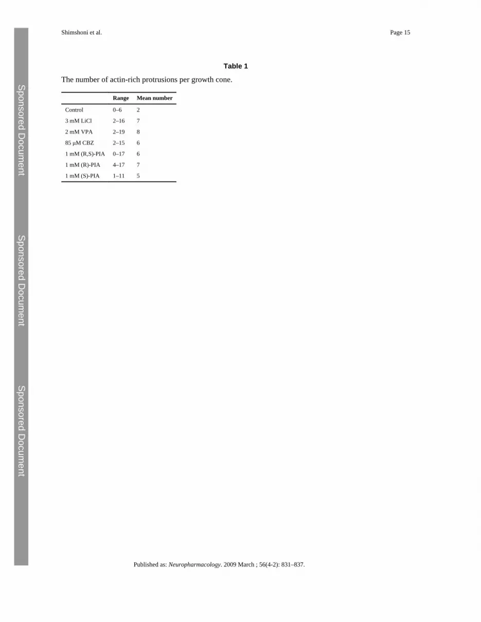
Growth cones of drug-treated neurons possessed an average of between 5 and 8 protrusionswith as many as 19 present on a single growth cone, whilst untreated neurons possessed anaverage of 2 with no more than 6 present on a single growth cone (Table 1; Fig. 4A). Theincrease in protrusions could arise either through increased initiation of their formation oraltered dynamics of their growth. We observed only a small increase in protrusion length onthe growth cone and no increase on those on the axon (Fig. 4B). This argues that althoughthere may be some change in their growth dynamics, the major effect of drug treatment is toincrease the rate of initiation of these F-actin structures.
4 DiscussionThe identity of the cellular target of mood stabilizers remains disputed. One argumentproposes that the Wnt signaling pathway is the important mood stabilizer target. This issupported by the observations that Li+ inhibits GSK-3 kinase activity and causes neuronalgrowth cone spreading with an associated change in microtubule dynamics (Goold et al.,1999; Klein and Melton, 1996; Ryves and Harwood, 2001). There is no doubt that GSK-3 isneurologically active, as GSK-3 hypomorphic mutations and GSK-3 inhibitors causebehavioural abnormalities in mice that are similar to those induced by treatment withantidepressant drugs (Beaulieu et al., 2004; Gould et al., 2004; Kaidanovich-Beilin et al.,2004; O'Brien et al., 2004). GSK-3 mediated signaling is also involved in mechanisms ofneuroprotection and has been associated with schizophrenia (Lovestone et al., 2007; Toledo
Shimshoni et al. Page 6
Published as: Neuropharmacology. 2009 March ; 56(4-2): 831–837.
Sponsored Docum
ent Sponsored D
ocument
Sponsored Docum
ent
et al., 2008). However, there is currently no direct evidence to link GSK-3 to bipolar mooddisorder. On the other hand, the less commonly used mood stabilizer CBZ also causesgrowth cone spreading, but with no change in microtubule structure or inhibition of GSK-3.CBZ has previously been shown to have no effect on β-catenin expression (Ryves et al.,2005), and we show here that CBZ does not induce TCF-mediated gene expression,eliminating any possibility of a β-catenin independent mechanism of stimulating Wntsignaling.
The situation with VPA is less clear. Initial reports suggested that VPA may directly inhibitGSK-3 (Chen et al., 1999), however a number of further investigations have demonstratedthis not to be the case (Eickholt et al., 2005; Phiel et al., 2001; Ryves et al., 2005). VPA hasbeen shown to alter gene expression via inhibition of histone deacetyalses (HDACs), and, insome but not all, cell contexts VPA can induce expression of β-catenin leading tostimulation of TCF-mediated gene expression (Phiel et al., 2001; Ryves et al., 2005).Previous reports, and those reported here, show that the effect of VPA on growth conemorphology is like that of CBZ (Williams et al., 2002) and, with the exception of one report(Hall et al., 2002), VPA and CBZ were shown to cause growth cone spreading without achange in microtubule dynamics. The VPA isomer (R,S)-PIA again has no effect on HDACactivity and only weak antiepileptic activity, but is a potent inducer of growth conespreading (Shimshoni et al., 2007a). We can now exclude the possibility that an enantiomerspecific effect on TCF-mediated gene expression was missed in this earlier experiment, andcan conclude without doubt that the VPA constitutional isomer (R,S)-PIA can induce growthcone spreading without activation of Wnt signaling. It remains to be seen how (R,S)-PIAperforms in animal models of bipolar disorder or patients.
To understand how the mood stabilizers, Li+, VPA and CBZ, and (R,S)-PIA cause changesin growth cone morphology, we examined underlying changes in the actin cytoskeleton. Wefound two effects common to all drugs. First, we found that growth cone spreading isassociated with localization of F-actin to the periphery of the cone. This is a feature of allspread growth cones, including those of the few observed in untreated control. Bothqualitative examination of images and quantitative measurements of the intensity ofphalloidin staining indicated that (R,S)-PIA induces elevated levels of F-actin. As (R,S)-PIAexhibits its effects at a lower concentration than VPA and therefore is the most potentinducer of growth cone spreading, these results suggest that drug treatment can drive localF-actin polymerization leading to changes in growth cone morphology.
Second, we find that all drugs induce a substantial increase in F-actin enriched protrusionsthat extend from the growth cone or its close vicinity. They are present at a low frequencyon untreated DRG neurons, and as there is only a marginal increase in length, but a largeincrease in number following drug treatment, we conclude that mood stabilizers induceformation of these protrusions rather than have a major effect on the stability of thosealready extant. The morphology, restricted distribution and lack of α-tubulin enrichmentindicate that these structures are not the same as the interstitial branching previouslyobserved following Li+, but not VPA and CBZ treatment (Williams et al., 2002). As they arelonger than 2 μm and enriched in F-actin, they resemble filopodia located on the axonalgrowth cone, which forms at the leading edge of motile cells or during early stages ofsynaptogenesis (Waites et al., 2005). However, unlike reports for filopodia, they can branchand in places become thickened. As a consequence the actual identity of these protrusions isunclear. Their presence does however indicate that (R,S)-PIA and mood stabilizers have asubstantial effect on the neuronal actin-based cytoskeleton.
As enlarged growth cones are observed on presynaptic terminals during synaptogenesis,axon bifurcation and during periods of sensing guidance cues from the environment
Shimshoni et al. Page 7
Published as: Neuropharmacology. 2009 March ; 56(4-2): 831–837.
Sponsored Docum
ent Sponsored D
ocument
Sponsored Docum
ent
(Eickholt et al., 2004; Williams et al., 2004), these findings support the hypothesis thatmood stabilizers modify the neural architecture. Such changes could lead to increased localneuronal interactions and connectivity, thereby reversing the reduced neuronal densityobserved in bipolar patients (Adler et al., 2005). This could occur via the longer-term eventsof neuronal morphogenesis. Alternatively functional changes could have more immediateeffects on synapse function of the presynaptic terminal and structural plasticity (Luo, 2002).
Although the actin cytoskeleton is not essential for axonal elongation, it is an importantcomponent of growth cone guidance. One possibility is that mood stabilizers alter responsesto extracellular signals leading to a change in the network of neuronal interactions. This re-wiring could occur during periods of neuronal development, including the establishment ofnew neuronal interactions following neurogenesis. Indeed several studies have demonstrateda reversal of the reduction in grey matter volume in the prefrontal cortex and an increasedneuronal and glial density, following treatment with mood stabilizers (Frey et al., 2004,2007). On a faster timescale, regulation of actin polymerization is also important for synapseformation of pre- and post-synaptic terminals, controlling both remodeling of the post-synaptic membrane and vesicle transport and the movement of dendritic spines, which formthe majority of the post-synaptic sites within the mammalian brain. It is possible that drug-mediated changes in actin dynamics may alter the processes governing synapse formation ofpre- and post-synaptic terminals, stability or function. Finally, as filopod formation is anearly stage of synaptogenesis of presynaptic terminals, the increase in actin-rich protrusionsobserved in drug-treated growth cones may reflect a switch from axonal growth to synapseformation.
In conclusion, this study using (R,S)-PIA, its individual enantiomers and mood stabilizersfurther indicates a Wnt independent change in neuronal behaviour. In contrast to earlierwork that focused on the effects of Li+ on microtubule dynamics, this current study arguesfor a common effect for mood stabilizers on the actin cytoskeleton, which may lead tochanges in neuronal morphology and plasticity in the CNS. (R,S)-PIA and its individualenantiomers are potent inducers of this effect, and suggest their potential as new moodstabilizers.
ReferencesAdler C.M. Levine A.D. DelBello M.P. Strakowski S.M. Changes in gray matter volume in patients
with bipolar disorder. Biol. Psychiatr.. 2005; 58:151–157.Atack J.R. Broughton H.B. Pollack S.J. Structure and mechanism of inositol monophosphatase. FEBS
Lett.. 1995; 361:1–7. [PubMed: 7890024]Beaulieu J.M. Sotnikova T.D. Yao W.D. Kockeritz L. Woodgett J.R. Gainetdinov R.R. Caron M.G.
Lithium antagonizes dopamine-dependent behaviors mediated by an AKT/glycogen synthase kinase3 signaling cascade. Proc. Natl. Acad. Sci. U.S.A.. 2004; 101:5099–5104. [PubMed: 15044694]
Berridge M.J. Downes C.P. Hanley M.R. Neural and developmental actions of lithium: a unifyinghypothesis. Cell. 1989; 59:411–419. [PubMed: 2553271]
Bottenstein J. Hayashi I. Hutchings S. Masui H. Mather J. McClure D.B. Ohasa S. Rizzino A. Sato G.Serrero G. Wolfe R. Wu R. The growth of cells in serum-free hormone-supplemented media.Methods Enzymol.. 1979; 58:94–109. [PubMed: 423795]
Chen G. Huang L.D. Jiang Y.M. Manji H.K. The mood-stabilizing agent valproate inhibits the activityof glycogen synthase kinase-3. J. Neurochem.. 1999; 72:1327–1330. [PubMed: 10037507]
Cryan J.F. Slattery D.A. Animal models of mood disorders: recent developments. Curr. Opin.Psychiatr.. 2007; 20:1–7.
Dent E.W. Gertler F.B. Cytoskeletal dynamics and transport in growth cone motility and axonguidance. Neuron. 2003; 40:209–227. [PubMed: 14556705]
Shimshoni et al. Page 8
Published as: Neuropharmacology. 2009 March ; 56(4-2): 831–837.
Sponsored Docum
ent Sponsored D
ocument
Sponsored Docum
ent
Eickholt B.J. Towers G.J. Ryves W.J. Eikel D. Adley K. Ylinen L.M. Chadborn N.H. Harwood A.J.Nau H. Williams R.S. Effects of valproic acid derivatives on inositol trisphosphate depletion,teratogenicity, glycogen synthase kinase-3beta inhibition, and viral replication: a screeningapproach for new bipolar disorder drugs derived from the valproic acid core structure. Mol.Pharmacol.. 2005; 67:1426–1433. [PubMed: 15687223]
Eickholt B.J. Williams R.S.B. Harwood A.J. Mood stabilizers and the cell biology of neuronal growthcones. Clin. Neurosci. Res.. 2004; 4:189–199.
Frey B.N. Andreazza A.C. Nery F.G. Martins M.R. Quevedo J. Soares J.C. Kapczinski F. The role ofhippocampus in the pathophysiology of bipolar disorder. Behav. Pharmacol.. 2007; 18:419–430.[PubMed: 17762510]
Frey B.N. Fonseca M.M. Machado-Vieira R. Soares J.C. Kapczinski F. Neuropathological andneurochemical abnormalities in bipolar disorder. Rev. Bras. Pisquiatr.. 2004; 26:180–188.
Goold R.G. Owen R. Gordon-Weeks P.R. Glycogen synthase kinase 3beta phosphorylation ofmicrotubule-associated protein 1B regulates the stability of microtubules in growth cones. J. CellSci.. 1999; 112(Pt 19):3373–3384. [PubMed: 10504342]
Gould T.D. Dow E.R. O'Donnell K.C. Chen G. Manji H.K. Targeting signal transduction pathways inthe treatment of mood disorders: recent insights into the relevance of the Wnt pathway. CNSNeurol. Disord. Drug Targets. 2007; 6:193–204. [PubMed: 17511616]
Gould T.D. Einat H. Bhat R. Manji H.K. AR-A014418, a selective GSK-3 inhibitor, producesantidepressant-like effects in the forced swim test. Int. J. Neuropsychopharmacol.. 2004; 7:387–390. [PubMed: 15315719]
Hall A.C. Brennan A. Goold R.G. Cleverley K. Lucas F.R. Gordon-Weeks P.R. Salinas P.C. Valproateregulates GSK-3-mediated axonal remodeling and synapsin I clustering in developing neurons.Mol. Cell. Neurosci.. 2002; 20:257–270. [PubMed: 12093158]
Hallcher L.M. Sherman W.R. The effects of lithium ion and other agents on the activity of myo-inositol-1-phosphatase from bovine brain. J. Biol. Chem.. 1980; 255:10896–10901. [PubMed:6253491]
Harwood A.J. Agam G. Search for a common mechanism of mood stabilizers. Biochem. Pharmacol..2003; 66:179–189. [PubMed: 12826261]
Kaidanovich-Beilin O. Milman A. Weizman A. Pick C.G. Eldar-Finkelman H. Rapid antidepressive-like activity of specific glycogen synthase kinase-3 inhibitor and its effect on beta-catenin inmouse hippocampus. Biol. Psychiatr.. 2004; 55:781–784.
Klein P.S. Melton D.A. A molecular mechanism for the effect of lithium on development. Proc. Natl.Acad. Sci. U.S.A.. 1996; 93:8455–8459. [PubMed: 8710892]
Korinek V. Barker N. Morin P.J. van Wichen D. de Weger R. Kinzler K.W. Vogelstein B. Clevers H.Constitutive transcriptional activation by a beta-catenin-Tcf complex in APC−/− colon carcinoma.Science. 1997; 275:1784–1787. [PubMed: 9065401]
Lovestone S. Davis D.R. Webster M.T. Kaech S. Brion J.P. Matus A. Anderton B.H. Lithium reducestau phosphorylation: effects in living cells and in neurons at therapeutic concentrations. Biol.Psychiatr.. 1999; 45:995–1003.
Lovestone S. Killick R. Di Forti M. Murray R. Schizophrenia as a GSK-3 dysregulation disorder.Trends Neurosci.. 2007; 30:142–149. [PubMed: 17324475]
Lucas F.R. Goold R.G. Gordon-Weeks P.R. Salinas P.C. Inhibition of GSK-3beta leading to the loss ofphosphorylated MAP-1B is an early event in axonal remodelling induced by WNT-7a or lithium.J. Cell Sci.. 1998; 111(Pt 10):1351–1361. [PubMed: 9570753]
Luo L. Actin cytoskeleton regulation in neuronal morphogenesis and structural plasticity. Annu. Rev.Cell Dev. Biol.. 2002; 18:601–635. [PubMed: 12142283]
Machado-Vieira R. Kapczinski F. Soares J.C. Perspectives for the development of animal models ofbipolar disorder. Prog. Neuropsychopharmacol. Biol. Psychiatr.. 2004; 28:209–224.
Mustelin T. Poso H. Iivanainen A. Andersson L.C. myo-Inositol reverses Li+-induced inhibition ofphosphoinositide turnover and ornithine decarboxylase induction during early lymphocyteactivation. Eur. J. Immunol.. 1986; 16:859–861. [PubMed: 3013648]
Shimshoni et al. Page 9
Published as: Neuropharmacology. 2009 March ; 56(4-2): 831–837.
Sponsored Docum
ent Sponsored D
ocument
Sponsored Docum
ent

O'Brien W.T. Harper A.D. Jove F. Woodgett J.R. Maretto S. Piccolo S. Klein P.S. Glycogen synthasekinase-3beta haploinsufficiency mimics the behavioral and molecular effects of lithium. J.Neurosci.. 2004; 24:6791–6798. [PubMed: 15282284]
Owen R. Gordon-Weeks P.R. Inhibition of glycogen synthase kinase 3beta in sensory neurons inculture alters filopodia dynamics and microtubule distribution in growth cones. Mol. Cell.Neurosci.. 2003; 23:626–637. [PubMed: 12932442]
Phiel C.J. Zhang F. Huang E.Y. Guenther M.G. Lazar M.A. Klein P.S. Histone deacetylase is a directtarget of valproic acid, a potent anticonvulsant, mood stabilizer, and teratogen. J. Biol. Chem..2001; 276:36734–36741. [PubMed: 11473107]
Ryves W.J. Dalton E.C. Harwood A.J. Williams R.S. GSK-3 activity in neocortical cells is inhibitedby lithium but not carbamazepine or valproic acid. Bipolar Disord.. 2005; 7:260–265. [PubMed:15898963]
Ryves W.J. Harwood A.J. Lithium inhibits glycogen synthase kinase-3 by competition for magnesium.Biochem. Biophys. Res. Commun.. 2001; 280:720–725. [PubMed: 11162580]
Sang H. Lu Z. Li Y. Ru B. Wang W. Chen J. Phosphorylation of tau by glycogen synthase kinase3beta in intact mammalian cells influences the stability of microtubules. Neurosci. Lett.. 2001;312:141–144. [PubMed: 11602330]
Shaltiel G. Shamir A. Shapiro J. Ding D. Dalton E. Bialer M. Harwood A.J. Belmaker R.H. GreenbergM.L. Agam G. Valproate decreases inositol biosynthesis. Biol. Psychiatr.. 2004; 56:868–874.
Shimshoni J.A. Dalton E.C. Jenkins A. Eyal S. Ewan K. Williams R.S. Pessah N. Yagen B. HarwoodA.J. Bialer M. The effects of central nervous system-active valproic acid constitutional isomers,cyclopropyl analogs, and amide derivatives on neuronal growth cone behavior. Mol. Pharmacol..2007; 71:884–892. [PubMed: 17167030]
Shimshoni J.A. Bialer M. Wlodarczyk B. Finnell R.H. Yagen B. Potent anticonvulsant urea derivativesof constitutional isomers of valproic acid. J. Med. Chem.. 2007; 50:6419–6427. [PubMed:17994680]
Silverstone P.H. Wu R.H. O'Donnell T. Ulrich M. Asghar S.J. Hanstock C.C. Chronic treatment withboth lithium and sodium valproate may normalize phosphoinositol cycle activity in bipolarpatients. Hum. Psychopharmacol.. 2002; 17:321–327. [PubMed: 12415549]
Toledo E.M. Colombres M. Inestrosa N.C. Wnt signaling in neuroprotection and stem celldifferentiation. Prog. Neurobiol.. 2008; 86(3):281–296. [PubMed: 18786602]
Waites C.L. Craig A.M. Garner C.C. Mechanisms of vertebrate synaptogenesis. Annu. Rev. Neurosci..2005; 28:251–274. [PubMed: 16022596]
Williams R. Ryves W.J. Dalton E.C. Eickholt B. Shaltiel G. Agam G. Harwood A.J. A molecular cellbiology of lithium. Biochem. Soc. Trans.. 2004; 32:799–802. [PubMed: 15494019]
Williams R.S. Cheng L. Mudge A.W. Harwood A.J. A common mechanism of action for three mood-stabilizing drugs. Nature. 2002; 417:292–295. [PubMed: 12015604]
Appendix Supplementary informationRefer to Web version on PubMed Central for supplementary material.
AcknowledgmentsThis study was supported by a Wellcome Trust program grant [07450] to AJH.
Shimshoni et al. Page 10
Published as: Neuropharmacology. 2009 March ; 56(4-2): 831–837.
Sponsored Docum
ent Sponsored D
ocument
Sponsored Docum
ent

Fig. 1.Chemical structures of compounds used in this study. VPA, valproic acid; (R,S)-propylisopropylacetic acid, (R,S)-PIA; (R)-propylisopropylacetic acid, (R)-PIA; (S)-propylisopropylacetic acid, (S)-PIA; carbamazepine, CBZ.
Shimshoni et al. Page 11
Published as: Neuropharmacology. 2009 March ; 56(4-2): 831–837.
Sponsored Docum
ent Sponsored D
ocument
Sponsored Docum
ent

Fig. 2.Induction of TCF-mediated gene expression. Cells were grown for 48 h in complete mediumin the presence of lithium (2 mM), CBZ (1 and 0.5 mM), VPA (3 mM), (R,S)-PIA (1 mM)or one of its two enantiomers, (R)-PIA (1 mM) and (S)-PIA (1 mM). The histograms showthe relative light unites for the corresponding drugs assayed in triplicates (Mean ± SD).
Shimshoni et al. Page 12
Published as: Neuropharmacology. 2009 March ; 56(4-2): 831–837.
Sponsored Docum
ent Sponsored D
ocument
Sponsored Docum
ent
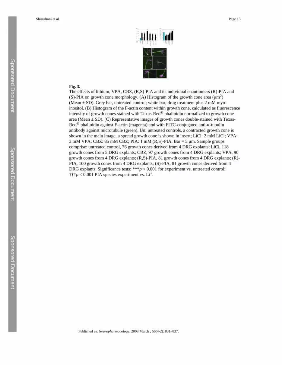
Fig. 3.The effects of lithium, VPA, CBZ, (R,S)-PIA and its individual enantiomers (R)-PIA and(S)-PIA on growth cone morphology. (A) Histogram of the growth cone area (μm2)(Mean ± SD). Grey bar, untreated control; white bar, drug treatment plus 2 mM myo-inositol. (B) Histogram of the F-actin content within growth cone, calculated as fluorescenceintensity of growth cones stained with Texas-Red® phalloidin normalized to growth conearea (Mean ± SD). (C) Representative images of growth cones double-stained with Texas-Red® phalloidin against F-actin (magenta) and with FITC-conjugated anti-α-tubulinantibody against microtubule (green). Un: untreated controls, a contracted growth cone isshown in the main image, a spread growth cone is shown in insert; LiCl: 2 mM LiCl; VPA:3 mM VPA; CBZ: 85 mM CBZ; PIA: 1 mM (R,S)-PIA. Bar = 5 μm. Sample groupscomprise: untreated control, 76 growth cones derived from 4 DRG explants; LiCl, 118growth cones from 5 DRG explants; CBZ, 97 growth cones from 4 DRG explants; VPA, 90growth cones from 4 DRG explants; (R,S)-PIA, 81 growth cones from 4 DRG explants; (R)-PIA, 100 growth cones from 4 DRG explants; (S)-PIA, 81 growth cones derived from 4DRG explants. Significance tests: ***p < 0.001 for experiment vs. untreated control;†††p < 0.001 PIA species experiment vs. Li+.
Shimshoni et al. Page 13
Published as: Neuropharmacology. 2009 March ; 56(4-2): 831–837.
Sponsored Docum
ent Sponsored D
ocument
Sponsored Docum
ent
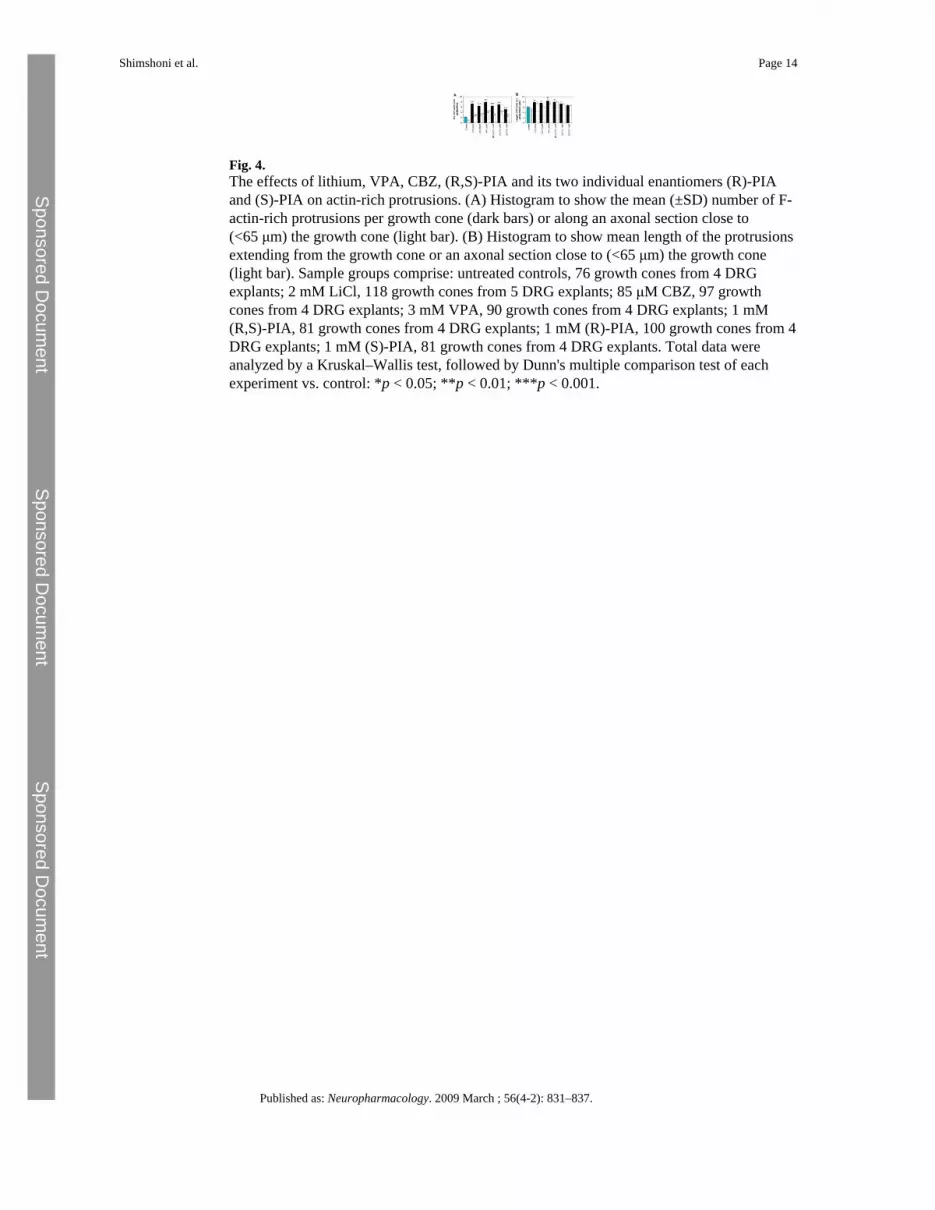
Fig. 4.The effects of lithium, VPA, CBZ, (R,S)-PIA and its two individual enantiomers (R)-PIAand (S)-PIA on actin-rich protrusions. (A) Histogram to show the mean (±SD) number of F-actin-rich protrusions per growth cone (dark bars) or along an axonal section close to(<65 μm) the growth cone (light bar). (B) Histogram to show mean length of the protrusionsextending from the growth cone or an axonal section close to (<65 μm) the growth cone(light bar). Sample groups comprise: untreated controls, 76 growth cones from 4 DRGexplants; 2 mM LiCl, 118 growth cones from 5 DRG explants; 85 μM CBZ, 97 growthcones from 4 DRG explants; 3 mM VPA, 90 growth cones from 4 DRG explants; 1 mM(R,S)-PIA, 81 growth cones from 4 DRG explants; 1 mM (R)-PIA, 100 growth cones from 4DRG explants; 1 mM (S)-PIA, 81 growth cones from 4 DRG explants. Total data wereanalyzed by a Kruskal–Wallis test, followed by Dunn's multiple comparison test of eachexperiment vs. control: *p < 0.05; **p < 0.01; ***p < 0.001.
Shimshoni et al. Page 14
Published as: Neuropharmacology. 2009 March ; 56(4-2): 831–837.
Sponsored Docum
ent Sponsored D
ocument
Sponsored Docum
ent
Sponsored Docum
ent Sponsored D
ocument
Sponsored Docum
ent
Shimshoni et al. Page 15
Table 1
The number of actin-rich protrusions per growth cone.
Range Mean number
Control 0–6 2
3 mM LiCl 2–16 7
2 mM VPA 2–19 8
85 μM CBZ 2–15 6
1 mM (R,S)-PIA 0–17 6
1 mM (R)-PIA 4–17 7
1 mM (S)-PIA 1–11 5
Published as: Neuropharmacology. 2009 March ; 56(4-2): 831–837.