reduction of nadph-oxidase activity ameliorates the cardiovascular phenotype in a mouse model of...
TRANSCRIPT
Reduction of NADPH-Oxidase Activity Ameliorates theCardiovascular Phenotype in a Mouse Model of Williams-Beuren SyndromeVictoria Campuzano1,2*, Maria Segura-Puimedon1,2, Verena Terrado1,2, Carolina Sanchez-Rodrıguez3,
Mathilde Coustets1, Mauricio Menacho-Marquez4, Julian Nevado2,5, Xose R. Bustelo4, Uta Francke6,
Luis A. Perez-Jurado1,2
1 Departament de Ciencies Experimentals i de la Salut, Universitat Pompeu Fabra, Barcelona, Spain, 2 Centro de Investigacion Biomedica en Red de Enfermedades Raras
(CIBERER), ISCIII, Madrid, Spain, 3 Unidad de Investigacion, Hospital Universitario de Getafe, Getafe, Spain, 4 Centro de Investigacion del Cancer e Instituto de Biologıa
Molecular del Cancer de Salamanca (IMBCC), CSIC–Universidad de Salamanca, Salamanca, Spain, 5 INGEMM-Instituto de Genetica Medica y Molecular/IdiPAZ, Madrid,
Spain, 6 Department of Genetics, Stanford University School of Medicine, Stanford, California, United States of America
Abstract
A hallmark feature of Williams-Beuren Syndrome (WBS) is a generalized arteriopathy due to elastin deficiency, presenting asstenoses of medium and large arteries and leading to hypertension and other cardiovascular complications. Deletion of afunctional NCF1 gene copy has been shown to protect a proportion of WBS patients against hypertension, likely throughreduced NADPH-oxidase (NOX)–mediated oxidative stress. DD mice, carrying a 0.67 Mb heterozygous deletion including theEln gene, presented with a generalized arteriopathy, hypertension, and cardiac hypertrophy, associated with elevatedangiotensin II (angII), oxidative stress parameters, and Ncf1 expression. Genetic (by crossing with Ncf1 mutant) and/orpharmacological (with ang II type 1 receptor blocker, losartan, or NOX inhibitor apocynin) reduction of NOX activitycontrolled hormonal and biochemical parameters in DD mice, resulting in normalized blood pressure and improvedcardiovascular histology. We provide strong evidence for implication of the redox system in the pathophysiology of thecardiovascular disease in a mouse model of WBS. The phenotype of these mice can be ameliorated by either genetic orpharmacological intervention reducing NOX activity, likely through reduced angII–mediated oxidative stress. Therefore,anti-NOX therapy merits evaluation to prevent the potentially serious cardiovascular complications of WBS, as well as inother cardiovascular disorders mediated by similar pathogenic mechanism.
Citation: Campuzano V, Segura-Puimedon M, Terrado V, Sanchez-Rodrıguez C, Coustets M, et al. (2012) Reduction of NADPH-Oxidase Activity Ameliorates theCardiovascular Phenotype in a Mouse Model of Williams-Beuren Syndrome. PLoS Genet 8(2): e1002458. doi:10.1371/journal.pgen.1002458
Editor: Andrew O. M. Wilkie, University of Oxford, United Kingdom
Received July 29, 2011; Accepted November 16, 2011; Published February 2, 2012
Copyright: � 2012 Campuzano et al. This is an open-access article distributed under the terms of the Creative Commons Attribution License, which permitsunrestricted use, distribution, and reproduction in any medium, provided the original author and source are credited.
Funding: This work was supported by grants from the Spanish Ministry of Science and Innovation of Health (FIS 07/0059 to VC, FIS 10/2512 to LAP-J, RD06/0020/0001 to XRB) and the VI Framework Programme of the European Union (LSHG-CT-2006-037627 to LAP-J). VC is a Miguel Servet FIS Investigator (CP04/00068). MS-Pis supported by a CIBERER Fellowship. MM-M is supported by the CSIC JAE-Doc program. The funders had no role in study design, data collection and analysis,decision to publish, or preparation of the manuscript.
Competing Interests: The authors have declared that no competing interests exist.
* E-mail: [email protected]
Introduction
Williams-Beuren syndrome (WBS [MIM 194050]) is a devel-
opmental disorder with multisystemic manifestations and a
prevalence of ,1/10,000 newborns, caused by a segmental
aneusomy of 1.55–1.83 Mb at chromosomal band 7q11.23, which
includes ELN (coding for elastin [MIM 130160]) and 25–27
additional genes [1,2]. The recurrent WBS deletion common to
most patients is mediated by nonallelic homologous recombination
between regional segmental duplications that flank the WBS
critical region [3]. In addition to distinctive craniofacial charac-
teristics and mild mental retardation with social disinhibition and
hyperacusis, a hallmark feature of WBS is a generalized
arteriopathy presenting as narrowing of the large elastic arteries
[4]. Histological characterization of arterial vessel walls of WBS
patients showed increased number and disorganized lamellar
structures, fragmented elastic fibers, and hypertrophy of smooth
muscle cells [5]. This large arterial vessel remodeling which is a
consequence of abnormalities in vascular development, is thought
to be responsible for the cardiovascular disease manifested in 84%
of WBS patients [4,6]. Identical vascular features, most promi-
nently supravalvular aortic stenosis, are also found in patients with
heterozygous deletions or disruptions of the ELN gene, implicating
elastin haploinsufficiency in this phenotype [5,7]. The arteriopathy
is the main cause of serious morbidity in WBS, including systemic
hypertension and possible complications such as stroke, cardiac
ischemia, and sudden death [8,9].
Animal models provide further evidence for elastin deficiency as
the main cause of cardiovascular disease in WBS, underscoring the
prominent role of the elastic matrix in the morphogenesis and
homeostasis of the vessel wall [10]. Heterozygous knockout mice
with only one copy of the Eln gene reproduce many of the
alterations observed in the WBS vascular phenotype [11,12].
Hypertension is a consistent feature of Eln+/2 mice, associated
with elevated plasma renin activity (PRA) and angiotensin II
(angII) levels, that can be blocked by the administration of angII
type 1 receptor (AT1R) antagonists [13]. In addition to direct
effects on the vasculature, many of the cellular actions of angII are
PLoS Genetics | www.plosgenetics.org 1 February 2012 | Volume 8 | Issue 2 | e1002458
mediated by the activation of the NADPH-oxidase (NOX), thus
stimulating the formation of reactive oxygen species (ROS).
Evidence is accumulating that increased oxidative stress has a
relevant pathophysiological role in cardiovascular disease, includ-
ing hypertension, atherosclerosis, and heart failure [14].
In WBS, the dosage of the NCF1 gene, encoding the p47phox
subunit of NOX, is a strong modifier of the risk of hypertension.
Hypertension was significantly less prevalent in patients whose
deletion included NCF1, indicating that hemizygosity for NCF1
was a protective factor against hypertension in WBS. Decreased
p47phox protein, superoxide anion production, and protein
nitrosylation levels, were all observed in cell lines from patients
hemizygous at NCF1 [15]. Reduced angII-mediated oxidative
stress in the vasculature was the proposed mechanism behind this
protective effect. Indeed, studies performed in Ncf1 knockout mice
have revealed that p47phox is one of the major effectors of angII
action. The administration of angII did not lead to increased
superoxide production or blood pressure elevation in homozygous
knockout animals, as it did in wild-type mice [16].
The aim of the present study was to evaluate whether oxidative
stress significantly contributes to the cardiovascular phenotype of a
mouse model for WBS, and whether reduction of NOX activity by
genetic modification and/or by pharmacological inhibition might
have a potential benefit in the rescue of this phenotype. By using
non-invasive blood pressure measurements, histological, biochem-
ical and molecular analyses, we have documented a negative
correlation between NOX activity and the cardiovascular
phenotype in a mouse model of WBS, as well as prevention of
many of the manifestations by using anti-NOX therapies.
Results
Cardiovascular phenotype of DD mice related to elevatedangII and oxidative stress
Previously reported mice bearing a heterozygous deletion of half
of the orthologous region of the WBS locus (0.67 Mb, from Limk1
to Trim50, including Eln), called DD, were used as a model for the
WBS cardiovascular phenotype [11,17].
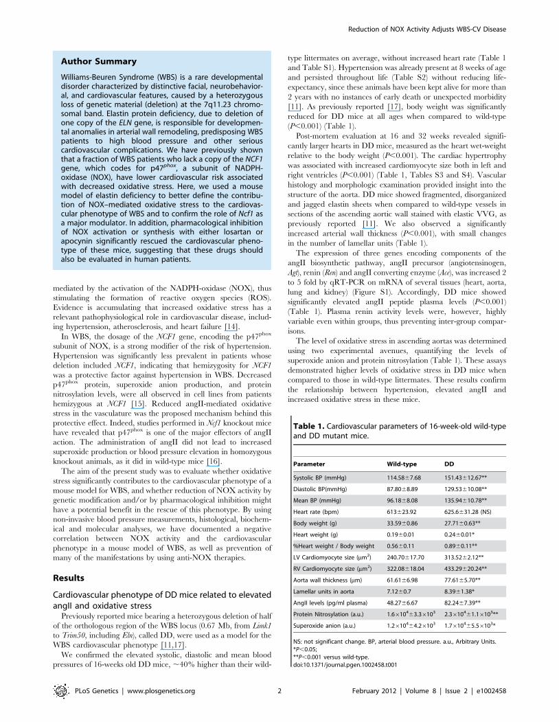
We confirmed the elevated systolic, diastolic and mean blood
pressures of 16-weeks old DD mice, ,40% higher than their wild-
type littermates on average, without increased heart rate (Table 1
and Table S1). Hypertension was already present at 8 weeks of age
and persisted throughout life (Table S2) without reducing life-
expectancy, since these animals have been kept alive for more than
2 years with no instances of early death or unexpected morbidity
[11]. As previously reported [17], body weight was significantly
reduced for DD mice at all ages when compared to wild-type
(P,0.001) (Table 1).
Post-mortem evaluation at 16 and 32 weeks revealed signifi-
cantly larger hearts in DD mice, measured as the heart wet-weight
relative to the body weight (P,0.001). The cardiac hypertrophy
was associated with increased cardiomyocyte size both in left and
right ventricles (P,0.001) (Table 1, Tables S3 and S4). Vascular
histology and morphologic examination provided insight into the
structure of the aorta. DD mice showed fragmented, disorganized
and jagged elastin sheets when compared to wild-type vessels in
sections of the ascending aortic wall stained with elastic VVG, as
previously reported [11]. We also observed a significantly
increased arterial wall thickness (P,0.001), with small changes
in the number of lamellar units (Table 1).
The expression of three genes encoding components of the
angII biosynthetic pathway, angII precursor (angiotensinogen,
Agt), renin (Ren) and angII converting enzyme (Ace), was increased 2
to 5 fold by qRT-PCR on mRNA of several tissues (heart, aorta,
lung and kidney) (Figure S1). Accordingly, DD mice showed
significantly elevated angII peptide plasma levels (P,0.001)
(Table 1). Plasma renin activity levels were, however, highly
variable even within groups, thus preventing inter-group compar-
isons.
The level of oxidative stress in ascending aortas was determined
using two experimental avenues, quantifying the levels of
superoxide anion and protein nitrosylation (Table 1). These assays
demonstrated higher levels of oxidative stress in DD mice when
compared to those in wild-type littermates. These results confirm
the relationship between hypertension, elevated angII and
increased oxidative stress in these mice.
Table 1. Cardiovascular parameters of 16-week-old wild-typeand DD mutant mice.
Parameter Wild-type DD
Systolic BP (mmHg) 114.5867.68 151.43612.67**
Diastolic BP(mmHg) 87.8068.89 129.53610.08**
Mean BP (mmHg) 96.1868.08 135.94610.78**
Heart rate (bpm) 613623.92 625.6631.28 (NS)
Body weight (g) 33.5960.86 27.7160.63**
Heart weight (g) 0.1960.01 0.2460.01*
%Heart weight / Body weight 0.5660.11 0.8960.11**
LV Cardiomyocyte size (mm2) 240.70617.70 313.5262.12**
RV Cardiomyocyte size (mm2) 322.08618.04 433.29620.24**
Aorta wall thickness (mm) 61.6166.98 77.6165.70**
Lamellar units in aorta 7.1260.7 8.3961.38*
AngII levels (pg/ml plasma) 48.2766.67 82.2467.39**
Protein Nitrosylation (a.u.) 1.6610463.36103 2.3610461.16103**
Superoxide anion (a.u.) 1.2610464.26103 1.7610465.56103*
NS: not significant change. BP, arterial blood pressure. a.u., Arbitrary Units.*P,0.05;**P,0.001 versus wild-type.doi:10.1371/journal.pgen.1002458.t001
Author Summary
Williams-Beuren Syndrome (WBS) is a rare developmentaldisorder characterized by distinctive facial, neurobehavior-al, and cardiovascular features, caused by a heterozygousloss of genetic material (deletion) at the 7q11.23 chromo-somal band. Elastin protein deficiency, due to deletion ofone copy of the ELN gene, is responsible for developmen-tal anomalies in arterial wall remodeling, predisposing WBSpatients to high blood pressure and other seriouscardiovascular complications. We have previously shownthat a fraction of WBS patients who lack a copy of the NCF1gene, which codes for p47phox, a subunit of NADPH-oxidase (NOX), have lower cardiovascular risk associatedwith decreased oxidative stress. Here, we used a mousemodel of elastin deficiency to better define the contribu-tion of NOX–mediated oxidative stress to the cardiovas-cular phenotype of WBS and to confirm the role of Ncf1 asa major modulator. In addition, pharmacological inhibitionof NOX activation or synthesis with either losartan orapocynin significantly rescued the cardiovascular pheno-type of these mice, suggesting that these drugs shouldalso be evaluated in human patients.
Reduction of NOX Activity Adjusts WBS-CV Disease
PLoS Genetics | www.plosgenetics.org 2 February 2012 | Volume 8 | Issue 2 | e1002458
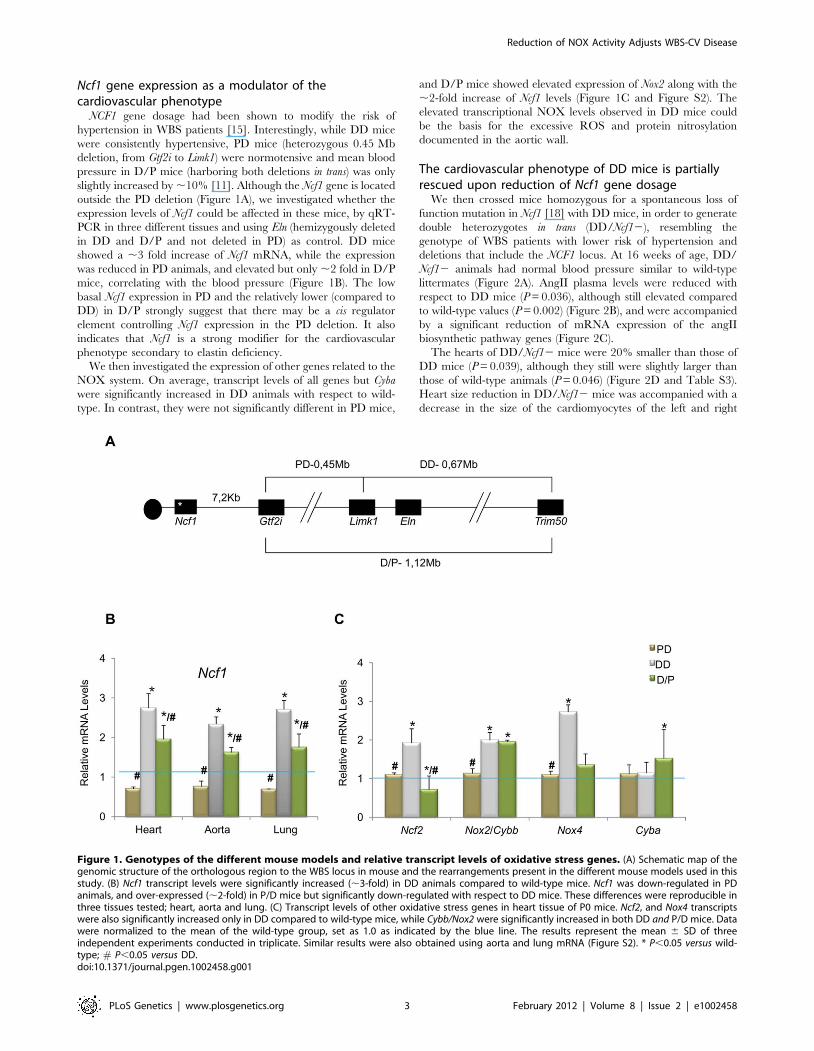
Ncf1 gene expression as a modulator of thecardiovascular phenotype
NCF1 gene dosage had been shown to modify the risk of
hypertension in WBS patients [15]. Interestingly, while DD mice
were consistently hypertensive, PD mice (heterozygous 0.45 Mb
deletion, from Gtf2i to Limk1) were normotensive and mean blood
pressure in D/P mice (harboring both deletions in trans) was only
slightly increased by ,10% [11]. Although the Ncf1 gene is located
outside the PD deletion (Figure 1A), we investigated whether the
expression levels of Ncf1 could be affected in these mice, by qRT-
PCR in three different tissues and using Eln (hemizygously deleted
in DD and D/P and not deleted in PD) as control. DD mice
showed a ,3 fold increase of Ncf1 mRNA, while the expression
was reduced in PD animals, and elevated but only ,2 fold in D/P
mice, correlating with the blood pressure (Figure 1B). The low
basal Ncf1 expression in PD and the relatively lower (compared to
DD) in D/P strongly suggest that there may be a cis regulator
element controlling Ncf1 expression in the PD deletion. It also
indicates that Ncf1 is a strong modifier for the cardiovascular
phenotype secondary to elastin deficiency.
We then investigated the expression of other genes related to the
NOX system. On average, transcript levels of all genes but Cyba
were significantly increased in DD animals with respect to wild-
type. In contrast, they were not significantly different in PD mice,
and D/P mice showed elevated expression of Nox2 along with the
,2-fold increase of Ncf1 levels (Figure 1C and Figure S2). The
elevated transcriptional NOX levels observed in DD mice could
be the basis for the excessive ROS and protein nitrosylation
documented in the aortic wall.
The cardiovascular phenotype of DD mice is partiallyrescued upon reduction of Ncf1 gene dosage
We then crossed mice homozygous for a spontaneous loss of
function mutation in Ncf1 [18] with DD mice, in order to generate
double heterozygotes in trans (DD/Ncf12), resembling the
genotype of WBS patients with lower risk of hypertension and
deletions that include the NCF1 locus. At 16 weeks of age, DD/
Ncf12 animals had normal blood pressure similar to wild-type
littermates (Figure 2A). AngII plasma levels were reduced with
respect to DD mice (P = 0.036), although still elevated compared
to wild-type values (P = 0.002) (Figure 2B), and were accompanied
by a significant reduction of mRNA expression of the angII
biosynthetic pathway genes (Figure 2C).
The hearts of DD/Ncf12 mice were 20% smaller than those of
DD mice (P = 0.039), although they still were slightly larger than
those of wild-type animals (P = 0.046) (Figure 2D and Table S3).
Heart size reduction in DD/Ncf12 mice was accompanied with a
decrease in the size of the cardiomyocytes of the left and right
Figure 1. Genotypes of the different mouse models and relative transcript levels of oxidative stress genes. (A) Schematic map of thegenomic structure of the orthologous region to the WBS locus in mouse and the rearrangements present in the different mouse models used in thisstudy. (B) Ncf1 transcript levels were significantly increased (,3-fold) in DD animals compared to wild-type mice. Ncf1 was down-regulated in PDanimals, and over-expressed (,2-fold) in P/D mice but significantly down-regulated with respect to DD mice. These differences were reproducible inthree tissues tested; heart, aorta and lung. (C) Transcript levels of other oxidative stress genes in heart tissue of P0 mice. Ncf2, and Nox4 transcriptswere also significantly increased only in DD compared to wild-type mice, while Cybb/Nox2 were significantly increased in both DD and P/D mice. Datawere normalized to the mean of the wild-type group, set as 1.0 as indicated by the blue line. The results represent the mean 6 SD of threeindependent experiments conducted in triplicate. Similar results were also obtained using aorta and lung mRNA (Figure S2). * P,0.05 versus wild-type; # P,0.05 versus DD.doi:10.1371/journal.pgen.1002458.g001
Reduction of NOX Activity Adjusts WBS-CV Disease
PLoS Genetics | www.plosgenetics.org 3 February 2012 | Volume 8 | Issue 2 | e1002458
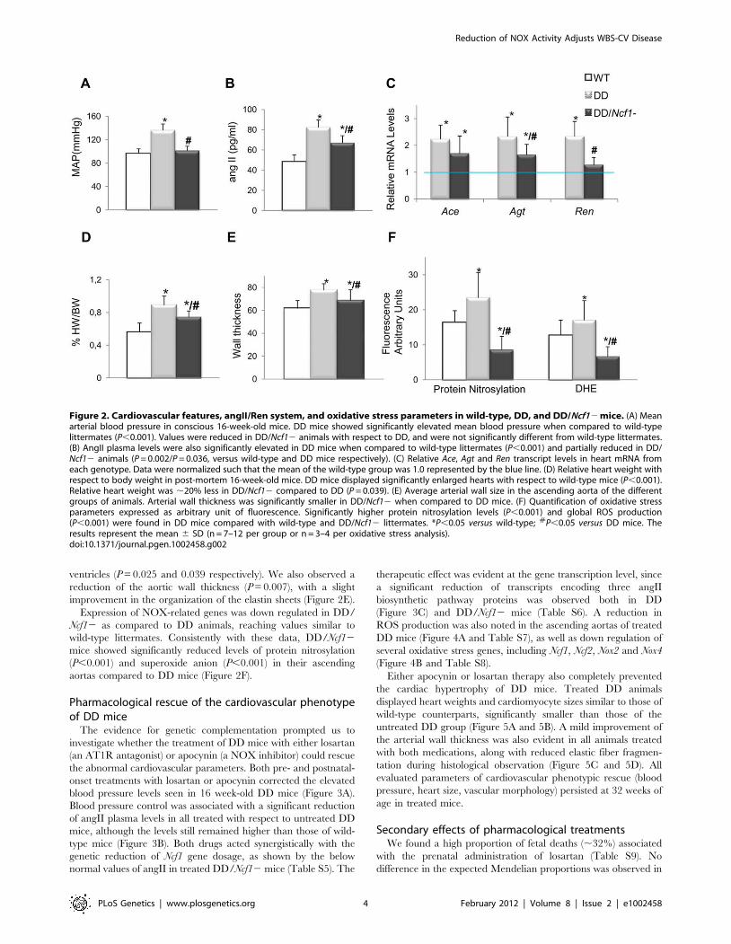
ventricles (P = 0.025 and 0.039 respectively). We also observed a
reduction of the aortic wall thickness (P = 0.007), with a slight
improvement in the organization of the elastin sheets (Figure 2E).
Expression of NOX-related genes was down regulated in DD/
Ncf12 as compared to DD animals, reaching values similar to
wild-type littermates. Consistently with these data, DD/Ncf12
mice showed significantly reduced levels of protein nitrosylation
(P,0.001) and superoxide anion (P,0.001) in their ascending
aortas compared to DD mice (Figure 2F).
Pharmacological rescue of the cardiovascular phenotypeof DD mice
The evidence for genetic complementation prompted us to
investigate whether the treatment of DD mice with either losartan
(an AT1R antagonist) or apocynin (a NOX inhibitor) could rescue
the abnormal cardiovascular parameters. Both pre- and postnatal-
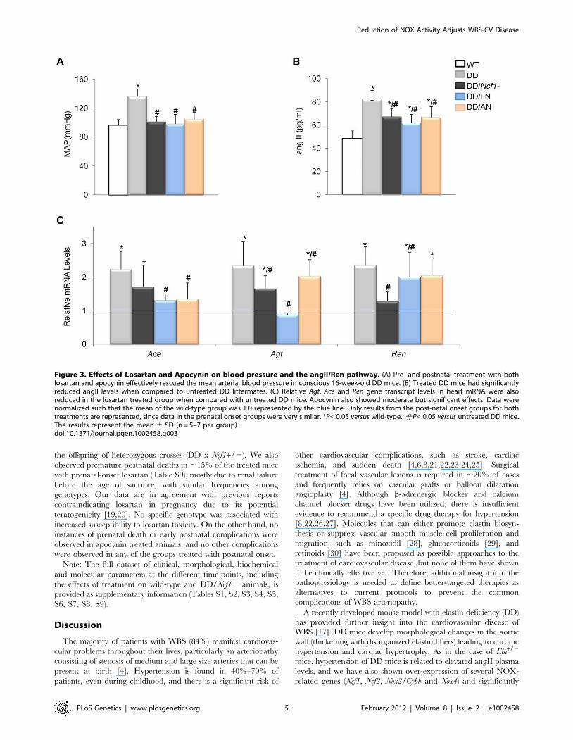
onset treatments with losartan or apocynin corrected the elevated
blood pressure levels seen in 16 week-old DD mice (Figure 3A).
Blood pressure control was associated with a significant reduction
of angII plasma levels in all treated with respect to untreated DD
mice, although the levels still remained higher than those of wild-
type mice (Figure 3B). Both drugs acted synergistically with the
genetic reduction of Ncf1 gene dosage, as shown by the below
normal values of angII in treated DD/Ncf12 mice (Table S5). The
therapeutic effect was evident at the gene transcription level, since
a significant reduction of transcripts encoding three angII
biosynthetic pathway proteins was observed both in DD
(Figure 3C) and DD/Ncf12 mice (Table S6). A reduction in
ROS production was also noted in the ascending aortas of treated
DD mice (Figure 4A and Table S7), as well as down regulation of
several oxidative stress genes, including Ncf1, Ncf2, Nox2 and Nox4
(Figure 4B and Table S8).
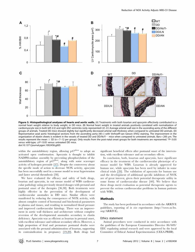
Either apocynin or losartan therapy also completely prevented
the cardiac hypertrophy of DD mice. Treated DD animals
displayed heart weights and cardiomyocyte sizes similar to those of
wild-type counterparts, significantly smaller than those of the
untreated DD group (Figure 5A and 5B). A mild improvement of
the arterial wall thickness was also evident in all animals treated
with both medications, along with reduced elastic fiber fragmen-
tation during histological observation (Figure 5C and 5D). All
evaluated parameters of cardiovascular phenotypic rescue (blood
pressure, heart size, vascular morphology) persisted at 32 weeks of
age in treated mice.
Secondary effects of pharmacological treatmentsWe found a high proportion of fetal deaths (,32%) associated
with the prenatal administration of losartan (Table S9). No
difference in the expected Mendelian proportions was observed in
Figure 2. Cardiovascular features, angII/Ren system, and oxidative stress parameters in wild-type, DD, and DD/Ncf12 mice. (A) Meanarterial blood pressure in conscious 16-week-old mice. DD mice showed significantly elevated mean blood pressure when compared to wild-typelittermates (P,0.001). Values were reduced in DD/Ncf12 animals with respect to DD, and were not significantly different from wild-type littermates.(B) AngII plasma levels were also significantly elevated in DD mice when compared to wild-type littermates (P,0.001) and partially reduced in DD/Ncf12 animals (P = 0.002/P = 0.036, versus wild-type and DD mice respectively). (C) Relative Ace, Agt and Ren transcript levels in heart mRNA fromeach genotype. Data were normalized such that the mean of the wild-type group was 1.0 represented by the blue line. (D) Relative heart weight withrespect to body weight in post-mortem 16-week-old mice. DD mice displayed significantly enlarged hearts with respect to wild-type mice (P,0.001).Relative heart weight was ,20% less in DD/Ncf12 compared to DD (P = 0.039). (E) Average arterial wall size in the ascending aorta of the differentgroups of animals. Arterial wall thickness was significantly smaller in DD/Ncf12 when compared to DD mice. (F) Quantification of oxidative stressparameters expressed as arbitrary unit of fluorescence. Significantly higher protein nitrosylation levels (P,0.001) and global ROS production(P,0.001) were found in DD mice compared with wild-type and DD/Ncf12 littermates. *P,0.05 versus wild-type; #P,0.05 versus DD mice. Theresults represent the mean 6 SD (n = 7–12 per group or n = 3–4 per oxidative stress analysis).doi:10.1371/journal.pgen.1002458.g002
Reduction of NOX Activity Adjusts WBS-CV Disease
PLoS Genetics | www.plosgenetics.org 4 February 2012 | Volume 8 | Issue 2 | e1002458
the offspring of heterozygous crosses (DD x Ncf1+/2). We also
observed premature postnatal deaths in ,15% of the treated mice
with prenatal-onset losartan (Table S9), mostly due to renal failure
before the age of sacrifice, with similar frequencies among
genotypes. Our data are in agreement with previous reports
contraindicating losartan in pregnancy due to its potential
teratogenicity [19,20]. No specific genotype was associated with
increased susceptibility to losartan toxicity. On the other hand, no
instances of prenatal death or early postnatal complications were
observed in apocynin treated animals, and no other complications
were observed in any of the groups treated with postnatal onset.
Note: The full dataset of clinical, morphological, biochemical
and molecular parameters at the different time-points, including
the effects of treatment on wild-type and DD/Ncf12 animals, is
provided as supplementary information (Tables S1, S2, S3, S4, S5,
S6, S7, S8, S9).
Discussion
The majority of patients with WBS (84%) manifest cardiovas-
cular problems throughout their lives, particularly an arteriopathy
consisting of stenosis of medium and large size arteries that can be
present at birth [4]. Hypertension is found in 40%–70% of
patients, even during childhood, and there is a significant risk of
other cardiovascular complications, such as stroke, cardiac
ischemia, and sudden death [4,6,8,21,22,23,24,25]. Surgical
treatment of focal vascular lesions is required in ,20% of cases
and frequently relies on vascular grafts or balloon dilatation
angioplasty [4]. Although b-adrenergic blocker and calcium
channel blocker drugs have been utilized, there is insufficient
evidence to recommend a specific drug therapy for hypertension
[8,22,26,27]. Molecules that can either promote elastin biosyn-
thesis or suppress vascular smooth muscle cell proliferation and
migration, such as minoxidil [28], glucocorticoids [29], and
retinoids [30] have been proposed as possible approaches to the
treatment of cardiovascular disease, but none of them have shown
to be clinically effective yet. Therefore, additional insight into the
pathophysiology is needed to define better-targeted therapies as
alternatives to current protocols to prevent the common
complications of WBS arteriopathy.
A recently developed mouse model with elastin deficiency (DD)
has provided further insight into the cardiovascular disease of
WBS [17]. DD mice develop morphological changes in the aortic
wall (thickening with disorganized elastin fibers) leading to chronic
hypertension and cardiac hypertrophy. As in the case of Eln+/2
mice, hypertension of DD mice is related to elevated angII plasma
levels, and we have also shown over-expression of several NOX-
related genes (Ncf1, Ncf2, Nox2/Cybb and Nox4) and significantly
Figure 3. Effects of Losartan and Apocynin on blood pressure and the angII/Ren pathway. (A) Pre- and postnatal treatment with bothlosartan and apocynin effectively rescued the mean arterial blood pressure in conscious 16-week-old DD mice. (B) Treated DD mice had significantlyreduced angII levels when compared to untreated DD littermates. (C) Relative Agt, Ace and Ren gene transcript levels in heart mRNA were alsoreduced in the losartan treated group when compared with untreated DD mice. Apocynin also showed moderate but significant effects. Data werenormalized such that the mean of the wild-type group was 1.0 represented by the blue line. Only results from the post-natal onset groups for bothtreatments are represented, since data in the prenatal onset groups were very similar. *P,0.05 versus wild-type.; #P,0.05 versus untreated DD mice.The results represent the mean 6 SD (n = 5–7 per group).doi:10.1371/journal.pgen.1002458.g003
Reduction of NOX Activity Adjusts WBS-CV Disease
PLoS Genetics | www.plosgenetics.org 5 February 2012 | Volume 8 | Issue 2 | e1002458
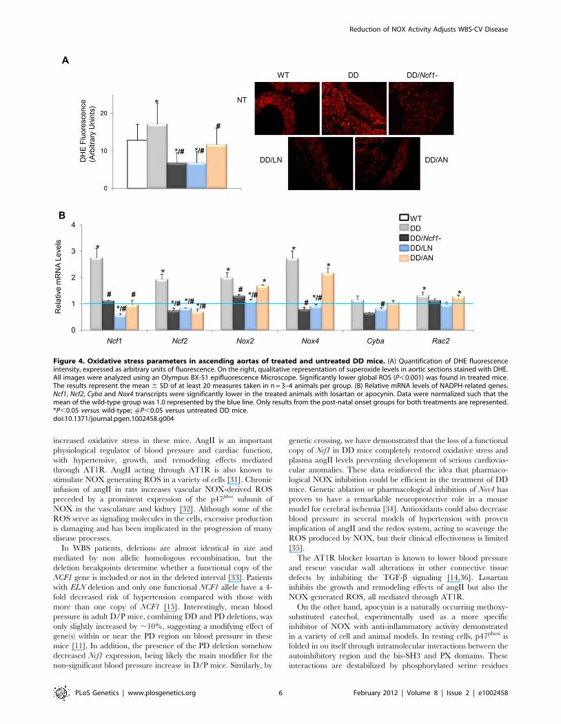
increased oxidative stress in these mice. AngII is an important
physiological regulator of blood pressure and cardiac function,
with hypertensive, growth, and remodeling effects mediated
through AT1R. AngII acting through AT1R is also known to
stimulate NOX generating ROS in a variety of cells [31]. Chronic
infusion of angII in rats increases vascular NOX-derived ROS
preceded by a prominent expression of the p47phox subunit of
NOX in the vasculature and kidney [32]. Although some of the
ROS serve as signaling molecules in the cells, excessive production
is damaging and has been implicated in the progression of many
disease processes.
In WBS patients, deletions are almost identical in size and
mediated by non allelic homologous recombination, but the
deletion breakpoints determine whether a functional copy of the
NCF1 gene is included or not in the deleted interval [33]. Patients
with ELN deletion and only one functional NCF1 allele have a 4-
fold decreased risk of hypertension compared with those with
more than one copy of NCF1 [15]. Interestingly, mean blood
pressure in adult D/P mice, combining DD and PD deletions, was
only slightly increased by ,10%, suggesting a modifying effect of
gene(s) within or near the PD region on blood pressure in these
mice [11]. In addition, the presence of the PD deletion somehow
decreased Ncf1 expression, being likely the main modifier for the
non-significant blood pressure increase in D/P mice. Similarly, by
genetic crossing, we have demonstrated that the loss of a functional
copy of Ncf1 in DD mice completely restored oxidative stress and
plasma angII levels preventing development of serious cardiovas-
cular anomalies. These data reinforced the idea that pharmaco-
logical NOX inhibition could be efficient in the treatment of DD
mice. Genetic ablation or pharmacological inhibition of Nox4 has
proven to have a remarkable neuroprotective role in a mouse
model for cerebral ischemia [34]. Antioxidants could also decrease
blood pressure in several models of hypertension with proven
implication of angII and the redox system, acting to scavenge the
ROS produced by NOX, but their clinical effectiveness is limited
[35].
The AT1R blocker losartan is known to lower blood pressure
and rescue vascular wall alterations in other connective tissue
defects by inhibiting the TGF-b signaling [14,36]. Losartan
inhibits the growth and remodeling effects of angII but also the
NOX generated ROS, all mediated through AT1R.
On the other hand, apocynin is a naturally occurring methoxy-
substituted catechol, experimentally used as a more specific
inhibitor of NOX with anti-inflammatory activity demonstrated
in a variety of cell and animal models. In resting cells, p47phox is
folded in on itself through intramolecular interactions between the
autoinhibitory region and the bis-SH3 and PX domains. These
interactions are destabilized by phosphorylated serine residues
Figure 4. Oxidative stress parameters in ascending aortas of treated and untreated DD mice. (A) Quantification of DHE fluorescenceintensity, expressed as arbitrary units of fluorescence. On the right, qualitative representation of superoxide levels in aortic sections stained with DHE.All images were analyzed using an Olympus BX-51 epifluorescence Microscope. Significantly lower global ROS (P,0.001) was found in treated mice.The results represent the mean 6 SD of at least 20 measures taken in n = 3–4 animals per group. (B) Relative mRNA levels of NADPH-related genes.Ncf1, Ncf2, Cyba and Nox4 transcripts were significantly lower in the treated animals with losartan or apocynin. Data were normalized such that themean of the wild-type group was 1.0 represented by the blue line. Only results from the post-natal onset groups for both treatments are represented.*P,0.05 versus wild-type; #P,0.05 versus untreated DD mice.doi:10.1371/journal.pgen.1002458.g004
Reduction of NOX Activity Adjusts WBS-CV Disease
PLoS Genetics | www.plosgenetics.org 6 February 2012 | Volume 8 | Issue 2 | e1002458
within the autoinhibitory region, allowing p47phox to adopt an
activated open conformation. Apocynin is thought to inhibit
NADPH-oxidase assembly by preventing phosphorylation of the
autoinhibitory region of p47phox, along with some scavenger
activity of hydrogen peroxide [37]. Despite the controversy about
the specific mode of action to decrease NOX activity, apocynin
has been successfully used in a mouse model to treat hypertension
and faster arterial thrombosis [38].
We have evaluated the efficacy and safety of both drugs,
losartan and apocynin, in our mouse model of WBS cardiovas-
cular pathology using previously titrated dosages with prenatal and
postnatal onset of the therapies [36,38]. Both treatments were
highly effective in the prevention of the development of
cardiovascular anomalies in DD mice. Similar effects were
manifested by reducing the consequences of NOX activity, with
almost complete control of hormonal and biochemical parameters
in plasma and tissues, and resulting in normalized blood pressure
and improved cardiovascular histology. There was an improve-
ment in aortic wall thickness and architecture without complete
reversion of the developmental anomalies secondary to elastin
deficiency. Apocynin was as efficient as losartan in prenatal onset,
with excellent tolerance and without secondary effects. However, a
high proportion of fetal and premature postnatal deaths were
associated with the prenatal administration of losartan, supporting
its contraindication in pregnancy [19,20]. Both drugs had
significant beneficial effects after postnatal onset of the interven-
tion, with excellent tolerance and no secondary effects.
In conclusion, both, losartan and apocynin, have significant
efficacy in the treatment of the cardiovascular phenotype of a
mouse model for WBS. Losartan is already approved for
human use, while apocynin has been used by inhaler in some
clinical trials [39]. The validation of apocynin for human use
and the development of additional specific inhibitors of NOX
are of great interest, given their potential therapeutic utility in
some forms of cardiovascular disease [40]. We believe that
these drugs merit evaluation as potential therapeutic agents to
prevent the serious cardiovascular problems in human patients
with WBS.
Methods
The study has been performed in accordance with the ARRIVE
guidelines, reporting of in vivo experiments (http://www.nc3rs.
org/ARRIVE).
Ethics statementAnimal procedures were conducted in strict accordance with
the guidelines of the European Communities Directive 86/609/
EEC regulating animal research and were approved by the local
Committee of Ethical Animal Experimentation (CEEA-PRBB).
Figure 5. Histopathological analyses of hearts and aortic walls. (A) Treatments with both losartan and apocynin effectively contributed to anormal heart weight relative to body weight, in DD mice. (B) Normal heart weight in treated animals positively correlated with normalization ofcardiomyocyte size in both left (LV) and right (RV) ventricles (only represented LV). (C) Average arterial wall size in the ascending aorta of the differentgroups of animals. Treated DD mice showed slightly but significantly decreased arterial wall thickness when compared to untreated DD animals. (D)Representative axial aortic histological sections from the ascending aorta (406) with Verhoeff-van Gieson (VVG) staining. The improvement in theorganization of elastin sheets is evident in the vessels of treated DD and DD/Ncf12 mice when compared to untreated animals. Bars = 200 mm. Theresults represent the mean 6 SD (n = 7–12 per group). Only results from the post-natal onset groups for both treatments are represented. *P,0.05versus wild-type; #P,0.05 versus untreated DD mice.doi:10.1371/journal.pgen.1002458.g005
Reduction of NOX Activity Adjusts WBS-CV Disease
PLoS Genetics | www.plosgenetics.org 7 February 2012 | Volume 8 | Issue 2 | e1002458
Animal modelsPreviously reported mice bearing a heterozygous deletion of half
of the orthologous region of the WBS locus on chromosomal band
5G1 (0.67 Mb from Limk1 to Trim50, including Eln), called DD for
distal deletion, were used as a model for the WBS cardiovascular
phenotype [11,17]. Mice with the proximal half-deletion of the
orthologous WBS locus (0.45 Mb from Limk1 to Gtf1i), called PD,
and the double mutants in trans (with homozygous Limk1 deletion),
D/P [17] were also used for some studies. Heterozygous DD
animals were crossed with mice bearing a homozygous loss of
function mutation of the Ncf1 gene (B6 (Cg)-Ncf1m1J)[18] to
obtain double mutants in the first generation (DD/Ncf12),
harbouring then a mutant allele (DD deletion and Ncf1 mutation)
in each chromosome (Figure 1A). All mice were bred on a majority
C57BL/6J background (97%). Tail clipping was performed within
4 weeks of birth to determine the genotype of each mouse using
PCR and appropriate primers (See primer sequences in Table
S10).
Animal treatmentsFifteen different groups of mice (7–15 littermate animals per
group, 5 groups per genotype: wild-type, DD or DD/Ncf12), were
used in this study for a total of n = 208. The 5 groups per genotype
corresponded to untreated animals (NT), treated with losartan
(Coozar, MSD) with prenatal (LP) or postnatal onset (LN), and
treated with apocynin (Sigma) with prenatal (AP) or postnatal
onset (AN). As previously described, drugs were administered in
the drinking water with final concentrations of 0.002 g/day for
losartan [36] and 2.561024 g/day for apocynin [41]. In the
groups of prenatal initiation, pregnant females started treatment at
14.5 dpc and therapy was continued throughout lactation.
Postnatal treatments started at 7 weeks of age. In both cases,
mice continued on oral therapy until 16 or 32 weeks of age, when
they were sacrificed. Drinking water with drugs were refreshed
every 3 days and protected from light by wrapping the drinking
water container with aluminum foil. We recorded drinking
volumes for untreated and treated mice in order to avoid any
interference in the drinking water because of drugs supplement
(Table S11).
Blood pressure measurementsSystolic, mean, and diastolic blood pressure were measured in
conscious male mice on three separate occasions by using a tail
cuff system (Non-Invasive Blood Pressure System, PanLab), while
holding the mice in a black box on a heated stage. In order to
improve measurement consistency, multiple sessions were per-
formed to train each mouse. At least 12 readings (4 per session)
were made for each mouse (n = 7–15 per group).
HistopathologyAnimals were sacrificed at two time points (16 or 32-week-old).
Immediately following sacrifice, all the organs in the thoracic cage
(thymus, lung, heart and aorta) were removed in block and fixed in
10% buffered formalin at 4uC for 16 hours. Hearts and aorta were
dissected, washed, and weighed (wet weight). Hearts and vessels
were processed for paraffin embedding. Wall thickness and
lamellar units were analyzed using 5 mm cross-sections of the
ascending aorta (transected immediately below the level of the
brachiocephalic artery) stained with Verhoeff-van Gieson (VVG)
to visualize elastic lamina. Wall thickness at 10 different
representative locations was measured and averaged by an
observer blinded to genotype and treatment arm for each mouse.
The number of medial lamellar units (MLUs) at 4 sites was
assessed and averaged by 2 separate blinded observers. These axial
cross-sections were imaged with an Olympus BXS1 microscope
with epifluorescence and phase-contrast optics equipped with the
Olympus DP71 camera, and images were captured with the CellB
Digital Imaging system software. MLUs counting and wall
thickness were quantified using Adobe Photoshop CS (Adobe
Systems).
Quantification of mRNARNA was extracted from the visceral organs of the thorax by
using TRIZOL reagent (Invitrogen) according to the manufac-
turer’s instructions, followed by a second spin columns (Qiagen)
purification. To avoid possible contamination of gDNA, all
samples were analyzed before conversion to cDNA using standard
PCR. In addition, primers were designed in different exons to
avoid undesired amplification. cDNA was prepared from 1 mg
total RNA using random hexamers and SuperScript II RNase H-
reverse transcriptase (Invitrogen). The expression of genes
involved in the angII biosynthetic pathway (Agt, Ren and Ace)
and NOX-related oxidative stress (Ncf1, Ncf2, Nox2/Cybb, Nox4,
Cyba and Rac2) were evaluated by quantitative real-time PCR
(qRT-PCR). After diluting the cDNA (from 1:10 to 1:100,
depending on the tissue), 5 ml were used as template for qRT-
PCR using an ABI5700 thermocycler (Applied Biosystems) with
the FastStart DNA Master SYBR Green Kit (Roche) and gene
specific primers. Characteristics of primers are given in Table S10.
Amplification of the Rps28 transcript served as RNA control for
relative quantification. Each sample and the corresponding
negative controls for each pair of primers were analyzed in
triplicate at least in two independent experiments. Threshold cycle
values were set manually and analyzed using the comparative
method [42].
Measurements of plasma angII levelsBlood was collected from the mouse heart into EDTA tubes
immediately after sacrifice. Plasma was collected after centrifuga-
tion at 1,500 g for 10 minutes and stored at 280uC until use.
AngII levels were determined with the Renin Fluorometric Assay
Kit Sensolyte 520 following the manufacturer’s instructions.
Superoxide anion and nitrotyrosine detectionFormalin-fixed, paraffin-embedded transverse sections (5 mm in
thickness) were mounted on polylysine-coated glass slides. After
blockade with 5% bovine serum albumin plus 0.1% Triton X-100
in phosphate-buffered saline overnight at 4uC, the sections were
incubated for 90 min at 37uC with the fluorescent probe DHE
(Calbiochem, Darmstadt, Germany). In the presence of O2-, DHE
is oxidized to ethidium, which intercalates with DNA, and yields
bright red fluorescence. After washing with PBS plus 0.1% Triton
X-100, sections were mounted and visualized by fluorescence
microscopy (Olympus BX51, Japan). DHE fluorescence intensity
was analyzed with NIH ImageJ software (v1.43, April 2010;U.S.
National Institutes of Health, Bethesda, MD) as previously
described [43]. The fluorescence intensity is proportional to the
amount of superoxide anion. Thereafter, sections were incubated
with 40,6-diamindino-2-fenilindol (DAPI) (300 nM) for 5 min at
37uC, reactive with fluorescent blue, marking the interlayer
between DNA base pairs of cell nuclei. DAPI staining of cell nuclei
helps detect true DHE staining (present in the nucleus) versus
nonspecific staining. The specificity of the immunostaining was
evaluated by the omission of the dye (negative controls). For the
quantification of fluorescence, we also subtracted the background
present in the negative control, in an attempt to eliminate any
Reduction of NOX Activity Adjusts WBS-CV Disease
PLoS Genetics | www.plosgenetics.org 8 February 2012 | Volume 8 | Issue 2 | e1002458

autofluorescence. All comparisons were made on cuts prepared
with the same experimental conditions and the same day.
The distribution of 3-nitrotyrosine residues, as an indirect
marker of peroxynitrite (ONOO-) production, was evaluated by
indirect immunofluorescence. In brief, arterial sections were
blocked for 2 h at 37uC and incubated overnight at 4uC with a
polyclonal anti-nitrotyrosine antibody (dilution 1:100; Chemicon
International, Temecula, CA, USA).
StatisticsAll data are presented as means 6 SD. Statistical analysis was
performed using ANOVA with a post hoc Bonferroni comparison
between multiple groups. In specific cases of two-group compar-
isons we performed t-test. Values of p,0.05 were considered
significant.
Supporting Information
Figure S1 Relative transcript levels of the Ace, Agt, and Ren genes
in several tissues of the DD and DD/Ncf1 mice. A significantly
increased expression of the three genes was observed in all tissues
tested of DD mice. DD/Ncf1 mice showed slightly increased Ace
and Agt expression, significantly lower than DD animals, and
normal Ren expression in all tissues. Data were normalized such
that the mean of the wild-type group was 1.0 represented by the
blue line. *P,0.05 versus wild-type.; #P,0.05 versus DD mice.
The results represent the mean 6 SD (n = 5–7 per group).
(TIF)
Figure S2 Relative transcript levels of the Ncf2, Nox2, Nox4 and
Cyba genes in heart, aorta, and lung of PD, DD, and D/P mice.
Significantly increased expression of three genes (Ncf2, Nox2 and
Nox4) is noted in all tissues of DD mice, with Cyba expression also
increased in lung. Elevated expression of Nox2 was also observed in
all tissues of D/P animals, along with mild elevation of Nox4 and
Cyba in the aorta. Data were normalized such that the mean of the
wild-type group was 1.0 represented by the blue line. *P,0.05
versus wild-type.; #P,0.05 versus untreated DD mice. The results
represent the mean 6 SD (n = 3–4 per group).
(TIF)
Table S1 Blood pressure measurements at 16 weeks of age.
Systolic, diastolic and mean blood pressure (Figure 2A and
Figure 3A) were recorded ‘‘in vivo’’ from 16-weeks-old mice.
Mean and SD values of the different groups according to each
genotype and intervention are shown. Statistical analysis was done
using ANOVA with a post hoc Bonferroni comparison among
multiple groups. P-values of the different comparisons are also
shown, with significant values displayed in bold. WT: wild-type;
DD: distal deletion; DD/Ncf12: double heterozygous for DD and
Ncf1 (in trans); NT: no treatment; LN: losartan postnatal; LP:
losartan prenatal; AN: apocynin postnatal; AP: apocynin prenatal.
(PDF)
Table S2 Mean blood pressure at 8 and 32 weeks. Mean blood
pressure was recorded ‘‘in vivo’’ from 8 and 32-weeks-old mice.
Mean and SD values of the different groups according to each
genotype and intervention are shown. Statistical analysis was done
using ANOVA with a post hoc Bonferroni comparison among
multiple groups. P-values of the different comparisons are also
shown, with significant values displayed in bold. WT: wild-type;
DD: distal deletion; DD/Ncf12: double heterozygous for DD and
Ncf1 (in trans); NT: no treatment; LN: losartan postnatal; LP:
losartan prenatal; AN: apocynin postnatal; AP: apocynin prenatal.
(PDF)
Table S3 Histopathology at 16 weeks. Histological parameters
of the cardiovascular system recorded in 16-weeks-old mice after
sacrifice; including the aortic wall thickness, the number of
lamellar units in the aortic wall, the proportion of heart weight
versus body weight and the cross sectional area of cardiomyocytes
in the left and right ventricles (Figure 2D, 2E and Figure 5). Mean
and SD values of the different groups according to each genotype
and intervention are shown. Statistical analysis was done using
ANOVA with a post hoc Bonferroni comparison among multiple
groups. P-values of the different comparisons are also shown, with
significant values displayed in bold. WT: wild-type; DD: distal
deletion; DD/Ncf12: double heterozygous for DD and Ncf1 (in
trans); NT: no treatment; LN: losartan postnatal; LP: losartan
prenatal; AN: apocynin postnatal; AP: apocynin prenatal.
(PDF)
Table S4 Histopathology at 32 weeks. Histological parameters
of the cardiovascular system recorded in 32-weeks-old mice after
sacrifice, including the aortic wall thickness, the number of
lamellar units in the aortic wall and the proportion of heart weight
versus body weight. Mean and SD values of the different groups
according to each genotype and intervention are shown. Statistical
analysis was done using ANOVA with a post hoc Bonferroni
comparison among multiple groups. P-values of the different
comparisons are also shown, with significant values displayed in
bold. WT: wild-type; DD: distal deletion; DD/Ncf12: double
heterozygous for DD and Ncf1 (in trans); NT: no treatment; LN:
losartan postnatal; LP: losartan prenatal; AN: apocynin postnatal;
AP: apocynin prenatal.
(PDF)
Table S5 AngII levels in plasma. AngII plasma levels were
recorded in mice 16 and 32-weeks-old (Figure 2B and Figure 3B).
Mean and SD values of the different groups according to each
genotype and intervention are shown. Statistical analyses were
done using ANOVA with a post hoc Bonferroni comparison
among multiple groups. P-values of the different comparisons are
also shown, with significant values displayed in bold. WT: wild-
type; DD: distal deletion; DD/Ncf12: double heterozygous for DD
and Ncf1 (in trans); NT: no treatment; LT: losartan treatment; AT:
apocynin treatment. Data in the groups with prenatal and
postnatal onset for both drugs were similar and have been joined
as single groups.
(PDF)
Table S6 Relative mRNA levels of angII/Ren pathway genes in
different tissues. Mean and SD values of mRNA levels of angII/
renin pathway genes recorded by qRT-PCR analysis in heart,
aorta and lung of 16-weeks-old mice (Figure 2C, Figure 3C and
Figure S1) and mean and SD values of mRNA levels of Ren gene
recorded by qRT-PCR analysis in kidneys of 16-weeks-old mice
(Figure S1). Each sample and the corresponding negative controls
for each pair of primers were analyzed in triplicate at least in two
independent experiments. Statistical analyses of two-group
comparisons were performed by t-test. P-values of the different
comparisons are also shown, with significant values displayed in
bold. WT: wild-type; DD: distal deletion; DD/Ncf12: double
heterozygous for DD and Ncf1 (in trans). NT: no treatment; LP:
losartan prenatal; AP: apocynin prenatal onset treatments.
(PDF)
Table S7 Quantification of protein nitrosylation and DHE
fluorescence in the aortic wall. Mean and SD values of
fluorescence intensity per area in the different groups of genotypes
and interventions are shown, expressed as arbitrary units of
fluorescence (Figure 2F and Figure 4A, 4B). Background effect was
Reduction of NOX Activity Adjusts WBS-CV Disease
PLoS Genetics | www.plosgenetics.org 9 February 2012 | Volume 8 | Issue 2 | e1002458
minimized by subtraction of the values obtained in negative
control samples (no DHE). All aortic wall sections studied were
prepared simultaneously and using identical experimental condi-
tions to avoid experimental biases. Statistical analyses were done
using ANOVA with a post hoc Bonferroni comparison among
multiple groups. P-values of the different comparisons are also
shown, with significant values displayed in bold. WT: wild-type;
DD: distal deletion; DD/Ncf12: double heterozygous for DD and
Ncf1 (in trans); NT: no treatment; LN: losartan postnatal; LP:
losartan prenatal; AN: apocynin postnatal; AP: apocynin prenatal.
(PDF)
Table S8 Relative mRNA levels of oxidative stress molecules in
heart, aorta and lung. Relative mRNA levels (mean and SD
values) of 5 NOX-related genes were recorded by qRT-PCR
analysis in hearts, aortas and lungs (Figure 1B, 1C and Figure 4B)
of 16-weeks-old mice. Each sample and the corresponding
negative control for each pair of primers were analyzed in
triplicate at least in two independent experiments. Statistical
analyses with two-group comparisons were performed by t-test. P-
values of the different comparisons are also shown, with significant
values displayed in bold. DD: distal deletion (from Limk1 to
Trim50); PD: proximal deletion (from Gtf2i to Limk1); D/P: double
heterozygous for DD and PD deletions (in trans). NT: no treatment;
LT: losartan treatment; AT: apocynin treatment.
(PDF)
Table S9 Secondary effects of pharmacological treatments.
Prenatal outcomes and premature postnatal deaths in treated
animals. We found a high proportion of fetal deaths (,32%) only
associated with the prenatal administration of losartan, calculated
by the expected number of pups born by mate. No differences
among genotypes were observed.
(PDF)
Table S10 Primer sequences and PCR conditions for qRT-PCR
and genotyping. The locus name, primer sequences, amplicon
size, genomic location and the optimal melting temperatures for
each specific primer are shown. Conditions for qRT-PCR are also
shown, following the recommendations of the MIQE guidelines.
(PDF)
Table S11 Recording of drinking volumes. We recorded daily
drinking volumes of all untreated and treated mice. The table
displays the mean daily volume drank per animal in ml. Up to 4
littermate animals were stocked per cage, regardless of their
genotype. No significant differences between groups were
observed. NT: no treatment; LN: losartan postnatal treatment;
AN: apocynin postnatal treatment.
(PDF)
Acknowledgments
The authors gratefully acknowledge Thais Freitas for helping us with the
histological analysis of samples and Gabriela Palacios for critical reading of
the manuscript.
Author Contributions
Conceived and designed the experiments: VC LAP-J. Performed the
experiments: VC MS-P VT MC MM-M CS-R. Analyzed the data: VC
LAP-J MS-P MC JN XRB UF. Contributed reagents/materials/analysis
tools: MM-M XRB CS-R JN UF. Wrote the paper: VC LAP-J XRB JN
UF.
References
1. Peoples R, Franke Y, Wang YK, Perez-Jurado L, Paperna T, et al. (2000) A
physical map, including a BAC/PAC clone contig, of the Williams-Beurensyndrome–deletion region at 7q11.23. Am J Hum Genet 66: 47–68.
2. Pober BR (2010) Williams-Beuren syndrome. N Engl J Med 362: 239–252.
3. Bayes M, Magano LF, Rivera N, Flores R, Perez Jurado LA (2003) Mutational
mechanisms of Williams-Beuren syndrome deletions. Am J Hum Genet 73:131–151.
4. Pober BR, Johnson M, Urban Z (2008) Mechanisms and treatment of
cardiovascular disease in Williams-Beuren syndrome. J Clin Invest 118:1606–1615.
5. O’Connor WN, Davis JB, Jr., Geissler R, Cottrill CM, Noonan JA, et al. (1985)
Supravalvular aortic stenosis. Clinical and pathologic observations in six
patients. Arch Pathol Lab Med 109: 179–185.
6. Rein AJ, Preminger TJ, Perry SB, Lock JE, Sanders SP (1993) Generalizedarteriopathy in Williams syndrome: an intravascular ultrasound study. J Am Coll
Cardiol 21: 1727–1730.
7. Tassabehji M, Metcalfe K, Donnai D, Hurst J, Reardon W, et al. (1997) Elastin:genomic structure and point mutations in patients with supravalvular aortic
stenosis. Hum Mol Genet 6: 1029–1036.
8. Broder K, Reinhardt E, Ahern J, Lifton R, Tamborlane W, et al. (1999)Elevated ambulatory blood pressure in 20 subjects with Williams syndrome.
Am J Med Genet 83: 356–360.
9. Wessel A, Gravenhorst V, Buchhorn R, Gosch A, Partsch CJ, et al. (2004) Riskof sudden death in the Williams-Beuren syndrome. Am J Med Genet A 127A:
234–237.
10. Dietz HC, Mecham RP (2000) Mouse models of genetic diseases resulting from
mutations in elastic fiber proteins. Matrix Biol 19: 481–488.
11. Goergen CJ, Li HH, Francke U, Taylor CA (2011) Induced chromosomedeletion in a Williams-Beuren syndrome mouse model causes cardiovascular
abnormalities. J Vasc Res 48: 119–129.
12. Li DY, Faury G, Taylor DG, Davis EC, Boyle WA, et al. (1998) Novel arterialpathology in mice and humans hemizygous for elastin. J Clin Invest 102:
1783–1787.
13. Faury G, Pezet M, Knutsen RH, Boyle WA, Heximer SP, et al. (2003)Developmental adaptation of the mouse cardiovascular system to elastin
haploinsufficiency. J Clin Invest 112: 1419–1428.
14. Lee MY, Griendling KK (2008) Redox signaling, vascular function, andhypertension. Antioxid Redox Signal 10: 1045–1059.
15. Del Campo M, Antonell A, Magano LF, Munoz FJ, Flores R, et al. (2006)
Hemizygosity at the NCF1 gene in patients with Williams-Beuren syndrome
decreases their risk of hypertension. Am J Hum Genet 78: 533–542.
16. Landmesser U, Spiekermann S, Dikalov S, Tatge H, Wilke R, et al. (2002)
Vascular oxidative stress and endothelial dysfunction in patients with chronicheart failure: role of xanthine-oxidase and extracellular superoxide dismutase.
Circulation 106: 3073–3078.
17. Li HH, Roy M, Kuscuoglu U, Spencer CM, Halm B, et al. (2009) Induced
chromosome deletions cause hypersociability and other features of Williams-Beuren syndrome in mice. EMBO Mol Med 1: 50–65.
18. Hultqvist M, Olofsson P, Holmberg J, Backstrom BT, Tordsson J, et al. (2004)Enhanced autoimmunity, arthritis, and encephalomyelitis in mice with a
reduced oxidative burst due to a mutation in the Ncf1 gene. Proc Natl AcadSci U S A 101: 12646–12651.
19. Alwan S, Polifka JE, Friedman JM (2005) Angiotensin II receptor antagonisttreatment during pregnancy. Birth Defects Res A Clin Mol Teratol 73: 123–130.
20. Quan A (2006) Fetopathy associated with exposure to angiotensin convertingenzyme inhibitors and angiotensin receptor antagonists. Early Hum Dev 82:
23–28.
21. Bird LM, Billman GF, Lacro RV, Spicer RL, Jariwala LK, et al. (1996) Sudden
death in Williams syndrome: report of ten cases. J Pediatr 129: 926–931.
22. Eronen M, Peippo M, Hiippala A, Raatikka M, Arvio M, et al. (2002)Cardiovascular manifestations in 75 patients with Williams syndrome. J Med
Genet 39: 554–558.
23. Giordano U, Turchetta A, Giannotti A, Digilio MC, Virgilii F, et al. (2001)
Exercise testing and 24-hour ambulatory blood pressure monitoring in childrenwith Williams syndrome. Pediatr Cardiol 22: 509–511.
24. Rose C, Wessel A, Pankau R, Partsch CJ, Bursch J (2001) Anomalies of theabdominal aorta in Williams-Beuren syndrome–another cause of arterial
hypertension. Eur J Pediatr 160: 655–658.
25. Wollack JB, Kaifer M, LaMonte MP, Rothman M (1996) Stroke in Williams
syndrome. Stroke 27: 143–146.
26. Cherniske EM, Carpenter TO, Klaiman C, Young E, Bregman J, et al. (2004)
Multisystem study of 20 older adults with Williams syndrome. Am J MedGenet A 131: 255–264.
27. Wessel A, Motz R, Pankau R, Bursch JH (1997) [Arterial hypertension and
blood pressure profile in patients with Williams-Beuren syndrome]. Z Kardiol
86: 251–257.
28. Tsoporis J, Keeley FW, Lee RM, Leenen FH (1998) Arterial vasodilation andvascular connective tissue changes in spontaneously hypertensive rats.
J Cardiovasc Pharmacol 31: 960–962.
29. Pierce RA, Mariencheck WI, Sandefur S, Crouch EC, Parks WC (1995)
Glucocorticoids upregulate tropoelastin expression during late stages of fetal
lung development. Am J Physiol 268: L491–500.
Reduction of NOX Activity Adjusts WBS-CV Disease
PLoS Genetics | www.plosgenetics.org 10 February 2012 | Volume 8 | Issue 2 | e1002458
30. McGowan SE, Doro MM, Jackson SK (1997) Endogenous retinoids increase
perinatal elastin gene expression in rat lung fibroblasts and fetal explants.Am J Physiol 273: L410–416.
31. Sowers JR (2002) Hypertension, angiotensin II, and oxidative stress. N Engl J Med
346: 1999–2001.32. Chabrashvili T, Tojo A, Onozato ML, Kitiyakara C, Quinn MT, et al. (2002)
Expression and cellular localization of classic NADPH oxidase subunits in thespontaneously hypertensive rat kidney. Hypertension 39: 269–274.
33. Cusco I, Corominas R, Bayes M, Flores R, Rivera-Brugues N, et al. (2008) Copy
number variation at the 7q11.23 segmental duplications is a susceptibility factorfor the Williams-Beuren syndrome deletion. Genome Res 18: 683–694.
34. Kleinschnitz C, Grund H, Wingler K, Armitage ME, Jones E, et al. (2010) Post-stroke inhibition of induced NADPH oxidase type 4 prevents oxidative stress and
neurodegeneration. PLoS Biol 8: e1000479. doi:10.1371/journal.pbio.1000479.35. Paravicini TM, Touyz RM (2006) Redox signaling in hypertension. Cardiovasc
Res 71: 247–258.
36. Habashi JP, Judge DP, Holm TM, Cohn RD, Loeys BL, et al. (2006) Losartan,an AT1 antagonist, prevents aortic aneurysm in a mouse model of Marfan
syndrome. Science 312: 117–121.
37. Drummond GR, Selemidis S, Griendling KK, Sobey CG (2011) Combating
oxidative stress in vascular disease: NADPH oxidases as therapeutic targets. NatRev Drug Discov 10: 453–471.
38. Adams GN, Larusch GA, Stavrou E, Zhou Y, Nieman MT, et al. (2011) Murine
prolylcarboxypeptidase depletion induces vascular dysfunction with hyperten-sion and faster arterial thrombosis. Blood.
39. Stefanska J, Sokolowska M, Sarniak A, Wlodarczyk A, Doniec Z, et al. (2010)Apocynin decreases hydrogen peroxide and nitrate concentrations in exhaled
breath in healthy subjects. Pulm Pharmacol Ther 23: 48–54.
40. Wind S, Beuerlein K, Eucker T, Muller H, Scheurer P, et al. (2010)Comparative pharmacology of chemically distinct NADPH oxidase inhibitors.
Br J Pharmacol 161: 885–898.41. Tang XN, Cairns B, Cairns N, Yenari MA (2008) Apocynin improves outcome
in experimental stroke with a narrow dose range. Neuroscience 154: 556–562.42. Livak KJ, Schmittgen TD (2001) Analysis of relative gene expression data using real-
time quantitative PCR and the 2(2Delta Delta C(T)) Method. Methods 25: 402–408.
43. Lau YE, Galligan JJ, Kreulen DL, Fink GD (2006) Activation of ETB receptorsincreases superoxide levels in sympathetic ganglia in vivo. Am J Physiol Regul
Integr Comp Physiol 290: R90–95.
Reduction of NOX Activity Adjusts WBS-CV Disease
PLoS Genetics | www.plosgenetics.org 11 February 2012 | Volume 8 | Issue 2 | e1002458