purdue university - iupui scholarworks
TRANSCRIPT
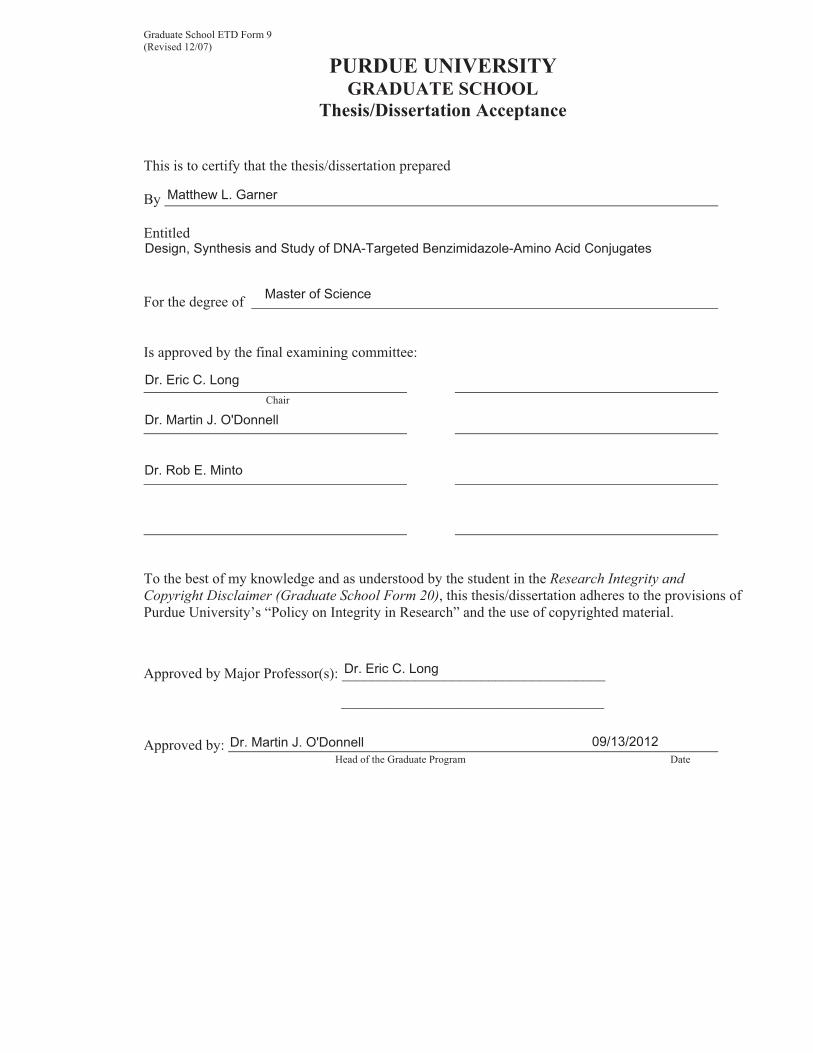
Graduate School ETD Form 9 (Revised 12/07)
PURDUE UNIVERSITY GRADUATE SCHOOL
Thesis/Dissertation Acceptance
This is to certify that the thesis/dissertation prepared
By
Entitled
For the degree of
Is approved by the final examining committee:
Chair
To the best of my knowledge and as understood by the student in the Research Integrity and Copyright Disclaimer (Graduate School Form 20), this thesis/dissertation adheres to the provisions of Purdue University’s “Policy on Integrity in Research” and the use of copyrighted material.
Approved by Major Professor(s): ____________________________________
____________________________________
Approved by: Head of the Graduate Program Date
Matthew L. Garner
Design, Synthesis and Study of DNA-Targeted Benzimidazole-Amino Acid Conjugates
Master of Science
Dr. Eric C. Long
Dr. Martin J. O'Donnell
Dr. Rob E. Minto
Dr. Eric C. Long
Dr. Martin J. O'Donnell 09/13/2012
Graduate School Form 20 (Revised 9/10)
PURDUE UNIVERSITY GRADUATE SCHOOL
Research Integrity and Copyright Disclaimer
Title of Thesis/Dissertation:
For the degree of Choose your degree
I certify that in the preparation of this thesis, I have observed the provisions of Purdue University Executive Memorandum No. C-22, September 6, 1991, Policy on Integrity in Research.*
Further, I certify that this work is free of plagiarism and all materials appearing in this thesis/dissertation have been properly quoted and attributed.
I certify that all copyrighted material incorporated into this thesis/dissertation is in compliance with the United States’ copyright law and that I have received written permission from the copyright owners for my use of their work, which is beyond the scope of the law. I agree to indemnify and save harmless Purdue University from any and all claims that may be asserted or that may arise from any copyright violation.
______________________________________ Printed Name and Signature of Candidate
______________________________________ Date (month/day/year)
*Located at http://www.purdue.edu/policies/pages/teach_res_outreach/c_22.html
Design, Synthesis and Study of DNA-Targeted Benzimidazole-Amino Acid Conjugates
Master of Science
Matthew L. Garner
09/13/2012
DESIGN, SYNTHESIS AND STUDY OF DNA-TARGETED
BENZIMIDAZOLE-AMINO ACID CONJUGATES
A Thesis
Submitted to the Faculty
of
Purdue University
by
Matthew L. Garner
In Partial Fulfillment of the
Requirements for the Degree
of
Master of Science
December 2012
Purdue University
Indianapolis, Indiana
ii
Dedicated to my family and friends

iii
ACKNOWLEDGMENTS
I would like to acknowledge my mentor Dr. Eric C. Long and thank him for
assisting and directing my trip through graduate school. I would also like to specially
thank Dr. Tax Georgiadis for his friendship and all his assistance in the laboratory
performing syntheses, purification, and analysis. My friend and fellow graduate student,
David Ames, was also very helpful through the process of purifying my compounds as
well as our trip through graduate courses, I am grateful to have him as a friend.
I would like to thank Dr. Martin J. O’Donnell and Dr. Robert E. Minto for serving
on my graduate committee. Dr. Karl Dria and Cary Pritchard were also both helpful in
training, advising, and troubleshooting instrumentation and are due considerable thanks.
Also, I would like to thank Kitty O’Doherty and Beverly Hewitt for all their assistance
through my time at IUPUI.
Dr. Ryan Denton was also a great friend and advisor through the time I spent
teaching labs for him and is due considerable thanks as well. Wai Ping Kam is another
friend gained during my time in graduate school that helped and offered me advice
throughout the process of teaching labs.
I would like to thank my parents, sister, family, and friends for all their love,
support, and encouragement throughout my time in graduate school. Finally, I would
like to thank my lovely girlfriend, Amanda Hardwick, for all her support and belief in me
throughout my time at IUPUI. I am truly blessed to have such wonderful people in my
life and without them I wouldn’t have been able to obtain this degree.
iv
TABLE OF CONTENTS
Page
LIST OF TABLES .......................................................................................................... vii
LIST OF FIGURES ....................................................................................................... viii
LIST OF ABBREVIATIONS ............................................................................................. x
ABSTRACT .................................................................................................................. xiii
CHAPTER 1. STRUCTURE OF B-FORM DNA AND MINOR GROOVE RECOGNITION BY LOW MOLECULAR WEIGHT COMPOUNDS ............ 1 1.1. Overview ......................................................................................................... 1
1.2. Introduction to DNA Structure .......................................................................... 2
1.2.1. Overview .............................................................................................. 2
1.2.2. Structure of B-Form DNA ..................................................................... 3
1.3. DNA Ligand Binding Modes............................................................................. 7
1.4. Examples of DNA Minor Groove Binding Ligands .......................................... 10
1.4.1. Netropsin ........................................................................................... 10
1.4.2. Distamycin ......................................................................................... 13
1.4.3. Synthetic Polyamides ......................................................................... 15
1.5. Benzimidazole-Based DNA Minor Groove Binders ........................................ 18
1.5.1. Hoechst 33258 ................................................................................... 18
1.5.2. Benzimidazole-Amidine Systems ....................................................... 21
1.6. Plan of Study ................................................................................................. 23
v
Page
1.7. List of References ......................................................................................... 25
CHAPTER 2. DESIGN AND SYNTHESIS OF AMINO ACID-BENZIMIDAZOLE- AMIDINE CONJUGATES ....................................................................... 30 2.1. Design of Amino Acid-Benzimidazole-Amidine Conjugates ........................... 30
2.2. Synthesis – General Considerations .............................................................. 31
2.2.1. Synthesis of 3,4-Diaminobenzamidoxime .......................................... 33
2.2.2. Benzimidazole-Amidines Lacking Amino Acid Diversity ..................... 34
2.2.3. Amino Acid-Benzimidazole-Amidine Conjugates ................................ 37
2.2.3.1. Single Amino Acid-Benzimidazole-Amidine Conjugates ..... 38
2.2.3.2. Dipeptide-Benzimidazole-Amidine Conjugates ................... 40
2.3. Summary ....................................................................................................... 42
2.4. Experimental Protocols .................................................................................. 43
2.4.1. Materials ........................................................................................... 43
2.4.2. Instruments ....................................................................................... 43
2.4.3. Syntheses ......................................................................................... 43
2.4.3.1. General Synthetic Considerations ...................................... 43
2.4.3.2. General Procedure for Synthesis of Model-BI-(+) ............... 44
2.4.3.3. General Procedure for Synthesis of Xaa-BI-(+) Conjugates ....................................................... 44 2.4.3.4. General Procedure for Synthesis of Xaa-Gly-BI-(+) Conjugates ................................................. 45 2.4.3.5. Synthesis of Diaminobenzamidoxime................................. 46
2.5. List of References .......................................................................................... 65
CHAPTER 3. PRELIMINARY SCREENING OF DNA BINDING ACTIVITY................... 68
3.1. Overview ....................................................................................................... 68
vi
Page
3.2. HT-FID Assay ................................................................................................ 68
3.2.1. HT-FID Assay Validation .................................................................... 70
3.3. HT-FID Analysis of Amino Acid-Benzimidazole-Amidine Conjugates ............. 71
3.3.1. HT-FID Analysis of Mono-(Amino Acid)-Benzimidazole-Amidine Conjugates ........................................................................................ 72 3.3.2. HT-FID Analysis of Dipeptide-Benzimidazole-Amidine Conjugates .... 73
3.4. Summary ....................................................................................................... 75
3.5. Experimental Protocols .................................................................................. 76
3.5.1. Materials ............................................................................................ 76
3.5.2. HT-FID Assay .................................................................................... 76
3.6. List of References ......................................................................................... 77
APPENDICES
Appendix A. 1H NMR Spectra ................................................................................ 78
Appendix B. Mass Spectra .................................................................................. 115
vii
LIST OF TABLES
Table Page 2.1. Mono-(amino acid)-benzimidazole-amidine conjugate m/z and yields ................... 39
2.2. Di-(amino acid)-benzimidazole-amidine conjugate m/z and yields ........................ 41
3.1. Zʹ-score analysis of netropsin and distamycin ....................................................... 71
viii
LIST OF FIGURES
Figure Page 1.1. The “Central Dogma of Molecular Biology” ............................................................. 3
1.2. Primary structure of DNA ........................................................................................ 4
1.3. A•T and G•C Watson-Crick base pairs of DNA ....................................................... 4
1.4. Structures of A, B, and Z-DNA ................................................................................ 5
1.5. Structure of the B-form DNA double helix ............................................................... 6 1.6. Ethidium bromide intercalated between two DNA base pairs .................................. 8
1.7. Structures of ethidium bromide and thiazole orange ............................................... 8
1.8. Structure of netropsin ............................................................................................ 10 1.9. Netropsin bound to the minor groove of 5ʹ-AATT DNA .......................................... 11
1.10. Hydrogen bonding observed between netropsin and the AATT oligonucleotide .... 12
1.11. Merged-bar FID histogram of netropsin at 0.75 and 1.5 μM .................................. 13
1.12. Structure of distamycin ......................................................................................... 14
1.13. Structures of polyamides bound to DNA: (A) 2:1 motif, (B) 1:1 motif ..................... 14
1.14. Merged-bar FID histogram of distamycin at 2.0 μM: (A) all 512 sequences, (B) top 50 sequences showing highest affinity ...................................................... 15 1.15. Lexitropsin 2-imidazole-distamycin with arrows indicating hydrogen accepting and donating groups in red and blue, respectively ................................................ 16 1.16. Pairing code illustrating the contacts of polyamides with minor groove. Note, the structure of the Hp extends a hydrogen deep into the groove to interact with sterically hindered thymine-O2 lone pair ........................................... 17 1.17. Structure of Hoechst 33258 .................................................................................. 19
ix
Figure Page 1.18. Merged-bar FID histogram of Hoechst 33258 at 2.0 μM: (A) all 512 oligonucleotide sequences, (B) top 50 sequences showing highest affinity ........... 20 1.19. Structures of (A) a simple Hoechst 33258 analogue and (B) a Hoechst- peptide conjugate ................................................................................................. 21 1.20. Structure of RT 29 ................................................................................................ 22
1.21. Structures of DB Series Compounds .................................................................... 22
1.22. Structure of model amino acid-phenyl-benzimidazole-amidine system ................. 24
2.1. Resin-bound benzimidazole amidine systems: (A) single amino acid- and (B) di-amino acid-benzimidazole-amidine conjugates where Xaa is any one of 20
naturally occurring amino acids (except Trp, Ser, Cys, or His for structure A). ...... 31 2.2. Structures of Rink amide resin and Wang resin, arrows indicate coupling sites .... 32 2.3. Solid-phase coupling of amino acid to Rink amide resin ....................................... 32 2.4. Solid-phase amidoxime reduction ......................................................................... 33 2.5. Synthesis of 3,4-diaminobenzamidoxime .............................................................. 34 2.6. 1H NMR of purified 3,4-diaminobenzamidoxime (1) in DMSO-d6 ........................... 34 2.7. Solid-phase synthesis of phenyl-benzimidazole-amidine ...................................... 35 2.8. 1H NMR of model-benzimidazole-amidine (2) in DMSO-d6 ................................... 37 2.9. Solid-phase synthesis of single amino acid-benzimidazole-amidine conjugates ... 39 2.10. Example 1H NMR of glycine-benzimidazole-amidine (3) in DMSO-d6 .................... 40 2.11. Example 1H NMR of glycine-glycine-benzimidazole-amidine (19) in DMSO-d6 ..... 41 3.1. Depiction of the HT-FID assay process ................................................................. 69 3.2. Relative binding of Xaa-BI-(+) conjugates in CT-DNA ........................................... 72 3.3. Relative binding of Xaa-Gly-BI-(+) conjugates in CT-DNA .................................... 73 3.4. Focused relative FID binding analysis of the most potent binders ......................... 74
x
LIST OF ABBREVIATIONS
ACN acetonitrile
Ala alanine
Arg arginine
Asn asparagine
Asp aspartic acid
BI benzimidazole
Boc di-tert-butyl dicarbonate
bp base pair
CT-DNA calf thymus DNA
Cys cysteine
DCM dichloromethane
DIC diisopropylcarbodiimide
DMF dimethylformamide
DMSO dimethyl sulfoxide
DNA deoxyribonucleic acid
EtBr ethidium bromide
EtOAc ethyl acetate
EtOH ethanol
Fmoc fluorenylmethoxycarbonyl
FID fluorescence intercalator displacement

xi
Gln glutamine
Glu glutamic acid
Gly glycine
His histidine
HOBt 1-hydroxybenzotriazole
HPLC high performance liquid chromatography
HT-FID high-throughput fluorescence intercalator displacement assay
Ile isoleucine
LC/MS liquid chromatography mass spectrometry
Leu leucine
Lys lysine
MeOH methanol
Met methionine
mRNA messenger ribonucleic acid
NMR nuclear magnetic resonance
Phe phenylalanine
Pro proline
Ser serine
TFA trifluoroacetic acid
Thr threonine
TLC thin-layer chromatography
TRIS tris(hydroxymethyl)aminomethane
Trp tryptophan
Tyr tyrosine
xii
UV ultraviolet
Val valine
Xaa(s) any amino acid(s)
Xaa-BI-(+) any amino acid-benzimidazole-amidine
xiii
ABSTRACT
Garner, Matthew L. M.S., Purdue University, December 2012. Design, Synthesis and Study of DNA-Targeted Benzimidazole-Amino Acid Conjugates. Major Professor: Eric C. Long.
The DNA minor groove continues to be an important biological target in the
development of anticancer, antiviral, and antimicrobial compounds. Among agents that
target the minor groove, studies of well-established benzimidazole-based DNA binders
such as Hoechst 33258 have made it clear that the benzimidazole-amidine portion of
these molecules promotes an efficient, site-selective DNA association. Building on the
beneficial attributes of existing benzimidazole-based DNA binding agents, a series of
benzimidazole-amino acid conjugates was synthesized to investigate their DNA
recognition and binding properties. In this series of compounds, the benzimidazole-
amidine moiety was utilized as a core DNA “anchoring” element accompanied by
different amino acids to provide structural diversity that may influence DNA binding
affinity and site-selectivity. Single amino acid conjugates of benzimidazole-amidines
were synthesized, as well as a series of conjugates containing 20 dipeptides with the
general structure Xaa-Gly. These conjugates were synthesized through a solid-phase
synthetic route building from a resin-bound amino acid (or dipeptide). The synthetic
steps involved: (1) the coupling of 4-formylbenzoic acid to the resin-bound amino acid
(via diisopropylcarbodiimide and hydroxybenzotriazole); followed by (2) introduction of a
3,4-diaminobenzamidoxime in the presence of 1,4-benzoquinone to construct the
benzimidazole ring; and, finally, (3) reduction of the resin-bound amidoxime functionality
to an amidine via treatment with 1M SnCl2·2H2O in DMF before cleavage of final product
from the resin. The synthetic route developed and employed was simple and
straightforward except for the final reduction that proved to be very arduous. All target
compounds were obtained in good yield (based upon weight), averaging 73% mono-
amino acid and 78% di-amino acid final compound upon cleavage from resin.
xiv
Ultimately, the DNA binding activities of the amino acid-benzimidazole-amidine
conjugates were analyzed using a fluorescent intercalator displacement (FID) assay and
calf thymus DNA as a substrate. The relative DNA binding affinities of both the mono-
and di-amino acid-benzimidazole-amidine conjugates were generally weaker than that of
netropsin and distamycin with the dipeptide conjugates showing stronger binding
affinities than the mono-amino acid conjugates. The dipeptide conjugates containing
amino acids with positively charged side chains, Lys-Gly-BI-(+) and Arg-Gly-BI-(+),
showed the strongest DNA binding affinities amongst all our synthesized conjugates.
1
CHAPTER 1. STRUCTURE OF B-FORM DNA AND MINOR GROOVE RECOGNITION
BY LOW MOLECULAR WEIGHT COMPOUNDS
1.1. Overview
The DNA minor groove has been an important focus of chemical and biological
studies since the elucidation of the structure of DNA and an understanding of the role of
DNA in the life cycle of a cell. The interaction of small molecules with DNA is also a
prolific area of study because many therapeutically important molecules bind reversibly
to nucleic acids.1-6 It is commonly believed that minor groove binding compounds
disrupt normal cellular functions by binding near or at promoter regions of genes, altering
transcription7 or disrupting DNA replication. Thus, much effort has been directed toward
the discovery of low molecular weight compounds that recognize and bind to DNA due to
their potential use as anticancer, antiviral, and antimicrobial drugs.7-10 In addition, the
development of sequence-specific and sequence-selective DNA binding molecules is a
research goal that is important for understanding nucleic acid molecular recognition due
to the ability of these agents to act as nucleic acid conformational probes and
footprinting reagents.11,12 In general, low molecular weight ligands recognize DNA using
a combination of weak intermolecular forces such as electrostatics, van der Waals
forces, and hydrogen bonding; DNA binding can ultimately occur through ligand
interactions with the minor groove, phosphodiester backbone, and stacked Watson-Crick
base pairs.
As will be discussed, the structural basis for the design of many man-made DNA
minor groove binding ligands originates from naturally occurring peptide-based
compounds such as netropsin, distamycin, actinomycin, and echinomycin.13-16 These
compounds have provided a starting point for many drug design efforts including
synthetic polyamides.17 Also, benzimidazole derivates18 have displayed exceptional
DNA binding abilities and may provide a useful moiety for drug design. Some important
features of molecules that bind to the minor groove of B-DNA like those mentioned
above are: (1) a crescent shape complementary to the curvature of the minor groove; (2)
2
positive charges that enhance electrostatic interactions; (3) inward-facing hydrogen-
bonding groups for sequence recognition; and (4) an unfused heterocyclic structure that
allows flexible structural optimization of the compound for minor groove interactions.19
These guidelines have aided in the design of minor groove binding heterocycles with
strong minor groove binding interactions and biological activities.20-21 While we note the
importance of shape complementarity, more recent studies also emphasize that the
shapes of compounds do not have to exactly match the curvature of the minor groove to
yield strong sequence-specific binding, as well as the usefulness of nitrogen containing
heterocycles for minor groove recognition.22 All of these factors were considered in the
design of our minor groove binding agents to be described herein.
This thesis will describe a series of mono- and di-amino acid-benzimidazole-
amidine conjugates designed to target the minor groove of B-form DNA. A phenyl-
benzimidazole core structure will be included to provide hydrogen-bonding sites as well
as allowing an overall molecular curvature that closely resembles that of the minor
groove. In addition, by introducing amino acids, we will place in position: (1) amide
bonds that can serve as auxiliary hydrogen-bonding sites to interact with the DNA minor
groove, and (2) side-chains that introduce structural and chemical diversity. Finally, an
amidine group will provide a positively charged moiety that can interact electrostatically
with the negatively charged phosphodiester backbone of DNA. Positively charged
moieties are usually attracted to A/T-rich regions of DNA that have a slightly increased
electrostatic potential than regions of G•C base pairs.
1.2. Introduction to DNA Structure
1.2.1. Overview
The “Central Dogma of Molecular Biology” (Figure 1.1) outlines the role of DNA
in living organisms23 and the role of DNA in replication and protein expression. DNA
contains all the genetic information that controls the synthesis and regulation of protein
expression in a cell. DNA has two main functions: (1) to provide a template for its own
replication during cell division and (2) to direct transcription of complementary strands of
messenger ribonucleic acid (mRNA) and other RNAs.24 Upon the initiation of protein
expression, DNA is initially transcribed into an mRNA template that is processed and
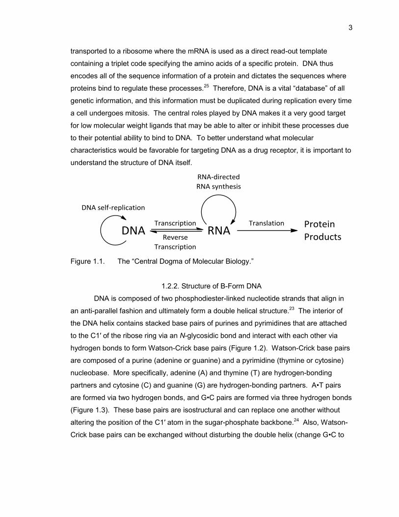
3
transported to a ribosome where the mRNA is used as a direct read-out template
containing a triplet code specifying the amino acids of a specific protein. DNA thus
encodes all of the sequence information of a protein and dictates the sequences where
proteins bind to regulate these processes.25 Therefore, DNA is a vital “database” of all
genetic information, and this information must be duplicated during replication every time
a cell undergoes mitosis. The central roles played by DNA makes it a very good target
for low molecular weight ligands that may be able to alter or inhibit these processes due
to their potential ability to bind to DNA. To better understand what molecular
characteristics would be favorable for targeting DNA as a drug receptor, it is important to
understand the structure of DNA itself.
Figure 1.1. The “Central Dogma of Molecular Biology.”
1.2.2. Structure of B-Form DNA
DNA is composed of two phosphodiester-linked nucleotide strands that align in
an anti-parallel fashion and ultimately form a double helical structure.23 The interior of
the DNA helix contains stacked base pairs of purines and pyrimidines that are attached
to the C1′ of the ribose ring via an N-glycosidic bond and interact with each other via
hydrogen bonds to form Watson-Crick base pairs (Figure 1.2). Watson-Crick base pairs
are composed of a purine (adenine or guanine) and a pyrimidine (thymine or cytosine)
nucleobase. More specifically, adenine (A) and thymine (T) are hydrogen-bonding
partners and cytosine (C) and guanine (G) are hydrogen-bonding partners. A•T pairs
are formed via two hydrogen bonds, and G•C pairs are formed via three hydrogen bonds
(Figure 1.3). These base pairs are isostructural and can replace one another without
altering the position of the C1′ atom in the sugar-phosphate backbone.24 Also, Watson-
Crick base pairs can be exchanged without disturbing the double helix (change G•C to
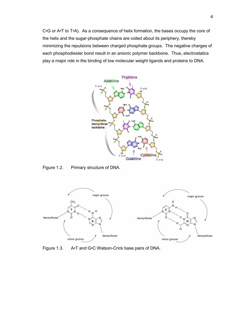
4
C•G or A•T to T•A). As a consequence of helix formation, the bases occupy the core of
the helix and the sugar-phosphate chains are coiled about its periphery, thereby
minimizing the repulsions between charged phosphate groups. The negative charges of
each phosphodiester bond result in an anionic polymer backbone. Thus, electrostatics
play a major role in the binding of low molecular weight ligands and proteins to DNA.
Figure 1.2. Primary structure of DNA.
Figure 1.3. A•T and G•C Watson-Crick base pairs of DNA.
5
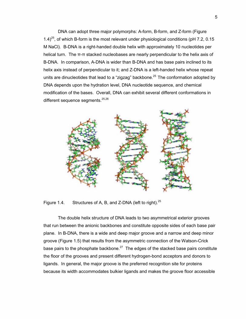
DNA can adopt three major polymorphs: A-form, B-form, and Z-form (Figure
1.4)25, of which B-form is the most relevant under physiological conditions (pH 7.2, 0.15
M NaCl). B-DNA is a right-handed double helix with approximately 10 nucleotides per
helical turn. The π-π stacked nucleobases are nearly perpendicular to the helix axis of
B-DNA. In comparison, A-DNA is wider than B-DNA and has base pairs inclined to its
helix axis instead of perpendicular to it; and Z-DNA is a left-handed helix whose repeat
units are dinucleotides that lead to a “zigzag” backbone.25 The conformation adopted by
DNA depends upon the hydration level, DNA nucleotide sequence, and chemical
modification of the bases. Overall, DNA can exhibit several different conformations in
different sequence segments.25,26
Figure 1.4. Structures of A, B, and Z-DNA (left to right).25
The double helix structure of DNA leads to two asymmetrical exterior grooves
that run between the anionic backbones and constitute opposite sides of each base pair
plane. In B-DNA, there is a wide and deep major groove and a narrow and deep minor
groove (Figure 1.5) that results from the asymmetric connection of the Watson-Crick
base pairs to the phosphate backbone.27 The edges of the stacked base pairs constitute
the floor of the grooves and present different hydrogen-bond acceptors and donors to
ligands. In general, the major groove is the preferred recognition site for proteins
because its width accommodates bulkier ligands and makes the groove floor accessible
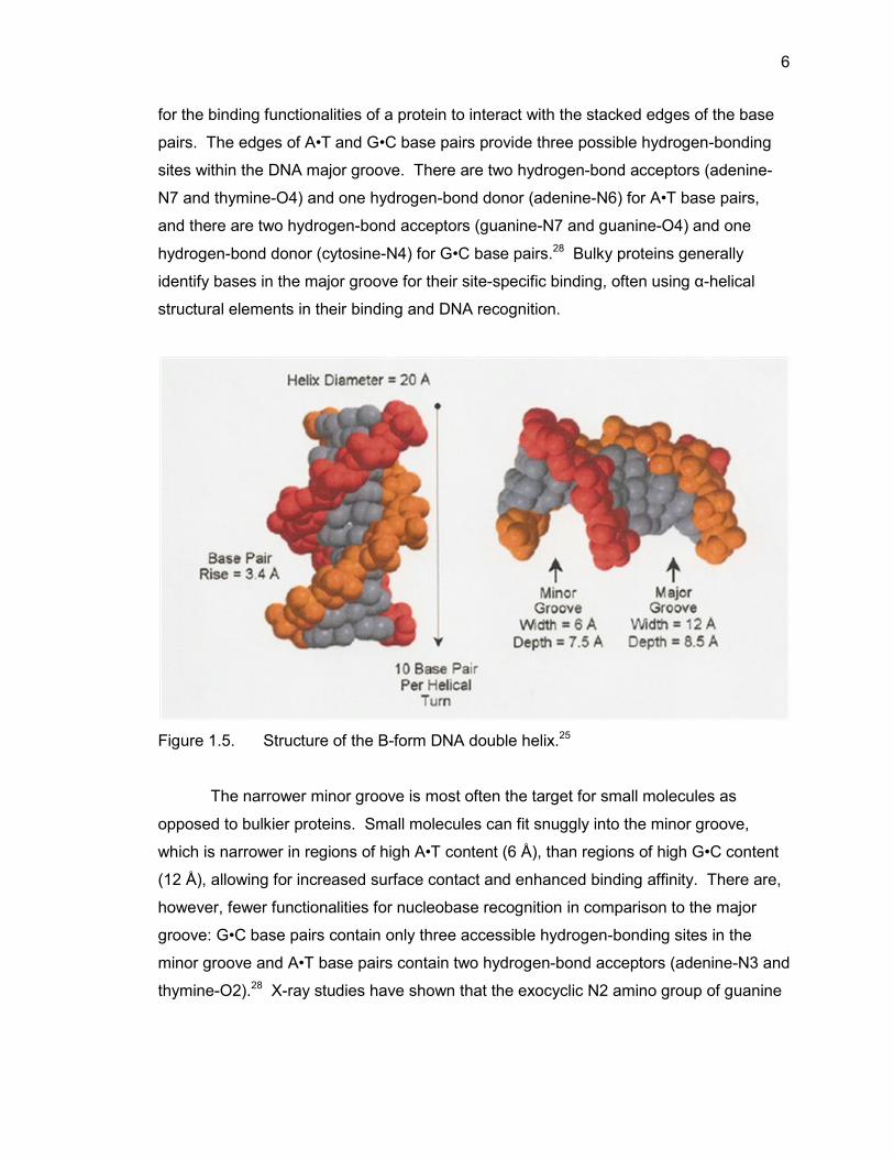
6
for the binding functionalities of a protein to interact with the stacked edges of the base
pairs. The edges of A•T and G•C base pairs provide three possible hydrogen-bonding
sites within the DNA major groove. There are two hydrogen-bond acceptors (adenine-
N7 and thymine-O4) and one hydrogen-bond donor (adenine-N6) for A•T base pairs,
and there are two hydrogen-bond acceptors (guanine-N7 and guanine-O4) and one
hydrogen-bond donor (cytosine-N4) for G•C base pairs.28 Bulky proteins generally
identify bases in the major groove for their site-specific binding, often using α-helical
structural elements in their binding and DNA recognition.
Figure 1.5. Structure of the B-form DNA double helix.25
The narrower minor groove is most often the target for small molecules as
opposed to bulkier proteins. Small molecules can fit snuggly into the minor groove,
which is narrower in regions of high A•T content (6 Å), than regions of high G•C content
(12 Å), allowing for increased surface contact and enhanced binding affinity. There are,
however, fewer functionalities for nucleobase recognition in comparison to the major
groove: G•C base pairs contain only three accessible hydrogen-bonding sites in the
minor groove and A•T base pairs contain two hydrogen-bond acceptors (adenine-N3 and
thymine-O2).28 X-ray studies have shown that the exocyclic N2 amino group of guanine
7
is often a hydrogen-bond donor group.29 However, this same amino group also appears
to disrupt the association of DNA minor groove binders in G/C-rich regions by protruding
from the floor of the groove, preventing a close association that would otherwise occur in
deeper A/T-rich regions. Conceptually, the three sites in G•C base pairs would compare
favorably to the two of A•T base pairs, but studies have demonstrated that many binders
prefer A•T sites. This suggests that hydrogen bonding is not the sole determinant for
sequence recognition within the DNA minor groove. It is likely that electrostatic potential
is of great importance for minor groove recognition as a series of A•T base pairs has a
greater negative electrostatic potential at the floor of the groove than that of G•C base
pairs.7 The more negative electrostatic potential of A•T base pairs is likely the reason
positively charged ligands prefer A/T-rich regions to G/C-rich regions.
Much effort has been expended towards understanding the structure of DNA and
its potential drug binding sites. It has been established that ligand-DNA binding
commonly occurs through a combination of electrostatics, van der Waals forces, and
hydrogen bonding. The negatively charged phosphodiester backbone forms complexes
with low molecular weight ligands with positive charges through electrostatic
interactions; the formation of the Watson-Crick base pairs results in the presence of
hydrophobic features along the walls of the groove. Knowledge of the numerous binding
sites in DNA and the methods known DNA binders use to bind to DNA aids our attempts
to develop new molecules with increased binding affinity and specificity. This thesis will
focus on utilizing low molecular weight ligand-DNA interactions of the minor groove in
our design of amino acid-benzimidazole-amidine conjugates as possible DNA minor
groove binding ligands.
1.3. DNA Ligand Binding Modes
There are several DNA-ligand binding modes, including (1) exterior surface
binding which is mainly electrostatically driven, (2) intercalation, and (3) groove binding
to either the major or minor groove. DNA intercalators are typically positively-charged,
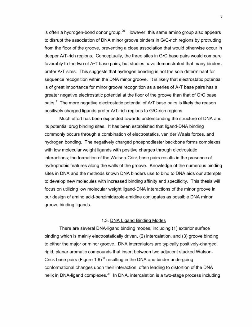
rigid, planar aromatic compounds that insert between two adjacent stacked Watson-
Crick base pairs (Figure 1.6)30 resulting in the DNA and binder undergoing
conformational changes upon their interaction, often leading to distortion of the DNA
helix in DNA-ligand complexes.31 In DNA, intercalation is a two-stage process including
8
(1) an initial diffusion-controlled association of the intercalator with the exterior of the
helix followed by (2) a slower insertion of the intercalator between stacked base pairs.
When intercalation occurs, the flanking base pairs must separate approximately 0.3 nm,
causing structural changes in the DNA which can lead to inhibition of transcription and
replication. Intercalators can also resemble another base pair and cause enzymes to
make errors in transcription, resulting in the addition of an extra base that alters the
triplet code generating an altered sequence. Therefore, many intercalators are powerful
mutagens and in some cases can be used in chemotherapeutic treatments to inhibit
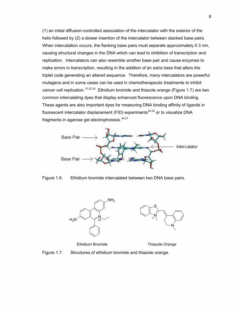
cancer cell replication.12,32,33 Ethidium bromide and thiazole orange (Figure 1.7) are two
common intercalating dyes that display enhanced fluorescence upon DNA binding.
These agents are also important dyes for measuring DNA binding affinity of ligands in
fluorescent intercalator displacement (FID) experiments34,35 or to visualize DNA
fragments in agarose gel electrophoresis.36,37
Figure 1.6. Ethidium bromide intercalated between two DNA base pairs.
Figure 1.7. Structures of ethidium bromide and thiazole orange.
9
There are many factors involved in DNA minor groove binding, thus only the
major influences will be discussed. The natural products netropsin and distamycin have
played an integral role in understanding these mechanisms of small molecule-DNA
minor groove recognition. An early crystal structure of netropsin bound to DNA revealed
that minor groove binding occurs when netropsin aligns along the groove cleft of B-DNA
and forms hydrogen bonds with the floor of the groove.38 Overall, small molecule DNA
recognition occurs through a combination of electrostatic interactions with the
phosphodiester backbone, hydrogen bonding to the nucleobases, van der Waals contact
with the walls of the groove, hydrophobic interactions, and steric hindrance to influence
the mode of ligand binding to DNA.39 The sugar-phosphate backbone of DNA, which is
negatively charged, is attractive to positively charged ligands which results in an
increase of ligand concentration near DNA from bulk solution. As stated earlier, A•T
regions of B-DNA have greater negative potential than those of G•C regions due to the
presence of electron rich thymine-O2 and adenine-N3 as well as the narrowed groove
width, making A•T regions targets for positively charged ligands. Therefore, minor
groove binding ligands typically have at least one positively charged group to enhance
its A•T site selectivity as well as binding affinity.
The width, structurally, of the groove around the helix can also play a part in the
minor groove binding of ligands to DNA. In regions of high A•T content, the groove is
narrower, whereas regions of high G•C content have a wider groove due, as noted
earlier, to the exocyclic group of guanine. An x-ray structure of netropsin bound to DNA
suggest the exocyclic group protrudes from the floor of the minor groove and causes
steric hindrance that interferes with binding in regions of high G•C content.40 Many
minor groove binders are elongated structures that contain multiple hydrogen-bonding
functionalities, therefore the narrower groove in A/T-rich regions contributes to the
hydrophobic contacts made with the surfaces of small molecules.41 The narrower
groove in A/T-rich regions aids in aligning small molecules so that hydrogen-bonding
groups are directly exposed to the floor of the groove, whereas the wider groove in G/C-
rich regions does not provide the tight fit to aid in binding by planar ligands.
10
1.4. Examples of DNA Minor Groove Binding Ligands
1.4.1. Netropsin
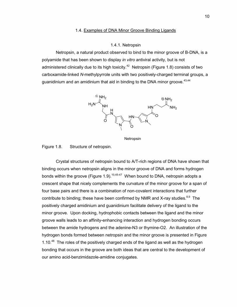
Netropsin, a natural product observed to bind to the minor groove of B-DNA, is a
polyamide that has been shown to display in vitro antiviral activity, but is not
administered clinically due to its high toxicity.42 Netropsin (Figure 1.8) consists of two
carboxamide-linked N-methylpyrrole units with two positively-charged terminal groups, a
guanidinium and an amidinium that aid in binding to the DNA minor groove.43,44
Figure 1.8. Structure of netropsin.
Crystal structures of netropsin bound to A/T-rich regions of DNA have shown that
binding occurs when netropsin aligns in the minor groove of DNA and forms hydrogen
bonds within the groove (Figure 1.9).10,45-47 When bound to DNA, netropsin adopts a
crescent shape that nicely complements the curvature of the minor groove for a span of
four base pairs and there is a combination of non-covalent interactions that further
contribute to binding; these have been confirmed by NMR and X-ray studies.8,9 The
positively charged amidinium and guanidinium facilitate delivery of the ligand to the
minor groove. Upon docking, hydrophobic contacts between the ligand and the minor
groove walls leads to an affinity-enhancing interaction and hydrogen bonding occurs
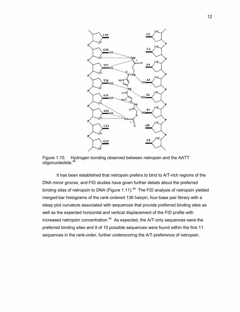
between the amide hydrogens and the adenine-N3 or thymine-O2. An illustration of the
hydrogen bonds formed between netropsin and the minor groove is presented in Figure
1.10.48 The roles of the positively charged ends of the ligand as well as the hydrogen
bonding that occurs in the groove are both ideas that are central to the development of
our amino acid-benzimidazole-amidine conjugates.
11
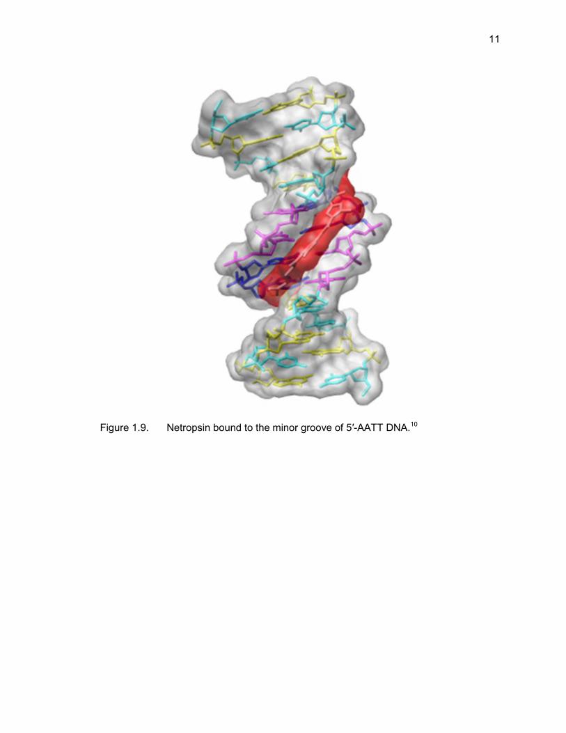
Figure 1.9. Netropsin bound to the minor groove of 5′-AATT DNA.10
12
Figure 1.10. Hydrogen bonding observed between netropsin and the AATT oligonucleotide.48
It has been established that netropsin prefers to bind to A/T-rich regions of the
DNA minor groove, and FID studies have given further details about the preferred
binding sites of netropsin to DNA (Figure 1.11).49 The FID analysis of netropsin yielded
merged-bar histograms of the rank-ordered 136 hairpin, four-base pair library with a
steep plot curvature associated with sequences that provide preferred binding sites as
well as the expected horizontal and vertical displacement of the FID profile with
increased netropsin concentration.49 As expected, the A/T-only sequences were the
preferred binding sites and 9 of 10 possible sequences were found within the first 11
sequences in the rank-order, further underscoring the A/T-preference of netropsin.
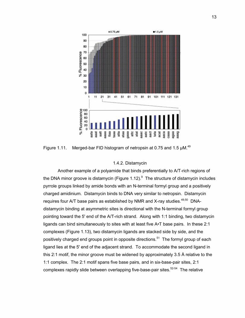
13
Figure 1.11. Merged-bar FID histogram of netropsin at 0.75 and 1.5 μM.49
1.4.2. Distamycin
Another example of a polyamide that binds preferentially to A/T-rich regions of
the DNA minor groove is distamycin (Figure 1.12).9 The structure of distamycin includes
pyrrole groups linked by amide bonds with an N-terminal formyl group and a positively
charged amidinium. Distamycin binds to DNA very similar to netropsin. Distamycin
requires four A/T base pairs as established by NMR and X-ray studies.49,50 DNA-
distamycin binding at asymmetric sites is directional with the N-terminal formyl group
pointing toward the 5' end of the A/T-rich strand. Along with 1:1 binding, two distamycin
ligands can bind simultaneously to sites with at least five A•T base pairs. In these 2:1
complexes (Figure 1.13), two distamycin ligands are stacked side by side, and the
positively charged end groups point in opposite directions.51 The formyl group of each
ligand lies at the 5' end of the adjacent strand. To accommodate the second ligand in
this 2:1 motif, the minor groove must be widened by approximately 3.5 Å relative to the
1:1 complex. The 2:1 motif spans five base pairs, and in six-base-pair sites, 2:1
complexes rapidly slide between overlapping five-base-pair sites.52-54 The relative
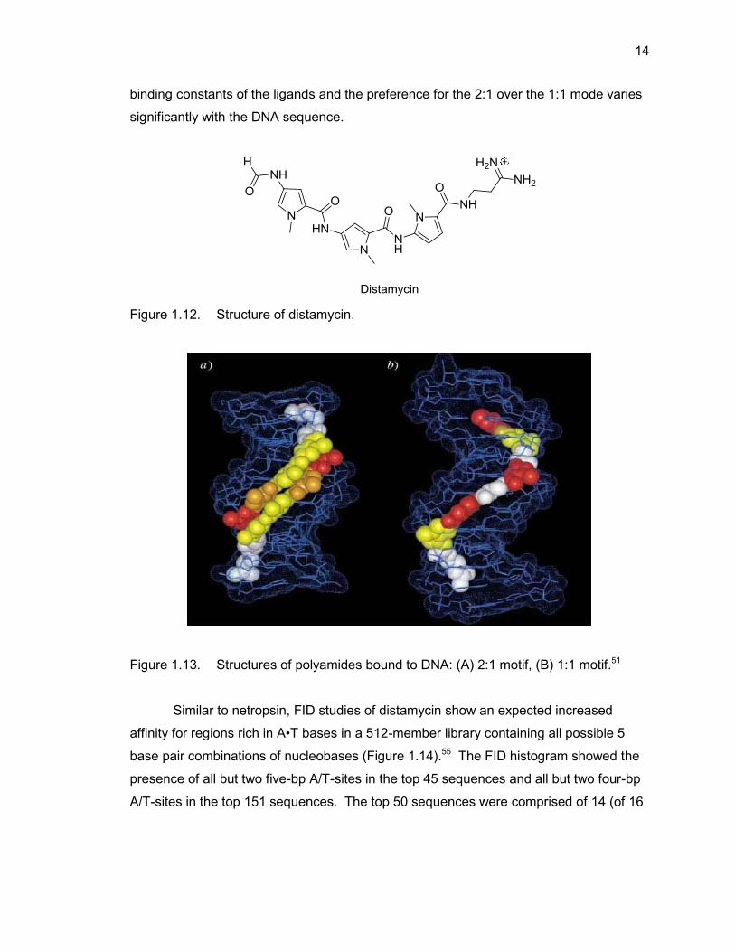
14
binding constants of the ligands and the preference for the 2:1 over the 1:1 mode varies
significantly with the DNA sequence.
Figure 1.12. Structure of distamycin.
Figure 1.13. Structures of polyamides bound to DNA: (A) 2:1 motif, (B) 1:1 motif.51
Similar to netropsin, FID studies of distamycin show an expected increased
affinity for regions rich in A•T bases in a 512-member library containing all possible 5
base pair combinations of nucleobases (Figure 1.14).55 The FID histogram showed the
presence of all but two five-bp A/T-sites in the top 45 sequences and all but two four-bp
A/T-sites in the top 151 sequences. The top 50 sequences were comprised of 14 (of 16
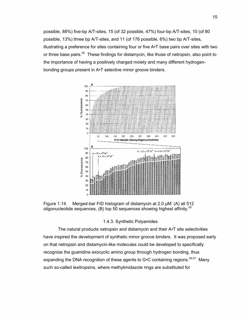
15
possible, 88%) five-bp A/T-sites, 15 (of 32 possible, 47%) four-bp A/T-sites, 10 (of 80
possible, 13%) three bp A/T-sites, and 11 (of 176 possible, 6%) two bp A/T-sites,
illustrating a preference for sites containing four or five A•T base pairs over sites with two
or three base pairs.55 These findings for distamycin, like those of netropsin, also point to
the importance of having a positively charged moiety and many different hydrogen-
bonding groups present in A•T selective minor groove binders.
Figure 1.14. Merged-bar FID histogram of distamycin at 2.0 μM: (A) all 512 oligonucleotide sequences, (B) top 50 sequences showing highest affinity.55
1.4.3. Synthetic Polyamides
The natural products netropsin and distamycin and their A•T site selectivities
have inspired the development of synthetic minor groove binders. It was proposed early
on that netropsin and distamycin-like molecules could be developed to specifically
recognize the guanidine exocyclic amino group through hydrogen bonding, thus
expanding the DNA recognition of these agents to G•C containing regions.56,57 Many
such so-called lexitropsins, where methylimidazole rings are substituted for
16
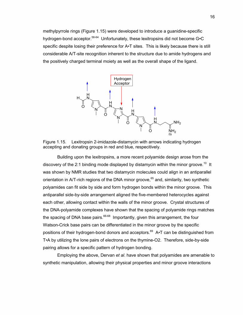
methylpyrrole rings (Figure 1.15) were developed to introduce a guanidine-specific
hydrogen-bond acceptor.58-64 Unfortunately, these lexitropsins did not become G•C
specific despite losing their preference for A•T sites. This is likely because there is still
considerable A/T-site recognition inherent to the structure due to amide hydrogens and
the positively charged terminal moiety as well as the overall shape of the ligand.
Figure 1.15. Lexitropsin 2-imidazole-distamycin with arrows indicating hydrogen accepting and donating groups in red and blue, respecitively.
Building upon the lexitropsins, a more recent polyamide design arose from the
discovery of the 2:1 binding mode displayed by distamycin within the minor groove.10 It
was shown by NMR studies that two distamycin molecules could align in an antiparallel
orientation in A/T-rich regions of the DNA minor groove,65 and, similarily, two synthetic
polyamides can fit side by side and form hydrogen bonds within the minor groove. This
antiparallel side-by-side arrangement aligned the five-membered heterocycles against
each other, allowing contact within the walls of the minor groove. Crystal structures of
the DNA-polyamide complexes have shown that the spacing of polyamide rings matches
the spacing of DNA base pairs.66-68 Importantly, given this arrangement, the four
Watson-Crick base pairs can be differentiated in the minor groove by the specific
positions of their hydrogen-bond donors and acceptors.69 A•T can be distinguished from
T•A by utilizing the lone pairs of electrons on the thymine-O2. Therefore, side-by-side
pairing allows for a specific pattern of hydrogen bonding.
Employing the above, Dervan et al. have shown that polyamides are amenable to
synthetic manipulation, allowing their physical properties and minor groove interactions
17
to be controlled. Moreover, over two decades of work has gone into the development of
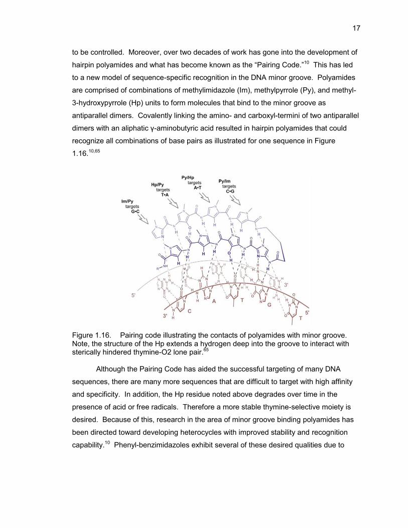
hairpin polyamides and what has become known as the “Pairing Code.”10 This has led
to a new model of sequence-specific recognition in the DNA minor groove. Polyamides
are comprised of combinations of methylimidazole (Im), methylpyrrole (Py), and methyl-
3-hydroxypyrrole (Hp) units to form molecules that bind to the minor groove as
antiparallel dimers. Covalently linking the amino- and carboxyl-termini of two antiparallel
dimers with an aliphatic γ-aminobutyric acid resulted in hairpin polyamides that could
recognize all combinations of base pairs as illustrated for one sequence in Figure
1.16.10,65
Figure 1.16. Pairing code illustrating the contacts of polyamides with minor groove. Note, the structure of the Hp extends a hydrogen deep into the groove to interact with sterically hindered thymine-O2 lone pair.65
Although the Pairing Code has aided the successful targeting of many DNA
sequences, there are many more sequences that are difficult to target with high affinity
and specificity. In addition, the Hp residue noted above degrades over time in the
presence of acid or free radicals. Therefore a more stable thymine-selective moiety is
desired. Because of this, research in the area of minor groove binding polyamides has
been directed toward developing heterocycles with improved stability and recognition
capability.10 Phenyl-benzimidazoles exhibit several of these desired qualities due to
18
their slightly different curvature and ability to recognize DNA through two sets of
hydrogen-bond donating groups.70 This curvature, as well as the possible hydrogen
bonding that can occur in the minor groove with the benzimidazole are key to including
benzimidazoles in our design of minor groove binding ligands.
1.5. Benzimidazole-Based DNA Minor Groove Binders
Phenyl-benzimidazole ring systems represent a structural framework that is
amenable to functionalization and imparts a curvature that complements the DNA minor
groove. Benzimidazole derivatives have been incorporated into the backbones of
polyamides in a manner that preserves hydrogen bonding contact with the minor
groove.71 Indeed, (1) the classic minor groove-binding Hoechst dyes are composed of
benzimidazole units and (2) hydroxybenzimidazole (Hz) and imidazopyridine (Ip) rings
have been included in polyamides without an amide linker between the rings. It has
been shown that Py-Hz and Py-Ip pairs are functionally identical to the five-membered
ring pairs Py-Hp and Py-Im.72 The Py-Hz pair has been shown to distinguish T/A from
A/T base pairs while the Py-Ip pair has been shown to distinguish G/C from C/G base
pairs. The benzimidazole containing polyamides have proven to be more chemically
stable and have been incorporated into effective minor groove binding DNA ligands.
1.5.1. Hoechst 33258
Hoechst 33258 (Figure 1.17) is a bis-benzimidazole comprised of two linked
benzimidazole groups with a phenol and methylpiperazine at either end. Hoechst 33258
has been employed as a chromosomal stain73 and a fluorescence indicator74 due to its
ability to be excited by ultraviolet light at ~350 nm and to exhibit enhanced fluorescence
upon binding to DNA. DNA footprinting and biophysical studies have shown Hoechst
33258 binds selectively to A•T sequences.74 Hoechst has been shown to exhibit
antitumor activity and was in phase I/II clincal trials against pancreatic carcinomas
before being abandoned due to its toxicity.75,76
19
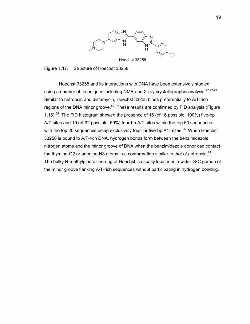
Figure 1.17. Structure of Hoechst 33258.
Hoechst 33258 and its interactions with DNA have been extensively studied
using a number of techniques including NMR and X-ray crystallographic analysis.72,77-79
Similar to netropsin and distamycin, Hoechst 33258 binds preferentially to A/T-rich
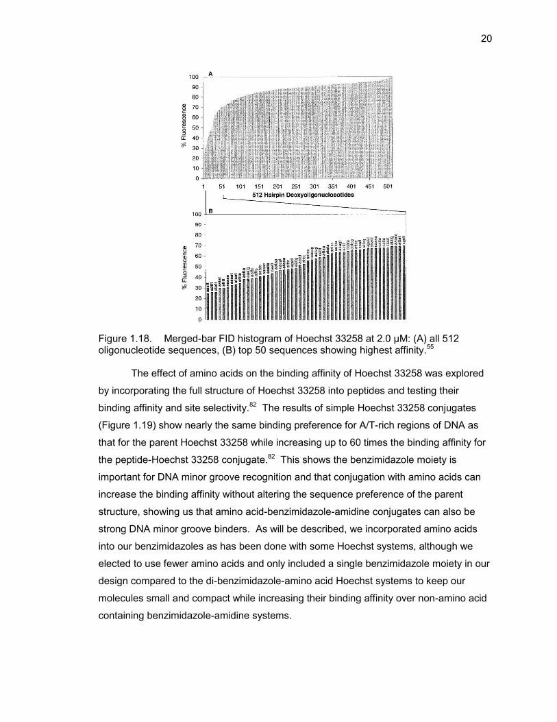
regions of the DNA minor groove.80 These results are confirmed by FID analysis (Figure
1.18).55 The FID histogram showed the presence of 16 (of 16 possible, 100%) five-bp
A/T-sites and 19 (of 32 possible, 59%) four-bp A/T-sites within the top 50 sequences
with the top 20 sequences being exclusively four- or five-bp A/T-sites.55 When Hoechst
33258 is bound to A/T-rich DNA, hydrogen bonds form between the benzimidazole
nitrogen atoms and the minor groove of DNA when the benzimidazole donor can contact
the thymine-O2 or adenine-N3 atoms in a conformation similar to that of netropsin.81
The bulky N-methylpiperazine ring of Hoechst is usually located in a wider G•C portion of
the minor groove flanking A/T-rich sequences without participating in hydrogen bonding.
20
Figure 1.18. Merged-bar FID histogram of Hoechst 33258 at 2.0 μM: (A) all 512 oligonucleotide sequences, (B) top 50 sequences showing highest affinity.55
The effect of amino acids on the binding affinity of Hoechst 33258 was explored
by incorporating the full structure of Hoechst 33258 into peptides and testing their
binding affinity and site selectivity.82 The results of simple Hoechst 33258 conjugates
(Figure 1.19) show nearly the same binding preference for A/T-rich regions of DNA as
that for the parent Hoechst 33258 while increasing up to 60 times the binding affinity for
the peptide-Hoechst 33258 conjugate.82 This shows the benzimidazole moiety is
important for DNA minor groove recognition and that conjugation with amino acids can
increase the binding affinity without altering the sequence preference of the parent
structure, showing us that amino acid-benzimidazole-amidine conjugates can also be
strong DNA minor groove binders. As will be described, we incorporated amino acids
into our benzimidazoles as has been done with some Hoechst systems, although we
elected to use fewer amino acids and only included a single benzimidazole moiety in our
design compared to the di-benzimidazole-amino acid Hoechst systems to keep our
molecules small and compact while increasing their binding affinity over non-amino acid
containing benzimidazole-amidine systems.
21
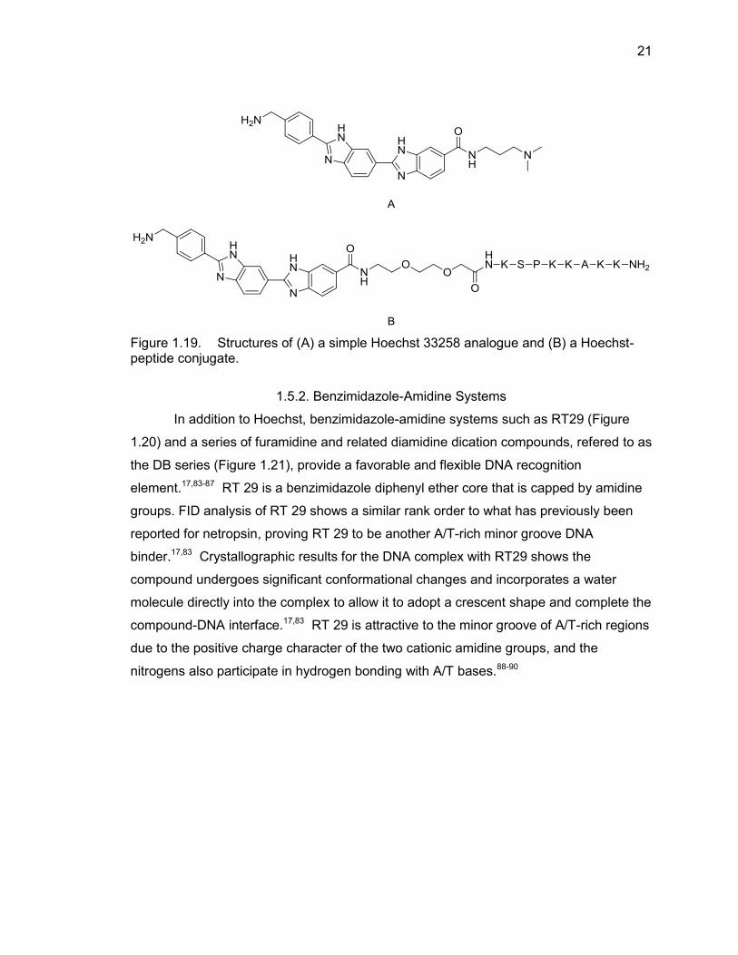
Figure 1.19. Structures of (A) a simple Hoechst 33258 analogue and (B) a Hoechst-peptide conjugate.
1.5.2. Benzimidazole-Amidine Systems
In addition to Hoechst, benzimidazole-amidine systems such as RT29 (Figure
1.20) and a series of furamidine and related diamidine dication compounds, refered to as
the DB series (Figure 1.21), provide a favorable and flexible DNA recognition
element.17,83-87 RT 29 is a benzimidazole diphenyl ether core that is capped by amidine
groups. FID analysis of RT 29 shows a similar rank order to what has previously been
reported for netropsin, proving RT 29 to be another A/T-rich minor groove DNA
binder.17,83 Crystallographic results for the DNA complex with RT29 shows the
compound undergoes significant conformational changes and incorporates a water
molecule directly into the complex to allow it to adopt a crescent shape and complete the
compound-DNA interface.17,83 RT 29 is attractive to the minor groove of A/T-rich regions
due to the positive charge character of the two cationic amidine groups, and the
nitrogens also participate in hydrogen bonding with A/T bases.88-90
22
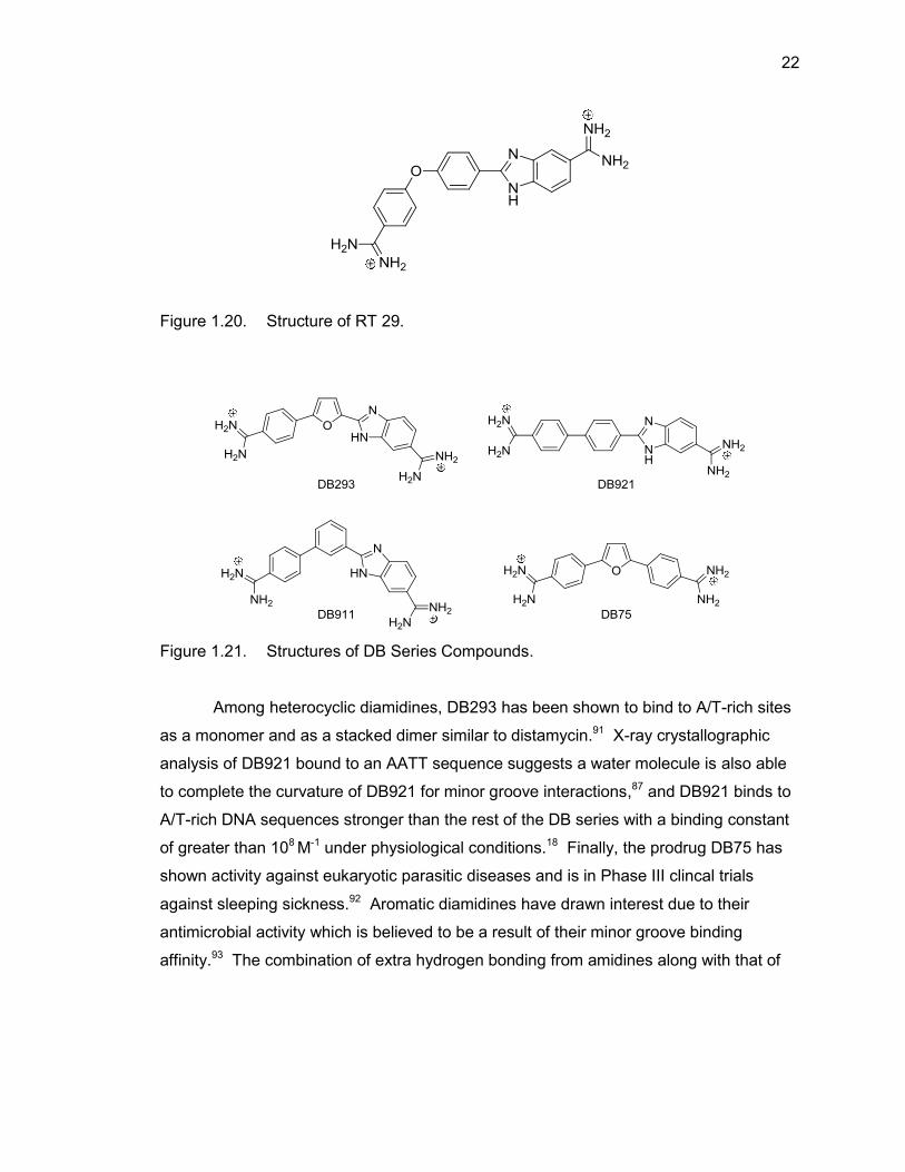
Figure 1.20. Structure of RT 29.
Figure 1.21. Structures of DB Series Compounds.
Among heterocyclic diamidines, DB293 has been shown to bind to A/T-rich sites
as a monomer and as a stacked dimer similar to distamycin.91 X-ray crystallographic
analysis of DB921 bound to an AATT sequence suggests a water molecule is also able
to complete the curvature of DB921 for minor groove interactions,87 and DB921 binds to
A/T-rich DNA sequences stronger than the rest of the DB series with a binding constant
of greater than 108 M-1 under physiological conditions.18 Finally, the prodrug DB75 has
shown activity against eukaryotic parasitic diseases and is in Phase III clincal trials
against sleeping sickness.92 Aromatic diamidines have drawn interest due to their
antimicrobial activity which is believed to be a result of their minor groove binding
affinity.93 The combination of extra hydrogen bonding from amidines along with that of
23
the benzimidazole make compounds containing these moieties strong targets for the
development of DNA minor groove binding agents.
In summary, we have introduced benzimidazole compounds, benzimidazole-
amidine systems, and benzimidazole-peptide conjugates. Benzimidazoles are good
minor groove A/T-rich region binding moieties due to their ability to hydrogen bond with
the floor of the the minor groove and that their size is not too bulky to be accommodated
by the minor groove. The positively charged amidine moiety is drawn to the more
negatively charged A/T regions of DNA and provides excellent affinity for these regions.
Peptides conjugated with these moieties can enhance the binding affinity. Therefore, we
hypothesize that amino acid-benzimidazole-amidine conjugates should be good DNA
minor groove binders due to the combination of these advantageous features.
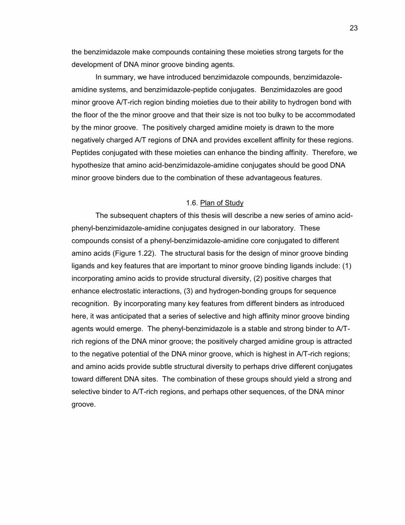
1.6. Plan of Study
The subsequent chapters of this thesis will describe a new series of amino acid-
phenyl-benzimidazole-amidine conjugates designed in our laboratory. These
compounds consist of a phenyl-benzimidazole-amidine core conjugated to different
amino acids (Figure 1.22). The structural basis for the design of minor groove binding
ligands and key features that are important to minor groove binding ligands include: (1)
incorporating amino acids to provide structural diversity, (2) positive charges that
enhance electrostatic interactions, (3) and hydrogen-bonding groups for sequence
recognition. By incorporating many key features from different binders as introduced
here, it was anticipated that a series of selective and high affinity minor groove binding
agents would emerge. The phenyl-benzimidazole is a stable and strong binder to A/T-
rich regions of the DNA minor groove; the positively charged amidine group is attracted
to the negative potential of the DNA minor groove, which is highest in A/T-rich regions;
and amino acids provide subtle structural diversity to perhaps drive different conjugates
toward different DNA sites. The combination of these groups should yield a strong and
selective binder to A/T-rich regions, and perhaps other sequences, of the DNA minor
groove.
24
Figure 1.22. Structure of model amino acid-phenyl-benzimidazole-amidine system.
The focus of this study was to develop the synthesis of the amino acid-
benzimidazole-amidine system that we have designed and to determine their relative
DNA binding affinities. All conjugates will be synthesized on solid-phase support and
their relative binding will be measured with the FID assay. Conjugates of this form have
never been explored, and the solid-phase synthesis of benzimidazole-amidine systems
has not been reported. Therefore, the synthetic protocol will be useful to supplement
solid-phase synthesis.
25
1.7. List of References
1. McKnight, R. E.; Gleason, A. B.; Keyes, J. A.; Sahabi, S. Bioorg. Med. Chem. Lett.
2007, 17, 1013.
2. Tse, W. C.; Boger, D. L. Chem. Biol. 2004, 11, 1607.
3. Haq, I. Arch. Biochem. Biophys. 2002, 403, 1.
4. Yang, X. L.; Wang, A. H-J. Pharmacol. Ther. 1999, 83, 181.
5. Chaires, I. B. Biopolymers 1997, 44, 201.
6. Hutchins, R. A.; Crenshaw, J. M.; Graves, D. E.; Denny, W. A. Biochemistry 2003,
42, 13754.
7. Neidle, S. Nat. Prod. Rep. 2001, 18, 291.
8. Neidle, S. Biopolymers 1997, 44, 105.
9. Geierstanger, B. H.; Wemmer, D. E. Ann. Rev. Biophys. Biomol. Struct. 1995, 24,
463.
10. Dervan, P. B. Bioorg. Med. Chem. 2001, 9, 2215.
11. Munde, M.; Ismail, M. A.; Arafa, R.; Peixoto, P.; Catharine J.; Collar, C. J.; Liu, Y.;
Hu, L. X.; David-Cordonnier, M.; Lansiaux, A.; Bailly, C.; Boykin, D. W.; Wilson, W.
D. J. Am. Chem. Soc. 2007, 129, 13732.
12. Hurley, L. H. Nat. Rev. Cancer 2002, 2, 188.
13. Gale, E. F.; Cundliffe, E.; Reynolds, P. E.; Richmond, M. H.; Waring, M. J. in: The
Molecular Basis of Antibitoic Action, 2nd Ed., Wiley & Sons, 1981.
14. Ughetto, G.; Wang, A. H-J.; Quigley, G. J.; va der Marel, G. A.; van Boom, J. H.;
Rich, A. Nucleic Acids Res. 1985, 13, 2305.
15. Wang, A. H-J.; Ughetto, G.; Quigley, G. J.; Hakoshima, T.; van der Marel, G. A.;
Science 1984, 225, 1115
16. Wang, A. H-J.; Ughetto, G.; Quigley, G. J.; Rich, A.; J. Biomol. Struct. Dyn. 1986, 4,
319.
17. Bremer, R. E.; Baird, E. E.; Dervan, P. B. Chem. Biol. 1998, 5, 119.
18. Tanious, F. A.; Laine, W.; Peixoto, P.; Bailly, C.; Goodwin, K. D.; Lewis, M. A.; Long,
E. C.; Georgiadis, M. M.; Tidwell, R. R.; Wilson, W. D. Biochemistry 2007, 46, 6944.
19. Bailly, C.; Waring, M. J. Nucleic Acids Res. 1998, 26, 4309.
20. Mathis, A. M.; Holman, J. L.; Sturk, L. M.; Boykin, D. W.; Tidwell, R. R.; Hall, J. E.
Antimicrobial Agents Chemother. 2006, 50, 2185.
26
21. Wilson, W. D.; Nguyen, B.; Tanious, F. A.; Mathis, A.; Hall, J.E.; Stephens, C. E.;
Boykin, D. W. Curr. Med. Chem. Anti-Cancer Agents 2005, 5, 389.
22. Nguyen, B.; Hamelberg, D.; Bailly, C.; Colson, P.; Stanek, J.; Brun, R.; Neidle, S.;
Wilson, W. D. Biophys. J. 2004, 86, 1028.
23. Crick, F. Nature 1970, 227, 561.
24. Voet, D.; Voet, J. G. Biochemistry, 2nd Ed., Wiley & Sons, 1995.
25. Ghosh, A.; Bansal, M. Acta. Crystallogr. D. Biol. Crystallogr. 2003, 59, 620.
26. Basu, H. S.; Feuerstein, B. G.; Zarling, D. A.; Shafer, R. H.; Marton, L. J. J. Biomol.
Struct. Dyn. 1988, 6, 299.
27. Steitz, T. A. Q. Rev. Biophys. 1990, 23, 205.
28. Seeman, N. C.; Rosenberg, J.M.; Rich, A. Proc. Natl. Acad. Sci. U.S.A. 1976, 73,
804.
29. Marky, L.A.; Breslauer, K. J. Proc. Natl. Acad. Sci. U.S.A. 1987, 84, 4359.
30. Lerman, L. S. J. Mol. Biol. 1961, 3, 18.
31. Huang, X.; Shullenberger, D. F.; Long, E. C. Biochem. Biophys. Res. Commun.
1994, 198, 712.
32. Waring, M. J. Annu. Rev. Biochem. 1981, 50, 159.
33. Chen, J.; Stubbe, J. Nat. Rev. 2005, 5, 102.
34. Boger, D. L.; Tse, W. C. Bioorg. Med. Chem. 2001, 9, 2511.
35. Tse, W. C.; Boger, D. L. Acc. Chem. Res. 2004, 37, 61.
36. Waring, M. J. J. Mol. Biol. 1965, 13, 269.
37. Crawford, L. V.; Waring, M. J. J. Mol. Biol. 1967, 25, 23.
38. Neidle, S. Ed. Oxford Handbook of Nucleic Acid Structure, Oxford University Press,
1984.
39. Neto, B. A.; Lapis, A. A. Molecules 2009, 14, 1725.
40. Kopka, M. L.; Yoon, C.; Goodsell, D.; Pjura, P.; Dickerson, R.E. J. Mol. Biol. 1985,
183, 553.
41. Koo, H-S.; Wu, H-M.; Crothers, D. M. Nature 1986, 320, 501.
42. Zimmer, C.; Wähnert, U. Prog. Biophys. Mol. Biol. 1986, 47, 31.
43. Patel, N.; Berglund, H.: Nilsson, L.; Rigler, R.; McLaughlin, L. W.; Gräslund, A. Eur.
J. Biochem. 1992, 203, 361.
44. Bailly, C.; Chaires, J. B. Bioconjugate Chem. 1998, 9, 513.
27
45. Zimmer, C. Prog. Nucleic Acid Res. Mol. Biol. 1975, 15, 285.
46. Taylor, J. S.; Schultz, P. G.; Dervan, P. B. Tetrahedron 1984, 40, 457.
47. Dervan, P. B. Science 1986, 232, 464.
48. Nunn, M. C.; Garman, E.; Neidle, S. Biochemistry 1997, 36, 4792.
49. Lewis, M. A.; Long, E. C. Bioorg. Med. Chem. 2006, 14, 3481.
50. Pelton, J. G.; Wemmer, D. E. Biochemistry 1988, 27, 8088.
51. Marques, M. A.; Doss, R. M.; Urbach, A. R.; Dervan, P. B.; Helvetica Chimica. Acta.
2002, 85, 4485.
52. Coil, M.; Fredrick, C. A.; Wang, A. H-J.; Rich, A. Proc. Natl. Acad. Sci. USA 1987,
84, 8385.
53. Fagan, P. A.; Wemmer, D. E. J. Am. Chem. Soc. 1992, 114, 1080.
54. Geierstanger, B. H.; Jacobsen, J. P.; Mrksich, M.; Dervan, P. B.; Wemmer, D. E.
Biochemistry 1994, 33, 3055.
55. Boger, D. L.; Fink, B. E.; Brunette, S. R.; Tse, W. C.; Hedrick, M. P. J. Am. Chem.
Soc. 2001, 123, 5878.
56. Kopka, M. L.; Yoon, C.; Goodsell, D.; Pjura, P.; Dickerson, R. E. Proc. Natl. Acad.
Sci. USA 1985, 82, 1376.
57. Burckhardt, G.; Luck, G.; Zimmer, C.; Shirl, J.; Krowicki, K.; Lown, J. W. Biochem.
Biophys. Acta. 1989, 1009, 11
58. Kissinger, K.; Krowicki, K.; Dabrowiak, J. C.; Lown, J. W. Biochemistry 1987, 26,
5590.
59. Lee, M.; Chang, D. K.; Hartley, J. A.; Pon, R. T.; Krowicki, K.; Lown, J. W.
Biochemistry 1988, 27, 445.
60. Lee, M.; Coulter, D. M.; Pon, R. T.; Krowicki, K.; Lown, J. W. Biomol. Struct. Dyn.
1988, 5, 1059.
61. Lee, M.; Hartley, J. A.; Pon, R. T.; Krowicki, K.; Lown, J. W. Nucleic Acids Res. 1988,
16, 665.
62. Lee, M.; Krowicki, K.; Hartley, J. A.; Pon, R. T.; Lown, J. W. J. Am. Chem. Soc. 1988,
110, 3641.
63. Lee, M.; Rhodes, A. L.; Wyatt, M. D.; Hartley, F. S. J. Biochemistry 1993, 32, 4237.
64. Kielkopf, C. L.; Baird, E. E.; Dervan, P. B.; Rees, D. C. Nat. Struct. Biol. 1998, 5,
104.
28
65. Dervan, P. B. Curr. Opin. Struct. Biol. 2003, 13, 284.
66. Kielkopf, C. L.; White, S.; Szewczyk, J. W.; Turner, J. M.; Baird, E. E.; Dervan, P. B.;
Rees, D. C. Science 1998, 282, 111.
67. Kielkopf, C. L.; Bremer, R. E.; White, S.; Szewczyk, J. W.; Turner, J. M.; Baird, E. E.;
Dervan, P. B.; Rees, C. C. J. Mol. Biol. 2000, 295, 557.
68. White, S.; Szewczyk, J. W.; Turner, J. M.; Baird, E. E.; Dervan, P. B. Nature 1998,
391, 468.
69. Fede, A.; Labhardt, A.; Bannwarth, W.; Leupin, W. Biochemistry 1991, 30, 11377.
70. Morgan, A. R.; Lee, J. S.; Pulleyblank, D. E.; Murray, N. L; Evans, D. H. Nucleic
Acids Res. 1979, 7, 3.
71. Renneberg D.; Dervan, P. B. J. Am. Chem. Soc. 2003, 125, 5707.
72. Harshman, K. D.; Dervan, P. B. Nucleic Acid Res. 1985, 13, 4825.
73. Coll, M.; Frederick, C. A.; Wang, A. H-J.; Rich, A. Proc. Natl. Acad. Sci. U.S.A. 1987,
84, 8385.
74. Teng, M.; Ladbury, J.E.; Chowdhry, B. Z.; Jenkins, T. C.; Chaires, J. B. J. Mol. Biol.
1997, 271, 244.
75. Chen, A. Y.; Yu, C.; Gatto, B.; Liu, L. F. Proc. Natl. Acad. Sci. 1993, 90, 8131.
76. Kraut, E. H.; Fleming, T.; Segal, M.; Neidhart, J. A.; Behrens, B. C.; MacDonald, J.
Invest. New Drugs 1991, 9, 95.
77. Spink, N.; Brown, D. G.; Skelly, J. V.; Neidle, S. Nucleic Acid Res. 1994, 22, 1607.
78. Searle, M. S.; Embrey, K. J. Nucleic Acid Res. 1990, 18, 3753.
79. Teng, M. K.; Usman, N.; Frederick, C. A.; Wang, J. Nucleic Acid Res. 2000, 16,
2671.
80. Quintana, J. R.; Lipanov, A. A.; Dickerson, R. E. Biochemistry 1991, 30, 10294.
81. Haq, I.; Ladbury, J. E.; Chowdhry, B. Z.; Jenkins, T. C.; Chaires, J. B. J. Mol. Biol.
1997, 271, 244.
82. Behrens, C.; Harrit, N.; Nielsen, P. E. Bioconj. Chem. 2001, 12, 1021.
83. Goodwin, K. D.; Lewis, M. A.; Tanious, F. A.; Tidwell, R. R.; Wilson, W. D.;
Georgiadis, M. M.; Long, E. C. J. Am. Chem. Soc. 2006, 128, 7846.
84. Lombardy, R. L.; Tanious, F. A.; Ramachandran, K.; Tidwell, R. R.; Wilson, W. D. J.
Med. Chem. 1996, 39, 1452.
29
85. Peixoto, P.; Liu, Y.; Depauw, S.; Hildebrand, M-P.; Boykin, D. W.; Bailly, C.; Wilson,
W. D.; David-Cordonnier, M. Nucleic Acids Res. 2008, 36, 3341.
86. Rahimian, M.; Kumar, A.; Say, M.; Bakunov, S. A.; Boykin, D. W.; Tidwell, R. R.;
Wilson, W. D. Biochemistry 2009, 48, 1573.
87. Miao, Y.; Lee, M. P. H.; Parkinson, G. N.; Batista-Parra, A.; Ismail, M. A.; Neidle, S.;
Boykin, D. W.; Wilson, W. D. Biochemistry 2005, 44, 14701.
88. Goodwin, K. D.; Long, E. C.; Georgiadis, M. M. Nucleic Acids Res. 2005, 33, 4106.
89. Clark, G. R.; Boykin, D. W.; Czarny, A.; Neidle, S. Nucleic Acids Res. 1997, 25,
1510.
90. Ismail, M. A.; Batista-Parra, A.; Miao, Y.; Wilson, W. D.; Wenzler, T.; Brun, R.;
Boykin, D. W. Bioorg. Med. Chem. 2005, 13, 6718.
91. Bailly, C.; Chessari, G.; Carrasco, C.; Joubert, A.; Mann, J.; Wilson, W. D.; Neidle, S.
Nucleic Acids Res. 2003, 31, 1514.
92. Ismail, M. A.; Arafa, R. K.; Brun, R.; Wenzler, T.; Miao, Y.; Wilson, W. D.; Generaux,
C.; Bridges, A.; Hall, J. E.; Boykin, D. W. J. Med. Chem. 2006, 49, 5324.
93. Ismail, M. A.; Brun, R.; Wenzler, T.; Tanious, F. A.; Wilson, W. D.; Boykin, D. W.
Bioorg. Med. Chem. 2004, 12, 5405.
30
CHAPTER 2. DESIGN AND SYNTHESIS OF AMINO ACID-BENZIMIDAZOLE-
AMIDINE CONJUGATES
2.1. Design of Amino Acid-Benzimidazole-Amidine Conjugates
Benzimidazoles and their derivatives are powerful antitumor, antifungal, and
antiviral compounds1-4 that have been employed extensively in the design of DNA minor
groove targeted compounds.5 The structure of the benzimidazole ring system has been
shown to be a good design element for recognition of the DNA minor groove due to the
presence of heteroatoms in the ring that promote hydrogen bonding and, as part of a
phenyl-benzimidazole, the overall curvature of these systems can closely resemble that
of the minor groove itself.6-9 Therefore, benzimidazoles provide a building block for
further structural variability, and they have been shown to exhibit excellent interactions
with the minor groove of DNA in many systems.10-17 Because of these qualities,
benzimidazoles were included in the design of our DNA minor groove targeted
compounds.
In addition to the benzimidazole moiety, amidines have been shown to be an
important pharmacophore in medicinal chemistry. More specifically, aromatic amidines
have been shown to function as arginine side-chain mimetics due to their favorable
spatial and cationic properties.18,19 Many of the previously mentioned DNA minor groove
binders, such as netropsin, distamycin, RT 29, and the DB series, include amidine
moieties. An amidine contributes a positive charge which aids in recognition of the minor
groove, more specifically, A/T-rich sites that have a greater negative electrostatic
potential.11,19-25 These desirable qualities led to the inclusion of the amidine moiety in
our design of DNA minor groove binding targets.
Amino acids were also included in our design to provide structural diversity to the
basic phenyl-benzimidazole structure. The differing side chains of each amino acid
provide subtle structural differences to the overall molecule which may lead to the
targeting of different DNA sequences. Individual amino acids or small chains of amino
acids should be compact enough to fit into the DNA minor groove, and these will contain
31
multiple amide bonds similar to the structures of netropsin, distamycin, and the
lexitropsins11,19-21,26-29 Because of these qualities, we have designed benzimidazole-
amidine systems that include a single amino acid and systems that include two amino
acids. The basic structures of our resin bound benzimidazole-amidine systems are
shown in Figure 2.1.
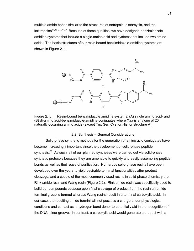
Figure 2.1. Resin-bound benzimidazole amidine systems: (A) single amino acid- and (B) di-amino acid-benzimidazole-amidine conjugates where Xaa is any one of 20 naturally occurring amino acids (except Trp, Ser, Cys, or His for structure A).
2.2. Synthesis – General Considerations
Solid-phase synthetic methods for the generation of amino acid conjugates have
become increasingly important since the development of solid-phase peptide
synthesis.30 As such, all of our planned syntheses were carried out via solid-phase
synthetic protocols because they are amenable to quickly and easily assembling peptide
bonds as well as their ease of purification. Numerous solid-phase resins have been
developed over the years to yield desirable terminal functionalities after product
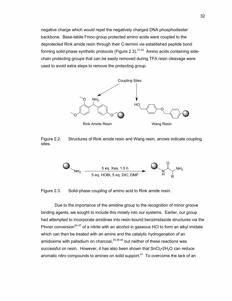
cleavage, and a couple of the most commonly used resins in solid-phase chemistry are
Rink amide resin and Wang resin (Figure 2.2). Rink amide resin was specifically used to
build our compounds because upon final cleavage of product from the resin an amide
terminal group is formed whereas Wang resins result in a terminal carboxylic acid. In
our case, the resulting amide termini will not possess a charge under physiological
conditions and can act as a hydrogen bond donor to potentially aid in the recognition of
the DNA minor groove. In contrast, a carboxylic acid would generate a product with a
32
negative charge which would repel the negatively charged DNA phosphodiester
backbone. Base-labile Fmoc-group protected amino acids were coupled to the
deprotected Rink amide resin through their C-termini via established peptide bond
forming solid-phase synthetic protocols (Figure 2.3).31-33 Amino acids containing side-
chain protecting groups that can be easily removed during TFA resin cleavage were
used to avoid extra steps to remove the protecting group.
Figure 2.2. Structures of Rink amide resin and Wang resin, arrows indicate coupling sites.
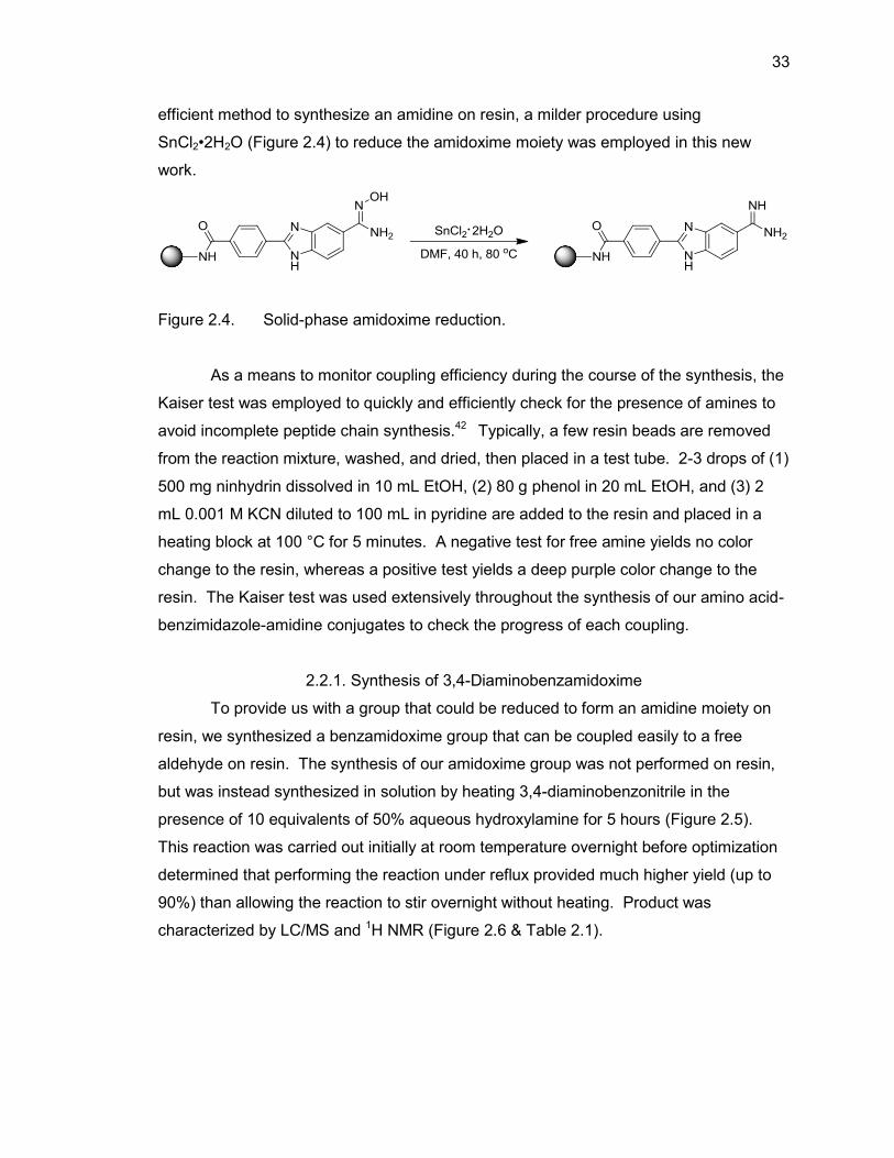
Figure 2.3. Solid-phase coupling of amino acid to Rink amide resin.
Due to the importance of the amidine group to the recognition of minor groove
binding agents, we sought to include this moiety into our systems. Earlier, our group
had attempted to incorporate amidines into resin-bound benzimidazole structures via the
Pinner conversion34-37 of a nitrile with an alcohol in gaseous HCl to form an alkyl imidate
which can then be treated with an amine and the catalytic hydrogenation of an
amidoxime with palladium on charcoal,25,38-40 but neither of these reactions was
successful on resin. However, it has also been shown that SnCl2•2H2O can reduce
aromatic nitro compounds to amines on solid support.41 To overcome the lack of an
33
efficient method to synthesize an amidine on resin, a milder procedure using
SnCl2•2H2O (Figure 2.4) to reduce the amidoxime moiety was employed in this new
work.
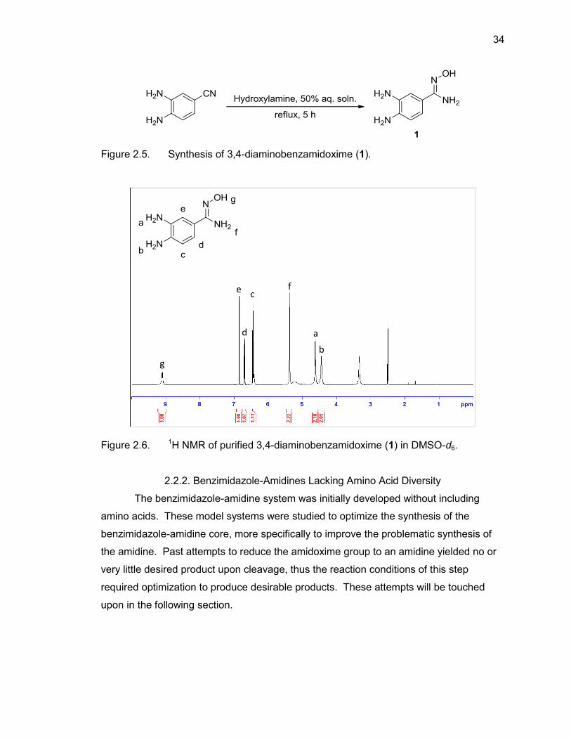
Figure 2.4. Solid-phase amidoxime reduction.
As a means to monitor coupling efficiency during the course of the synthesis, the
Kaiser test was employed to quickly and efficiently check for the presence of amines to
avoid incomplete peptide chain synthesis.42 Typically, a few resin beads are removed
from the reaction mixture, washed, and dried, then placed in a test tube. 2-3 drops of (1)
500 mg ninhydrin dissolved in 10 mL EtOH, (2) 80 g phenol in 20 mL EtOH, and (3) 2
mL 0.001 M KCN diluted to 100 mL in pyridine are added to the resin and placed in a
heating block at 100 °C for 5 minutes. A negative test for free amine yields no color
change to the resin, whereas a positive test yields a deep purple color change to the
resin. The Kaiser test was used extensively throughout the synthesis of our amino acid-
benzimidazole-amidine conjugates to check the progress of each coupling.
2.2.1. Synthesis of 3,4-Diaminobenzamidoxime
To provide us with a group that could be reduced to form an amidine moiety on
resin, we synthesized a benzamidoxime group that can be coupled easily to a free
aldehyde on resin. The synthesis of our amidoxime group was not performed on resin,
but was instead synthesized in solution by heating 3,4-diaminobenzonitrile in the
presence of 10 equivalents of 50% aqueous hydroxylamine for 5 hours (Figure 2.5).
This reaction was carried out initially at room temperature overnight before optimization
determined that performing the reaction under reflux provided much higher yield (up to
90%) than allowing the reaction to stir overnight without heating. Product was
characterized by LC/MS and 1H NMR (Figure 2.6 & Table 2.1).
34
Figure 2.5. Synthesis of 3,4-diaminobenzamidoxime (1).
Figure 2.6. 1H NMR of purified 3,4-diaminobenzamidoxime (1) in DMSO-d6.
2.2.2. Benzimidazole-Amidines Lacking Amino Acid Diversity
The benzimidazole-amidine system was initially developed without including
amino acids. These model systems were studied to optimize the synthesis of the
benzimidazole-amidine core, more specifically to improve the problematic synthesis of
the amidine. Past attempts to reduce the amidoxime group to an amidine yielded no or
very little desired product upon cleavage, thus the reaction conditions of this step
required optimization to produce desirable products. These attempts will be touched
upon in the following section.
a
b
c
d
e f
g
35
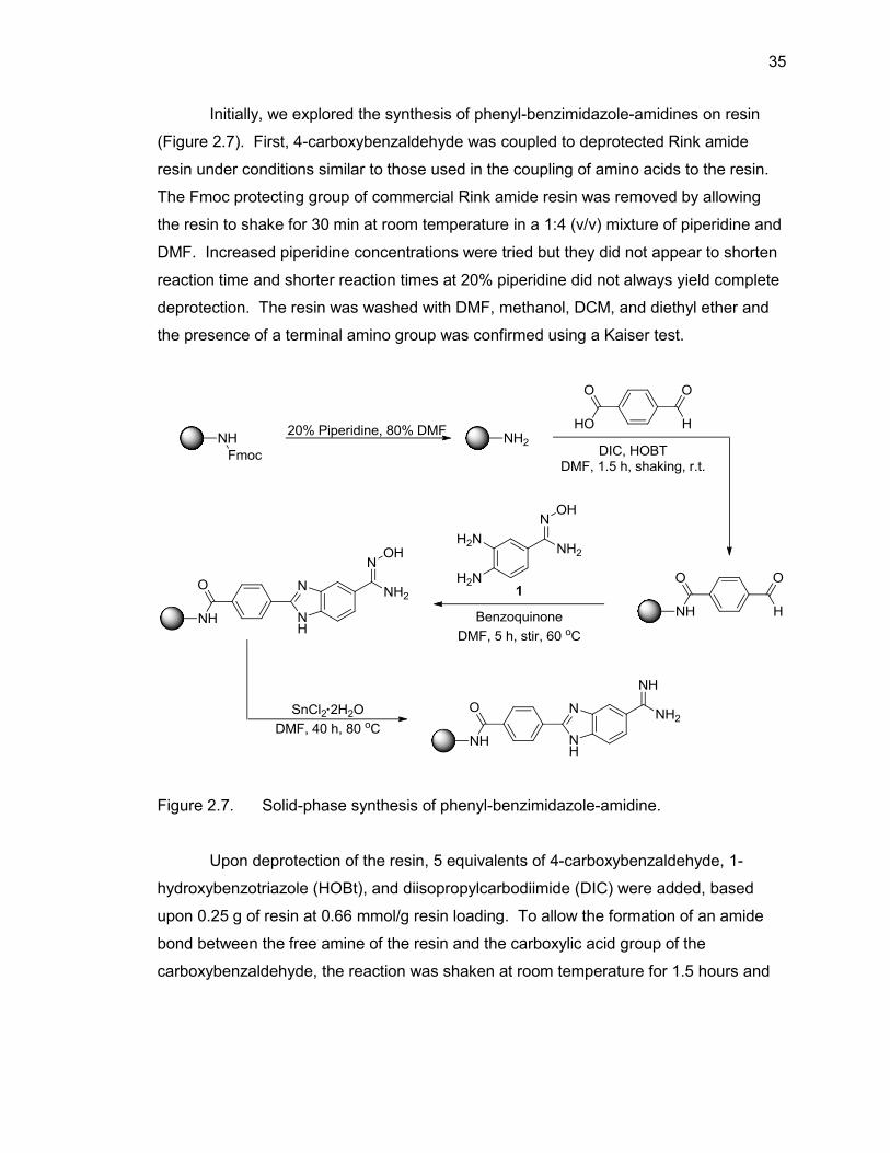
Initially, we explored the synthesis of phenyl-benzimidazole-amidines on resin
(Figure 2.7). First, 4-carboxybenzaldehyde was coupled to deprotected Rink amide
resin under conditions similar to those used in the coupling of amino acids to the resin.
The Fmoc protecting group of commercial Rink amide resin was removed by allowing
the resin to shake for 30 min at room temperature in a 1:4 (v/v) mixture of piperidine and
DMF. Increased piperidine concentrations were tried but they did not appear to shorten
reaction time and shorter reaction times at 20% piperidine did not always yield complete
deprotection. The resin was washed with DMF, methanol, DCM, and diethyl ether and
the presence of a terminal amino group was confirmed using a Kaiser test.
Figure 2.7. Solid-phase synthesis of phenyl-benzimidazole-amidine.
Upon deprotection of the resin, 5 equivalents of 4-carboxybenzaldehyde, 1-
hydroxybenzotriazole (HOBt), and diisopropylcarbodiimide (DIC) were added, based
upon 0.25 g of resin at 0.66 mmol/g resin loading. To allow the formation of an amide
bond between the free amine of the resin and the carboxylic acid group of the
carboxybenzaldehyde, the reaction was shaken at room temperature for 1.5 hours and
36
monitored via Kaiser test. When purple resin beads were present, indicating an
incomplete coupling, a second coupling of five equivalents of 4-carboxybenzaldehyde
was performed. Initially, the amount of 4-carboxybenzaldehyde added was increased up
to 10 times excess in an attempt to complete the coupling in the initial 1.5 hours, or
shorter if possible, but there was no discernible difference observed versus using five
equivalents. Reaction times up to 5 hours were also attempted, but the coupling was
still incomplete. Finally, it was decided that doing a double coupling with 1.5 hour
reaction times was the most efficient way to proceed with this step of the reaction. The
resin was again washed with DMF, methanol, DCM, and diethyl ether and the completed
coupling of the carboxybenzaldehyde to the resin was confirmed by Kaiser test.
Next, the benzimidazole ring was formed via the condensation of the aldehyde
(from 4-carboxybenzaldehyde) with 3,4-diaminobenzamidoxime. Ten equivalents of the
amidoxime-containing diaminobenzene 1 was allowed to react with the resin-linked
aldehyde for five hours at 60 °C. In an attempt to conserve the diaminobenzene,
syntheses using five equivalents were attempted but did not yield complete coupling in
five hours and required overnight reactions to reach completion. Also, differing reaction
times were attempted with shorter times showing incomplete coupling via LC/MS
(mixture of product and starting material) and longer reaction times showing no
difference to five hours. The reaction was also conducted at room temperature but
would typically take up to 24 hours to show complete coupling. After the formation of the
benzimidazole was complete, resins were washed with DMF, methanol, DCM, and
diethyl ether and the amidoxime group was reduced to the desired amidine moiety.
The reduction of resin-bound amidoxime to resin-bound amidine proved to be the
most difficult reaction leading to final product, but was eventually achieved by adding 15
equivalents of 1 M SnCl2•2H2O in DMF to the resin-bound benzimidazole amidoxime and
allowing the reaction to proceed at 70 °C for over 40 hours. To increase the reaction
yield, a new aliquot of the SnCl2 solution was added to the reaction mixture every 4
hours. Initially performing the reaction with only one aliquot of 1 M SnCl2•2H2O led to
the reaction mixture thickening to the point that stirring would no longer occur and upon
cleavage, very little or no desired product was seen. Increasing the concentration and/or
volume of 1 M SnCl2•2H2O did not alleviate this problem and usually resulted in less
than 25% yield. Eventually, additional aliquots of 1 M SnCl2•2H2O were added to the
37
reaction vessel and it was determined that adding a new aliquot every four hours
throughout the day would prevent the reaction mixture from thickening and yielded
desired product upon cleavage. Shorter durations of the entire reaction were attempted
but at 24 hours there was still a large amount of unreacted amidoxime present, so 40
hours remained the overall length of reaction time.
Upon the final amidine formation, the phenyl-benzimidazole-amidine was cleaved
from the resin with a 1:1 solution of TFA:DCM. The TFA and DCM were evaporated
under a flow of nitrogen gas and the resulting residue was dissolved in methanol and
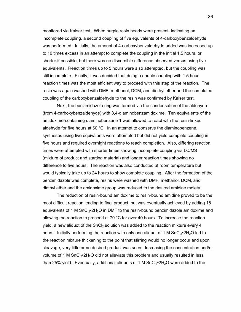
passed through silica then concentrated leading to a 76% yield. Product was
characterized by LC/MS and 1H NMR (Figure 2.8 & Table 2.2).
Figure 2.8. 1H NMR of model-benzimidazole-amidine (2) in DMSO-d6.
2.2.3. Amino Acid-Benzimidazole-Amidine Conjugates
Having confirmed the ability to synthesize phenyl-benzimidazole-amidines on
resin in good yield, we proceeded to synthesize amino acid-containing benzimidazole-
amidines. Two classes of amino acid-benzimidazole-amidines were synthesized: (1)
containing only a single amino acid (Xaa) and (2) a dipeptide containing an amino acid
linked via a glycine to the benzimidazole-amidine portion (Xaa-Gly). The glycine residue
e i a b c
f g d h
38
was added to provide space between the benzimidazole-amidine portion and the varied
amino acid.
2.2.3.1. Single Amino Acid-Benzimidazole-Amidine Conjugates
The syntheses of these conjugates followed the same route described previously
with the addition of amino acid couplings directly to Rink amide resin preceding the
coupling of 4-carboxybenzaldehyde (Figure 2.9). Once resin deprotection was
confirmed via Kaiser test, five equivalents of the desired amino acid, based upon 0.25 g
of resin at 0.66 mmol/g resin loading was shaken at room temperature for 1.5 hours.
Reaction times and reagent concentrations that were tested with 4-
carboxybenzaldehyde were also used with the amino acids but it was also seen that
higher concentrations did not make the reaction proceed faster. After washing the resin,
a Kaiser test was performed to verify the coupling of the amino acid to the resin. If free
amine was present, a second coupling of five equivalents of amino acid was performed
for another 1.5 hours. Generally, amino acids with side chains that contained protecting
groups (Trp, Ser, Thr, Asn, Gln, Tyr, Cys, Lys, Arg, His, Asp, and Glu) required a double
coupling, whereas amino acids with no protecting groups (Gly, Ala, Val, Leu, Ile, Met,
Pro, and Phe) on their side chains did not require a double coupling. Once the Kaiser
test was negative for free amine, the resin-bound amino acid was liberated of its
protecting Fmoc group via shaking in 20% piperidine, 80% DMF for 30 minutes. Once
deprotection was confirmed via Kaiser test, 4-carboxybenzaldehyde was coupled to the
resin and the benzimidazole and amidine moieties were formed as previously described.
After cleavage from resin, the Xaa-benzimidazole-amidines were characterized via
LC/MS and 1H NMR. Isolated, lyophilized product yields and LC/MS results are reported
in Table 2.3. A sample 1H NMR of glycine-benzimidazole-amidine is shown in Figure
2.10 and Table 2.4.
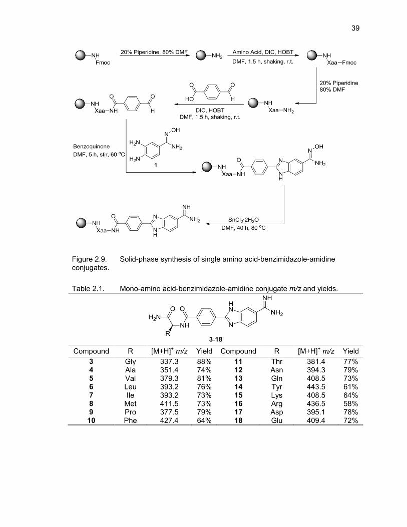
39
Figure 2.9. Solid-phase synthesis of single amino acid-benzimidazole-amidine conjugates.
Table 2.1. Mono-amino acid-benzimidazole-amidine conjugate m/z and yields.
Compound R [M+H]+ m/z Yield Compound R [M+H]+ m/z Yield
3 Gly 337.3 88% 11 Thr 381.4 77% 4 Ala 351.4 74% 12 Asn 394.3 79% 5 Val 379.3 81% 13 Gln 408.5 73% 6 Leu 393.2 76% 14 Tyr 443.5 61% 7 Ile 393.2 73% 15 Lys 408.5 64% 8 Met 411.5 73% 16 Arg 436.5 58% 9 Pro 377.5 79% 17 Asp 395.1 78% 10 Phe 427.4 64% 18 Glu 409.4 72%
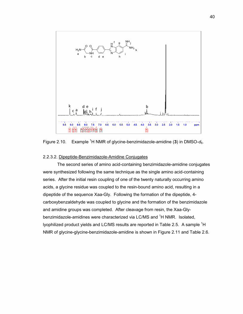
40
Figure 2.10. Example 1H NMR of glycine-benzimidazole-amidine (3) in DMSO-d6.
2.2.3.2. Dipeptide-Benzimidazole-Amidine Conjugates
The second series of amino acid-containing benzimidazole-amidine conjugates
were synthesized following the same technique as the single amino acid-containing
series. After the initial resin coupling of one of the twenty naturally occurring amino
acids, a glycine residue was coupled to the resin-bound amino acid, resulting in a
dipeptide of the sequence Xaa-Gly. Following the formation of the dipeptide, 4-
carboxybenzaldehyde was coupled to glycine and the formation of the benzimidazole
and amidine groups was completed. After cleavage from resin, the Xaa-Gly-
benzimidazole-amidines were characterized via LC/MS and 1H NMR. Isolated,
lyophilized product yields and LC/MS results are reported in Table 2.5. A sample 1H
NMR of glycine-glycine-benzimidazole-amidine is shown in Figure 2.11 and Table 2.6.
k c a
d g
e h
i f j b
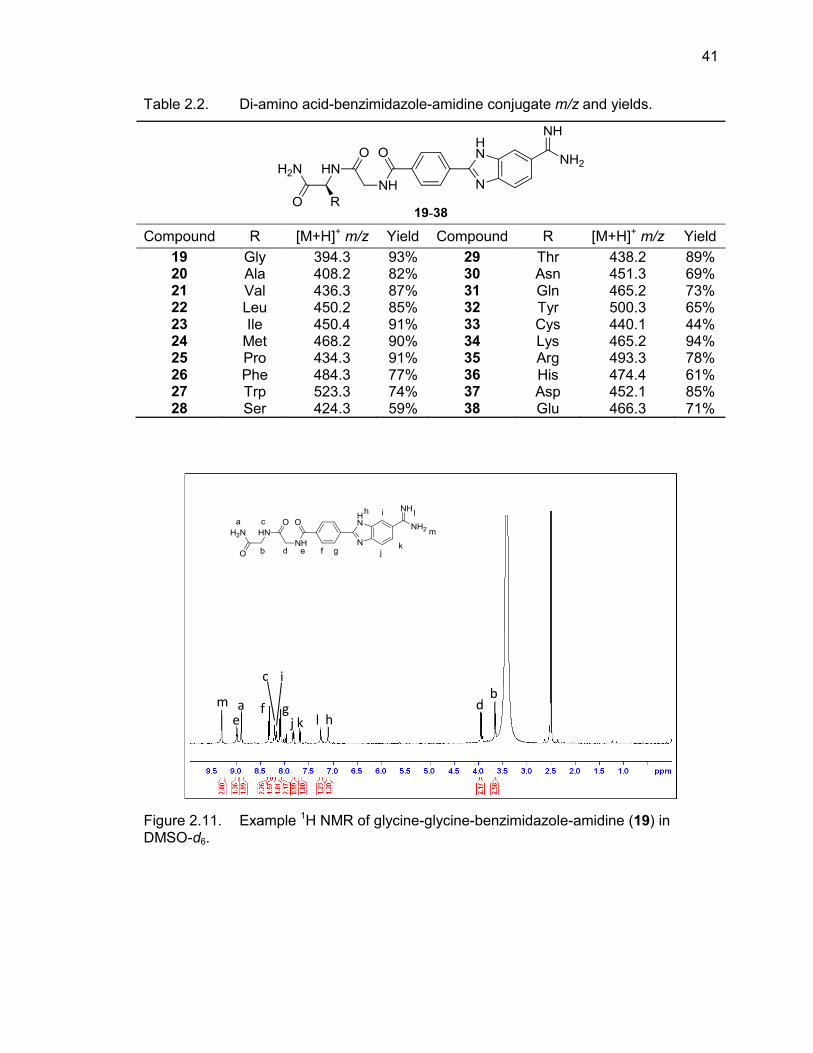
41
Table 2.2. Di-amino acid-benzimidazole-amidine conjugate m/z and yields.
Compound R [M+H]+ m/z Yield Compound R [M+H]+ m/z Yield
19 Gly 394.3 93% 29 Thr 438.2 89% 20 Ala 408.2 82% 30 Asn 451.3 69% 21 Val 436.3 87% 31 Gln 465.2 73% 22 Leu 450.2 85% 32 Tyr 500.3 65% 23 Ile 450.4 91% 33 Cys 440.1 44% 24 Met 468.2 90% 34 Lys 465.2 94% 25 Pro 434.3 91% 35 Arg 493.3 78% 26 Phe 484.3 77% 36 His 474.4 61% 27 Trp 523.3 74% 37 Asp 452.1 85% 28 Ser 424.3 59% 38 Glu 466.3 71%
Figure 2.11. Example 1H NMR of glycine-glycine-benzimidazole-amidine (19) in DMSO-d6.
m e a f
c i
g j k l h
d b
42
2.3. Summary
A new protocol for synthesizing amino acid-benzimidazole-amidine conjugates
via solid-phase chemistry was developed. Sixteen single amino acid-containing phenyl-
benzimidazole-amidine conjugates and 20 dipeptide phenyl-benzimidazole-amidine
conjugates were synthesized using this method. Despite numerous attempts,
tryptophan-, serine-, cysteine-, and histidine-containing single amino acid conjugates
could not be successfully synthesized. The likely cause for failure in the synthesis of
these amino acid conjugates occurred during the reduction step. All of the Kaiser tests
throughout the synthesis of these conjugates showed the couplings were going to
completion as expected, and LC/MS of several intermediate products showed the
desired masses were present and confirmed that the amino acid, aldehyde, and
diaminobenzene-amidoxime couplings went to completion. There was no sign of failed
coupling until cleavage after the reduction. Also, the quality of the amino acids should
not be of concern as the same sources were used to synthesize the dipeptides without
issue. It is possible that complex side chains were not stable to the reduction conditions
without another amino acid stabilizing them or that steric hindrance prevented the
reduction from proceeding.
The most difficult aspect of the synthesis was the formation of the amidine group.
The Pinner synthesis and the catalytic hydrogenation of an amidoxime were not
successful on resin. To overcome this, a milder procedure using SnCl2•2H2O to reduce
the amidoxime moiety was employed. The synthesis of our amidoxime group was not
performed on resin, but was instead synthesized in solution by heating 3,4-
diaminobenzonitrile in the presence of 10 equivalents of 50% aqueous hydroxylamine for
5 hours. The resulting diamino-amidoxime was coupled to the resin and reduced using
SnCl2•2H2O. Although the reaction was time-consuming, this proved to be a much more
efficient method of forming the desired amidine on resin than previously described
methods.
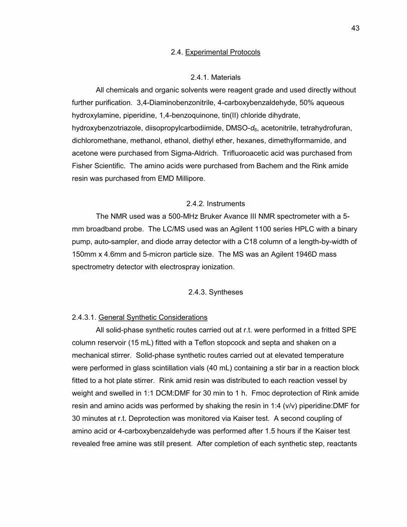
43
2.4. Experimental Protocols
2.4.1. Materials
All chemicals and organic solvents were reagent grade and used directly without
further purification. 3,4-Diaminobenzonitrile, 4-carboxybenzaldehyde, 50% aqueous
hydroxylamine, piperidine, 1,4-benzoquinone, tin(II) chloride dihydrate,
hydroxybenzotriazole, diisopropylcarbodiimide, DMSO-d6, acetonitrile, tetrahydrofuran,
dichloromethane, methanol, ethanol, diethyl ether, hexanes, dimethylformamide, and
acetone were purchased from Sigma-Aldrich. Trifluoroacetic acid was purchased from
Fisher Scientific. The amino acids were purchased from Bachem and the Rink amide
resin was purchased from EMD Millipore.
2.4.2. Instruments
The NMR used was a 500-MHz Bruker Avance III NMR spectrometer with a 5-
mm broadband probe. The LC/MS used was an Agilent 1100 series HPLC with a binary
pump, auto-sampler, and diode array detector with a C18 column of a length-by-width of
150mm x 4.6mm and 5-micron particle size. The MS was an Agilent 1946D mass
spectrometry detector with electrospray ionization.
2.4.3. Syntheses
2.4.3.1. General Synthetic Considerations
All solid-phase synthetic routes carried out at r.t. were performed in a fritted SPE
column reservoir (15 mL) fitted with a Teflon stopcock and septa and shaken on a
mechanical stirrer. Solid-phase synthetic routes carried out at elevated temperature
were performed in glass scintillation vials (40 mL) containing a stir bar in a reaction block
fitted to a hot plate stirrer. Rink amid resin was distributed to each reaction vessel by
weight and swelled in 1:1 DCM:DMF for 30 min to 1 h. Fmoc deprotection of Rink amide
resin and amino acids was performed by shaking the resin in 1:4 (v/v) piperidine:DMF for
30 minutes at r.t. Deprotection was monitored via Kaiser test. A second coupling of
amino acid or 4-carboxybenzaldehyde was performed after 1.5 hours if the Kaiser test
revealed free amine was still present. After completion of each synthetic step, reactants
44
were drained and the resin was washed with DMF (3 x 5 mL), methanol (3 x 5 mL), DCM
(3 x 5 mL), and diethyl ether (3 x 5 mL) and the solvents were removed by filtration using
a vacuum system. All reactions were performed under the presence of nitrogen.
2.4.3.2. General Procedure for Synthesis of Model-BI-(+)
Deprotected Rink amide resin (0.25 g, 0.66 mmol/g) was treated with 4-
carboxybenzaldehyde (0.8250 mmol, 123.8 mg), HOBt (0.8250 mmol, 111.5 mg), and
DIC (0.8250 mmol, 104.1 mg) in 15 mL DMF and coupled to the deprotected Rink amide
resin. The mixture was shaken on a mechanical stirrer at room temperature for 1.5 h.
Completion of the coupling was confirmed by the Kaiser test. 3,4-
Diaminobenzamidoxime (1.650 mmol, 274.2 mg) and 1,4-benzoquinone (0.165 mmol,
17.8 mg) were dissolved in 15 mL DMF and coupled to resin-bound phenylaldehyde in a
scintillation vial for 5 h at 60 °C using an IKA-Werke mechanical stirrer and heating
block. Tin(II) chloride dehydrate (2.475 mmol, 558.5 mg) was dissolved in DMF (2.5 mL)
and reacted with the resin-linked amidoxime using an IKA-Werke mechanical stirrer and
heating block at 80 °C over 40 h with new aliquots of the Tin(II) chloride solution being
added to the reaction mixture every 4 h. The product was cleaved from the resin via
shaking in a 1:1 solution of TFA:DCM for 1.5 h. The resin was removed by filtration and
rinsed with TFA and the filtrate-containing product was collected in tared vials, then the
TFA and DCM were evaporated with a stream of nitrogen until only a couple of mL
remained, which was then precipitated into cold ether. The resulting solid was dissolved
in MeOH and initially purified via flushing through a silica column (MeOH) to remove bulk
impurities and then HPLC purified (A: H2O, 0.1% TFA; B: ACN, 0.1% TFA. Gradient: 0
min, 80% A; 2 min, 80% A; 20 min, 20% A; 25 min, 0% A; 30 min, 0% A; 32 min, 80% A;
35 min, 80% A). Product was confirmed via LC/MS and 1H NMR.
2.4.3.3. General Procedure for Synthesis of Xaa-BI-(+) Conjugates
Deprotected Rink amide resin (0.25 g, 0.66 mmol/g) was treated with an amino
acid (0.825 mmol), HOBt (0.8250 mmol, 111.5 mg), and DIC (0.8250 mmol, 104.1 mg) in
15 mL DMF. The mixture was shaken on a mechanical stirrer at room temperature for
1.5 h. Completion of the coupling was confirmed by the Kaiser test. 4-
Carboxybenzaldehyde (0.8250 mmol, 123.8 mg), HOBt (0.8250 mmol, 111.5 mg), and
45
DIC (0.8250 mmol, 104.1 mg) were dissolved in 15 mL DMF and coupled to deprotected
resin-bound amino acid on a mechanical stirrer at room temperature for 1.5 h.
Completion of the coupling was confirmed by the Kaiser test. 3,4-
Diaminobenzamidoxime (1.650 mmol, 274.2 mg) and 1,4-benzoquinone (0.165 mmol,
17.8 mg) were dissolved in 15 mL DMF and coupled to resin-bound phenylaldehyde in a
scintillation vial for 5 h at 60 °C using an IKA-Werke mechanical stirrer and heating
block. Tin(II) chloride dehydrate (2.475 mmol, 558.5 mg) was dissolved in DMF (2.5 mL)
and reacted with the resin-linked amidoxime using an IKA-Werke mechanical stirrer and
heating block at 80 °C over 40 h with new aliquots of the Tin(II) chloride solution being
added to the reaction mixture every 4 h. The product was cleaved from the resin via
shaking in a 1:1 solution of TFA:DCM for 1.5 h. The resin was removed by filtration and
rinsed with TFA and the filtrate-containing product was collected in tared vials, then the
TFA and DCM were evaporated with a stream of nitrogen until only a couple of mL
remained, which was then precipitated into cold ether. The resulting solid was dissolved
in MeOH and initially purified via flushing through a silica column (MeOH) to remove bulk
impurities and then HPLC purified (A: H2O, 0.1% TFA; B: ACN, 0.1% TFA. Gradient: 0
min, 80% A; 2 min, 80% A; 20 min, 20% A; 25 min, 0% A; 30 min, 0% A; 32 min, 80% A;
35 min, 80% A). Product was confirmed via LC/MS and 1H NMR.
2.4.3.4. General Procedure for Synthesis of Xaa-Gly-BI-(+) Conjugates
Deprotected Rink amide resin (0.25 g, 0.66 mmol/g) was treated with an amino
acid (0.825 mmol), HOBt (0.8250 mmol, 111.5 mg), and DIC (0.8250 mmol, 104.1 mg) in
15 mL DMF for 1.5 h. The mixture was shaken on a mechanical stirrer at room
temperature for 1.5 h. Completion of the coupling was confirmed by the Kaiser test.
Glycine (0.8250 mmol, 254.3 mg), HOBt (0.8250 mmol, 111.5 mg), and DIC (0.8250
mmol, 104.1 mg) were dissolved in 15 mL DMF and coupled to deprotected resin-bound
amino acid on a mechanical stirrer at room temperature for 1.5 h. 4-
Carboxybenzaldehyde (0.8250 mmol, 123.8 mg), HOBt (0.8250 mmol, 111.5 mg), and
DIC (0.8250 mmol, 104.1 mg) were dissolved in 15 mL DMF and coupled to the
deprotected terminal glycine on a mechanical stirrer at room temperature for 1.5 h. 3,4-
Diaminobenzamidoxime (1.650 mmol, 274.2 mg) and 1,4-benzoquinone (0.165 mmol,
17.8 mg) were dissolved in 15 mL DMF and coupled to resin-bound phenylaldehyde in a
46
scintillation vial for 5 h at 60 °C using an IKA-Werke mechanical stirrer and heating
block. Tin(II) chloride dehydrate (2.475 mmol, 558.5 mg) was dissolved in DMF (2.5 mL)
and reacted with the resin-linked amidoxime using an IKA-Werke mechanical stirrer and
heating block at 80 °C over 40 h with new aliquots of the Tin(II) chloride solution being
added to the reaction mixture every 4 h. The product was cleaved from the resin via
shaking in a 1:1 solution of TFA:DCM for 1.5 h. The resin was removed by filtration and
rinsed with TFA and the filtrate-containing product was collected in tared vials, then the
TFA and DCM were evaporated with a stream of nitrogen until only a couple of mL
remained, which was then precipitated into cold ether. The resulting solid was dissolved
in MeOH and initially purified via flushing through a silica column (MeOH) to remove bulk
impurities and then HPLC purified (A: H2O, 0.1% TFA; B: ACN, 0.1% TFA. Gradient: 0
min, 80% A; 2 min, 80% A; 20 min, 20% A; 25 min, 0% A; 30 min, 0% A; 32 min, 80% A;
35 min, 80% A). Product was confirmed via LC/MS and 1H NMR.
2.4.3.5. Synthesis of Diaminobenzamidoxime
3,4-Diamino-N'-hydroxybenzimidamide, (1)
Compound 1 was synthesized by mixing 3,4-diaminobenzonitrile (7.5 mmol, 1.0
g) in ethanol (20 mL) and 20 equivalents 50% aqueous hydroxylamine (150 mmol, 4.95
g) and refluxing for 5 hours. The ethanol was evaporated under reduced pressure. The
compound was purified via column chromatography [EtOAc:MeOH (90:10)] and
confirmed via LC/MS and 1H NMR. (1.06 g, 85%); 1H NMR (DMSO-d6); δ4.45 (s, 2H),
4.63 (s, 2H), 5.38 (s, 2H), 6.46 (d, J = 8 Hz, 1H), 6.71 (dd, J = 8.5 Hz, 1H), 6.85 (s, 1H),
9.11 (s, 1H); LC/MS calcd for [M+H]+: C7H10N4O 167.1, found 167.3.
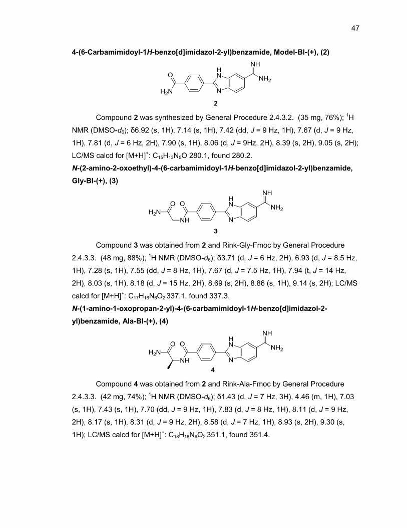
47
4-(6-Carbamimidoyl-1H-benzo[d]imidazol-2-yl)benzamide, Model-BI-(+), (2)
Compound 2 was synthesized by General Procedure 2.4.3.2. (35 mg, 76%); 1H
NMR (DMSO-d6); δ6.92 (s, 1H), 7.14 (s, 1H), 7.42 (dd, J = 9 Hz, 1H), 7.67 (d, J = 9 Hz,
1H), 7.81 (d, J = 6 Hz, 2H), 7.90 (s, 1H), 8.06 (d, J = 9Hz, 2H), 8.39 (s, 2H), 9.05 (s, 2H);
LC/MS calcd for [M+H]+: C15H13N5O 280.1, found 280.2.
N-(2-amino-2-oxoethyl)-4-(6-carbamimidoyl-1H-benzo[d]imidazol-2-yl)benzamide,
Gly-BI-(+), (3)
Compound 3 was obtained from 2 and Rink-Gly-Fmoc by General Procedure
2.4.3.3. (48 mg, 88%); 1H NMR (DMSO-d6); δ3.71 (d, J = 6 Hz, 2H), 6.93 (d, J = 8.5 Hz,
1H), 7.28 (s, 1H), 7.55 (dd, J = 8 Hz, 1H), 7.67 (d, J = 7.5 Hz, 1H), 7.94 (t, J = 14 Hz,
2H), 8.03 (s, 1H), 8.18 (d, J = 15 Hz, 2H), 8.69 (s, 2H), 8.86 (s, 1H), 9.14 (s, 2H); LC/MS
calcd for [M+H]+: C17H16N6O2 337.1, found 337.3.
N-(1-amino-1-oxopropan-2-yl)-4-(6-carbamimidoyl-1H-benzo[d]imidazol-2-
yl)benzamide, Ala-BI-(+), (4)
Compound 4 was obtained from 2 and Rink-Ala-Fmoc by General Procedure
2.4.3.3. (42 mg, 74%); 1H NMR (DMSO-d6); δ1.43 (d, J = 7 Hz, 3H), 4.46 (m, 1H), 7.03
(s, 1H), 7.43 (s, 1H), 7.70 (dd, J = 9 Hz, 1H), 7.83 (d, J = 8 Hz, 1H), 8.11 (d, J = 9 Hz,
2H), 8.17 (s, 1H), 8.31 (d, J = 9 Hz, 2H), 8.58 (d, J = 7 Hz, 1H), 8.93 (s, 2H), 9.30 (s,
1H); LC/MS calcd for [M+H]+: C18H18N6O2 351.1, found 351.4.
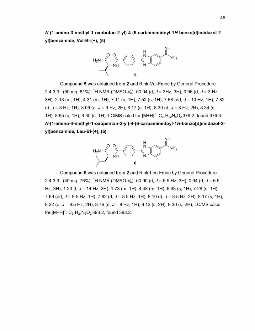
48
N-(1-amino-3-methyl-1-oxobutan-2-yl)-4-(6-carbamimidoyl-1H-benzo[d]imidazol-2-
yl)benzamide, Val-BI-(+), (5)
Compound 5 was obtained from 2 and Rink-Val-Fmoc by General Procedure
2.4.3.3. (50 mg, 81%); 1H NMR (DMSO-d6); δ0.94 (d, J = 3Hz, 3H), 0.96 (d, J = 3 Hz,
3H), 2.13 (m, 1H), 4.31 (m, 1H), 7.11 (s, 1H), 7.52 (s, 1H), 7.68 (dd, J = 10 Hz, 1H), 7.82
(d, J = 8 Hz, 1H), 8.09 (d, J = 9 Hz, 2H), 8.17 (s, 1H), 8.30 (d, J = 8 Hz, 2H), 8.34 (s,
1H), 8.95 (s, 1H), 9.30 (s, 1H); LC/MS calcd for [M+H]+: C20H22N6O2 379.2, found 379.3.
N-(1-amino-4-methyl-1-oxopentan-2-yl)-4-(6-carbamimidoyl-1H-benzo[d]imidazol-2-
yl)benzamide, Leu-BI-(+), (6)
Compound 6 was obtained from 2 and Rink-Leu-Fmoc by General Procedure
2.4.3.3. (49 mg, 76%); 1H NMR (DMSO-d6); δ0.90 (d, J = 6.5 Hz, 3H), 0.94 (d, J = 6.5
Hz, 3H), 1.23 (t, J = 14 Hz, 2H), 1.73 (m, 1H), 4.48 (m, 1H), 6.93 (s, 1H), 7.28 (s, 1H),
7.69 (dd, J = 9.5 Hz, 1H), 7.82 (d, J = 8.5 Hz, 1H), 8.10 (d, J = 8.5 Hz, 2H), 8.17 (s, 1H),
8.32 (d, J = 8.5 Hz, 2H), 8.76 (d, J = 8 Hz, 1H), 9.12 (s, 2H), 9.30 (s, 2H); LC/MS calcd
for [M+H]+: C21H24N6O2 393.2, found 393.2.
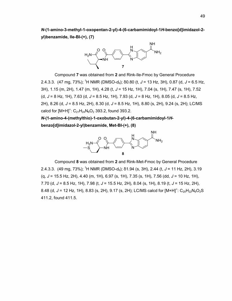
49
N-(1-amino-3-methyl-1-oxopentan-2-yl)-4-(6-carbamimidoyl-1H-benzo[d]imidazol-2-
yl)benzamide, Ile-BI-(+), (7)
Compound 7 was obtained from 2 and Rink-Ile-Fmoc by General Procedure
2.4.3.3. (47 mg, 73%); 1H NMR (DMSO-d6); δ0.80 (t, J = 13 Hz, 3H), 0.87 (d, J = 6.5 Hz,
3H), 1.15 (m, 2H), 1.47 (m, 1H), 4.28 (t, J = 15 Hz, 1H), 7.04 (s, 1H), 7.47 (s, 1H), 7.52
(d, J = 8 Hz, 1H), 7.63 (d, J = 8.5 Hz, 1H), 7.93 (d, J = 8 Hz, 1H), 8.05 (d, J = 8.5 Hz,
2H), 8.26 (d, J = 8.5 Hz, 2H), 8.30 (d, J = 8.5 Hz, 1H), 8.80 (s, 2H), 9.24 (s, 2H); LC/MS
calcd for [M+H]+: C21H24N6O2 393.2, found 393.2.
N-(1-amino-4-(methylthio)-1-oxobutan-2-yl)-4-(6-carbamimidoyl-1H-
benzo[d]imidazol-2-yl)benzamide, Met-BI-(+), (8)
Compound 8 was obtained from 2 and Rink-Met-Fmoc by General Procedure
2.4.3.3. (49 mg, 73%); 1H NMR (DMSO-d6); δ1.94 (s, 3H), 2.44 (t, J = 11 Hz, 2H), 3.19
(q, J = 15.5 Hz, 2H), 4.40 (m, 1H), 6.97 (s, 1H), 7.35 (s, 1H), 7.56 (dd, J = 10 Hz, 1H),
7.70 (d, J = 8.5 Hz, 1H), 7.98 (t, J = 15.5 Hz, 2H), 8.04 (s, 1H), 8.19 (t, J = 15 Hz, 2H),
8.48 (d, J = 12 Hz, 1H), 8.83 (s, 2H), 9.17 (s, 2H); LC/MS calcd for [M+H]+: C20H22N6O2S
411.2, found 411.5.
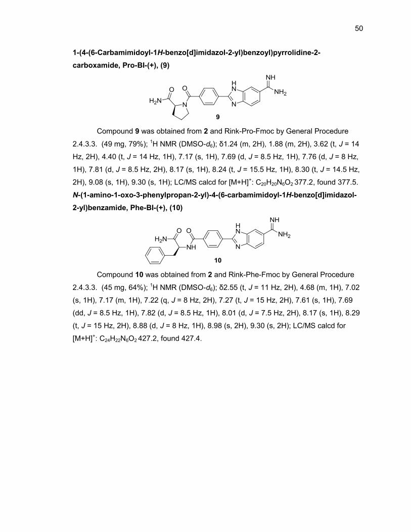
50
1-(4-(6-Carbamimidoyl-1H-benzo[d]imidazol-2-yl)benzoyl)pyrrolidine-2-
carboxamide, Pro-BI-(+), (9)
Compound 9 was obtained from 2 and Rink-Pro-Fmoc by General Procedure
2.4.3.3. (49 mg, 79%); 1H NMR (DMSO-d6); δ1.24 (m, 2H), 1.88 (m, 2H), 3.62 (t, J = 14
Hz, 2H), 4.40 (t, J = 14 Hz, 1H), 7.17 (s, 1H), 7.69 (d, J = 8.5 Hz, 1H), 7.76 (d, J = 8 Hz,
1H), 7.81 (d, J = 8.5 Hz, 2H), 8.17 (s, 1H), 8.24 (t, J = 15.5 Hz, 1H), 8.30 (t, J = 14.5 Hz,
2H), 9.08 (s, 1H), 9.30 (s, 1H); LC/MS calcd for [M+H]+: C20H20N6O2 377.2, found 377.5.
N-(1-amino-1-oxo-3-phenylpropan-2-yl)-4-(6-carbamimidoyl-1H-benzo[d]imidazol-
2-yl)benzamide, Phe-BI-(+), (10)
Compound 10 was obtained from 2 and Rink-Phe-Fmoc by General Procedure
2.4.3.3. (45 mg, 64%); 1H NMR (DMSO-d6); δ2.55 (t, J = 11 Hz, 2H), 4.68 (m, 1H), 7.02
(s, 1H), 7.17 (m, 1H), 7.22 (q, J = 8 Hz, 2H), 7.27 (t, J = 15 Hz, 2H), 7.61 (s, 1H), 7.69
(dd, J = 8.5 Hz, 1H), 7.82 (d, J = 8.5 Hz, 1H), 8.01 (d, J = 7.5 Hz, 2H), 8.17 (s, 1H), 8.29
(t, J = 15 Hz, 2H), 8.88 (d, J = 8 Hz, 1H), 8.98 (s, 2H), 9.30 (s, 2H); LC/MS calcd for
[M+H]+: C24H22N6O2 427.2, found 427.4.
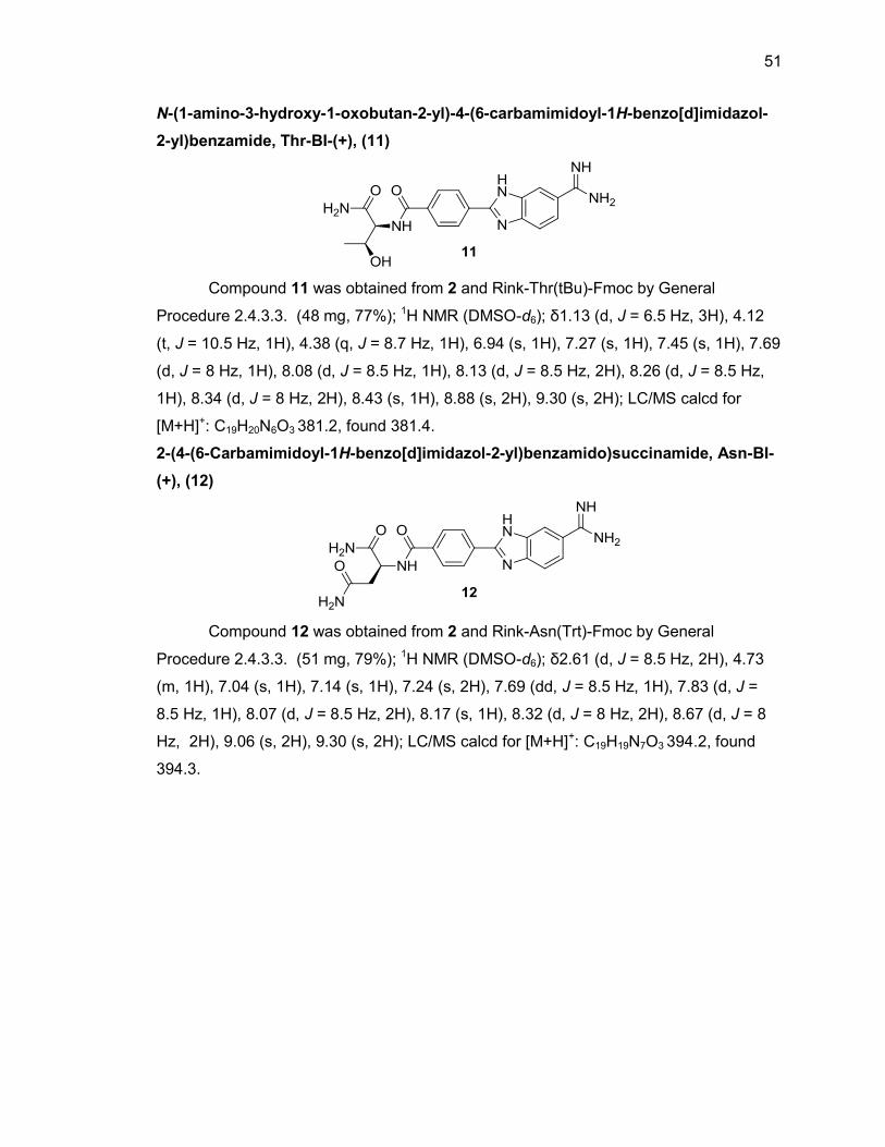
51
N-(1-amino-3-hydroxy-1-oxobutan-2-yl)-4-(6-carbamimidoyl-1H-benzo[d]imidazol-
2-yl)benzamide, Thr-BI-(+), (11)
Compound 11 was obtained from 2 and Rink-Thr(tBu)-Fmoc by General
Procedure 2.4.3.3. (48 mg, 77%); 1H NMR (DMSO-d6); δ1.13 (d, J = 6.5 Hz, 3H), 4.12
(t, J = 10.5 Hz, 1H), 4.38 (q, J = 8.7 Hz, 1H), 6.94 (s, 1H), 7.27 (s, 1H), 7.45 (s, 1H), 7.69
(d, J = 8 Hz, 1H), 8.08 (d, J = 8.5 Hz, 1H), 8.13 (d, J = 8.5 Hz, 2H), 8.26 (d, J = 8.5 Hz,
1H), 8.34 (d, J = 8 Hz, 2H), 8.43 (s, 1H), 8.88 (s, 2H), 9.30 (s, 2H); LC/MS calcd for
[M+H]+: C19H20N6O3 381.2, found 381.4.
2-(4-(6-Carbamimidoyl-1H-benzo[d]imidazol-2-yl)benzamido)succinamide, Asn-BI-
(+), (12)
Compound 12 was obtained from 2 and Rink-Asn(Trt)-Fmoc by General
Procedure 2.4.3.3. (51 mg, 79%); 1H NMR (DMSO-d6); δ2.61 (d, J = 8.5 Hz, 2H), 4.73
(m, 1H), 7.04 (s, 1H), 7.14 (s, 1H), 7.24 (s, 2H), 7.69 (dd, J = 8.5 Hz, 1H), 7.83 (d, J =
8.5 Hz, 1H), 8.07 (d, J = 8.5 Hz, 2H), 8.17 (s, 1H), 8.32 (d, J = 8 Hz, 2H), 8.67 (d, J = 8
Hz, 2H), 9.06 (s, 2H), 9.30 (s, 2H); LC/MS calcd for [M+H]+: C19H19N7O3 394.2, found
394.3.
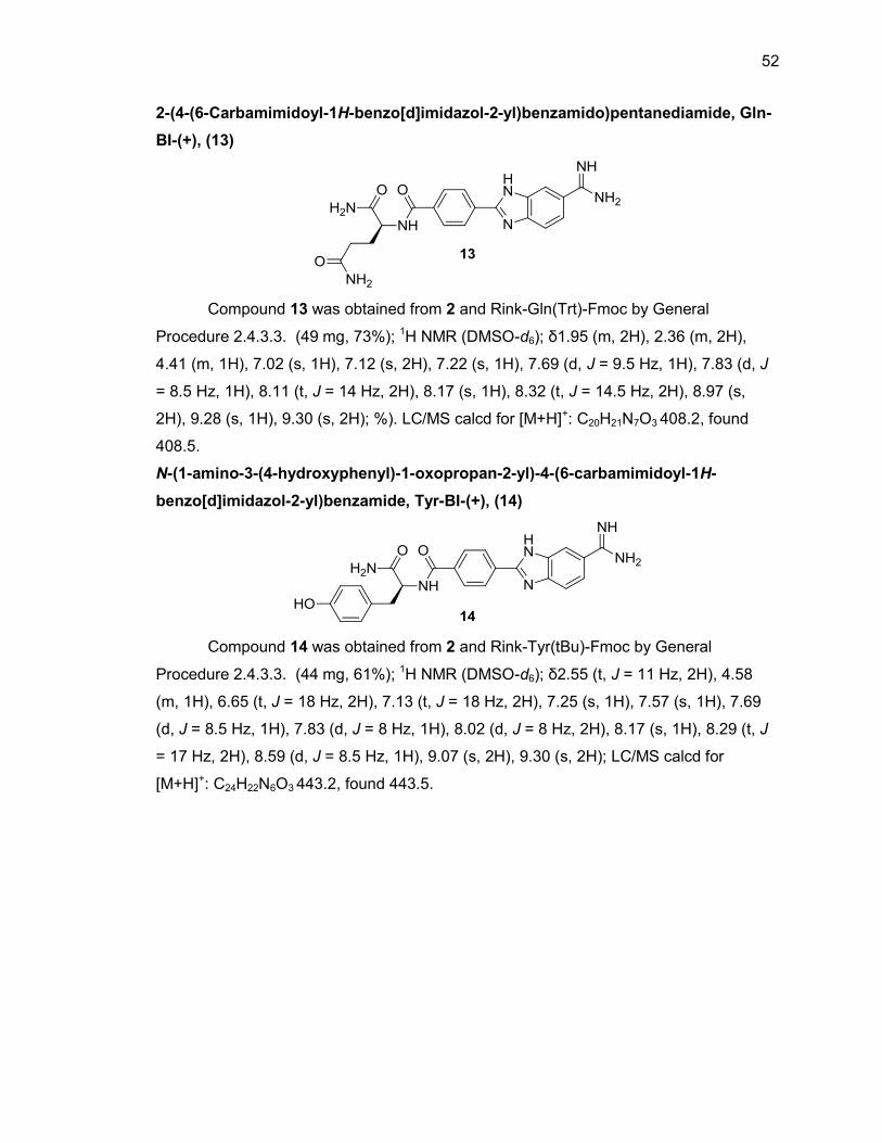
52
2-(4-(6-Carbamimidoyl-1H-benzo[d]imidazol-2-yl)benzamido)pentanediamide, Gln-
BI-(+), (13)
Compound 13 was obtained from 2 and Rink-Gln(Trt)-Fmoc by General
Procedure 2.4.3.3. (49 mg, 73%); 1H NMR (DMSO-d6); δ1.95 (m, 2H), 2.36 (m, 2H),
4.41 (m, 1H), 7.02 (s, 1H), 7.12 (s, 2H), 7.22 (s, 1H), 7.69 (d, J = 9.5 Hz, 1H), 7.83 (d, J
= 8.5 Hz, 1H), 8.11 (t, J = 14 Hz, 2H), 8.17 (s, 1H), 8.32 (t, J = 14.5 Hz, 2H), 8.97 (s,
2H), 9.28 (s, 1H), 9.30 (s, 2H); %). LC/MS calcd for [M+H]+: C20H21N7O3 408.2, found
408.5.
N-(1-amino-3-(4-hydroxyphenyl)-1-oxopropan-2-yl)-4-(6-carbamimidoyl-1H-
benzo[d]imidazol-2-yl)benzamide, Tyr-BI-(+), (14)
Compound 14 was obtained from 2 and Rink-Tyr(tBu)-Fmoc by General
Procedure 2.4.3.3. (44 mg, 61%); 1H NMR (DMSO-d6); δ2.55 (t, J = 11 Hz, 2H), 4.58
(m, 1H), 6.65 (t, J = 18 Hz, 2H), 7.13 (t, J = 18 Hz, 2H), 7.25 (s, 1H), 7.57 (s, 1H), 7.69
(d, J = 8.5 Hz, 1H), 7.83 (d, J = 8 Hz, 1H), 8.02 (d, J = 8 Hz, 2H), 8.17 (s, 1H), 8.29 (t, J
= 17 Hz, 2H), 8.59 (d, J = 8.5 Hz, 1H), 9.07 (s, 2H), 9.30 (s, 2H); LC/MS calcd for
[M+H]+: C24H22N6O3 443.2, found 443.5.
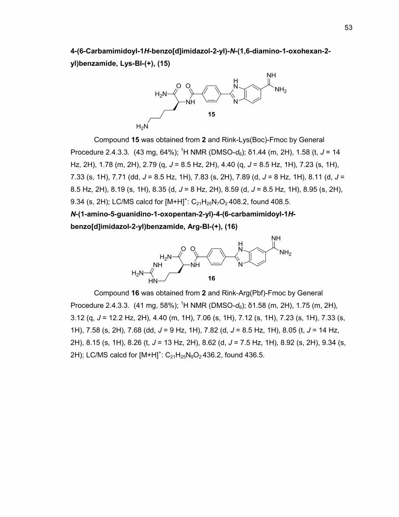
53
4-(6-Carbamimidoyl-1H-benzo[d]imidazol-2-yl)-N-(1,6-diamino-1-oxohexan-2-
yl)benzamide, Lys-BI-(+), (15)
Compound 15 was obtained from 2 and Rink-Lys(Boc)-Fmoc by General
Procedure 2.4.3.3. (43 mg, 64%); 1H NMR (DMSO-d6); δ1.44 (m, 2H), 1.58 (t, J = 14
Hz, 2H), 1.78 (m, 2H), 2.79 (q, J = 8.5 Hz, 2H), 4.40 (q, J = 8.5 Hz, 1H), 7.23 (s, 1H),
7.33 (s, 1H), 7.71 (dd, J = 8.5 Hz, 1H), 7.83 (s, 2H), 7.89 (d, J = 8 Hz, 1H), 8.11 (d, J =
8.5 Hz, 2H), 8.19 (s, 1H), 8.35 (d, J = 8 Hz, 2H), 8.59 (d, J = 8.5 Hz, 1H), 8.95 (s, 2H),
9.34 (s, 2H); LC/MS calcd for [M+H]+: C21H25N7O2 408.2, found 408.5.
N-(1-amino-5-guanidino-1-oxopentan-2-yl)-4-(6-carbamimidoyl-1H-
benzo[d]imidazol-2-yl)benzamide, Arg-BI-(+), (16)
Compound 16 was obtained from 2 and Rink-Arg(Pbf)-Fmoc by General
Procedure 2.4.3.3. (41 mg, 58%); 1H NMR (DMSO-d6); δ1.58 (m, 2H), 1.75 (m, 2H),
3.12 (q, J = 12.2 Hz, 2H), 4.40 (m, 1H), 7.06 (s, 1H), 7.12 (s, 1H), 7.23 (s, 1H), 7.33 (s,
1H), 7.58 (s, 2H), 7.68 (dd, J = 9 Hz, 1H), 7.82 (d, J = 8.5 Hz, 1H), 8.05 (t, J = 14 Hz,
2H), 8.15 (s, 1H), 8.26 (t, J = 13 Hz, 2H), 8.62 (d, J = 7.5 Hz, 1H), 8.92 (s, 2H), 9.34 (s,
2H); LC/MS calcd for [M+H]+: C21H25N9O2 436.2, found 436.5.
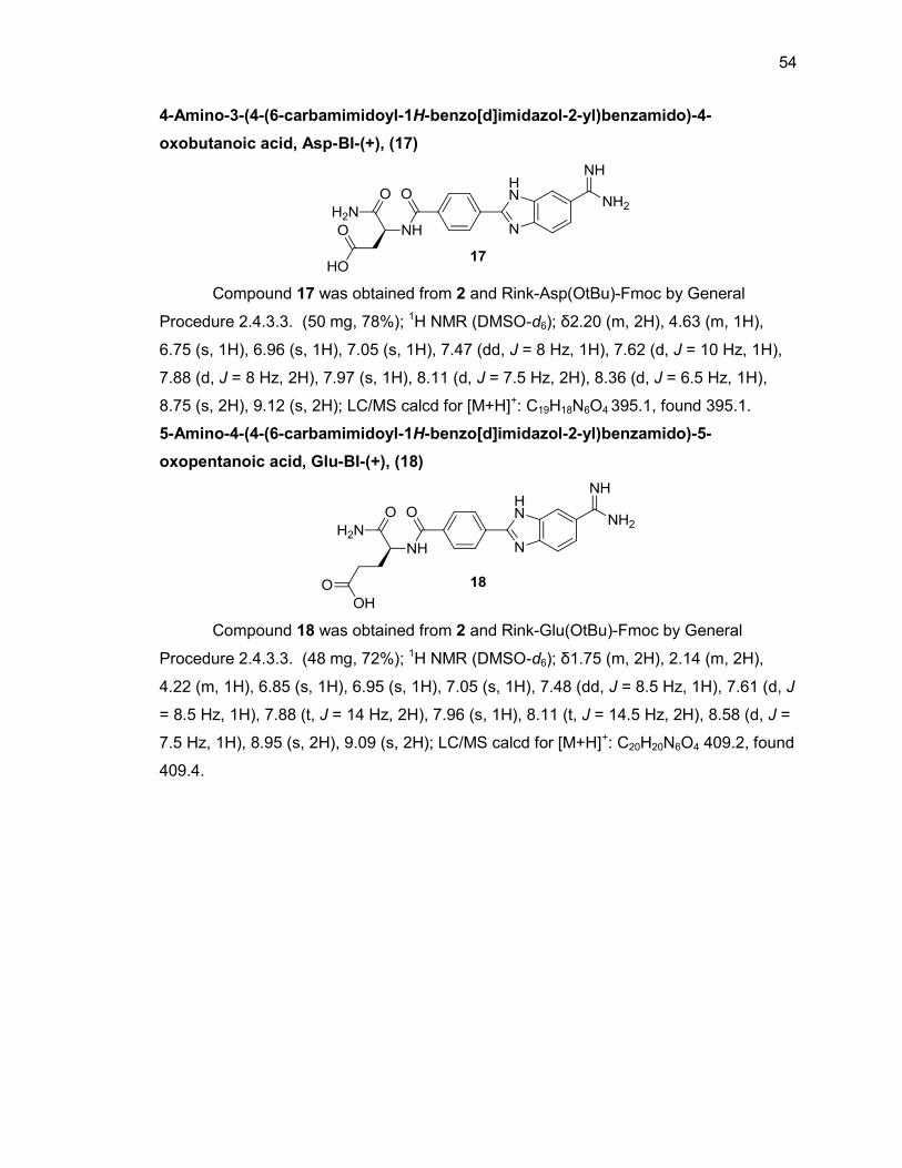
54
4-Amino-3-(4-(6-carbamimidoyl-1H-benzo[d]imidazol-2-yl)benzamido)-4-
oxobutanoic acid, Asp-BI-(+), (17)
Compound 17 was obtained from 2 and Rink-Asp(OtBu)-Fmoc by General
Procedure 2.4.3.3. (50 mg, 78%); 1H NMR (DMSO-d6); δ2.20 (m, 2H), 4.63 (m, 1H),
6.75 (s, 1H), 6.96 (s, 1H), 7.05 (s, 1H), 7.47 (dd, J = 8 Hz, 1H), 7.62 (d, J = 10 Hz, 1H),
7.88 (d, J = 8 Hz, 2H), 7.97 (s, 1H), 8.11 (d, J = 7.5 Hz, 2H), 8.36 (d, J = 6.5 Hz, 1H),
8.75 (s, 2H), 9.12 (s, 2H); LC/MS calcd for [M+H]+: C19H18N6O4 395.1, found 395.1.
5-Amino-4-(4-(6-carbamimidoyl-1H-benzo[d]imidazol-2-yl)benzamido)-5-
oxopentanoic acid, Glu-BI-(+), (18)
Compound 18 was obtained from 2 and Rink-Glu(OtBu)-Fmoc by General
Procedure 2.4.3.3. (48 mg, 72%); 1H NMR (DMSO-d6); δ1.75 (m, 2H), 2.14 (m, 2H),
4.22 (m, 1H), 6.85 (s, 1H), 6.95 (s, 1H), 7.05 (s, 1H), 7.48 (dd, J = 8.5 Hz, 1H), 7.61 (d, J
= 8.5 Hz, 1H), 7.88 (t, J = 14 Hz, 2H), 7.96 (s, 1H), 8.11 (t, J = 14.5 Hz, 2H), 8.58 (d, J =
7.5 Hz, 1H), 8.95 (s, 2H), 9.09 (s, 2H); LC/MS calcd for [M+H]+: C20H20N6O4 409.2, found
409.4.
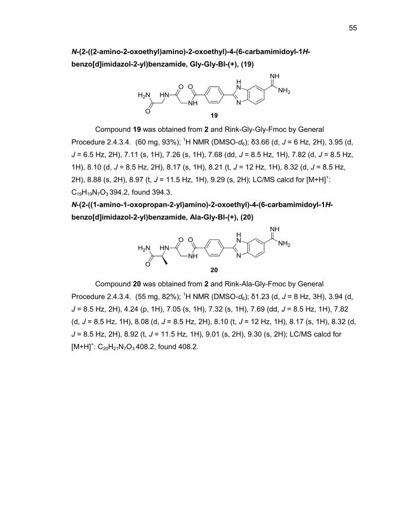
55
N-(2-((2-amino-2-oxoethyl)amino)-2-oxoethyl)-4-(6-carbamimidoyl-1H-
benzo[d]imidazol-2-yl)benzamide, Gly-Gly-BI-(+), (19)
Compound 19 was obtained from 2 and Rink-Gly-Gly-Fmoc by General
Procedure 2.4.3.4. (60 mg, 93%); 1H NMR (DMSO-d6); δ3.66 (d, J = 6 Hz, 2H), 3.95 (d,
J = 6.5 Hz, 2H), 7.11 (s, 1H), 7.26 (s, 1H), 7.68 (dd, J = 8.5 Hz, 1H), 7.82 (d, J = 8.5 Hz,
1H), 8.10 (d, J = 8.5 Hz, 2H), 8.17 (s, 1H), 8.21 (t, J = 12 Hz, 1H), 8.32 (d, J = 8.5 Hz,
2H), 8.88 (s, 2H), 8.97 (t, J = 11.5 Hz, 1H), 9.29 (s, 2H); LC/MS calcd for [M+H]+:
C19H19N7O3 394.2, found 394.3.
N-(2-((1-amino-1-oxopropan-2-yl)amino)-2-oxoethyl)-4-(6-carbamimidoyl-1H-
benzo[d]imidazol-2-yl)benzamide, Ala-Gly-BI-(+), (20)
Compound 20 was obtained from 2 and Rink-Ala-Gly-Fmoc by General
Procedure 2.4.3.4. (55 mg, 82%); 1H NMR (DMSO-d6); δ1.23 (d, J = 8 Hz, 3H), 3.94 (d,
J = 8.5 Hz, 2H), 4.24 (p, 1H), 7.05 (s, 1H), 7.32 (s, 1H), 7.69 (dd, J = 8.5 Hz, 1H), 7.82
(d, J = 8.5 Hz, 1H), 8.08 (d, J = 8.5 Hz, 2H), 8.10 (t, J = 12 Hz, 1H), 8.17 (s, 1H), 8.32 (d,
J = 8.5 Hz, 2H), 8.92 (t, J = 11.5 Hz, 1H), 9.01 (s, 2H), 9.30 (s, 2H); LC/MS calcd for
[M+H]+: C20H21N7O3 408.2, found 408.2.
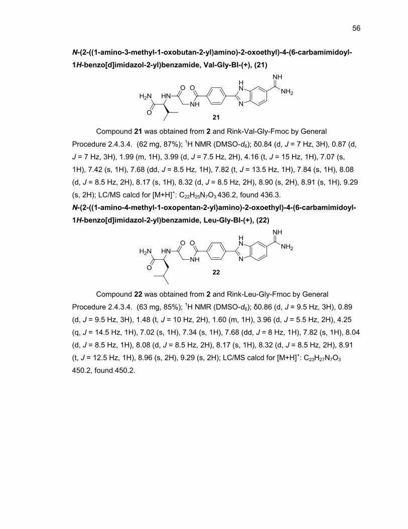
56
N-(2-((1-amino-3-methyl-1-oxobutan-2-yl)amino)-2-oxoethyl)-4-(6-carbamimidoyl-
1H-benzo[d]imidazol-2-yl)benzamide, Val-Gly-BI-(+), (21)
Compound 21 was obtained from 2 and Rink-Val-Gly-Fmoc by General
Procedure 2.4.3.4. (62 mg, 87%); 1H NMR (DMSO-d6); δ0.84 (d, J = 7 Hz, 3H), 0.87 (d,
J = 7 Hz, 3H), 1.99 (m, 1H), 3.99 (d, J = 7.5 Hz, 2H), 4.16 (t, J = 15 Hz, 1H), 7.07 (s,
1H), 7.42 (s, 1H), 7.68 (dd, J = 8.5 Hz, 1H), 7.82 (t, J = 13.5 Hz, 1H), 7.84 (s, 1H), 8.08
(d, J = 8.5 Hz, 2H), 8.17 (s, 1H), 8.32 (d, J = 8.5 Hz, 2H), 8.90 (s, 2H), 8.91 (s, 1H), 9.29
(s, 2H); LC/MS calcd for [M+H]+: C22H25N7O3 436.2, found 436.3.
N-(2-((1-amino-4-methyl-1-oxopentan-2-yl)amino)-2-oxoethyl)-4-(6-carbamimidoyl-
1H-benzo[d]imidazol-2-yl)benzamide, Leu-Gly-BI-(+), (22)
Compound 22 was obtained from 2 and Rink-Leu-Gly-Fmoc by General
Procedure 2.4.3.4. (63 mg, 85%); 1H NMR (DMSO-d6); δ0.86 (d, J = 9.5 Hz, 3H), 0.89
(d, J = 9.5 Hz, 3H), 1.48 (t, J = 10 Hz, 2H), 1.60 (m, 1H), 3.96 (d, J = 5.5 Hz, 2H), 4.25
(q, J = 14.5 Hz, 1H), 7.02 (s, 1H), 7.34 (s, 1H), 7.68 (dd, J = 8 Hz, 1H), 7.82 (s, 1H), 8.04
(d, J = 8.5 Hz, 1H), 8.08 (d, J = 8.5 Hz, 2H), 8.17 (s, 1H), 8.32 (d, J = 8.5 Hz, 2H), 8.91
(t, J = 12.5 Hz, 1H), 8.96 (s, 2H), 9.29 (s, 2H); LC/MS calcd for [M+H]+: C23H27N7O3
450.2, found 450.2.
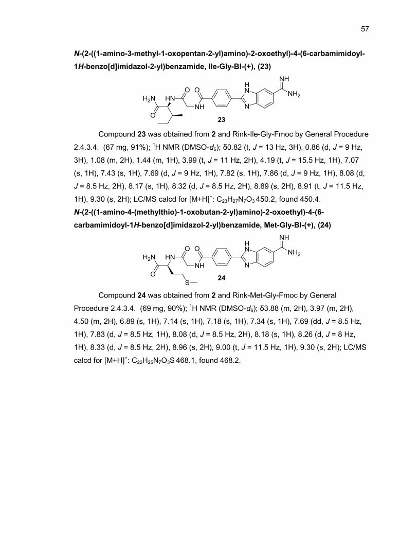
57
N-(2-((1-amino-3-methyl-1-oxopentan-2-yl)amino)-2-oxoethyl)-4-(6-carbamimidoyl-
1H-benzo[d]imidazol-2-yl)benzamide, Ile-Gly-BI-(+), (23)
Compound 23 was obtained from 2 and Rink-Ile-Gly-Fmoc by General Procedure
2.4.3.4. (67 mg, 91%); 1H NMR (DMSO-d6); δ0.82 (t, J = 13 Hz, 3H), 0.86 (d, J = 9 Hz,
3H), 1.08 (m, 2H), 1.44 (m, 1H), 3.99 (t, J = 11 Hz, 2H), 4.19 (t, J = 15.5 Hz, 1H), 7.07
(s, 1H), 7.43 (s, 1H), 7.69 (d, J = 9 Hz, 1H), 7.82 (s, 1H), 7.86 (d, J = 9 Hz, 1H), 8.08 (d,
J = 8.5 Hz, 2H), 8.17 (s, 1H), 8.32 (d, J = 8.5 Hz, 2H), 8.89 (s, 2H), 8.91 (t, J = 11.5 Hz,
1H), 9.30 (s, 2H); LC/MS calcd for [M+H]+: C23H27N7O3 450.2, found 450.4.
N-(2-((1-amino-4-(methylthio)-1-oxobutan-2-yl)amino)-2-oxoethyl)-4-(6-
carbamimidoyl-1H-benzo[d]imidazol-2-yl)benzamide, Met-Gly-BI-(+), (24)
Compound 24 was obtained from 2 and Rink-Met-Gly-Fmoc by General
Procedure 2.4.3.4. (69 mg, 90%); 1H NMR (DMSO-d6); δ3.88 (m, 2H), 3.97 (m, 2H),
4.50 (m, 2H), 6.89 (s, 1H), 7.14 (s, 1H), 7.18 (s, 1H), 7.34 (s, 1H), 7.69 (dd, J = 8.5 Hz,
1H), 7.83 (d, J = 8.5 Hz, 1H), 8.08 (d, J = 8.5 Hz, 2H), 8.18 (s, 1H), 8.26 (d, J = 8 Hz,
1H), 8.33 (d, J = 8.5 Hz, 2H), 8.96 (s, 2H), 9.00 (t, J = 11.5 Hz, 1H), 9.30 (s, 2H); LC/MS
calcd for [M+H]+: C22H25N7O3S 468.1, found 468.2.
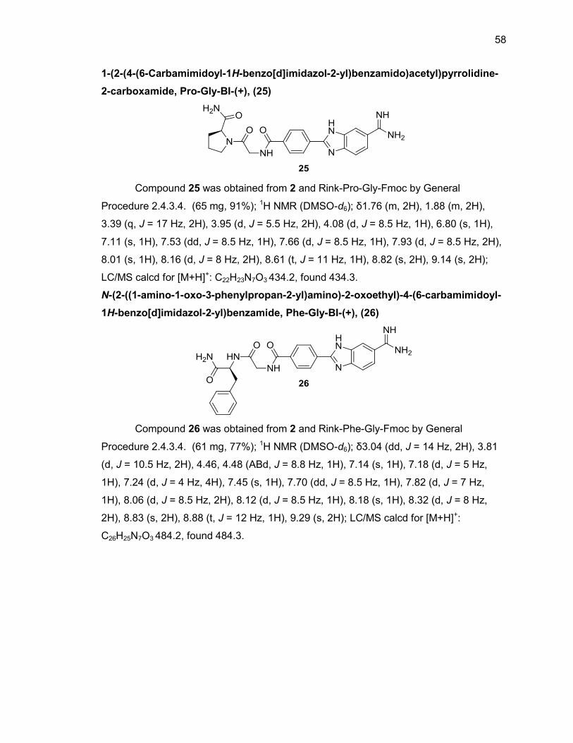
58
1-(2-(4-(6-Carbamimidoyl-1H-benzo[d]imidazol-2-yl)benzamido)acetyl)pyrrolidine-
2-carboxamide, Pro-Gly-BI-(+), (25)
Compound 25 was obtained from 2 and Rink-Pro-Gly-Fmoc by General
Procedure 2.4.3.4. (65 mg, 91%); 1H NMR (DMSO-d6); δ1.76 (m, 2H), 1.88 (m, 2H),
3.39 (q, J = 17 Hz, 2H), 3.95 (d, J = 5.5 Hz, 2H), 4.08 (d, J = 8.5 Hz, 1H), 6.80 (s, 1H),
7.11 (s, 1H), 7.53 (dd, J = 8.5 Hz, 1H), 7.66 (d, J = 8.5 Hz, 1H), 7.93 (d, J = 8.5 Hz, 2H),
8.01 (s, 1H), 8.16 (d, J = 8 Hz, 2H), 8.61 (t, J = 11 Hz, 1H), 8.82 (s, 2H), 9.14 (s, 2H);
LC/MS calcd for [M+H]+: C22H23N7O3 434.2, found 434.3.
N-(2-((1-amino-1-oxo-3-phenylpropan-2-yl)amino)-2-oxoethyl)-4-(6-carbamimidoyl-
1H-benzo[d]imidazol-2-yl)benzamide, Phe-Gly-BI-(+), (26)
Compound 26 was obtained from 2 and Rink-Phe-Gly-Fmoc by General
Procedure 2.4.3.4. (61 mg, 77%); 1H NMR (DMSO-d6); δ3.04 (dd, J = 14 Hz, 2H), 3.81
(d, J = 10.5 Hz, 2H), 4.46, 4.48 (ABd, J = 8.8 Hz, 1H), 7.14 (s, 1H), 7.18 (d, J = 5 Hz,
1H), 7.24 (d, J = 4 Hz, 4H), 7.45 (s, 1H), 7.70 (dd, J = 8.5 Hz, 1H), 7.82 (d, J = 7 Hz,
1H), 8.06 (d, J = 8.5 Hz, 2H), 8.12 (d, J = 8.5 Hz, 1H), 8.18 (s, 1H), 8.32 (d, J = 8 Hz,
2H), 8.83 (s, 2H), 8.88 (t, J = 12 Hz, 1H), 9.29 (s, 2H); LC/MS calcd for [M+H]+:
C26H25N7O3 484.2, found 484.3.
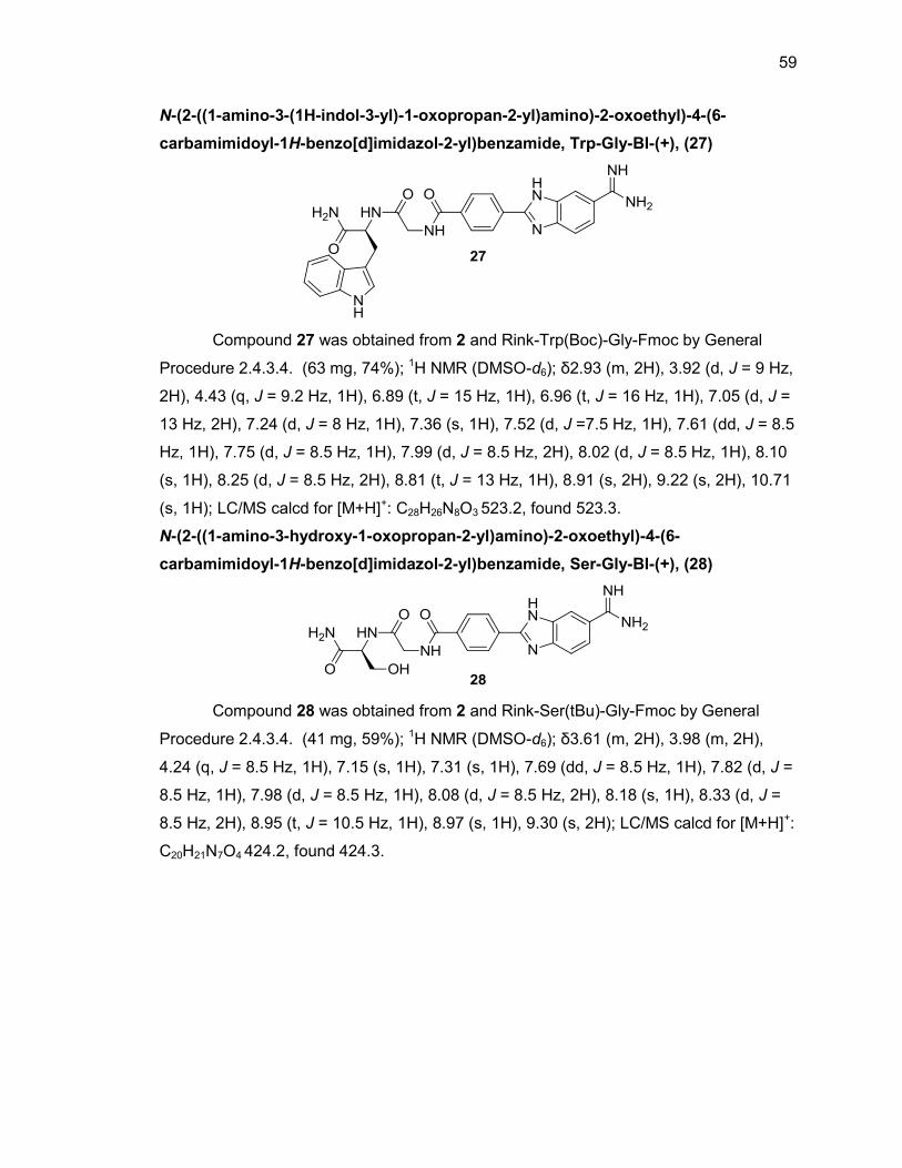
59
N-(2-((1-amino-3-(1H-indol-3-yl)-1-oxopropan-2-yl)amino)-2-oxoethyl)-4-(6-
carbamimidoyl-1H-benzo[d]imidazol-2-yl)benzamide, Trp-Gly-BI-(+), (27)
Compound 27 was obtained from 2 and Rink-Trp(Boc)-Gly-Fmoc by General
Procedure 2.4.3.4. (63 mg, 74%); 1H NMR (DMSO-d6); δ2.93 (m, 2H), 3.92 (d, J = 9 Hz,
2H), 4.43 (q, J = 9.2 Hz, 1H), 6.89 (t, J = 15 Hz, 1H), 6.96 (t, J = 16 Hz, 1H), 7.05 (d, J =
13 Hz, 2H), 7.24 (d, J = 8 Hz, 1H), 7.36 (s, 1H), 7.52 (d, J =7.5 Hz, 1H), 7.61 (dd, J = 8.5
Hz, 1H), 7.75 (d, J = 8.5 Hz, 1H), 7.99 (d, J = 8.5 Hz, 2H), 8.02 (d, J = 8.5 Hz, 1H), 8.10
(s, 1H), 8.25 (d, J = 8.5 Hz, 2H), 8.81 (t, J = 13 Hz, 1H), 8.91 (s, 2H), 9.22 (s, 2H), 10.71
(s, 1H); LC/MS calcd for [M+H]+: C28H26N8O3 523.2, found 523.3.
N-(2-((1-amino-3-hydroxy-1-oxopropan-2-yl)amino)-2-oxoethyl)-4-(6-
carbamimidoyl-1H-benzo[d]imidazol-2-yl)benzamide, Ser-Gly-BI-(+), (28)
Compound 28 was obtained from 2 and Rink-Ser(tBu)-Gly-Fmoc by General
Procedure 2.4.3.4. (41 mg, 59%); 1H NMR (DMSO-d6); δ3.61 (m, 2H), 3.98 (m, 2H),
4.24 (q, J = 8.5 Hz, 1H), 7.15 (s, 1H), 7.31 (s, 1H), 7.69 (dd, J = 8.5 Hz, 1H), 7.82 (d, J =
8.5 Hz, 1H), 7.98 (d, J = 8.5 Hz, 1H), 8.08 (d, J = 8.5 Hz, 2H), 8.18 (s, 1H), 8.33 (d, J =
8.5 Hz, 2H), 8.95 (t, J = 10.5 Hz, 1H), 8.97 (s, 1H), 9.30 (s, 2H); LC/MS calcd for [M+H]+:
C20H21N7O4 424.2, found 424.3.
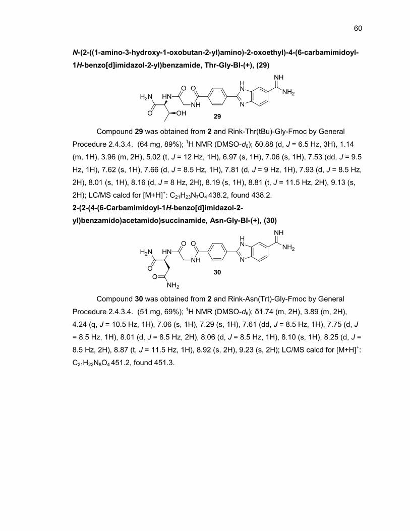
60
N-(2-((1-amino-3-hydroxy-1-oxobutan-2-yl)amino)-2-oxoethyl)-4-(6-carbamimidoyl-
1H-benzo[d]imidazol-2-yl)benzamide, Thr-Gly-BI-(+), (29)
Compound 29 was obtained from 2 and Rink-Thr(tBu)-Gly-Fmoc by General
Procedure 2.4.3.4. (64 mg, 89%); 1H NMR (DMSO-d6); δ0.88 (d, J = 6.5 Hz, 3H), 1.14
(m, 1H), 3.96 (m, 2H), 5.02 (t, J = 12 Hz, 1H), 6.97 (s, 1H), 7.06 (s, 1H), 7.53 (dd, J = 9.5
Hz, 1H), 7.62 (s, 1H), 7.66 (d, J = 8.5 Hz, 1H), 7.81 (d, J = 9 Hz, 1H), 7.93 (d, J = 8.5 Hz,
2H), 8.01 (s, 1H), 8.16 (d, J = 8 Hz, 2H), 8.19 (s, 1H), 8.81 (t, J = 11.5 Hz, 2H), 9.13 (s,
2H); LC/MS calcd for [M+H]+: C21H23N7O4 438.2, found 438.2.
2-(2-(4-(6-Carbamimidoyl-1H-benzo[d]imidazol-2-
yl)benzamido)acetamido)succinamide, Asn-Gly-BI-(+), (30)
Compound 30 was obtained from 2 and Rink-Asn(Trt)-Gly-Fmoc by General
Procedure 2.4.3.4. (51 mg, 69%); 1H NMR (DMSO-d6); δ1.74 (m, 2H), 3.89 (m, 2H),
4.24 (q, J = 10.5 Hz, 1H), 7.06 (s, 1H), 7.29 (s, 1H), 7.61 (dd, J = 8.5 Hz, 1H), 7.75 (d, J
= 8.5 Hz, 1H), 8.01 (d, J = 8.5 Hz, 2H), 8.06 (d, J = 8.5 Hz, 1H), 8.10 (s, 1H), 8.25 (d, J =
8.5 Hz, 2H), 8.87 (t, J = 11.5 Hz, 1H), 8.92 (s, 2H), 9.23 (s, 2H); LC/MS calcd for [M+H]+:
C21H22N8O4 451.2, found 451.3.
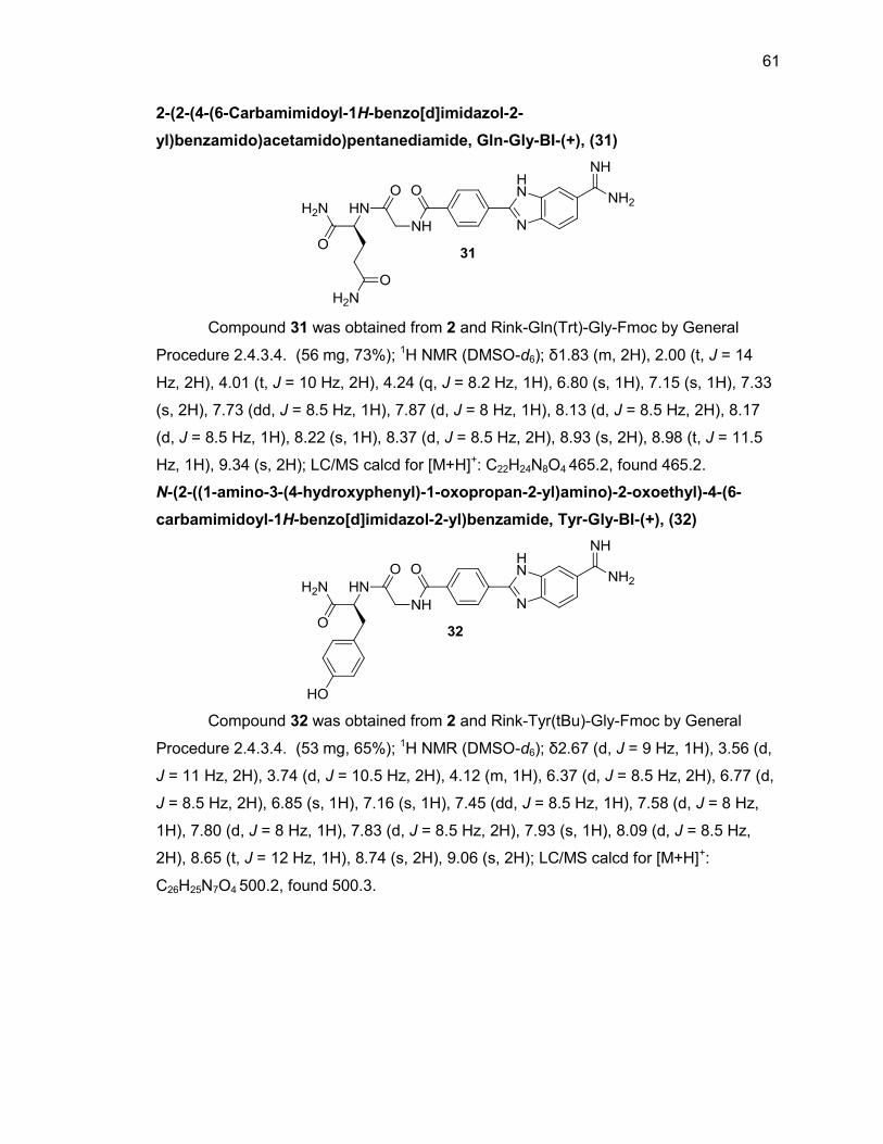
61
2-(2-(4-(6-Carbamimidoyl-1H-benzo[d]imidazol-2-
yl)benzamido)acetamido)pentanediamide, Gln-Gly-BI-(+), (31)
Compound 31 was obtained from 2 and Rink-Gln(Trt)-Gly-Fmoc by General
Procedure 2.4.3.4. (56 mg, 73%); 1H NMR (DMSO-d6); δ1.83 (m, 2H), 2.00 (t, J = 14
Hz, 2H), 4.01 (t, J = 10 Hz, 2H), 4.24 (q, J = 8.2 Hz, 1H), 6.80 (s, 1H), 7.15 (s, 1H), 7.33
(s, 2H), 7.73 (dd, J = 8.5 Hz, 1H), 7.87 (d, J = 8 Hz, 1H), 8.13 (d, J = 8.5 Hz, 2H), 8.17
(d, J = 8.5 Hz, 1H), 8.22 (s, 1H), 8.37 (d, J = 8.5 Hz, 2H), 8.93 (s, 2H), 8.98 (t, J = 11.5
Hz, 1H), 9.34 (s, 2H); LC/MS calcd for [M+H]+: C22H24N8O4 465.2, found 465.2.
N-(2-((1-amino-3-(4-hydroxyphenyl)-1-oxopropan-2-yl)amino)-2-oxoethyl)-4-(6-
carbamimidoyl-1H-benzo[d]imidazol-2-yl)benzamide, Tyr-Gly-BI-(+), (32)
Compound 32 was obtained from 2 and Rink-Tyr(tBu)-Gly-Fmoc by General
Procedure 2.4.3.4. (53 mg, 65%); 1H NMR (DMSO-d6); δ2.67 (d, J = 9 Hz, 1H), 3.56 (d,
J = 11 Hz, 2H), 3.74 (d, J = 10.5 Hz, 2H), 4.12 (m, 1H), 6.37 (d, J = 8.5 Hz, 2H), 6.77 (d,
J = 8.5 Hz, 2H), 6.85 (s, 1H), 7.16 (s, 1H), 7.45 (dd, J = 8.5 Hz, 1H), 7.58 (d, J = 8 Hz,
1H), 7.80 (d, J = 8 Hz, 1H), 7.83 (d, J = 8.5 Hz, 2H), 7.93 (s, 1H), 8.09 (d, J = 8.5 Hz,
2H), 8.65 (t, J = 12 Hz, 1H), 8.74 (s, 2H), 9.06 (s, 2H); LC/MS calcd for [M+H]+:
C26H25N7O4 500.2, found 500.3.
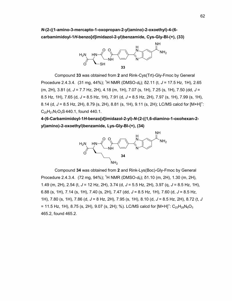
62
N-(2-((1-amino-3-mercapto-1-oxopropan-2-yl)amino)-2-oxoethyl)-4-(6-
carbamimidoyl-1H-benzo[d]imidazol-2-yl)benzamide, Cys-Gly-BI-(+), (33)
Compound 33 was obtained from 2 and Rink-Cys(Trt)-Gly-Fmoc by General
Procedure 2.4.3.4. (31 mg, 44%); 1H NMR (DMSO-d6); δ2.11 (t, J = 17.5 Hz, 1H), 2.65
(m, 2H), 3.81 (d, J = 7.7 Hz, 2H), 4.18 (m, 1H), 7.07 (s, 1H), 7.25 (s, 1H), 7.50 (dd, J =
8.5 Hz, 1H), 7.65 (d, J = 8.5 Hz, 1H), 7.91 (d, J = 8.5 Hz, 2H), 7.97 (s, 1H), 7.99 (s, 1H),
8.14 (d, J = 8.5 Hz, 2H), 8.79 (s, 2H), 8.81 (s, 1H), 9.11 (s, 2H); LC/MS calcd for [M+H]+:
C20H21N7O3S 440.1, found 440.1.
4-(6-Carbamimidoyl-1H-benzo[d]imidazol-2-yl)-N-(2-((1,6-diamino-1-oxohexan-2-
yl)amino)-2-oxoethyl)benzamide, Lys-Gly-BI-(+), (34)
Compound 34 was obtained from 2 and Rink-Lys(Boc)-Gly-Fmoc by General
Procedure 2.4.3.4. (72 mg, 94%); 1H NMR (DMSO-d6); δ1.10 (m, 2H), 1.30 (m, 2H),
1.49 (m, 2H), 2.54 (t, J = 12 Hz, 2H), 3.74 (d, J = 5.5 Hz, 2H), 3.97 (q, J = 8.5 Hz, 1H),
6.88 (s, 1H), 7.14 (s, 1H), 7.40 (s, 2H), 7.47 (dd, J = 8.5 Hz, 1H), 7.60 (d, J = 8.5 Hz,
1H), 7.80 (s, 1H), 7.86 (d, J = 8 Hz, 2H), 7.95 (s, 1H), 8.10 (d, J = 8.5 Hz, 2H), 8.72 (t, J
= 11.5 Hz, 1H), 8.75 (s, 2H), 9.07 (s, 2H); %). LC/MS calcd for [M+H]+: C23H28N8O3
465.2, found 465.2.
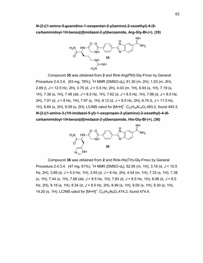
63
N-(2-((1-amino-5-guanidino-1-oxopentan-2-yl)amino)-2-oxoethyl)-4-(6-
carbamimidoyl-1H-benzo[d]imidazol-2-yl)benzamide, Arg-Gly-BI-(+), (35)
Compound 35 was obtained from 2 and Rink-Arg(Pbf)-Gly-Fmoc by General
Procedure 2.4.3.4. (63 mg, 78%); 1H NMR (DMSO-d6); δ1.30 (m, 2H), 1.53 (m, 2H),
2.89 (t, J = 12.5 Hz, 2H), 3.75 (d, J = 5.5 Hz, 2H), 4.03 (m, 1H), 6.93 (s, 1H), 7.19 (s,
1H), 7.38 (s, 1H), 7.48 (dd, J = 8.5 Hz, 1H), 7.62 (d, J = 8.5 Hz, 1H), 7.88 (d, J = 8.5 Hz,
2H), 7.91 (d, J = 8 Hz, 1H), 7.97 (s, 1H), 8.12 (d, J = 8.5 Hz, 2H), 8.74 (t, J = 11.5 Hz,
1H), 8.84 (s, 2H), 9.09 (s, 2H); LC/MS calcd for [M+H]+: C23H28N10O3 493.2, found 493.3.
N-(2-((1-amino-3-(1H-imidazol-5-yl)-1-oxopropan-2-yl)amino)-2-oxoethyl)-4-(6-
carbamimidoyl-1H-benzo[d]imidazol-2-yl)benzamide, His-Gly-BI-(+), (36)
Compound 36 was obtained from 2 and Rink-His(Trt)-Gly-Fmoc by General
Procedure 2.4.3.4. (47 mg, 61%); 1H NMR (DMSO-d6); δ2.95 (m, 1H), 3.18 (d, J = 10.5
Hz, 2H), 3.89 (d, J = 5.5 Hz, 1H), 3.93 (d, J = 6 Hz, 2H), 4.54 (m, 1H), 7.33 (s, 1H), 7.38
(s, 1H), 7.44 (s, 1H), 7.69 (dd, J = 8.5 Hz, 1H), 7.83 (d, J = 8.5 Hz, 1H), 8.08 (d, J = 8.5
Hz, 2H), 8.18 (s, 1H), 8.34 (d, J = 8.5 Hz, 2H), 8.99 (s, 1H), 9.00 (s, 1H), 9.30 (s, 1H),
14.20 (s, 1H); LC/MS calcd for [M+H]+: C23H23N9O3 474.2, found 474.4.
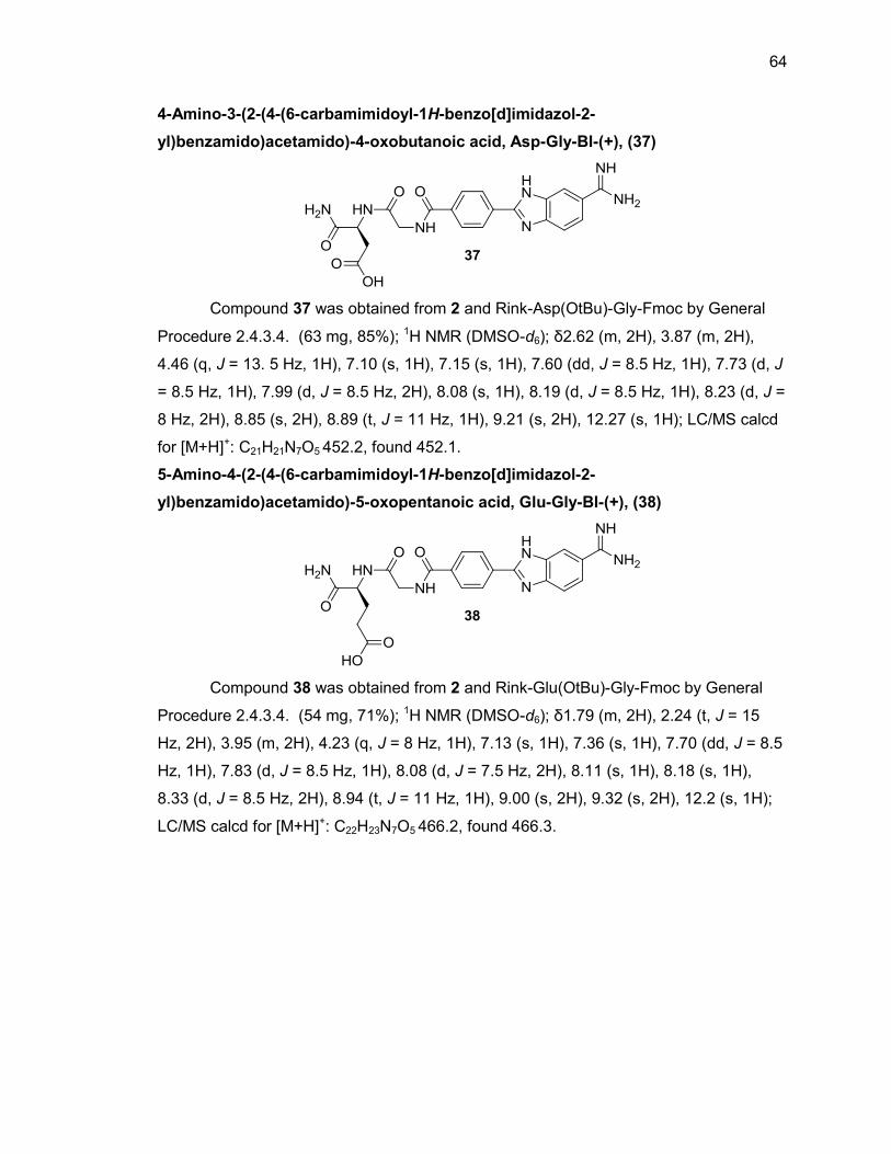
64
4-Amino-3-(2-(4-(6-carbamimidoyl-1H-benzo[d]imidazol-2-
yl)benzamido)acetamido)-4-oxobutanoic acid, Asp-Gly-BI-(+), (37)
Compound 37 was obtained from 2 and Rink-Asp(OtBu)-Gly-Fmoc by General
Procedure 2.4.3.4. (63 mg, 85%); 1H NMR (DMSO-d6); δ2.62 (m, 2H), 3.87 (m, 2H),
4.46 (q, J = 13. 5 Hz, 1H), 7.10 (s, 1H), 7.15 (s, 1H), 7.60 (dd, J = 8.5 Hz, 1H), 7.73 (d, J
= 8.5 Hz, 1H), 7.99 (d, J = 8.5 Hz, 2H), 8.08 (s, 1H), 8.19 (d, J = 8.5 Hz, 1H), 8.23 (d, J =
8 Hz, 2H), 8.85 (s, 2H), 8.89 (t, J = 11 Hz, 1H), 9.21 (s, 2H), 12.27 (s, 1H); LC/MS calcd
for [M+H]+: C21H21N7O5 452.2, found 452.1.
5-Amino-4-(2-(4-(6-carbamimidoyl-1H-benzo[d]imidazol-2-
yl)benzamido)acetamido)-5-oxopentanoic acid, Glu-Gly-BI-(+), (38)
Compound 38 was obtained from 2 and Rink-Glu(OtBu)-Gly-Fmoc by General
Procedure 2.4.3.4. (54 mg, 71%); 1H NMR (DMSO-d6); δ1.79 (m, 2H), 2.24 (t, J = 15
Hz, 2H), 3.95 (m, 2H), 4.23 (q, J = 8 Hz, 1H), 7.13 (s, 1H), 7.36 (s, 1H), 7.70 (dd, J = 8.5
Hz, 1H), 7.83 (d, J = 8.5 Hz, 1H), 8.08 (d, J = 7.5 Hz, 2H), 8.11 (s, 1H), 8.18 (s, 1H),
8.33 (d, J = 8.5 Hz, 2H), 8.94 (t, J = 11 Hz, 1H), 9.00 (s, 2H), 9.32 (s, 2H), 12.2 (s, 1H);
LC/MS calcd for [M+H]+: C22H23N7O5 466.2, found 466.3.
65
2.5. List of References
1. Denny, W. A.; Rewcastle, G. W.; Baguly, B. C. J. Med. Chem. 1990, 33, 814.
2. Goker, H.; Kus, C.; Boykin, D. W.; Yildiz, S.; Altanlar, N. Bioorg. Med. Chem. 2002,
10, 2589.
3. Seth, P. P.; Jefferson, E. A.; Risen, L. M.; Osgood, S. A. Bioorg. Med. Chem. Lett.
2003, 13, 1669.
4. Nare, B.; Liu, Z.; Prichard, R. K.; George, E. Biochem. Pharmacol. 1994, 48, 2215.
5. Mahaimeed, H. A. Int. Med. Res. 1997, 25, 175.
6. Renneberg D; Dervan, P. B. J. Am. Chem. Soc. 2003, 125, 5707.
7. Harshman, K. D.; Dervan, P. B. Nucleic Acid Res. 1985, 13, 4825.
8. Marques, M. A.; Doss, R. M.; Foister, S.; Dervan, P. B. J. Am. Chem. Soc. 2004,
126, 10339.
9. Viger, A.; Dervan, P. B. Bioorg. Med. Chem. 2006, 14, 853.
10. Wilson, W. D.; Nguyen, B.; Tanious, F. A.; Mathis, A.; Hall, J. E.; Stephens, C. E.;
Boykin, D. W. Curr. Med. Chem. Anti-Cancer Agents 2005, 5, 389.
11. Miao, Y.; Michael P. H. Lee, M. P. H.; Parkinson, G. N.; Batista-Parra, A.; Ismail, M.
A.; Neidle, S.; Boykin, D. W.; Wilson, W. D. Biochemistry 2005, 44, 14701.
12. Neidle, S. Nat. Prod. Rep. 2001, 18, 291.
13. Joubert, A.; Sun, X. W.; Johansson, E.; Bailly, C.; Mann, J.; Neidle, S. Biochemistry
2003, 42, 5984.
14. Tanious, F. A.; Hamelberg, D.; Bailly, C.; Czarny, A.; Boykin, D. W.; Wilson, W. D. J.
Am. Chem. Soc. 2004, 126, 143.
15. Haq, I.; Ladbury, J. E.; Chowdhry, B. Z.; Jenkins, T. C.; Chaires, J. B. J. Mol. Biol.
1997, 271, 244.
16. Satz, A. L.; Bruice, T. C. Acc. Chem. Res. 2002, 35, 86.
17. Haq, I. Arch. Biochem. Biophys. 2002, 403, 1.
18. Zablocki, J. A.; Miyano, M.; Garland, R. B.; Pireh, D.; Schretzman, L.; Rao, S. N.;
Lindmark, R. J.; Panzer-Knodle, S. G.; Nicholson, N. S.; Taite, B. B.; Salyers, A. K.;
King, L. W.; Campion, J. G.; Feigen, L. P. J. Med. Chem. 1993, 36, 1811.
19. Peterlin-Mašič, L.; D. Kikelj, D. Tetrahedron 2001, 57, 7073.
20. Patel, N.; Berglund, H.: Nilsson, L.; Rigler, R.; McLaughlin, L. W.; Gräslund, A. Eur.
J. Biochem. 1992, 203, 361.
66
21. Bailly, C.; Chaires, J. B. Bioconj. Chem. 1998, 9, 513.
22. Geierstanger, B. H.; Jacobsen, J. P.; Mrksich, M.; Dervan, P. B.; Wemmer, D. E.
Biochemistry 1994, 33, 3055.
23. Goodwin, K. D.; Long, E. C.; Georgiadis, M. M. Nucleic Acids Res. 2005, 33, 4106.
24. Clark, G. R.; Boykin, D. W.; Czarny, A.; Neidle, S. Nucleic Acids Res. 1997, 25,
1510.
25. Ismail, M. A.; Batista-Parra, A.; Miao, Y.; Wilson, W. D.; Wenzler, T.; Brun, R.;
Boykin, D. W. Bioorg. Med. Chem. 2005, 13, 6718.
26. Dervan, P. B. Curr. Opin. Struct. Biol. 2003, 13, 284.
27. Kielkopf, C. L.; White, S.; Szewczyk, J. W.; Turner, J. M.; Baird, E. E.; Dervan, P. B.;
Rees, D. C. Science 1998, 282, 111.
28. Kielkopf, C. L.; Bremer, R. E.; White, S.; Szewczyk, J. W.; Turner, J. M.; Baird, E. E.;
Dervan, P. B.; Rees, C. C. J. Mol Biol. 2000, 295, 557.
29. White, S.; Szewczyk, J. W.; Turner, J. M.; Baird, E. E.; Dervan, P. B. Nature 1998,
391, 468.
30. Merrifield, R. B. J. Amer. Chem. Soc. 1963, 85, 2149.
31. Chan, W. C.; White, P. D. Fmoc Solid Phase Peptide Synthesis, Oxford University
Press, 2000.
32. Stewart, J. M.; Young, J. D.; Solid Phase Peptide Synthesis, 2nd Ed, Pierce Chemical
Company, 1984.
33. Peptide Synthesis, Merck, 2009.
34. Parrish, R. F.; Straus, J. W.; Paulson, J. D.; Polakoski, K. L.; Tidwell, R. R.; Geratz,
J. D.; Stevens, F. M. J. Med. Chem. 1978, 21, 1132.
35. Goker, H.; Alp, M.; Yǐldǐz, S. Molecules 2005, 10, 1377.
36. Wendt, M. D.; Rockway, T. W.; Geyer, A.; McClellan, W.; Weitzberg, M.; Zhao, X. M.;
Mantei, R.; Nienaber, V. L.; Stewart, K.; Klinghofer, V.; Giranda, V. L. J. Med. Chem.
2004, 47, 303.
37. Xue, C. B.; Rafalski, M.; Roderick, J.; Eyermann, C. J.; Mousa, S.; Olson, R. E.;
DeGrado, W. F. Bioorg. Med. Chem. Lett. 1996, 6, 339.
38. Ismail, M. A.; Brun, R.; Wenzler, T.; Tanious, F. A.; Wilson, W. D.; Boykin, D. W.
Bioorg. Med. Chem. 2004, 12, 5405.
39. Anbazhagan, M.; Boykin, D. W.; Chad E. Stephens, C. E.; Synthesis 2003, 16, 2467.
67
40. Judkins, B. D.; Allen, D. G.; Cook, T. A.; Evans, B.; Sardharwala, T. E. Synth.
Commun. 1996, 26, 4351.
41. Dörwald, Z. F.; Organic Synthesis on Solid Phase, 2nd ed., Wiley-VCH: Weinheim,
2002.
42. Kaiser, E.; Colescott, R. L.; Bossinger, C. D.; Cook, P. I. Anal. Biochem. 1970, 34,
595.
68
CHAPTER 3. PRELIMINARY SCREENING OF DNA BINDING ACTIVITY
3.1. Overview
A new series of amino acid-benzimidazole-amidine and di-amino acid-
benzimidazole-amidine conjugates were designed and synthesized in our laboratory. To
analyze the relative DNA binding affinity of these compounds, a microplate-based
fluorescence intercalator displacement (HT-FID) assay was used. This chapter will
introduce the HT-FID assay developed by Boger et al. and illustrate its validation
process. We will use calf thymus DNA along with ethidium bromide as a fluorescence
indicator to establish the relative binding affinity of our amino acid-benzimidazole-
amidine conjugates.1-5
3.2. HT-FID Assay
There are a number of techniques such as footprinting and affinity cleavage6
used to analyze the DNA-binding interactions of small molecules; unfortunately, most of
these methods are challenging and time-consuming,7 making analysis of large catalogs
of compounds difficult. The HT-FID assay (Figure 3.1)8, addresses the ease-of-use
challenges of other methods and utilizes DNA treated with ethidium bromide to yield
fluorescence upon DNA-ethidium bromide binding. Addition of DNA-binding compound
displaces the ethidium bromide, resulting in a decrease in the fluorescence. The percent
fluorescence decrease is directly related to the extent of DNA-ligand binding and can
show the relative binding affinity of our synthesized compounds. FID is commonly
employed using a library of all possible 4- or 5-bp hairpin oligonucleotides to establish
the DNA-ligand binding site-selectivity of the target compound, but calf thymus DNA
(CT-DNA) has the advantage of being a cost-effective high-throughput substrate
approximating all possible combinations of base pairs and can be used to establish the
relative DNA binding of a group of compounds.1 It is important to note the analysis of
our amino acid-benzimidazole-amidine conjugates is preliminary, and as such, the focus
of this section is on the relative binding as determined using CT-DNA rather than
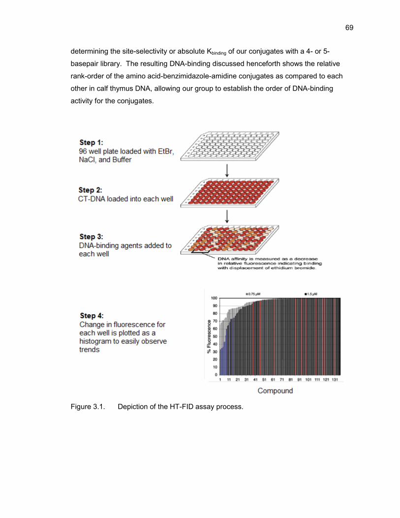
69
determining the site-selectivity or absolute Kbinding of our conjugates with a 4- or 5-
basepair library. The resulting DNA-binding discussed henceforth shows the relative
rank-order of the amino acid-benzimidazole-amidine conjugates as compared to each
other in calf thymus DNA, allowing our group to establish the order of DNA-binding
activity for the conjugates.
Figure 3.1. Depiction of the HT-FID assay process.
70
The HT-FID assay using CT-DNA has been used to analyze a commercially-
available chemical library of 60,000 compounds to determine which of the compounds
exhibited DNA-binding ability.1 Although a determination of site selectivity was not our
focus, the FID assay has been validated by analyzing the DNA binding site selectivity of
several well studied A/T-rich minor groove binding agents such as netropsin, distamycin,
Hoechst 33258, and RT 294 and the assay could be used in the future to further analyze
the specific binding sites of our compounds. The HT-FID assay using CT-DNA has the
potential to be widely applicable in the discovery of new DNA binding agents, making it
an ideal choice to quickly establish the relative binding of our amino acid-benzimidazole-
amidine conjugates.
3.2.1. HT-FID Assay Validation
Due to its well-behaved performance in the FID assay, netropsin can be used as
a compound in the validation of the assay.8,9 Netropsin has previously been evaluated
using the FID assay with both 4- and 5-bp libraries of oligonucleotides as well as with
CT-DNA using ethidium bromide as the reporting fluorophores, thus providing sufficient
information to validate the assay. Therefore, netropsin was analyzed along with our
amino acid-benzimidazole-amidine conjugates to validate the assay. Distamycin,
another compound that has been extensively studied using the 4- and 5-bp libraries was
also used in our validation to provide a second well-studied DNA binder for comparison
of our conjugates. For our validation, netropsin and distamycin were carried out in the
assay side-by-side with our conjugates at concentrations of 0.75, 1.5, and 3.0 μM with
10 μM CT-DNA and 4.5 μM ethidium bromide, as previously described.1
To assess the validity of our assay using ethidium bromide-bound calf thymus
DNA, Z′ scores10 were determined using netropsin and distamycin as positive controls
due to their well understood binding and wells containing a buffer blank were used as a
negative control. To define a screening window for the HT-FID assay, µc- and µc+ are
denoted as the means of the negative control signal and positive control signal,
respectively. The standard deviations of the signals are denoted as σc- and σc+,
respectively. The Z′-factor can be used for quality assessment in assay development
and optimization and is appropriate for evaluating the overall quality of the assay. For
example, if the Z′ value is small (negative or near zero), the assay conditions have not
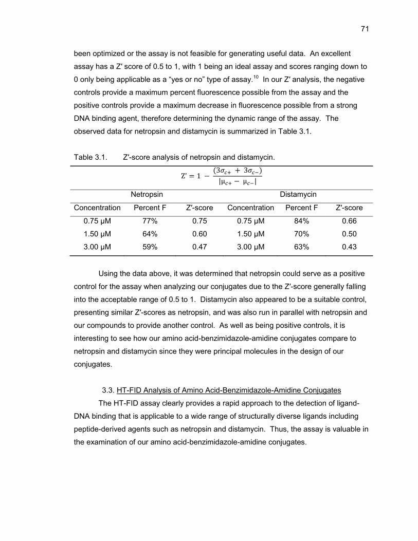
71
been optimized or the assay is not feasible for generating useful data. An excellent
assay has a Z′ score of 0.5 to 1, with 1 being an ideal assay and scores ranging down to
0 only being applicable as a “yes or no” type of assay.10 In our Z′ analysis, the negative
controls provide a maximum percent fluorescence possible from the assay and the
positive controls provide a maximum decrease in fluorescence possible from a strong
DNA binding agent, therefore determining the dynamic range of the assay. The
observed data for netropsin and distamycin is summarized in Table 3.1.
Table 3.1. Z′-score analysis of netropsin and distamycin.
Netropsin Distamycin
Concentration Percent F Z′-score Concentration Percent F Z′-score
0.75 μM 77% 0.75 0.75 μM 84% 0.66
1.50 μM 64% 0.60 1.50 μM 70% 0.50
3.00 μM 59% 0.47 3.00 μM 63% 0.43
Using the data above, it was determined that netropsin could serve as a positive
control for the assay when analyzing our conjugates due to the Z′-score generally falling
into the acceptable range of 0.5 to 1. Distamycin also appeared to be a suitable control,
presenting similar Z′-scores as netropsin, and was also run in parallel with netropsin and
our compounds to provide another control. As well as being positive controls, it is
interesting to see how our amino acid-benzimidazole-amidine conjugates compare to
netropsin and distamycin since they were principal molecules in the design of our
conjugates.
3.3. HT-FID Analysis of Amino Acid-Benzimidazole-Amidine Conjugates
The HT-FID assay clearly provides a rapid approach to the detection of ligand-
DNA binding that is applicable to a wide range of structurally diverse ligands including
peptide-derived agents such as netropsin and distamycin. Thus, the assay is valuable in
the examination of our amino acid-benzimidazole-amidine conjugates.
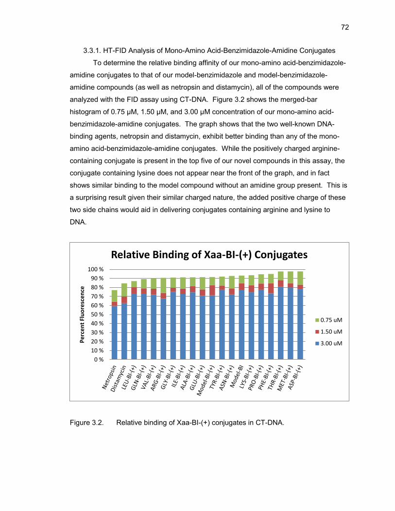
72
3.3.1. HT-FID Analysis of Mono-Amino Acid-Benzimidazole-Amidine Conjugates
To determine the relative binding affinity of our mono-amino acid-benzimidazole-
amidine conjugates to that of our model-benzimidazole and model-benzimidazole-
amidine compounds (as well as netropsin and distamycin), all of the compounds were
analyzed with the FID assay using CT-DNA. Figure 3.2 shows the merged-bar
histogram of 0.75 µM, 1.50 µM, and 3.00 µM concentration of our mono-amino acid-
benzimidazole-amidine conjugates. The graph shows that the two well-known DNA-
binding agents, netropsin and distamycin, exhibit better binding than any of the mono-
amino acid-benzimidazole-amidine conjugates. While the positively charged arginine-
containing conjugate is present in the top five of our novel compounds in this assay, the
conjugate containing lysine does not appear near the front of the graph, and in fact
shows similar binding to the model compound without an amidine group present. This is
a surprising result given their similar charged nature, the added positive charge of these
two side chains would aid in delivering conjugates containing arginine and lysine to
DNA.
Figure 3.2. Relative binding of Xaa-BI-(+) conjugates in CT-DNA.
0 %
10 %
20 %
30 %
40 %
50 %
60 %
70 %
80 %
90 %
100 %
Pe
rce
nt
Flu
ore
sce
nce
Relative Binding of Xaa-BI-(+) Conjugates
0.75 uM
1.50 uM
3.00 uM
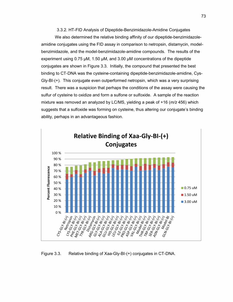
73
3.3.2. HT-FID Analysis of Dipeptide-Benzimidazole-Amidine Conjugates
We also determined the relative binding affinity of our dipeptide-benzimidazole-
amidine conjugates using the FID assay in comparison to netropsin, distamycin, model-
benzimidazole, and the model-benzimidazole-amidine compounds. The results of the
experiment using 0.75 µM, 1.50 µM, and 3.00 µM concentrations of the dipeptide
conjugates are shown in Figure 3.3. Initially, the compound that presented the best
binding to CT-DNA was the cysteine-containing dipeptide-benzimidazole-amidine, Cys-
Gly-BI-(+). This conjugate even outperformed netropsin, which was a very surprising
result. There was a suspicion that perhaps the conditions of the assay were causing the
sulfur of cysteine to oxidize and form a sulfone or sulfoxide. A sample of the reaction
mixture was removed an analyzed by LC/MS, yielding a peak of +16 (m/z 456) which
suggests that a sulfoxide was forming on cysteine, thus altering our conjugate’s binding
ability, perhaps in an advantageous fashion.
Figure 3.3. Relative binding of Xaa-Gly-BI-(+) conjugates in CT-DNA.
0 %
10 %
20 %
30 %
40 %
50 %
60 %
70 %
80 %
90 %
100 %
Pe
rce
nt
Flu
ore
sce
nce
Relative Binding of Xaa-Gly-BI-(+) Conjugates
0.75 uM
1.50 uM
3.00 uM
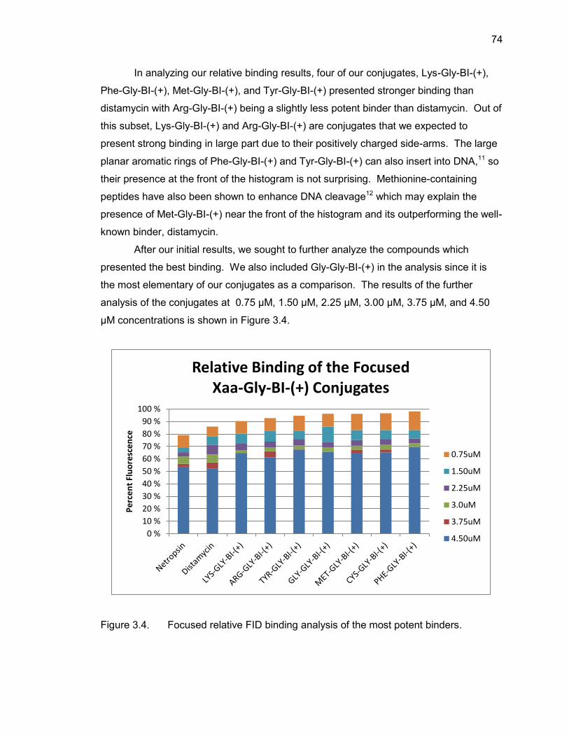
74
In analyzing our relative binding results, four of our conjugates, Lys-Gly-BI-(+),
Phe-Gly-BI-(+), Met-Gly-BI-(+), and Tyr-Gly-BI-(+) presented stronger binding than
distamycin with Arg-Gly-BI-(+) being a slightly less potent binder than distamycin. Out of
this subset, Lys-Gly-BI-(+) and Arg-Gly-BI-(+) are conjugates that we expected to
present strong binding in large part due to their positively charged side-arms. The large
planar aromatic rings of Phe-Gly-BI-(+) and Tyr-Gly-BI-(+) can also insert into DNA,11 so
their presence at the front of the histogram is not surprising. Methionine-containing
peptides have also been shown to enhance DNA cleavage12 which may explain the
presence of Met-Gly-BI-(+) near the front of the histogram and its outperforming the well-
known binder, distamycin.
After our initial results, we sought to further analyze the compounds which
presented the best binding. We also included Gly-Gly-BI-(+) in the analysis since it is
the most elementary of our conjugates as a comparison. The results of the further
analysis of the conjugates at 0.75 µM, 1.50 µM, 2.25 µM, 3.00 µM, 3.75 µM, and 4.50
µM concentrations is shown in Figure 3.4.
Figure 3.4. Focused relative FID binding analysis of the most potent binders.
0 %
10 %
20 %
30 %
40 %
50 %
60 %
70 %
80 %
90 %
100 %
Pe
rce
nt
Flu
ore
sce
nce
Relative Binding of the Focused Xaa-Gly-BI-(+) Conjugates
0.75uM
1.50uM
2.25uM
3.0uM
3.75uM
4.50uM
75
Upon further analysis, netropsin and distamycin present the strongest DNA-
binding ability as expected and the two conjugates containing positively-charged side
arms, Lys-Gly-BI-(+) and Arg-Gly-BI-(+), have the strongest binding of all our
conjugates. The presence of lysine- and arginine-containing conjugates at the front of
the histogram, outperforming all of our other dipeptide-conjugates is expected as they
both contain a positively charged side-arm that should aid in delivering the conjugate to
A/T-rich regions of DNA. It is possible that our initial analysis did not show these
expected results due to an operator error in addition of the compounds to the well or that
over the course of the incubation, the conjugates were oxidized as seen with Cys-Gly-BI-
(+) or they decomposed in some manner, skewing the expected results.
3.4. Summary
The dipeptide-benzimidazole-amidine conjugates appear to have better DNA-
binding ability than the mono-amino acid-benzimidazole-amidine conjugates as a couple
of these conjugates actually outperformed distamycin in the FID assay. Upon further
review, it does appear that both netropsin and distamycin are superior to any of our
conjugates, as is expected. However, having conjugates that even come close to the
DNA binding ability of these two compounds speaks to the great power of the amino
acid-benzimidazole-amidine system and may warrant further synthesis and analysis to
find stronger dipeptides and possibly tripeptides. It also needs to be noted that the
assay using CT-DNA only shows relative binding compared to each other compound
and in no way shows site preference since CT-DNA is made up of a mixture of possible
base-pair sequences. Also, CT-DNA is a mixture of A/T base-pairs to G/C base-pairs,
thus the known A/T-binders netropsin and distamycin will only be able to bind to a
portion of the available DNA. This may also apply to our conjugates as they are
designed to be A/T-binders, but further analysis of the site-selectivity of each conjugate
will be needed and is beyond the scope of the analysis that we intended to perform.
76
3.5. Experimental Protocols
3.5.1. Materials
Netropsin (ε296 = 21,500 M-1cm-1) and distamycin (ε303 = 37,000 M-1cm-1) were
purchased from Sigma-Aldrich and quantitated spectroscopically and materials used in
buffer solution, NaCl and tris(hydroxymethyl)-aminomethane (TRIS), were also
purchased from Sigma-Aldrich. Ethidium bromide (ε476 = 5680 M-1cm-1) was purchased
from Fluka and quantitated spectroscopically. Calf thymus DNA (ε260 = 6300 M-1cm-1)
was purchased from Invitrogen and the concentration of the CT-DNA was confirmed by
ultraviolet-visible spectroscopy. All amino acid-benzimidazole-amidine conjugates were
synthesized through previously detailed solid-phase protocols in our laboratory. All
experiments were carried out on a Varian Cary Eclipse Spectrophotometer fluorescence
plate reader.
3.5.2. HT-FID Assay
Each well of a Costar black 96-well plate was loaded with TRIS buffer along with
ethidium bromide (150 μL of 5.5 μM EtBr, 120 mM NaCl, and 12 mM TRIS). Calf
thymus DNA was then added to each well (25 μL of 11.1 μM CT-DNA in H2O). Final
concentrations in each well were 1.5 μM CT-DNA, 4.5 μM EtBr, and 0.75 to 3.00 μM of
DNA-binding agent. The final buffer solution consists of 100 mM NaCl, 10 mM TRIS, pH
7.4. After incubation at 25 °C for 30 minutes, each well was read (average of three
readings) on the fluorescence plate reader (λEx 545 nm, λEm 595 nm). Compound
assessments were conducted in triplicate with each well acting as its own control well
(no agent = 100% fluorescence, no DNA = 0% fluorescence) and fluorescence readings
are reported as a percent fluorescence decrease relative to the control wells.
The above protocol was followed for the focused analysis of dipeptide-
benzimidazole-amidine conjugates except for the range of compound concentrations
was 0.75 to 4.50 μM.
77
3.6. List of References
1. Glass, L. S.; Bapat, A.; Kelley, M. R.; Georgiadis, M. M.; Long, E. C.; Bioorg. Med.
Chem. Lett. 2010, 20, 1685.
2. Goodwin, K. D.; Lewis, M. A.; Tanious F. A.; Tidwell, R. R.; Wilson, W. D.;
Georgiadis, M. M.; Long, E. C. J. Am. Chem. Soc. 2006, 128, 7846.
3. Lewis, M. A.; Long, E. C. Bioorg. Med. Chem. 2006, 14, 3481.
4. Tanious, F. A.; Laine, W.; Peixoto, P.; Bailly, C.; Goodwin, K. D.; Lewis, M. A.; Long,
E. C.; Georgiadis, M. M.; Tidwell, R. R.; Wilson, W. D. Biochemistry 2007, 46, 6944.
5. Glass, L. S.; Nguyen, B.; Goodwin, K. D.; Dardonville, C.; Wilson, W. D.; Long, E. C.;
Georgiadis, M. M. Biochemistry 2009, 48, 5943.
6. Boger, D. L.; Tse, W. C. Bioorg. Med. Chem. 2001, 9, 2511.
7. Boger, D. L.; Fink, B. E.; Hedrick, M. P. J. Am. Chem. Soc. 2000, 122, 6382.
8. Tse, W. C.; Boger, D. L. Acc. Chem. Res. 2004, 37, 61.
9. Boger, D. L.; Fink, B. E.; Brunette, S. R.; Tse, W. C.; Hedrick, M. P. J. Am. Chem.
Soc. 2001, 123, 5878.
10. Zhang, J. H.; Chung, T. D.; Oldenburg, K. R. J. Biomol. Screen. 1999, 4, 67.
11. Chen, Y.; Rice, P. A. Annu. Rev. Biophys. Biomol. Struct. 2003, 32, 135.
12. Huang, X.; Pieczko, M. E.; Long, E. C. Biochemistry 1999, 38, 2160.

APPENDICES
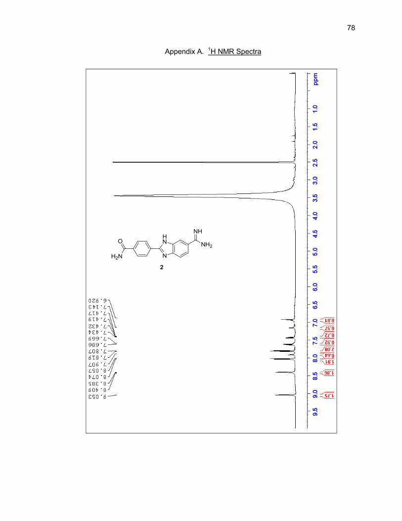
78
Appendix A. 1H NMR Spectra
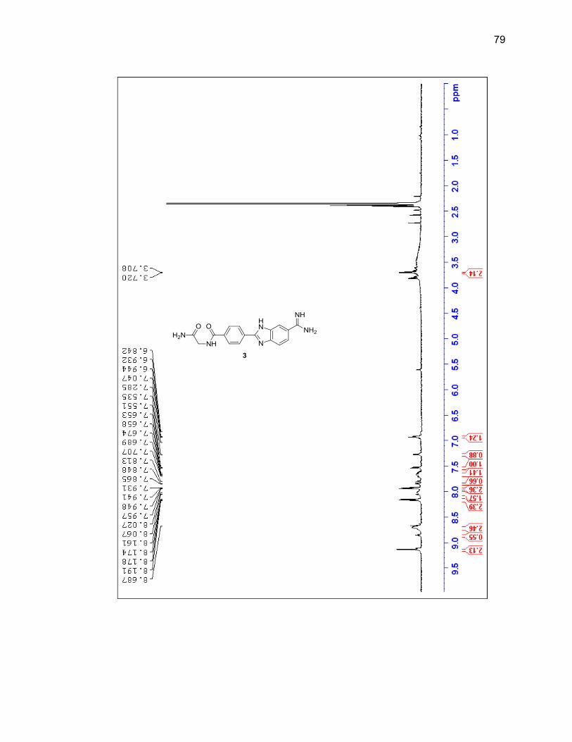
79
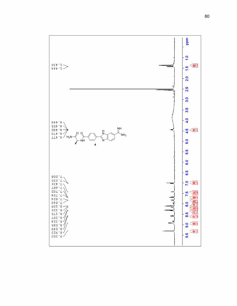
80
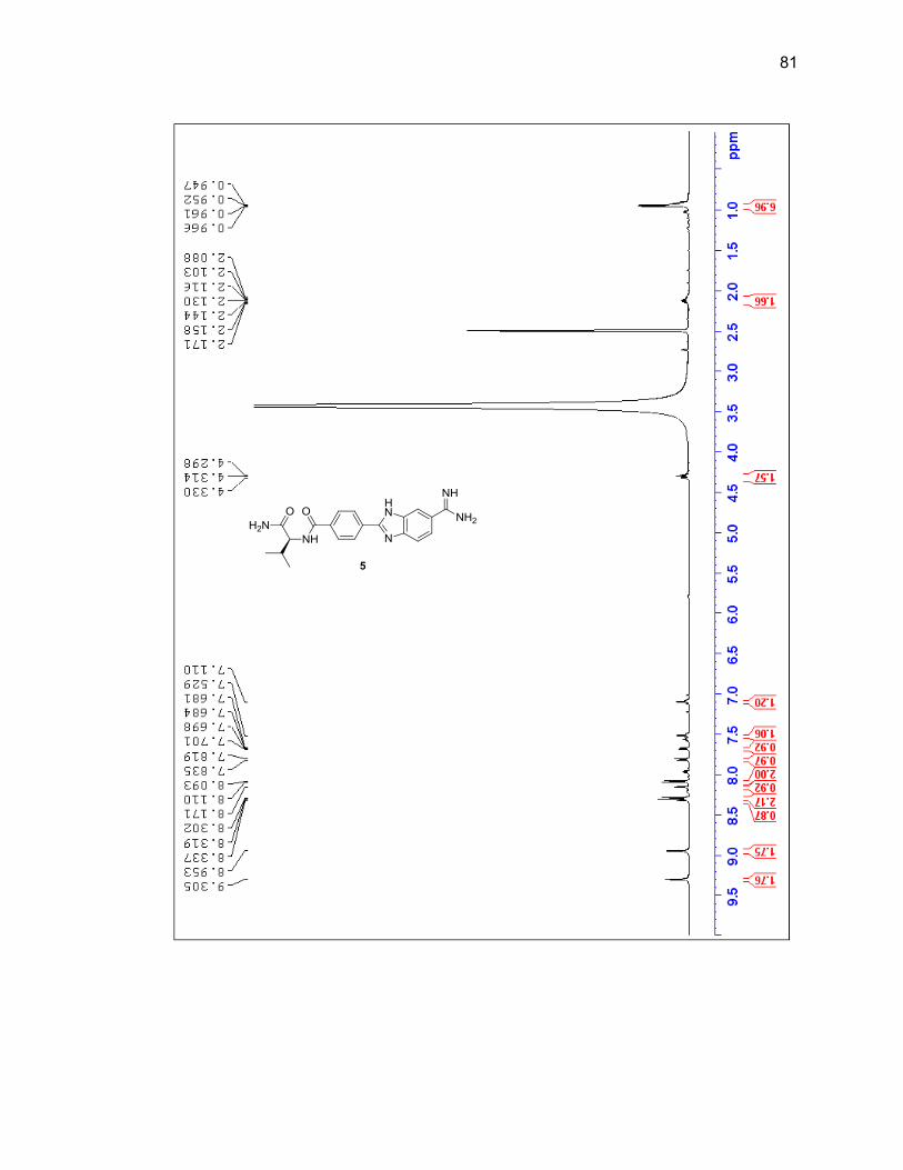
81
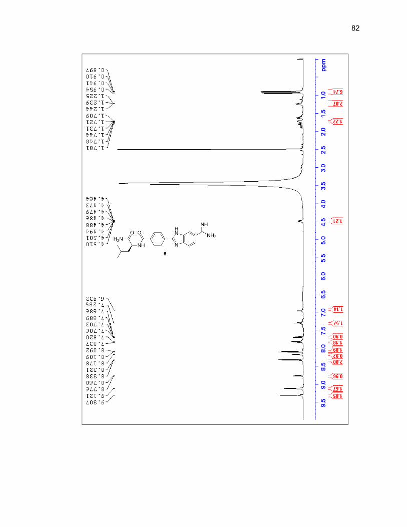
82
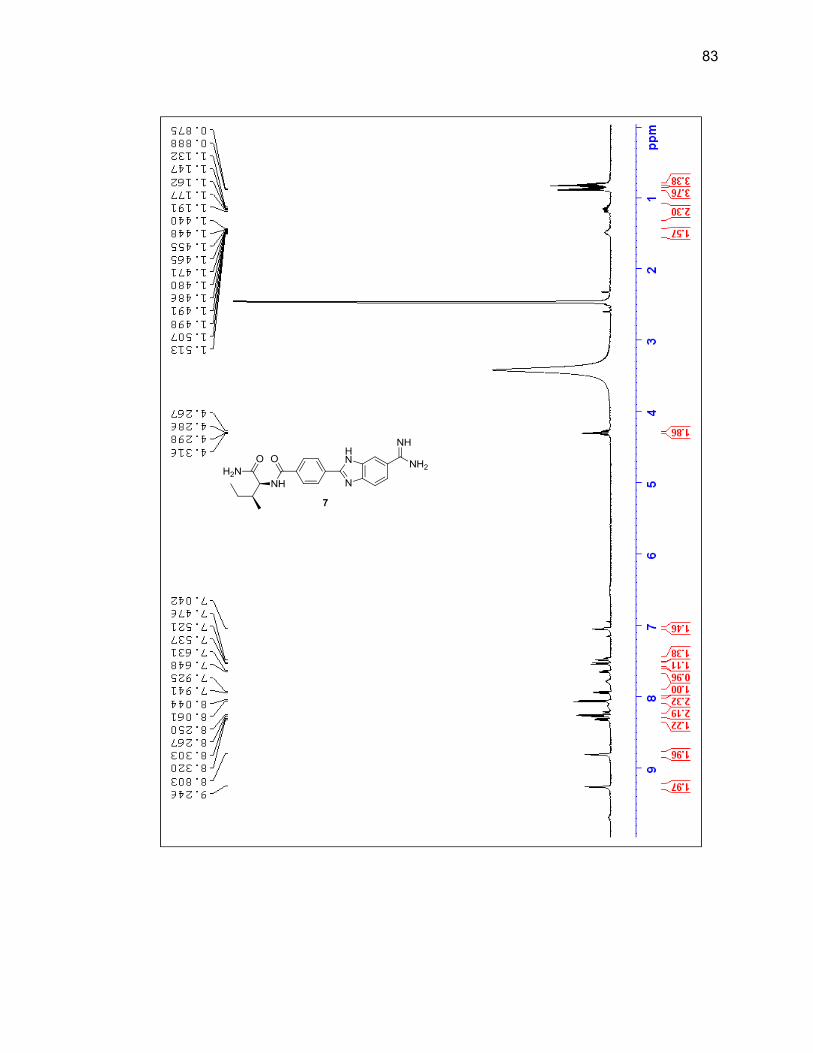
83
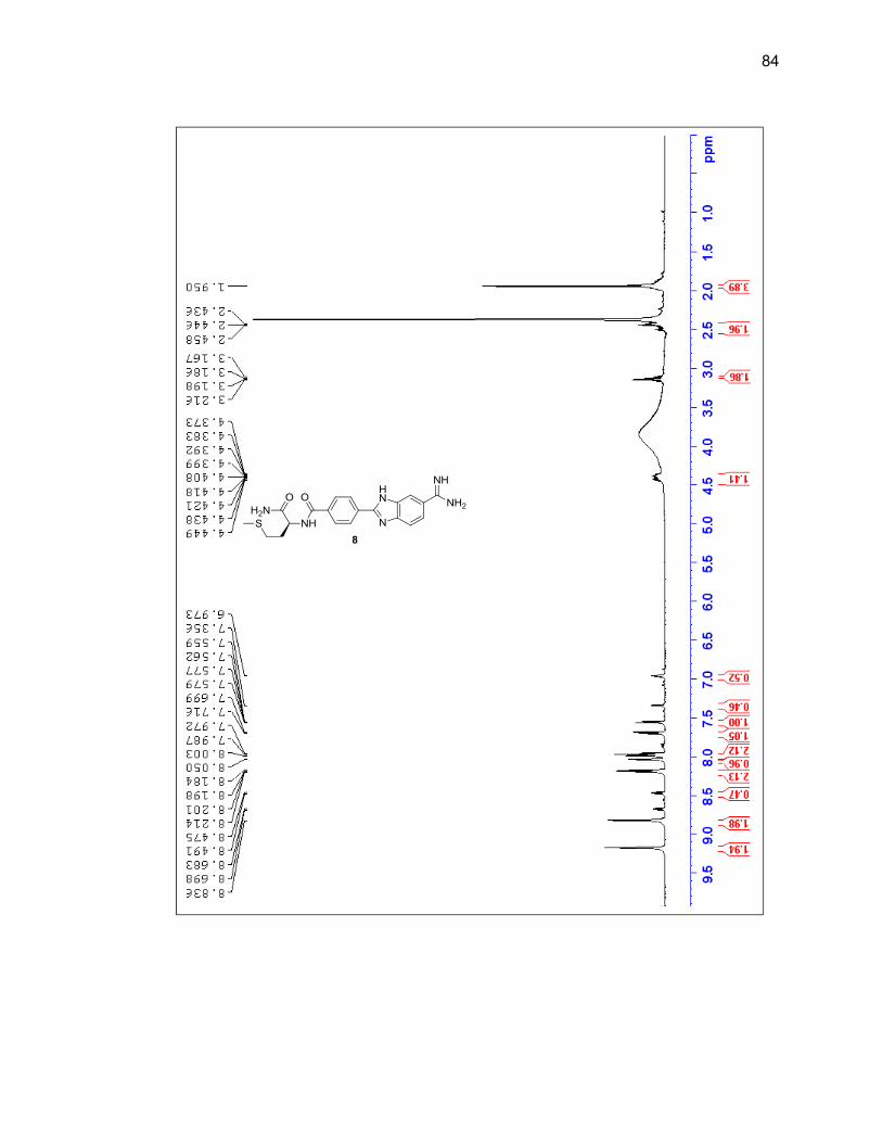
84
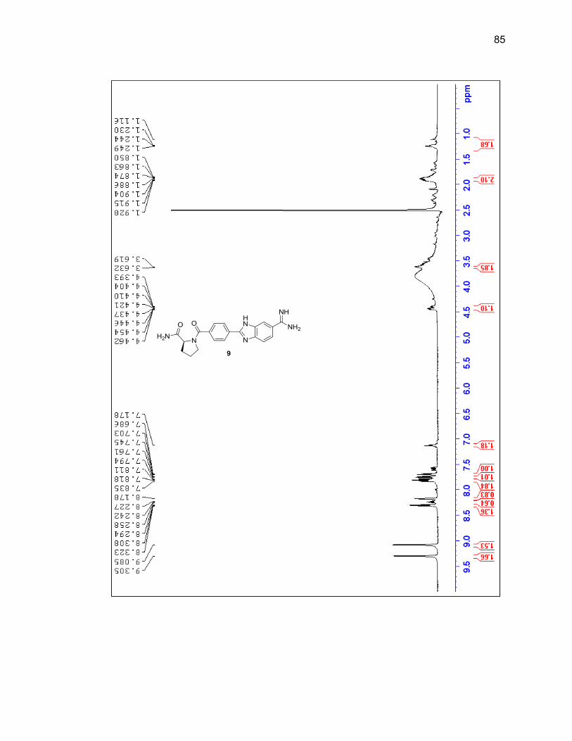
85
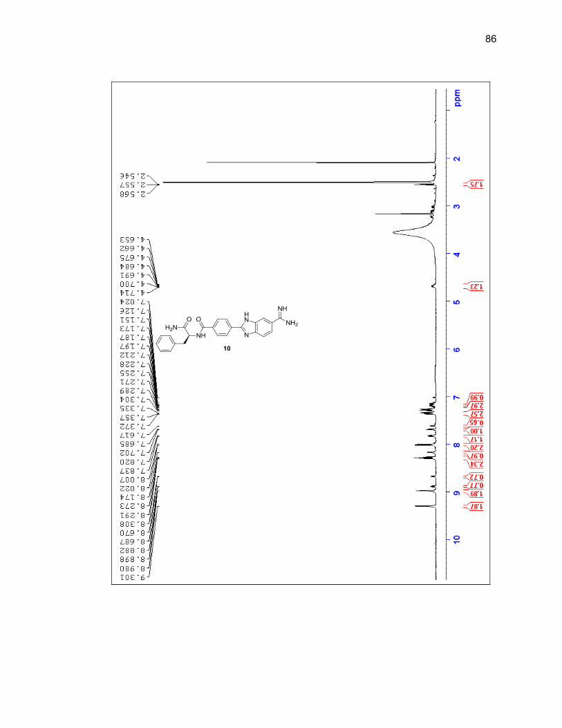
86
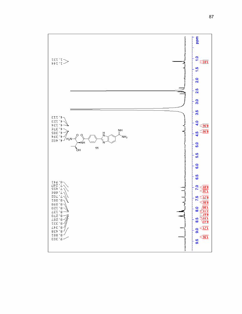
87
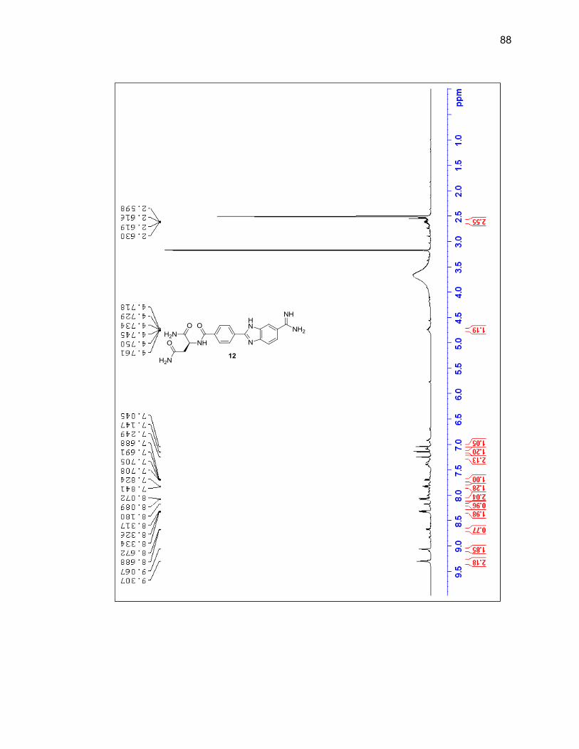
88
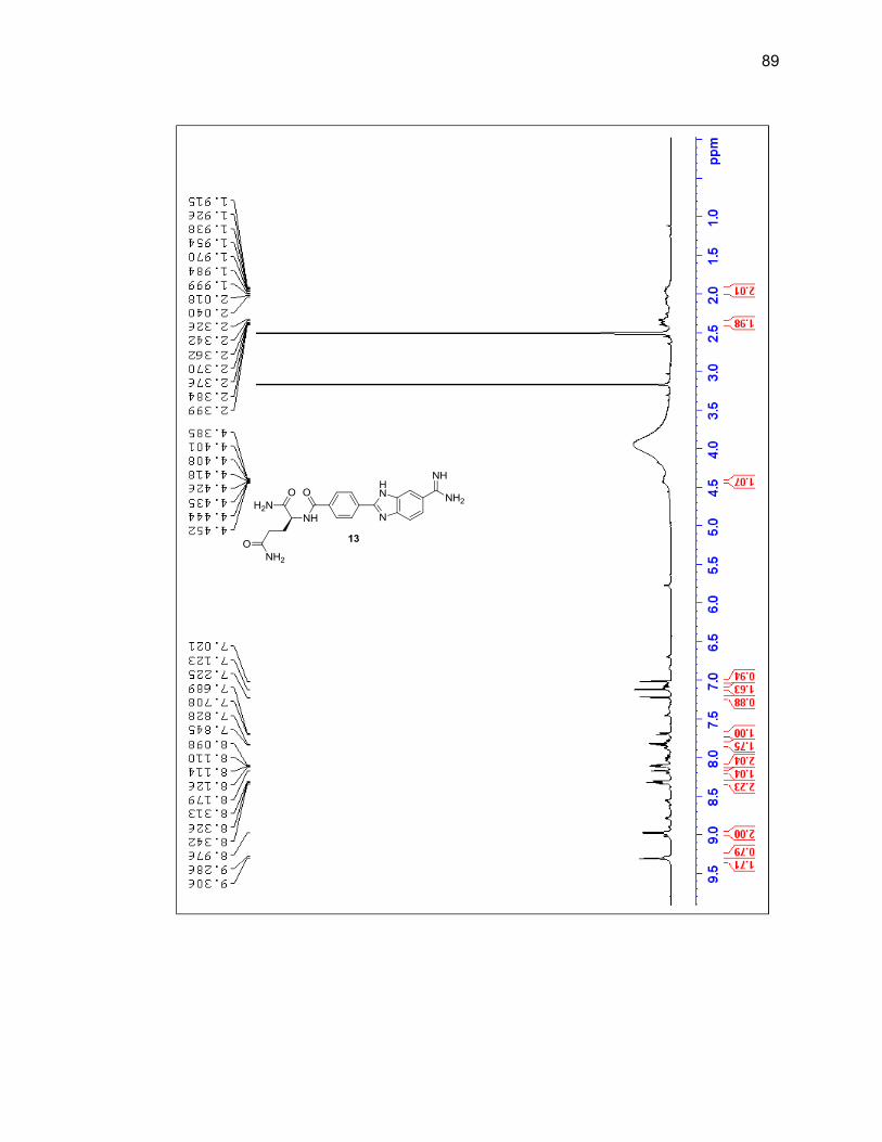
89
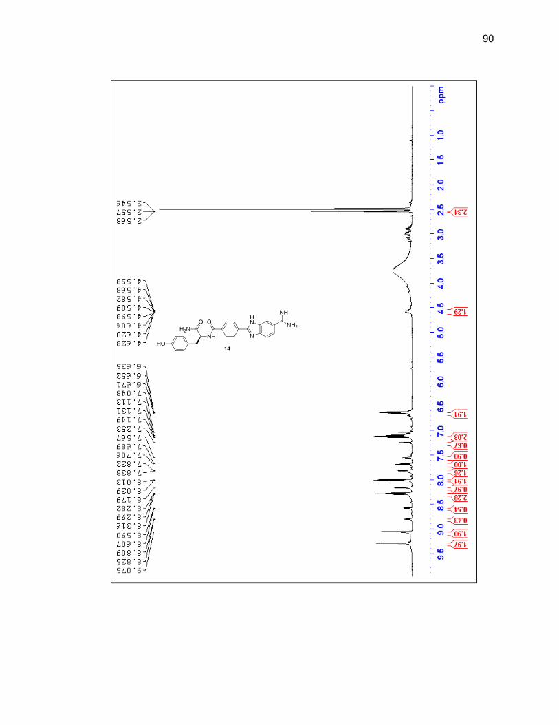
90
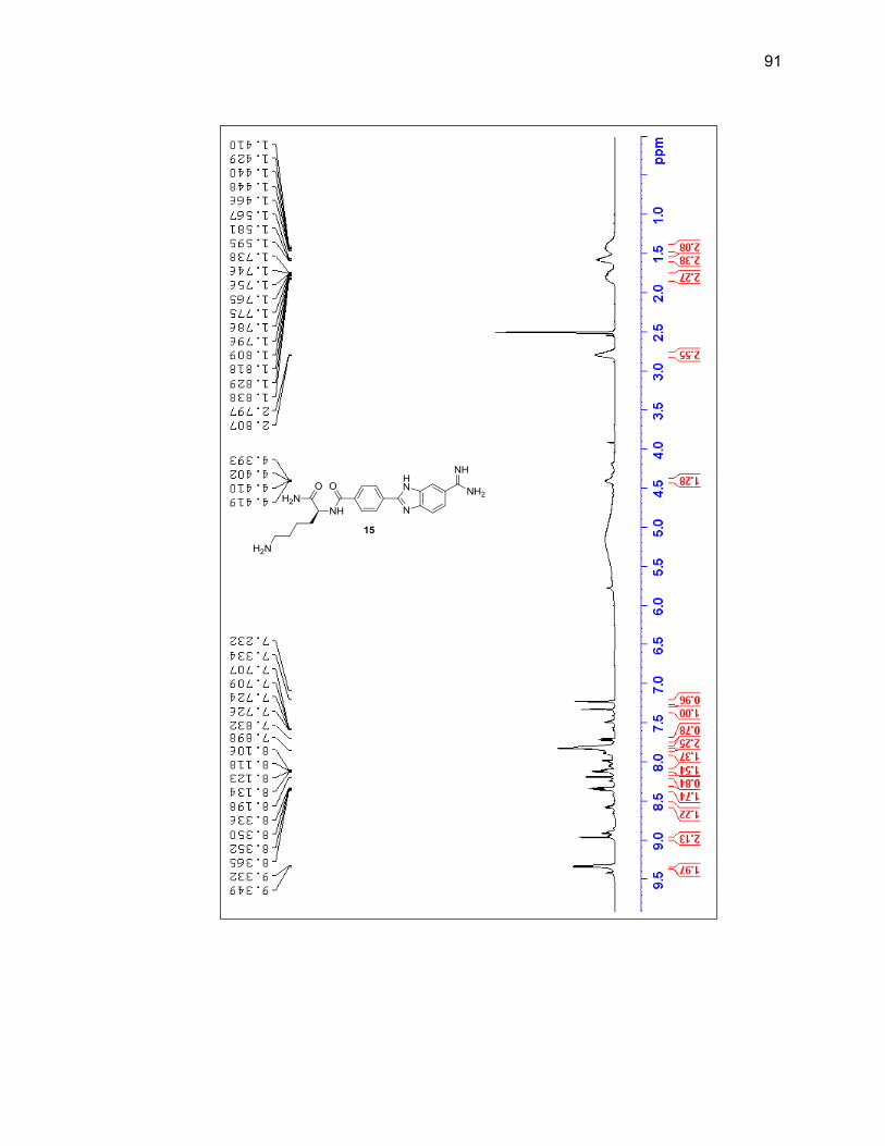
91
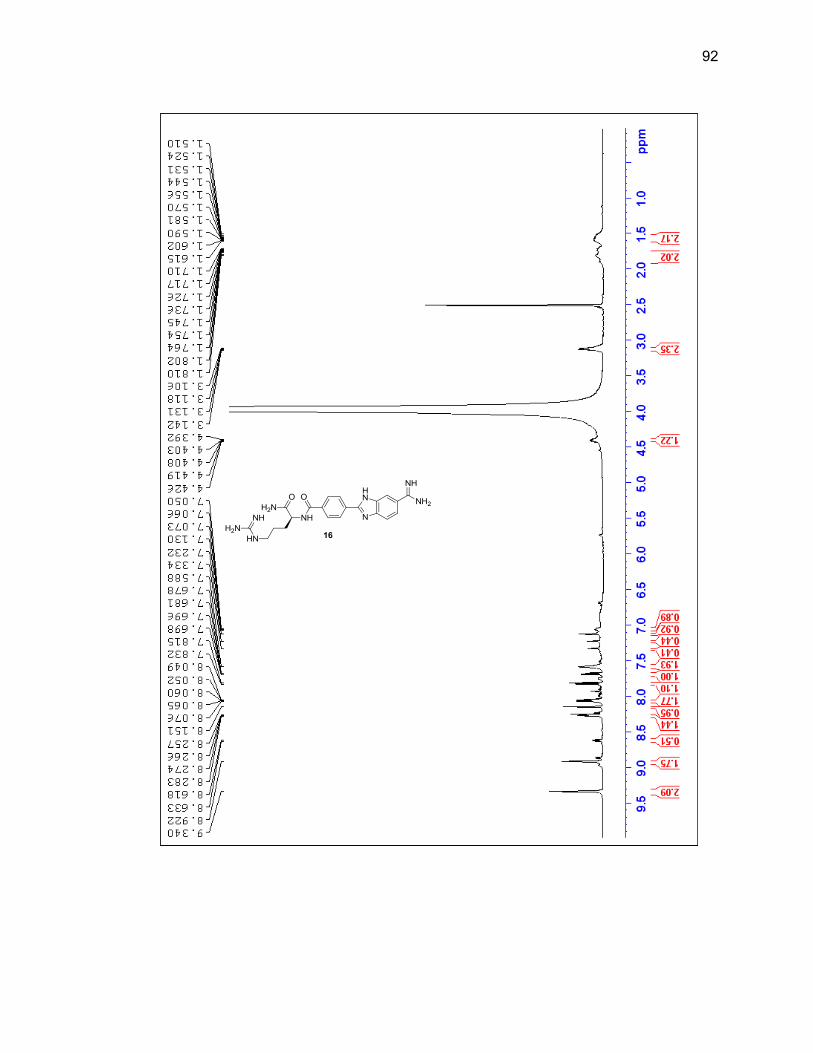
92
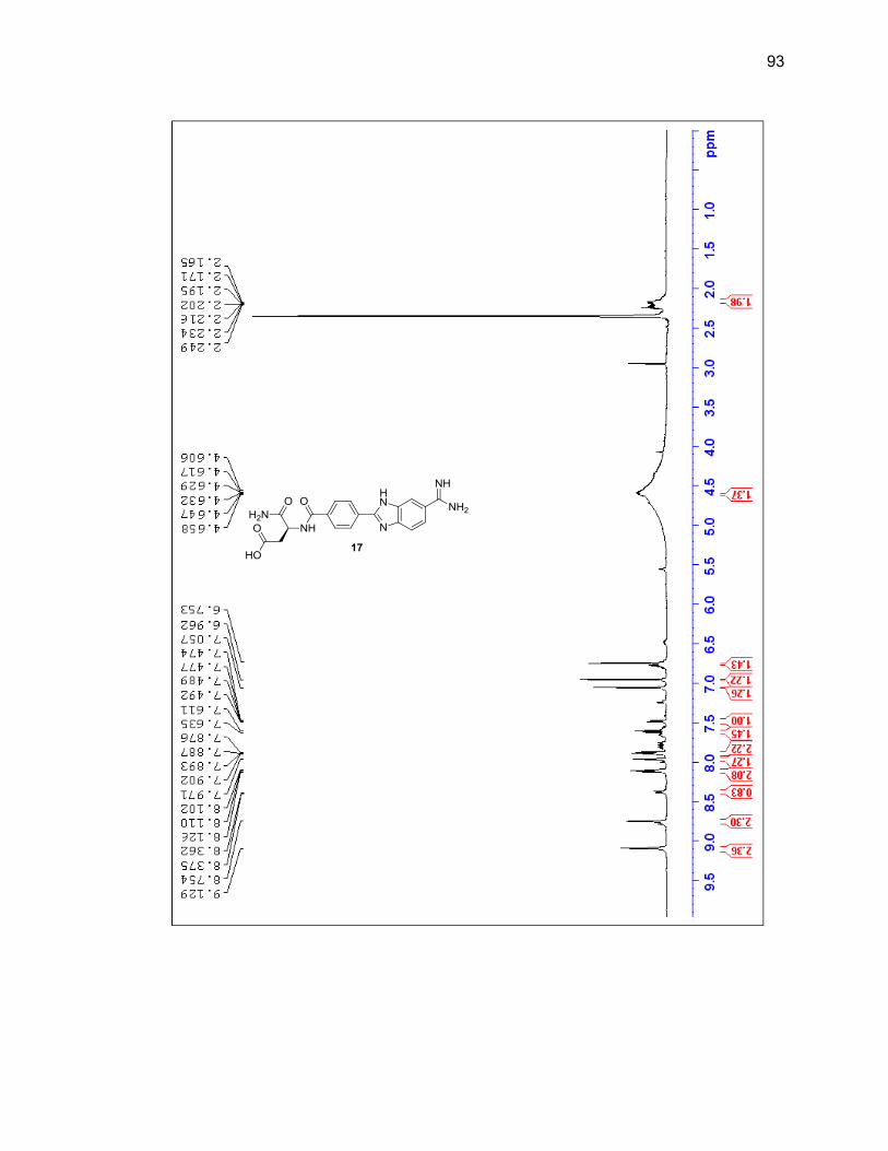
93
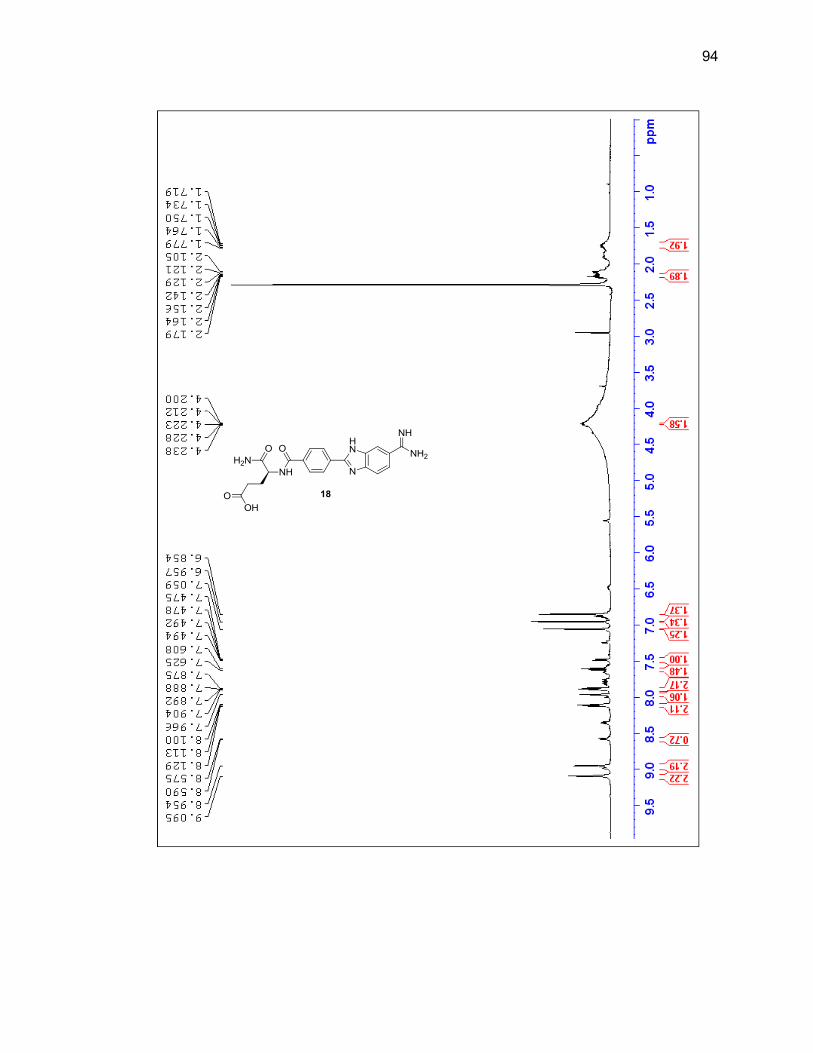
94
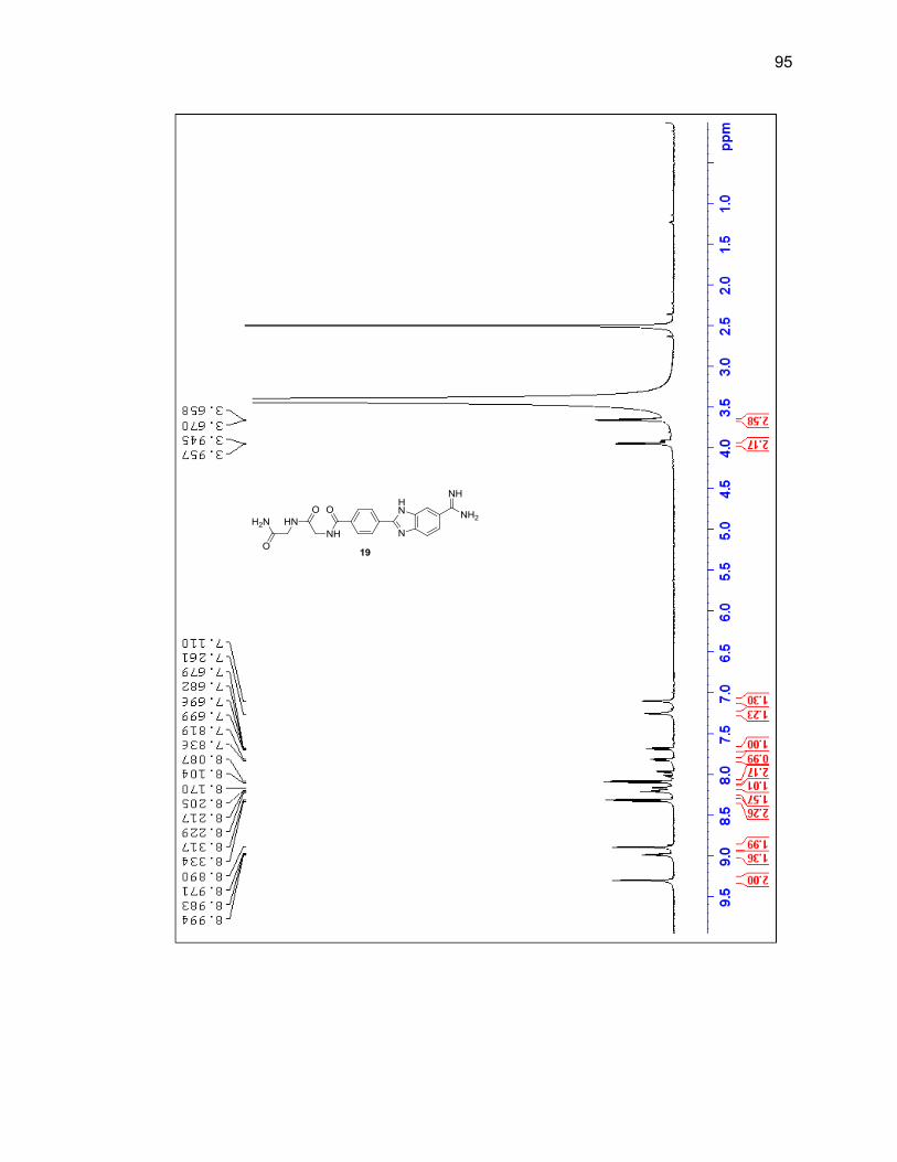
95
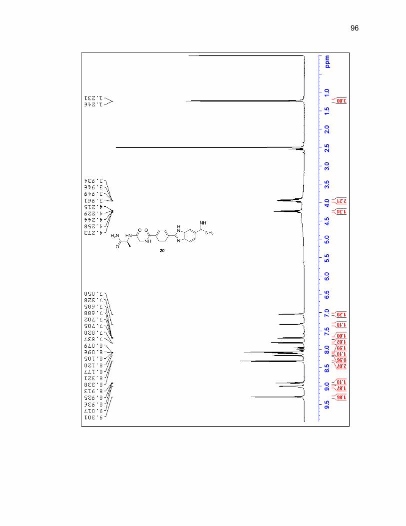
96
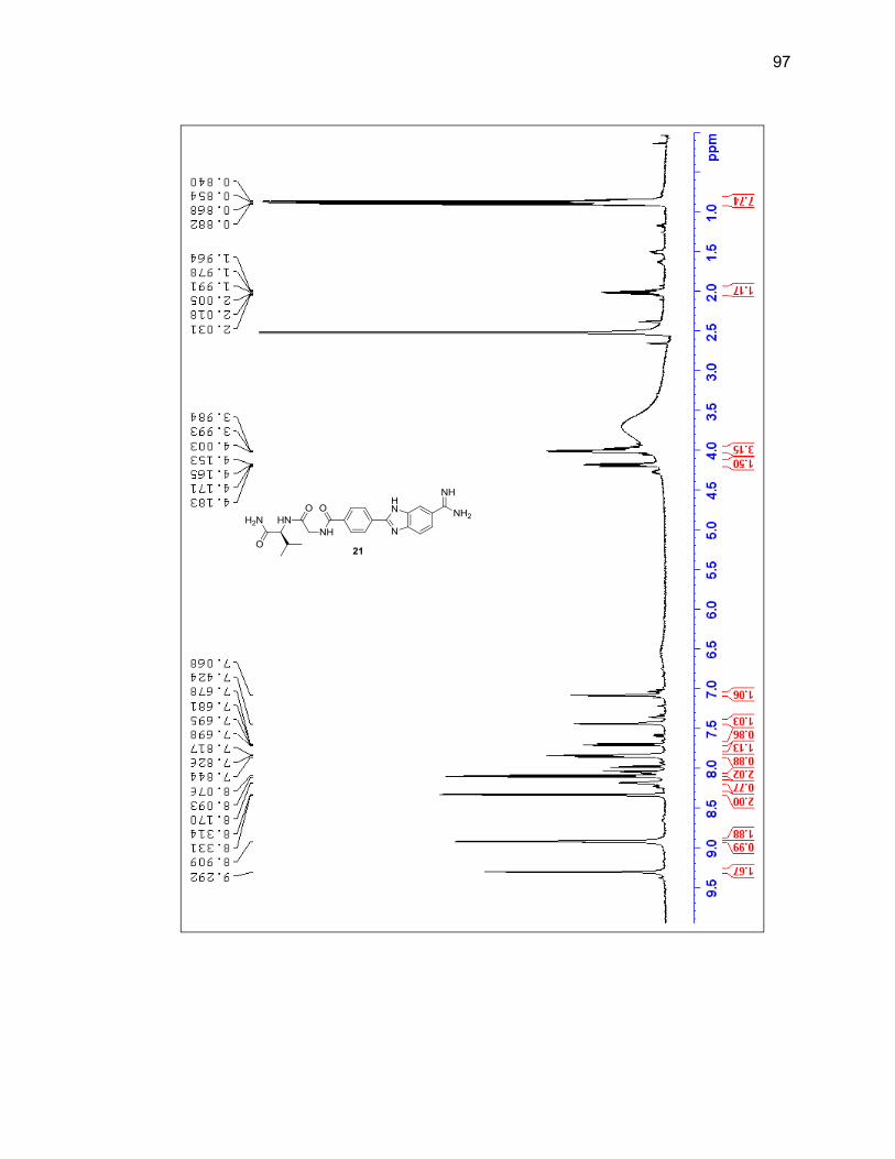
97
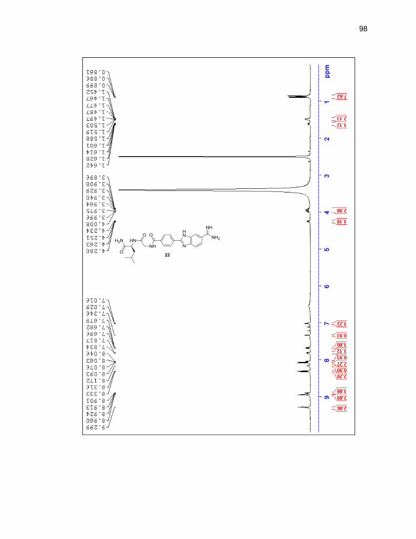
98
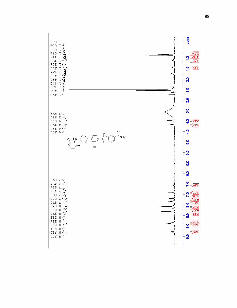
99
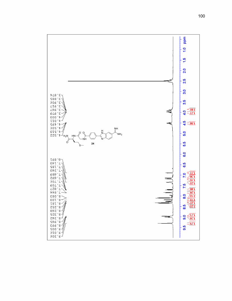
100
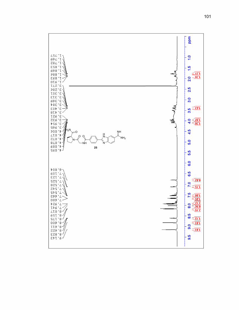
101
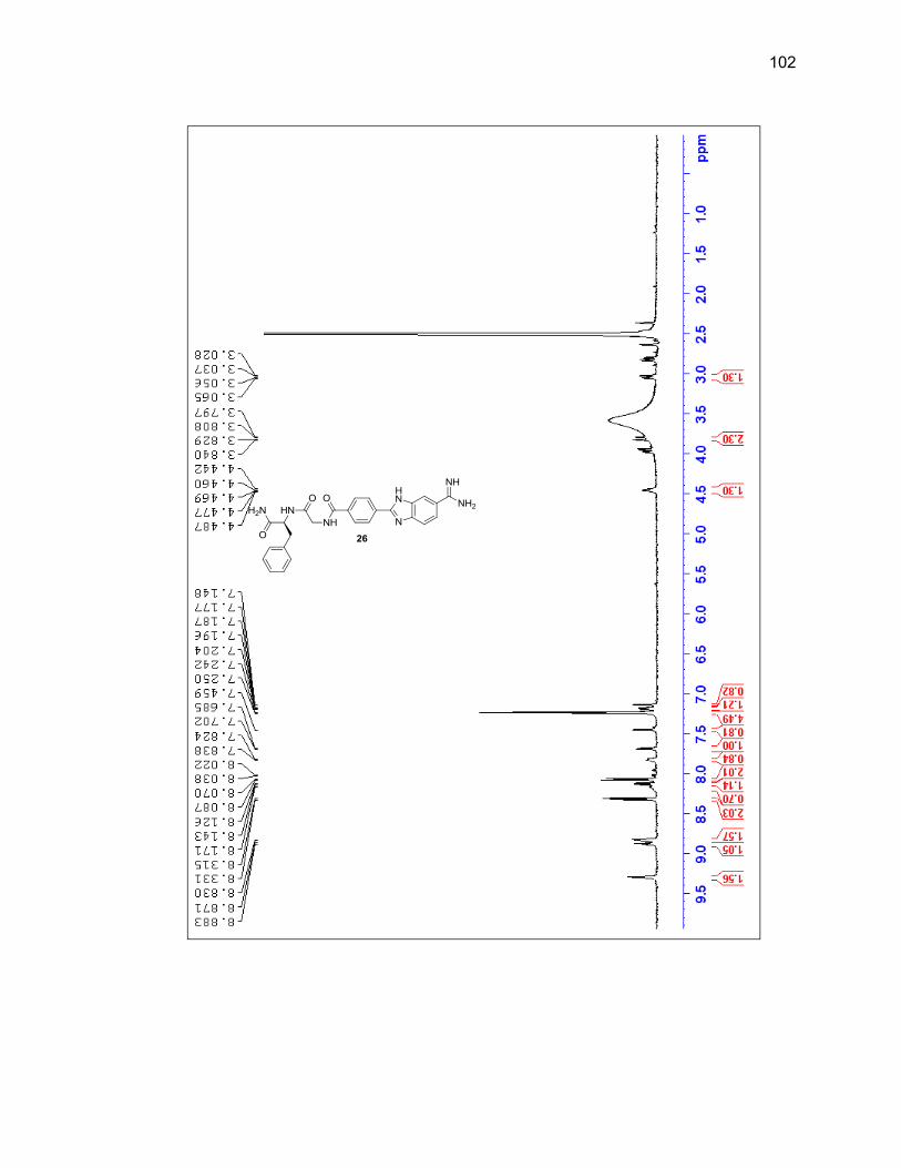
102
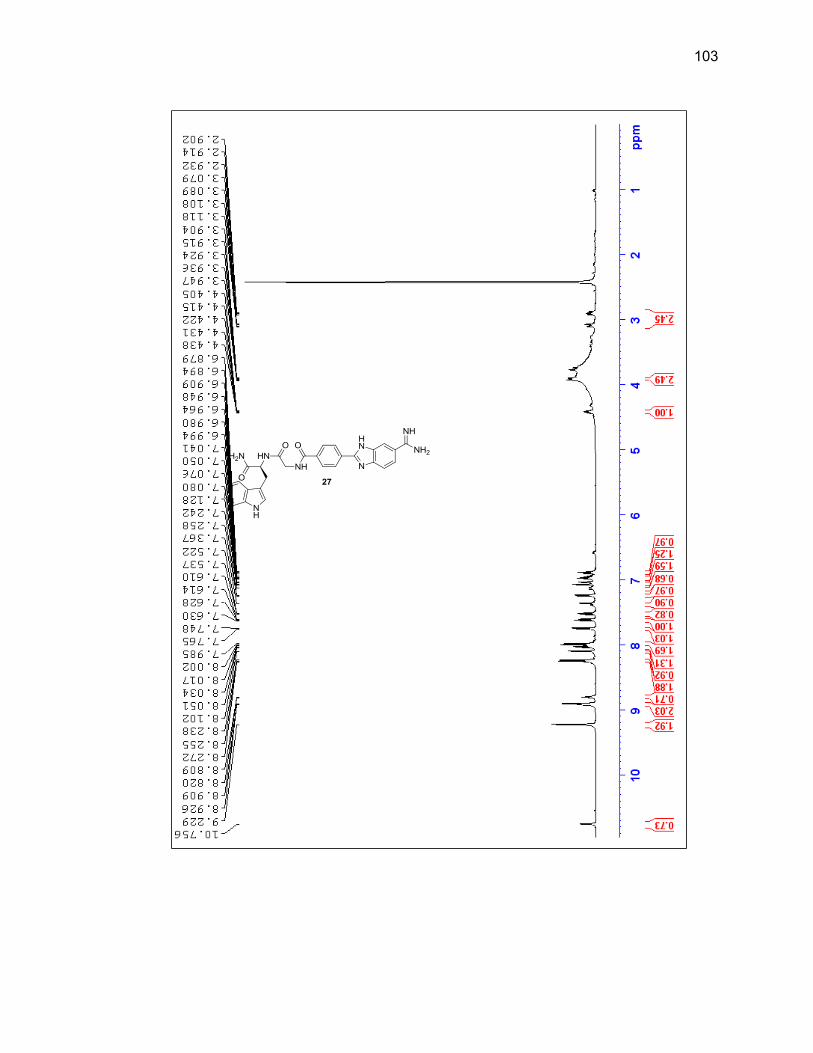
103
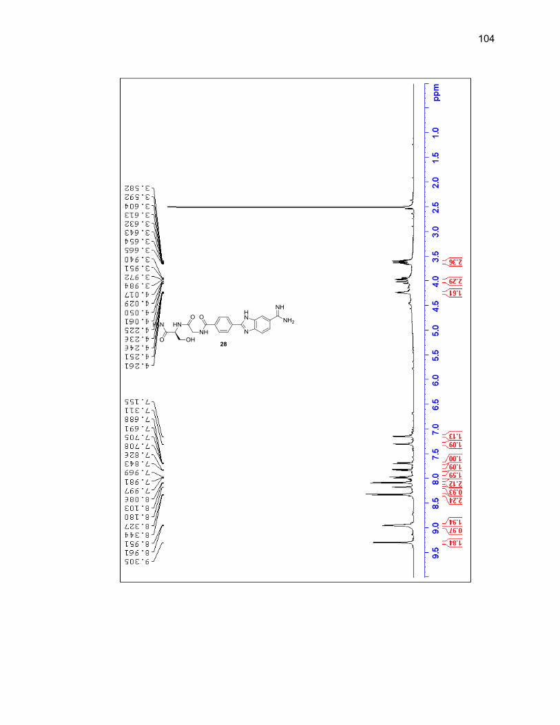
104
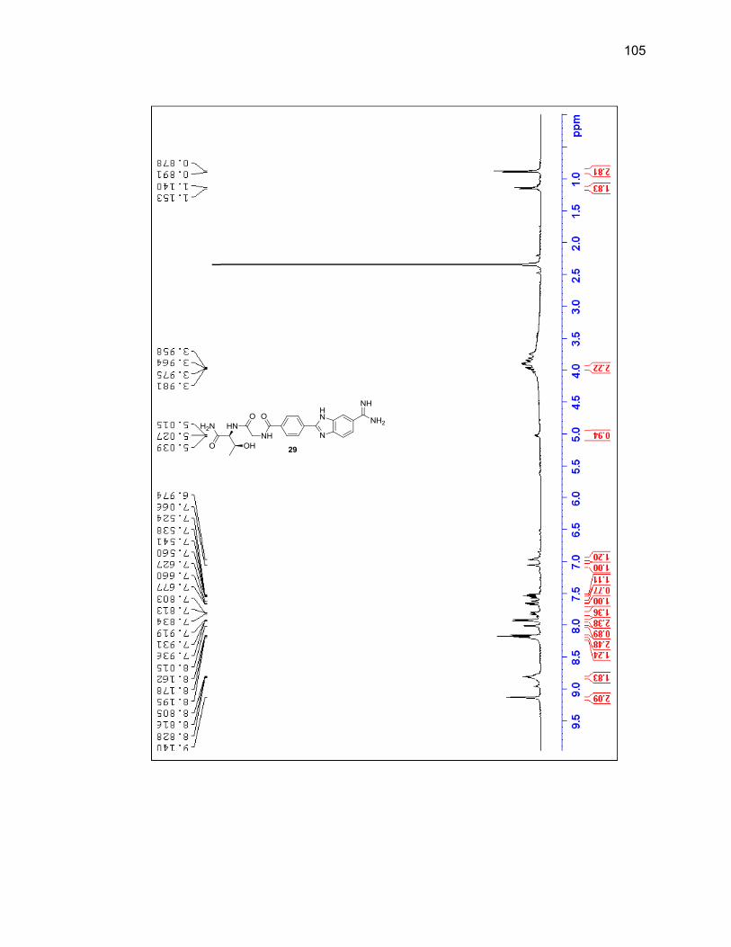
105
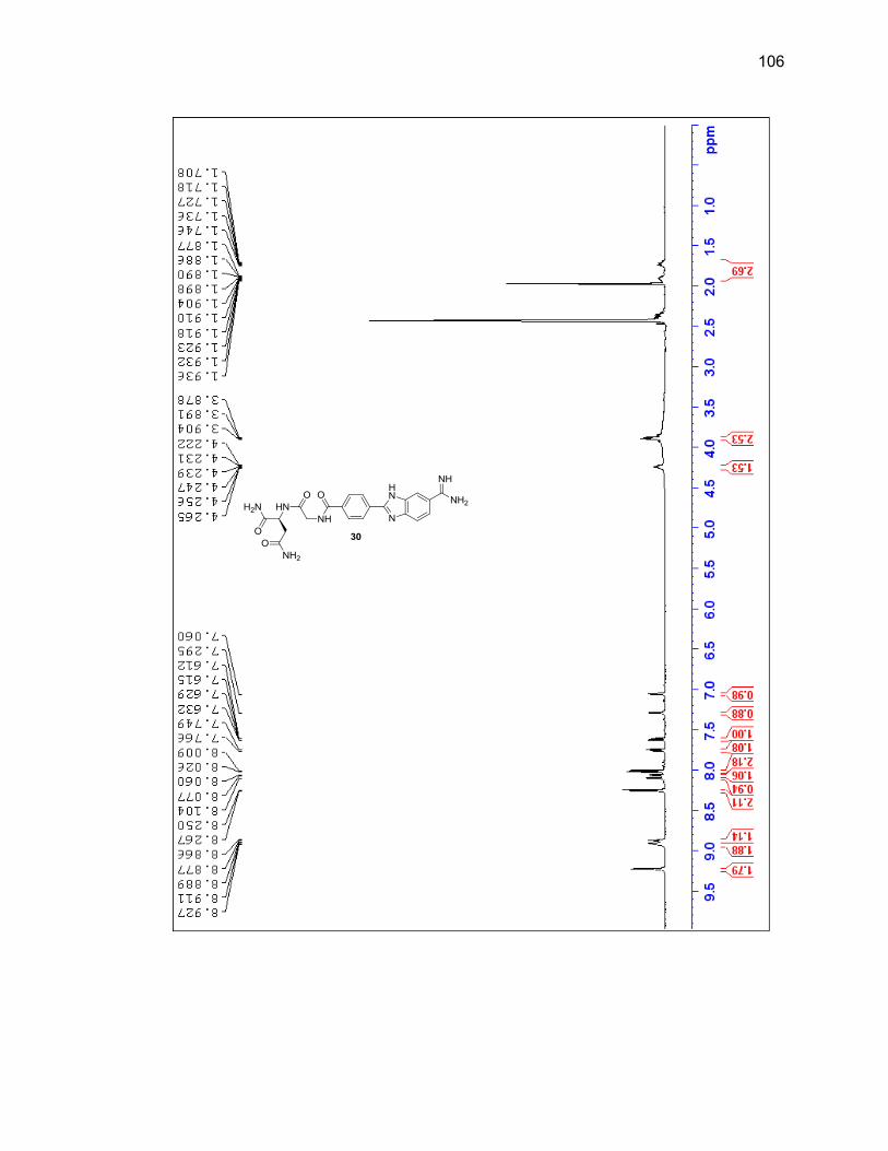
106
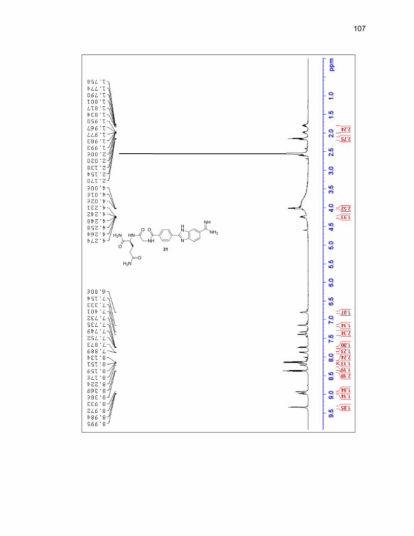
107
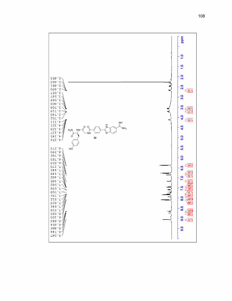
108
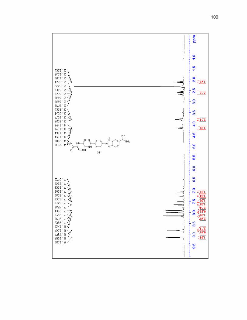
109
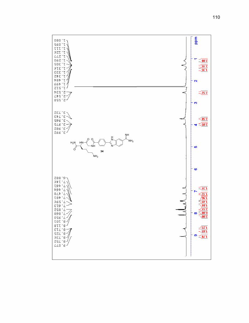
110
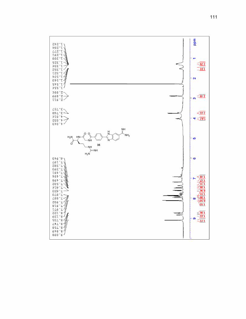
111
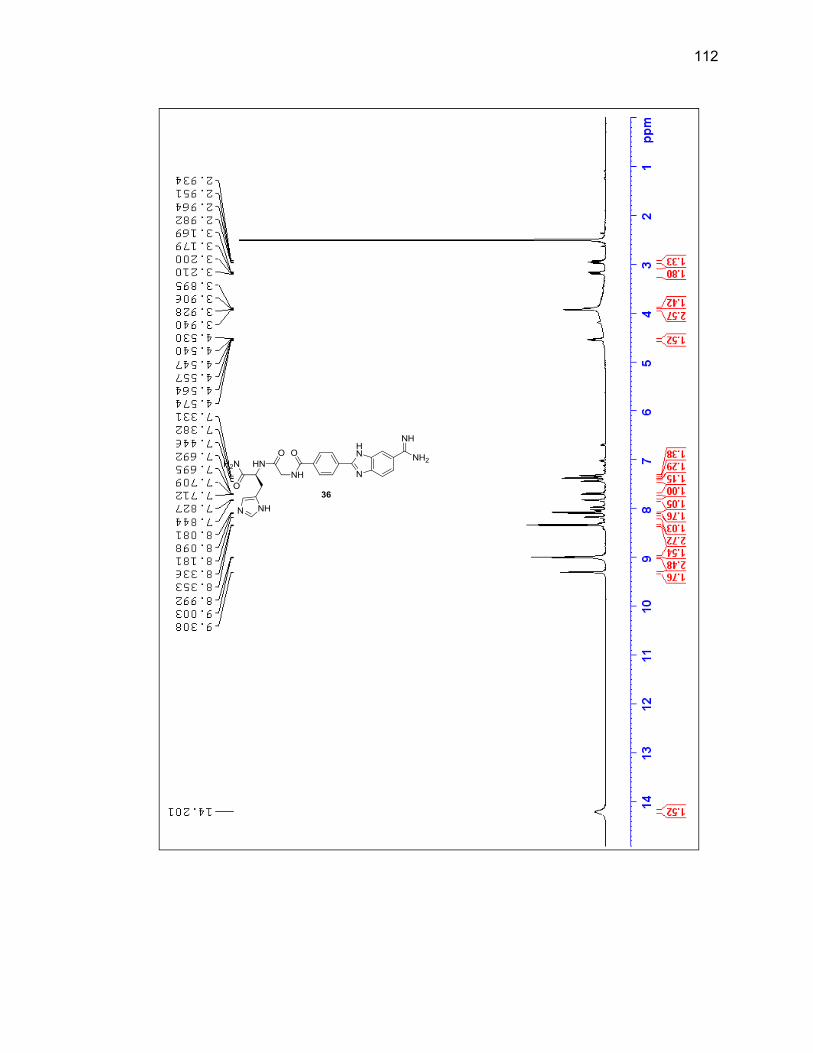
112
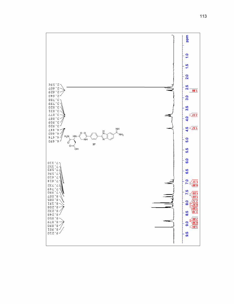
113
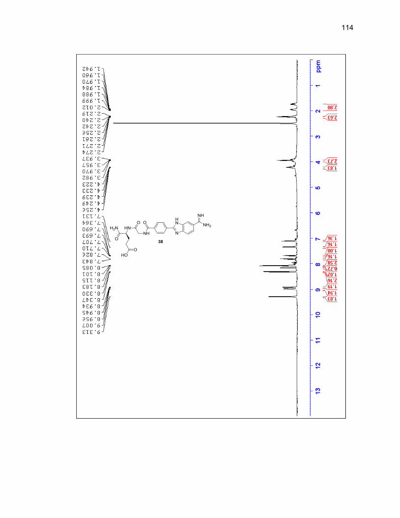
114
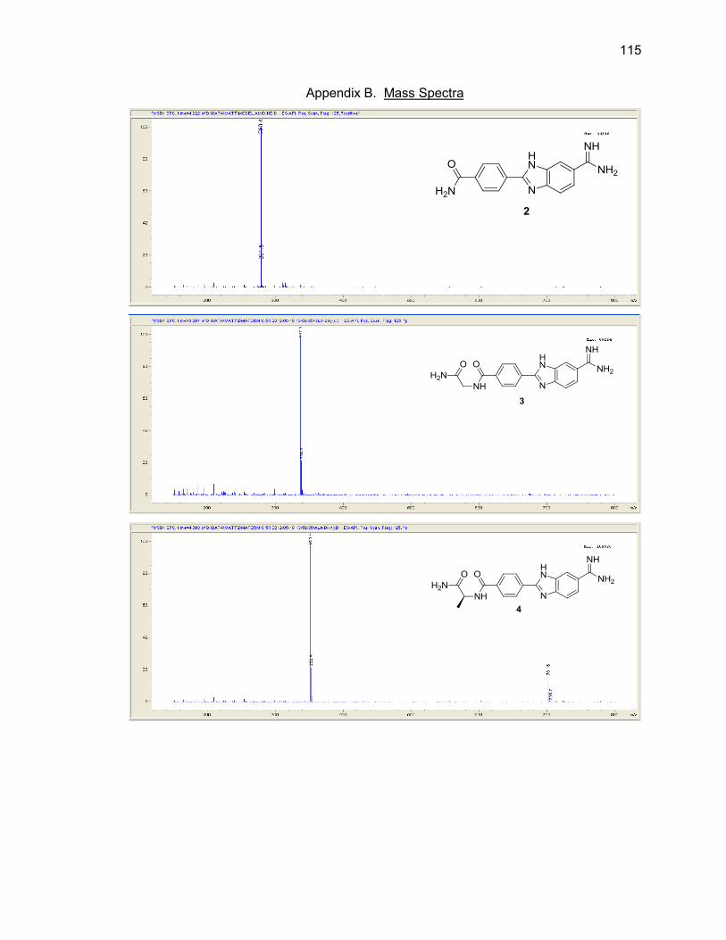
115
Appendix B. Mass Spectra
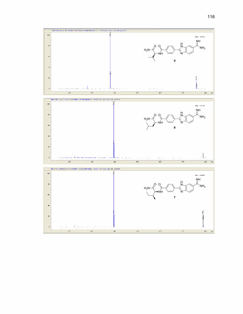
116
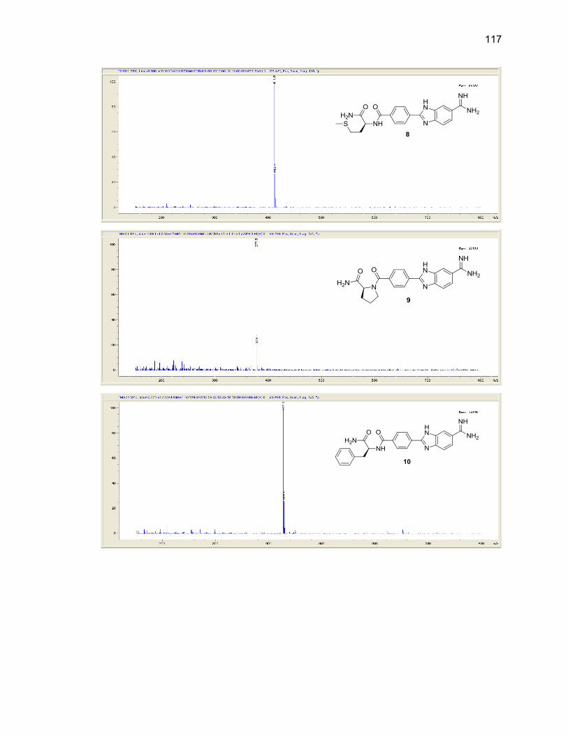
117
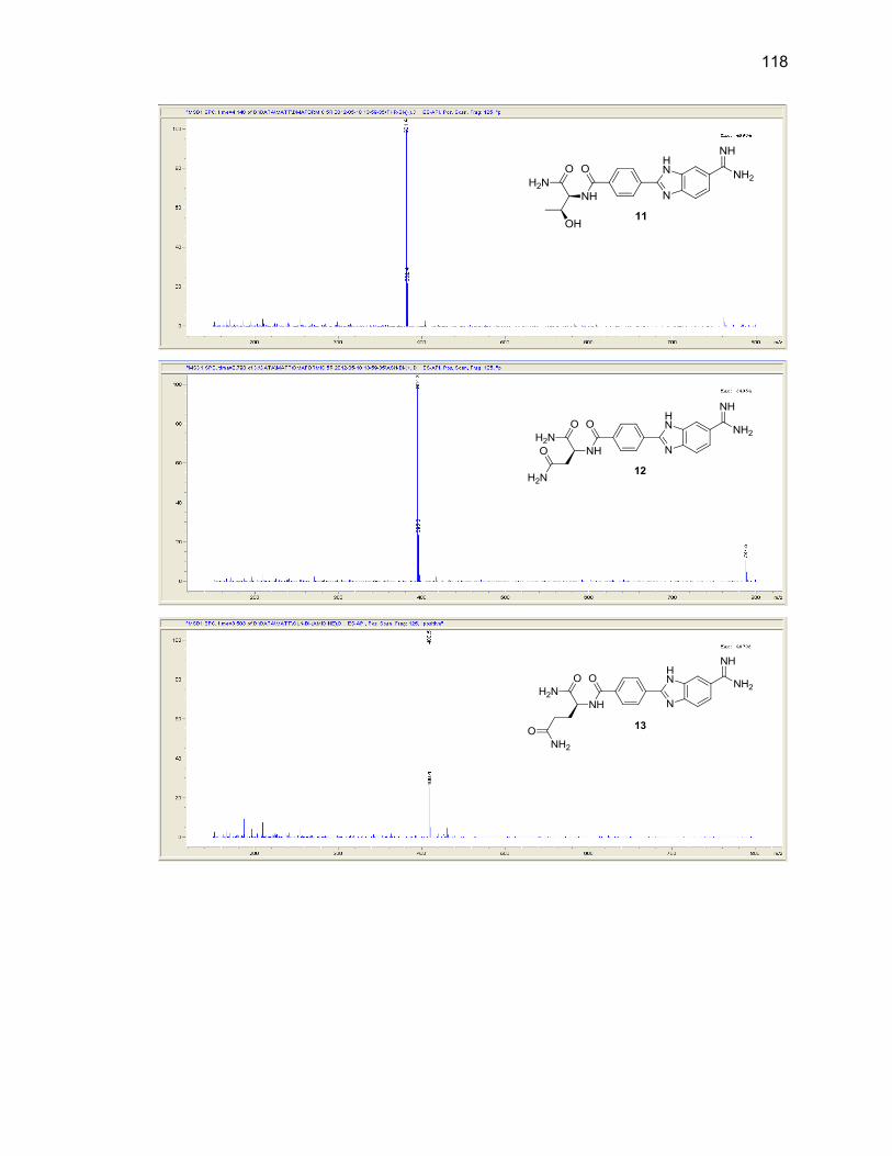
118
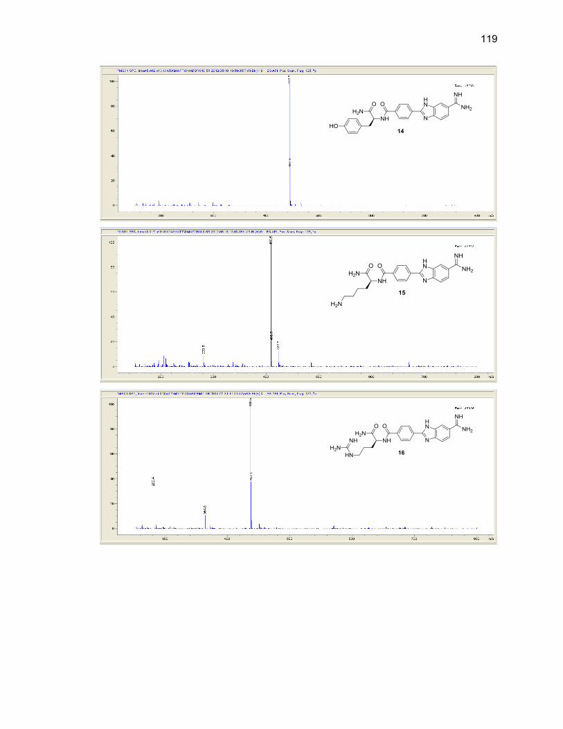
119
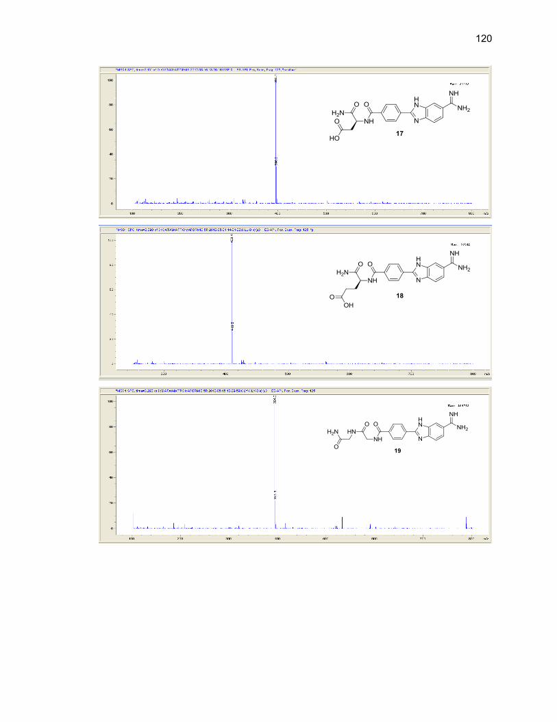
120
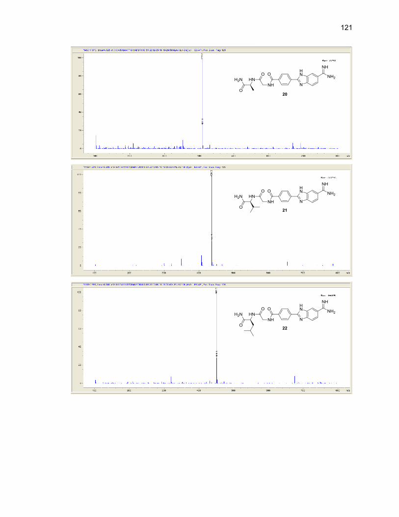
121
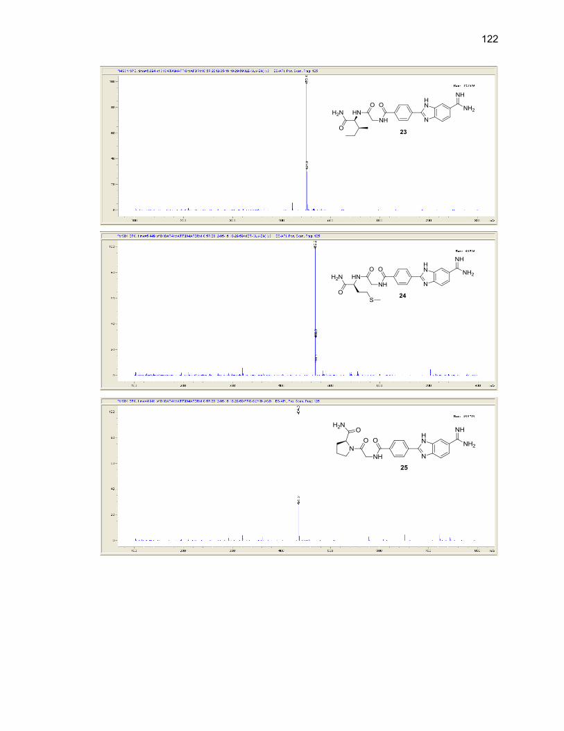
122

123

124
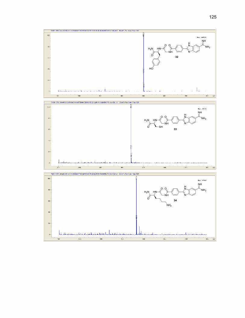
125
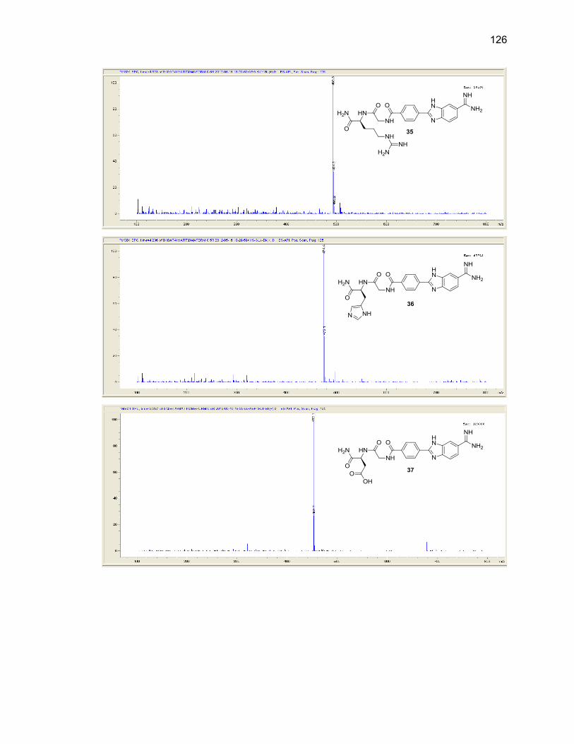
126
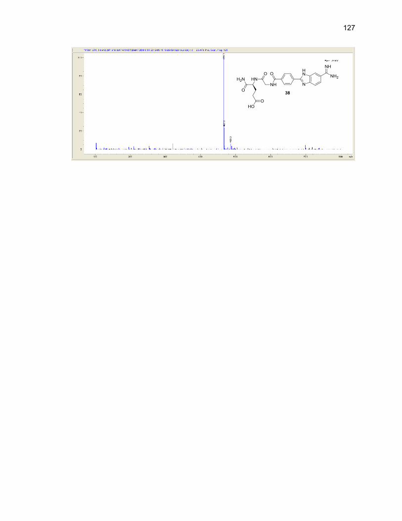
127