an evidence based - iupui scholarworks
TRANSCRIPT
ACCEPTED MANUSCRIPT
ACCEPTED MANUSCRIP
T
Assessing Copy Number Aberrations and Copy Neutral Loss of Heterozygosity Across the
Genome as Best Practice: An Evidence Based Review of Clinical Utility from the Cancer
Genomics Consortium (CGC) Working Group for Myelodysplastic Syndrome,
Myelodysplastic/Myeloproliferative and Myeloproliferative Neoplasms
Rashmi Kanagal-Shamanna1*
Jennelle C. Hodge2
Tracy Tucker3
Shashi Shetty4
Ashwini Yenamandra5
Amanda Dixon-McIver6
Christine Bryke7
Emma Huxley8
Patrick A. Lennon9
Gordana Raca10
Xinjie Xu11
Sally Jeffries8
Fabiola Quintero-Rivera12
Patricia T. Greipp13
Marilyn L. Slovak14
M. Anwar Iqbal14
Min Fang15*
1Department of Hematopathology, The University of Texas M.D. Anderson Cancer Center,
Houston TX, USA
2Department of Medical and Molecular Genetics, Indiana University School of Medicine,
Indianapolis, IN; Department of Pediatrics, University of California Los Angeles, Los Angeles,
CA; Department of Pathology and Laboratory Medicine, Cedars-Sinai Medical Center, Los
Angeles, CA, USA
3Department of Pathology and Laboratory Medicine, Cancer Genetics Laboratory, British
Columbia Cancer Agency, Vancouver, BC Canada
___________________________________________________________________
This is the author's manuscript of the article published in final edited form as:Kanagal-Shamanna, R., Hodge, J. C., Tucker, T., Shetty, S., Yenamandra, A., Dixon-McIver, A., … Fang, M. (2018). Assessing Copy Number Aberrations and Copy Neutral Loss of Heterozygosity Across the Genome as Best Practice: An Evidence Based Review of Clinical Utility from the Cancer Genomics Consortium (CGC) Working Group for Myelodysplastic Syndrome, Myelodysplastic/Myeloproliferative and Myeloproliferative Neoplasms. Cancer Genetics. https://doi.org/10.1016/j.cancergen.2018.07.003

ACCEPTED MANUSCRIPT
ACCEPTED MANUSCRIP
T
4Department of Pathology, UHCMC, University Hospitals and Case Western Reserve
University, Cleveland, OH, USA
5Department of Pathology, Microbiology and Immunology, Vanderbilt University Medical
Center, Nashville, TN, USA
6IGENZ, Auckland, New Zealand
7Department of Pathology, Beth Israel Deaconess Medical Center, Harvard Medical School,
Boston, MA, USA
8West Midlands Regional Genetics Laboratory, Birmingham Women’s and Children’s NHS
Foundation Trust, Birmingham, UK
9PathGroup, Nashville TN, USA
10Department of Pathology and Laboratory Medicine, Children’s Hospital of Los Angeles, Los
Angeles, California
11University of Utah, ARUP Laboratories, Salt Lake City, UT
12Department of Pathology and Laboratory Medicine, UCLA Clinical Genomics Center,
University of California Los Angeles, Los Angeles, CA, USA
13Department of Laboratory Medicine and Pathology, Genomics Laboratory, Mayo Clinic,
Rochester, MN, USA
14TriCore Reference Laboratories/University of New Mexico, Albuquerque, NM, USA
15University of Rochester Medical Center, Rochester, NY, USA
16Fred Hutchinson Cancer Research Center and University of Washington, Seattle, WA, USA
* Corresponding authors
Email address: [email protected]
Email address: [email protected]
Downloaded for Anonymous User (n/a) at Indiana University - Ruth Lilly Medical Library from ClinicalKey.com by Elsevier on October 16, 2018.For personal use only. No other uses without permission. Copyright ©2018. Elsevier Inc. All rights reserved.
ACCEPTED MANUSCRIPT
ACCEPTED MANUSCRIP
T
Highlights
Assessment of clinically significant copy number alterations (CNAs) and copy-neutral
loss-of-heterozygosity (CN-LOH) in myeloid malignancies by chromosomal microarray
(CMA) can improve diagnostic yield, refine risk-stratification and provide genomic
information to guide therapy. The Cancer Genomics Consortium (CGC) Working Group
for Myeloid Neoplasms performed an extensive systematic examination of the peer-
reviewed literature to evaluate the clinical value of CMA testing in the workup of
myelodysplastic syndrome (MDS), myelodysplastic/myeloproliferative neoplasms
(MDS/MPN) and myeloproliferative neoplasms (MPN). Based on the evidence, this
review describes the specific clinical scenarios where CMA can complement the
information obtained by current standard-of-care testing modalities. An example of a
testing algorithm illustrating how CMA can be incorporated in selected settings within
the backbone of the current testing guidelines is provided. In addition, the current review
provides an exhaustive list of recurrent CNAs and CN-LOH observed in these myeloid
neoplasms and their clinical significance.
Downloaded for Anonymous User (n/a) at Indiana University - Ruth Lilly Medical Library from ClinicalKey.com by Elsevier on October 16, 2018.For personal use only. No other uses without permission. Copyright ©2018. Elsevier Inc. All rights reserved.
ACCEPTED MANUSCRIPT
ACCEPTED MANUSCRIP
T
List of Acronyms
aCGH, array-based comparative genomic hybridization
CN-LOH, copy neutral loss of heterozygosity
MDS, myelodysplastic syndrome
MPN, myeloproliferative neoplasm
CMA, Chromosomal Microarray
CGAT, chromosome genomic array testing
RARS-T (Refractory anemia with ring sideroblasts and thrombocytosis
SNP-A, single-nucleotide polymorphism array
NCCN, National Comprehensive Cancer Network
WHO, World Health Organization
NGS, Next Generation Sequencing
FISH, Fluorescence In Situ Hybridization
Downloaded for Anonymous User (n/a) at Indiana University - Ruth Lilly Medical Library from ClinicalKey.com by Elsevier on October 16, 2018.For personal use only. No other uses without permission. Copyright ©2018. Elsevier Inc. All rights reserved.
ACCEPTED MANUSCRIPT
ACCEPTED MANUSCRIP
T
Abstract
Multiple studies have demonstrated the utility of chromosomal microarray (CMA) testing to
identify clinically significant copy number alterations (CNAs) and copy-neutral loss-of-
heterozygosity (CN-LOH) in myeloid malignancies. However, guidelines for integrating CMA
as a standard practice for diagnostic evaluation, assessment of prognosis and predicting treatment
response are still lacking. CMA has not been recommended for clinical work-up of myeloid
malignancies by the WHO 2016 or the NCCN 2017 guidelines but is a suggested test by the
European LeukaemiaNet 2013 for the diagnosis of primary myelodysplastic syndrome (MDS).
The Cancer Genomics Consortium (CGC) Working Group for Myeloid Neoplasms
systematically reviewed peer-reviewed literature to determine the power of CMA in 1)
improving diagnostic yield, 2) refining risk stratification, and 3) providing additional genomic
information to guide therapy. In this manuscript, we summarize the evidence base for the clinical
utility of array testing in the workup of MDS, myelodysplastic/myeloproliferative neoplasms
(MDS/MPN) and myeloproliferative neoplasms (MPN). This review provides a list of recurrent
CNAs and CN-LOH noted in this disease spectrum and describes the clinical significance of the
aberrations and how they complement gene mutation findings by sequencing. Furthermore, for
new or suspected diagnosis of MDS or MPN, we present suggestions for integrating genomic
testing methods (CMA and mutation testing by next generation sequencing) into the current
standard-of-care clinical laboratory testing (karyotype, FISH, morphology, and flow).
Keywords: Copy Number Aberrations, Copy Neutral Loss of Heterozygosity, Microarray,
Myelodysplastic Syndrome, Myeloproliferative Neoplasm, Next-generation Sequencing
Downloaded for Anonymous User (n/a) at Indiana University - Ruth Lilly Medical Library from ClinicalKey.com by Elsevier on October 16, 2018.For personal use only. No other uses without permission. Copyright ©2018. Elsevier Inc. All rights reserved.

ACCEPTED MANUSCRIPT
ACCEPTED MANUSCRIP
T
Introduction
The integration of genetic data into the clinical and pathological assessment of myeloid
neoplasms underscores the expanding role of genomic changes in the diagnosis, prognosis,
classification and therapeutic implications of precision medicine. Myeloid neoplasms include
myelodysplastic syndrome (MDS), myelodysplastic/ myeloproliferative neoplasm (MDS/MPN),
myeloproliferative neoplasm (MPN) and acute myeloid leukemia (AML). The myelodysplastic
syndromes (MDS) comprise a very heterogeneous group of clonal myeloid disorders
characterized by peripheral blood cytopenias, a bone marrow aspirate/biopsy showing dysplasia
in one or more hematopoietic lineages and/or the presence of characteristic chromosome
abnormalities [1, 2]. In addition, karyotype is a critical component of the International Prognostic
Scoring System (IPSS), the gold standard used to predict overall survival and risk of AML
transformation in primary MDS patients [3]. The recently revised IPSS or IPSS-R refined the
cytogenetics categories listed in the original IPSS and provided “greater weight” to the
cytogenetic categories, underscoring the importance of genetic-based testing in the myeloid
malignancies [4]. However, karyotype analysis only detects chromosome abnormalities in ~ 50%
of primary MDS patients. Thus, to further improve the genetic diagnostic and prognostic
precision in MDS and identify therapeutic targets, molecular genetic assays such as CMAs and
NGS are needed. MPNs are clonal hematopoietic disorders characterized by proliferation of one
or more of the myeloid lineages, while MDS/MPNs have features of both MDS and MPN at the
time of initial presentation [1]. Per current NCCN guidelines for MPN, the diagnosis of MPN is
based on the 2016 WHO criteria and requires a combination of clinical, laboratory, cytogenetics,
and molecular testing [1, 5, 6]. For chronic myeloid leukemia (CML), defined by the presence of
BCR/ABL1 rearrangement, RT-PCR or FISH with or without conventional karyotype are
recommended for diagnosis. For BCR/ABL1-negative MPNs, in the absence of mutations of
JAK2, MPL and CALR, chromosomal abnormalities can represent markers of clonality.
Similarly, diagnosis of certain subcategories of MDS/MPN such as chronic myelomonocytic
leukemia is facilitated by detection of chromosomal abnormalities, especially in the absence of
diagnostic morphologic features [1].
Downloaded for Anonymous User (n/a) at Indiana University - Ruth Lilly Medical Library from ClinicalKey.com by Elsevier on October 16, 2018.For personal use only. No other uses without permission. Copyright ©2018. Elsevier Inc. All rights reserved.
ACCEPTED MANUSCRIPT
ACCEPTED MANUSCRIP
T
Numerous studies described below have demonstrated the utility of chromosomal microarray
(CMA) testing to identify copy number alterations (CNAs) and copy neutral loss of
heterozygosity (CN-LOH) in myeloid malignancies for diagnostic evaluation and assessment of
prognosis; certain CN-LOH have significant therapeutic implications due to underlying
mutations that could be potential therapeutic targets or predict treatment response. However,
CMA, also known as array comparative genome hybridization (aCGH), single nucleotide
polymorphism array (SNP-A), chromosome genomic array testing (CGAT), DNA microarray
testing, genomic array or simply referred to as array, is still not a standard of practice across all
cancer care institutions. Assessment of genomic aberrations by CMA testing has not been
addressed by the WHO 2016 or the NCCN guidelines for clinical work-up of hematological
malignancies [1, 7, 8]; European LeukaemiaNet 2013 has suggested the use of CMA testing for
the diagnosis of primary MDS [9]. However, at this time, there are no specific guidelines
available for clinical utilization (i.e., when and how to perform CMA analysis).
To evaluate the clinical utility of CMA in hematological malignancies, the Cancer Genomics
Consortium (CGC) Working Group for Myeloid Neoplasms was formed comprising
cytogenetics, molecular genetics, and pathology experts under the auspices of the CGC. An
extensive systematic examination of the peer-reviewed literature was performed to evaluate the
clinical value of CMA and to identify the recurrent CNAs and CN-LOH in various myeloid
malignancies. According to the 2016 WHO classification, diseases reviewed in this manuscript
include MDS, MDS/MPN, and MPN including CML. For each recurrent CNA (gain or loss) or
CN-LOH, the clinical significance of the affected gene(s) in various myeloid disorders and their
corresponding impact on clinical management were assessed.
Here, we present the evidence base for the clinical utility of array testing in myeloid neoplasms
(MDS, MDS/MPN and MPN), and provide suggestions for clinical utilization and methodology
considerations.
Downloaded for Anonymous User (n/a) at Indiana University - Ruth Lilly Medical Library from ClinicalKey.com by Elsevier on October 16, 2018.For personal use only. No other uses without permission. Copyright ©2018. Elsevier Inc. All rights reserved.
ACCEPTED MANUSCRIPT
ACCEPTED MANUSCRIP
T
Materials and Methods
Literature search and review
A literature search was performed for articles on PubMed using a combination of the following
terminologies: “MDS; MPN; MDS/MPN; chronic myelomonocytic leukemia (CMML) and
myeloid neoplasms” with “microarray; SNP array; array CGH; loss of heterozygosity/LOH;
uniparental disomy/UPD; copy number. A total of 66 peer-reviewed articles were reviewed in-
depth up to 2017. These studies utilized one of the three common microarray platforms, namely,
Agilent copy number (CN) or CN+SNP arrays, Affymetrix CN+SNP arrays, or Illumina-SNP
arrays. The following data from each of the articles were collected: type of study, array platform,
total number of cases in the study, disease type and WHO sub-classification whenever available;
time point of testing during the disease course, criteria for making the calls (gains/ losses/ CN-
LOH), diagnostic yield, recurrent CNA and CN-LOH findings and their clinical significance
(diagnostic/ prognostic/ therapeutic targets) and their role in clonal evolution and disease
transformation from MDS or MPN to AML. The primary literature was also evaluated to identify
the spectrum of recurrently affected genomic regions and genes, regardless of known clinical
significance, in MDS, MDS/MPN or MPN as ascertained through chromosomal microarray
analysis. Review articles and articles related to primary or secondary AML cases were
excluded.
Recurrent CNA and CN-LOH detected across the myeloid neoplasms were retrieved. Clinical
significance was based on the utility for a) diagnosis; b) prognostication; c) predictive marker for
therapeutics (targeted agents or precision medicine); and/or d) correlation of other clinical-
pathological findings of interest, e.g., morphologic subtypes, flow cytometry immunophenotype,
association with somatic mutations, microRNAs, etc. Because variable criteria for aberrant CNA
and CN-LOH calls were used in the literature, we applied the following consistent inclusion
criteria for the purpose of this review to obtain comparable data across all articles: included
CNAs generally ≥100 Kb in size and CN-LOH regions of ≥ 10 Mb and telomeric for CN-LOH
regions that occurred in ≥ 2 patients in a single study unless of known clinical significance or
proven somatic by paired germline tissue array analysis. For each recurrent CNA and CN-LOH,
gene content (if known), disease type and clinical significance were recorded.
Downloaded for Anonymous User (n/a) at Indiana University - Ruth Lilly Medical Library from ClinicalKey.com by Elsevier on October 16, 2018.For personal use only. No other uses without permission. Copyright ©2018. Elsevier Inc. All rights reserved.

ACCEPTED MANUSCRIPT
ACCEPTED MANUSCRIP
T
The level of evidence for clinical significance of CNAs was assigned as follows: Level 1, well
established: present in current WHO classification (adapted from IPSS [3]/IPSS-R [4]for MDS)
[1, 2, 4] and/or professional practice guidelines (NCCN for MDS [7] and MPN [5] and
International MDS/MPN Working Group's recommendations for MDS/MPN [10]); Level 2,
emerging: defined here as recurrent (≥5 cases) in well-powered studies with expert consensus;
and Level 3, other recurrent abnormalities present in either ≥5 cases that do not meet levels 1 or
2 or in ≥2 cases of deletion that overlaps a myeloid-associated gene with previously described
loss-of-function mutations. The level of evidence for clinical significance of CN-LOH was
assigned as follows: Level 1, well established, present in current WHO classification and/or
professional practice guidelines; Level 2, emerging, defined here as present in ≥2 cases,
including a known myeloid gene from NCCN guidelines (22 genes), ≥10 MB, and at least one
study proved the affected region is not germline; and Level 3, other recurrent abnormalities
present in ≥2 cases that do not meet levels 1 or 2 and includes at least one known myeloid gene
from a non-NCCN guidelines source (99 genes) and ≥10 MB [11].
Results
The results of this analysis are organized into different sub-headings for the sake of clarity:
I. Evidence of improved diagnostic yield by CMA in myeloid neoplasms
a. Detection of CNAs
b. Detection of CN-LOH
c. Utility in cases with non-informative karyotype
II. Summary of the disease-based prognostic and therapeutic implications of CMA
findings:
a. Myeloid disorders classified per 2016 WHO classification: MDS, MDS/MPN and
MPN(CML and BCR/ABL1 negative MPNs)
b. Myeloid disorders with specific genetic abnormalities: del(5q), TET2 alterations,
TP53 mutations, Trisomy 8 and del(20q)
c. Bone marrow failure syndrome (BMFS)
d. Precursor myeloid entities: idiopathic cytopenias of undetermined significance
(ICUS), idiopathic dysplasia of undetermined significance (IDUS), clonal
Downloaded for Anonymous User (n/a) at Indiana University - Ruth Lilly Medical Library from ClinicalKey.com by Elsevier on October 16, 2018.For personal use only. No other uses without permission. Copyright ©2018. Elsevier Inc. All rights reserved.
ACCEPTED MANUSCRIPT
ACCEPTED MANUSCRIP
T
cytopenias of undetermined significance (CCUS) and clonal hematopoiesis of
indeterminate potential (CHIP)
III. Important Pre-analytical and Post-analytical considerations for CMA and limitations
of CMA testing
a. Peripheral Blood vs. Bone Marrow
b. Formalin fixed paraffin embedded (FFPE) material
IV. Limitations of CMA
I. CMA facilitates improved diagnostic yield in myeloid neoplasms
a. Detection of CNAs
The overall detection rate by CMA in all myeloid neoplasms ranged between 19 and 83%. In
patients with normal karyotype, the detection rate ranged between 33% and 62% [12]. In patients
with both normal karyotype and normal FISH, CMA detection rate was 25% whereas in patients
with normal FISH, karyotype and NGS studies, the detection rate was 10% (6 of 59) of patients
in a large study [13]. The higher detection rate of CMA is due to its ability to detect sub-
microscopic CNAs beyond the resolution of karyotype and FISH. In addition, CNAs detected by
CMA are potentially targetable by on-label and off-label FDA approved therapies in 46% of
patients with myeloid malignancies [13]. The results are summarized in Table 1. Recurrent
CNAs include gain of chromosomes 1p, 8, 9p, 13 and deletions of 4q, 5q, 7q, 11q, 12p, 17p, 20q,
21q, among others (see Table 2-4 for the complete list).
Overall, CMA identified 54% cryptic/submicroscopic CNAs in myeloid malignancies with
normal/ non-informative karyotype [14, 15]. Of those with normal karyotype (study sample size
ranged between 33 patients to over 200), detection rate was 15%-40.1% [16-21]. The median
sizes of CNAs were 0.3 Mb and 0.625 Mb for deletions and duplications, respectively [17]. Of
particular importance, TET2 deletion, noted in 5.6% of myeloid malignancies, is cytogenetically
cryptic in 50% of cases. CMA is helpful to identify TET2 deletions since FISH is not routinely
performed in clinical labs [22, 23]. The concordance between FISH and CMA for TET2 deletions
was 100% [24].
Downloaded for Anonymous User (n/a) at Indiana University - Ruth Lilly Medical Library from ClinicalKey.com by Elsevier on October 16, 2018.For personal use only. No other uses without permission. Copyright ©2018. Elsevier Inc. All rights reserved.
ACCEPTED MANUSCRIPT
ACCEPTED MANUSCRIP
T
Focusing only on MDS, we selected studies with unbiased patient cohorts of at least 30 WHO-
defined MDS patients regardless of karyotype and IPSS/IPSS-R risk scores to enable a more
accurate estimation of CMA abnormality rate. The review revealed 1) an overall detection rate
ranging between 28% and 83% [12, 14, 16, 21, 25-31]; 2) detection rate ranging between 10%-
80% in patients with normal karyotype [12, 14, 16, 18, 19, 21, 25, 26, 29-32]; 3) additional
aberrations identified in MDS patients with del(5q) or del(7q) [33, 34]; and 4) a detection rate of
up to 50% in MDS cases with unsuccessful cytogenetics [31, 35].
b. Detection of CN-LOH
One of the most important advantages of CMA is the identification of CN-LOH that cannot be
detected using any other standard laboratory techniques. CN-LOH is a frequent chromosomal
lesion in MDS, CMML, and MDS/MPN [36] and could involve almost any chromosome (Table
2). The overall frequency of CN-LOH in myeloid neoplasms ranged between 6% and 41%
(Table 1) although the frequency in MDS was much lower than in MDS/MPN [31, 36]. Akagi
et al reported that 32% of AML/MDS patients with normal karyotype had CN-LOH with a
median size of 30.91 Mb [17]; Heinrichs et al reported CN-LOH in 15% of MDS patients, with
all CN-LOH validated as somatic by comparison to buccal cells. The latter study concluded that
the presence of acquired CN-LOH helped in making the diagnosis of MDS based on
identification of a clonal genetic abnormality [16]. The presence of 4q24 CN-LOH correlated
with myeloproliferative features and was mostly noted in MDS/MPN whereas 4q24
microdeletions were more common in MDS (enriched in MDS with ring sideroblasts and
multilineage dysplasia sub-category) and secondary AML (sAML) [23, 37]. CN-LOH of 17p
was noted in 18% of 72 newly diagnosed MDS patients with complex chromosomal alterations,
all of which had a TP53 mutation [38].
Identification of CN-LOH is a marker of clonality and pinpoints a possible underlying
homozygous gene mutation; for example, CN-LOH of 1p, 11q, 9p, 13q and 17p are associated
with mutations in KIT/NRAS, CBL, JAK2, FLT3 and TP53 genes, respectively; homozygous
mutations in the latter four genes have been associated with disease progression [23, 36, 39-41].
The pathogenic significance can be inferred by the identification of characteristic
clinicopathological findings associated with specific CN-LOH: advanced MDS/AML in the
Downloaded for Anonymous User (n/a) at Indiana University - Ruth Lilly Medical Library from ClinicalKey.com by Elsevier on October 16, 2018.For personal use only. No other uses without permission. Copyright ©2018. Elsevier Inc. All rights reserved.
ACCEPTED MANUSCRIPT
ACCEPTED MANUSCRIP
T
presence of 17p CN-LOH; mixed MDS/MPN, monocytosis and a high propensity for AML
transformation in the presence of 11q CN-LOH [36]. Furthermore, homozygous mutations due to
CN-LOH, such as JAK2 mutations with 9p CN-LOH, FLT3 ITD mutations due to 13q CN-LOH,
TP53 mutations due to 17p CN-LOH, and CBL mutations due to 11q CN-LOH have been
associated with disease progression [36, 39-41]. Aside from the mutations, CN-LOH by itself
can confer poor prognosis, as shown by poor outcome in MDS patients with CN-LOH of 7q,
similar to MDS with del(7q) [12, 16, 38, 41].
c. Utility in cases with non-informative karyotype (failed or less than 20 metaphases)
Gondek et al. reported CNAs in up to 44% of myeloid neoplasms with non-informative
karyotype [12]. Arenillas et al. identified abnormalities in 50% of patients including CN-LOH of
3q in addition to common abnormalities of 5q, 7, and 8 [35]. The authors identified significant
differences in overall survival (OS) between IPSS and IPSS-R cytogenetic risk groups that were
calculated based on the CNA data obtained from SNP arrays [35]. CMA can also help refine the
nature of ambiguous cytogenetic findings,, such as additional material (add), marker and ring
chromosomes (mar, ring), double minutes (dmin) which can represent amplification (e.g.
MLL/KMT2A, MYC) and delineate the breakpoints in chromosomal rearrangements [42].
Summary: Taken together, these data emphasize the considerable diagnostic yield of CMA in
detecting submicroscopic CNAs and CN-LOH in myeloid neoplasms. Specifically, because
CMA recapitulates most of the findings of karyotype studies in normal and non-informative
(failed or limited growth) cases, it adds diagnostic value. In addition, CN-LOH pinpoints regions
harboring possible homozygous mutations.
II. Summary of the disease-based prognostic and therapeutic implications of CMA
findings
Because of the heterogeneous nature of the disease subtypes included in this review, we detail
the relevant prognostic and therapeutic implications of CMA findings within various disease
entities based on WHO classification, including MDS, MDS/MPN (CMML and MDS/MPN-U),
CML, and BCR/ABL1-negative (Ph-negative) MPNs, as shown below. Bone marrow failure
Downloaded for Anonymous User (n/a) at Indiana University - Ruth Lilly Medical Library from ClinicalKey.com by Elsevier on October 16, 2018.For personal use only. No other uses without permission. Copyright ©2018. Elsevier Inc. All rights reserved.
ACCEPTED MANUSCRIPT
ACCEPTED MANUSCRIP
T
syndrome (BMFS) and precursor MDS are also discussed separately. Table 1 highlights the
literature review results, and Table 2-4 lists all recurrent CNAs or CN-LOH reported to date. In
both tables, we classify the types of recurrent CMA findings based on the level of evidence as
defined in Methods.
a. Summary of the disease-based prognostic and therapeutic implications of CMA findings in
myeloid disorders classified per 2016 WHO classification
Myelodysplastic syndrome (MDS)
Multiple studies have shown that detection of additional aberrations by CMA in patients
diagnosed with MDS has prognostic value [14, 26, 42, 43]. Tiu et al. showed that the outcome of
patients with chromosomal defects detected by either karyotype or array was worse than that of
patients in whom no lesions were detected for OS (16 vs 43 months; P ≤ 0.0001), event-free
survival (EFS) (12 vs 20 months; P = 0.0006), and progression-free survival (PFS) (11 vs 17
months; P =0.002) [14]. Regardless of prior karyotype, survival of patients with new defects
uncovered by array testing was significantly inferior compared to patients with a negative result
[14]. Multivariable analysis showed that the presence of new array-detected lesions and an
increased number of such lesions (2 vs 1 or none) were independent predictors of inferior OS and
EFS in patients with MDS and related myeloid malignancies [14]. Due to higher yield of
chromosomal abnormalities, Tiu and colleagues suggested that CMA testing facilitates better
prognostic stratification of MDS using the IPSS scoring system leading to significant impact on
treatment selection. Within MDS patients with IPSS intermediate-1 risk group, the survival
curves for patients with and without additional abnormalities by CMA diverged (median survival
28 versus 9 months, P=0.03) [12]. Within the low-risk IPSS groups, patients with additional
CMA–detected defects had worse OS although EFS or PFS did not differ; this finding did not
extend to the high-risk group [14]. Further, total genomic aberrations (TGA) measured by CMA
can further stratify MDS patients with both low and high IPSS/IPSS-R scores [26, 44].
The prognostic impact of CNAs in MDS with normal karyotype was also confirmed by Thiel et
al. among 107 patients from the German (Duesseldorf) registry [18]. A total of 43 (40.1%) MDS
cases revealed both common recurrent (4q, 5q, 7q, 21q) and other individual CNAs. The median
Downloaded for Anonymous User (n/a) at Indiana University - Ruth Lilly Medical Library from ClinicalKey.com by Elsevier on October 16, 2018.For personal use only. No other uses without permission. Copyright ©2018. Elsevier Inc. All rights reserved.
ACCEPTED MANUSCRIPT
ACCEPTED MANUSCRIP
T
survival among the patient group without CNAs was 56 months in comparison to 20 months in
the group with CNAs (P=0.002) [18]. A few other papers that focused on MDS with normal
karyotype did not show significant prognostic impact by multivariable analysis, presumably due
to small sample size [19, 20]. Nevertheless, 20% of low-risk (good or very good) MDS cases
had a major cryptic CNA [21]; therefore the risk category was modified for more accurate
stratification of these patients.
A common concern is whether CMA-detected abnormalities convey the same prognostic effect
of well-defined karyotypic abnormalities. Gondek et al. compared the survival outcome among
patient groups with 1) normal CMA testing results, 2) previously known deletion 7/7q by
karyotype, and 3) those with normal karyotype but new cryptic lesions of chromosome 7
detected by CMA (including 7q deletions and CN-LOH). The patients with new cryptic lesions
by CMA showed similar outcomes as the patients with previously known deletion 7/7q; as
expected, their outcome was significantly worse than patients with normal karyotype by
karyotype and CMA (median survival 6 vs 8 vs 39 months, respectively, P=0.002) [12].
In terms of predictive markers in MDS, the best known is del(5q). Patients with this abnormality
respond well to lenalidomide. As an example, CMA helped to identify cryptic
del(5)(q31.3q33.2) (12 Mb) in a patient [19] whose WHO diagnostic classification was
subsequently changed from MDS-RA to 5q- syndrome (included in the commonly deleted
region); neither karyotype analysis nor MDS FISH probes using the most common 5q- probe
targeting EGR1 at 5q31 could identify del(5)(q31.3q33.2). However, a FISH probe targeting the
more distal region of 5q33 was able to confirm the CMA finding [19].
Low-risk vs high-risk MDS (based on IPSS or IPSS-R)
Identification of cryptic aberrations using CMA analysis can facilitate prognostic stratification in
lower-risk IPSS patients [45]. 20% of MDS patients with low-risk (good or very good) had a
major cryptic CNA [21]. Within low-risk MDS (IPSS < 1), Starczynowski et al. showed that the
presence of aberrations of more than 3Mb was associated with a lower OS and more frequent
transformation to AML [26]. In a large series of 119 low-risk MDS patients, there was a
correlation between a higher IPSS score and presence of CNAs. Specifically, deletions were
Downloaded for Anonymous User (n/a) at Indiana University - Ruth Lilly Medical Library from ClinicalKey.com by Elsevier on October 16, 2018.For personal use only. No other uses without permission. Copyright ©2018. Elsevier Inc. All rights reserved.
ACCEPTED MANUSCRIPT
ACCEPTED MANUSCRIP
T
associated with higher IPSS scores compared with amplifications (p=0.007) [45]. Although
univariate analysis showed that deletions and IPSS scores correlated with OS, only IPSS scores
retained prognostic significance by multivariate analysis [45]. In low-risk MDS patients with
normal karyotype, a significantly shorter OS was observed for patients with additional
aberrations compared to patients without additional aberrations (p=0.017) [18]. Similar findings
were observed independently in another study where MDS patients with low-risk IPSS with
additional CMA abnormalities had worse OS (but not EFS or PFS) [14].
In addition, CMA improved patient stratification even in high-risk MDS patients. The detection
rate of CMA abnormalities was much higher (up to 80% for new aberrations not identified by
karyotype) in MDS patients with abnormal karyotype [46, 47]. In a study on high-risk MDS
patients treated with azacitidine, identification of CMA abnormalities greater than 100 Mb
correlated with worse OS [44]. Within high-risk MDS/AML patients with del(5q) or highly
complex karyotypes, the amount of genetic rearrangements and fragmentation status had an
effect on outcome and response to treatment [48]. Specifically, total genomic aberration size
(<200 Mb) was predictive of improved OS. Within these patients, TP53 mutation was associated
with therapy refractoriness only if accompanied by heavily rearranged chromosomes [48]. In
newly diagnosed MDS patients with complex chromosomal aberrations, CN-LOH of 17p (~18%
of patients) correlated with aggressive clinical course [38]. Thus, CMA analysis has a significant
prognostication value in both low-risk and high-risk MDS.
Summary: CMA adds prognostic value in MDS patients with normal karyotype and in MDS
patients with low or intermediate IPSS-R risk, especially when on the interface of an IPSS-R
range, by providing genomic-based evidence (CNAs or CN-LOH) to either upgrade or
downgrade risk to optimize patient management.
Myelodysplastic/ Myeloproliferative neoplasms (MDS/MPN)
Within MDS/MPN, chromosomal aberrations were detected in 75% of patients by CMA as
opposed to 37% by conventional cytogenetic studies [12]. Recurrent CNAs included gains of
chromosomes 8 and 21q and losses of 4q, 5q, 7q, 12p, 13q, 17p, and 20q (Table 2). The overall
survival of patients with MDS/MPN and sAML with additional lesions by arrays was lower than
Downloaded for Anonymous User (n/a) at Indiana University - Ruth Lilly Medical Library from ClinicalKey.com by Elsevier on October 16, 2018.For personal use only. No other uses without permission. Copyright ©2018. Elsevier Inc. All rights reserved.
ACCEPTED MANUSCRIPT
ACCEPTED MANUSCRIP
T
patients with normal karyotype and array results [12]. When patients with MDS/MPD-U who
progressed to AML were compared to those with a stable course of the disease, CMA showed, as
expected, a greater number of lesions detected in the first group; however, no survival difference
was noted between patients with or without previously cryptic defects, likely due to the small
sample size [43].
In addition to cryptic CNAs, CN-LOH is frequently observed in MDS/MPN and often as a
solitary lesion and may represent clonality [37] (seeTable 3 for details). CN-LOH was more
frequent in patients without a JAK2 mutation (frequently involved chromosome 11) compared to
MDS/MPN patients with a JAK2 mutation (frequently involved chromosome 9) [43]. Dunbar et
al. reported frequent CN-LOH in both CMML (48%) and MDS/MPN-unclassifiable (38%) and
also in secondary AML arising from MDS/MPN [36]. The authors discovered novel mutations in
the CBL gene at 11q23.3 in 58% of patients [36] thereby establishing CN-LOH of chromosome
11q as an important clue to homozygous CBL mutation [36]. Similarly, Jankowska et al. found
that CN-LOH of chromosome 4q was also frequent in MDS/MPN and in secondary AML arising
from MDS/MPN including CMML; however, it was absent in RARS-T (Refractory anemia with
ring sideroblasts and thrombocytosis) or atypical CML. In contrast, microdeletions of 4q24 were
noted in MDS [23]. CN-LOH of 4q was associated with TET2 mutations in all cases, but TET2
mutations were less frequent in cases with microdeletions. Morphologically, myeloproliferative
features were apparent in cases with CN-LOH of 4q and not in deletion of 4q (TET2), suggesting
that either CN-LOH of 4q or TET2 mutation conferred these features [23].
Summary: MDS/MPN patients could benefit from CMA because of high CN-LOH frequency in
this disease group, which cannot be otherwise detected, and the additional CMA lesions have
significant survival impact and are associated with disease progression.
Myeloproliferative Neoplasms (MPN)
Chronic myelogenous leukemia (CML)
CNAs are not infrequent in CML even in chronic phase. Four studies of unique CML patients
have been reported with CMA analysis with a total of 259 patients, 214 of which also had a
Downloaded for Anonymous User (n/a) at Indiana University - Ruth Lilly Medical Library from ClinicalKey.com by Elsevier on October 16, 2018.For personal use only. No other uses without permission. Copyright ©2018. Elsevier Inc. All rights reserved.
ACCEPTED MANUSCRIPT
ACCEPTED MANUSCRIP
T
karyotype [15, 49-52]. Overall, CMA identified 121 CNAs in 84 patients and the one study that
assessed LOH identified 65 LOH regions (>3Mb) in 19 patients [49].
In one large study, CMA detected abnormalities in 21% with the size ranging between 0.1 and 52
Mb [49]. Submicroscopic deletions at 9q34 and 22q11.2 were seen in 12%, with half occurring
right at the BCR or ABL1 breakpoint. 1p CN-LOH and 9p CN-LOH (JAK2 mutation positive)
were seen in one patient each, but are known to be recurrent [49]. Another study also showed a
detection rate of 24% in chronic phase CML patients; recurrent losses of 9q34 and 22q11.2 were
noted at t(9;22) breakpoints [15].
Nowak et al. explored the genomic alterations in tyrosine kinase inhibitor (TKI) resistant CML
patients. In addition to t(9;22), 26 of 45 (57.8%) patients had an abnormal CMA result. On
average, there were 1.68 CNAs per TKI-resistant patient. These included a total of 36 deletions,
29 duplications, and 9 types of CN-LOH. Recurrent lesions in this cohort included 1p and 19q.
The common secondary findings at time of TKI resistance were extra BCR/ABL1, trisomy 8 and
deletion of TP53 [50].
Boultwood et al. explored a gamut of chromosomal alterations during disease progression in 41
CML patients using array testing. Twelve of the 41 patients in this cohort had paired samples in
chronic and blast phases. Overall, 75.6% patients showed abnormalities by array, including
unique findings of 41 CN-LOH and 9 CN gain in 27 patients with available karyotype for
comparison. However, most CN-LOH were not convincing because a low-resolution 50K array
was used and the cut-off was set below 5Mb, unless lesions were noted only during the blast
phase of the paired-sample analysis. Recurrent deletions >1 Mb involved chromosomes 12p and
17p (TP53). Mutation in ASXL1 exon 12 was detected in 15% patients in both chronic and blast
crisis phase. Of note, all patients in this cohort were of pre-imatinib era [51].
Summary: Although CMA could identify many additional clonal findings in CML patients,
especially those at the time of TKI resistance and disease progression, no clear prognostic and
predictive value of CMA findings has been established to date.
Downloaded for Anonymous User (n/a) at Indiana University - Ruth Lilly Medical Library from ClinicalKey.com by Elsevier on October 16, 2018.For personal use only. No other uses without permission. Copyright ©2018. Elsevier Inc. All rights reserved.
ACCEPTED MANUSCRIPT
ACCEPTED MANUSCRIP
T
BCR/ABL1-negative MPN
CMA is able to detect all clonal abnormalities seen in BCR/ABL1-negative MPN by karyotype,
such as +1q (14%), gain or loss of 6p (7%), +8, and deletions of 12p, 13q and 20q in primary
myelofibrosis [53, 54]. In addition, frequent additional alterations uniquely detected by CMA
included 6p CN-LOH (12.5%), 9p gain/CN-LOH (18.8%), and 22q deletion (12.5%) [54]. In
MPN and MDS/MPN, 9p CN-LOH was the most common, accounting for 41% overall and
100% in polycythemia vera (PV) [55]. BCR/ABL1-negative MPN with homozygous JAK2
mutations had frequent 9p CN-LOH while those with heterozygous JAK2 mutations had no
detectable 9p CN-LOH [43]. Recurrent CN-LOH of 1p associated with MPL mutations in
essential thrombocythemia (ET) and 11q CN-LOH associated with CBL mutations have been
reported in myelofibrosis.
The main concern for MPN patients is disease progression, either to myelofibrosis or to acute
leukemia. Several studies compared the genomic profiles of stable disease vs. progression
among MPN patients. In MPNs, the average number of aberrations increased over the course of
disease progression (3 vs. 0.6 in patients with and without progression, respectively). When
excluding 9p CN-LOH, the incidence of genomic changes (both CNA and CN-LOH) was
significantly higher in patients with disease progression than in patients without disease
progression (63% and 0%, respectively, p=0.01) [55]. Similarly, Thoennissen et al. reported up
to 3-fold more genomic changes in MPN at the time of leukemic progression compared to
chronic phase (p<0.001) [56]. Rumi et al. also demonstrated that disease progression of PV or
ET to either secondary myelofibrosis or AML was associated with a significant increase in the
number of chromosomal aberrations, and no change in the mutant allele burden of JAK2
mutation [39]. This was also true in patient without CN-LOH of 9p [39]. In a series of 408
samples, Klampfl et al. reported that changes involving 1q and 9p were strongly associated with
secondary myelofibrosis or progression to accelerated phase whereas, changes involving
chromosomes 1q, 3q, 5q, 6p, 7p, 7q, 19q, and 22q were associated with post-MPN AML when
compared to chronic phase MPN [57]. Thoennissen et al. reported trisomy 8 or 8q24
amplification was almost exclusively detected in JAK2V617F negative patients with MPN blast
phase [56]. Also, CN-LOH of either 7q or 9p including homozygous JAK2V617F was related to
decreased survival after leukemic transformation (P=0.01 and P=0.016, respectively) [56].
Downloaded for Anonymous User (n/a) at Indiana University - Ruth Lilly Medical Library from ClinicalKey.com by Elsevier on October 16, 2018.For personal use only. No other uses without permission. Copyright ©2018. Elsevier Inc. All rights reserved.
ACCEPTED MANUSCRIPT
ACCEPTED MANUSCRIP
T
Among patients with progression, 80% showed a CMA abnormality at baseline. All patients with
9p CN-LOH as a sole abnormality did not progress, suggesting this was a favorable marker [55]
even though a higher JAK2 mutant burden (>50%) in PV has been reported to associate with a
higher risk of developing myelofibrosis [58]. Nevertheless, AML transformation arose in either
the clone with 9p CN-LOH and homozygous JAK2 mutation or a new JAK2-negative clone with
normal chromosome 9 [43]. In PV patients, 9p aberrations (either as CN-LOH and/or gain) were
associated with progression to post-PV MF, and this may result in a higher JAK2 mutant allele
burden [39]. More importantly, there was a significant association between the acquisition of
aberrations of chromosome 5, 7, or 17p and progression to blast phase [39]. The presence of one
or more of these aberrations was independently associated with reduced overall survival from the
time of diagnosis of MPN (HR 18, 95% CI 1.9–164, P = 0.011) and progression to AML (OR
5.9, 95% CI 1.2–27.7, P = 0.006) [39].
Puda et al. compared the CNAs between secondary AML or blast transformation of MPN and
chronic phase of MPN or MDS. Within secondary AML or blast transformation of MPN, the
detection rate was 83.1%; recurrent CN-LOH, according to descending frequency, included 9p,
11q, 17p, 1p or 22q, 4q or 19q, and 6p. Deletions of polycomb repressive complex 2 (PRC2)
members were significantly enriched in secondary AML compared with chronic phase MPN or
MDS: JARID2 on 6p, AEBP2 on 12p, SUZ12 on 17q, and EZH2 on 7q; in contrast, PRC2
sequence mutations were rare, thereby suggesting that deletions were the main type of defect of
PRC2 loci in myeloid malignancies [59].
Summary: CMA testing in the workup of BCR/ABL1-negative MPN has clinical value. The
detection of increasing number of genomic lesions was associated with disease progression, and
CN-LOH was common. Specific changes were associated with the type of progression (1q/9p
with myelofibrosis and 3q, 5q, 6p, 7p, 7q, 19q, and 22q with post-MPN AML), and acquisition
of certain abnormalities (5, 7, or 17p) was independently associated with survival. It is helpful
that 80% of patients with progression showed CMA abnormalities at baseline. Therefore, CMA
can be helpful to identify patients who are more likely to progress.
Downloaded for Anonymous User (n/a) at Indiana University - Ruth Lilly Medical Library from ClinicalKey.com by Elsevier on October 16, 2018.For personal use only. No other uses without permission. Copyright ©2018. Elsevier Inc. All rights reserved.

ACCEPTED MANUSCRIPT
ACCEPTED MANUSCRIP
T
b. Summary of the disease-based prognostic and therapeutic implications of CMA findings in
myeloid disorders with specific genetic abnormalities
Del(5q)
Monosomy 5 or deletion 5q abnormalities are frequent in myeloid malignancies. Chromosome 5
abnormalities were identified in approximately 440 cases in this review series by karyotype
/FISH (n=390) or CMA (n=440) with a concordance of ~90% between these techniques [12, 14,
15, 19, 26, 28, 29, 33-35, 39, 43, 46, 48, 60-64]. A total of 14 cases (3.2%) were missed by either
CMA (n=4) or karyotype/FISH (n=10) and array was able to identify chromosome 5
abnormalities in 43 (11%) cases in which karyotype analysis failed.
Deletion of 5q as the sole abnormality in primary MDS is associated with a good cytogenetic risk
in IPSS-R. From our review, of the 392 abnormal karyotypes, 209 had a 5q abnormality as the
sole abnormality by karyotype analysis; but not all studies reported on the analysis of regions
outside of 5q by CMA. For the studies that addressed additional aberrations identified by CMA
compared to karyotype, 100 cases showed sole 5q abnormalities by karyotype compared to only
53 cases by CMA. This finding could potentially upgrade the IPSS-R determined using
conventional karyotype. One study that assessed these patients for response to lenalidomide
showed no significant difference in the response of patients with 5q deletions as the sole
abnormality and those with additional abnormalities detected by CMA [60].
Despite the lack of correlation with treatment response, one study identified significant
differences in OS between patients with del(5q) as the sole abnormality by karyotype (median
OS = 34 months) compared to 5q abnormalities (loss and CN-LOH) identified by CMA only (OS
= 15 months) [14]. Furthermore, using CMA to refine 5q deletion breakpoints, Stengel et al.
have shown in MDS, MPN and MDS/MPN cases that the size of 5q deletion correlated with the
number of additional CNAs detected by array, and TP53 mutations were correlated with a larger
del(5q) size and with disease progression and worse prognosis [62].
According to the WHO 2016 recommendation, in patients with isolated del(5q), which may
include one additional abnormality with the exception of del(7q) or monosomy 7, testing for
Downloaded for Anonymous User (n/a) at Indiana University - Ruth Lilly Medical Library from ClinicalKey.com by Elsevier on October 16, 2018.For personal use only. No other uses without permission. Copyright ©2018. Elsevier Inc. All rights reserved.
ACCEPTED MANUSCRIPT
ACCEPTED MANUSCRIP
T
TP53 mutation is recommended to identify an adverse subset of del(5q) syndrome [1]. TP53
mutations/deletions are markers of clonal progression and predictors associated with a poor
response to lenalidomide and an increased risk of AML transformation in del(5q) patients [65-
67].
Summary: Almost 50% of cases with del(5q) as the sole abnormality had additional chromosome
aberrations identified by CMA. As only one study has addressed the response to treatment,
further studies are necessary to potentially identify particular chromosomal regions that could
predict response to therapy.
TET2 alterations
TET2 alterations (deletion, CN-LOH, and mutations) are evident in every type of myeloid
malignancies (Table 2). TET2 deletions are cytogenetically cryptic in 50% of cases, and need
either FISH or CMA for identification [24]. In a study on 893 adult patients with myeloid
malignancies, using FISH, TET2 deletion was found in 5.2% AML, 4.8% MDS, 6.9% CMML,
and 6.3% MPN [24]. By using CMA, the size of the TET2 deletions was variable, ranging
between 0.6 and 17.2 Mb. While concordance between FISH and CMA is high, CMA has the
advantage of detecting CN-LOH. In TET2-deleted patients, TET2 mutations were detected in
19/37 (51.4%) by NGS, including 10/14 (71.4%) CMML, 6/16 (37.5%) AML, 2/4 (50%) MDS
and 1/3 (33%) MPN. JAK2 V617F was detected in 6/18 TET2 deleted patients (33%). CBL
mutation was also found in 2/36 (5.5%) patients. In de novo AML (n=301), TET2 deletion was
associated with intermediate- and poor-risk cytogenetics; among patients with intermediate-risk
cytogenetics, TET2 alteration had worse OS and EFS [24].
Importantly, alterations of TET2 could be a potential marker associated with response to
demethylation agents. Specifically, clonal TET2 mutations (>10% variant allele frequency) were
associated with improved response to treatment with hypomethylating agents, although there was
no improvement in overall survival [68-70]. The response was more robust in the absence of
ASXL1 mutation [68]. Similarly, mutations in TET2 and/or DNMT3A independently predicted
better response to DNA methyltransferase inhibitors [71].
Downloaded for Anonymous User (n/a) at Indiana University - Ruth Lilly Medical Library from ClinicalKey.com by Elsevier on October 16, 2018.For personal use only. No other uses without permission. Copyright ©2018. Elsevier Inc. All rights reserved.
ACCEPTED MANUSCRIPT
ACCEPTED MANUSCRIP
T
Summary: CMA can help uncover TET2 deletion as it is cytogenetically cryptic in 50% of cases
and FISH is not routinely performed. TET2 deletion is associated with TET2 mutation, which
could be a marker of improved response to hypomethylating agents.
TP53 mutations
A number of studies have reported deletions, mutations and CN-LOH of the short arm of
chromosome 17p encompassing the TP53 gene as a recurrent abnormality in MDS (Table 1 and
2). TP53 mutations were frequently associated with MDS with del(5q) and complex cytogenetic
abnormalities [72]. In a study of 106 patients with MDS, MDS/MPN, and MPN associated with
deletion 5q, using CMA, the size of the deletion ranged between 16 and 119 Mb with a median
of 70 Mb. In that study, the highest mutation frequency was reported in TP53 (overall frequency
31%, frequency in MDS was 36%) followed by JAK2 (23%) and DNMT3A (18%). While there
was no significant differences in size of the deletions between the various WHO defined entities,
cases with larger deletions (defined as ≥70 Mb) had a significantly higher frequency of TP53
mutations [62]. In a separate study of 72 newly diagnosed MDS patients with complex
chromosomal abnormalities, 17p CN-LOH was detected by CMA in 18% of the patients,
distributed as follows: 38.4% RAEB-2, 46.1% MDS-AML, in 7.6% of RCMD and 7.6% of
MDS-unclassified. CMA characterized the average size of the CN-LOH region to 8.2–20.8 Mb
encompassing the TP53 gene. All of the 17p CN-LOH patients in this study also had mutations
of TP53 [38]. Within high-risk MDS/AML patients with del(5q) with and without additional
cytogenetic abnormalities treated with a sequential combination of azacitidine and lenalidomide,
TP53 mutations were associated with increased genomic instability, and the total number of
genomic alterations <200 Mb was predictive of improved OS (p=0.046), while TP53 mutations
by itself did not predict response to therapy. Further, TP53 mutated patients showed therapy
refractoriness only when accompanied by heavily rearranged chromosomes, while TP53 mutated
patients without heavily rearranged chromosomes responded to treatment [48].
Summary: CMA study has shown that cases with 17p CN-LOH were often accompanied by
homozygous mutations of the TP53 gene. LOH (deletion, mutation, or CN-LOH) at 17p was
strongly associated with a complex karyotype and deletions of 5q and 7q. In a newly diagnosed
MDS patient, this could trigger a rapid clonal evolution with high risk [38].
Downloaded for Anonymous User (n/a) at Indiana University - Ruth Lilly Medical Library from ClinicalKey.com by Elsevier on October 16, 2018.For personal use only. No other uses without permission. Copyright ©2018. Elsevier Inc. All rights reserved.

ACCEPTED MANUSCRIPT
ACCEPTED MANUSCRIP
T
Trisomy 8 and del(20q)
Trisomy 8 and del(20q) are most common among sole cytogenetic abnormalities but are not
diagnostic of MDS in the absence of morphological dysplasia. In one study, CMA analysis on
trisomy 8 MDS/AML patients revealed additional submicroscopic CNAs in 40% of cases,
including a recurrent 12p deletion encompassing the ETV6 locus [73]. A possible association was
reported between IDH mutations and trisomy 8 [74]. Two or more additional CNAs/CN-LOH
identified by CMA in these patients would reclassify a patient from intermediate-risk to high-risk
because of complex karyotype, defined as 3 or more clonal abnormalities [75]. In a study of 306
MDS patients, the commonly deleted region (CDR) for del(20q) was defined as 4.6 Mb in size
encompassing 96 genes, flanked by PTPRT at 20q13.11 and EYA2 at 20q13.12 [76]. CMA
analysis on 30 of these patients showed no significant difference in deletion size in early or
advanced MDS cases. Additional aberrations equal to or greater than 3 by karyotype were seen
in 10.2% of patients with significantly worse 2-year OS (0% vs. 87.7%, HR 27.5, p=0.003) by
multivariate analysis. Sequence analysis identified mutations in U2AF1 (20%), SRSF2 (19.5%),
ASXL1 (16.3%), RUNX1 (8.9%), and SF3B1 (5%); only ASXL1 mutation status had a significant
impact on prognosis (2-year OS of 45.5% vs. 87.9% with and without ASXL1 mutation,
respectively, p=0.002). ASXL1 mutations typically occurred outside of CDR and were associated
with advanced MDS [76].
Summary: CMA may identify abnormalities in addition to trisomy 8 or del(20q) in patients with
MDS or suspected MDS. Aside from complex karyotype, the prognosis of these additional
findings is unclear. Larger studies are necessary to determine if additional abnormalities,
identified by CMA but not by karyotype, impact prognosis.
c. Summary of the disease-based prognostic and therapeutic implications of CMA findings in
bone marrow failure syndrome (BMFS)
Revised 4th
edition of WHO classification has recognized myeloid neoplasms with germline
predisposition [1]. CMA testing is especially useful in these settings as cells from patients with
bone marrow failure syndromes such as aplastic anemia (AA) are typically hard to grow in vitro
Downloaded for Anonymous User (n/a) at Indiana University - Ruth Lilly Medical Library from ClinicalKey.com by Elsevier on October 16, 2018.For personal use only. No other uses without permission. Copyright ©2018. Elsevier Inc. All rights reserved.
ACCEPTED MANUSCRIPT
ACCEPTED MANUSCRIP
T
[77]. CMA can detect additional aberrations in AA patients and hypocellular MDS beyond
karyotype. Serial longitudinal CMA analyses were able to identify monosomy 7 in four AA
patients, consistent with progression, earlier than karyotype analysis [77]. CN-LOH of 6p
involving the HLA locus was a frequent abnormality seen in 11% of AA patients [78, 79]. CN-
LOH of 6p is a mechanism for cells evading the immune system, rather than a malignant process
and is often associated with multiple clones. None of the AA patients with 6p CN-LOH
developed MDS or additional MDS-related cytogenetic abnormalities [79]. However, none
achieved long-term remission with immune suppressive therapy either [79]. An important
consequence to note is that HLA typing of peripheral blood could give inaccurate or ambiguous
results if CN-LOH 6p is present.
A comprehensive CMA analysis was performed on 91 patients with various BMFS including AA
with and without PNH, Fanconi Anemia (FA), Dyskeratosis Congenita, Diamond Blackfan
Anemia, Shwachman Diamond Syndrome, severe congenital neutropenia and BMFS that could
not be classified. CMA facilitated identification of a number of pathogenic abnormalities (low-
level acquired CNAs, CN-LOH, and inherited regions of homozygosity) that were not identified
by conventional karyotype. Using CMA, delineation of the breakpoint of ring chromosome 21
was possible in a case of RUNX1 haploinsufficiency. Further, inherited regions of homozygosity
(ROH) were frequent in BMFS, and were located in the regions containing genes with autosomal
recessive mutations, such as FANCA mutation in FA, DOCK8 mutations on chromosome 9 in
primary combined immunodeficiency syndrome. Sequential CMA analysis in 25 patients at
different time points including diagnosis, routine follow-up and relapse showed that 2 of 4
relapsed patients had acquired CN-LOH. Hence, the authors suggested that repeating CMA at the
time of suspected disease relapse had the highest yield. Interestingly, acquired CN-LOH was
significantly more frequent in patients with acquired AA (aAA) than in the combined category of
non-aAA BMFS patients (p<0.01). The most frequent acquired CN-LOH in aAA involved 6p.
Other less common aberrations included 5q15qter, 6q12qter and 15q12qter, and these were
mostly small clones [78].
Some patients with familial platelet disorders with germline RUNX1 mutations have deletions
encompassing multiple exons that cannot be detected by targeted NGS-based testing. In such
Downloaded for Anonymous User (n/a) at Indiana University - Ruth Lilly Medical Library from ClinicalKey.com by Elsevier on October 16, 2018.For personal use only. No other uses without permission. Copyright ©2018. Elsevier Inc. All rights reserved.
ACCEPTED MANUSCRIPT
ACCEPTED MANUSCRIP
T
situations, CMA testing is one of the easiest methods of identification of such alterations, as
illustrated in a patient in one study [80]. Further, CMA testing can help in detection of acquired
secondary somatic alterations in these patients that often accompanies transformation to
MDS/AML. In a study by Antony-Debre et al., 2 of 9 patients with familial platelet disorder with
germline RUNX1 mutations who transformed to AML had CN-LOH of chromosome 21 that was
only detectable by CMA [81].
Summary: A key question in the evaluation of BMFS patients is differential diagnosis of MDS or
transformation to MDS/AML. Literature evidence suggests that CMA is a valuable tool to
address this question. Another differential diagnosis is AA or hypoplastic MDS. The presence of
CN-LOH of 6p would suggest AA.
d. Summary of the disease-based prognostic and therapeutic implications of CMA findings in
precursor myeloid entities of ICUS, IDUS, CCUS, and CHIP
CMA and targeted mutation profiling have identified MDS-associated alterations in the
hematopoietic cells of normal individuals signifying that acquired somatic alterations were not
restricted to patients with myeloid neoplasms [82-84]. Additionally, some patients with
suspected MDS present with unexplained cytopenias and others present with dysplasia without
cytopenias and fail to meet the standardized morphologic parameters of MDS. Because these
individuals do not meet the WHO diagnostic criteria for MDS or other hematological disease,
provisional descriptive entities have been introduced into clinical practice to classify these
diagnostically challenging patients. These entities include: a) patients with persistent peripheral
blood unexplained cytopenias, normal bone marrow morphology, but no clonal karyotypic and
MDS-associated mutations are classified as having idiopathic cytopenias of undetermined
significance (ICUS); b) patients with a dysplastic bone marrow without cause, no or mild
cytopenias and no clonal aberrations are classified as having idiopathic dysplasia of
undetermined significance (IDUS); c) Clonal cytopenias of undetermined significance (CCUS)
describes individuals with idiopathic cytopenias and clonal hematopoiesis (MDS-associated
mutations and/or CMA-defined or non-MDS defining cytogenetic lesions), and d) clonal
hematopoiesis of indeterminate potential (CHIP) has been proposed for patients with a clonal
Downloaded for Anonymous User (n/a) at Indiana University - Ruth Lilly Medical Library from ClinicalKey.com by Elsevier on October 16, 2018.For personal use only. No other uses without permission. Copyright ©2018. Elsevier Inc. All rights reserved.
ACCEPTED MANUSCRIPT
ACCEPTED MANUSCRIP
T
alteration/mutation associated with hematologic neoplasia without cytopenias or dysplasia [82,
85-88].
Currently, little is known about the natural history of these preclinical entities but some degree of
risk for either MDS or other hematologic malignancy is inferred. Molecular characterization of
ICUS/IDUS cases using CMA and mutation profiling report a subset of these diagnostically
ambiguous patients have acquired MDS-associated alterations (reported range, 35%-62%) [21,
89, 90]. Allen et al. reported 5 of 12 IDUS/ICUS patients with normal cytogenetics showed
clonal aberrations by array, indicating reclassification to CCUS. With a median of 28 months of
follow-up, 3 of 3 CCUS patients reevaluated by subsequent bone marrow evaluation met the
criteria for MDS or MDS/MPN within 6 months. Among the seven patients who did show clonal
hematopoiesis, three ICUS patients subsequently met the criteria for MDS within 9 months. In a
prospective study, Kwok et al. [89] studied 144 patients with unexplained cytopenias in which
35% of ICUS patients carried a somatic mutation or chromosome aberration indicative of clonal
hematopoiesis. In a different study designed to distinguish preclinical MDS from healthy
individuals, Cargo and colleagues [90] reported 63 of 69 (91%) ICUS/CCUS patients showed
either an array-based abnormality (23%) or a MDS-associated mutation in their non-diagnostic
marrow. The number of mutations and the variant allele fraction (VAF)/clonal size were notably
greater in the ICUS patients vs. healthy individuals. For the 59 ICUS patient with a follow-up
marrow sample, 39 patients eventually progressed to MDS or AML.
The risk of progression for CCUS patients is unknown but suggested to be between CHIP and
MDS. Because CHIP involves a mutated hematopoietic stem cell or immature progenitor cell,
CHIP is currently viewed a precursor state for a broad range of hematopoietic neoplasms with a
rate of progression to a hematologic neoplasm in the 0.5% to 1% per year, similar to the
transition of monoclonal gammopathy of undetermined significance (MGUS) to multiple
myeloma [88].
Summary: The high frequency of CNAs/CN-LOH by CMA and somatic MDS-associated
mutations recently reported in precursor myeloid entities provide potential objective markers of
Downloaded for Anonymous User (n/a) at Indiana University - Ruth Lilly Medical Library from ClinicalKey.com by Elsevier on October 16, 2018.For personal use only. No other uses without permission. Copyright ©2018. Elsevier Inc. All rights reserved.

ACCEPTED MANUSCRIPT
ACCEPTED MANUSCRIP
T
disease. As these pre-malignant clinical entities evolve, molecular genetic testing is warranted
for ambiguous morphology/diagnostically challenging patients.
III. Important Pre-analytical and Post-analytical considerations for CMA and
limitations of CMA testing
a. Peripheral Blood vs. Bone Marrow
For myeloid malignancies, studies have demonstrated a high level of concordance (95%)
between CMA aberrations detected in peripheral blood (PB) and bone marrow (BM) [29]. Some
authors have suggested that PB granulocytes may be a viable option for patients in which bone
marrow/ karyotype analysis cannot be performed [61]. This is particularly helpful in elderly
patients or in patients with fibrotic marrow and dry taps. Two studies have demonstrated that
CNAs can be more readily identified in CD34+ cells compared to granulocytes or whole
mononuclear cells [16, 61]. There is a significant correlation between gene aberrations detected
in CD34+ cells of MDS patients by CMA with gene expression data [91]. However, isolating
CD34+ cells is usually not feasible for routine clinical testing. Furthermore, in order to
distinguish somatic from germline aberrations, it would be ideal to test BM samples with buccal
DNA as matched germline control [16, 92, 93]. Again, this is not always feasible in a clinical
laboratory. Therefore, some laboratories may consider testing for remission samples instead.
b. Formalin fixed paraffin embedded (FFPE) material
FFPE tissue in the haematological setting offers an alternative tissue source when other more
easily extracted tissue such as bone marrow aspirate samples are not available. FFPE tissue is an
excellent source of DNA, despite the detrimental effect that the fixatives used can have on the
quality of the nucleic acids retrieved [94-97]. Many studies have demonstrated comparable
performance between DNA obtained from FFPE samples and fresh frozen tissue, especially with
modified protocols, and have shown concordance of CNVs obtained from FFPE and fresh frozen
specimens [94, 98, 99]. CMA testing is reliable from FFPE specimens if optimised protocols are
used [100, 101], ensuring minimum DNA quality, adequate input quantity and sufficient tumor
burden. Decalcified FFPE specimens are especially challenging. Nevertheless, Stevens-Kroef et
al. demonstrated a high concordance between CMA on decalcified BM biopsy samples and
Downloaded for Anonymous User (n/a) at Indiana University - Ruth Lilly Medical Library from ClinicalKey.com by Elsevier on October 16, 2018.For personal use only. No other uses without permission. Copyright ©2018. Elsevier Inc. All rights reserved.

ACCEPTED MANUSCRIPT
ACCEPTED MANUSCRIP
T
karyotyping on corresponding BM aspirates with a much higher resolution [102]. This is
particularly valuable in obtaining clinically important genomic information in MDS cases with
fibrosis and dry tap lacking cytogenetic data [102].
IV. Limitations of CMA
A variable number of aberrations were missed by CMA compared to karyotype or FISH, mostly
due to low level clones, depending on the resolution of CMA testing and coverage [12, 26, 35,
47, 61]. There are many commercial CMA platforms available and each should be independently
validated in the laboratory to establish the limit of detection. The detection limit varies
considerably in the studies reviewed as a consequence of biases and variability including disease
selection, metaphase chromosome abnormality selection, differing CMA platforms, differing
calling criteria and whether CMA analysis was performed blind to the metaphase chromosome
analysis. In one study 142 cases of deletion 5q identified by metaphase chromosome analysis
were identified by CMA analysis [33]. This contrasts to another study where 4/30 (13%) cases
with CNAs of del(5q), del(7q), del(20q) and del(17p) by metaphase chromosome analysis with
clonal levels of between ~10 to 15% remained undetected by CMA [28]. The detection limits of
CMA and metaphase analysis are difficult to compare since karyotype by chromosome banding
analysis evaluates only dividing cells while CMA analysis evaluates the entire cell pool.
CMA analysis can only detect copy number changes and CN-LOH. Rearrangements that are
genuinely balanced such as translocations and inversions, having no loss or gain of genetic
material at the molecular level, cannot be detected using CMA technology, although one study
estimated that 7% of abnormal CMA cases had an “unbalanced translocation” [42]. This implies
that aberrations that appear balanced by metaphase chromosome analysis may be unbalanced by
CMA technology due to sub-microscopic CNAs at one or more of the rearrangement
breakpoints. Indeed, balanced rearrangements by karyotype have been shown to be unbalanced at
the sub-microscopic level in 37% cases by CMA [103]. These imbalances may highlight the
presence of a recurrent rearrangement, such as a CNV near the MECOM gene may represent a
rearrangement of the MECOM gene. In this literature review, with the exception of CML, most
studies reported either none or one case where a balanced rearrangement was missed; this
Downloaded for Anonymous User (n/a) at Indiana University - Ruth Lilly Medical Library from ClinicalKey.com by Elsevier on October 16, 2018.For personal use only. No other uses without permission. Copyright ©2018. Elsevier Inc. All rights reserved.
ACCEPTED MANUSCRIPT
ACCEPTED MANUSCRIP
T
included one case of an inv(3)(q21q26) [104]. In MDS, other than inv(3) present in 1%, the
common, recurrent chromosome abnormalities and prognostically significant abnormalities
named in IPSS-R are copy number changes [4]. Therefore, CMA is ideal for MDS, MDS/MPN,
and Ph-negative MPN. In general, if balanced rearrangements are also expected in the cancer
type being studied, CMA should be supplemented by karyotype analysis and/or FISH [103].
Another limitation of CMA includes the inability to identify independent clones and the
complexity of the sub-clones, i.e., clonal architecture that is obtainable by karyotype analysis.
While clonal evolution can be deduced, this complexity may not be captured by CMA analysis
alone [47, 105].
Discussion
The recent implementation of CMA into clinical laboratories has been an exciting and practical
advancement in cytogenetic testing since the introduction of FISH technology as a new
molecular cytogenetic tool in the 1990s. Despite the heterogeneity in study design, patient
cohort, disease stage, CMA platform used, and criteria for aberration calls, the overall clinical
diagnostic impact and advantages of CMA in MDS, MPN and MDS/MPN is evident. The CMA
technology has a considerably higher resolution for whole genome coverage; for example, a
CMA platform with a functional resolution of 10-20 Kb has at least a 1000-fold increase in
resolution when compared to karyotype analysis (5-10 Mb). It also allows for more precise
identification of genes involved in genomic abnormalities. There is no requirement for dividing
cells, and both fresh and formalin-fixed paraffin-embedded tissues could be used. It showed
equivalent diagnostic yield for MDS between blood and marrow, which is especially helpful.
CMA is generally considered technically less laborious and more cost-effective than karyotype
with great potential for multiplexing and automation. However, evidence for clinical utility is
badly needed for routine utilization of this test by hemato-oncology providers and for better
insurance coverage to benefit our patients.
Key questions from providers typically include: (1) Are deletions and gains detected by CMA
the same in terms of diagnosis and prognosis as those detected by karyotype and FISH? (2)
What does a finding of CN-LOH mean to my patient? (3) How is CMA different from NGS
testing? Are they redundant? (4) Is CMA testing recommended by NCCN-guidelines? (5) Is
Downloaded for Anonymous User (n/a) at Indiana University - Ruth Lilly Medical Library from ClinicalKey.com by Elsevier on October 16, 2018.For personal use only. No other uses without permission. Copyright ©2018. Elsevier Inc. All rights reserved.
ACCEPTED MANUSCRIPT
ACCEPTED MANUSCRIP
T
CMA paid by insurance companies? (6) How do I use these new results? We will highlight the
answers to these questions as we summarize the evidence base below.
Here, we present literature-based evidence on the application of CMA as a clinical diagnostic
test in the work-up of MDS, MPN and MDS/MPN in specific clinical settings. We provide an
example of a diagnostic testing algorithm for genomic testing in these neoplasms based on the
complementary nature of CMA testing with current diagnostic modalities (Figure 1).
Summary of Evidence Base:
Table 1 summarizes the most important or unique CMA findings and their clinical utility in each
disease entity while Tables 2-4 provides a comprehensive list of all the recurrent CMA findings
reported in the literature based on our review. Diagnostic yield is generally high across the
myeloid neoplasms (up to 80%) and is around 50% among those with normal or failed
karyotype. Even for patients with an abnormal karyotype, CMA can detect additional
aberrations in approximately half of the cases. The number of CMA abnormalities is usually
low, with a median between 1 and 2. Higher numbers are typically associated with advanced
disease, disease progression, or high-risk disease. The concordance between FISH and CMA is
excellent for CNAs, and the diagnostic and prognostic impact of the deletions and gains
identified by CMA is equivalent to those by karyotype and FISH even in the setting of a clinical
trial [12, 18, 21, 24, 106].
In specific clinical scenarios, CMA testing offers advantages over routine karyotype as detailed
below and summarized in Figure 1A. CMA is particularly useful in myeloid neoplasms with
insufficient (<20 metaphases) or failed karyotype either due to the poor quality of the specimen
or due to factors inherent to the disease condition such as bone marrow failure. In such
situations, CMA testing of DNA extracted from residual cell pellets, aspirate smears or touch
preps or FFPE sections is an alternative to karyotype and/or FISH.
CN-LOH is prevalent in MDS/MPN and BCR-ABL1-negative MPN, with a reported frequency
between 6%-41%. Currently, CMA is the only feasible technique available for identification of
Downloaded for Anonymous User (n/a) at Indiana University - Ruth Lilly Medical Library from ClinicalKey.com by Elsevier on October 16, 2018.For personal use only. No other uses without permission. Copyright ©2018. Elsevier Inc. All rights reserved.

ACCEPTED MANUSCRIPT
ACCEPTED MANUSCRIP
T
CN-LOH. In addition to being a clonal marker, identification of CN-LOH can direct appropriate
mutation analysis of specific genes of clinical significance.
In the setting of normal karyotype, CMA provides additional information of clinical value.
Additional aberrations detected by CMA over conventional karyotype are associated with worse
survival in MDS patients [14, 26, 42, 43], MDS/MPN [12], and MPN [39] and also with disease
progression to secondary leukemia or myelofibrosis [39, 55, 57, 59, 63, 107]. Additional CMA-
detected genomic aberrations and total genomic aberration measured by CMA can be used for
further risk-stratification in both low and high IPSS/IPSS-R risk MDS patient groups [12, 14, 26,
44]. Therefore, CMA can be helpful in low or intermediate-risk MDS patients, especially when
straddling the prognostic range to more accurately assess the prognostic risk based on objective
genetic data.
CMA can detect potential markers of clonality in diagnostically challenging settings. These
include cases with ambiguous morphology not diagnostic of a myeloid neoplasm, recently
recognized as precursor myeloid entities, to differentiate BMFS from hypoplastic MDS, and
assess progression of BMFS to MDS.
Predictive markers remain scant even with the help of CMA. Specifically, response to
lenalidomide among MDS patients with del(5q) and TKI resistance among CML patients did not
correlate with CMA findings even though additional CNAs found by array and TP53
mutations/17p deletions are associated with disease progression and worse prognosis [60, 62].
However, CMA is useful in detecting cryptic TET2 deletions and 4q CN-LOH. Recent evidence
suggests that TET2 alterations could be a potential marker for response to demethylation agents,
especially in the absence of ASXL1 mutation [68-70].
Current Practice of CMA in Myeloid Neoplasms (expert opinion)
Overall, CMA is extremely useful for work-up of patients with MDS, MDS/MPN, and Ph-
negative MPN along with karyotype, FISH and gene mutation analysis, where CNAs and CN-
LOH are the most common abnormalities in these diseases and balanced chromosomal
abnormalities do not have a major role. CMA provides a whole genome view at 1000x
Downloaded for Anonymous User (n/a) at Indiana University - Ruth Lilly Medical Library from ClinicalKey.com by Elsevier on October 16, 2018.For personal use only. No other uses without permission. Copyright ©2018. Elsevier Inc. All rights reserved.
ACCEPTED MANUSCRIPT
ACCEPTED MANUSCRIP
T
resolution, from a gene to chromosome level in a single assay that does not require cell cultures.
CMA facilitates detection of submicroscopic CNAs below the resolution of conventional
karyotyping and adds precision with regards to breakpoint locations and the gene content.
Accurate definition of breakpoints can suggest haploinsufficiency or disruption of a key gene
and provide direction for appropriate mutation testing if clinically relevant. Most importantly,
CMA is the only technique that allows detection of large CN-LOH, some of which are of
prognostic value. This finding can also trigger mutational analysis of target genes of potentially
predictive significance. In cases where karyotyping fails or cannot be performed, CMA allows
detection of diagnostic and prognostic CNAs. For MDS, there is a growing body of literature
challenging the value of FISH following an adequate, normal karyotype analysis (He et al. 2016,
Coleman et al. 2012, Jiang et al. 2011), prompting the recent Center for Medicare Services
(CMS) proposal to eliminate coverage for MDS FISH when karyotype is successful. This further
emphasizes the clinical utility of CMA in MDS. Limitations of CMA include inability to detect
balanced rearrangements (noted in about 5% of the MDS patients), higher limit of detection than
FISH, and reduced ability to fully resolve individual clones.
Many clinical laboratories have implemented CMA for hematological malignancies. The
robustness of CMA offers great confidence in reporting new clonal findings which aid in
diagnosis and prognosis. With a resolution of 10-20 Kb, CMA cannot detect point mutations and
indels except those at the SNP loci covered by the array design. NGS is ideal to detect single
nucleotide point mutations and small indels up to 1 Kb. NGS may also infer copy numbers but
are not yet as accurately as CMA. Therefore, NGS and CMA can be utilized as complementary
genomic testing tools. Mutation analysis, karyotype and FISH are recommended in practice
guidelines available for myeloid neoplasms. However, CMA has not been explicitly mentioned,
which raises concerns among providers regarding the value of CMA.
We present an example of an evidence-based testing algorithm to help promote the best clinical
practice for diagnostic workup of MDS, MPN or MDS/MPN neoplasms by integrating CMA into
current standard-of-care clinical laboratory testing tools (karyotype, FISH, morphology and
flow) (Figure 1B). Many variations of this testing strategy can be applied based on local
institutional standard of care. Initial evaluation typically includes karyotype analysis along with
Downloaded for Anonymous User (n/a) at Indiana University - Ruth Lilly Medical Library from ClinicalKey.com by Elsevier on October 16, 2018.For personal use only. No other uses without permission. Copyright ©2018. Elsevier Inc. All rights reserved.

ACCEPTED MANUSCRIPT
ACCEPTED MANUSCRIP
T
morphology, and whenever possible, multiparameter flow cytometry immunophenotypic
analysis. NGS panel testing consisting of genes recommended by the NCCN guidelines is
usually performed. We suggest that CMA can be performed in specific scenarios of clinical
significance: Anytime karyotype is unsuccessful, CMA is recommended. In MDS, if the
karyotype is successful with an abnormal karyotype with a poor or very poor cytogenetics group
classified per IPSS-R, no CMA testing is needed; patients are likely to receive aggressive or
clinical trial regimens regardless of CMA findings. If the karyotype is successful with a very
good, good or intermediate cytogenetics risk group, CMA is most helpful in patients with normal
karyotype or in patients with cytogenetic risk-scores with uncertain IPSS/IPSS-R to accurately
risk-stratify the patient. Baseline FISH for the key stem clone abnormality based on the
karyotype or CMA findings could be considered to identify an informative probe for future
monitoring of minimal residual disease after therapy.
Establishing a diagnosis of MDS may be challenging in the absence of diagnostic morphologic
findings or a MDS-defining cytogenetic abnormality. In such patients, identification of CNAs, or
CN-LOH in the regions of myeloid-related genes would trigger a more rigorous follow-up
schedule in comparison to patients lacking these adverse prognostic markers. Our example MDS
testing algorithm includes testing for ambiguous morphology/diagnostically challenging patients.
Future large-scale prospective studies should assist in further refining the spectrum and number
of specific mutations/chromosome aberrations involved in preclinical risk progression.
For Ph-negative MPN, CMA may be helpful as a reflex to normal or failed karyotype while
mutation testing appropriate for the specific subtype diseases should be carried out; for instance,
JAK2 for PV and ET, CALR and MPL for myelofibrosis, and CSF3R for chronic neutrophilic
leukemia.
In CML, however, although higher numbers of chromosome abnormalities detected by CMA in
addition to t(9;22) are clearly associated with blast crisis (76% vs. 58% in chronic phase), they
have no predictive value for TKI resistance. Therefore, CMA is not currently of high value in
this scenario. Quantitative real-time PCR is the preferred monitoring method; hence a baseline
Downloaded for Anonymous User (n/a) at Indiana University - Ruth Lilly Medical Library from ClinicalKey.com by Elsevier on October 16, 2018.For personal use only. No other uses without permission. Copyright ©2018. Elsevier Inc. All rights reserved.
ACCEPTED MANUSCRIPT
ACCEPTED MANUSCRIP
T
Q-PCR for BCR/ABL1 is essential. Occasionally, Q-PCR may be uninformative due to rare
rearrangement breakpoints and FISH would be preferred for monitoring instead.
The proposed algorithm may prove to be cost effective when CMA replaces extensive FISH
panels routinely performed at diagnosis and that mutation testing could be by single- or
multiplex-PCR or NGS depending on the availability at various clinical settings.
Summary of cost/reimbursement information and published social economic consideration
Payers, insurance companies and Medicare have their own policies based on research to
determine reimbursement approvals and rates. For example, Aetna considers CMA testing
medically necessary for evaluating histologically equivocal Spitzoid melanocytic neoplasms
(http://www.aetna.com/cpb/medical/data/700_799/0787.html). Furthermore, NCCN guideline on
“Melanoma” (Version 3.2015) states to “Consider use of comparative genomic hybridization
(CGH) or fluorescence in situ hybridization (FISH) for histologically equivocal lesions”. The
majority of such policy decisions is driven by consensus statements from professional societies
or published clinical utility literature. For example, an NGS cancer panel was approved by
Palmetto (a contractor for the Centers for Medicare and Medicaid Services) for patients with
advanced non-small cell lung cancer based on ASCO guidelines. However, there have been no
clear guidelines about when to use CMA testing in the context of hematological malignancies.
Therefore, practicing laboratories have received widely varied rates of reimbursement for CMA.
The Cancer Genomics Consortium (CGC) Working Group for Myeloid Neoplasms, in an
international effort, has evaluated through a systematical peer-reviewed literature review, the
clinical utility of CNA and CN-LOH evaluation by CMA in the non-AML myeloid neoplasms,
and developed a working algorithm for optimal genome-wide testing by CMA. The data indicate
such testing is appropriate in some scenarios for clinical management with equitable
reimbursement similar to other molecular based testing is warranted in the light of this clinical
utility information.
Downloaded for Anonymous User (n/a) at Indiana University - Ruth Lilly Medical Library from ClinicalKey.com by Elsevier on October 16, 2018.For personal use only. No other uses without permission. Copyright ©2018. Elsevier Inc. All rights reserved.

ACCEPTED MANUSCRIPT
ACCEPTED MANUSCRIP
T
Future directions
It is an exciting time when new technologies like CMA, NGS, exome sequencing, and whole
genome sequencing are providing increasingly higher-resolution and cost-effective clinical test
options for precision medicine. CMA is likely a cost-effective alternative to FISH panels (when
karyotype is inadequate) for studies of copy number changes and allelic imbalances that provides
more information. It is a whole-genome analysis approach unlike most of the current NGS panels
that would only detect changes in the targeted regions. NGS is invaluable in mutation
characterization, but the limitations of NGS panel based testing to detect CNVs in somatic/tumor
samples and the advantages of using CMA for this purpose has been widely studied by various
groups. Furthermore, CN-LOH analysis can be cumbersome with NGS panel testing [96, 97].
Exome and whole-genome sequencing can help close the current gap of identifying genomic
aberrations in the range between 1 Kb and 100 Kb in clinical use. Eventually whole-genome
sequencing or whole-exome sequencing will allow accurate assessment of CNAs and CN-LOH.
Currently, CMA offers the best high-resolution tool for this application.
This review clearly identified areas where we should further demonstrate the clinical utility of
CMA. Precursor myeloid entities were only recently recognized. Diagnosis based on genomic
findings in the absence of morphological evidence remains to be established and require
additional larger studies. Many questions remain to be answered in myeloid disorders. Should
CN-LOH be considered provisional evidence of a myeloid neoplasm as advocated by Gronseth
et al [108]? What is the significance of CN-LOH in various chromosome arms? How are they
correlated with mutations or other structural aberrations important for diagnosis and prognosis?
Last but not least, which of the recurrent findings may serve as predictive markers? Clinical
laboratories should join forces to address these questions by multicenter studies and clinical trials
that include karyotype, FISH, NGS and CMA in order to establish the best genetic testing
algorithm. One of the goals of CGC is to facilitate efforts in this direction by bringing multiple
institutions and professional organizations under one umbrella.
Acknowledgements
Downloaded for Anonymous User (n/a) at Indiana University - Ruth Lilly Medical Library from ClinicalKey.com by Elsevier on October 16, 2018.For personal use only. No other uses without permission. Copyright ©2018. Elsevier Inc. All rights reserved.

ACCEPTED MANUSCRIPT
ACCEPTED MANUSCRIP
T
We sincerely thank Andrew Evans, MD, hematopathologist at University of Rochester Medical
Center and Joachim Deeg, MD, hematooncologist at Fred Hutchinson Cancer Research Center
and University of Washington, for presubmission review of the algorithm and the manuscript and
for their valuable input. Both Drs. Min Fang and M. Anwar Iqbal are senior authors on this
manuscript.
Conflict of interest
None
Downloaded for Anonymous User (n/a) at Indiana University - Ruth Lilly Medical Library from ClinicalKey.com by Elsevier on October 16, 2018.For personal use only. No other uses without permission. Copyright ©2018. Elsevier Inc. All rights reserved.
ACCEPTED MANUSCRIPT
ACCEPTED MANUSCRIP
T
Table 1. Evidence for the clinical utility of chromosomal microarray testing (CMA) in myeloid disorders excluding acute myeloid
leukemia
Disease
Overall
CMA
detection
rate
Key and unique
CMA aberrations
Altered
gene(s) Impact References
MDS
28-83%
(Normal
karyotype
only: 11-
39%)
Total genomic alteration Prognostic poor survival [26, 31, 35, 44, 48]
1p CN-LOH Prognostic for progression to AML [14, 25, 36, 60, 104]
1q gain Recurrent [14, 21, 30, 104]
4q loss TET2 Prognostic for poor survival [14, 21, 23, 24, 46]
4q CN-LOH TET2 Prognostic for poor survival [12, 14, 16, 21, 30, 35-37, 45, 63, 109]
5q loss 5q loss “size” prognostic for
progression to AML [14, 15, 21, 26, 33, 62, 104, 110]
7q loss CUX1, EZH2 Prognostic for poor survival [14, 15, 18, 30, 32, 38, 45, 60, 63, 78,
102, 104, 107, 110]
7q CN-LOH Recurrent [12, 14, 21, 25, 30, 36, 48, 91, 109]
Downloaded for Anonymous User (n/a) at Indiana University - Ruth Lilly Medical Library from ClinicalKey.com by Elsevier on October 16, 2018.For personal use only. No other uses without permission. Copyright ©2018. Elsevier Inc. All rights reserved.
ACCEPTED MANUSCRIPT
ACCEPTED MANUSCRIP
T
11q CN-LOH CBL Prognostic/ recurrent [12, 14, 15, 25, 35, 36, 63, 104]
12p loss ETV6 Recurrent [14, 16, 30, 32, 46]
13q loss ?RB1 Recurrent [14, 21, 32, 35, 104]
17p loss TP53 Recurrent [14, 30, 34, 46, 102]
17p CN-LOH TP53 Diagnostic for advanced MDS/sAML [21, 30, 35, 36, 38]
20q loss Recurrent [14, 60, 61, 64, 102, 107, 110]
21q CN-LOH or deletion RUNX1 Prognostic for progression to AML [14, 18, 32, 46, 60, 91]
MDS/MPN 73%/NA 4q CN-LOH TET2 Recurrent [12, 20, 23, 63, 64]
7q CN-LOH Likely CUX1 Recurrent [12, 20, 36, 60, 63]
11q CN-LOH CBL Recurrent [12, 20, 23, 36, 43]
MPN >56%/NA 1q gain Recurrent [39, 54, 55]
4q loss TET2 Prognostic for progression to AML [24, 57]
9p CN-LOH JAK2 Predictive for JAK2 inhibitors;
Prognostic for PV progression to MF [39, 43, 54, 55, 111]
14q CN-LOH Presence of CNAs/CN-LOH prognostic
for progression to AML [39, 54, 55, 60]
20q loss Recurrent [39, 54, 112]
CML 21-24%/NA 17p loss TP53 Recurrent, progression, associated
with TKI resistance [50, 51]
Downloaded for Anonymous User (n/a) at Indiana University - Ruth Lilly Medical Library from ClinicalKey.com by Elsevier on October 16, 2018.For personal use only. No other uses without permission. Copyright ©2018. Elsevier Inc. All rights reserved.
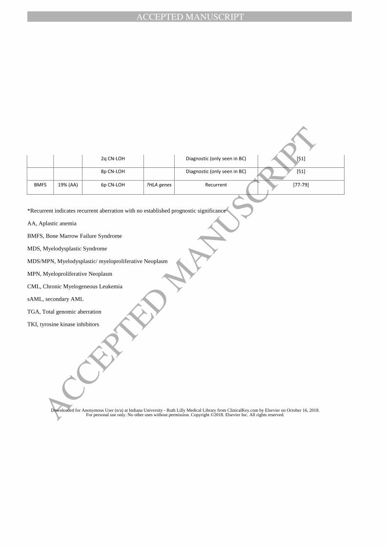
ACCEPTED MANUSCRIPT
ACCEPTED MANUSCRIP
T
2q CN-LOH Diagnostic (only seen in BC) [51]
8p CN-LOH Diagnostic (only seen in BC) [51]
BMFS 19% (AA) 6p CN-LOH ?HLA genes Recurrent [77-79]
*Recurrent indicates recurrent aberration with no established prognostic significance
AA, Aplastic anemia
BMFS, Bone Marrow Failure Syndrome
MDS, Myelodysplastic Syndrome
MDS/MPN, Myelodysplastic/ myeloproliferative Neoplasm
MPN, Myeloproliferative Neoplasm
CML, Chronic Myelogeneous Leukemia
sAML, secondary AML
TGA, Total genomic aberration
TKI, tyrosine kinase inhibitors
Downloaded for Anonymous User (n/a) at Indiana University - Ruth Lilly Medical Library from ClinicalKey.com by Elsevier on October 16, 2018.For personal use only. No other uses without permission. Copyright ©2018. Elsevier Inc. All rights reserved.

ACCEPTED MANUSCRIPT
ACCEPTED MANUSCRIP
T
Table 2. A comprehensive list of CNAs and CN-LOH of known or likely clinical significance in MDS detected by CMA testing
Chromosome Disease Abnormality
Type (Gain, Loss, CN-LOH)
Region Relevant Genes (if
known) Clinical
Significance*
Level of
Evidence
References
1 MDS Gain 1p36.33-p33 MPL Recurrent 3
[15] [14, 61]
1 MDS CN-LOH 1p MPL Recurrent 2 [14, 25, 36,
60]
1 MDS Gain 1q Recurrent 2 [14, 21, 30,
104]
2 MDS CN-LOH 2pter-2p13.3 DNMT3A Recurrent 2 [14, 62, 63,
113]
3 MDS CN-LOH 3q21.3-qter MECOM, GATA2 Recurrent 3 [14, 16, 19,
35, 60]
4 MDS Loss 4q24 TET2 T*** 2 [14, 18, 21, 23, 24, 32,
46]
4 MDS CN-LOH 4q12-qter TET2 Recurrent 2
[12, 14, 16, 21, 29, 30, 36, 37, 45,
63, 109]
5 MDS Gain 5p Suggestive of i(5p) with 5q
del Recurrent 3 [14]
5 MDS Loss 5q RPS14 D, P (Good when
isolated) 1
[14-16, 18, 19, 21, 26,
30, 33-35, 38, 42, 45, 48, 61-64, 102, 104, 110,
114]
Downloaded for Anonymous User (n/a) at Indiana University - Ruth Lilly Medical Library from ClinicalKey.com by Elsevier on October 16, 2018.For personal use only. No other uses without permission. Copyright ©2018. Elsevier Inc. All rights reserved.

ACCEPTED MANUSCRIPT
ACCEPTED MANUSCRIP
T
7 MDS Loss 7q EZH2, CUX1 D, P
(Intermediate) 1
[14, 15, 18, 19, 26, 30, 32, 34, 38, 45, 48, 60,
78, 102, 104, 110]
7 MDS CN-LOH 7q21.11-qter EZH2, CUX1 Recurrent 2
[12, 14, 16, 19, 21, 25, 30, 36, 48,
91, 109]
7 MDS Loss (Monosomy) 7 Whole Chromosome D, P (Poor) 1
[34, 38, 45, 63, 78, 102,
104, 107, 110]
8 MDS Gain (Trisomy) 8 Whole Chromosome P
(Intermediate)** 1
[14, 21, 30, 34, 46, 48, 60, 61, 73,
78, 102, 110]
9 MDS Gain 9p JAK2 Recurrent 3 [14, 30, 46]
9 MDS CN-LOH 9pter-p24.2 JAK2 Recurrent 2 [14, 31, 35]
11 MDS Loss 11q14.1-q24.3 CBL D, P (Very Good) 1 [14, 61]
11 MDS CN-LOH 11q13.3-qter CBL Recurrent 2 [12, 14, 15, 25, 35, 36,
63, 104, 107]
11 MDS Gain (Trisomy
and q-arm) 11 / 11q CBL Recurrent 3
[14, 16, 26, 30, 63]
12 MDS Loss 12p ETV6 D, P (Good) 1 [14, 16, 30,
32, 46]
12 MDS CN-LOH 12pter-p11.23 ETV6 Recurrent 2 [35, 63]
13 MDS Loss 13q RB1 D, P
(Intermediate) 2
[14, 21, 35, 104]
13 MDS CN-LOH 13q12.3-qter FLT3, RB1 Recurrent 3 [14, 36, 63]
13 MDS Gain (Trisomy) 13 Whole Chromosome Recurrent 3 [14]
Downloaded for Anonymous User (n/a) at Indiana University - Ruth Lilly Medical Library from ClinicalKey.com by Elsevier on October 16, 2018.For personal use only. No other uses without permission. Copyright ©2018. Elsevier Inc. All rights reserved.
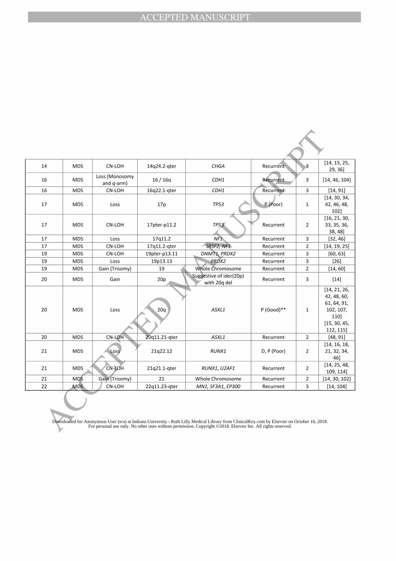
ACCEPTED MANUSCRIPT
ACCEPTED MANUSCRIP
T
14 MDS CN-LOH 14q24.2-qter CHGA Recurrent 3 [14, 15, 25,
29, 36]
16 MDS Loss (Monosomy
and q-arm) 16 / 16q CDH1 Recurrent 3 [14, 46, 104]
16 MDS CN-LOH 16q22.1-qter CDH1 Recurrent 3 [14, 91]
17 MDS Loss 17p TP53 P (Poor) 1 [14, 30, 34, 42, 46, 48,
102]
17 MDS CN-LOH 17pter-p11.2 TP53 Recurrent 2 [16, 21, 30, 33, 35, 36,
38, 48]
17 MDS Loss 17q11.2 NF1 Recurrent 3 [32, 46]
17 MDS CN-LOH 17q11.2-qter SRSF2, NF1 Recurrent 2 [14, 19, 25]
19 MDS CN-LOH 19pter-p13.11 DNMT1, PRDX2 Recurrent 3 [60, 63]
19 MDS Loss 19p13.13 PRDX2 Recurrent 3 [26]
19 MDS Gain (Trisomy) 19 Whole Chromosome Recurrent 2 [14, 60]
20 MDS Gain 20p Suggestive of ider(20p)
with 20q del Recurrent 3 [14]
20 MDS Loss 20q ASXL1 P (Good)** 1
[14, 21, 26, 42, 48, 60, 61, 64, 91, 102, 107,
110] [15, 30, 45, 112, 115]
20 MDS CN-LOH 20q11.21-qter ASXL1 Recurrent 2 [48, 91]
21 MDS Loss 21q22.12 RUNX1 D, P (Poor) 2 [14, 16, 18, 21, 32, 34,
46]
21 MDS CN-LOH 21q21.1-qter RUNX1, U2AF1 Recurrent 2 [14, 25, 48, 109, 114]
21 MDS Gain (Trisomy) 21 Whole Chromosome Recurrent 2 [14, 30, 102]
22 MDS CN-LOH 22q11.23-qter MN1, SF3A1, EP300 Recurrent 3 [14, 104]
Downloaded for Anonymous User (n/a) at Indiana University - Ruth Lilly Medical Library from ClinicalKey.com by Elsevier on October 16, 2018.For personal use only. No other uses without permission. Copyright ©2018. Elsevier Inc. All rights reserved.

ACCEPTED MANUSCRIPT
ACCEPTED MANUSCRIP
T
Legend: D- diagnostic significance; P-prognostic significance; T- therapeutic significance.
Recurrent indicates recurrent aberration with no established prognostic significance.
* Clinical significance based on WHO classification using IPSS-R (Greenberg et al., Blood 2012; Schanz et al., J Clin Oncol 2011).
** Isolated trisomy 8 or del(20q) are not diagnostic of MDS in the absence of morphologic findings of disease.
***Potential marker for responsiveness to hypomethylating agents or DNA methyltransferase inhibitors (Bejar et al., Blood 124:2705-
12, 2014;Traina et al., Leukemia 28:78-87, 2014).
Downloaded for Anonymous User (n/a) at Indiana University - Ruth Lilly Medical Library from ClinicalKey.com by Elsevier on October 16, 2018.For personal use only. No other uses without permission. Copyright ©2018. Elsevier Inc. All rights reserved.
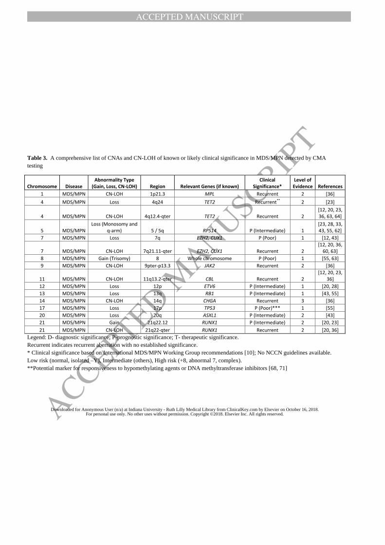
ACCEPTED MANUSCRIPT
ACCEPTED MANUSCRIP
T
Table 3. A comprehensive list of CNAs and CN-LOH of known or likely clinical significance in MDS/MPN detected by CMA
testing
Chromosome Disease Abnormality Type
(Gain, Loss, CN-LOH) Region Relevant Genes (if known) Clinical
Significance* Level of
Evidence References
1 MDS/MPN CN-LOH 1p21.3 MPL Recurrent 2 [36]
4 MDS/MPN Loss 4q24 TET2 Recurrent** 2 [23]
4 MDS/MPN CN-LOH 4q12.4-qter TET2 Recurrent 2 [12, 20, 23, 36, 63, 64]
5 MDS/MPN Loss (Monosomy and
q-arm) 5 / 5q RPS14 P (Intermediate) 1 [23, 28, 33, 43, 55, 62]
7 MDS/MPN Loss 7q EZH2, CUX1 P (Poor) 1 [12, 43]
7 MDS/MPN CN-LOH 7q21.11-qter EZH2, CUX1 Recurrent 2 [12, 20, 36,
60, 63]
8 MDS/MPN Gain (Trisomy) 8 Whole chromosome P (Poor) 1 [55, 63]
9 MDS/MPN CN-LOH 9pter-p13.3 JAK2 Recurrent 2 [36]
11 MDS/MPN CN-LOH 11q13.2-qter CBL Recurrent 2 [12, 20, 23,
36]
12 MDS/MPN Loss 12p ETV6 P (Intermediate) 1 [20, 28]
13 MDS/MPN Loss 13q RB1 P (Intermediate) 1 [43, 55]
14 MDS/MPN CN-LOH 14q CHGA Recurrent 3 [36]
17 MDS/MPN Loss 17p TP53 P (Poor)*** 1 [55]
20 MDS/MPN Loss 20q ASXL1 P (Intermediate) 2 [43]
21 MDS/MPN Gain 21q22.12 RUNX1 P (Intermediate) 2 [20, 23]
21 MDS/MPN CN-LOH 21q22-qter RUNX1 Recurrent 2 [20, 36]
Legend: D- diagnostic significance; P-prognostic significance; T- therapeutic significance.
Recurrent indicates recurrent aberration with no established significance.
* Clinical significance based on International MDS/MPN Working Group recommendations [10]; No NCCN guidelines available.
Low risk (normal, isolated –Y), Intermediate (others), High risk (+8, abnormal 7, complex).
**Potential marker for responsiveness to hypomethylating agents or DNA methyltransferase inhibitors [68, 71]
Downloaded for Anonymous User (n/a) at Indiana University - Ruth Lilly Medical Library from ClinicalKey.com by Elsevier on October 16, 2018.For personal use only. No other uses without permission. Copyright ©2018. Elsevier Inc. All rights reserved.

ACCEPTED MANUSCRIPT
ACCEPTED MANUSCRIP
T
***Haploinsufficiency of 17p as part of an isolated isochromosome may be a distinct disease entity with further increased risk of
AML progression relative to 17p loss in a complex karyotype.
Downloaded for Anonymous User (n/a) at Indiana University - Ruth Lilly Medical Library from ClinicalKey.com by Elsevier on October 16, 2018.For personal use only. No other uses without permission. Copyright ©2018. Elsevier Inc. All rights reserved.
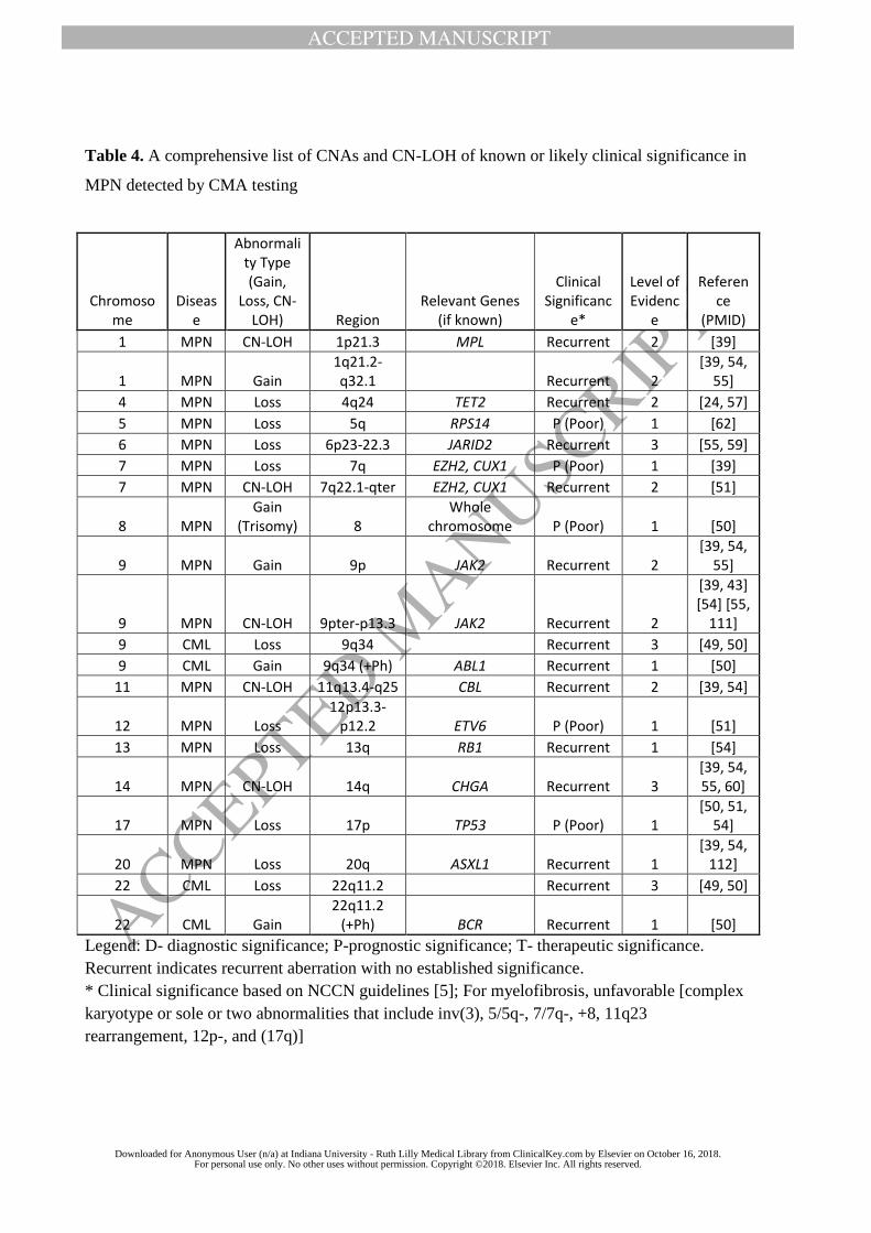
ACCEPTED MANUSCRIPT
ACCEPTED MANUSCRIP
T
Table 4. A comprehensive list of CNAs and CN-LOH of known or likely clinical significance in
MPN detected by CMA testing
Legend: D- diagnostic significance; P-prognostic significance; T- therapeutic significance.
Recurrent indicates recurrent aberration with no established significance.
* Clinical significance based on NCCN guidelines [5]; For myelofibrosis, unfavorable [complex
karyotype or sole or two abnormalities that include inv(3), 5/5q-, 7/7q-, +8, 11q23
rearrangement, 12p-, and (17q)]
Chromosome
Disease
Abnormality Type (Gain,
Loss, CN-LOH) Region
Relevant Genes (if known)
Clinical Significanc
e*
Level of Evidenc
e
Reference
(PMID)
1 MPN CN-LOH 1p21.3 MPL Recurrent 2 [39]
1 MPN Gain 1q21.2-q32.1 Recurrent 2
[39, 54, 55]
4 MPN Loss 4q24 TET2 Recurrent 2 [24, 57]
5 MPN Loss 5q RPS14 P (Poor) 1 [62]
6 MPN Loss 6p23-22.3 JARID2 Recurrent 3 [55, 59]
7 MPN Loss 7q EZH2, CUX1 P (Poor) 1 [39]
7 MPN CN-LOH 7q22.1-qter EZH2, CUX1 Recurrent 2 [51]
8 MPN Gain
(Trisomy) 8 Whole
chromosome P (Poor) 1 [50]
9 MPN Gain 9p JAK2 Recurrent 2 [39, 54,
55]
9 MPN CN-LOH 9pter-p13.3 JAK2 Recurrent 2
[39, 43] [54] [55,
111]
9 CML Loss 9q34 Recurrent 3 [49, 50]
9 CML Gain 9q34 (+Ph) ABL1 Recurrent 1 [50]
11 MPN CN-LOH 11q13.4-q25 CBL Recurrent 2 [39, 54]
12 MPN Loss 12p13.3-
p12.2 ETV6 P (Poor) 1 [51]
13 MPN Loss 13q RB1 Recurrent 1 [54]
14 MPN CN-LOH 14q CHGA Recurrent 3 [39, 54, 55, 60]
17 MPN Loss 17p TP53 P (Poor) 1 [50, 51,
54]
20 MPN Loss 20q ASXL1 Recurrent 1 [39, 54,
112]
22 CML Loss 22q11.2 Recurrent 3 [49, 50]
22 CML Gain 22q11.2
(+Ph) BCR Recurrent 1 [50]
Downloaded for Anonymous User (n/a) at Indiana University - Ruth Lilly Medical Library from ClinicalKey.com by Elsevier on October 16, 2018.For personal use only. No other uses without permission. Copyright ©2018. Elsevier Inc. All rights reserved.
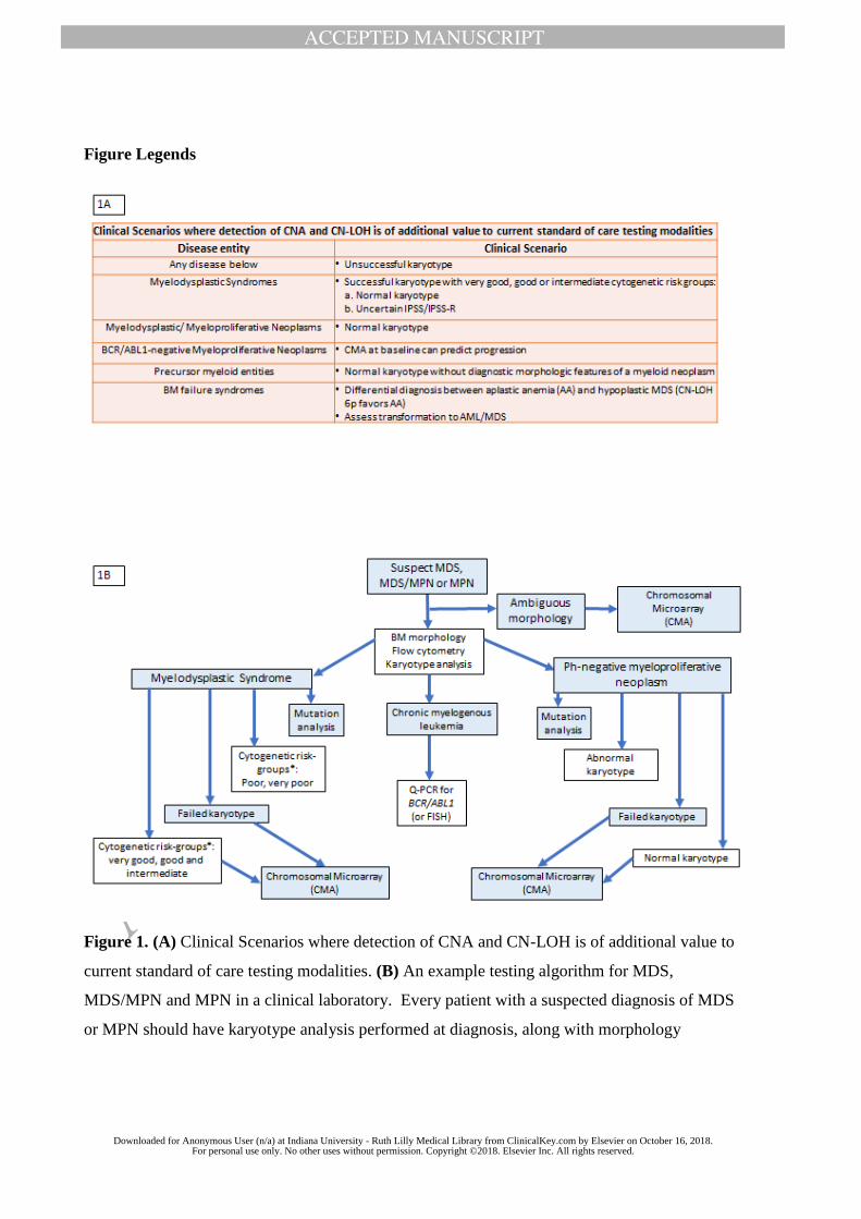
ACCEPTED MANUSCRIPT
ACCEPTED MANUSCRIP
T
Figure Legends
Figure 1. (A) Clinical Scenarios where detection of CNA and CN-LOH is of additional value to
current standard of care testing modalities. (B) An example testing algorithm for MDS,
MDS/MPN and MPN in a clinical laboratory. Every patient with a suspected diagnosis of MDS
or MPN should have karyotype analysis performed at diagnosis, along with morphology
Downloaded for Anonymous User (n/a) at Indiana University - Ruth Lilly Medical Library from ClinicalKey.com by Elsevier on October 16, 2018.For personal use only. No other uses without permission. Copyright ©2018. Elsevier Inc. All rights reserved.
ACCEPTED MANUSCRIPT
ACCEPTED MANUSCRIP
T
evaluation, flow cytometry analysis and mutation analysis (if considered appropriate and
feasible). In the case of CML with t(9;22) by karyotype, quantitative real-time PCR is the
preferred monitoring test. For patients with suspected MDS, MDS/MPN or Ph-negative MPN
diagnoses, chromosomal microarray testing (CMA) is recommended if the karyotype is
unsuccessful; for MDS patients with successful karyotype showing very good, good or
intermediate cytogenetic risk-groups per IPSS-R stratification, CMA is especially helpful in
patients with normal karyotype and in patients with uncertain IPSS/IPSS-R risk score to further
assist with diagnostic uncertainty and risk stratification**. In MDS patients when del(5q) is
detected by karyotype as a sole abnormality or along with one additional abnormality other than
del(7q)/-7, 2016 WHO classification system recommends performing TP53 mutation studies for
identifying those patients with poor response to lenalidomide. The genes in the mutation analysis
panel should be based on the NCCN guideline and may be tailored to the needs and technology
available at each institution. SF3B1, ASXL1, RUNX1, EZH2, ETV6 and TP53 for MDS and
JAK2, CALR, MPL, TET2 for MPN are typical examples. If feasible, when clonal copy number
alterations (CNAs) are identified by either CMA or karyotype, a baseline FISH for the key stem
clone could be considered to establish an informative FISH marker for future monitoring of
minimal residual disease post-treatment. In patients with ambiguous morphology, CMA is
helpful for identification of clonal markers.
* Cytogenetic risk-groups classified per IPSS-R [2, 4]
** At this time, IPSS-R is based on karyotype assessed by chromosome banding analysis only.
Further multi-center studies to comprehensively evaluate the prognostic impact of additional
findings by genomic arrays compared to IPSS-R need to be undertaken.
Downloaded for Anonymous User (n/a) at Indiana University - Ruth Lilly Medical Library from ClinicalKey.com by Elsevier on October 16, 2018.For personal use only. No other uses without permission. Copyright ©2018. Elsevier Inc. All rights reserved.

ACCEPTED MANUSCRIPT
ACCEPTED MANUSCRIP
T
References
[1] Arber DA, Orazi A, Hasserjian R, Thiele J, Borowitz MJ, Le Beau MM, et al. The 2016 revision to the World Health Organization classification of myeloid neoplasms and acute leukemia. Blood 2016;127:2391-405. [2] Schanz J, Tuchler H, Sole F, Mallo M, Luno E, Cervera J, et al. New comprehensive cytogenetic scoring system for primary myelodysplastic syndromes (MDS) and oligoblastic acute myeloid leukemia after MDS derived from an international database merge. J Clin Oncol 2012;30:820-9. [3] Greenberg P, Cox C, LeBeau MM, Fenaux P, Morel P, Sanz G, et al. International scoring system for evaluating prognosis in myelodysplastic syndromes. Blood 1997;89:2079-88. [4] Greenberg PL, Tuechler H, Schanz J, Sanz G, Garcia-Manero G, Sole F, et al. Revised international prognostic scoring system for myelodysplastic syndromes. Blood 2012;120:2454-65. [5] Mesa R, Jamieson C, Bhatia R, Deininger MW, Gerds AT, Gojo I, et al. Myeloproliferative Neoplasms, Version 2.2017, NCCN Clinical Practice Guidelines in Oncology. J Natl Compr Canc Netw 2016;14:1572-611. [6] Mesa RA, Jamieson C, Bhatia R, Deininger MW, Fletcher CD, Gerds AT, et al. NCCN Guidelines Insights: Myeloproliferative Neoplasms, Version 2.2018. J Natl Compr Canc Netw 2017;15:1193-207. [7] Greenberg PL, Stone RM, Al-Kali A, Barta SK, Bejar R, Bennett JM, et al. Myelodysplastic Syndromes, Version 2.2017, NCCN Clinical Practice Guidelines in Oncology. J Natl Compr Canc Netw 2017;15:60-87. [8] Mesa RA. The new NCCN guidelines for the management of myelofibrosis. Clin Adv Hematol Oncol 2017;15:193-5. [9] Malcovati L, Hellstrom-Lindberg E, Bowen D, Ades L, Cermak J, Del Canizo C, et al. Diagnosis and treatment of primary myelodysplastic syndromes in adults: recommendations from the European LeukemiaNet. Blood 2013;122:2943-64. [10] Mughal TI, Cross NC, Padron E, Tiu RV, Savona M, Malcovati L, et al. An International MDS/MPN Working Group's perspective and recommendations on molecular pathogenesis, diagnosis and clinical characterization of myelodysplastic/myeloproliferative neoplasms. Haematologica 2015;100:1117-30. [11] Papaemmanuil E, Gerstung M, Malcovati L, Tauro S, Gundem G, Van Loo P, et al. Clinical and biological implications of driver mutations in myelodysplastic syndromes. Blood 2013;122:3616-27; quiz 99. [12] Gondek LP, Tiu R, O'Keefe CL, Sekeres MA, Theil KS, Maciejewski JP. Chromosomal lesions and uniparental disomy detected by SNP arrays in MDS, MDS/MPD, and MDS-derived AML. Blood 2008;111:1534-42. [13] Mukherjee S, Sathanoori M, Ma Z, Andreatta M, Lennon PA, Wheeler SR, et al. Addition of chromosomal microarray and next generation sequencing to FISH and classical cytogenetics enhances genomic profiling of myeloid malignancies. Cancer Genet 2017;216-217:128-41. [14] Tiu RV, Gondek LP, O'Keefe CL, Elson P, Huh J, Mohamedali A, et al. Prognostic impact of SNP array karyotyping in myelodysplastic syndromes and related myeloid malignancies. Blood 2011;117:4552-60. [15] Huh J, Jung CW, Kim HJ, Kim YK, Moon JH, Sohn SK, et al. Different characteristics identified by single nucleotide polymorphism array analysis in leukemia suggest the need for different application strategies depending on disease category. Genes Chromosomes Cancer 2013;52:44-55. [16] Heinrichs S, Kulkarni RV, Bueso-Ramos CE, Levine RL, Loh ML, Li C, et al. Accurate detection of uniparental disomy and microdeletions by SNP array analysis in myelodysplastic syndromes with normal cytogenetics. Leukemia 2009;23:1605-13.
Downloaded for Anonymous User (n/a) at Indiana University - Ruth Lilly Medical Library from ClinicalKey.com by Elsevier on October 16, 2018.For personal use only. No other uses without permission. Copyright ©2018. Elsevier Inc. All rights reserved.
ACCEPTED MANUSCRIPT
ACCEPTED MANUSCRIP
T
[17] Akagi T, Ogawa S, Dugas M, Kawamata N, Yamamoto G, Nannya Y, et al. Frequent genomic abnormalities in acute myeloid leukemia/myelodysplastic syndrome with normal karyotype. Haematologica 2009;94:213-23. [18] Thiel A, Beier M, Ingenhag D, Servan K, Hein M, Moeller V, et al. Comprehensive array CGH of normal karyotype myelodysplastic syndromes reveals hidden recurrent and individual genomic copy number alterations with prognostic relevance. Leukemia 2011;25:387-99. [19] Merkerova MD, Bystricka D, Belickova M, Krejcik Z, Zemanova Z, Polak J, et al. From cryptic chromosomal lesions to pathologically relevant genes: integration of SNP-array with gene expression profiling in myelodysplastic syndrome with normal karyotype. Genes Chromosomes Cancer 2012;51:419-28. [20] Palomo L, Xicoy B, Garcia O, Mallo M, Adema V, Cabezon M, et al. Impact of SNP array karyotyping on the diagnosis and the outcome of chronic myelomonocytic leukemia with low risk cytogenetic features or no metaphases. Am J Hematol 2016;91:185-92. [21] Evans AG, Ahmad A, Burack WR, Iqbal MA. Combined comparative genomic hybridization and single-nucleotide polymorphism array detects cryptic chromosomal lesions in both myelodysplastic syndromes and cytopenias of undetermined significance. Mod Pathol 2016;29:1183-99. [22] Chou W-C, Chou S-C, Liu C-Y, Chen C-Y, Hou H-A, Kuo Y-Y, et al. TET2 mutation is an unfavorable prognostic factor in acute myeloid leukemia patients with intermediate-risk cytogenetics. Blood 2011;118:3803-10. [23] Jankowska AM, Szpurka H, Tiu RV, Makishima H, Afable M, Huh J, et al. Loss of heterozygosity 4q24 and TET2 mutations associated with myelodysplastic/myeloproliferative neoplasms. Blood 2009;113:6403-10. [24] Bacher U, Weissmann S, Kohlmann A, Schindela S, Alpermann T, Schnittger S, et al. TET2 deletions are a recurrent but rare phenomenon in myeloid malignancies and are frequently accompanied by TET2 mutations on the remaining allele. Br J Haematol 2012;156:67-75. [25] Gondek LP, Haddad AS, O'Keefe CL, Tiu R, Wlodarski MW, Sekeres MA, et al. Detection of cryptic chromosomal lesions including acquired segmental uniparental disomy in advanced and low-risk myelodysplastic syndromes. Exp Hematol 2007;35:1728-38. [26] Starczynowski DT, Vercauteren S, Telenius A, Sung S, Tohyama K, Brooks-Wilson A, et al. High-resolution whole genome tiling path array CGH analysis of CD34+ cells from patients with low-risk myelodysplastic syndromes reveals cryptic copy number alterations and predicts overall and leukemia-free survival. Blood 2008;112:3412-24. [27] Makishima H, Rataul M, Gondek LP, Huh J, Cook JR, Theil KS, et al. FISH and SNP-A karyotyping in myelodysplastic syndromes: improving cytogenetic detection of del(5q), monosomy 7, del(7q), trisomy 8 and del(20q). Leuk Res 2010;34:447-53. [28] Slovak ML, Smith DD, Bedell V, Hsu YH, O'Donnell M, Forman SJ, et al. Assessing karyotype precision by microarray-based comparative genomic hybridization in the myelodysplastic/myeloproliferative syndromes. Mol Cytogenet 2010;3:23. [29] Mohamedali AM, Gaken J, Ahmed M, Malik F, Smith AE, Best S, et al. High concordance of genomic and cytogenetic aberrations between peripheral blood and bone marrow in myelodysplastic syndrome (MDS). Leukemia 2015;29:1928-38. [30] Hu Q, Chu Y, Song Q, Yao Y, Yang W, Huang S. The prevalence of chromosomal aberrations associated with myelodysplastic syndromes in China. Ann Hematol 2016;95:1241-8. [31] Yeung CCS, McElhone S, Chen XY, Ng D, Storer BE, Deeg HJ, et al. Impact of copy neutral loss of heterozygosity and total genome aberrations on survival in myelodysplastic syndrome. Mod Pathol 2017.
Downloaded for Anonymous User (n/a) at Indiana University - Ruth Lilly Medical Library from ClinicalKey.com by Elsevier on October 16, 2018.For personal use only. No other uses without permission. Copyright ©2018. Elsevier Inc. All rights reserved.
ACCEPTED MANUSCRIPT
ACCEPTED MANUSCRIP
T
[32] Volkert S, Haferlach T, Holzwarth J, Zenger M, Kern W, Staller M, et al. Array CGH identifies copy number changes in 11% of 520 MDS patients with normal karyotype and uncovers prognostically relevant deletions. Leukemia 2016;30:257-60. [33] Jerez A, Gondek LP, Jankowska AM, Makishima H, Przychodzen B, Tiu RV, et al. Topography, clinical, and genomic correlates of 5q myeloid malignancies revisited. J Clin Oncol 2012;30:1343-9. [34] Zhang R, Kim YM, Wang X, Li Y, Lu X, Sternenberger AR, et al. Genomic Copy Number Variations in the Myelodysplastic Syndrome and Acute Myeloid Leukemia Patients with del(5q) and/or -7/del(7q). Int J Med Sci 2015;12:719-26. [35] Arenillas L, Mallo M, Ramos F, Guinta K, Barragan E, Lumbreras E, et al. Single nucleotide polymorphism array karyotyping: a diagnostic and prognostic tool in myelodysplastic syndromes with unsuccessful conventional cytogenetic testing. Genes Chromosomes Cancer 2013;52:1167-77. [36] Dunbar AJ, Gondek LP, O'Keefe CL, Makishima H, Rataul MS, Szpurka H, et al. 250K single nucleotide polymorphism array karyotyping identifies acquired uniparental disomy and homozygous mutations, including novel missense substitutions of c-Cbl, in myeloid malignancies. Cancer Res 2008;68:10349-57. [37] Mohamedali AM, Smith AE, Gaken J, Lea NC, Mian SA, Westwood NB, et al. Novel TET2 mutations associated with UPD4q24 in myelodysplastic syndrome. J Clin Oncol 2009;27:4002-6. [38] Svobodova K, Zemanova Z, Lhotska H, Novakova M, Podskalska L, Belickova M, et al. Copy number neutral loss of heterozygosity at 17p and homozygous mutations of TP53 are associated with complex chromosomal aberrations in patients newly diagnosed with myelodysplastic syndromes. Leuk Res 2016;42:7-12. [39] Rumi E, Harutyunyan A, Elena C, Pietra D, Klampfl T, Bagienski K, et al. Identification of genomic aberrations associated with disease transformation by means of high-resolution SNP array analysis in patients with myeloproliferative neoplasm. Am J Hematol 2011;86:974-9. [40] Kralovics R, Passamonti F, Buser AS, Teo SS, Tiedt R, Passweg JR, et al. A gain-of-function mutation of JAK2 in myeloproliferative disorders. N Engl J Med 2005;352:1779-90. [41] O'Keefe C, McDevitt MA, Maciejewski JP. Copy neutral loss of heterozygosity: a novel chromosomal lesion in myeloid malignancies. Blood 2010;115:2731-9. [42] Bajaj R, Xu F, Xiang B, Wilcox K, Diadamo AJ, Kumar R, et al. Evidence-based genomic diagnosis characterized chromosomal and cryptic imbalances in 30 elderly patients with myelodysplastic syndrome and acute myeloid leukemia. Mol Cytogenet 2011;4:3. [43] Gondek LP, Dunbar AJ, Szpurka H, McDevitt MA, Maciejewski JP. SNP array karyotyping allows for the detection of uniparental disomy and cryptic chromosomal abnormalities in MDS/MPD-U and MPD. PLoS One 2007;2:e1225. [44] Cluzeau T, Moreilhon C, Mounier N, Karsenti JM, Gastaud L, Garnier G, et al. Total genomic alteration as measured by SNP-array-based molecular karyotyping is predictive of overall survival in a cohort of MDS or AML patients treated with azacitidine. Blood Cancer J 2013;3:e155. [45] Mohamedali A, Gaken J, Twine NA, Ingram W, Westwood N, Lea NC, et al. Prevalence and prognostic significance of allelic imbalance by single-nucleotide polymorphism analysis in low-risk myelodysplastic syndromes. Blood 2007;110:3365-73. [46] Kolquist KA, Schultz RA, Furrow A, Brown TC, Han JY, Campbell LJ, et al. Microarray-based comparative genomic hybridization of cancer targets reveals novel, recurrent genetic aberrations in the myelodysplastic syndromes. Cancer Genet 2011;204:603-28. [47] Hartmann L, Stephenson CF, Verkamp SR, Johnson KR, Burnworth B, Hammock K, et al. Detection of clonal evolution in hematopoietic malignancies by combining comparative genomic hybridization and single nucleotide polymorphism arrays. Clin Chem 2014;60:1558-68.
Downloaded for Anonymous User (n/a) at Indiana University - Ruth Lilly Medical Library from ClinicalKey.com by Elsevier on October 16, 2018.For personal use only. No other uses without permission. Copyright ©2018. Elsevier Inc. All rights reserved.
ACCEPTED MANUSCRIPT
ACCEPTED MANUSCRIP
T
[48] Ganster C, Shirneshan K, Salinas-Riester G, Braulke F, Schanz J, Platzbecker U, et al. Influence of total genomic alteration and chromosomal fragmentation on response to a combination of azacitidine and lenalidomide in a cohort of patients with very high risk MDS. Leuk Res 2015;39:1079-87. [49] Huh J, Jung CW, Kim JW, Kim HJ, Kim SH, Shin MG, et al. Genome-wide high density single-nucleotide polymorphism array-based karyotyping improves detection of clonal aberrations including der(9) deletion, but does not predict treatment outcomes after imatinib therapy in chronic myeloid leukemia. Ann Hematol 2011;90:1255-64. [50] Nowak D, Ogawa S, Muschen M, Kato M, Kawamata N, Meixel A, et al. SNP array analysis of tyrosine kinase inhibitor-resistant chronic myeloid leukemia identifies heterogeneous secondary genomic alterations. Blood 2010;115:1049-53. [51] Boultwood J, Perry J, Zaman R, Fernandez-Santamaria C, Littlewood T, Kusec R, et al. High-density single nucleotide polymorphism array analysis and ASXL1 gene mutation screening in chronic myeloid leukemia during disease progression. Leukemia 2010;24:1139-45. [52] Hosoya N, Sanada M, Nannya Y, Nakazaki K, Wang L, Hangaishi A, et al. Genomewide screening of DNA copy number changes in chronic myelogenous leukemia with the use of high‐resolution array‐based comparative genomic hybridization. Genes, Chromosomes and Cancer 2006;45:482-94. [53] Hussein K, Van Dyke DL, Tefferi A. Conventional cytogenetics in myelofibrosis: literature review and discussion. Eur J Haematol 2009;82:329-38. [54] Singh NR, Morris CM, Koleth M, Wong K, Ward CM, Stevenson WS. Polyploidy in myelofibrosis: analysis by cytogenetic and SNP array indicates association with advancing disease. Mol Cytogenet 2013;6:59. [55] Hahm C, Huh HJ, Mun YC, Seong CM, Chung WS, Huh J. Genomic aberrations of myeloproliferative and myelodysplastic/myeloproliferative neoplasms in chronic phase and during disease progression. Int J Lab Hematol 2015;37:181-9. [56] Thoennissen NH, Krug UO, Lee DH, Kawamata N, Iwanski GB, Lasho T, et al. Prevalence and prognostic impact of allelic imbalances associated with leukemic transformation of Philadelphia chromosome-negative myeloproliferative neoplasms. Blood 2010;115:2882-90. [57] Klampfl T, Harutyunyan A, Berg T, Gisslinger B, Schalling M, Bagienski K, et al. Genome integrity of myeloproliferative neoplasms in chronic phase and during disease progression. Blood 2011;118:167-76. [58] Passamonti F, Rumi E, Pietra D, Elena C, Boveri E, Arcaini L, et al. A prospective study of 338 patients with polycythemia vera: the impact of JAK2 (V617F) allele burden and leukocytosis on fibrotic or leukemic disease transformation and vascular complications. Leukemia 2010;24:1574-9. [59] Puda A, Milosevic JD, Berg T, Klampfl T, Harutyunyan AS, Gisslinger B, et al. Frequent deletions of JARID2 in leukemic transformation of chronic myeloid malignancies. Am J Hematol 2012;87:245-50. [60] Sugimoto Y, Sekeres MA, Makishima H, Traina F, Visconte V, Jankowska A, et al. Cytogenetic and molecular predictors of response in patients with myeloid malignancies without del[5q] treated with lenalidomide. J Hematol Oncol 2012;5:4. [61] Vercauteren SM, Sung S, Starczynowski DT, Lam WL, Bruyere H, Horsman DE, et al. Array comparative genomic hybridization of peripheral blood granulocytes of patients with myelodysplastic syndrome detects karyotypic abnormalities. Am J Clin Pathol 2010;134:119-26. [62] Stengel A, Kern W, Haferlach T, Meggendorfer M, Haferlach C. The 5q deletion size in myeloid malignancies is correlated to additional chromosomal aberrations and to TP53 mutations. Genes Chromosomes Cancer 2016;55:777-85. [63] Flach J, Dicker F, Schnittger S, Schindela S, Kohlmann A, Haferlach T, et al. An accumulation of cytogenetic and molecular genetic events characterizes the progression from MDS to secondary AML: an analysis of 38 paired samples analyzed by cytogenetics, molecular mutation analysis and SNP microarray profiling. Leukemia 2011;25:713-8.
Downloaded for Anonymous User (n/a) at Indiana University - Ruth Lilly Medical Library from ClinicalKey.com by Elsevier on October 16, 2018.For personal use only. No other uses without permission. Copyright ©2018. Elsevier Inc. All rights reserved.
ACCEPTED MANUSCRIPT
ACCEPTED MANUSCRIP
T
[64] da Silva FB, Machado-Neto JA, Bertini V, Velloso E, Ratis CA, Calado RT, et al. Single-nucleotide polymorphism array (SNP-A) improves the identification of chromosomal abnormalities by metaphase cytogenetics in myelodysplastic syndrome. J Clin Pathol 2017;70:435-42. [65] Jadersten M, Saft L, Smith A, Kulasekararaj A, Pomplun S, Gohring G, et al. TP53 mutations in low-risk myelodysplastic syndromes with del(5q) predict disease progression. J Clin Oncol 2011;29:1971-9. [66] Gaballa MR, Besa EC. Myelodysplastic syndromes with 5q deletion: pathophysiology and role of lenalidomide. Ann Hematol 2014;93:723-33. [67] Komrokji RS, Padron E, Ebert BL, List AF. Deletion 5q MDS: molecular and therapeutic implications. Best Pract Res Clin Haematol 2013;26:365-75. [68] Bejar R, Lord A, Stevenson K, Bar-Natan M, Perez-Ladaga A, Zaneveld J, et al. TET2 mutations predict response to hypomethylating agents in myelodysplastic syndrome patients. Blood 2014;124:2705-12. [69] Itzykson R, Kosmider O, Cluzeau T, Mansat-De Mas V, Dreyfus F, Beyne-Rauzy O, et al. Impact of TET2 mutations on response rate to azacitidine in myelodysplastic syndromes and low blast count acute myeloid leukemias. Leukemia 2011;25:1147-52. [70] Diesch J, Zwick A, Garz AK, Palau A, Buschbeck M, Gotze KS. A clinical-molecular update on azanucleoside-based therapy for the treatment of hematologic cancers. Clin Epigenetics 2016;8:71. [71] Traina F, Visconte V, Elson P, Tabarroki A, Jankowska AM, Hasrouni E, et al. Impact of molecular mutations on treatment response to DNMT inhibitors in myelodysplasia and related neoplasms. Leukemia 2014;28:78-87. [72] Kulasekararaj AG, Smith AE, Mian SA, Mohamedali AM, Krishnamurthy P, Lea NC, et al. TP53 mutations in myelodysplastic syndrome are strongly correlated with aberrations of chromosome 5, and correlate with adverse prognosis. Br J Haematol 2013;160:660-72. [73] Paulsson K, Heidenblad M, Strombeck B, Staaf J, Jonsson G, Borg A, et al. High-resolution genome-wide array-based comparative genome hybridization reveals cryptic chromosome changes in AML and MDS cases with trisomy 8 as the sole cytogenetic aberration. Leukemia 2006;20:840-6. [74] Caramazza D, Lasho TL, Finke CM, Gangat N, Dingli D, Knudson RA, et al. IDH mutations and trisomy 8 in myelodysplastic syndromes and acute myeloid leukemia. Leukemia 2010;24:2120-2. [75] Dohner H, Estey E, Grimwade D, Amadori S, Appelbaum FR, Buchner T, et al. Diagnosis and management of AML in adults: 2017 ELN recommendations from an international expert panel. Blood 2017;129:424-47. [76] Bacher U, Haferlach T, Schnittger S, Zenger M, Meggendorfer M, Jeromin S, et al. Investigation of 305 patients with myelodysplastic syndromes and 20q deletion for associated cytogenetic and molecular genetic lesions and their prognostic impact. British journal of haematology 2014;164:822-33. [77] Afable MG, 2nd, Wlodarski M, Makishima H, Shaik M, Sekeres MA, Tiu RV, et al. SNP array-based karyotyping: differences and similarities between aplastic anemia and hypocellular myelodysplastic syndromes. Blood 2011;117:6876-84. [78] Babushok DV, Xie HM, Roth JJ, Perdigones N, Olson TS, Cockroft JD, et al. Single nucleotide polymorphism array analysis of bone marrow failure patients reveals characteristic patterns of genetic changes. Br J Haematol 2014;164:73-82. [79] Betensky M, Babushok D, Roth JJ, Mason PJ, Biegel JA, Busse TM, et al. Clonal evolution and clinical significance of copy number neutral loss of heterozygosity of chromosome arm 6p in acquired aplastic anemia. Cancer Genet 2016;209:1-10. [80] Kanagal-Shamanna R, Loghavi S, DiNardo CD, Medeiros LJ, Garcia-Manero G, Jabbour E, et al. Bone marrow pathologic abnormalities in familial platelet disorder with propensity for myeloid malignancy and germline RUNX1 mutation. Haematologica 2017;102:1661-70.
Downloaded for Anonymous User (n/a) at Indiana University - Ruth Lilly Medical Library from ClinicalKey.com by Elsevier on October 16, 2018.For personal use only. No other uses without permission. Copyright ©2018. Elsevier Inc. All rights reserved.
ACCEPTED MANUSCRIPT
ACCEPTED MANUSCRIP
T
[81] Antony-Debre I, Duployez N, Bucci M, Geffroy S, Micol JB, Renneville A, et al. Somatic mutations associated with leukemic progression of familial platelet disorder with predisposition to acute myeloid leukemia. Leukemia 2016;30:999-1002. [82] Jaiswal S, Fontanillas P, Flannick J, Manning A, Grauman PV, Mar BG, et al. Age-related clonal hematopoiesis associated with adverse outcomes. N Engl J Med 2014;371:2488-98. [83] Genovese G, Kahler AK, Handsaker RE, Lindberg J, Rose SA, Bakhoum SF, et al. Clonal hematopoiesis and blood-cancer risk inferred from blood DNA sequence. N Engl J Med 2014;371:2477-87. [84] Xie M, Lu C, Wang J, McLellan MD, Johnson KJ, Wendl MC, et al. Age-related mutations associated with clonal hematopoietic expansion and malignancies. Nat Med 2014;20:1472-8. [85] Valent P, Bain BJ, Bennett JM, Wimazal F, Sperr WR, Mufti G, et al. Idiopathic cytopenia of undetermined significance (ICUS) and idiopathic dysplasia of uncertain significance (IDUS), and their distinction from low risk MDS. Leuk Res 2012;36:1-5. [86] Valent P, Horny HP. Minimal diagnostic criteria for myelodysplastic syndromes and separation from ICUS and IDUS: update and open questions. Eur J Clin Invest 2009;39:548-53. [87] Steensma DP. Dysplasia has A differential diagnosis: distinguishing genuine myelodysplastic syndromes (MDS) from mimics, imitators, copycats and impostors. Curr Hematol Malig Rep 2012;7:310-20. [88] Steensma DP, Bejar R, Jaiswal S, Lindsley RC, Sekeres MA, Hasserjian RP, et al. Clonal hematopoiesis of indeterminate potential and its distinction from myelodysplastic syndromes. Blood 2015;126:9-16. [89] Kwok B, Hall JM, Witte JS, Xu Y, Reddy P, Lin K, et al. MDS-associated somatic mutations and clonal hematopoiesis are common in idiopathic cytopenias of undetermined significance. Blood 2015;126:2355-61. [90] Cargo CA, Rowbotham N, Evans PA, Barrans SL, Bowen DT, Crouch S, et al. Targeted sequencing identifies patients with preclinical MDS at high risk of disease progression. Blood 2015;126:2362-5. [91] Nowak D, Nolte F, Mossner M, Nowak V, Baldus CD, Hopfer O, et al. Genome-wide DNA-mapping of CD34+ cells from patients with myelodysplastic syndrome using 500K SNP arrays identifies significant regions of deletion and uniparental disomy. Exp Hematol 2009;37:215-24. [92] Jacoby MA, Walter MJ. Detection of copy number alterations in acute myeloid leukemia and myelodysplastic syndromes. Expert Rev Mol Diagn 2012;12:253-64. [93] Heinrichs S, Li C, Look AT. SNP array analysis in hematologic malignancies: avoiding false discoveries. Blood 2010;115:4157-61. [94] Jacobs S, Thompson ER, Nannya Y, Yamamoto G, Pillai R, Ogawa S, et al. Genome-wide, high-resolution detection of copy number, loss of heterozygosity, and genotypes from formalin-fixed, paraffin-embedded tumor tissue using microarrays. Cancer Res 2007;67:2544-51. [95] Lips EH, Dierssen JW, van Eijk R, Oosting J, Eilers PH, Tollenaar RA, et al. Reliable high-throughput genotyping and loss-of-heterozygosity detection in formalin-fixed, paraffin-embedded tumors using single nucleotide polymorphism arrays. Cancer Res 2005;65:10188-91. [96] Thompson ER, Herbert SC, Forrest SM, Campbell IG. Whole genome SNP arrays using DNA derived from formalin-fixed, paraffin-embedded ovarian tumor tissue. Hum Mutat 2005;26:384-9. [97] Bertoncini P, Le Chevalier S, Lavenus S, Layrolle P, Louarn G. Early adhesion of human mesenchymal stem cells on TiO(2) surfaces studied by single-cell force spectroscopy measurements. J Mol Recognit 2012;25:262-9. [98] Wang Y, Carlton VE, Karlin-Neumann G, Sapolsky R, Zhang L, Moorhead M, et al. High quality copy number and genotype data from FFPE samples using Molecular Inversion Probe (MIP) microarrays. BMC Med Genomics 2009;2:8.
Downloaded for Anonymous User (n/a) at Indiana University - Ruth Lilly Medical Library from ClinicalKey.com by Elsevier on October 16, 2018.For personal use only. No other uses without permission. Copyright ©2018. Elsevier Inc. All rights reserved.
ACCEPTED MANUSCRIPT
ACCEPTED MANUSCRIP
T
[99] Oosting J, Lips EH, van Eijk R, Eilers PH, Szuhai K, Wijmenga C, et al. High-resolution copy number analysis of paraffin-embedded archival tissue using SNP BeadArrays. Genome Res 2007;17:368-76. [100] Joosse SA, van Beers EH, Nederlof PM. Automated array-CGH optimized for archival formalin-fixed, paraffin-embedded tumor material. BMC Cancer 2007;7:43. [101] Little SE, Vuononvirta R, Reis-Filho JS, Natrajan R, Iravani M, Fenwick K, et al. Array CGH using whole genome amplification of fresh-frozen and formalin-fixed, paraffin-embedded tumor DNA. Genomics 2006;87:298-306. [102] Stevens-Kroef MJ, Hebeda KM, Verwiel ET, Kamping EJ, van Cleef PH, Kuiper RP, et al. Microarray-based genomic profiling and in situ hybridization on fibrotic bone marrow biopsies for the identification of numerical chromosomal abnormalities in myelodysplastic syndrome. Mol Cytogenet 2015;8:33. [103] Peterson JF, Aggarwal N, Smith CA, Gollin SM, Surti U, Rajkovic A, et al. Integration of microarray analysis into the clinical diagnosis of hematological malignancies: How much can we improve cytogenetic testing? Oncotarget 2015;6:18845-62. [104] Xu X, Johnson EB, Leverton L, Arthur A, Watson Q, Chang FL, et al. The advantage of using SNP array in clinical testing for hematological malignancies--a comparative study of three genetic testing methods. Cancer Genet 2013;206:317-26. [105] Schoumans J, Suela J, Hastings R, Muehlematter D, Rack K, van den Berg E, et al. Guidelines for genomic array analysis in acquired haematological neoplastic disorders. Genes Chromosomes Cancer 2016;55:480-91. [106] Stevens-Kroef MJ, Olde Weghuis D, ElIdrissi-Zaynoun N, van der Reijden B, Cremers EMP, Alhan C, et al. Genomic array as compared to karyotyping in myelodysplastic syndromes in a prospective clinical trial. Genes Chromosomes Cancer 2017;56:524-34. [107] Barresi V, Palumbo GA, Musso N, Consoli C, Capizzi C, Meli CR, et al. Clonal selection of 11q CN-LOH and CBL gene mutation in a serially studied patient during MDS progression to AML. Leuk Res 2010;34:1539-42. [108] Gronseth C, McElhone S, Scott B, Yeung C, Fang M. Detection of cnLOH as a sole abnormality in the diagnosis of myelodysplastic syndrome. CAP Today 2016:82-6. [109] Larsson N, Lilljebjorn H, Lassen C, Johansson B, Fioretos T. Myeloid malignancies with acquired trisomy 21 as the sole cytogenetic change are clinically highly variable and display a heterogeneous pattern of copy number alterations and mutations. Eur J Haematol 2012;88:136-43. [110] Kunze K, Gamerdinger U, Lessig-Owlanj J, Sorokina M, Brobeil A, Tur MK, et al. Detection of an activated JAK3 variant and a Xq26.3 microdeletion causing loss of PHF6 and miR-424 expression in myelodysplastic syndromes by combined targeted next generation sequencing and SNP array analysis. Pathol Res Pract 2014;210:369-76. [111] Stegelmann F, Bullinger L, Griesshammer M, Holzmann K, Habdank M, Kuhn S, et al. High-resolution single-nucleotide polymorphism array-profiling in myeloproliferative neoplasms identifies novel genomic aberrations. Haematologica 2010;95:666-9. [112] Huh J, Tiu RV, Gondek LP, O'Keefe CL, Jasek M, Makishima H, et al. Characterization of chromosome arm 20q abnormalities in myeloid malignancies using genome-wide single nucleotide polymorphism array analysis. Genes Chromosomes Cancer 2010;49:390-9. [113] Hahm C, Mun YC, Seong CM, Han SH, Chung WS, Huh J. Single nucleotide polymorphism array-based karyotyping in acute myeloid leukemia or myelodysplastic syndrome with trisomy 8 as the sole chromosomal abnormality. Acta Haematol 2013;129:154-8. [114] Noronha TR, Rohr SS, Chauffaille Mde L. Identifying the similarities and differences between single nucleotide polymorphism array (SNPa) analysis and karyotyping in acute myeloid leukemia and myelodysplastic syndromes. Rev Bras Hematol Hemoter 2015;37:48-54.
Downloaded for Anonymous User (n/a) at Indiana University - Ruth Lilly Medical Library from ClinicalKey.com by Elsevier on October 16, 2018.For personal use only. No other uses without permission. Copyright ©2018. Elsevier Inc. All rights reserved.

ACCEPTED MANUSCRIPT
ACCEPTED MANUSCRIP
T
[115] Bacher U, Haferlach T, Schnittger S, Zenger M, Meggendorfer M, Jeromin S, et al. Investigation of 305 patients with myelodysplastic syndromes and 20q deletion for associated cytogenetic and molecular genetic lesions and their prognostic impact. Br J Haematol 2014;164:822-33.
Downloaded for Anonymous User (n/a) at Indiana University - Ruth Lilly Medical Library from ClinicalKey.com by Elsevier on October 16, 2018.For personal use only. No other uses without permission. Copyright ©2018. Elsevier Inc. All rights reserved.