osteocytic fgf23 and its kidney function - iupui scholarworks
TRANSCRIPT
REVIEWpublished: 28 August 2020
doi: 10.3389/fendo.2020.00592
Frontiers in Endocrinology | www.frontiersin.org 1 August 2020 | Volume 11 | Article 592
Edited by:
Lilian Irene Plotkin,
School of Medicine, Indiana University
Bloomington, United States
Reviewed by:
Paola Divieti Pajevic,
Boston University, United States
Jan Josef Stepan,
Charles University, Czechia
*Correspondence:
Kenneth E. White
Specialty section:
This article was submitted to
Bone Research,
a section of the journal
Frontiers in Endocrinology
Received: 15 June 2020
Accepted: 20 July 2020
Published: 28 August 2020
Citation:
Agoro R, Ni P, Noonan ML and
White KE (2020) Osteocytic FGF23
and Its Kidney Function.
Front. Endocrinol. 11:592.
doi: 10.3389/fendo.2020.00592
Osteocytic FGF23 and Its KidneyFunctionRafiou Agoro 1, Pu Ni 1, Megan L. Noonan 1 and Kenneth E. White 1,2*
1Department of Medical and Molecular Genetics, Indiana University School of Medicine, Indianapolis, IN, United States,2Medicine/Division of Nephrology, Indiana University School of Medicine, Indianapolis, IN, United States
Osteocytes, which represent up to 95% of adult skeletal cells, are deeply embedded
in bone. These cells exhibit important interactive abilities with other bone cells such
as osteoblasts and osteoclasts to control skeletal formation and resorption. Beyond
this local role, osteocytes can also influence the function of distant organs due to the
presence of their sophisticated lacunocanalicular system, which connects osteocyte
dendrites directly to the vasculature. Through these networks, osteocytes sense changes
in circulating metabolites and respond by producing endocrine factors to control
homeostasis. One critical function of osteocytes is to respond to increased blood
phosphate and 1,25(OH)2 vitamin D (1,25D) by producing fibroblast growth factor-23
(FGF23). FGF23 acts on the kidneys through partner fibroblast growth factor receptors
(FGFRs) and the co-receptor Klotho to promote phosphaturia via a downregulation of
phosphate transporters, as well as the control of vitamin D metabolizing enzymes to
reduce blood 1,25D. In the first part of this review, we will explore the signals involved in
the positive and negative regulation of FGF23 in osteocytes. In the second portion, we
will bridge bone responses with the review of current knowledge on FGF23 endocrine
functions in the kidneys.
Keywords: osteocyte, FGF23, FGF23 signaling, Klotho, kidney
INTRODUCTION
The mammalian skeleton is formed by several types of bone which interconnect to providestructural support for the body. In adult humans, the reference value for the skeleton weightis 10.5 kg (1, 2) representing up to 15% of the average body weight. Bone mass is dynamicallyregulated during a lifetime, and subjected to changes with uncontrollable parameters such as age,gender, genetics, and ethnicity; as well as controllable factors such as lifestyle behaviors includingphysical activity levels, smoking and alcohol consumption patterns, and diet (3, 4). Beyond itsimportant role to enable mobility and provide needed support and structure to the body, bonerepresents an important reservoir of several minerals including phosphate and calcium, both ofwhich are required for proper mineral metabolism and cellular functions (5).
Bone is a mineralized connective tissue formed with three primary cell types that direct intrinsicskeletal properties: osteoclasts, which resorb mineralized bone; osteoblasts, which form the bonematrix; and osteocytes, which are considered terminally differentiated osteoblasts (6, 7). Althoughmorphologically and functionally distinct in the bone, osteoclasts, osteoblasts, and osteocytes areinterdependent and produce growth factors to support each other’s functions as well as respondingin a coordinated manner to metabolic demands, physical stimuli, and structural duties (8). Thethree main bone cells are derived from two distinct lineages; osteoblasts and osteocytes derive from
Agoro et al. FGF23 and Its Kidney Function
pluripotent mesenchymal stem cells and share the sameprogenitors as fibroblasts and adipocytes (9, 10), whereasosteoclasts are derived from hematopoietic progenitors in themonocyte and macrophage lineage (11–13).
The osteocytes, which represent the majority of adult skeletalcells, are deeply embedded with abilities to communicate locallywith osteoblasts and osteoclasts. This function is necessary tocontrol skeletal formation as orchestrated by osteoblasts, andbone resorption dictated by osteoclasts, as well as controllingthe physiological function of distant organs such as the kidney.Taking advantage of its dendrites, which connect to thevasculature and give these atypical cells direct access to thecirculation, the osteocyte can send and receive signals with thevascularized organs. Among the important osteocyte-secretedfactors is fibroblast growth factor-23 (FGF23). Once producedand secreted by osteocytes, this hormone preferentially acts onkidney and parathyroid glands to regulate phosphate and vitaminD homeostasis. FGF23 activity mainly occurs through thebinding of FGF23 to FGF receptor (likely FGFR1), which requiresthe presence of its membrane and/or soluble co-receptor Klothofor a potent FGF23-induced downstream signaling cascade(14). In this review, the stimulative and repressive regulatorymechanisms involved in FGF23 production and processing inosteocytes will be discussed. We will also bridge the control ofFGF23 in osteocytes with highlighting the key signaling pathwaysinvolved in phosphate, 1,25D, calcium, and sodium metabolisminduced by FGF23 in the kidneys.
THE OSTEOCYTE: A CRITICAL BONE ANDENDOCRINE CELL
As the most prevalent cell in bone, osteocytes have importantroles both within, and outside the skeleton. The osteocytesare considered as major orchestrators of skeletal activity; thesecells can sense and integrate mechanical and chemical stimulifrom the microenvironment with the goal to properly regulatebone formation and resorption. Osteocytes derive from matureand matrix-producing terminally differentiated osteoblasts (6).During their last phase of differentiation, mature osteoblastsbecome embedded in the matrix and generate cellular extensions,which are future osteocyte dendrites. To establish a sophisticatedand complex network called the lacunocanalicular system, thedendrites of the newly formed osteocytes are fastened with thedendrites of existing osteocytes through a multitude of canaliculi(15). Even after the terminal differentiation of mature osteoblaststo generate osteocytes, the latter remain active in contributingto bone remodeling. For instance, osteocytes produce sclerostin(SOST), which binds to low-density lipoprotein receptor-relatedprotein (Lrp)5/6, and neutralizes the anabolic Wnt/beta-cateninpathway (16, 17), thus negatively regulating bone formation.Osteocytes also produce the receptor activator of nuclear factor-κB ligand (RANKL) which stimulates osteoclastogenesis, thuspromoting bone resorption (18, 19).
In bone, osteocytes are bathed in canalicular fluid thatdelivers and exchanges nutrients, circulating factors, mechanicalsignals, and oxygen between the circulation and the “fixed and
embedded” osteocytes (20). During the last decade, the osteocytelacunocanalicular network has gained tremendous attentionbecause of accumulating and convincing evidence describingosteocytes as amajor endocrine cell, and its role in the productionof critical hormones targeting several organs. One of the mostimportant osteocyte-secreted factors is FGF23. This hormonewas first characterized as a mammalian “phosphatonin” byidentifying stabilizing mutations in the FGF23 gene in patientswith autosomal dominant hypophosphatemic rickets (ADHR), arenal phosphate wasting disorder (21).
FGF23 PRODUCTION AND CLEAVAGE INOSTEOCYTES
FGF23 is a phosphaturic hormone derived and secreted primarilyby bone osteocytes. Mature, bioactive FGF23 is physiologicallydesigned to target the kidney to regulate phosphate and vitaminD homeostasis; and, in a feedback mechanism to control bonemineralization and FGF23 production (22, 23). The mechanismsof FGF23 regulation in osteocytes are not fully understood.Several breakthroughs have been made that greatly improved ourknowledge on the mechanisms of osteocytic FGF23 upregulationor downregulation through differential signaling pathways, aswell as the pathophysiological response to multiple stimuli. Inaddition to several mechanisms involved in the transcriptionalregulation of FGF23, another layer of FGF23 regulation in boneis the ability of the mature protein to be proteolytically cleavedwithin osteocytes to generate inactive FGF23 fragments before itssecretion into the bloodstream (24).
Regulation of Osteocytic FGF23 byPhosphate, FGF23, FGFR Activation, andKlothoCirculating levels of phosphate control FGF23 productionin mammals (25, 26). Although the mechanisms of FGF23regulation by phosphate are not fully understood, recent studieshave implicated the type III sodium phosphate co-transporterPiT2 (Slc20a2) as being required for mediating phosphate-dependent FGF23 production (27). Indeed, in vivo studies usingdietary protocols in PiT2 knock out mice showed that thedeletion of PiT2 results in “inappropriately” normal intact,bioactive FGF23 in the circulation in response to high or lowphosphate diet, which should normally increase or decreaseFGF23, respectively (27). Using an ex vivo system of cultured longbone shafts, parallel studies showed that the PiT2 KO bone shaftsfailed to undergo Pi-induced FGF23 production, illustrating thatPiT2 could be required for FGF23 induction in mouse bone (27).These new findings provided interesting insight underlying thephosphate-dependent regulation of FGF23 secretion, perhaps viaPiT2 regulating phosphate uptake in osteocytes (27) (Figure 1).
In another study, using proteomic analysis to identifypotential upstream sensors in response to elevated phosphatethrough dietary intervention, the FGFR1 isoform FGFR1c wasshown to be activated through the phosphorylation of FGFRsubstrate 2α (FRS2α, on tyrosine 196) under high phosphateconditions, but in the absence of an FGF ligand. These
Frontiers in Endocrinology | www.frontiersin.org 2 August 2020 | Volume 11 | Article 592
Agoro et al. FGF23 and Its Kidney Function
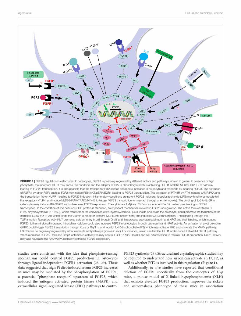
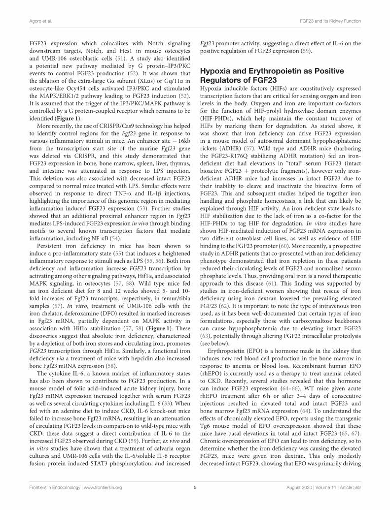
FIGURE 1 | FGF23 regulation in osteocytes. In osteocytes, FGF23 is positively regulated by different factors and pathways (shown in green). In presence of high
phosphate, the receptor FGFR1 may sense this condition and the adaptor FRS2α is phosphorylated thus activating FGFR1 and the MEK/pERK/EGR1 pathway
leading to FGF23 transcription. It is also possible that the transporter PiT2 senses phosphate increases in osteocyte and responds by inducing FGF23. The activation
of FGFR1 by other FGFs such as FGF2 may induce Pi3K/AKT/pERK/EGR1 leading to FGF23 upregulation. The activation of PTH1R by PTH induces cAMP/PKA and
the transcription factor NURR1 leading to FGF23 induction. Inflammatory conditions are potent FGF23 inducers; lipopolysaccharide (LPS) may bind to osteocyte toll
like receptor 4 (TLR4) and induce MyD88/IRAK/TRAF6/NF-κB to trigger FGF23 transcription (or may act through anemia/hypoxia). The binding of IL-6 to IL-6R in
osteocytes may induce JAK/STAT3 and subsequent FGF23 expression. The cytokines IL-1β and TNF-α can induce NF-κB in osteocytes leading to FGF23
transcription. In the condition of iron deficiency, HIF protein is stabilized, an important mechanism involved in FGF23 upregulation. The active form of vitamin D
(1,25-dihydroxyvitamin D; 1,25D), which results from the conversion of 25-hydroxyvitamin D (25D) inside or outside the osteocyte, could promote the formation of the
complex 1,25D-VDR-RXR which binds the vitamin D receptor element (VDRE, not shown here) and induces FGF23 transcription. The signaling through the
TGF-β-Activin Receptors ALK4/5/7 promotes calcium entry in cell through Orai1 and this process activates calcineurin and NFAT and their binding, which induces
FGF23. Lithium-induced increased intracellular calcium could also increase FGF23 in osteocytes through calcineurin and NFAT activity. An activation of a yet unknown
GPRC could trigger FGF23 transcription through XLαs or Gq/11α and inositol 1,4,5-trisphosphate (IP3) which may activate PKC and stimulate the MAPK pathway.
FGF23 can be negatively regulated by other elements and pathways (shown in red). For instance, insulin can bind to IGFR1 and induce PI3K/AKT/FOXO1 pathway
which represses FGF23. Phex and Dmp1 activities in osteocytes may control FGFR1/PI3KR1/GRB and cell differentiation to restrain FGF23 production. Dmp1 activity
may also neutralize the FAK/MAPK pathway restricting FGF23 expression.
studies were consistent with the idea that phosphate-sensingmechanisms could control FGF23 production in osteocytesthrough ligand-independent FGFR1 activation (28, 29). Thesedata suggested that high Pi diet-induced serum FGF23 increasesin mice may be mediated by the phosphorylation of FGFR1,a potential “phosphate receptor” upstream of FGF23, whichinduced the mitogen activated protein kinase (MAPK) andextracellular signal-regulated kinase (ERK) pathways to control
FGF23 synthesis (28). Structural and crystallographic studiesmaybe required to understand how an ion can activate an FGFR, aswell as whether PiT2 is involved in this regulation (Figure 1).
Additionally, in vivo studies have reported that conditionaldeletion of FGFR1 specifically from the osteocytes of Hypmice, a mouse model of X-linked hypophosphatemia (XLH)that exhibits elevated FGF23 production, improves the ricketsand osteomalacia phenotype of these mice in association
Frontiers in Endocrinology | www.frontiersin.org 3 August 2020 | Volume 11 | Article 592
Agoro et al. FGF23 and Its Kidney Function
with a decrease in both FGF23 bone mRNA and circulatingprotein. These mice also undergo an alleviation of theirhypophosphatemic status, as well as an increase of plasma1,25D levels (30). Furthermore, promoter studies identified thatthe activation of FGFR1 signaling through FGF2 is dependenton the PI3K-AKT pathway in the MC3T3-E1 osteoblastic cellline, providing evidence that Fgf23 gene promoter activityis induced by FGFR1 activation (30). In addition to geneticinterventions, pharmacologic-based studies have shown thatactivation of FGFR1 induces FGF23 production and leads tohypophosphatemia, whereas the inhibition of FGFR signalingattenuates FGF23 production (31) (Figure 1).
Another study by Hori et al. investigated whether phosphatecould induce reactive oxygen species (ROS) in vitro, leadingto increased FGF23 expression. Using the osteoblastic cell lineUMR-106, these investigators showed that elevated phosphatein the culture media enhances the production of ROS, and thathydrogen peroxide further boosts FGF23 production in a dose-dependent manner, an effect which can be neutralized by aninhibitor of NADPH oxidase (32). These discoveries suggestedthat in vitro phosphate directly enhanced FGF23 transcriptionby stimulating NADPH-induced ROS production and the MEK-ERK pathway. How this translates to the signaling in theosteocyte in vivo will be an interesting facet to this potentialregulatory network.
Circulating FGF23 ismarkedly elevated during chronic kidneydisease (CKD), and this is associated with poor long-termoutcomes. By investigating the regulation of FGF23 throughFGFR1, Hassan et al. demonstrated that the activation of FGFR1is essential for the high levels of FGF23 observed during bothacute and chronic uremia in mice and rats (33). In addition,in mice treated with the receptor FGFR1 inhibitor PD173074by oral gavage followed with an acute kidney injury inductionusing folic acid, the prevailing increased FGF23 was reducedby 50% in calvaria and led to a complete prevention of thecirculating intact FGF23 rises (33). In a more prolonged uremiccondition using a diet with adenine plus high-phosphorus for14 days to induce CKD, which resulted in high levels of Fgf23mRNA and serum FGF23 in rats, an oral gavage interventionwith PD173074 given during the last 2 days of treatmentreduced FGF23 induction by 75% in calvaria and completelynormalized circulating FGF23 (33). Therefore, the FGFRs mayplay an important role in osteocyte-bloodstream communicationto control FGF23 production.
Asmentioned above, FGF23 signals via the interaction with itsreceptor FGFR1c and its specific co-receptor Klotho (14, 34). Arecent investigation has shown that Klotho could be detected inosteocytes (35) suggesting that FGF23 may be one of the ligandsthat activates FGFR1 in osteocytes, thereby regulating the FGF23gene transcription in a positive feedback loop. The expression ofKlotho in osteocytes has been suggested to contribute to boneformation and bone volume increases coupled with enhancedosteoblast activity (35). Although Klotho has been detected inbone (35), the kidney, parathyroid glands and brain remainthe primary organs with abundant expression of Klotho tothe best of our current knowledge (36–38). However, it ispossible that in response to pathological circumstances, bone cells
could enhance Klotho expression, thus modulating bone FGFR1signaling via FGF23.
Through proteolytic cleavage of the membrane-bound klotho(mKL) (39–42), a soluble form of Klotho (sKL) can be liberatedinto the circulation (43, 44). Soluble Klotho has been describedto have a direct role to regulate Wnt and MAPK pathways inosteoblastic UMR-106 cells in concert with the presence of FGF23(45). Indeed, in vitro studies showed that a co-treatment ofUMR-106 cells with FGF23 and soluble Klotho activated MAPKsignaling, leading to an increase of Dickkopf-1 (DKK1) protein,a soluble inhibitor of Wnt/beta-catenin signaling (45). Theinduction of Dkk1 through FGF23/FGFR/sKL was shown to bedependent upon the MAPK pathway since the inhibition of thispathway using theMEK inhibitor U0126 completely abrogated p-ERK/ERK induction and abolished downstreamDkk1 expression(45). The binding of the secreted Dkk1 to the receptors Frizzled(Fz) and Lrp5/6 thus promoted the phosphorylation of β-cateninand inactivated the Wnt pathway in osteoblasts (45). Otherstudies have shown that a treatment of UMR-106 cells withsoluble Klotho and FGF23 dose-dependently increased MAPKand Egr1 mRNA responses, an effect which was FGFR- andMEK-dependent, and led to FGF23 upregulation (46). Futurestudies are needed to confirm potential expression of Klothoin osteocytes and under some circumstances whether FGF23binding to FGFR1-Klotho complex in osteocytes could induceFGF23 expression.
Signals Involved in the Positive Regulationof FGF23 by Inflammation and IronInflammation is a complex phenomenon involving multipleimmune and non-immune cells which cooperate to respondto endogenous and exogenous events by secreting specificpro-inflammatory and/or anti-inflammatory factors. Pro-inflammatory stimuli such as the cytokines tumor necrosis factoralpha (TNFα), interleukin 1β (IL-1β), the tumor necrosis factor-like weak inducer of apoptosis (TWEAK), and the bacterialcomponent lipopolysaccharide (LPS) have all been shown todose-dependently upregulate FGF23 in the osteocyte-like cellline IDG-SW3 (47). Particularly, TNF and IL-1β induce FGF23expression via nuclear factor kappa-light-chain-enhancer ofactivated B cells (NF-κB) (47), a major transcription factorcomplex involved in the control of cytokines and in themediation of multiple pro-inflammatory cellular responses.The serine/threonine kinase p38 mitogen-activated proteinkinase (p38MAPK), which is activated by several cellular stressstimuli and involved in the transcriptional activity of NF-κB, isanother positive regulator of FGF23 in bone cells (48). Otherinvestigations have shown that the serine/threonine kinaseprotein kinase C (PKC), which drives FGF23 expression inresponse to phorbol ester 12-O-tetradecanoylphorbol-13-acetate(PMA), can be suppressed by 75% in the presence of the PKCα/βinhibitor Go6976. These studies suggested that PKC is a positiveregulator of FGF23 synthesis in IDG-SW3 osteocytic cellsvia NF-κB (49). Furthermore, Notch signaling, which can bemediated by pro-inflammatory cytokines such as TNF-α (50),has been described by Tamamura et al., to positively regulate
Frontiers in Endocrinology | www.frontiersin.org 4 August 2020 | Volume 11 | Article 592
Agoro et al. FGF23 and Its Kidney Function
FGF23 expression which colocalizes with Notch signalingdownstream targets, Notch, and Hes1 in mouse osteocytesand UMR-106 osteoblastic cells (51). A study also identifieda potential new pathway mediated by G protein–IP3/PKCevents to control FGF23 production (52). It was shown thatthe ablation of the extra-large Gα subunit (XLαs) or Gq/11α inosteocyte-like Ocy454 cells activated IP3/PKC and stimulatedthe MAPK/ERK1/2 pathway leading to FGF23 induction (52).It is assumed that the trigger of the IP3/PKC/MAPK pathway iscontrolled by a G protein-coupled receptor which remains to beidentified (Figure 1).
More recently, the use of CRISPR/Cas9 technology has helpedto identify control regions for the Fgf23 gene in response tovarious inflammatory stimuli in mice. An enhancer site − 16kbfrom the transcription start site of the murine Fgf23 genewas deleted via CRISPR, and this study demonstrated thatFGF23 expression in bone, bone marrow, spleen, liver, thymus,and intestine was attenuated in response to LPS injection.This deletion was also associated with decreased intact FGF23compared to normal mice treated with LPS. Similar effects wereobserved in response to direct TNF-α and IL-1β injections,highlighting the importance of this genomic region in mediatinginflammation-induced FGF23 expression (53). Further studiesshowed that an additional proximal enhancer region in Fgf23mediates LPS-induced FGF23 expression in vivo through bindingmotifs to several known transcription factors that mediateinflammation, including NF-κB (54).
Persistent iron deficiency in mice has been shown toinduce a pro-inflammatory state (55) that induces a heightenedinflammatory response to stimuli such as LPS (55, 56). Both irondeficiency and inflammation increase FGF23 transcription byactivating among other signaling pathways, Hif1α, and associatedMAPK signaling, in osteocytes (57, 58). Wild type mice fedan iron deficient diet for 8 and 12 weeks showed 5- and 10-fold increases of Fgf23 transcripts, respectively, in femur/tibiasamples (57). In vitro, treatment of UMR-106 cells with theiron chelator, deferoxamine (DFO) resulted in marked increasesin Fgf23 mRNA, partially dependent on MAPK activity inassociation with Hif1α stabilization (57, 58) (Figure 1). Thesediscoveries suggest that absolute iron deficiency, characterizedby a depletion of both iron stores and circulating iron, promotesFGF23 transcription through Hif1α. Similarly, a functional irondeficiency via a treatment of mice with hepcidin also increasedbone Fgf23 mRNA expression (58).
The cytokine IL-6, a known marker of inflammatory stateshas also been shown to contribute to FGF23 production. In amouse model of folic acid–induced acute kidney injury, boneFgf23 mRNA expression increased together with serum FGF23as well as several circulating cytokines including IL-6 (33). Whenfed with an adenine diet to induce CKD, IL-6 knock-out micefailed to increase bone Fgf23 mRNA, resulting in an attenuationof circulating FGF23 levels in comparison to wild-type mice withCKD; these data suggest a direct contribution of IL-6 to theincreased FGF23 observed during CKD (59). Further, ex vivo andin vitro studies have shown that a treatment of calvaria organcultures and UMR-106 cells with the IL-6/soluble IL-6 receptorfusion protein induced STAT3 phosphorylation, and increased
Fgf23 promoter activity, suggesting a direct effect of IL-6 on thepositive regulation of FGF23 expression (59).
Hypoxia and Erythropoietin as PositiveRegulators of FGF23Hypoxia inducible factors (HIFs) are constitutively expressedtranscription factors that are critical for sensing oxygen and ironlevels in the body. Oxygen and iron are important co-factorsfor the function of HIF-prolyl hydroxylase domain enzymes(HIF-PHDs), which help maintain the constant turnover ofHIFs by marking them for degradation. As stated above, itwas shown that iron deficiency can drive FGF23 expressionin a mouse model of autosomal dominant hypophosphatemicrickets (ADHR) (57). Wild type and ADHR mice (harboringthe FGF23-R176Q stabilizing ADHR mutation) fed an iron-deficient diet had elevations in “total” serum FGF23 (intactbioactive FGF23 + proteolytic fragments), however only iron-deficient ADHR mice had increases in intact FGF23 due totheir inability to cleave and inactivate the bioactive form ofFGF23. This and subsequent studies helped tie together ironhandling and phosphate homeostasis, a link that can likely beexplained through HIF activity. An iron-deficient state leads toHIF stabilization due to the lack of iron as a co-factor for theHIF-PHDs to tag HIF for degradation. In vitro studies haveshown HIF-mediated induction of FGF23 mRNA expression intwo different osteoblast cell lines, as well as evidence of HIFbinding to the FGF23 promoter (60).More recently, a prospectivestudy in ADHR patients that co-presented with an iron deficiencyphenotype demonstrated that iron repletion in these patientsreduced their circulating levels of FGF23 and normalized serumphosphate levels. Thus, providing oral iron is a novel therapeuticapproach to this disease (61). This finding was supported bystudies in iron-deficient women showing that rescue of irondeficiency using iron dextran lowered the prevailing elevatedFGF23 (62). It is important to note the type of intravenous ironused, as it has been well-documented that certain types of ironformulations, especially those with carboxymaltose backbonescan cause hypophosphatemia due to elevating intact FGF23(63), potentially through altering FGF23 intracellular proteolysis(see below).
Erythropoietin (EPO) is a hormone made in the kidney thatinduces new red blood cell production in the bone marrow inresponse to anemia or blood loss. Recombinant human EPO(rhEPO) is currently used as a therapy to treat anemia relatedto CKD. Recently, several studies revealed that this hormonecan induce FGF23 expression (64–66). WT mice given acuterhEPO treatment after 6 h or after 3–4 days of consecutiveinjections resulted in elevated total and intact FGF23 andbone marrow Fgf23 mRNA expression (64). To understand theeffects of chronically elevated EPO, reports using the transgenicTg6 mouse model of EPO overexpression showed that thesemice have basal elevations in total and intact FGF23 (65, 67).Chronic overexpression of EPO can lead to iron deficiency, so todetermine whether the iron deficiency was causing the elevatedFGF23, mice were given iron dextran. This only modestlydecreased intact FGF23, showing that EPO was primarily driving
Frontiers in Endocrinology | www.frontiersin.org 5 August 2020 | Volume 11 | Article 592
Agoro et al. FGF23 and Its Kidney Function
FGF23 expression (65). In healthy human subjects, total FGF23was elevated 24 h after a single rhEPO dose with no changesin intact FGF23 (66). In another study, a small population ofanemic patients with normal kidney function were given a singledose of rhEPO. This increased total and intact FGF23 over thespan of 12–18 h after the injection (64). Few in vivo studieshave examined the effect of curing anemia of CKD on mineralmetabolism. Most recently, a single rhEPO injection in CKDmice did increase total serum FGF23 after 6 and 24 h but had noeffect on intact FGF23 (65).
To mitigate potential adverse effects of high rhEPO treatment,new strategies are leveraging the HIF system by creatinginhibitors of the HIF prolyl hydroxylases (HIF-PHDs) calledHIF-PHDs inhibitors (HIF-PHI). These therapeutics havebecome increasingly important for anemic patients with CKD,where they promote endogenous EPO production by stabilizingHIFs. This class of drug also reduce hepcidin levels to increaseiron utilization in tissues thereby creating a synergistic effectin providing iron availability to newly forming red blood cells(68). Recent studies have shown in normal mice that severalHIF-PHIs can regulate Fgf23 expression. For instance, FG-4592(Roxadustat) was shown to increase bone Fgf23 mRNA andcirculating intact FGF23 (58, 66). Elevations in total and intactFGF23 were observed in WT mice treated with the HIF-PHIBAY 85-3934 (Molidustat) after 6 h that returned to baseline by24 h. This study showed that elevated EPO precedes increases inFGF23 in response to HIF-PHI BAY85-3934, suggesting that thecause for elevated FGF23 in response to this treatment was dueto elevated EPO. This was confirmed by treating HIF-PHI micewith an EPO-neutralizing antibody, which completely attenuatedthe increased serum total FGF23 (69). A recent paper, however,showed that EPO and HIF-PHI treatment of anemic mice withCKD resulted in suppressed FGF23. This study suggested thatunder conditions of anemia, as opposed to mice with normaliron homeostasis, rescuing iron utilization during CKD may bea more potent suppressor of FGF23 than EPO and HIF-PHIsare stimulators (70). Although EPO and HIF-PHI can induceFGF23, further studies are required to delineate the mechanismsdirecting these components on osteocytic FGF23 production.
Signals Involved in the TranscriptionalRegulation of FGF23 by PTHParathyroid hormone (PTH) is a hormone secreted by theparathyroid glands that regulates calcium utilization throughits effects on bone, kidney, and intestine (71–73). DuringCKD, a secondary hyperparathyroidism occurs in whichPTH is excessively secreted, in response to factors such ashyperphosphatemia, hypocalcemia, and low 1,25D levels, topotentially promote elevated FGF23 (74). Using bioinformaticsand chromatin immunoprecipitation assays, studies havereported that Nurr1 is an essential transcription factor involvedin FGF23 upregulation in response to PTH in UMR-106 cells.Furthermore, in a mouse model of CKD, the administration ofa calcimimetic, which is known to activate the calcium-sensingreceptor in different tissues (75) with the goal to attenuate PTHlevels and actions, reduced FGF23 concentrations as well as bone
Nurr1 mRNA and protein levels (76). To test the relationshipsbetween PTH and FGF23, a mouse model with constitutiveactivation of PTH receptor (PTHR) signaling in osteocytes wasused by Rhee et al. in a report that showed that PTHR activationincreased FGF23 expression in vivo and in vitro through cAMPand Wnt-dependent mechanisms (77). In addition, Knab et al.showed that PTH-induced increases in FGF23 expression werePKA-dependent in osteocyte-like cells, suggesting that FGF23production is regulated by the cAMP/PKA/Nurr1 pathway inresponse to PTH (78) (Figure 1).
Signals Involved in the Positive Regulationof FGF23 by TGF-β, Calcineurin, and NFATTGF-β has been reported to regulate the extracellular matrixby activating ROS, which increases calcium influx and activatescalcineurin (79–81). Using UMR-106 cells, TGF-β2 has beendescribed to enhance store-operated Ca2+ entry (SOCE)and induce the stimulation of FGF23, an effect significantlyattenuated by both the inhibitor of TGF-β type I receptor activinreceptor-like kinases (ALK5, ALK4, and ALK7) SB431542 andSOCE inhibitor 2-APB (82). Recent investigation has shownthat the synthesis of FGF23 in UMR-106 cells can be inducedby SOCE through Orai1 (83). In addition, the Ca2+ entryactivates the phosphatase calcineurin, which dephosphorylatesnuclear factor of activated T cells (NFAT) thereby stimulatingits transcriptional activity and targeting the Fgf23 gene. Thissuggested that FGF23 may be positively regulated by calcineurin-NFAT signaling (84). Further analyses confirmed that eitherthe inhibition of calcineurin using ciclosporin A (CsA) andtacrolimus (FK-506) or the blocking of the interaction betweencalcineurin and NFAT using the inhibitor INCA-6 reduced theabundance of Fgf23 transcripts as well as FGF23 protein (84).Additionally, lithium, a widely used drug for the treatment ofmood disorders and known to modify Ca2+ signaling, stimulatedthe release of FGF23, partially through NF-κB dependent up-regulation of Orai1 transcription and SOCE in UMR-106 cells(85) (Figure 1).
Signals Involved in the PositiveTranscriptional Regulation of FGF23 byCalcitriolEarly FGF23 physiological studies demonstrated that theadministration of 1,25D dose-dependently increased both serumFGF23 as well as serum inorganic phosphorus in normal rats(25). In wild type mice with normal renal function, injection ofcalcitriol increased serum FGF23 levels 1-week post treatmentby 15-fold. Calcidiol (25-hydroxyvitamin D), although with alesser effect could also induce FGF23 in normal mice (86). In amouse model of adenine diet-induced CKD, a 5 weeks-regimeninduced a 40-fold increase of circulating FGF23. However, in thebackground of a global deletion of Cyp27b1, the gene encodingthe enzyme vitamin D 1-α-hydroxylase involved in the formationof calcitriol (1(OH)ase−/− mice), only a 2-fold circulating FGF23was observed with this treatment, suggesting a contribution ofcalcitriol to the increased FGF23. At the bone compartment level,a specific deletion of Cyp27b1 in osteoblasts reduced FGF23
Frontiers in Endocrinology | www.frontiersin.org 6 August 2020 | Volume 11 | Article 592
Agoro et al. FGF23 and Its Kidney Function
induction in long bones from 58-fold in normal mice treatedwith a 5-weeks adenine diet to a 10-fold induction, suggesting apotential role of a local osteoblastic vitamin D conversion processin the induction of bone FGF23. This attenuation of increasedFGF23 was independent of a potential reduction in PTH levelssince plasma PTH remained elevated in response to the adeninediet-induced CKD in the mice with global deletion of Cyp27b1,as well as those with osteoblast-specific deletion of Cyp27b1 (86)(Figure 1).
NEGATIVE REGULATORS OF FGF23
Dentin Matrix Acidic Phosphoprotein 1 andPhosphate-Regulating Gene WithHomologies to Endopeptidases on the XChromosomeThe disease XLH is characterized by hypophosphatemia andimpaired mineralization caused by mutations of the phosphate-regulating gene with homologies to endopeptidases on the Xchromosome (PHEX). Loss of PHEX leads to the overproductionof FGF23 in osteocytes, causing hypophosphatemia with bonemineralization impairment, and thus bone fragility. Similarto XLH, recessive loss-of-function mutations in the dentinmatrix protein-1 (DMP1) gene, a member of small integrin-binding ligand N-linked glycoprotein (SIBLING) proteins,is responsible for a human phosphate wasting and impairedbone mineralization disease termed autosomal recessivehypophosphatemic rickets type 1 (ARHR1). It was shown thata lack of DMP1 in both humans and mice markedly increasedFGF23 expression in bone (87).
To gain insight into the mechanisms by which PHEXmutations upregulate FGF23 expression, studies have beendesigned to investigate the local effects in bone from a mousemodel of XLH (Hyp mice) in a normal hormonal environmentin comparison to the function of wild type bone in the abnormalmetabolic environment of Hyp mice. Using a surgical procedureto perform intramuscular bone cross-transplantations betweenwild-type and Hyp mice, a study found that increased FGF23expression in Hyp bone results from intrinsic PHEX deficiencyfrom bone, since FGF23 was increased in Hyp osteocytes beforeand after explantation into WT mice but was not increased inWT osteocytes after explantation into Hyp mice. This evidencesuggested that the mechanisms whereby PHEX mutations leadto increased FGF23 expression in osteocytes is intrinsic tobone (88).
Similar to the phenotype resulting from PHEX inactivation,the inactivation of Dmp1 in mice resulted in equivalent intrinsicbone mineralization defects associated with increased FGF23expression in osteocytes (89–91). Using cortical bone isolatedfrom 12-days old WT, Hyp, Dmp1−/−, and Hyp/Dmp1−/−
mice to perform a genome-wide microarray analysis, thephosphatidylinositol 3-kinase regulatory α subunit (PIK3R1)and growth factor receptor-bound protein 2 (GRB2) pathwayswere identified as potential common signaling controlled byPHEX and DMP1 to regulate FGF23 promoter activity throughFGFs/FGFR in osteocytes (91, 92) (Figure 1). These findings
highlight that the activation of FGFRs which contribute toFGF23 production in osteocytes may be independent fromthe phosphate sensing pathways described above. Additionally,recent in vivo and in vitro studies suggested a direct negativeregulation of DMP1 in FGF23 expression in osteocytes byactivating FAK-mediated MAPK signaling, thus coordinating theextracellular environment of osteocytic lacunae as well as bonemetabolism (93) (Figure 1). The creation of the floxed-Fgf23mouse has recently emerged as a critical tool to understandFGF23 function in vivo (94). To this end, flox-Fgf23 mice weremated to the global eIIa-cre which mimicked the phenotypeof the Fgf23-KO mouse. Fgf23 was also specifically deletedfrom either the osteoblast lineage using the Col2.3 promoter todrive Cre expression, or from late osteoblasts/osteocytes usingthe Dmp1-Cre. This resulted in ∼50% reduction of iFGF23,with compromised ability to respond to changes in phosphate(94), demonstrating the specificity of osteoblast/osteocyte FGF23production in response to metabolic changes.
Negative Regulation of FGF23 by Insulinand Insulin-Like Growth Factor 1Recent studies reported insulin and insulin-like growth factor1 (IGF1) as negative regulators of FGF23 production in vitroas well as in mice and humans (95). In vitro, insulin andIGF1 down-regulated FGF23 production by inhibiting thetranscription factor forkhead box protein O1 (FOXO1) throughphosphoinositide 3-kinase (PI3K)/protein kinase B (PKB)/Aktsignaling in UMR-106 cells (95). In vivo, insulin deficiencyresulted in an increase of serum FGF23 concentrations in mice,which was reversed by insulin administration. Interestingly, inwomen subjects, an increase in plasma insulin levels followingan oral glucose administration correlated negatively with plasmaFGF23 concentrations (95) (Figure 1).
FGF23 CLEAVAGE: A PHYSIOLOGICALAND ENDOGENOUS MECHANISM TOATTENUATE FGF23 BIOACTIVITY
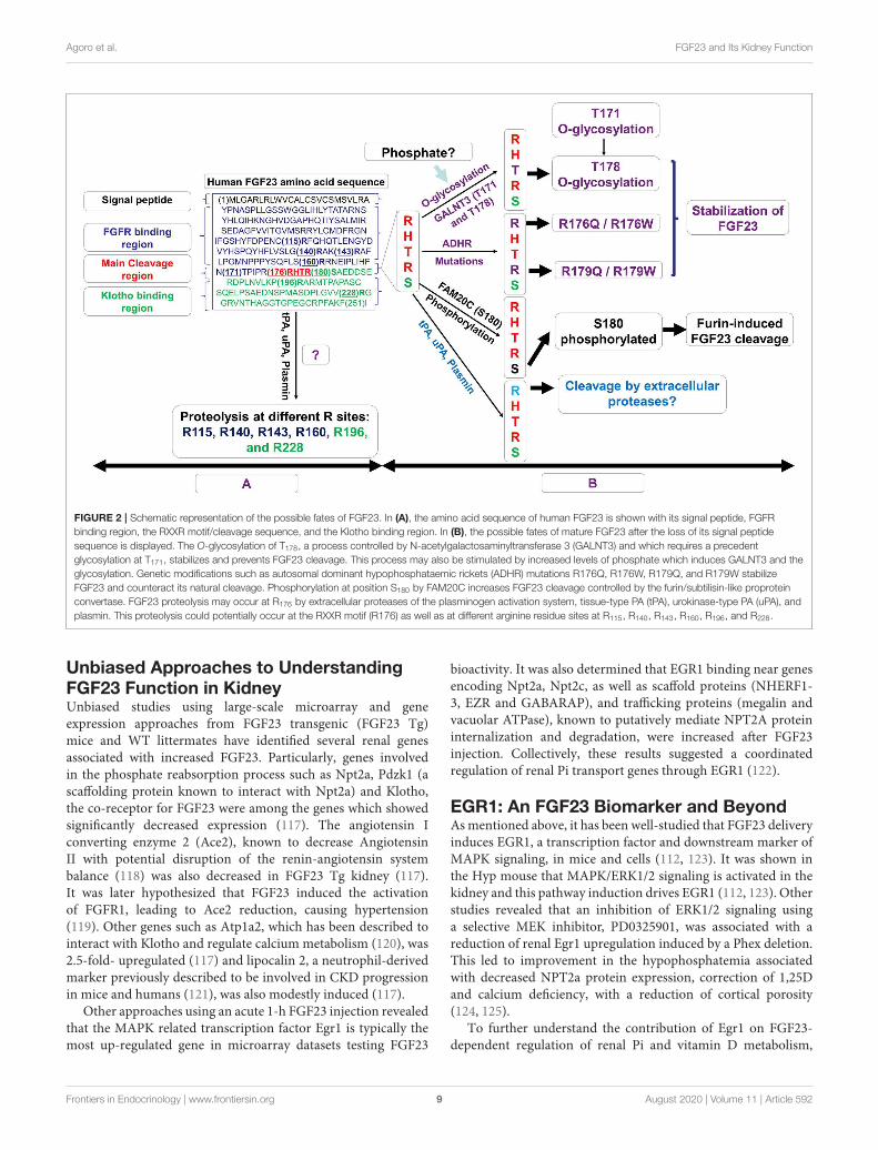
FGF23 is synthesized as a 251-amino acid protein, primarilyin osteocytes. The FGF23 signal peptide is represented by thefirst N-terminal 24 aa. The cleavage of the signal peptide resultsin a release of a mature peptide that can be secreted as thebioactive hormone, referred to as “intact” FGF23 (“iFGF23”).To control the bioactivity of iFGF23, the protein can becleaved at the subtilisin-like proprotein convertase (SPC) siteR176HTR179/S180AE (RXXR/SAE motif) generating at leasttwo fragments identified as an N-terminal fragment whichis structurally similar to other FGF family members and amore unique C-terminal tail (21, 96). The proteolytic cleavageof excess iFGF23 represents a critical secondary regulatorymechanism to maintain stable serum iFGF23 and normalserum phosphate. The absence of this proteolytic activity inhumans through the FGF23 gene mutations R176Q, R176W,R179Q, and R179W are causative for autosomal dominanthypophosphatemic rickets (ADHR), characterized by elevatedintact FGF23 and hypophosphatemia (21) (Figure 2). In mice
Frontiers in Endocrinology | www.frontiersin.org 7 August 2020 | Volume 11 | Article 592
Agoro et al. FGF23 and Its Kidney Function
carrying the ADHR point mutation R176Q-Fgf23, in response toabsolute iron deficiency using dietary intervention, the iFGF23levels were elevated due to the stabilization of the bioactiveFGF23 (57). This elevated iFGF23 condition in response toiron-deficient diet in ADHR mice caused similar phenotypesas observed in ADHR/XLH patients, such as alterations ingenes controlling phosphate reabsorption and 1,25D production,and a hypophosphatemic bone disease (57). These findingssuggested that iron status is a synergistic factor of the ADHRphenotype, and that ADHR is a disease of gene-environmentinteractions (57).
The cleavage of FGF23 is controlled by the serineendoprotease furin, a subtilisin-like convertase, at the siteR179/S180 in response to several stimuli including PTH (78).Previous investigations have shown that an iron deficientstate also promotes iFGF23 cleavage leading to increasedsecretion of FGF23 fragments but normal iFGF23 in WTmice (57, 58). In HeLa Cells, it was described that an irondeficient state induces furin upregulation via the stabilizationof Hif1α (97), a similar mechanism which occurs in osteocytes.Posttranslational modifications of iFGF23 can occur via theO-glycosylation of Thr178 in the furin proprotein processingmotif RHT178R179, which stabilized FGF23 (98) (Figure 2).This glycosylation process at Thr178 is controlled by theenzymatic actions of N-acetylgalactosaminyltransferase3 (GALNT3) which can be upregulated under highphosphate conditions potentially via the control of FGFR1cactivation and the induction of the transcriptional activatorsearly growth response 1 (EGR1) and ETS variant 5(ETV5) (28).
In a recent study, Thr178 was identified as a poor substratesite with limited glycosylation acceptance, likely a protectivemechanism to prevent cellular resistances to FGF23 cleavage.Interestingly, Thr178 glycosylation was shown to require aprevious glycosylation at Thr171 before generating a furin-resistant and secreted stable iFGF23 (99) (Figure 2). Thesenew discoveries suggest that GALNT3 specificity for FGF23and its ability to control circulating levels of bioactiveFGF23 is a control point achieved by FGF23 being a ratherpoor substrate for this enzyme (99). In contrast to the O-glycosylation induced by GALNT3 which stabilizes FGF23,the phosphorylation at position S180 by the kinase FAM20Cinhibited O-glycosylation of FGF23, thus promoting FGF23cleavage (98) (Figure 2). Indeed, recessive inactivating mutationsin human FAM20C cause ARHR type 3 (ARHR3; Rainessyndrome), consistent with its role as an FGF23 de-stabilizer(100, 101).
Besides furin which is thought to cleave FGF23 mainlyintracellularly, the extracellular proteases of the plasminogenactivation system, tissue-type PA (tPA), and urokinase-type PA(uPA), as well as plasmin may also display proteolytic activityon FGF23 protein at the RXXR motif (R176), with potentialadditional cleavages at arginine residues R114, R140, R143, R160,R196, and R228 (102). Of note, Klotho knock out mice aswell as mice with acute kidney injury, which both exhibitelevated levels of intact FGF23, also display elevated plasminogenactivator inhibitor-1 (103, 104). Thus, the proteolysis of FGF23
by tPA, uPA, and plasmin may potentially regulate the levelsof active FGF23 thus in part, controlling phosphate andmineral homeostasis.
These collective studies demonstrated that FGF23 proteincleavage is a dynamic process which can be adjusted afterproduction at the cell level with phosphorylation andglycosylation dictating the levels of bioactive FGF23 dependingupon the osteocyte cell state. Certainly, future studies are neededto understand the regulation of GALNT3, furin, FAM20C, andpotentially the enzymes of the plasminogen activation system inthe coordinated control of FGF23 production and bioactivity.
FGF23 RENAL SIGNAL TRANSDUCTION
In contrast to paracrine FGFs, such as FGF1 and FGF2, endocrineFGFs such as FGF23 lack the heparin-binding domain intheir C-terminus, which enables escape from the osteocyte cellmatrix after secretion and their actions on distant target organsincluding kidney (14, 105). FGF23 acts primarily on kidney topromote phosphaturia and parallel reductions in 1,25D (14, 34).The phosphaturic activity of FGF23 is critical in CKD, preventingand delaying hyperphosphatemia and vascular calcificationsas FGF23 progressively rises with the loss of renal function(106). In a study among patients undergoing hemodialysis,high serum phosphate levels across a quartile of patients (>5.5mg/dl) was associated with a 20% increase in the multivariableadjusted risk of death, as compared with normal levels (3.5–4.5 mg/dl) (107). These findings underscore the importanceof controlling circulating phosphate in kidney disease patients.During CKD, as kidney function decreases with a progressivedecrease of glomerular filtration rate (GFR), the kidney losesfunctioning nephrons decreasing the overall excretion capabilityof the kidney. The dramatic rise of FGF23 during CKD islikely to attempt to boost the excretory capacity of the existingnephrons to maintain normal serum phosphate levels. This islikely the primary reason why early stage CKD patients exhibitnormal serum phosphate over much of the disease course [forreview (108)].
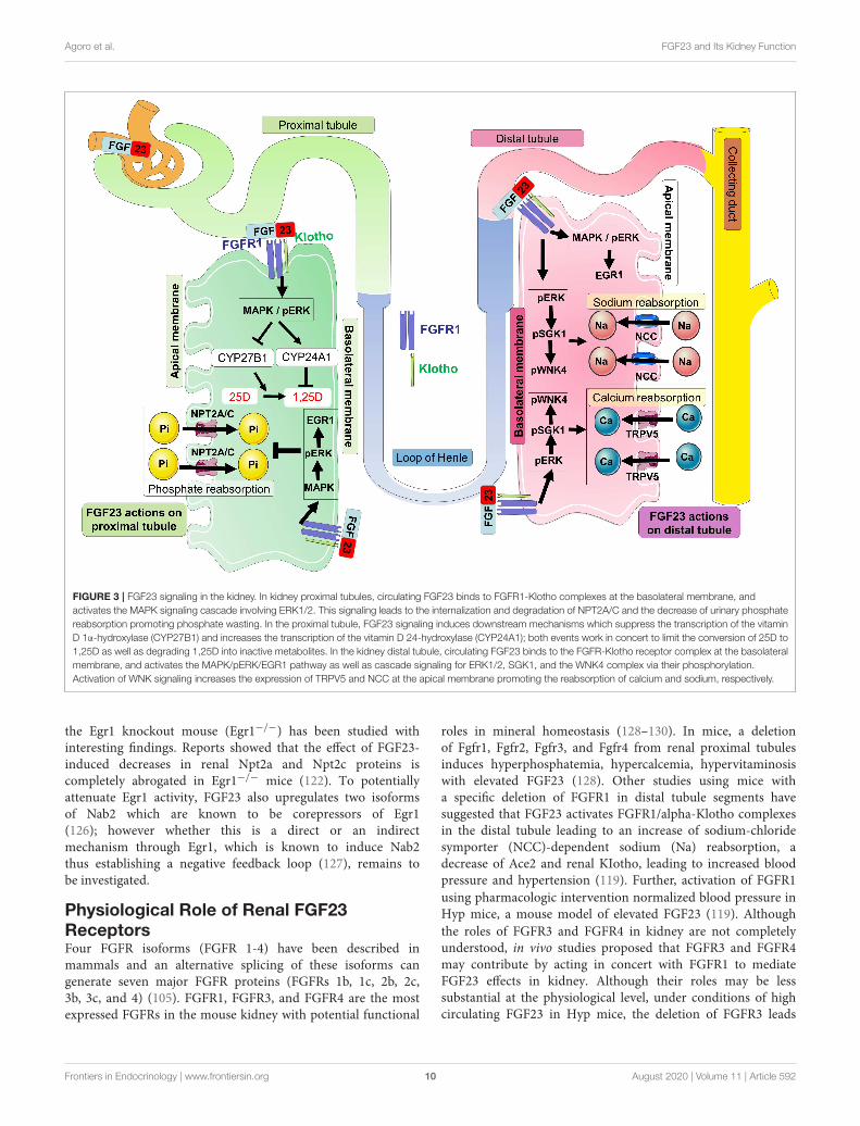
FGF23 actions are likely mediated by FGF receptors FGFR1c,FGFR3c, and FGFR4 and the co-receptor Klotho, a single-passtransmembrane protein highly expressed in kidney (109–113).FGF23 binds to FGFR1c and the interaction between thesetwo proteins with the co-receptor Klotho (which dramaticallyincreases the binding affinity of the complex FGF23-FGFR-Klotho) triggers potent FGF23 signaling and activity (114). Inkidney, FGF23 signaling on the basolateral side of proximalnephron cells causes the internalization of the sodium-dependentphosphate co-transporters NPT2A and NPT2C from the apicalsurface. These actions decrease phosphate reabsorption processesfrom the kidney (115) (Figure 3). A global genetic deletion ofFGF23 in mice resulted in severe hyperphosphatemia, due to theabsence of the FGF23-mediated phosphaturia mechanism (116).In addition, FGF23 signaling regulates vitamin Dmetabolism viathe modulation of the vitamin D metabolic enzymes expressionas well as regulating calcium and sodium reabsorption processes(Figure 3).
Frontiers in Endocrinology | www.frontiersin.org 8 August 2020 | Volume 11 | Article 592
Agoro et al. FGF23 and Its Kidney Function
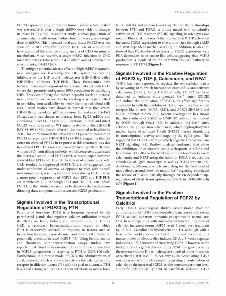
FIGURE 2 | Schematic representation of the possible fates of FGF23. In (A), the amino acid sequence of human FGF23 is shown with its signal peptide, FGFR
binding region, the RXXR motif/cleavage sequence, and the Klotho binding region. In (B), the possible fates of mature FGF23 after the loss of its signal peptide
sequence is displayed. The O-glycosylation of T178, a process controlled by N-acetylgalactosaminyltransferase 3 (GALNT3) and which requires a precedent
glycosylation at T171, stabilizes and prevents FGF23 cleavage. This process may also be stimulated by increased levels of phosphate which induces GALNT3 and the
glycosylation. Genetic modifications such as autosomal dominant hypophosphataemic rickets (ADHR) mutations R176Q, R176W, R179Q, and R179W stabilize
FGF23 and counteract its natural cleavage. Phosphorylation at position S180 by FAM20C increases FGF23 cleavage controlled by the furin/subtilisin-like proprotein
convertase. FGF23 proteolysis may occur at R176 by extracellular proteases of the plasminogen activation system, tissue-type PA (tPA), urokinase-type PA (uPA), and
plasmin. This proteolysis could potentially occur at the RXXR motif (R176) as well as at different arginine residue sites at R115, R140, R143, R160, R196, and R228.
Unbiased Approaches to UnderstandingFGF23 Function in KidneyUnbiased studies using large-scale microarray and geneexpression approaches from FGF23 transgenic (FGF23 Tg)mice and WT littermates have identified several renal genesassociated with increased FGF23. Particularly, genes involvedin the phosphate reabsorption process such as Npt2a, Pdzk1 (ascaffolding protein known to interact with Npt2a) and Klotho,the co-receptor for FGF23 were among the genes which showedsignificantly decreased expression (117). The angiotensin Iconverting enzyme 2 (Ace2), known to decrease AngiotensinII with potential disruption of the renin-angiotensin systembalance (118) was also decreased in FGF23 Tg kidney (117).It was later hypothesized that FGF23 induced the activationof FGFR1, leading to Ace2 reduction, causing hypertension(119). Other genes such as Atp1a2, which has been described tointeract with Klotho and regulate calcium metabolism (120), was2.5-fold- upregulated (117) and lipocalin 2, a neutrophil-derivedmarker previously described to be involved in CKD progressionin mice and humans (121), was also modestly induced (117).
Other approaches using an acute 1-h FGF23 injection revealedthat the MAPK related transcription factor Egr1 is typically themost up-regulated gene in microarray datasets testing FGF23
bioactivity. It was also determined that EGR1 binding near genesencoding Npt2a, Npt2c, as well as scaffold proteins (NHERF1-3, EZR and GABARAP), and trafficking proteins (megalin andvacuolar ATPase), known to putatively mediate NPT2A proteininternalization and degradation, were increased after FGF23injection. Collectively, these results suggested a coordinatedregulation of renal Pi transport genes through EGR1 (122).
EGR1: An FGF23 Biomarker and BeyondAsmentioned above, it has been well-studied that FGF23 deliveryinduces EGR1, a transcription factor and downstream marker ofMAPK signaling, in mice and cells (112, 123). It was shown inthe Hyp mouse that MAPK/ERK1/2 signaling is activated in thekidney and this pathway induction drives EGR1 (112, 123). Otherstudies revealed that an inhibition of ERK1/2 signaling usinga selective MEK inhibitor, PD0325901, was associated with areduction of renal Egr1 upregulation induced by a Phex deletion.This led to improvement in the hypophosphatemia associatedwith decreased NPT2a protein expression, correction of 1,25Dand calcium deficiency, with a reduction of cortical porosity(124, 125).
To further understand the contribution of Egr1 on FGF23-dependent regulation of renal Pi and vitamin D metabolism,
Frontiers in Endocrinology | www.frontiersin.org 9 August 2020 | Volume 11 | Article 592
Agoro et al. FGF23 and Its Kidney Function
FIGURE 3 | FGF23 signaling in the kidney. In kidney proximal tubules, circulating FGF23 binds to FGFR1-Klotho complexes at the basolateral membrane, and
activates the MAPK signaling cascade involving ERK1/2. This signaling leads to the internalization and degradation of NPT2A/C and the decrease of urinary phosphate
reabsorption promoting phosphate wasting. In the proximal tubule, FGF23 signaling induces downstream mechanisms which suppress the transcription of the vitamin
D 1α-hydroxylase (CYP27B1) and increases the transcription of the vitamin D 24-hydroxylase (CYP24A1); both events work in concert to limit the conversion of 25D to
1,25D as well as degrading 1,25D into inactive metabolites. In the kidney distal tubule, circulating FGF23 binds to the FGFR-Klotho receptor complex at the basolateral
membrane, and activates the MAPK/pERK/EGR1 pathway as well as cascade signaling for ERK1/2, SGK1, and the WNK4 complex via their phosphorylation.
Activation of WNK signaling increases the expression of TRPV5 and NCC at the apical membrane promoting the reabsorption of calcium and sodium, respectively.
the Egr1 knockout mouse (Egr1−/−) has been studied withinteresting findings. Reports showed that the effect of FGF23-induced decreases in renal Npt2a and Npt2c proteins iscompletely abrogated in Egr1−/− mice (122). To potentiallyattenuate Egr1 activity, FGF23 also upregulates two isoformsof Nab2 which are known to be corepressors of Egr1(126); however whether this is a direct or an indirectmechanism through Egr1, which is known to induce Nab2thus establishing a negative feedback loop (127), remains tobe investigated.
Physiological Role of Renal FGF23ReceptorsFour FGFR isoforms (FGFR 1-4) have been described inmammals and an alternative splicing of these isoforms cangenerate seven major FGFR proteins (FGFRs 1b, 1c, 2b, 2c,3b, 3c, and 4) (105). FGFR1, FGFR3, and FGFR4 are the mostexpressed FGFRs in the mouse kidney with potential functional
roles in mineral homeostasis (128–130). In mice, a deletionof Fgfr1, Fgfr2, Fgfr3, and Fgfr4 from renal proximal tubulesinduces hyperphosphatemia, hypercalcemia, hypervitaminosiswith elevated FGF23 (128). Other studies using mice witha specific deletion of FGFR1 in distal tubule segments havesuggested that FGF23 activates FGFR1/alpha-Klotho complexesin the distal tubule leading to an increase of sodium-chloridesymporter (NCC)-dependent sodium (Na) reabsorption, adecrease of Ace2 and renal KIotho, leading to increased bloodpressure and hypertension (119). Further, activation of FGFR1
using pharmacologic intervention normalized blood pressure inHyp mice, a mouse model of elevated FGF23 (119). Although
the roles of FGFR3 and FGFR4 in kidney are not completelyunderstood, in vivo studies proposed that FGFR3 and FGFR4may contribute by acting in concert with FGFR1 to mediateFGF23 effects in kidney. Although their roles may be lesssubstantial at the physiological level, under conditions of highcirculating FGF23 in Hyp mice, the deletion of FGFR3 leads
Frontiers in Endocrinology | www.frontiersin.org 10 August 2020 | Volume 11 | Article 592
Agoro et al. FGF23 and Its Kidney Function
to a feedback stimulation of Fgf23 mRNA expression in bone(129, 131), suggesting complex kidney-bone crosstalk.
The co-receptor KL is required for high-affinity FGF23activity in the kidney. Whole-nephron deletion of Klothoin mice results in renal FGF23 resistance, characterized byhypervitaminosis D, hyperphosphatemia, and other phenotypesthat may resemble premature aging (39, 132). However, provisionof a low phosphate diet to KL-null mice reversed the severephenotypes, showing that the majority of KL-null phenotypesare due to extreme phosphate imbalances (133, 134). Klotho ishighly expressed in the distal tubule segments of the nephronin comparison to its relatively modest expression in proximaltubules (123) although the phosphate reabsorption occursprimarily in the proximal tubules due to an abundant expressionof the sodium phosphate transporters Npt2a and Npt2c (135–137). To delineate and identify the main effector sites of FGF23actions in kidney, recent elegant studies have been performedusing mouse models with nephron segment-specific deletionof Klotho in concert with a full characterization of mineralmetabolism of these mice (138, 139). Olauson et al. generated amouse model with deletion of Klotho in distal tubular segments(Ksp-KL2/2) which was characterized as fertile with a normalgross phenotype despite a disrupted mineral metabolism. Thesephenotypes were in contrast to Klotho-null mice which are notfertile in addition to undergoing premature death and severevascular calcifications (138). By using immunohistochemistryanalysis, investigators showed that partial deletion of Klothoin distal tubule resulted in hyperphosphatemia with elevatedplasma FGF23 and increased Npt2a protein expression in theproximal tubule apical membrane (138). In other studies whereKlotho was conditionally deleted from renal proximal tubule,the mineral metabolism phenotype was variable dependingupon the strategies used to perform specific Klotho deletion.Indeed, in the studies of Ide et al., only a mild phenotype onmineral metabolism with a decrease of urinary phosphate wasobserved when Klotho was deleted from proximal tubulesusing three different proximal tubule specific Cre transgenicmice: Kap-Cre (kidney androgen-regulated protein), Slc34a1-Cre (sodium phosphate cotransporter-2a1) or Pepck-Cre(phosphoenolpyruvate carboxykinase) (139). The latter, harborselevated serum iFGF23 and a slight increase in Npt2a protein(139). In contrast, other studies using an inducible promoter-Cre, Ndrg1-Cre, to delete Klotho from proximal tubules revealeda more pronounced effect on mineral metabolism with markedlyelevated iFGF23 and hyperphosphatemia (128). The phenotypicdifferences across these mouse models could be explained by thevariability in Cre-mediated recombination efficiency, which canbe factored by the cell-type specificity of Cre expression, the Creexpression efficiency in specific cell types, and the recombinationfeasibility from different genetic modification strategies.
It was later shown that FGF23 signaling can cross-talk withPTH signaling to control mineral metabolism. A deletion ofboth PTH1R and Klotho from the kidney proximal tubule(PT-PTH1R/KL−/− mice) led to a severe disturbance ofmineral metabolism including hyperphosphatemia at baselineand increased circulating phosphate in response to high
phosphate diet (140). The hyperphosphatemia observed in PT-PTH1RKL−/− mice was associated with elevated circulatingFGF23, PTH, decreased circulating 1,25D and increased Npt2a(140). These new data underscore that FGF23 and PTHsignaling pathways can interact in kidney thus coordinating renalphosphate handling in the proximal tubule (140, 141).
Using animal models, studies confirmed over recent yearsthat FGF23 is a negative regulator of 1,25D production. Indeed,FGF23 potently inhibits the expression of the 25(OH)D-1α-hydroxylase CYP27B1 in the renal proximal tubule whilestimulating in contrario the expression of the vitamin D catabolicenzyme CYP24A1 at the mRNA level (23, 142–144). Mice withglobal deletion of the Fgf23 gene displayed elevations in serum1,25D by 2-4 fold (144). Since FGF23 acts as a requisite partnerwith its co-receptor Klotho to control mineral metabolism,Klotho ablation in mice resulted in a strikingly similar phenotypeto the Fgf23-null mice, including increased serum levels of1,25D associated with increased renal Cyp27b1 expression(39, 145). Interestingly, the premature aging-like phenotypeof Fgf23−/− and Klotho−/− mice can be completely rescuedusing a genetic approach to ablate 1,25D synthesis throughthe generation of double mutant Fgf23−/−/1α(OH)ase−/− andKlotho−/−/1α(OH)ase−/− mice (146, 147). These data suggestedthat increased vitamin D played a major role in the abnormalmineral ion metabolism and soft-tissue anomalies observed inFgf23−/− and Klotho−/− mice. Although Klotho deletion resultsin hypervitaminosis, and the kidney is the predominant organexpressing Klotho, studies using targeted deletion of Klothoin the proximal or distal tubule segment of the nephron haveshown an overall modest effects on circulating vitamin D levels(138, 139), likely due to endocrine compensation.
It has also been described that the deletion of vitamin Dreceptor (VDR) in Fgf23−/− and Klotho−/− mice rescued thesemice from an early lethality phenotype due to the absence ofvitamin D signaling causing reduced phosphate absorption (148,149). Therefore, Fgf23−/−/VDR1/1 and Kl−/−/VDR1/1 doublemutant mice can be used to examine the roles of FGF23 andKlotho at older ages by keeping these mice on a rescue diet richin calcium, phosphorus, and lactose (150, 151) with the goal ofpreventing hypocalcemia and severe hyperparathyroidism due tothe non-functioning VDR status.
Study of the Fgf23−/−/VDR1/1 and Kl−/−/VDR1/1 miceshowed that the deletion of Fgf23 or Klotho leads to a decreasein the membrane abundance of NCC (the sodium chloridecotransporter) in the kidney distal tubule and subsequently todecreased Na+ reabsorption (152). In contrast, treatment ofWT mice with FGF23 over 5 days upregulated distal tubularNCC resulting in increased Na+ reabsorption and increasedblood Na+ concentrations (152). Using Hyp mice with elevatedFGF23, studies have also shown that these mice have increaseddistal tubular Na+ uptake and membrane abundance of NCC(152). It was explored whether the effects of FGF23 on NCCexpression in kidney may potentially drive physiological changesincluding hypertension and heart hypertrophy in a αKlotho-dependent manner. The inhibition of NCC using chlorothiazideabrogated FGF23-induced heart hypertrophy suggesting that
Frontiers in Endocrinology | www.frontiersin.org 11 August 2020 | Volume 11 | Article 592
Agoro et al. FGF23 and Its Kidney Function
FGF23 may act as a potential regulator of renal Na+ reabsorptionwith downstream consequences, although patients with FGF23-related gain or loss of function mutations primarily showmore severe defects in phosphate metabolism. Another mineralthat may be regulated by FGF23 in distal tubule is calcium(153). In this regard, studies have shown that renal calciumreabsorption and renal membrane abundance of TRPV5 arereduced in Fgf23−/−/VDR1/1 and Kl−/−/VDR1/1 doublemutant mice (153).
Renal FGF23 Signal TransductionAlthough FGFR1c, 3c, and 4 are ubiquitously expressed, Klothoexpression is predominantly expressed in specific tissues such askidney renal tubules, parathyroid gland, and choroid plexus ofbrain, suggesting that these organs are the physiological targetsfor FGF23-mediated endocrine actions (39, 154, 155). FGF23preferentially binds to FGFR1c and Klotho, and this complexinitiates FGFR1c signal transduction via the cytoplasmic adaptorFRS2α (156), which activates the FRS2α/Ras/MAPK pathway(157) (Figure 3). In vitro studies using human embryonic kidneycells (HEK293), which endogenously express FGFRs but notKlotho (158), showed that the presence of soluble Klotho (sKL) ormembrane-bound Klotho (mKL) is required for FGF23-inducedMAPK activity, which was assessed by pERK1/2 induction andEGR1 mRNA expression (157, 159). Although initial studiessuggested that mKL and sKL share a common function ofmediating FGF23-induced FRS2α/Ras/MAPK signaling, recentfindings suggested that FGF23 responses were quantitativelydifferent depending on mKL or sKL availability (159). In vivostudies, potentially using genetic targeting to isolate the biologicaleffects of mKL from sKL will be required to deepen ourunderstanding of these interactions.
In the absence of Klotho, in HEK293 cells, FGF23 alone canactivate pPLCγ and pAKT, and these activities are completelyneutralized by the presence of Klotho (159, 160). These studiesfurther suggested that FGF23 preferentially induced FGFR1csignaling via Klotho. However, in the absence of Klotho,high concentrations of FGF23 can activate FGFR4 (157). Theactivation of FGFR4 was shown to induce PLCγ-catalyzedproduction of diacylglycerol and inositol 1,4,5-triphosphatethat increased cytoplasmic calcium levels, thereby activatingseveral calcium-sensing signal mediators, including the proteinphosphatase calcineurin (157). The activation of calcineurindephosphorylates the transcription factor NFAT, which permitsits translocation into the nucleus to modulate the expression ofspecific target genes (161). This FGFR4-mediated effect may playa key role in cardiac hypertrophy through FGFR4 during highlyelevated FGF23 in CKD (157, 162).
The phosphaturic action of FGF23 in kidney proximaltubule and actions in the distal tubule may occur primarilythrough FGFR1c, the main “phosphaturic” FGFR expressed inboth segments and colocalized with Klotho (123, 129). A C-terminal FGF23 peptide antagonist has been developed recentlyto block FGF23 signaling by inhibiting tyrosine phosphorylationof FRS2α and downstream activation of the MAPK cascades(34). Studies in Hyp mice using this novel peptide confirmedthat the inhibition of FGF23 signaling in kidney upregulates
the expression of the sodium-phosphate cotransporters Npt2aand Npt2c, coupled with the alleviation of the observedhypophosphatemia (34). In a mouse model of CKD, this FGF23antagonist peptide has been shown to rescue the prevailinganemia (163).
Beside the effects of FGF23 on phosphate homeostasis,FGF23 signaling has been described to promote renal calciumreabsorption through the TRPV5 channel. Indeed, the apicalmembrane abundance of TRPV5 in renal distal tubules could beregulated by the binding of FGF23 to FGFR-Klotho complexeswhich activated a signaling pathway implicating ERK1/2, SGK1,and WNK4. This signaling pathway led to the increase ofintracellular transport of fully glycosylated TRPV5 from theGolgi apparatus to the apical plasma membrane, thus decreasingthe renal loss of calcium (153). In distal convoluted tubule, theERK1/2-SGK1-WNK4 signaling pathway leads to WNK4 serinephosphorylation at residue 71 and kinase activation. FGF23promoted the physical interaction between NCC and WNK4,increasing NCC membrane abundance, and would promotesodium reabsorption (152). Additional studies of the actions ofFGF23 and Klotho specifically within the kidney distal tubule arerequired to determine the full extent of kidney FGF23 bioactivity.
CONCLUSION
The hormone FGF23 is mainly produced by osteocyteswith the ability to target distant organs such as kidney. Inlate osteoblasts and osteocytes, FGF23 can be upregulatedby elevated phosphate, anemia, inflammation, PTH and1,25D; and downregulated by hypophosphatemia, insulin,and insulin-like growth factor 1. Although not coveredhere, studies have shown that FGF23 can be producedat lower levels by other cells such as immune cells, bonemarrow erythroid cells and other tissues such as liver inresponse to diverse stimuli (106, 164). The posttranslationalmodifications of FGF23 protein via O-glycosylation andphosphorylation controls the proteolytic cleavage ofmature FGF23 protein which dictates biologically activeFGF23 concentrations.
The binding of FGF23 to FGFR1-Klotho complexes inthe kidney has been shown to induce a signaling cascadethrough MAPK which controls mineral metabolism. Thesignals induced by FGF23 in kidney downregulate theexpression of Npt2a/c leading to decreased phosphatereabsorption in proximal tubules, and upregulation ofTPRV5 and NCC, potentially promoting calcium andsodium reabsorption, respectively, in the distal tubule.Alterations of FGF23 expression in osteocytes, FGF23processing, and FGF23 activity cause severe endocrinepathologies resulting in rare and common diseases. Thus,further understanding the mechanisms controlling FGF23production in osteocytes and bioactivity in kidney will lead toimproved patient outcomes.
In summary, although much is known regarding FGF23regulation and actions, gaps in our knowledge exist. Theseinclude the potential contributions of bone cells such as
Frontiers in Endocrinology | www.frontiersin.org 12 August 2020 | Volume 11 | Article 592
Agoro et al. FGF23 and Its Kidney Function
osteoblasts and osteoclasts in the regulation of osteocytic FGF23,and it remains unclear whether aged osteocytes (mature cellsand deeply embedded in the mineralized bone matrix) aremore effective in terms of upregulating FGF23 in responseto physiological and pathological changes vs. early osteocytes.Finally, whether FGF23 can target other cell-types in the kidneybeyond its defined actions on proximal and distal tubulesremains unknown, thus future investigation could examine theeffects of FGF23 on renal immune cells such as macrophagesand regulatory T cells, critical for the control of renalinflammation and kidney remodeling during acute kidney injuryand CKD.
AUTHOR CONTRIBUTIONS
RA, PN, MN, and KW wrote and edited the manuscript andfigures. All authors contributed to the article and approved thesubmitted version.
FUNDING
The authors would like to acknowledge NIH grants R21-AR059278, R01-DK112958, and R01- HL145528 (KW); T32-HL007910 and F31-DK122679 (MN); and The David WeaverProfessorship (KW).
REFERENCES
1. ICRP. Basic Anatomical and Physiological Data for use in Radiological
Protection—The Skeleton. Kidlington; Oxford; Elsevier Science (1995).
2. Avtandilashvili M, Tolmachev SY. Modeling the skeleton weight
of an adult caucasian man. Health Phys. (2019) 117:149–
55. doi: 10.1097/HP.0000000000000881
3. Mosekilde L. Age-related changes in bone mass, structure, and
strength–effects of loading. Z Rheumatol. (2000) 59(Suppl.1):1–
9. doi: 10.1007/s003930070031
4. Cahman KDGF. Age-Related Changes in Bone Mass. Encyclopedia of Food
Sciences and Nutrition. 2nd ed. Cambridge, MA: Academic Press (2003).
p. 6,000.
5. Bonjour JP. Calcium and phosphate: a duet of ions
playing for bone health. J Am Coll Nutr. (2011)
30(5Suppl.1):438S−48S. doi: 10.1080/07315724.2011.10719988
6. Dallas SL, Bonewald LF. Dynamics of the transition from
osteoblast to osteocyte. Ann N Y Acad Sci. (2010) 1192:437–
43. doi: 10.1111/j.1749-6632.2009.05246.x
7. Downey PA, Siegel MI. Bone biology and the clinical implications for
osteoporosis. Phys Ther. (2006) 86:77–91. doi: 10.1093/ptj/86.1.77
8. Lerner UH. Osteoblasts, osteoclasts, and osteocytes: unveiling their intimate-
associated responses to applied orthodontic forces. Semin Orthodontics.
(2012) 18:237–48. doi: 10.1053/j.sodo.2012.06.002
9. Aubin JE. Bone stem cells. J Cell Biochem Suppl. (1998) 30–1:73–82.
doi: 10.1002/(SICI)1097-464472:30/31+ <73::AID-JCB11>3.0.CO;2-L
10. Pittenger MF, Mackay AM, Beck SC, Jaiswal RK, Douglas R, Mosca JD,
et al. Multilineage potential of adult humanmesenchymal stem cells. Science.
(1999) 284:143–7. doi: 10.1126/science.284.5411.143
11. Ash P, Loutit JF, Townsend KM. Osteoclasts derived from haematopoietic
stem cells. Nature. (1980) 283:669–70. doi: 10.1038/283669a0
12. Scheven BA, Visser JW, Nijweide PJ. In vitro osteoclast generation from
different bone marrow fractions, including a highly enriched haematopoietic
stem cell population. Nature. (1986) 321:79–81. doi: 10.1038/321079a0
13. Tondravi MM, McKercher SR, Anderson K, Erdmann JM, Quiroz
M, Maki R, et al. Osteopetrosis in mice lacking haematopoietic
transcription factor PU.1. Nature. (1997) 386:81–4. doi: 10.1038/386
081a0
14. Goetz R, Mohammadi M. Exploring mechanisms of FGF signalling through
the lens of structural biology. Nat Rev Mol Cell Biol. (2013) 14:166–
80. doi: 10.1038/nrm3528
15. Dallas SL, Veno PA, Tiede-Lewis LM. Live cell imaging
of bone cell and organ cultures. Methods Mol Biol. (2019)
1914:467–506. doi: 10.1007/978-1-4939-8997-3_27
16. Balemans W, Ebeling M, Patel N, Van Hul E, Olson P, Dioszegi M,
et al. Increased bone density in sclerosteosis is due to the deficiency
of a novel secreted protein (SOST). Hum Mol Genet. (2001) 10:537–
43. doi: 10.1093/hmg/10.5.537
17. Brunkow ME, Gardner JC, Van Ness J, Paeper BW, Kovacevich BR, Proll
S, et al. Bone dysplasia sclerosteosis results from loss of the SOST gene
product, a novel cystine knot-containing protein. Am J Hum Genet. (2001)
68:577–89. doi: 10.1086/318811
18. Nakashima T, Hayashi M, Fukunaga T, Kurata K, Oh-Hora M, Feng JQ,
et al. Evidence for osteocyte regulation of bone homeostasis through RANKL
expression. Nat Med. (2011) 17:1231–4. doi: 10.1038/nm.2452
19. Xiong J, Piemontese M, Thostenson JD, Weinstein RS, Manolagas SC,
O’Brien CA. Osteocyte-derived RANKL is a critical mediator of the increased
bone resorption caused by dietary calcium deficiency. Bone. (2014) 66:146–
54. doi: 10.1016/j.bone.2014.06.006
20. Knothe Tate ML, Niederer P, Knothe U. In vivo tracer
transport through the lacunocanalicular system of rat bone in
an environment devoid of mechanical loading. Bone. (1998)
22:107–17. doi: 10.1016/S8756-3282(97)00234-2
21. Consortium A. Autosomal dominant hypophosphataemic rickets
is associated with mutations in FGF23. Nat Genet. (2000)
26:345–8. doi: 10.1038/81664
22. Perwad F, Zhang MY, Tenenhouse HS, Portale AA. Fibroblast growth factor
23 impairs phosphorus and vitamin D metabolism in vivo and suppresses
25-hydroxyvitamin D-1alpha-hydroxylase expression in vitro. Am J Physiol
Renal Physiol. (2007) 293:F1577–83. doi: 10.1152/ajprenal.00463.2006
23. Shimada T, HasegawaH, Yamazaki Y,Muto T, Hino R, Takeuchi Y, et al. FGF-
23 is a potent regulator of vitamin Dmetabolism and phosphate homeostasis.
J Bone Miner Res. (2004) 19:429–35. doi: 10.1359/JBMR.0301264
24. Wolf M, White KE. Coupling fibroblast growth factor 23 production and
cleavage: iron deficiency, rickets, and kidney disease. Curr Opin Nephrol
Hypertens. (2014) 23:411–9. doi: 10.1097/01.mnh.0000447020.74593.6f
25. Saito H, Maeda A, Ohtomo S, Hirata M, Kusano K, Kato S, et al. Circulating
FGF-23 is regulated by 1alpha,25-dihydroxyvitamin D3 and phosphorus in
vivo. J Biol Chem. (2005) 280:2543–9. doi: 10.1074/jbc.M408903200
26. Perwad F, Azam N, Zhang MY, Yamashita T, Tenenhouse HS, Portale
AA. Dietary and serum phosphorus regulate fibroblast growth factor
23 expression and 1,25-dihydroxyvitamin D metabolism in mice.
Endocrinology. (2005) 146:5358–64. doi: 10.1210/en.2005-0777
27. Bon N, Frangi G, Sourice S, Guicheux J, Beck-Cormier S, Beck L. Phosphate-
dependent FGF23 secretion is modulated by PiT2/Slc20a2. Mol Metab.
(2018) 11:197–204. doi: 10.1016/j.molmet.2018.02.007
28. Takashi Y, Kosako H, Sawatsubashi S, Kinoshita Y, Ito N, Tsoumpra MK,
et al. Activation of unliganded FGF receptor by extracellular phosphate
potentiates proteolytic protection of FGF23 by its O-glycosylation. Proc Natl
Acad Sci USA. (2019) 116:11418–27. doi: 10.1073/pnas.1815166116
29. Takashi Y, Fukumoto S. Phosphate-sensing and regulatory
mechanism of FGF23 production. J Endocrinol Invest. (2020)
43:877–83. doi: 10.1007/s40618-020-01205-9
30. Xiao Z, Huang J, Cao L, Liang Y, Han X, Quarles LD. Osteocyte-
specific deletion of Fgfr1 suppresses FGF23. PLoS ONE. (2014)
9:e104154. doi: 10.1371/journal.pone.0104154
31. Wu AL, Feng B, Chen MZ, Kolumam G, Zavala-Solorio J,
Wyatt SK, et al. Antibody-mediated activation of FGFR1 induces
FGF23 production and hypophosphatemia. PLoS ONE. (2013)
8:e57322. doi: 10.1371/journal.pone.0057322
Frontiers in Endocrinology | www.frontiersin.org 13 August 2020 | Volume 11 | Article 592
Agoro et al. FGF23 and Its Kidney Function
32. Hori M, Kinoshita Y, Taguchi M, Fukumoto S. Phosphate enhances Fgf23
expression through reactive oxygen species in UMR-106 cells. J Bone Miner
Metab. (2016) 34:132–9. doi: 10.1007/s00774-015-0651-9
33. Hassan A, Durlacher K, Silver J, Naveh-Many T, Levi R. The fibroblast
growth factor receptor mediates the increased FGF23 expression in acute
and chronic uremia. Am J Physiol Renal Physiol. (2016) 310:F217–
21. doi: 10.1152/ajprenal.00332.2015
34. Goetz R, Nakada Y, Hu MC, Kurosu H, Wang L, Nakatani T, et al. Isolated
C-terminal tail of FGF23 alleviates hypophosphatemia by inhibiting FGF23-
FGFR-Klotho complex formation. Proc Natl Acad Sci USA. (2010) 107:407–
12. doi: 10.1073/pnas.0902006107
35. Komaba H, Kaludjerovic J, Hu DZ, Nagano K, Amano K, Ide N, et al. Klotho
expression in osteocytes regulates bone metabolism and controls bone
formation. Kidney Int. (2017) 92:599–611. doi: 10.1016/j.kint.2017.02.014
36. Avin KG, Coen PM, Huang W, Stolz DB, Sowa GA, Dube JJ, et al. Skeletal
muscle as a regulator of the longevity protein, Klotho. Front Physiol. (2014)
5:189. doi: 10.3389/fphys.2014.00189
37. Drueke TB, Massy ZA. Circulating Klotho levels: clinical relevance and
relationship with tissue Klotho expression. Kidney Int. (2013) 83:13–
5. doi: 10.1038/ki.2012.370
38. Lim K, Groen A, Molostvov G, Lu T, Lilley KS, Snead D, et al. α-klotho
expression in human tissues. J Clin Endocrinol Metab. (2015) 100:E1308–
18. doi: 10.1210/jc.2015-1800
39. Kuro-o M, Matsumura Y, Aizawa H, Kawaguchi H, Suga T, Utsugi T, et al.
Mutation of the mouse klotho gene leads to a syndrome resembling ageing.
Nature. (1997) 390:45–51. doi: 10.1038/36285
40. Matsumura Y, Aizawa H, Shiraki-Iida T, Nagai R, Kuro-o M, Nabeshima Y.
Identification of the human klotho gene and its two transcripts encoding
membrane and secreted klotho protein. Biochem Biophys Res Commun.
(1998) 242:626–30.
41. Bloch L, Sineshchekova O, Reichenbach D, Reiss K, Saftig P, Kuro-o M,
et al. Klotho is a substrate for alpha-, beta- and gamma-secretase. FEBS Lett.
(2009) 583:3221–4. doi: 10.1016/j.febslet.2009.09.009
42. Chen CD, Podvin S, Gillespie E, Leeman SE, Abraham CR. Insulin
stimulates the cleavage and release of the extracellular domain of Klotho
by ADAM10 and ADAM17. Proc Natl Acad Sci USA. (2007) 104:19796–
801. doi: 10.1073/pnas.0709805104
43. Lindberg K, Amin R, Moe OW, Hu MC, Erben RG, Ostman Wernerson A,
et al. The kidney is the principal organ mediating klotho effects. J Am Soc
Nephrol. (2014) 25:2169–75. doi: 10.1681/ASN.2013111209
44. Hu MC, Shi M, Zhang J, Addo T, Cho HJ, Barker SL, et al. Renal production,
uptake, and handling of circulating αKlotho. J Am Soc Nephrol. (2016)
27:79–90. doi: 10.1681/ASN.2014101030
45. Carrillo-Lopez N, Panizo S, Alonso-Montes C, Roman-Garcia P, Rodriguez
I, Martinez-Salgado C, et al. Direct inhibition of osteoblastic Wnt pathway
by fibroblast growth factor 23 contributes to bone loss in chronic
kidney disease. Kidney Int. (2016) 90:77–89. doi: 10.1016/j.kint.2016.
01.024
46. Hum JM, O’Bryan LM, Tatiparthi AK, Cass TA, Clinkenbeard EL, Cramer
MS, et al. Chronic hyperphosphatemia and vascular calcification are reduced
by stable delivery of soluble klotho. J Am Soc Nephrol. (2017) 28:1162–
74. doi: 10.1681/ASN.2015111266
47. Ito N, Wijenayaka AR, Prideaux M, Kogawa M, Ormsby RT, Evdokiou A,
et al. Regulation of FGF23 expression in IDG-SW3 osteocytes and human
bone by pro-inflammatory stimuli. Mol Cell Endocrinol. (2015) 399:208–
18. doi: 10.1016/j.mce.2014.10.007
48. Ewendt F, Foller M. p38MAPK controls fibroblast growth factor 23
(FGF23) synthesis in UMR106-osteoblast-like cells and in IDG-SW3
osteocytes. J Endocrinol Invest. (2019) 42:1477–83. doi: 10.1007/s40618-019-
01073-y
49. Bar L, Hase P, Foller M. PKC regulates the production
of fibroblast growth factor 23 (FGF23). PLoS ONE. (2019)
14:e0211309. doi: 10.1371/journal.pone.0211309
50. Maniati E, Bossard M, Cook N, Candido JB, Emami-Shahri N, Nedospasov
SA, et al. Crosstalk between the canonical NF-κB and Notch signaling
pathways inhibits Pparγ expression and promotes pancreatic cancer
progression inmice. J Clin Invest. (2011) 121:4685–99. doi: 10.1172/JCI45797
51. Tamamura Y, Sakamoto K, Katsube KI, Yamaguchi A. Notch signaling is
involved in Fgf23 upregulation in osteocytes. Biochem Biophys Res Commun.
(2019) 518:233–8. doi: 10.1016/j.bbrc.2019.08.038
52. He Q, Shumate LT, Matthias J, Aydin C, Wein MN, Spatz JM,
et al. A G protein-coupled, IP3/protein kinase C pathway controlling
the synthesis of phosphaturic hormone FGF23. JCI Insight. (2019)
4:125007. doi: 10.1172/jci.insight.125007
53. Onal M, Carlson AH, Thostenson JD, Benkusky NA, Meyer MB, Lee SM,
et al. A novel distal enhancer mediates inflammation-, PTH-, and early onset
murine kidney disease-induced expression of the mouse Fgf23 gene. JBMR
Plus. (2018) 2:32–47. doi: 10.1002/jbm4.10023
54. Lee SM, Carlson AH, Onal M, Benkusky NA, Meyer MB, Pike JW. A
control region near the fibroblast growth factor 23 gene mediates response to
phosphate, 1,25(OH)2D3, and LPS in vivo. Endocrinology. (2019) 160:2877–
91. doi: 10.1210/en.2019-00622
55. Pagani A, Nai A, Corna G, Bosurgi L, Rovere-Querini P, Camaschella C, et al.
Low hepcidin accounts for the pro-inflammatory status associated with iron
deficiency. Blood. (2011) 118:736–46. doi: 10.1182/blood-2011-02-337212
56. Agoro R, Taleb M, Quesniaux VFJ, Mura C. Cell iron
status influences macrophage polarization. PLoS ONE. (2018)
13:e0196921. doi: 10.1371/journal.pone.0196921
57. Farrow EG, Yu X, Summers LJ, Davis SI, Fleet JC, Allen MR, et al.
Iron deficiency drives an autosomal dominant hypophosphatemic rickets
(ADHR) phenotype in fibroblast growth factor-23 (Fgf23) knock-in mice.
Proc Natl Acad Sci USA. (2011) 108:E1146–55. doi: 10.1073/pnas.1110905108
58. David V, Martin A, Isakova T, Spaulding C, Qi L, Ramirez V, et al.
Inflammation and functional iron deficiency regulate fibroblast growth
factor 23 production. Kidney Int. (2016) 89:135–46. doi: 10.1038/ki.2015.290
59. Durlacher-Betzer K, Hassan A, Levi R, Axelrod J, Silver J, Naveh-Many
T. Interleukin-6 contributes to the increase in fibroblast growth factor 23
expression in acute and chronic kidney disease. Kidney Int. (2018) 94:315–
25. doi: 10.1016/j.kint.2018.02.026
60. Zhang Q, Doucet M, Tomlinson RE, Han X, Quarles LD, Collins MT,
et al. The hypoxia-inducible factor-1α activates ectopic production of
fibroblast growth factor 23 in tumor-induced osteomalacia. Bone Res. (2016)
4:16011. doi: 10.1038/boneres.2016.11
61. Imel EA, Liu Z, Coffman M, Acton D, Mehta R, Econs MJ. Oral iron
replacement normalizes fibroblast growth factor 23 in iron-deficient patients
with autosomal dominant hypophosphatemic rickets. J Bone Miner Res.
(2019) 35:231–8. doi: 10.1002/jbmr.3878
62. Wolf M, Koch TA, Bregman DB. Effects of iron deficiency anemia and
its treatment on fibroblast growth factor 23 and phosphate homeostasis in
women. J Bone Miner Res. (2013) 28:1793–803. doi: 10.1002/jbmr.1923
63. Klein K, Asaad S, Econs M, Rubin JE. Severe FGF23-
based hypophosphataemic osteomalacia due to ferric
carboxymaltose administration. BMJ Case Rep. (2018)
2018:222851. doi: 10.1136/bcr-2017-222851
64. Clinkenbeard EL, Hanudel MR, Stayrook KR, Appaiah HN, Farrow EG, Cass
TA, et al. Erythropoietin stimulates murine and human fibroblast growth
factor-23, revealing novel roles for bone and bone marrow. Haematologica.
(2017) 102:e427–30. doi: 10.3324/haematol.2017.167882
65. Hanudel MR, Eisenga MF, Rappaport M, Chua K, Qiao B, Jung G, et al.
Effects of erythropoietin on fibroblast growth factor 23 in mice and humans.
Nephrol Dial Transplant. (2018) 34:2057–65. doi: 10.1093/ndt/gfy189
66. Daryadel A, Bettoni C, Haider T, Imenez Silva PH, Schnitzbauer U,
Pastor-Arroyo EM, et al. Erythropoietin stimulates fibroblast growth
factor 23 (FGF23) in mice and men. Pflugers Arch. (2018) 470:1569–
82. doi: 10.1007/s00424-018-2171-7
67. Daryadel A, Natale L, Seebeck P, Bettoni C, Schnitzbauer U, Gassmann
M, et al. Elevated FGF23 and disordered renal mineral handling with
reduced bone mineralization in chronically erythropoietin over-expressing
transgenic mice. Sci Rep. (2019) 9:14989. doi: 10.1038/s41598-019-51577-z
68. Haase VH. HIF-prolyl hydroxylases as therapeutic targets in
erythropoiesis and iron metabolism. Hemodial Int. (2017)
21(Suppl.1):S110–24. doi: 10.1111/hdi.12567
69. Flamme I, Ellinghaus P, Urrego D, Kruger T. FGF23 expression in
rodents is directly induced via erythropoietin after inhibition of
Frontiers in Endocrinology | www.frontiersin.org 14 August 2020 | Volume 11 | Article 592
Agoro et al. FGF23 and Its Kidney Function
hypoxia inducible factor proline hydroxylase. PLoS ONE. (2017)
12:e0186979. doi: 10.1371/journal.pone.0186979
70. Noonan ML, Clinkenbeard EL, Ni P, Swallow EA, Tippen SP, Agoro R, et al.
Erythropoietin and a hypoxia-inducible factor prolyl hydroxylase inhibitor
(HIF-PHDi) lowers FGF23 in a model of chronic kidney disease (CKD).
Physiol Rep. (2020) 8:e14434. doi: 10.14814/phy2.14434
71. Qin L, Raggatt LJ, Partridge NC. Parathyroid hormone: a double-edged
sword for bone metabolism. Trends Endocrinol Metab. (2004) 15:60–
5. doi: 10.1016/j.tem.2004.01.006
72. Hou YC, Lu CL, Lu KC. Mineral bone disorders in chronic kidney disease.
Nephrology. (2018) 23(Suppl.4):88–94. doi: 10.1111/nep.13457
73. Murray TM, Rao LG, Divieti P, Bringhurst FR. Parathyroid hormone
secretion and action: evidence for discrete receptors for the carboxyl-
terminal region and related biological actions of carboxyl- terminal ligands.
Endocr Rev. (2005) 26:78–113. doi: 10.1210/er.2003-0024
74. Hruska KA, Seifert M. Pathophysiology of chronic kidney disease mineral
bone disorder (CKD-MBD). In: Rosen JC, editor. Primer on the Metabolic
Bone Diseases and Disorders of Mineral Metabolism. Hoboken, NJ: Wiley-
Blackwell (2013). p. 632–9.
75. Goodman WG. Calcimimetic agents and secondary hyperparathyroidism:
treatment and prevention. Nephrol Dial Transplant. (2002) 17:204–
7. doi: 10.1093/ndt/17.2.204
76. Meir T, Durlacher K, Pan Z, Amir G, RichardsWG, Silver J, et al. Parathyroid
hormone activates the orphan nuclear receptor Nurr1 to induce FGF23
transcription. Kidney Int. (2014) 86:1106–15. doi: 10.1038/ki.2014.215
77. Rhee Y, Bivi N, Farrow E, Lezcano V, Plotkin LI, White KE, et al. Parathyroid
hormone receptor signaling in osteocytes increases the expression of
fibroblast growth factor-23 in vitro and in vivo. Bone. (2011) 49:636–
43. doi: 10.1016/j.bone.2011.06.025
78. Knab VM, Corbin B, Andrukhova O, Hum JM, Ni P, Rabadi S,
et al. Acute parathyroid hormone injection increases C-terminal but not
intact fibroblast growth factor 23 levels. Endocrinology. (2017) 158:1130–
9. doi: 10.1210/en.2016-1451
79. Ishiyama N, Shibata H, Kanzaki M, Shiozaki S, Miyazaki J, Kobayashi
I, et al. Calcium as a second messenger of the action of transforming
growth factor-β on insulin secretion. Mol Cell Endocrinol. (1996) 117:1–
6. doi: 10.1016/0303-7207(95)03726-8
80. McGowan TA, Madesh M, Zhu Y, Wang L, Russo M, Deelman L, et al.
TGF-beta-induced Ca(2+) influx involves the type III IP(3) receptor and
regulates actin cytoskeleton. Am J Physiol Renal Physiol. (2002) 282:F910–
20. doi: 10.1152/ajprenal.00252.2001
81. Gooch JL, Gorin Y, Zhang BX, Abboud HE. Involvement of
calcineurin in transforming growth factor-β-mediated regulation
of extracellular matrix accumulation. J Biol Chem. (2004)
279:15561–70. doi: 10.1074/jbc.M308759200
82. Feger M, Hase P, Zhang B, Hirche F, Glosse P, Lang F, et al. The production
of fibroblast growth factor 23 is controlled by TGF-β2. Sci Rep. (2017)
7:4982. doi: 10.1038/s41598-017-05226-y
83. Zhang B, Yan J, Umbach AT, Fakhri H, Fajol A, Schmidt S, et al. NFκB-
sensitive Orai1 expression in the regulation of FGF23 release. J Mol Med.
(2016) 94:557–66. doi: 10.1007/s00109-015-1370-3
84. Bar L, Grossmann C, Gekle M, Foller M. Calcineurin inhibitors regulate
fibroblast growth factor 23 (FGF23) synthesis. Naunyn Schmiedebergs Arch
Pharmacol. (2017) 390:1117–23. doi: 10.1007/s00210-017-1411-2
85. Zhang B, Yan J, Schmidt S, Salker MS, Alexander D, Foller M, et al. Lithium-
sensitive store-operated Ca2+ entry in the regulation of FGF23 release.
Neurosignals. (2015) 23:34–48. doi: 10.1159/000442602
86. Nguyen-Yamamoto L, Karaplis AC, St-Arnaud R, Goltzman D.
Fibroblast growth factor 23 regulation by systemic and local osteoblast-
synthesized 1,25-dihydroxyvitamin D. J Am Soc Nephrol. (2017)
28:586–97. doi: 10.1681/ASN.2016010066
87. Feng JQ, Ward LM, Liu S, Lu Y, Xie Y, Yuan B, et al. Loss of DMP1
causes rickets and osteomalacia and identifies a role for osteocytes in mineral
metabolism. Nat Genet. (2006) 38:1310–5. doi: 10.1038/ng1905
88. Liu S, Tang W, Zhou J, Vierthaler L, Quarles LD. Distinct roles for intrinsic
osteocyte abnormalities and systemic factors in regulation of FGF23 and
bone mineralization in Hyp mice. Am J Physiol Endocrinol Metab. (2007)
293:E1636–44. doi: 10.1152/ajpendo.00396.2007
89. Liu S, Zhou J, Tang W, Jiang X, Rowe DW, Quarles LD. Pathogenic role
of Fgf23 in Hyp mice. Am J Physiol Endocrinol Metab. (2006) 291:E38–
49. doi: 10.1152/ajpendo.00008.2006
90. Liu S, Zhou J, Tang W, Menard R, Feng JQ, Quarles LD. Pathogenic
role of Fgf23 in Dmp1-null mice. Am J Physiol Endocrinol Metab. (2008)
295:E254–61. doi: 10.1152/ajpendo.90201.2008
91. Martin A, Liu S, David V, Li H, Karydis A, Feng JQ, et al. Bone proteins
PHEX and DMP1 regulate fibroblastic growth factor Fgf23 expression in
osteocytes through a common pathway involving FGF receptor (FGFR)
signaling. FASEB J. (2011) 25:2551–62. doi: 10.1096/fj.10-177816
92. Liu S, Tang W, Fang J, Ren J, Li H, Xiao Z, et al. Novel regulators of
Fgf23 expression and mineralization in Hyp bone. Mol Endocrinol. (2009)
23:1505–18. doi: 10.1210/me.2009-0085
93. Lee JW, Yamaguchi A, Iimura T. Functional heterogeneity of osteocytes in
FGF23 production: the possible involvement of DMP1 as a direct negative
regulator. Bonekey Rep. (2014) 3:543. doi: 10.1038/bonekey.2014.38
94. Clinkenbeard EL, Cass TA, Ni P, Hum JM, Bellido T, Allen MR, et al.
Conditional deletion of murine Fgf23: interruption of the normal skeletal
responses to phosphate challenge and rescue of genetic hypophosphatemia. J
Bone Miner Res. (2016) 31:1247–57. doi: 10.1002/jbmr.2792
95. Bar L, FegerM, Fajol A, Klotz LO, Zeng S, Lang F, et al. Insulin suppresses the
production of fibroblast growth factor 23 (FGF23). Proc Natl Acad Sci USA.
(2018) 115:5804–9. doi: 10.1073/pnas.1800160115
96. Gribaa M, Younes M, Bouyacoub Y, Korbaa W, Ben Charfeddine I,
Touzi M, et al. An autosomal dominant hypophosphatemic rickets
phenotype in a Tunisian family caused by a new FGF23 missense
mutation. J Bone Miner Metab. (2010) 28:111–5. doi: 10.1007/s00774-009-
0111-5
97. Silvestri L, Pagani A, Camaschella C. Furin-mediated release of soluble
hemojuvelin: a new link between hypoxia and iron homeostasis. Blood.
(2008) 111:924–31. doi: 10.1182/blood-2007-07-100677
98. Tagliabracci VS, Engel JL, Wiley SE, Xiao J, Gonzalez DJ, Nidumanda
Appaiah H, et al. Dynamic regulation of FGF23 by Fam20C phosphorylation,
GalNAc-T3 glycosylation, and furin proteolysis. Proc Natl Acad Sci USA.
(2014) 111:5520–5. doi: 10.1073/pnas.1402218111
99. de Las Rivas M, Paul Daniel EJ, Narimatsu Y, Companon I, Kato
K, Hermosilla P, et al. Molecular basis for fibroblast growth factor
23 O-glycosylation by GalNAc-T3. Nat Chem Biol. (2020) 16:351–
60. doi: 10.1038/s41589-019-0444-x
100. Rafaelsen SH, Raeder H, Fagerheim AK, Knappskog P, Carpenter
TO, Johansson S, et al. Exome sequencing reveals FAM20c mutations
associated with fibroblast growth factor 23-related hypophosphatemia,
dental anomalies, and ectopic calcification. J Bone Miner Res. (2013)
28:1378–85. doi: 10.1002/jbmr.1850
101. Liu P, Ma S, Zhang H, Liu C, Lu Y, Chen L, et al. Specific ablation of
mouse Fam20C in cells expressing type I collagen leads to skeletal defects
and hypophosphatemia. Sci Rep. (2017) 7:3590. doi: 10.1038/s41598-017-
03960-x
102. Eren M, Place AT, Thomas PM, Flevaris P, Miyata T, Vaughan DE.
PAI-1 is a critical regulator of FGF23 homeostasis. Sci Adv. (2017)
3:e1603259. doi: 10.1126/sciadv.1603259
103. Takeshita K, Yamamoto K, Ito M, Kondo T, Matsushita T, Hirai M,
et al. Increased expression of plasminogen activator inhibitor-1 with fibrin
deposition in a murine model of aging, “Klotho” mouse. Semin Thromb
Hemost. (2002) 28:545–54. doi: 10.1055/s-2002-36699
104. Eren M, Boe AE, Murphy SB, Place AT, Nagpal V, Morales-Nebreda
L, et al. PAI-1-regulated extracellular proteolysis governs senescence and
survival in Klotho mice. Proc Natl Acad Sci USA. (2014) 111:7090–
5. doi: 10.1073/pnas.1321942111
105. Itoh N, Nakayama Y, Konishi M. Roles of FGFs as paracrine or endocrine
signals in liver development, health, and disease. Front Cell Dev Biol. (2016)
4:30. doi: 10.3389/fcell.2016.00030
106. Clinkenbeard EL, Noonan ML, Thomas JC, Ni P, Hum JM, Aref
M, et al. Increased FGF23 protects against detrimental cardio-renal
consequences during elevated blood phosphate in CKD. JCI Insight. (2019)
4:123817. doi: 10.1172/jci.insight.123817
107. Gutierrez OM, Mannstadt M, Isakova T, Rauh-Hain JA, Tamez H,
Shah A, et al. Fibroblast growth factor 23 and mortality among
Frontiers in Endocrinology | www.frontiersin.org 15 August 2020 | Volume 11 | Article 592
Agoro et al. FGF23 and Its Kidney Function
patients undergoing hemodialysis. N Engl J Med. (2008) 359:584–
92. doi: 10.1056/NEJMoa0706130
108. John GB, Cheng CY, Kuro-o M. Role of Klotho in aging,
phosphate metabolism, and CKD. Am J Kidney Dis. (2011)
58:127–34. doi: 10.1053/j.ajkd.2010.12.027
109. Gattineni J, Twombley K, Goetz R, Mohammadi M, Baum M. Regulation
of serum 1,25(OH)2 vitamin D3 levels by fibroblast growth factor 23 is
mediated by FGF receptors 3 and 4. Am J Physiol Renal Physiol. (2011)
301:F371–7. doi: 10.1152/ajprenal.00740.2010
110. Gattineni J, Alphonse P, Zhang Q, Mathews N, Bates CM, Baum M.
Regulation of renal phosphate transport by FGF23 is mediated by
FGFR1 and FGFR4. Am J Physiol Renal Physiol. (2014) 306:F351–
8. doi: 10.1152/ajprenal.00232.2013
111. Kurosu H, Ogawa Y,MiyoshiM, YamamotoM, Nandi A, Rosenblatt KP, et al.
Regulation of fibroblast growth factor-23 signaling by klotho. J Biol Chem.
(2006) 281:6120–3. doi: 10.1074/jbc.C500457200
112. Urakawa I, Yamazaki Y, Shimada T, Iijima K, Hasegawa H, Okawa K, et al.
Klotho converts canonical FGF receptor into a specific receptor for FGF23.
Nature. (2006) 444:770–4. doi: 10.1038/nature05315
113. Nabeshima Y. [Discovery of alpha-Klotho and FGF23 unveiled new insight
into calcium and phosphate homeostasis]. Clin Calcium. (2008) 18:923–34.
114. Chen G, Liu Y, Goetz R, Fu L, Jayaraman S, Hu MC, et al. α-Klotho is a non-
enzymatic molecular scaffold for FGF23 hormone signalling. Nature. (2018)
553:461–6. doi: 10.1038/nature25451
115. Larsson T, Marsell R, Schipani E, Ohlsson C, Ljunggren O, Tenenhouse
HS, et al. Transgenic mice expressing fibroblast growth factor 23 under
the control of the α1(I) collagen promoter exhibit growth retardation,
osteomalacia, and disturbed phosphate homeostasis. Endocrinology. (2004)
145:3087–94. doi: 10.1210/en.2003-1768
116. Sitara D, Razzaque MS, Hesse M, Yoganathan S, Taguchi T, Erben
RG, et al. Homozygous ablation of fibroblast growth factor-23 results
in hyperphosphatemia and impaired skeletogenesis, and reverses
hypophosphatemia in Phex-deficient mice. Matrix Biol. (2004)
23:421–32. doi: 10.1016/j.matbio.2004.09.007
117. Marsell R, Krajisnik T, Goransson H, Ohlsson C, Ljunggren O, Larsson
TE, et al. Gene expression analysis of kidneys from transgenic mice
expressing fibroblast growth factor-23. Nephrol Dial Transplant. (2008)
23:827–33. doi: 10.1093/ndt/gfm672
118. Wysocki J, Gonzalez-Pacheco FR, Batlle D. Angiotensin-converting enzyme
2: possible role in hypertension and kidney disease. Curr Hypertens Rep.
(2008) 10:70–7. doi: 10.1007/s11906-008-0014-1
119. Han X, Ross J, Kolumam G, Pi M, Sonoda J, King G, et al. Cardiovascular
effects of renal distal tubule deletion of the FGF receptor 1 gene. J Am Soc
Nephrol. (2018) 29:69–80. doi: 10.1681/ASN.2017040412
120. Imura A, Tsuji Y, Murata M, Maeda R, Kubota K, Iwano A, et al. α-
Klotho as a regulator of calcium homeostasis. Science. (2007) 316:1615–
8. doi: 10.1126/science.1135901
121. Viau A, El Karoui K, Laouari D, Burtin M, Nguyen C, Mori K, et al. Lipocalin
2 is essential for chronic kidney disease progression in mice and humans. J
Clin Invest. (2010) 120:4065–76. doi: 10.1172/JCI42004
122. Portale AA, Zhang MY, David V, Martin A, Jiao Y, Gu W,
et al. Characterization of FGF23-dependent Egr-1 cistrome
in the mouse renal proximal tubule. PLoS ONE. (2015)
10:e0142924. doi: 10.1371/journal.pone.0142924
123. Farrow EG, Davis SI, Summers LJ, White KE. Initial FGF23-mediated
signaling occurs in the distal convoluted tubule. J Am Soc Nephrol. (2009)
20:955–60. doi: 10.1681/ASN.2008070783
124. Zhang MY, Ranch D, Pereira RC, Armbrecht HJ, Portale AA, Perwad
F. Chronic inhibition of ERK1/2 signaling improves disordered bone
and mineral metabolism in hypophosphatemic (Hyp) mice. Endocrinology.
(2012) 153:1806–16. doi: 10.1210/en.2011-1831
125. Ranch D, Zhang MY, Portale AA, Perwad F. Fibroblast growth factor 23
regulates renal 1,25-dihydroxyvitamin D and phosphate metabolism via
the MAP kinase signaling pathway in Hyp mice. J Bone Miner Res. (2011)
26:1883–90. doi: 10.1002/jbmr.401
126. Fukuda T, Kanomata K, Nojima J, Urakawa I, Suzawa T, Imada M,
et al. FGF23 induces expression of two isoforms of NAB2, which are
corepressors of Egr-1. Biochem Biophys Res Commun. (2007) 353:147–
51. doi: 10.1016/j.bbrc.2006.12.011
127. Kumbrink J, Gerlinger M, Johnson JP. Egr-1 induces the expression
of its corepressor nab2 by activation of the nab2 promoter thereby
establishing a negative feedback loop. J Biol Chem. (2005) 280:42785–
93. doi: 10.1074/jbc.M511079200
128. Takeshita A, Kawakami K, Furushima K, Miyajima M, Sakaguchi K.
Central role of the proximal tubular αKlotho/FGF receptor complex in
FGF23-regulated phosphate and vitamin D metabolism. Sci Rep. (2018)
8:6917. doi: 10.1038/s41598-018-25087-3
129. Liu S, Vierthaler L, Tang W, Zhou J, Quarles LD. FGFR3 and FGFR4 do
not mediate renal effects of FGF23. J Am Soc Nephrol. (2008) 19:2342–
50. doi: 10.1681/ASN.2007121301
130. Gattineni J, Bates C, Twombley K, Dwarakanath V, Robinson ML, Goetz R,
et al. FGF23 decreases renal NaPi-2a and NaPi-2c expression and induces
hypophosphatemia in vivo predominantly via FGF receptor 1. Am J Physiol
Renal Physiol. (2009) 297:F282–91. doi: 10.1152/ajprenal.90742.2008
131. Li H, Martin A, David V, Quarles LD. Compound deletion of Fgfr3 and Fgfr4
partially rescues the Hyp mouse phenotype. Am J Physiol Endocrinol Metab.
(2011) 300:E508–17. doi: 10.1152/ajpendo.00499.2010
132. Nakatani T, Ohnishi M, Razzaque MS. Inactivation of klotho function
induces hyperphosphatemia even in presence of high serum fibroblast
growth factor 23 levels in a genetically engineered hypophosphatemic (Hyp)
mouse model. FASEB J. (2009) 23:3702–11. doi: 10.1096/fj.08-123992
133. Morishita K, Shirai A, Kubota M, Katakura Y, Nabeshima Y, Takeshige
K, et al. The progression of aging in klotho mutant mice can be
modified by dietary phosphorus and zinc. J Nutr. (2001) 131:3182–
8. doi: 10.1093/jn/131.12.3182
134. Ohnishi M, Razzaque MS. Dietary and genetic evidence for
phosphate toxicity accelerating mammalian aging. FASEB J. (2010)
24:3562–71. doi: 10.1096/fj.09-152488
135. Murer H, Hernando N, Forster I, Biber J. Proximal tubular
phosphate reabsorption: molecular mechanisms. Physiol Rev. (2000)
80:1373–409. doi: 10.1152/physrev.2000.80.4.1373
136. Ohkido I, Segawa H, Yanagida R, Nakamura M, Miyamoto K.
Cloning, gene structure and dietary regulation of the type-IIc
Na/Pi cotransporter in the mouse kidney. Pflugers Arch. (2003)
446:106–15. doi: 10.1007/s00424-003-1010-6
137. Prie D, Urena Torres P, Friedlander G. Latest findings in phosphate
homeostasis. Kidney Int. (2009) 75:882–9. doi: 10.1038/ki.2008.643
138. Olauson H, Lindberg K, Amin R, Jia T, Wernerson A,
Andersson G, et al. Targeted deletion of Klotho in kidney distal
tubule disrupts mineral metabolism. J Am Soc Nephrol. (2012)
23:1641–51. doi: 10.1681/ASN.2012010048
139. Ide N, Olauson H, Sato T, Densmore MJ, Wang H, Hanai JI, et al.
In vivo evidence for a limited role of proximal tubular Klotho in renal
phosphate handling. Kidney Int. (2016) 90:348–62. doi: 10.1016/j.kint.2016.
04.009
140. Ide N, Ye R, Courbebaisse M, Olauson H, DensmoreMJ, Larsson TE, et al. In
vivo evidence for an interplay of FGF23/Klotho/PTH axis on the phosphate
handling in renal proximal tubules. Am J Physiol Renal Physiol. (2018)
315:F1261–70. doi: 10.1152/ajprenal.00650.2017
141. Andrukhova O, Streicher C, Zeitz U, Erben RG. Fgf23 and parathyroid
hormone signaling interact in kidney and bone.Mol Cell Endocrinol. (2016)
436:224–39. doi: 10.1016/j.mce.2016.07.035
142. Shimada T, Mizutani S, Muto T, Yoneya T, Hino R, Takeda S,
et al. Cloning and characterization of FGF23 as a causative factor of
tumor-induced osteomalacia. Proc Natl Acad Sci USA. (2001) 98:6500–
5. doi: 10.1073/pnas.101545198
143. Saito H, Kusano K, Kinosaki M, Ito H, Hirata M, Segawa H, et al. Human
fibroblast growth factor-23 mutants suppress Na+-dependent phosphate co-
transport activity and 1α,25-dihydroxyvitamin D3 production. J Biol Chem.
(2003) 278:2206–11. doi: 10.1074/jbc.M207872200
144. Shimada T, Kakitani M, Yamazaki Y, Hasegawa H, Takeuchi Y, Fujita T,
et al. Targeted ablation of Fgf23 demonstrates an essential physiological role
of FGF23 in phosphate and vitamin D metabolism. J Clin Invest. (2004)
113:561–8. doi: 10.1172/JCI200419081
Frontiers in Endocrinology | www.frontiersin.org 16 August 2020 | Volume 11 | Article 592
Agoro et al. FGF23 and Its Kidney Function
145. Nakatani T, Sarraj B, Ohnishi M, Densmore MJ, Taguchi T, Goetz R, et al.
In vivo genetic evidence for klotho-dependent, fibroblast growth factor 23
(Fgf23) -mediated regulation of systemic phosphate homeostasis. FASEB J.
(2009) 23:433–41. doi: 10.1096/fj.08-114397
146. Razzaque MS, Sitara D, Taguchi T, St-Arnaud R, Lanske B. Premature
aging-like phenotype in fibroblast growth factor 23 null mice is a vitamin
D-mediated process. FASEB J. (2006) 20:720–2. doi: 10.1096/fj.05-5432fje
147. Ohnishi M, Nakatani T, Lanske B, Razzaque MS. Reversal of mineral
ion homeostasis and soft-tissue calcification of klotho knockout mice
by deletion of vitamin D 1α-hydroxylase. Kidney Int. (2009) 75:1166–
72. doi: 10.1038/ki.2009.24
148. Hesse M, Frohlich LF, Zeitz U, Lanske B, Erben RG. Ablation of vitamin
D signaling rescues bone, mineral, and glucose homeostasis in Fgf-23
deficient mice. Matrix Biol. (2007) 26:75–84. doi: 10.1016/j.matbio.2006.
10.003
149. Anour R, Andrukhova O, Ritter E, Zeitz U, Erben RG. Klotho lacks a
vitamin D independent physiological role in glucose homeostasis, bone
turnover, and steady-state PTH secretion in vivo. PLoS ONE. (2012)
7:e31376. doi: 10.1371/journal.pone.0031376
150. Li YC, Amling M, Pirro AE, Priemel M, Meuse J, Baron
R, et al. Normalization of mineral ion homeostasis by
dietary means prevents hyperparathyroidism, rickets, and
osteomalacia, but not alopecia in vitamin D receptor-ablated
mice. Endocrinology. (1998) 139:4391–6. doi: 10.1210/endo.139.
10.6262
151. Erben RG, Soegiarto DW, Weber K, Zeitz U, Lieberherr M, Gniadecki
R, et al. Deletion of deoxyribonucleic acid binding domain of the
vitamin D receptor abrogates genomic and non-genomic functions of
vitamin D. Mol Endocrinol. (2002) 16:1524–37. doi: 10.1210/mend.16.
7.0866
152. Andrukhova O, Slavic S, Smorodchenko A, Zeitz U, Shalhoub V, Lanske B,
et al. FGF23 regulates renal sodium handling and blood pressure. EMBOMol
Med. (2014) 6:744–59. doi: 10.1002/emmm.201303716
153. Andrukhova O, Smorodchenko A, EgerbacherM, Streicher C, Zeitz U, Goetz
R, et al. FGF23 promotes renal calcium reabsorption through the TRPV5
channel. EMBO J. (2014) 33:229–46. doi: 10.1002/embj.201284188
154. Ben-Dov IZ, Galitzer H, Lavi-Moshayoff V, Goetz R, Kuro-oM,Mohammadi
M, et al. The parathyroid is a target organ for FGF23 in rats. J Clin Invest.
(2007) 117:4003–8. doi: 10.1172/JCI32409
155. Olauson H, Mencke R, Hillebrands JL, Larsson TE. Tissue
expression and source of circulating αKlotho. Bone. (2017)
100:19–35. doi: 10.1016/j.bone.2017.03.043
156. Xu Y, Sun Z. Regulation of S-formylglutathione hydrolase by the anti-aging
gene klotho. Oncotarget. (2017) 8:88259–75. doi: 10.18632/oncotarget.19111
157. Grabner A, Amaral AP, Schramm K, Singh S, Sloan A, Yanucil
C, et al. Activation of cardiac fibroblast growth factor receptor
4 causes left ventricular hypertrophy. Cell Metab. (2015)
22:1020–32. doi: 10.1016/j.cmet.2015.09.002
158. Xiao Z, Riccardi D, Velazquez HA, Chin AL, Yates CR, Carrick JD, et al. A
computationally identified compound antagonizes excess FGF-23 signaling
in renal tubules and a mouse model of hypophosphatemia. Sci Signal. (2016)
9:ra113. doi: 10.1126/scisignal.aaf5034
159. Xiao Z, King G, Mancarella S, Munkhsaikhan U, Cao L, Cai C, et al. FGF23
expression is stimulated in transgenic α-Klotho longevity mouse model. JCI
Insight. (2019) 4:132820. doi: 10.1172/jci.insight.132820
160. Han X, Cai C, Xiao Z, Quarles LD. FGF23 induced left ventricular
hypertrophy mediated by FGFR4 signaling in the myocardium is
attenuated by soluble Klotho in mice. J Mol Cell Cardiol. (2020) 138:66–
74. doi: 10.1016/j.yjmcc.2019.11.149
161. Crabtree GR, Olson EN. NFAT signaling: choreographing the social lives of
cells. Cell. (2002) 109(Suppl.):S67–79. doi: 10.1016/S0092-8674(02)00699-2
162. Faul C, Amaral AP, Oskouei B, Hu MC, Sloan A, Isakova T, et al.
FGF23 induces left ventricular hypertrophy. J Clin Invest. (2011) 121:4393–
408. doi: 10.1172/JCI46122
163. Agoro R, Montagna A, Goetz R, Aligbe O, Singh G, Coe LM, et al. Inhibition
of fibroblast growth factor 23 (FGF23) signaling rescues renal anemia. FASEB
J. (2018) 32:3752–64. doi: 10.1096/fj.201700667R
164. Agoro R, Park MY, Le Henaff C, Jankauskas S, Gaias A, Chen G,
et al. C-FGF23 peptide alleviates hypoferremia during acute inflammation.
Haematologica. (2020). doi: 10.3324/haematol.2019.237040. [Epub ahead of
print].
Conflict of Interest: KW receives royalties from Kyowa-Hakko-Kirin
Pharmaceutics, Inc., and research funding from Akebia-Keryx, both of which had
no part in the study design, data collection and analysis, decision to publish, or
preparation of this manuscript.
The remaining authors declare that the research was conducted in the absence of
any commercial or financial relationships that could be construed as a potential
conflict of interest.
Copyright © 2020 Agoro, Ni, Noonan and White. This is an open-access article
distributed under the terms of the Creative Commons Attribution License (CC BY).
The use, distribution or reproduction in other forums is permitted, provided the
original author(s) and the copyright owner(s) are credited and that the original
publication in this journal is cited, in accordance with accepted academic practice.
No use, distribution or reproduction is permitted which does not comply with these
terms.
Frontiers in Endocrinology | www.frontiersin.org 17 August 2020 | Volume 11 | Article 592