pub1p c-terminal rrm domain interacts with tif4631p through a conserved region neighbouring the...
TRANSCRIPT
Pub1p C-Terminal RRM Domain Interacts with Tif4631pthrough a Conserved Region Neighbouring the Pab1pBinding SiteClara M. Santiveri.¤, Yasmina Mirassou., Palma Rico-Lastres, Santiago Martınez-Lumbreras., Jose
Manuel Perez-Canadillas*
Department of Biological Physical Chemistry, Instituto de Quımica-Fısica ‘‘Rocasolano’’, CSIC, Madrid, Spain
Abstract
Pub1p, a highly abundant poly(A)+ mRNA binding protein in Saccharomyces cerevisiae, influences the stability andtranslational control of many cellular transcripts, particularly under some types of environmental stresses. We have studiedthe structure, RNA and protein recognition modes of different Pub1p constructs by NMR spectroscopy. The structure of theC-terminal RRM domain (RRM3) shows a non-canonical N-terminal helix that packs against the canonical RRM fold in anoriginal fashion. This structural trait is conserved in Pub1p metazoan homologues, the TIA-1 family, defining a new class ofRRM-type domains that we propose to name TRRM (TIA-1 C-terminal domain-like RRM). Pub1p TRRM and the N-terminalRRM1-RRM2 tandem bind RNA with high selectivity for U-rich sequences, with TRRM showing additional preference for UA-rich ones. RNA-mediated chemical shift changes map to b-sheet and protein loops in the three RRMs. Additionally, NMRtitration and biochemical in vitro cross-linking experiments determined that Pub1p TRRM interacts specifically with the N-terminal region (1–402) of yeast eIF4G1 (Tif4631p), very likely through the conserved Box1, a short sequence motifneighbouring the Pab1p binding site in Tif4631p. The interaction involves conserved residues of Pub1p TRRM, which definea protein interface that mirrors the Pab1p-Tif4631p binding mode. Neither protein nor RNA recognition involves the novelN-terminal helix, whose functional role remains unclear. By integrating these new results with the current knowledge aboutPub1p, we proposed different mechanisms of Pub1p recruitment to the mRNPs and Pub1p-mediated mRNA stabilization inwhich the Pub1p/Tif4631p interaction would play an important role.
Citation: Santiveri CM, Mirassou Y, Rico-Lastres P, Martınez-Lumbreras S, Perez-Canadillas JM (2011) Pub1p C-Terminal RRM Domain Interacts with Tif4631pthrough a Conserved Region Neighbouring the Pab1p Binding Site. PLoS ONE 6(9): e24481. doi:10.1371/journal.pone.0024481
Editor: Mick F. Tuite, University of Kent, United Kingdom
Received January 20, 2011; Accepted August 11, 2011; Published September 8, 2011
Copyright: � 2011 Santiveri et al. This is an open-access article distributed under the terms of the Creative Commons Attribution License, which permitsunrestricted use, distribution, and reproduction in any medium, provided the original author and source are credited.
Funding: This work was supported by C.S.I.C. (http://www.csic.es/) Special Intramural project [200880I088], MICINN (http://www.micinn.es/) project [CTQ2008-00080/BQU] and ‘‘Comunidad Autonoma de Madrid’’ (http://www.madrimasd.org/) project [CCG07-CSIC GEN2333]. SML is holder of an undergraduate contract from the‘‘Consejeria de Educacion de la Comunidad Autonoma de Madrid’’ (http://www.madrimasd.org/) and the European Social Fund (http://ec.europa.eu/employment_social/esf/index_en.htm). YM is holder of a Predoctoral ‘‘Junta de Ampliacion de Estudios (JAE)’’ contract (http://www.csic.es/web/guest/programa-jae).The funders had no role in study design, data collection and analysis, decision to publish, or preparation of the manuscript.
Competing Interests: The authors have declared that no competing interests exist.
* E-mail: [email protected]
¤ Current address: Department of Chemical and Physical Biology, Centro de Investigaciones Biologicas, CSIC, Madrid, Spain
. These authors contributed equally to this work.
Introduction
RNA binding proteins (RBP) play essential regulatory roles
during different stages of mRNA metabolism. Poly(A)-binding
protein 1 (Pab1p) [1,2] and poly(U)-binding protein 1 (Pub1p)
[3,4] are among the most abundant mRNA binding proteins in
Saccharomyces cerevisiae. These two proteins recognise RNA sequenc-
es with high affinity and specificity by using modular architectures
that contain several RNA binding domains, a strategy commonly
used by RBPs. Pab1p and Pub1p contain respectively, four and
three RNA-recognition motifs (RRMs), a 80–150 amino acids
domain very abundant in higher eukaryotes [5,6,7]. RRMs can
also function as protein-protein recognition domains, as has been
already shown in several complex structures (reviewed in [7]). The
second RRM of Pab1p interacts with Tif4631p and Tif4632p, the
eIF4G1 and eIF4G2 homologues in yeast [8,9], promoting the
formation of a ‘‘closed-loop’’ structure for the mRNA, which
stimulates translation initiation by facilitating mRNA recruitment
to pre-initiation complexes [10,11,12]. The ‘‘closed-loop’’ mRNP
is recruited to the 43S pre-initiation complex (43S PIC) to form the
48S PIC and represents a crucial checkpoint in the regulation of
translation initiation (recently reviewed in [13]). Proteins that bind
to eIF4E or Pab1p can lead to destabilization of the ‘‘closed-loop’’
mRNP causing translation initiation arrest [13,14].
Pub1p biological function antagonizes the Pab1p one. This
protein is not associated with polysomes, but rather seems to form
part of the translationally inactive mRNAs pool [3]. Pub1p is
homologous to mammalian HuR and TIA-1/TIAR proteins,
which modulate mRNA stability by blocking different degradation
pathways. Pub1p also causes changes in mRNA turnover, majorly
stabilization [15], which is regulated by Pub1p recognition of
RNA sequences in the 39- and 59-UTRs. For example, Pub1p
recognises stabilizer elements (STE) in the 59-UTR protecting
GCN4 and YAP1 mRNAs against the NMD pathway [16], but
can also bind ARE-sequences in the 3-UTR preventing dead-
enylation-meditated mRNAs degradation [17]. Moreover, Pub1p
PLoS ONE | www.plosone.org 1 September 2011 | Volume 6 | Issue 9 | e24481
and TIA-1 proteins play also an important role in translational
repression [18,19]. Protein expression arrest is an universal
mechanism of response to stress (recently reviewed in [20]) and
is frequently regulated by cellular signalling pathways that lead to
post-translational modification of proteins involved in translation
initiation: eIF2a and 4E-BPs among them [13]. Microscopically,
stress-induced translational arrest causes the accumulation of most
of the mRNAs into cytoplasmic RNA granular structures such as
stress granules (SG) and processing bodies (PB) in both yeast and
mammalian cells [21,22,23,24,25,26]. In general, cytoplasmic
mRNPs are in a dynamic equilibrium between actively translated
forms (i.e. polysomes) or arrested states (i.e. SG, PB and other
RNA granules). Different stressors cause inhibition of translation
initiation at different stages, altering this balance and causing the
accumulation of different types of RNA stress granules. Formation
of stress granules has been reported in Saccharomyces cerevisiae upon
glucose starvation (also known as EGP-bodies for eIF4E,G and
Pab1p) [27,28,29,30], robust heat shock [31], arsenite [30],
sodium azide [32] and high ethanol levels [33]. Pub1p is required
for the formation of the glucose deprivation RNA granules (i.e.
EGP-bodies) [25,27,29]. In addition to Pub1p, this type of RNPs
contain Pab1p, Tif4631p, Tif4632p, Tif45p, Pbp1p, Ngr1p,
Ygr250c, Gbp2p and Nrp1p proteins, but lack components of
the 43S pre-initiation complex (including the 40S ribosomal
subunit and eIF3), an important difference with the mammalian
SG [22,23]. Heat shock stress granules have been recently
reported in yeast [31] and are compositionally more similar to
mammalian SG. However their mechanism of assemble is
independent of eIF2a phosphorylation, in contrast to the
mammalian ones. Like in the EGP-bodies the heat shock stress
granules contain Pub1p. The influence of this protein in the
nucleation of SG is likely due to the presence of a prion-like C-
terminal domain, which in the case of TIA-1 has been probed to
be necessary for in the SG formation [34,35,36]. Nam8p and
Ngr1p, the other S. cerevisiae TIA-1 homologues [4,37], also
contain this prion-like domain. Nam8p plays a role in mRNA
splicing [38] that resembles that of TIA-1 [37,39,40,41,42] while
Ngr1p is the yeast homolog of TIAR [29].
Here we have studied the structure and protein-RNA and
protein-protein recognition properties of different regions of
Pub1p by NMR. The protein contains two RRMs at the N-
terminus whose X-ray structure has been recently reported [43].
We have assigned the NMR spectra of this region and determined
that it binds prefentially poly(U) sequences with both RRMs
participating in the RNA recognition. We have also determined
the solution structure of the C-terminal RRM domain, which
adopts an atypical fold with a novel N-terminal helix that is chiefly
stabilized by the interactions with a key tryptophan residue.
Remarkably, these unique features seem to be exclusive of the
Pub1p/TIA-1 protein family. This single RRM displays low
micromolar affinity for U-rich and UA-rich sequences with a slight
preference for the latter ones and the recognition mediated by the
b-sheet and the loops. Furthermore we have discovered that the
Pub1p C-terminal domain recognises specifically the N-terminal
region (residues 1–402) of Tif4631p. We have characterized this
interaction by NMR spectroscopy, following changes in signals of
both Pub1p and Tif4631p, and by in vitro protein-protein cross-
linking experiments with glutaraldehyde. NMR chemical shift
mapping experiments identify a novel protein-protein recognition
surface, which involves residues conserved in Pub1p/TIA-1
proteins. Comparative cross-linking experiments on a Pub1p
mutant confirm the interaction and validate the NMR-identified
interface. Additional NMR experiments that monitor Tif4631p
(1–402) signals allowed us to assign the Pub1p binding site to a
conserved peptide box (Box1) that contains the single Trp residue
(Trp 95) of the molecule. The new structural information is
integrated with previous biochemical data to propose possible
models of Pub1p-mediated mRNA stabilization and translational
control
Results
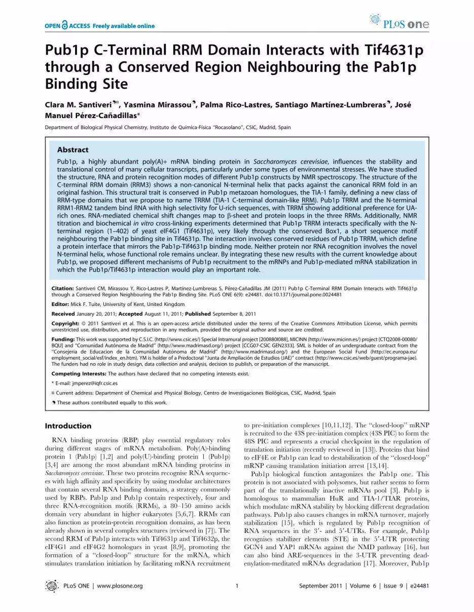
NMR characterization of Pub1p constructsSaccharomyces cerevisiae Pub1p is a 453 residue protein that
contains three RRM domains. We cloned three fragments of this
protein: Pub1p R13 containing RRM1, RRM2 and RRM3
domains (residues 32–414), Pub1p R12 including RRM1 and
RRM2 domains (residues 32–242) and Pub1p R3 that contains
RRM3 (residues 315–414). The constructs were expressed as
thioredoxin A fusions (N-terminal) to enhance protein expression
and solubility. Nevertheless, only Pub1p R12 and Pub1p R3
expressed as soluble proteins and remained so after tag removal.
Pub1p R3 is less soluble (300 mM) than the Pup1p R12, and
becomes even less soluble at pH below 7.0, maybe due to
contributions of histidine side chains titrating in this pH range.
Pub1p R13 construct expressed into inclusion bodies, perhaps due
to the asparagine/methionine-rich region in the RRM2-RRM3
linker. Other aggregation-prone sequences (like the N- and C-
terminal poly-glutamine regions) were not included in the designs.
Remarkably, prion-like regions are required for mammalian stress
granule assembly in TIA-1 [34,36], the metazoan homolog of
Pub1p.
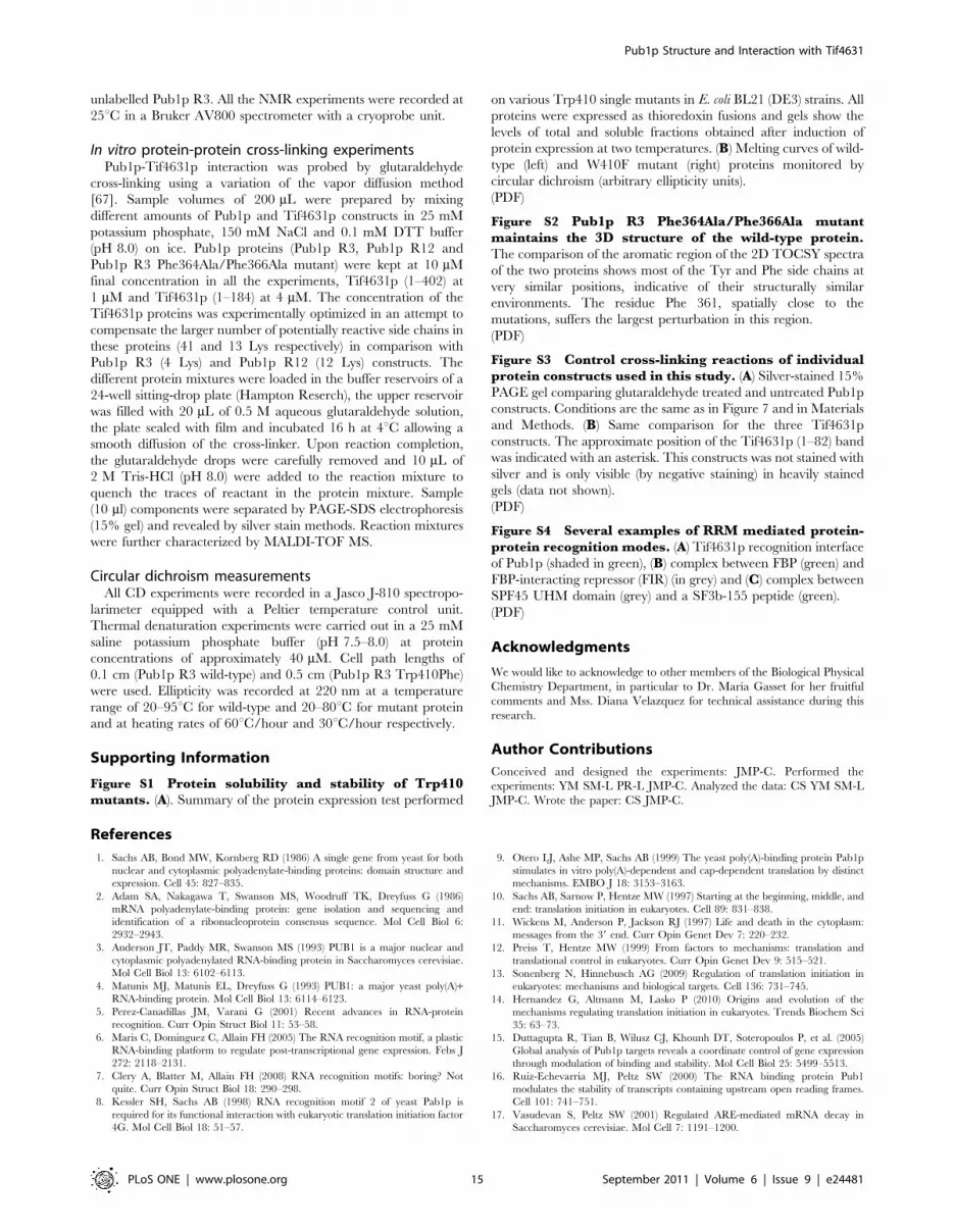
Pub1p R12 and Pub1p R3 constructs are monomeric and well-
folded according to the appearance of their 1H-15N HSQC spectra
(Figure 1), proton amide T2 relaxation measurements, gel filtration
chromatography and, in the case of Pub1p R3, sedimentation
equilibrium ultracentrifugation experiments (data not shown). The
chemical shift index CSI [44] provides evidences about the
secondary structure contents of these proteins (Figure 1C). Cross-
peaks corresponding to linker residues between RRM1 and
RRM2 (149–157) are missing in the 1H-15N HQSC spectrum,
evidencing that this segment might be conformationally flexible in
the ms to ms time scale. Interestingly, the structure of a Pub1p
construct similar to ours (residues 75–240) shows that this linker
participates in the formation of a crystallographic swapped dimer
[43]. Our studies found no evidence about the presence of such
dimer in solution, in agreement with the analysis of gel filtration
data performed by the authors of crystal structure. Nevertheless,
the line broadening effect might be explained with the hypothesis
of a chemical equilibrium between a major monomeric and a
minor dimeric forms occurring in an intermediate time scale (ms
to ms). Finally, the first N-terminal 41 residues (32–72) of Pub1p
R12 (not included in the X-ray construct) are conformationally
disordered and highly mobile as derived from the chemical shifts
and sharp line shapes.
For Pub1p R3, the most remarkable feature extracted from the13C chemical shift analysis is the prediction of a non-canonical
helical region right before the first strand of RRM3. Hence we
decided to pursue a full structural determination of this protein
domain.
Pub1p R3 fold defines a novel RRM structural classPub1p R3 NMR structure (PDB: 2LA4) was calculated from 1968
NOE-derived distance restraints and 129 angular restraints obtained
from the analysis of selected 13C chemical shifts (Table 1). Excluding
the first ten residues of the construct and the last two, which can be
regarded as disordered, the structure (Figure 2) is of high quality with
very low RMSD values across the 20-confomers-ensemble (0.38 A
Pub1p Structure and Interaction with Tif4631
PLoS ONE | www.plosone.org 2 September 2011 | Volume 6 | Issue 9 | e24481
for backbone atoms and 0.85 A for heavy atoms). Within the folded
region, 83% of the side chains of residues different from Ala, Gly and
Pro are ordered (RMSD#630u for the dihedral angle x1) and ten of
them are also well-ordered at level of angle x2.
Pub1p C-terminal domain adopts a typical a/b RRM-type fold
with a b1a1b2b3a2 topology [5,6,7,45], but with some key features
in comparison with other RRM domains. First, the b-sheet is
distorted in Pub1p R3, and the last strand b4 is missing. Instead
there is a short b-hairpin (400–405) right after the helix a2 (386–
396), previously reported in other RRM structures [7] and, more
importantly, a non-canonical N-terminal a-helix (labelled here as
a0: 326–333). This novel helix is stabilised by extensive contacts
with helix a2 and the C-terminal residues (Figure 2C), chiefly Trp
410, which is at the centre of a hydrophobic cluster (Val 329, Ile
332, Ile 333, Ala 336, Pro 337, Val 340, His 386, Ala 390 and Ile
393) that defines the orientation of the new helix. The side chain
imino proton of this residue forms a H-bond with the carbonyl
group of Ile 332 that drives Trp 410 out of its normal position as
part of the b-sheet, causing a local distortion of it and hindering
the formation of the fourth strand. Consistent with its structural
role, Trp 410 is strictly conserved across different fungal Pub1p
proteins (Figure 2D), Ngr1p, Nam8p (two Pub1p homologues in
yeast), TIA-1 and TIAR proteins. The sequence alignment along
these proteins allowed us to identify a conserved motif (Trp-Gly-
[Arg/Lys]) which is the hallmark of the family.
We pursued a thorough study of the role of Trp 410 by
analysing the properties of a set of 11 mutants including
replacements by aromatic (Tyr, Phe and His), hydrophobic (Leu,
Ile and Pro), polar (Asn, Ser, Thr) and small (Ala and Gly) side
chains. All mutants exhibited poor solubility when over-expressed
at 37uC, a situation that is partially alleviated when expressed at
20uC (Figure S1A), however the correlation with the amino acid
type is unclear. The poor solubility hindered the isolation and
purification of these proteins and some of the soluble ones at low
temperature turned to be highly unstable during the purification
(i.e. Trp410Ile). We were only able to obtain enough quantities to
perform further biophysical studies for the Trp410Phe mutant.
The comparison of the melting curves of the wild-type and mutant
proteins followed by circular dichroism shows that the later is
about 10uC less stable (Figure S1B). The large decrease in DHm
together with the more shallow profile of the curve is indicative of
a less co-operative unfolding mechanism for the mutant, perhaps
Figure 1. NMR spectra of Pub1p constructs and secondary structure prediction. (A) 1H-15N HSQC spectrum of the Pub1p R12 construct(0.5 mM DTT, 25 mM NaCl, 25 mM potassium phosphate buffer, pH 6.5) at 25uC. (B) 1H-15N HSQC spectrum of the Pub1p R3 domain (0.5 mM DTT,25 mM NaCl, 25 mM potassium phosphate buffer, pH 7.0) at 25uC. (C) Consensus chemical-shift index [44] calculated for both Pub1p R12 (black) andPub1p R3 (white) proteins using the CSI program (http://www.bionmr.ualberta.ca/bds/software/csi/latest/csi.html). The predicted a-helical and b-sheet regions are marked in the histogram with a and b, respectively.doi:10.1371/journal.pone.0024481.g001
Pub1p Structure and Interaction with Tif4631
PLoS ONE | www.plosone.org 3 September 2011 | Volume 6 | Issue 9 | e24481

attributable to the absence of the N-terminal helix. The overall
conclusion of this study is that the Trp 410 position is critical for
protein solubility and stability. The buried hydrogen bond Trp
410 Ne1-Ile 332 CO seems to be the key interaction that enforces a
tryptophan side chain at this position.
To summarize: the C-terminal (Trp-Gly-[Arg/Lys]) motif, the
distorted three-stranded b-sheet and the new N-terminal helix
represent novel features that are likely conserved in the RRM3
domains of Pub1p/TIA-1 proteins and represent the hallmarks of
a new class of RRMs that we propose to name TRRM (TIA-1 C-
terminal domain like RRM).
Pub1p R12 recognises poly(U) with high affinity andspecificity
Pub1p binds poly(U) sequences in vitro [3] and we decided to
investigate which regions of the protein are responsible for this
activity and specificity. First we explored the RNA binding of the
Pup1p R12 construct by NMR titration experiments (Figure 3).
We tested three different probes: U14, A14 and (UA)7, which have
very similar composition to that of the RNA motifs found in the
UTRs of Pub1p-bound mRNAs [15]. The U14 probe binds with
high affinity in the slow-exchange regime, a behaviour typical of
protein-RNA complexes with dissociation constants up to 250 nM
[46]. Therefore, our data demonstrate that the first two RRMs are
sufficient to account for the polyuridine binding activity of Pub1p
[3]. In contrast, A14 probe titrates in the fast chemical shift
exchange regime, typical of protein-RNA complexes with affinity
higher than 15 mM [46]. However the chemical shift perturbations
are negligible (Figure 3) at equimolecular ratio (100 mM
protein:RNA), indicating that the bound form is scarcely
populated at the working concentration (i.e. KD.100 mM).
Finally, the (UA)7 probe titrates in the intermediate chemical shift
time scale which is characterized by signal disappearance (by
exchange broadening) upon titration and even at stoichoimetric
conditions. This behaviour is typical of protein-RNA complexes
with KD ranging between 400 nM and 2 mM [46].
Mapping of chemical shift perturbations (CSP) onto the X-ray
structure of Pub1p RMM1-RMM2 construct (residues 75–240)
[43] reveals that both RRM participate in RNA recognition
(Figure 3C). In this case, unlike other well-known examples
[47,48,49,50], it is not clear whether the linker region participates
in the interaction, as the assignment of these residues remains
elusive in the complex. The largest changes are concentrated on
the canonical recognition interfaces (b-sheets), hence we might
expect a recognition mode similar to other RRM-containing
proteins that bind polyuridine tracks [47,49,51,52]. Strikingly, the
CSP effect propagates beyond the boundaries defined for the X-
ray construct as we could detect RNA-dependent changes up to
the residue Ala 63 (Figure 3A). This evidences the importance of
intrinsically unstructured regions like this (residues 63–74) in
ligand binding. Remarkably, Pub1p fungal orthologues show a
totally conserved tetrapeptide motif (GGRE in positions 69–71) in
this region. Although the way it participates in protein-RNA
recognition remains as an open question.
Pub1p R3 recognises U-rich and UA-rich RNAspreferentially
We also investigated RNA binding by Pub1p R3 through1H-15N HSQC-based NMR titration experiments with several
RNA probes (Table 2). Pub1p R3 forms complexes in the
chemical shift fast exchange regime for uracyl-free RNA probes
(i.e. A14, (AG)4, (AC)4 and C8) and in fast exchange with line
broadening, or ‘‘insufficient fast exchange’’ [53], for uracyl-
containing RNAs [U10, U12, U14, (UA)6, (UA)7 and (CU)6]
(Figure 4). The first group of complexes display binding affinities
around 70 mM or weaker (Table 2). KD values were calculated by
a global fitting of the NMR signals with Ddav.0.1 ppm (Equation
1) with a non-linear least squares protocol. The same fitting
procedure was followed for the second group of RNA probes but
excluding the crosspeaks experiencing ‘‘insufficient fast ex-
change’’ for which equation 2 (see Materials and Methods) can
not be applied [53]. The U-rich (U14, U12, U10) and UA-rich
series [(UA)7, (UA)6] display KD values around 1-order of
magnitude tighter than (CU)6 and at least two-orders tighter than
the A-rich sequences of the first group (Table 2). Furthermore we
found that U14 and (UA)7 form 2:1 protein-RNA complexes
whereas U12 and (UA)6 form 1:1 complexes. Thus, we can
conclude that Pub1p R3 binds an RNA stretch no longer than 6
bases, close to the longest ssRNA bound by an isolated RRM
domain [54].
The three-dimensional distribution of the RNA-induced CSP is
similar for all the RNA probes (Figure 4B), with the exception of
Asn 347 side chain (Figure 4A). The Nd2-Hd21 and Nd2-Hd22
peaks suffer variations between 0.2 and 0.4 ppm for U14, (UA)7and (CU)6 complexes, but hardly any change for the A14 one.
Residue Asn 347 is in the b1-a1 loop next to Phe 351, that also
changes in the uracyl-containing RNA probes. Hence we propose
that this loop contains an uracyl recognition hot spot in the
protein, with Asn 347 side chain probably making specific contacts
with the base moiety. A hydrogen bond between Asn 347 Hd26and the O4 of the uracyl would explain base specificity at this
position, with Phe 351 making stacking interactions to the base.
Interestingly, neither Asn 347 nor Phe 351 are conserved in TIA-1
proteins (Figure 2D), thus a different RNA selectivity might be
expected for these proteins. The C-terminal tetrapeptide Gly 411-
Lys 412-Glu 413-Arg 414 experiences very large CSP in the
Table 1. Summary of NMR restraints and structural statisticsfor the ensemble of 20 lowest energy structures of Pub1p R3(residues 315–414).
Distance restraints
Intraresidue 277
Sequential 440
Medium range (2#|i2j|#4) 421
Long range (|i2j|$5) 830
All 1968
Averaged NOE violations per structure (A) 0.004560.0005
Dihedral angle restraints
w 61
y 68
All 129
Averaged dihedral angle violations per structure (deg) 0.3260.08
Averaged pair-wise RMSD (A)
Backbone atoms (324–412) 0.3860.08
All heavy atoms (324–412) 0.8560.10
Ramachandran plot
Residues in most favoured regions 76.4%
Residues in additional allowed regions 21.7%
Residues in generously allowed regions 1.2%
Residues in disallowed regions 0.6%
doi:10.1371/journal.pone.0024481.t001
Pub1p Structure and Interaction with Tif4631
PLoS ONE | www.plosone.org 4 September 2011 | Volume 6 | Issue 9 | e24481

complexes and are surely involved in RNA binding (Figure 4B and
4C). The remaining changes are localized in the b-sheet, the usual
RNA docking platform in most RRMs [7]. Interestingly, neither
Trp 410 nor a0 helix residues suffer significant perturbations, thus
we can conclude that this novel element is not participating in
RNA recognition.
Pub1p R3 binds (UA)7 probes with low micromolar affinity,
which is the typical affinity range displayed by single RRM
domains recognising single strand RNA fragments with some
degree of specificity [51,55], but still weaker than those displaying
a high degree of specificity [54]. The recently published structure
of CUG-BP1 RRM3 [55] is probably a close model of Pub1p R3
Figure 2. The NMR structure of Pub1p R3 shows novel features. (A) Backbone atom superposition of the Pub1p R3 20 NMR calculatedstructures with the lowest target function (PDB:2LA4). (B) Ribbon plot of one representative NMR structure of Pub1p R3 with secondary structureelements numbered and depicted in green (b-strands) and red (a-helices). (C) Detailed view of the structure around a0 and a2 helices. Trp 410 andother residues that form the hydrophobic core are labeled and hydrogen bonds are represented as dashed donor-to-acceptor lines. (D) Sequencealignment of fungal Pub1p (Sc: Saccharomyces cerevisiae, Kl: Kluyveromyces lactis, Ag: Ashbya gossypii, Dh: Debaryomyces hansenii, Vp:Vanderwaltozyma polyspora, Ca: Candida albicans and Cg: Candida glabrata), human TIA-1/TIAR and Saccharomyces cerevisiae Nam8p and Ngr1pproteins. Secondary structure elements in Pub1p R3 structure are indicated on the top of the panel and the sequences have been highlighted indifferent shades of purple according to the level of conservation found across Pub1p/TIA-1 proteins. Typical RNP1 and RNP2 motifs found in RRMdomains are also indicated. A histogram showing the percentage of accessible surface area (ASA) per residue is plotted on the bottom of the figure.Bars corresponding to residues involved in (UA)7 binding (Ddav$0.1 ppm. See text and Figure 4) are highlighted in red and those taking part of thehydrophobic core of a0 helix are depicted in green.doi:10.1371/journal.pone.0024481.g002
Pub1p Structure and Interaction with Tif4631
PLoS ONE | www.plosone.org 5 September 2011 | Volume 6 | Issue 9 | e24481

Figure 3. RNA recognition by Pub1p R12. (A) Superposition of the 1H-15N HSQC spectra of the free Pub1p R12 protein (100 mM; in black) and thecomplex with U14 (100 mM each, gray) (left). Histogram showing the weighted averaged 1H and 15N amide chemical shift changes, Ddav (right). (B)Superposition of the 1H-15N HSQC spectrum of Pub1p R12 (100 mM) in the absence (black) and in the presence (gray) of 100 mM A14 (left). Histogramdisplaying the averaged 1H and 15N amide chemical shift changes for this complex (right). Assignments of the residues 149–157 are missing for thefree protein and for the complexes. (C) Red color-coded mapping of chemical shift changes observed upon U14 titration on the X-ray structure ofPub1p RRM1-RRM2 [43] (PDB: 3MD3). Unassigned residues in the linker between RRM1 and RRM2 are colored in yellow.doi:10.1371/journal.pone.0024481.g003
Pub1p Structure and Interaction with Tif4631
PLoS ONE | www.plosone.org 6 September 2011 | Volume 6 | Issue 9 | e24481

binding mode, as this protein recognises a related sequence (UG)3with similar affinity (KD = 1.3 mM) than Pub1p R3.
In conclusion, we found that Pub1p contains two separated
RNA recognition platforms (RRM1-RRM2 and RRM3) which
bind U-rich and UA-rich sequences with higher affinity than A-
rich ones. The recognition of 39-UTR by Pub1p might be
commanded by the N-terminal region (which binds RNA U-
stretches with greater affinity) with the C-terminal one playing an
auxiliary role in the selection of the transcripts.
Pub1p RRM3 interacts with Box1 region of Tif4631p(eIF4G)
The TRRM domain has been exclusively found in Pub1p, TIA-
1 and TIAR proteins, suggesting that it may play a unique role in
the biological function of this family of proteins. Proteomic studies
have identified Pub1p-binding proteins, which includes eIF4G
yeast homologues Tif4631p, and Tif4632p [56,57]. The
Tif4631p/Pub1p interaction has not been studied in detail yet,
however it has been proposed to be involved in translation control
of the MFA1 transcript in response to carbon source changes [19].
Additionally, the Pub1p and Tif4631p are essential components of
the EGP-bodies generated upon glucose starvation [29], robust
heat shock stress granules [31] and their homologues (TIA-1/
eIF4G) are constituents of mammalian stress granules
[21,22,26,58,59]. Tif4631p is a scaffold protein with a C-terminal
part that contains eIF4A- and eIF4E-interacting domains
(Figure 5A). Structures of these complexes have been reported
[60,61]. Much less is known for the N-terminal part that is
predicted to be intrinsically unstructured and includes the Pabp1
binding region [62]. We overexpressed a Tif4631p construct
corresponding to this region (residues 1–402), obtaining a soluble
protein that, as previously shown [63], migrates at apparently
higher molecular weight in the SDS-PAGE gels. Nevertheless, the
protein identity was confirmed by mass spectroscopy and the
recombinant protein shows the typical 1H-15N HSQC NMR
spectrum of an intrinsically unstructured protein, sharp and very
low dispersed signals (Figure 6). However, the existence of regions
with some local secondary structure propensity cannot be entirely
ruled out. First, we tested if Tif4631p (1–402) interacts with any of
our Pub1p constructs by 1H-15N HQSC titration experiments,
mixing unlabelled Tif4631p protein with 15N-labelled Pub1p R3
or Pub1p R12 in approximately 2:1 molar ratio. The comparison
of the CSP for the two Pub1p constructs (Figure 5B) showed that
Tif4631p (1–402) interacts specifically with Pub1p TRRM
domain. The magnitude of the CSP is small but non-randomly
distributed, suggesting that the interaction is weak but specific.
Similar weak but specific effects are observed in the NMR
titrations in the human eIF4A/4G/4H helicase complex [64]. The
Tif4631p binding site is formed by residues of strand b3, helix a1
and the linker between them (Figure 5C) and includes conserved
Phe 364 and Phe 366 (in fungi). The side chain amide groups of
Gln 362 and Asn 363 show significant CSP upon binding
(Figure 5C), maybe because make specific interactions across the
interface (i.e. hydrogen bonds). Among these four residues only
Phe 364 is conserved in TIA-1, thus if an equivalent interaction
with eIF4G exists it is likely to be topologically different.
Next, we analysed the perturbations on the Tif4631p NMR
spectra by mixing unlabelled Pub1p R3 in a 2:1 molar excess with15N-labelled Tif4631p (1–402) (Figure 6). This approach provides
a complementary view of the interaction by showing the effect on
Tif4631p. The high overlapping of Tif4631p 1H-15N HSQC
spectrum masks most of the expected changes, although the lower-
left crosspeak of the spectra shows changes upon binding
(Figure 6B). Because of its unique position, this signal could
luckily be assigned to Trp 95 Ne1-He1, the only Trp residue of
Tif4631p (1–402). This evidences that Pub1p R3 either contacts
this residue or at least is in its vicinity. Even more, the CPS
changes are accompanied by differential changes in the signal line
widths of the individual components of the Ne1-He1 multiplet. In
the free state all the four signals showed similar intensity and line
widths, a situation typical of a highly mobile group (i.e. intrinsically
disordered protein) (Figure 6A). However, upon binding, all the
components suffer line broadening, which is higher for the left
components of the multiplet. This differential effect is attributable
to the interference of NMR relaxation mechanisms (dipole-dipole
and chemical shift anisotropy), popularly known as the TROSY
effect [65]. This change on the relaxation properties of the Trp
signal undoubtedly shows that this side chain experiences a
reduction in mobility attribuitable to the association with Pub1p
R3. NMR data, CSP and the changes in line width, demonstrate
that Pub1p R3 binding site overlaps with Tif4631p Box1
(Figure 5A), a highly conserved region in Saccharomyces eIF4G
homologs [66] close to the Pab1p binding site.
To complement the NMR analysis, we decided to perform
independent biochemical experiments. First, we chose affinity
capture experiments by GST tagging either Tif4631p or Pub1p
constructs, but we faced technical problems due to resin-induced
protein precipitation for some Pub1p constructs, that interfere
with the outcome of the experiment. As an alternative, we
performed in vitro protein-protein cross-linking experiments using
glutaraldehyde. This time, the large number of potentially
reactive groups in Tif4631p (1–402) (48 Lys side chains) resulted
in a large amount of unspecific reactions (self cross-linking
mainly). We optimized the reaction by keeping protein concen-
trations below 10 mM and adjusting the Tif4631p/Pub1p ratio to
the ratio of Lys on each protein (see Materials and Methods for
further details). We also followed an adaptation of a vapour
diffusion method that ensures a slow addition of the cross-linking
agent [67] and minimizes unspecific reactions. The formation of
the corresponding adducts was monitored by PAGE-SDS. Three
different Tif4631p constructs were used: the original Tif4631p
(1–402) and two shorter forms Tif4631p (1–184) and Tif4631p
(1–82). On the other side, we used the Pub1p R12 and Pub1p R3
constructs and a double mutant of Pub1p R3 Phe364Ala/
Phe366Ala that targets key residues of Tif4631p binding site
identified by NMR. The results are summarized in Figure 7 and
confirm the conclusions obtained from the NMR analysis.
Tif4631p (1–402) and (1–184), that contain Box1, form adducts
with Pub1p R3, but not with Pub1p R12 or with Pub1p R3
double mutant. The lost of binding in the mutant is highly
revealing as demonstrates the direct involvement of Phe 364 and
Phe 366 in the interaction. An alternative explanation arguing
that mutations cause Pub1p R3 unfolding could be plausible, but
the high resemblance between Pub1p R3 mutant and wild-type
NMR spectra show that their structure must be similar (Figure
S2). Unfortunately, Tif4631p (1–82) can not be stained with silver
(Figure S3B, lines 3 and 6) and is only visible by negative staining
on overexposed silver stain gels (data not shown). Nevertheless,
none of the Pub1p variants form adducts with this construct,
showing that the so-called RNA1 region in Tif4631p [66]
(Figure 5A) does not interact with Pub1p.
In conclusion, our study about the molecular recognition
properties of Pub1p shows a very versatile protein capable to
recognise Tif4136p and RNA using structurally compatible
interfaces (Figure 4C and 5C). However, there are novel structural
features discovered, such as the conserved N-terminal helix in
Pub1p R3, whose function is still poorly understood, opening new
avenues for future research.
Pub1p Structure and Interaction with Tif4631
PLoS ONE | www.plosone.org 7 September 2011 | Volume 6 | Issue 9 | e24481

Discussion
The novel C-terminal RRM domain is conserved inPub1p/TIA-1 protein family
ELAV-like HuR and TIA-1/TIAR proteins have been
proposed to be the metazoan homologues of Pub1p on the basis
of its similar functional role in mRNA metabolism. However, the
structure of Pub1p R3 shows novel sequence features (the Trp-
Gly-[Arg/Lys] motif), which are present in TIA-1 proteins but not
in HuR ones, supporting that TIA-1/TIAR are the true
evolutionary relatives of Pub1p. An unpublished structure of the
third domain of TIAR protein has been deposited in the PDB data
bank (ID 1X4G) and it also contains the novel N-terminal helix
constituting, together with our structure, the only two cases of
RRMs having this non-canonical secondary structure element. We
propose to name this new class of domains as TRRM (TIA-1 C-
terminal domain like RRM). The conservation of this structural
feature might be correlated with a biological function common in
TIA-1 and Pub1p proteins. In metazoans, TIA-1 is involved in
stress granules formation [22,34,58] and in alternative splicing
[37,40,41,42]. In yeast, Pub1p and Ngr1p (TIAR homolog) are
components of different cytoplasmic RNA granules (recently
reviewed in [26]) while Nam8p is involved in RNA splicing
[38,68]. Although the level of conservation of the Trp-Gly-[Arg/
Lys] motif is paramount among the Pub1p/TIA-1 family, Ngr1p
and Nam8p sequences lack the characteristic amphipatic profile in
the region corresponding to the hypothetical N-terminal helix
(Figure 2D), suggesting that this element is not present in these
proteins. We have attempted to overexpress the TRRM domain of
Ngr1p but it proved to be highly insoluble (data not shown),
maybe because the absence of the N-terminal helix exposes the
hydrophobic cluster beneath it. Similarly the destabilization of the
same region in the Pub1p Trp410 mutants (Figure S1) could also
decrease the solubility, perhaps due to a similar effect.
Pub1p is not the only RNA binding protein found in
cytoplasmic RNA granules that contain non-canonical RRM fold.
The second RRM of TDP-43, a component of some types of
mammalian stress granules [69,70,71], has an additional b-strand
in the b-sheet), which is involved in protein dimerization [72]. The
TDP-43 dimer interface includes helix a1 and self-recognition is
guided by hydrogen bonds. It could be possible that Pub1p novel
N-terminal helix functions similarly, however we have not found
evidences by NMR, gel filtration and analytical ultracentrifugation
of such behaviour. Moreover, the cross-linking experiments, which
are very efficient in capturing transient interactions, did not detect
the presence of a Pub1p TRRM dimer (compare lines 1 and 4 in
Figure S3A).
Pub1p R3 binds Tif4631p using a similar interface toother protein-binding RRMs
A detailed structural analysis, in combination with the
alignment of TIA-1/Pub1p TRRM sequences, identifies con-
served residues in the opposite face to the RNA interface (Figure 8).
The backside of Pub1p TRRM is shaped by the contacts between
the three helices of the structure, which define a negatively
charged interface with two grooves and three ridges (Figure 8).
Two conserved residues (Asp 330 and Glu 387) characterize the
shallow groove (not present in previous RRM structures) defined
by helix a0 and helix a2; with the hydrophobic Pro 338 placed at
the bottom of the imaginary valley. His 386 side chain, also in the
region, is allowed to change to Arg or Lys, conserving a positive
character that seems to be key for the p-cation interaction with
Trp 410 and hence for protein stability (Figure 2C). The second
groove, defined by helix a1 and helix a2, is wider and deeper and
contains conserved hydrophobic side chains (Figure 8) and one or
two negatively charged side chains on helix a1. An structurally
equivalent region is involved in recognition of Trp-containing
peptides in U2AF homology motifs (UHM), a well-known family
of RRMs capable to interact with proteins and RNAs simulta-
neously [73] (Figure S4). In a more recent example, the same
groove is used for protein FIR to recognise an a-helical segment of
FUS through a novel protein-protein binding mode [74] (Figure
S4). Like in the later case, Tif4631p interacts with Pub1p TRRM
by using the a1/a2 groove, but also the linker connecting a1 and
b3 and part of b3, suggesting a possible novel mechanism of RRM-
mediated protein recognition. Our study identifies Box1 as the
Pub1p interacting region in Tif4631p. This peptide motif contains
a totally conserved tryptophan in Saccharomyces eIF4G homologs
([66] and Figure 5A), therefore the interaction might partially
resemble that of the UHM. Interestingly, Pabp1 does interact with
Tif4631p through its RRM2 domain [9,75], using an interface
similar to that of Pub1p TRRM. In this case, Pab1p targets the
conserved peptide Box3 in Tif4631p, which neighbours the Pub1p
binding site (Figure 5A). The structure of the Pab1p/Tif4631p
complex has not been solved, however the recognition of
mammalian eIF4G1 by the rotavirus NSP3 protein, a functional
homolog of PABP1, provides solid evidences on the binding mode
[75]. Protein-protein recognition would involve two conserved
residues in eIF4G1 (Ile 140 and Ile 142) that presumably pack
against Phe 119 and Phe 122 of PABP1 RRM2. Equivalent
residues can be found in Pab1p RRM2 and Tif4631p Box3
suggesting that the binding mode is conserved across species. We
have experimentally proved that Pub1p Phe 364 and Phe 366
(structurally equivalent to PABP1 Phe 119 and 122) are involved
in recognition (Figure 5C). Tif4631p Box1 does not contain
conserved Ile residues, but rather has a series of aromatic side
chains (Figure 5A) that would presumably participate in Pub1p
binding. All these observations suggest that Pub1p and Pab1p
share a similar Tif4631p recognition mechanism. Giving the high
Table 2. Intervals of KD valuesa for binding of RNAoligonucleotides to Pub1p R3 calculated at the 95%confidence level.
RNA KD (mM) Stoichiometry (Protein:RNA)
U14 1.2–3.9 2:1
U12 n.d 1:1
U10 n.d 1:1
(UA)7 ,1.0 2:1
(UA)6 n.d. 1:1
A14 70–88 2:1
(CU)6 16–22 1:1
(GU)4b n.d. n.d.
(AG)4 78–100 1:1
(AC)4c very weak 1:1
C8c very weak n.d.
aAll the resonances showing Ddav.0.1 ppm upon RNA binding were used inthe data fitting to derive KD values.
bThe KD values could not be determined due to precipitation of the complexduring the titration experiments.
cThe KD values could not be determined due to incomplete saturation of theprotein signals.
n.d. not determined.doi:10.1371/journal.pone.0024481.t002
Pub1p Structure and Interaction with Tif4631
PLoS ONE | www.plosone.org 8 September 2011 | Volume 6 | Issue 9 | e24481

Figure 4. RNA recognition by Pub1p R3. (A) Superposition of a selected region of the 1H-15N HSQC spectra of the Pub1p R3 domain in theabsence of ligand (black) and in the presence of increasing amounts of U14 (green), A14 (red), (UA)7 (purple) and (CU)6 (blue) RNA ligands. Resonanceassignments are indicated on the top spectrum. All the spectra were acquired at 100 mM protein concentration in 0.5 mM DTT, 25 mM NaCl, 25 mMpotassium phosphate buffer (pH 7.0) and 25uC. (B) Averaged 1H and 15N amide chemical shift changes (Ddav) observed in the 1H-15N HSQC spectrum
Pub1p Structure and Interaction with Tif4631
PLoS ONE | www.plosone.org 9 September 2011 | Volume 6 | Issue 9 | e24481

level of conservation of Box1 in Tif4632p, it is reasonable to think
that Pub1p recognises it in a similar fashion.
Determinants of Pub1p recruitment to the mRNPsAccording to chemical shift perturbation data, Pub1p TRRM
might be able to recognise Tif4631p and RNA specifically and
simultaneously. The Pub1p-binding box in Tif4631p is close to the
Pab1p binding site, suggesting the possibility that both proteins
could work cooperatively to recognise more than one sequence
motif in the UTRs. Alternatively, it could be possible that Pub1p
recruitment to the mRNPs was commanded by the interaction
with Tif4631p, in an RNA-independent manner. We find
interesting to discuss about the biological implications of these
new structural findings and to see the degree of agreement with
current models about different aspects of mRNA metabolism.
Pub1p is loaded to the nuclear mRNPs and then shuttled to the
cytoplasm, where is part of the fraction of translationally inactive
mRNPs [3]. The latter suggests that the protein is probably
removed from the mRNPs at some stage of the translational
initiation [13], or perhaps later during the pioneer round of
translation [76], although this issue has not been yet experimen-
tally addressed. Different types of stressors cause the interruption
of the normal translation initiation flow path, causing the
accumulation of pre-initiation complexes (PIC) that eventually
aggregate into different types of cytoplasmic granules. It is known
in yeast and mammals that Pub1p/TIA-1 proteins are compo-
nents of some of these granules ([26] and references herein). In
mammals, the most likely mechanism of TIA-1 mediated SG
nucleation involves its C-terminal prion-like region [34,36]. Pub1p
also contains an equivalent C-terminal region and additionally a
Met/Asn-rich region between RRM2 and RRM3 that could play
a similar role. The accumulation of stalled PICs at a stage that
they still contain TIA-1/Pub1p, would trigger their aggregation
into RNA granules (e.g. EGP-bodies or SG) above a certain
concentration threshold.
Pub1p promotes stabilization of mRNAs against ARE-mediated
deadenylation decay and NMD pathways, which occur in the P-
bodies. Thus, it is likely that Pub1p binding prevents (or delays) the
localization of a subset of mRNPS into P-bodies. Hence it seems
important to unravel which are the determinants of Pub1p
recruitment to these transcripts. A genome-wide study identified
368 cellular transcripts bound to Pub1p and, among them, 53
whose stability is decreased in the absence of Pub1p [15]. Most of
these mRNAs encode proteins that act in ribosomal biogenesis and
cellular metabolism. Furthermore, after a re-examination of these
data we have found that the Pub1p-bound dataset is enriched in
transcripts derived from intron-containing genes (48 out of 368).
Surprisingly the percentage increases up to 40% in the 53-subset
(transcripts which are simultaneously bound and stabilized by
Pub1p), suggesting that splicing modulates Pub1p-mediated
stabilization, probably by promoting its recruitment to the nascent
transcript. TIA-1 is a component of metazoan spliceosome
[37,39,40,41,42] but this biological function is performed (at least
partially) by Nam8p in yeast [38], rather than by Pub1p. We
propose three possible mechanisms of Pub1p recruitment to the
nascent mRNP in a splicing-dependent manner: binding to known
spliocesome components (i.e. Lms2p, Lms4p, Sto1p, Tif4631p)
[77], replacement of Nam8p and specific recognition of RNA
trans-acting elements in introns or exon junctions (typically U-rich
tracks).
Apart from the splicing-dependent mechanism there should be
an alternative Pub1p recruitment mechanism to account for
transcripts derived from intronless genes. We agree that such
procedure might depend on the cooperative recognition of RNA
regulatory elements in the UTRs [15]. A-rich, U-rich and UA-rich
motifs are overrepresented in the 39- and 59-UTRs of Pub1p-
bound mRNAs [15]. Since Pub1p has low affinity for A-rich
oligonucleotides, these authors proposed that additional RNA-
binding proteins could act in coordination with Pub1p to achieve
A-rich recognition. If Pub1p interacts with Tif4631p in vivo
through a similar mode to that described here, transcripts
containing A-rich and U-rich motifs could be recognised in a
cooperative manner by Pub1p and Pab1p thanks to the
simultaneous interaction of both proteins with neighbouring sites
in Tif4631p. The model does not exclude the participation of
other RBPs shuttle proteins like Hrp1p, Npl3p, Hrb1p or Gbp2p,
some of which can be tethered to eIF4G, in a similar way than
Pub1p and Pab1p does, to Tif4631p.
Materials and Methods
DNA cloning and site-directed mutagenesisDNA fragments corresponding to wild-type constructs were
amplified from Saccharomyces cerevisiae genomic DNA (Novagen) by
PCR using high fidelity DNA polymerase KOD (Novagen) and
specific DNA primers (Sigma, IDT). The products were purified,
digested with the corresponding restriction enzymes and ligated
into a home-modified pET28 (Novagen) vector that contains: an
N-terminal thioredoxin A fusion tag, for enhanced expression and
solubility, an internal 66His tag for affinity purification and a
TEV protease site, for fusion tag removal. Three constructs of S.
cerevisiae Pub1p protein were obtained: Pub1p R13 that spans over
three RRM domains (residues 32–414), Pub1p R12 containing
domains RRM1 and RRM2 (residues 32–242) and Pub1p R3 that
comprises domain RRM3 (315–414). The three Saccharomyces
cerevisiae Tif4631p constructs (residues 1–402, 1–184 and 1–82)
were cloned following equivalent methods.
The Trp410 library of mutants was generated in the Pub1p R3
framework by using the QuickChange site-directed mutagenesisHkit (Stratagene) and a DNA primer with a random base
composition of codon 410 (59-G GTG CTC GAG TTA TCT
TTC CTT ACC RNN ACC GGT TCT CAA GTT TC-39;
R = A or G and N = A,G,C or T). Pub1p R3 Phe364Ala/
Phe366Ala double mutant was generated using an equivalent
protocol.
Protein expression and purificationPlasmids corresponding to wild-type and mutant proteins were
transformed in E. coli BL21(DE3) (Novagen) chemically competent
cells and expressed in kanamycin (Sigma-Aldrich) containing
(30 mg/l) LB medium. For isotopic labelling, a K-MOPS derived
minimal medium [78] was supplemented with 15NH4Cl (1 g/l)
and/or 13C-glucose (4 g/l). Bacterial cultures were typically grown
at 37uC until OD600 = 0.6, transferred to a shaker at 20uC and
of Pub1p R3 (100 mM) in the presence of a 4-fold excess of the corresponding RNA target (color code matching that of panel A). An asterisk indicatesthat Phe 351 resonance broadens out beyond detection upon (UA)7 binding. Secondary structure elements are depicted on the top graph. (C) CPK(left) and cartoon (right) representations of the Pub1p R3 NMR structure. Residues have been shaded in red range according their Ddav values (fromwhite = 0.0 to bright red =Ddav
max) for the titration point with 400 mM U14 (upper panel in B). Only side chains of amino acids with Ddav$0.1 ppm areshown onto the cartoon image for clarity. The figures were generated with the program PyMOL v0.98 (DeLano Scientific LLC, Palo Alto, CA, USA).doi:10.1371/journal.pone.0024481.g004
Pub1p Structure and Interaction with Tif4631
PLoS ONE | www.plosone.org 10 September 2011 | Volume 6 | Issue 9 | e24481

Pub1p Structure and Interaction with Tif4631
PLoS ONE | www.plosone.org 11 September 2011 | Volume 6 | Issue 9 | e24481

induced overnight with 0.5 mM IPTG (Sigma-Aldrich) upon
temperature stabilization. Cells were harvested by centrifugation
(30 minutes at 3000 g), resuspended in buffer (20 mM potassium
phosphate pH 8.0, 300 mM NaCl, 0.1% b-mercaptoethanol)
containing protease inhibitors (Roche) and lysed by French-press
(Thermo Scientific) or sonication. Lysates were pelleted at high
speed (30 minutes at 15000 g) to separate cell debris and other
insoluble components. Wild-type protein constructs (Pub1p R12,
Pub1p R3 and Tif4631p) and Pub1p mutants were first purified by
metal affinity chromatography (HisTrapTM HP 5 ml (GE
Healthcare)). Fractions coming from this column that contain
the fusion protein were pooled together, dialysed (overnight) at
4uC against 20 mM potassium phosphate pH 8.0 buffer (plus
0.1% b-mercaptoethanol) and simultaneously digested with
homemade TEV protease (100–200 mg/ml). After complete
digestion, the samples were re-applied to the nickel column and
the target proteins were collected in the flow through. In the final
step, proteins were purified and concentrated by ion-exchange
chromatography: Pub1p R12 and Tif4631p with an anion
exchange column (MonoQ 5 ml, GE Healthcare) and Pub1p R3
with a cation exchange one (SP 5 ml, GE Healthcare). In either
case, proteins were eluted with a linear salt gradient (from 0 to
1 M NaCl) in a buffer containing 20 mM Tris-HCl pH 8.0 and
1 mM DTT. Pub1p mutant proteins were purified with similar
methodologies than wild-type proteins. Sample purity and
homogeneity was checked by PAGE-SDS and mass spectrometry.
NMR spectral assignment and structure calculationProtein samples for NMR spectroscopy were approximately
500–800 mM for Pub1p R12 and 300 mM for Pub1p R3 in 90%
H2O, 10% D2O, 0.1% sodium azide containing 25 mM
potassium phosphate buffer (pH 6.5–7.0), 25 mM NaCl, and
0.5 mM DTT. All experiments were recorded in Bruker AV600
and AV800 NMR spectrometers equipped with cryoprobes.
Sodium 2,2-dimethyl-2-silapentane-5-sulphonate (DSS) was used
as an internal chemical shift reference. The assignment of the
backbone 1H, 15N and 13C atoms was achieved by following the
standard methodology. The 3D experiments HNCO, HNCA,
HN(CO)CA, CBCA(CO)HN and CBCANH [79], recorded in the
AV800 spectrometer, were manually assigned for Pub1p R12. An
equivalent set of experiments, plus HN(CA)CO [79], was acquired
for the Pub1p R3 construct, and the spectra were assigned by
following a supervised automatic assignment protocol [80]. 3D
HC(C)H-TOCSY and (H)CCH-TOCSY experiments [81] were
recorded to assign the side chain resonances of Pub1p R3
construct. NMR spectra processing was done with NMRPipe [82]
whereas spectral viewing and analysis were done with the program
CcpNmr Analysis [83]. The completeness of the assignment for
the Pub1p R3 construct extends to 95%, while only backbone13C/15N resonances were assigned for the Pub1p R12 construct.
For Pub1p R3, a total of 1968 NOE-derived distance
constraints were extracted from the 2D-NOESY [84] and 15N-
edited-3D-NOESY [85] spectra, both with mixing times of 80 ms.
Additional 129 dihedral angular constraints were extracted from
the analysis of 13C (Ca ,C9 and Cb) chemical shifts [86] and
incorporated to the structure calculation. The structure calculation
was performed by following an standard torsion angle dynamics
protocol implemented in the program CYANA 2.1 [87].
Coordinates of 50 conformers were randomly generated and then
subjected to the restrained simulated annealing protocol. The 20
lowest target function conformers with no distance violations
larger than 0.2 A and no angle violations larger than 5.0u were
selected as representative of the structure of the protein (PDB
code: 2LA4 and BMRB code: 17502). MOLMOL [88] and
PyMOL v0.98 (DeLano Scientific LLC, Palo Alto, CA, USA)
molecular graphics packages were used for the analysis and
representation of the structures.
Protein-RNA interactions analysis by NMRAll RNA probes were chemically synthesized and purchased from
IDT (Integrated DNA technologies). All samples for NMR titration
studies were prepared by simultaneously dialyzing against the same
protein buffer (see above for other NMR experiments). Samples
were prepared without D2O in a 4 mm NMR tube (typically 300 ml)
and then inserting this into a 5 mm tube that contains D2O for
referencing the lock signal. In the case of Pub1p R12, 1H-15N
HSQC experiments were recorded at 1:1 protein:RNA ratio
(100 mM concentration each) for the probes: U14, A14 and (UA)7.
Assignment of the spectrum of the A14 complex was made by simple
comparison with the free protein spectrum, whereas the Pub1p-U14
complex spectrum was assigned with the aid of triple resonance
experiments (3D HNCA and CBCA(CO)HN) acquired with a
concentrated 1:1 sample (500 mM). Pub1p R3 RNA binding affinity
and selectivity was investigated with eleven RNA probes: U14, U12,
U10, A14, (UA)7, (UA)6, (CU)6, (AG)4, (AC)4, (GU)4 and C8.
Experimental series were performed starting at the highest
RNA:protein ratio (typically 400 mM:100 mM) and the following
points prepared by dilution of this sample with a stock of 15N
labelled protein at the same concentration and buffer composition,
thus the protein concentration is kept fixed. Six to eight titration
points were typically recorded for each RNA probe at protein:RNA
ratios ranging between 4:1 and 0.03:1. The weighted averaged 1H
and 15N amide chemical shift changes were calculated according to
the following formula:
Ddav~
ffiffiffiffiffiffiffiffiffiffiffiffiffiffiffiffiffiffiffiffiffiffiffiffiffiffiffiffiffiffiffiffiffiffiffiffiffiffiffiffiffiffiffiffiffiffiffiffiffiffiffiffiffiffiffiffiffiffiffiDd1Hð Þ2z Dd15N=5
� �2� �
|0:5
s; DdX ~ dobs
x {dfreex
� �ð1Þ
The Ddav values measured in experiments at constant protein
concentration (P0) were plotted versus the total ligand concentration
values (L0) and fitted to the following equation to obtain the
maximum chemical shift change (Ddmax) and the dissociation
constant (KD) [89]:
Ddav~Ddmax
KDzL0zP0ð Þ{ffiffiffiffiffiffiffiffiffiffiffiffiffiffiffiffiffiffiffiffiffiffiffiffiffiffiffiffiffiffiffiffiffiffiffiffiffiffiffiffiffiffiffiffiffiffiffiffiKDzL0zP0ð Þ{4P0L0
p2P0
ð2Þ
Because this equation applies only in the fast exchange limit
[89], 1H-15N HSQC peaks experiencing line broadening during
Figure 5. Monitoring Tif4631p recognition on the Pub1p NMR spectra. (A) Domain organization of Tif4631p (up) displaying recentlydescribed Box1, 2 and 3 in the N-terminus of the protein [66]. Schematic representation of the largest Tif4631p construct used in this study (middle)and pattern of sequence conservation of Box1 region in different Saccharomyces species [66]. (B) CSP of Pub1p R12 and Pub1p R3 backbone amideresonances (combined 1H and 15N values) caused by Tif4631p (1–402) titration (in an ,2:1 excess). Data sets were recorded independently for bothPub1p constructs at similar concentration and protein-protein ratios. (C). Comparison of the free and Tif4631p-bound Pub1p R3 1H-15N HSQC spectrain the region of the Gln and Asn side chains (left). Chemical shift mapping of the Tif4631p binding site (right), with residues changing more than0.025 ppm depicted in magenta.doi:10.1371/journal.pone.0024481.g005
Pub1p Structure and Interaction with Tif4631
PLoS ONE | www.plosone.org 12 September 2011 | Volume 6 | Issue 9 | e24481

the titration were excluded from the KD calculation. The
remaining peaks showing Ddav.0.1 ppm between the free and
the RNA-bound protein at the highest RNA/protein ratio were fit
globally (KD global parameter; Ddmax local parameter for each
curve) to equation (2) by a non-linear least squares method with
the software package GraphPad Prism version 5.00 for Mac
(GraphPad Software, San Diego California USA, www.graphpad.
com).
Protein-protein interactions analysis by NMRSamples for the protein constructs Pub1p R12, Pub1p R3 and
Tif4631p (1–402) were dialyzed simultaneously against 25 mM
potassium phosphate buffer (pH 7.0, 25 mM NaCl, 0.1 mM
DTT) for 24 hours at 4uC twice, to guarantee no differences in
buffer composition. Binding was monitored on the 1H-15N HSQC
spectra of Pub1p R12 and Pub1p R3 upon titration with different
amounts of unlabeled Tif4631p (1–402). Pub1p constructs were
prepared at 100 mM concentration and Tif4631p (1–402) was
added at different ratios: 1:2 for the Pub1p R12-Tif4631p (1–402)
complex and 1:1.9, 1:0.8 and 1:0.4 for the Pub1p R3-Tif4631p (1–
402) one. The chemical shift perturbations were calculated as
weighted average of 1H and 15N amide chemical shift changes
(equation (1)). A complementary NMR titration experiment was
done by monitoring changes in the 1H-15N HSQC spectrum of
100 mM 15N-labelled Tif4631p (1–402) upon addition of 120 mM
Figure 6. Monitoring Pub1p recognition on the Tif4631p NMRspectra. (A) 1H-15N HSQC spectrum of the construct Tif4631p (1–402)showing the characteristic low dispersion of an intrinsically unstruc-tured protein. Signal corresponding to the side chain of Trp 95 has beenlabelled. (B) Chemical shift perturbation on the Trp 95 He1-Ne1crosspeak: free Tif4631p protein in black and a complex with Pub1p R3in red (see Materials and Methods for details). (C) Analysis of line shapevariations caused by Pub1p binding in the multiplet components of theTrp 95 He1-Ne1 signal. Coupled 1H-15N HSQC (up) and 1D (down)spectra have been included for comparison. The low field componentsof the multiplet experience further line broadening in the spectra of thebound form (marked with arrows) in comparison with that in the freeform (black). This effect is linked to a reduction in mobility due to thecomplex formation (see text for further details). Tif4631p concentrationand buffer conditions are identical in both free and bound spectra,
Figure 7. Biochemical in vitro cross-linking experimentsprobing the interaction between Pub1p and Tif4631p. Thepotential interaction among different combinations of Pub1p andTif4631p constructs has been assayed by cross-linking with glutaralde-hyde using a vapour diffusion method (see Materials and Methods fordetails). The silver-stained 15% PAGE gel shows the protein composi-tion of different cross-linking reactions: Pub1p and Tif4631p constructshave been marked with white and black asterisks and detected adductswith red ones. Construct Tif4631p (1–82) was inefficiently stained withsilver (lines 7,8 and 9), although its approximate position of (as seen incoomassie stained gels) has been included for reference. Minor bandsobserved bellow the Tif4631p (1–402), Tif4631 (1–184) and Pub1p R12ones, correspond to intramolecular cross-linking reactions and/ordegradation products. Equivalent bands are found in the controlexperiments (Figure S3).doi:10.1371/journal.pone.0024481.g007
which have acquired with same parameters and represented at equalcontour levels.doi:10.1371/journal.pone.0024481.g006
Pub1p Structure and Interaction with Tif4631
PLoS ONE | www.plosone.org 13 September 2011 | Volume 6 | Issue 9 | e24481
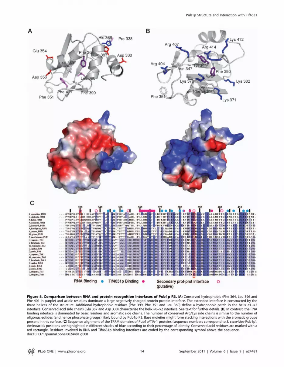
Figure 8. Comparison between RNA and protein recognition interfaces of Pub1p R3. (A) Conserved hydrophobic (Phe 364, Leu 396 andPhe 401 in purple) and acidic residues dominate a large negatively charged protein-protein interface. The extended interface is constructed by thethree helices of the structure. Additional hydrophobic residues (Phe 399, Phe 351 and Leu 360) define a hydrophobic patch in the helix a1–a2interface. Conserved acid side chains (Glu 387 and Asp 330) characterize the helix a0–a2 interface. See text for further details. (B) In contrast, the RNAbinding interface is dominated by basic residues and aromatic side chains. The number of conserved Arg/Lys side chains is similar to the number ofoligonucleotides (and hence phosphate groups) likely bound by Pub1p R3. Base moieties might form stacking interactions with the aromatic groupspresent in this surface. (C) Sequence alignment of the TRRM domains of Pub1p/TIA-1 proteins (sequence numbers correspond to S. cerevisiae Pub1p).Aminoacids positions are highlighted in different shades of blue according to their percentage of identity. Conserved acid residues are marked with ared rectangle. Residues involved in RNA and Tif4631p binding interfaces are coded by the corresponding symbol above the sequence.doi:10.1371/journal.pone.0024481.g008
Pub1p Structure and Interaction with Tif4631
PLoS ONE | www.plosone.org 14 September 2011 | Volume 6 | Issue 9 | e24481
unlabelled Pub1p R3. All the NMR experiments were recorded at
25uC in a Bruker AV800 spectrometer with a cryoprobe unit.
In vitro protein-protein cross-linking experimentsPub1p-Tif4631p interaction was probed by glutaraldehyde
cross-linking using a variation of the vapor diffusion method
[67]. Sample volumes of 200 mL were prepared by mixing
different amounts of Pub1p and Tif4631p constructs in 25 mM
potassium phosphate, 150 mM NaCl and 0.1 mM DTT buffer
(pH 8.0) on ice. Pub1p proteins (Pub1p R3, Pub1p R12 and
Pub1p R3 Phe364Ala/Phe366Ala mutant) were kept at 10 mM
final concentration in all the experiments, Tif4631p (1–402) at
1 mM and Tif4631p (1–184) at 4 mM. The concentration of the
Tif4631p proteins was experimentally optimized in an attempt to
compensate the larger number of potentially reactive side chains in
these proteins (41 and 13 Lys respectively) in comparison with
Pub1p R3 (4 Lys) and Pub1p R12 (12 Lys) constructs. The
different protein mixtures were loaded in the buffer reservoirs of a
24-well sitting-drop plate (Hampton Reserch), the upper reservoir
was filled with 20 mL of 0.5 M aqueous glutaraldehyde solution,
the plate sealed with film and incubated 16 h at 4uC allowing a
smooth diffusion of the cross-linker. Upon reaction completion,
the glutaraldehyde drops were carefully removed and 10 mL of
2 M Tris-HCl (pH 8.0) were added to the reaction mixture to
quench the traces of reactant in the protein mixture. Sample
(10 ml) components were separated by PAGE-SDS electrophoresis
(15% gel) and revealed by silver stain methods. Reaction mixtures
were further characterized by MALDI-TOF MS.
Circular dichroism measurementsAll CD experiments were recorded in a Jasco J-810 spectropo-
larimeter equipped with a Peltier temperature control unit.
Thermal denaturation experiments were carried out in a 25 mM
saline potassium phosphate buffer (pH 7.5–8.0) at protein
concentrations of approximately 40 mM. Cell path lengths of
0.1 cm (Pub1p R3 wild-type) and 0.5 cm (Pub1p R3 Trp410Phe)
were used. Ellipticity was recorded at 220 nm at a temperature
range of 20–95uC for wild-type and 20–80uC for mutant protein
and at heating rates of 60uC/hour and 30uC/hour respectively.
Supporting Information
Figure S1 Protein solubility and stability of Trp410mutants. (A). Summary of the protein expression test performed
on various Trp410 single mutants in E. coli BL21 (DE3) strains. All
proteins were expressed as thioredoxin fusions and gels show the
levels of total and soluble fractions obtained after induction of
protein expression at two temperatures. (B) Melting curves of wild-
type (left) and W410F mutant (right) proteins monitored by
circular dichroism (arbitrary ellipticity units).
(PDF)
Figure S2 Pub1p R3 Phe364Ala/Phe366Ala mutantmaintains the 3D structure of the wild-type protein.The comparison of the aromatic region of the 2D TOCSY spectra
of the two proteins shows most of the Tyr and Phe side chains at
very similar positions, indicative of their structurally similar
environments. The residue Phe 361, spatially close to the
mutations, suffers the largest perturbation in this region.
(PDF)
Figure S3 Control cross-linking reactions of individualprotein constructs used in this study. (A) Silver-stained 15%
PAGE gel comparing glutaraldehyde treated and untreated Pub1p
constructs. Conditions are the same as in Figure 7 and in Materials
and Methods. (B) Same comparison for the three Tif4631p
constructs. The approximate position of the Tif4631p (1–82) band
was indicated with an asterisk. This constructs was not stained with
silver and is only visible (by negative staining) in heavily stained
gels (data not shown).
(PDF)
Figure S4 Several examples of RRM mediated protein-protein recognition modes. (A) Tif4631p recognition interface
of Pub1p (shaded in green), (B) complex between FBP (green) and
FBP-interacting repressor (FIR) (in grey) and (C) complex between
SPF45 UHM domain (grey) and a SF3b-155 peptide (green).
(PDF)
Acknowledgments
We would like to acknowledge to other members of the Biological Physical
Chemistry Department, in particular to Dr. Marıa Gasset for her fruitful
comments and Mss. Diana Velazquez for technical assistance during this
research.
Author Contributions
Conceived and designed the experiments: JMP-C. Performed the
experiments: YM SM-L PR-L JMP-C. Analyzed the data: CS YM SM-L
JMP-C. Wrote the paper: CS JMP-C.
References
1. Sachs AB, Bond MW, Kornberg RD (1986) A single gene from yeast for both
nuclear and cytoplasmic polyadenylate-binding proteins: domain structure and
expression. Cell 45: 827–835.
2. Adam SA, Nakagawa T, Swanson MS, Woodruff TK, Dreyfuss G (1986)
mRNA polyadenylate-binding protein: gene isolation and sequencing and
identification of a ribonucleoprotein consensus sequence. Mol Cell Biol 6:
2932–2943.
3. Anderson JT, Paddy MR, Swanson MS (1993) PUB1 is a major nuclear and
cytoplasmic polyadenylated RNA-binding protein in Saccharomyces cerevisiae.
Mol Cell Biol 13: 6102–6113.
4. Matunis MJ, Matunis EL, Dreyfuss G (1993) PUB1: a major yeast poly(A)+RNA-binding protein. Mol Cell Biol 13: 6114–6123.
5. Perez-Canadillas JM, Varani G (2001) Recent advances in RNA-protein
recognition. Curr Opin Struct Biol 11: 53–58.
6. Maris C, Dominguez C, Allain FH (2005) The RNA recognition motif, a plastic
RNA-binding platform to regulate post-transcriptional gene expression. Febs J
272: 2118–2131.
7. Clery A, Blatter M, Allain FH (2008) RNA recognition motifs: boring? Not
quite. Curr Opin Struct Biol 18: 290–298.
8. Kessler SH, Sachs AB (1998) RNA recognition motif 2 of yeast Pab1p is
required for its functional interaction with eukaryotic translation initiation factor
4G. Mol Cell Biol 18: 51–57.
9. Otero LJ, Ashe MP, Sachs AB (1999) The yeast poly(A)-binding protein Pab1p
stimulates in vitro poly(A)-dependent and cap-dependent translation by distinct
mechanisms. EMBO J 18: 3153–3163.
10. Sachs AB, Sarnow P, Hentze MW (1997) Starting at the beginning, middle, and
end: translation initiation in eukaryotes. Cell 89: 831–838.
11. Wickens M, Anderson P, Jackson RJ (1997) Life and death in the cytoplasm:
messages from the 39 end. Curr Opin Genet Dev 7: 220–232.
12. Preiss T, Hentze MW (1999) From factors to mechanisms: translation and
translational control in eukaryotes. Curr Opin Genet Dev 9: 515–521.
13. Sonenberg N, Hinnebusch AG (2009) Regulation of translation initiation in
eukaryotes: mechanisms and biological targets. Cell 136: 731–745.
14. Hernandez G, Altmann M, Lasko P (2010) Origins and evolution of the
mechanisms regulating translation initiation in eukaryotes. Trends Biochem Sci
35: 63–73.
15. Duttagupta R, Tian B, Wilusz CJ, Khounh DT, Soteropoulos P, et al. (2005)
Global analysis of Pub1p targets reveals a coordinate control of gene expression
through modulation of binding and stability. Mol Cell Biol 25: 5499–5513.
16. Ruiz-Echevarria MJ, Peltz SW (2000) The RNA binding protein Pub1
modulates the stability of transcripts containing upstream open reading frames.
Cell 101: 741–751.
17. Vasudevan S, Peltz SW (2001) Regulated ARE-mediated mRNA decay in
Saccharomyces cerevisiae. Mol Cell 7: 1191–1200.
Pub1p Structure and Interaction with Tif4631
PLoS ONE | www.plosone.org 15 September 2011 | Volume 6 | Issue 9 | e24481
18. Lopez de Silanes I, Galban S, Martindale JL, Yang X, Mazan-Mamczarz K,et al. (2005) Identification and functional outcome of mRNAs associated with
RNA-binding protein TIA-1. Mol Cell Biol 25: 9520–9531.
19. Vasudevan S, Garneau N, Tu Khounh D, Peltz SW (2005) p38 mitogen-
activated protein kinase/Hog1p regulates translation of the AU-rich-element-bearing MFA2 transcript. Mol Cell Biol 25: 9753–9763.
20. Spriggs KA, Bushell M, Willis AE (2010) Translational regulation of geneexpression during conditions of cell stress. Mol Cell 40: 228–237.
21. Buchan JR, Parker R (2009) Eukaryotic stress granules: the ins and outs oftranslation. Mol Cell 36: 932–941.
22. Anderson P, Kedersha N (2009) RNA granules: post-transcriptional andepigenetic modulators of gene expression. Nat Rev Mol Cell Biol 10: 430–436.
23. Anderson P, Kedersha N (2009) Stress granules. Curr Biol 19: R397–398.
24. Moser JJ, Fritzler MJ (2010) Cytoplasmic ribonucleoprotein (RNP) bodies and
their relationship to GW/P bodies. Int J Biochem Cell Biol 42: 828–843.
25. Lui J, Campbell SG, Ashe MP (2010) Inhibition of translation initiationfollowing glucose depletion in yeast facilitates a rationalization of mRNA
content. Biochem Soc Trans 38: 1131–1136.
26. Thomas MG, Loschi M, Desbats MA, Boccaccio GL (2011) RNA granules: the
good, the bad and the ugly. Cell Signal 23: 324–334.
27. Hoyle NP, Castelli LM, Campbell SG, Holmes LE, Ashe MP (2007) Stress-
dependent relocalization of translationally primed mRNPs to cytoplasmic
granules that are kinetically and spatially distinct from P-bodies. J Cell Biol 179:65–74.
28. Brengues M, Parker R (2007) Accumulation of polyadenylated mRNA, Pab1p,
eIF4E, and eIF4G with P-bodies in Saccharomyces cerevisiae. Mol Biol Cell 18:
2592–2602.
29. Buchan JR, Muhlrad D, Parker R (2008) P bodies promote stress granule
assembly in Saccharomyces cerevisiae. J Cell Biol 183: 441–455.
30. Swisher KD, Parker R (2010) Localization to, and effects of Pbp1, Pbp4, Lsm12,
Dhh1, and Pab1 on stress granules in Saccharomyces cerevisiae. PLoS One 5:e10006.
31. Grousl T, Ivanov P, Frydlova I, Vasicova P, Janda F, et al. (2009) Robust heat
shock induces eIF2alpha-phosphorylation-independent assembly of stress
granules containing eIF3 and 40S ribosomal subunits in budding yeast,Saccharomyces cerevisiae. J Cell Sci 122: 2078–2088.
32. Buchan JR, Yoon JH, Parker R (2011) Stress-specific composition, assembly andkinetics of stress granules in Saccharomyces cerevisiae. J Cell Sci 124: 228–239.
33. Kato K, Yamamoto Y, Izawa S (2011) Severe ethanol stress induces assembly ofstress granules in Saccharomyces cerevisiae. Yeast 28: 339–347.
34. Kedersha NL, Gupta M, Li W, Miller I, Anderson P (1999) RNA-bindingproteins TIA-1 and TIAR link the phosphorylation of eIF-2 alpha to the
assembly of mammalian stress granules. J Cell Biol 147: 1431–1442.
35. Kedersha N, Cho MR, Li W, Yacono PW, Chen S, et al. (2000) Dynamic
shuttling of TIA-1 accompanies the recruitment of mRNA to mammalian stressgranules. J Cell Biol 151: 1257–1268.
36. Gilks N, Kedersha N, Ayodele M, Shen L, Stoecklin G, et al. (2004) Stressgranule assembly is mediated by prion-like aggregation of TIA-1. Mol Biol Cell
15: 5383–5398.
37. Gal-Mark N, Schwartz S, Ram O, Eyras E, Ast G (2009) The pivotal roles of
TIA proteins in 59 splice-site selection of alu exons and across evolution. PLoSGenet 5: e1000717.
38. Puig O, Gottschalk A, Fabrizio P, Seraphin B (1999) Interaction of the U1snRNP with nonconserved intronic sequences affects 59 splice site selection.
Genes Dev 13: 569–580.
39. Black DL (2003) Mechanisms of alternative pre-messenger RNA splicing. Annu
Rev Biochem 72: 291–336.
40. Forch P, Puig O, Kedersha N, Martinez C, Granneman S, et al. (2000) The
apoptosis-promoting factor TIA-1 is a regulator of alternative pre-mRNAsplicing. Mol Cell 6: 1089–1098.
41. Forch P, Puig O, Martinez C, Seraphin B, Valcarcel J (2002) The splicingregulator TIA-1 interacts with U1-C to promote U1 snRNP recruitment to 59
splice sites. EMBO J 21: 6882–6892.
42. Izquierdo JM, Majos N, Bonnal S, Martinez C, Castelo R, et al. (2005)
Regulation of Fas alternative splicing by antagonistic effects of TIA-1 and PTBon exon definition. Mol Cell 19: 475–484.
43. Li H, Shi H, Wang H, Zhu Z, Li X, et al. (2010) Crystal structure of the two N-terminal RRM domains of Pub1 and the poly(U)-binding properties of Pub1.
J Struct Biol 171: 291–297.
44. Wishart DS, Sykes BD (1994) Chemical shifts as a tool for structure
determination. Methods Enzymol 239: 363–392.
45. Nagai K, Oubridge C, Ito N, Avis J, Evans P (1995) The RNP domain: a
sequence-specific RNA-binding domain involved in processing and transport ofRNA. Trends Biochem Sci 20: 235–240.
46. Dominguez C, Schubert M, Duss O, Ravindranathan S, Allain FH (2010)Structure determination and dynamics of protein-RNA complexes by NMR
spectroscopy. Prog Nucl Magn Reson Spectrosc;in press.
47. Wang X, Tanaka Hall TM (2001) Structural basis for recognition of AU-rich
element RNA by the HuD protein. Nat Struct Biol 8: 141–145.
48. Perez-Canadillas JM (2006) Grabbing the message: structural basis of mRNA
39UTR recognition by Hrp1. EMBO J 25: 3167–3178.
49. Handa N, Nureki O, Kurimoto K, Kim I, Sakamoto H, et al. (1999) Structural
basis for recognition of the tra mRNA precursor by the Sex-lethal protein.Nature 398: 579–585.
50. Deo RC, Bonanno JB, Sonenberg N, Burley SK (1999) Recognition ofpolyadenylate RNA by the poly(A)-binding protein. Cell 98: 835–845.
51. Auweter SD, Oberstrass FC, Allain FH (2007) Solving the structure of PTB incomplex with pyrimidine tracts: an NMR study of protein-RNA complexes of
weak affinities. J Mol Biol 367: 174–186.
52. Oberstrass FC, Auweter SD, Erat M, Hargous Y, Henning A, et al. (2005)
Structure of PTB bound to RNA: specific binding and implications for splicing
regulation. Science 309: 2054–2057.
53. Sudmeir JL, Evelhoch JL, Jonsson NB-H (1980) Dependence of NMR lineshape
analysis upon chemical rates and mechanisms: Implications for enzyme histidinetitrations. J Magn Reson 40: 377–390.
54. Auweter SD, Fasan R, Reymond L, Underwood JG, Black DL, et al. (2006)Molecular basis of RNA recognition by the human alternative splicing factor
Fox-1. EMBO J 25: 163–173.
55. Tsuda K, Kuwasako K, Takahashi M, Someya T, Inoue M, et al. (2009)
Structural basis for the sequence-specific RNA-recognition mechanism ofhuman CUG-BP1 RRM3. Nucleic Acids Res 37: 5151–5166.
56. Gavin AC, Bosche M, Krause R, Grandi P, Marzioch M, et al. (2002)Functional organization of the yeast proteome by systematic analysis of protein
complexes. Nature 415: 141–147.
57. Tarassov K, Messier V, Landry CR, Radinovic S, Serna Molina MM, et al.
(2008) An in vivo map of the yeast protein interactome. Science 320: 1465–1470.
58. Anderson P, Kedersha N (2008) Stress granules: the Tao of RNA triage. TrendsBiochem Sci 33: 141–150.
59. Anderson P, Kedersha N (2006) RNA granules. J Cell Biol 172: 803–808.
60. Schutz P, Bumann M, Oberholzer AE, Bieniossek C, Trachsel H, et al. (2008)
Crystal structure of the yeast eIF4A-eIF4G complex: an RNA-helicasecontrolled by protein-protein interactions. Proc Natl Acad Sci U S A 105:
9564–9569.
61. Gross JD, Moerke NJ, von der Haar T, Lugovskoy AA, Sachs AB, et al. (2003)
Ribosome loading onto the mRNA cap is driven by conformational couplingbetween eIF4G and eIF4E. Cell 115: 739–750.
62. Tarun SZ, Jr., Wells SE, Deardorff JA, Sachs AB (1997) Translation initiationfactor eIF4G mediates in vitro poly(A) tail-dependent translation. Proc Natl
Acad Sci U S A 94: 9046–9051.
63. Berset C, Zurbriggen A, Djafarzadeh S, Altmann M, Trachsel H (2003) RNA-
binding activity of translation initiation factor eIF4G1 from Saccharomyces
cerevisiae. RNA 9: 871–880.
64. Marintchev A, Edmonds KA, Marintcheva B, Hendrickson E, Oberer M, et al.
(2009) Topology and regulation of the human eIF4A/4G/4H helicase complexin translation initiation. Cell 136: 447–460.
65. Pervushin K, Riek R, Wider G, Wuthrich K (1997) Attenuated T2 relaxation bymutual cancellation of dipole-dipole coupling and chemical shift anisotropy
indicates an avenue to NMR structures of very large biological macromoleculesin solution. Proc Natl Acad Sci U S A 94: 12366–12371.
66. Park EH, Walker SE, Lee JM, Rothenburg S, Lorsch JR, et al. (2011) Multiple
elements in the eIF4G1 N-terminus promote assembly of eIF4G1*PABP mRNPsin vivo. EMBO J 30: 302–316.
67. Fadouloglou VE, Kokkinidis M, Glykos NM (2008) Determination of proteinoligomerization state: two approaches based on glutaraldehyde crosslinking.
Anal Biochem 373: 404–406.
68. Zhang D, Rosbash M (1999) Identification of eight proteins that cross-link to
pre-mRNA in the yeast commitment complex. Genes Dev 13: 581–592.
69. Liu-Yesucevitz L, Bilgutay A, Zhang YJ, Vanderwyde T, Citro A, et al. (2010)
Tar DNA binding protein-43 (TDP-43) associates with stress granules: analysisof cultured cells and pathological brain tissue. PLoS One 5: e13250.
70. Freibaum BD, Chitta RK, High AA, Taylor JP (2010) Global analysis of TDP-43 interacting proteins reveals strong association with RNA splicing and
translation machinery. J Proteome Res 9: 1104–1120.
71. Colombrita C, Zennaro E, Fallini C, Weber M, Sommacal A, et al. (2009) TDP-
43 is recruited to stress granules in conditions of oxidative insult. J Neurochem
111: 1051–1061.
72. Kuo PH, Doudeva LG, Wang YT, Shen CK, Yuan HS (2009) Structural
insights into TDP-43 in nucleic-acid binding and domain interactions. NucleicAcids Res 37: 1799–1808.
73. Kielkopf CL, Lucke S, Green MR (2004) U2AF homology motifs: proteinrecognition in the RRM world. Genes Dev 18: 1513–1526.
74. Cukier CD, Hollingworth D, Martin SR, Kelly G, Diaz-Moreno I, et al. (2010)Molecular basis of FIR-mediated c-myc transcriptional control. Nat Struct Mol
Biol.
75. Groft CM, Burley SK (2002) Recognition of eIF4G by rotavirus NSP3 reveals a
basis for mRNA circularization. Mol Cell 9: 1273–1283.
76. Maquat LE, Tarn WY, Isken O (2010) The pioneer round of translation:
features and functions. Cell 142: 368–374.
77. Warkocki Z, Odenwalder P, Schmitzova J, Platzmann F, Stark H, et al. (2009)
Reconstitution of both steps of Saccharomyces cerevisiae splicing with purified
spliceosomal components. Nat Struct Mol Biol 16: 1237–1243.
78. Neidhardt FC, Bloch PL, Smith DF (1974) Culture medium for enterobacteria.
J Bacteriol 119: 736–747.
79. Sattler M, Schleucher J, Griesinger C (1999) Heteronuclear multidimensional
NMR experiments for the structure determination of proteins in solutionemploying pulsed field gradients. Prog Nucl Magn Reson Spectrosc 34: 93–158.
80. Jung YS, Zweckstetter M (2004) Mars – robust automatic backbone assignmentof proteins. J Biomol NMR 30: 11–23.
Pub1p Structure and Interaction with Tif4631
PLoS ONE | www.plosone.org 16 September 2011 | Volume 6 | Issue 9 | e24481
81. Kay LE, Xu GY, Singer AU, Muhandiram DR, Forman-Kay JD (1993) A
Gradient-Enhanced HCCH-TOCSY Experiment for Recording Side-Chain 1Hand 13C Correlations in H2O Samples of Proteins. Journal of Magnetic
Resonance, Series B 101: 333–337.
82. Delaglio F, Grzesiek S, Vuister GW, Zhu G, Pfeifer J, et al. (1995) NMRPipe: amultidimensional spectral processing system based on UNIX pipes. J Biomol
NMR 6: 277–293.83. Vranken WF, Boucher W, Stevens TJ, Fogh RH, Pajon A, et al. (2005) The
CCPN data model for NMR spectroscopy: development of a software pipeline.
Proteins 59: 687–696.84. Kumar A, Ernst RR, Wuthrich K (1980) A two-dimensional nuclear Overhauser
enhancement (2D NOE) experiment for the elucidation of complete proton-proton cross-relaxation networks in biological macromolecules. Biochem
Biophys Res Commun 95: 1–6.
85. Palmer AG, Cavanagh J, Byrd RA, Rance M (1992) Sensitivity improvement in
three-dimensional heteronuclear correlation NMR spectroscopy. Journal of
Magnetic Resonance 96: 416–424.
86. Cornilescu G, Delaglio F, Bax A (1999) Protein backbone angle restraints from
searching a database for chemical shift and sequence homology. J Biomol NMR
13: 289–302.
87. Guntert P, Mumenthaler C, Wuthrich K (1997) Torsion angle dynamics for
NMR structure calculation with the new program DYANA. J Mol Biol 273:
283–298.
88. Koradi R, Billeter M, Wuthrich K (1996) MOLMOL: a program for display and
analysis of macromolecular structures. J Mol Graph 14: 51–55, 29–32.
89. Fielding L (2007) NMR methods for the determination of protein-ligand
dissociation constants. Prog Nucl Magn Reson Spectrosc 51.
Pub1p Structure and Interaction with Tif4631
PLoS ONE | www.plosone.org 17 September 2011 | Volume 6 | Issue 9 | e24481