the c-terminal region of hprp8 interacts with the conserved gu dinucleotide at the 5′ splice site
TRANSCRIPT

1999 5: 167-179RNA J L Reyes, E H Gustafson, H R Luo, et al. dinucleotide at the 5' splice site.The C-terminal region of hPrp8 interacts with the conserved GU
References http://rnajournal.cshlp.org/content/5/2/167#related-urls
Article cited in:
serviceEmail alerting
click heretop right corner of the article orReceive free email alerts when new articles cite this article - sign up in the box at the
http://rnajournal.cshlp.org/subscriptions go to: RNATo subscribe to
© 1999 RNA Society
Cold Spring Harbor Laboratory Press on July 13, 2011 - Published by rnajournal.cshlp.orgDownloaded from
The C-terminal region of hPrp8 interacts with theconserved GU dinucleotide at the 59 splice site
JOSÉ L. REYES,1 E. HILARY GUSTAFSON, 1 HONGBO R. LUO,2 MELISSA J. MOORE, 2
and MARIA M. KONARSKA 1
1The Rockefeller University, New York, New York 10021, USA2Howard Hughes Medical Institute, Brandeis University, Department of Biochemistry,Waltham, Massachusetts 02454, USA
ABSTRACT
A U5 snRNP protein, hPrp8, forms a UV-induced crosslink with the 5 9 splice site (59SS) RNA within splicing complexB assembled in trans - as well as in cis -splicing reactions. Both yeast and human Prp8 interact with the 5 9SS, branchsite, polypyrimidine tract, and 3 9SS during splicing. To begin to define functional domains in Prp8 we have mappedthe site of the 5 9SS crosslink within the hPrp8 protein. Immunoprecipitation analysis limited the site of crosslink tothe C-terminal 50–60-kDa segment of hPrp8. In addition, size comparison of the crosslink-containing peptides gen-erated with different proteolytic reagents with the pattern of fragments predicted from the hPrp8 sequence allowed formapping of the crosslink to a stretch of five amino acids in the C-terminal portion of hPrp8 (positions 1894–1898). Thesite of the 5 9SS:hPrp8 crosslink falls within a segment spanning the previously defined polypyrimidine tract recog-nition domain in yPrp8, suggesting that an overlapping region of Prp8 may be involved both in the 5 9SS and poly-pyrimidine tract recognition events. In the context of other known interactions of Prp8, these results suggest that thisprotein may participate in formation of the catalytic center of the spliceosome.
Keywords: 5 9 splice site; pre-mRNA splicing; Prp8; RNA:protein interaction; UV-crosslinking
INTRODUCTION
Removal of introns from eukaryotic pre-mRNA is cata-lyzed by a multicomponent complex termed the splice-osome+A set of small nuclear ribonucleoprotein (snRNP)particles U1, U2, and U4/U5/U6 snRNPs, accompa-nied by a number of protein factors, bind to pre-mRNAin a stepwise fashion (reviewed in Moore et al+, 1993;Nilsen, 1994)+
Correct splicing of mRNA precursors depends on theprecise recognition of sequence elements that directassembly of the spliceosome and represent the sub-strates for the reaction+ One of them, the 59 splice site(59SS), defines the exon/intron junction and representsthe substrate for the first step of splicing+ The initialbase pairing between U1 snRNA and the 59SS formedin splicing complex E is subsequently disrupted andreplaced by the interaction of the 59SS with U4/U5/U6snRNP within splicing complex B+ A specific base-pairing interaction between nucleotides in the con-served ACAGAG sequence of U6 snRNA and the intronbases at the 59SS was demonstrated both by geneticand biochemical analyses (Kandels-Lewis & Séraphin,
1993; Konforti et al+, 1993; Lesser & Guthrie, 1993;Sontheimer & Steitz, 1993)+ Furthermore, exon se-quences at the 59SS are thought to interact with theconserved loop 1 in U5 snRNA (see Newman, 1997)+In yeast, this interaction is dispensable for the first, butessential for the second catalytic step in vitro (O’Keefeet al+, 1996)+ Finally, the most highly conserved ele-ment in the 59SS sequence, the GU dinucleotide at the59 end of the intron, is implicated in interactions with U6and U2 snRNAs (Sontheimer & Steitz, 1993; Kim &Abelson, 1996; Luukkonen & Séraphin, 1998a, 1998b)+The U5 snRNP-specific yeast Prp8 protein and its mam-malian homolog hPrp8 (also termed p220) interact withthe 59SS, branch site, polypyrimidine tract, and 39SSregions (e+g+,Wyatt et al+, 1992; MacMillan et al+, 1994;Teigelkamp et al+, 1995a; Chiara et al+, 1996; Reyeset al+, 1996; Umen & Guthrie, 1996)+ Thus, Prp8 is theonly splicing factor known to interact with all the impor-tant pre-mRNA sequences+ Its role in the recognition ofthe 59SS is the subject of the present study+
A simplified in vitro system in HeLa cell nuclear ex-tracts was used to study interactions between the 59SSand spliceosomal components+ A short RNA oligonu-cleotide comprising the 59SS consensus sequence(59SS RNA, A5G/GUAAGUAdTdC3, where / repre-sents the exon/intron junction), in the presence of a
Reprint requests to: Maria M+ Konarska, The Rockefeller Univer-sity, 1230 York Avenue, New York, New York 10021, USA; e-mail:konarsk@rockvax+rockefeller+edu+
RNA (1999), 5:167–179+ Cambridge University Press+ Printed in the USA+Copyright © 1999 RNA Society+
167
Cold Spring Harbor Laboratory Press on July 13, 2011 - Published by rnajournal.cshlp.orgDownloaded from

longer RNA containing the branch site, polypyrimidinetract, and 39SS (39SS RNA), induces formation of splic-ing complex B and undergoes both steps of splicing intrans (Konforti & Konarska, 1995)+ Upon UV irradiationof these trans-splicing reactions, the 59SS RNA be-comes crosslinked to hPrp8 within splicing complex B+The site of crosslink maps to the highly conserved GUdinucleotide at the 59 end of the intron (Reyes et al+,1996)+ This interaction is remarkably specific since sub-stitution of the uridine residue within the GU with thy-midine or 5-iodo-uridine diminishes the 59SS RNAinteraction with hPrp8+ This effect is correlated with adecreased splicing efficiency of the modified 59SS RNAin the trans-splicing assay (Reyes et al+, 1996)+
Here we show that the same crosslink can also bedetected in cis-splicing reactions and that the 59SSRNA:hPrp8 interaction occurs only in the context of the59SS sequence capable of other important interactionswithin the spliceosome+ These data suggest a highlyspecific recognition of the GU dinucleotide by hPrp8 inthe broader context of multiple interactions betweenthe 59SS and other spliceosomal components+ Thus, toidentify the region of hPrp8 involved in this interactionwith the 59SS, we have mapped the site of the 59SScrosslink within the hPrp8 amino acid sequence+ First,we have used an antibody against the C-terminalsegment of hPrp8 to narrow down the location of thecrosslink to the C-terminal 50–60-kDa region+ Second,we have performed an extensive series of enzymaticand chemical proteolytic digestions of the 59SS:hPrp8crosslink+ Size comparison of the resulting crosslink-containing peptides with the hPrp8 sequence allowedfor a more precise mapping of the crosslink to a stretchof 5 amino acids (positions 1894–1898) in the C-terminalregion of the protein+ The site of the 59SS:hPrp8 cross-link corresponds to a segment within the previouslydefined polypyrimidine tract recognition domain in yPrp8(Umen & Guthrie, 1996), suggesting that an overlap-ping region of Prp8 may also be involved in the 59SSrecognition+
RESULTS
hPrp8 crosslinks to the 5 9 splice site GUdinucleotide in cis -spliceosomes
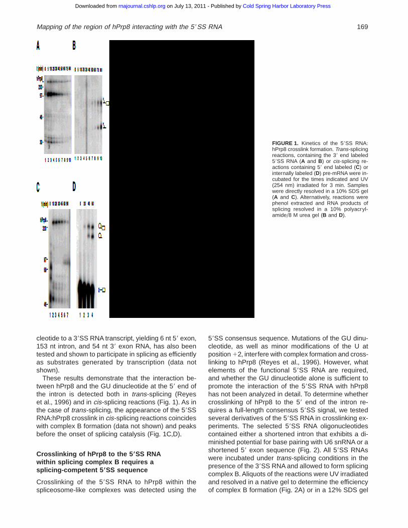
To further characterize the interaction between hPrp8and the GU dinucleotide at the 59 end of the intron, weanalyzed the time course of appearance of the 59SS:hPrp8 crosslink in trans-splicing reactions (Fig+ 1A)+The 3 min irradiation with 254-nm UV light (as opposedto 15 min used previously; Reyes et al+, 1996) yieldslower levels of the 59SS RNA:hPrp8 crosslink but moreprecisely reflects the selected time points of incuba-tion+ After irradiation, the crosslinked products weredirectly resolved in a 10% acrylamide/SDS gel+ The59SS RNA:hPrp8 crosslink appears early in the reac-
tion (5 min), reaches a maximal level at 10 min, andthen slowly decreases over time (Fig+ 1A)+ The major;97-kDa crosslink detected in these reactions is notstably associated with spliceosomes because it is notdetected in complex B resolved in native gels (Reyeset al+, 1996) and it can be separated from splicing com-plexes upon centrifugation through glycerol cushions(data not shown and Sha et al+, 1998)+ The 59SS RNA:hPrp8 crosslinking profile parallels that of the splicingcomplex B formation (data not shown), consistent withthe finding that interaction of hPrp8 with the 59SS RNAoccurs within complex B (Reyes et al+, 1996)+ How-ever, the efficiency of crosslinking decreases prior tothe appearance of splicing intermediates and products(Fig+ 1B), indicating that the 59SS RNA:hPrp8 inter-action is disrupted or changed at the later stages ofsplicing+ Since even minor changes in the local physi-cal environment may affect crosslinking, this loss ofcrosslink formation does not necessarily indicate thatthe 59SS:hPrp8 interaction is discontinued after com-plex B formation+ In fact, hPrp8 was shown to interactwith the 59 exon nucleotides even at the stage of splic-ing catalysis (Wyatt et al+, 1992)+
Because the cis- and trans-splicing systems may dif-fer in the details of the 59SS recognition, we testedwhether the GU dinucleotide:hPrp8 interaction observedin trans-splicing reactions can also be detected in thecontext of a full-length pre-mRNA cis-splicing sub-strate+ We have constructed a cis-splicing pre-mRNAbased on the bimolecular substrates used in trans-splicing reactions+ This substrate (213 nt) was gener-ated by a DNA oligonucleotide-assisted ligation (Moore& Sharp, 1992) of the standard 59SS RNA oligonucle-otide (A5G/GUAAGUAdT) 32P-labeled at the 59 end,with the 39SS RNA transcript containing sequences de-rived from the second intron (145 nt) and third exon(54 nt) of the rabbit b-globin gene+ Splicing reactionsusing such a 59 end-labeled pre-mRNA were UV irra-diated for 3 min and subjected to RNase A digestionprior to resolving the crosslinked products in a 10%acrylamide/SDS gel (Fig+ 1C)+ Upon digestion withRNase A, only the crosslink products formed upstreamof position U12 (p*A5G/GU) are detectable+ A clearlyvisible 59SS RNA:hPrp8 crosslink present in RNaseA-treated reactions (Fig+ 1C) is not detectable uponRNase T1 treatment (data not shown), effectively lim-iting the site of crosslinking within the RNA to the GUdinucleotide at the 59 end of the intron+ Splicing effi-ciency cannot be accurately determined using the 59end-labeled substrate since only formation of the ligated-exons product can be monitored+ To follow the accu-mulation of lariat intermediates and products (Fig+ 1D),a uniformly 32P-labeled pre-mRNA transcript contain-ing 20 nt 59 exon, 191 nt intron, and 54 nt 39 exon ofb-globin was prepared by transcription (see Materialsand Methods)+ In addition, a pre-mRNA generated byligation of the 39 end 32P-labeled 59SS RNA oligonu-
168 J.L. Reyes et al.
Cold Spring Harbor Laboratory Press on July 13, 2011 - Published by rnajournal.cshlp.orgDownloaded from
cleotide to a 39SS RNA transcript, yielding 6 nt 59 exon,153 nt intron, and 54 nt 39 exon RNA, has also beentested and shown to participate in splicing as efficientlyas substrates generated by transcription (data notshown)+
These results demonstrate that the interaction be-tween hPrp8 and the GU dinucleotide at the 59 end ofthe intron is detected both in trans-splicing (Reyeset al+, 1996) and in cis-splicing reactions (Fig+ 1)+ As inthe case of trans-splicing, the appearance of the 59SSRNA:hPrp8 crosslink in cis-splicing reactions coincideswith complex B formation (data not shown) and peaksbefore the onset of splicing catalysis (Fig+ 1C,D)+
Crosslinking of hPrp8 to the 5 9SS RNAwithin splicing complex B requires asplicing-competent 5 9SS sequence
Crosslinking of the 59SS RNA to hPrp8 within thespliceosome-like complexes was detected using the
59SS consensus sequence+ Mutations of the GU dinu-cleotide, as well as minor modifications of the U atposition 12, interfere with complex formation and cross-linking to hPrp8 (Reyes et al+, 1996)+ However, whatelements of the functional 59SS RNA are required,and whether the GU dinucleotide alone is sufficient topromote the interaction of the 59SS RNA with hPrp8has not been analyzed in detail+ To determine whethercrosslinking of hPrp8 to the 59 end of the intron re-quires a full-length consensus 59SS signal, we testedseveral derivatives of the 59SS RNA in crosslinking ex-periments+ The selected 59SS RNA oligonucleotidescontained either a shortened intron that exhibits a di-minished potential for base pairing with U6 snRNA or ashortened 59 exon sequence (Fig+ 2)+ All 59SS RNAswere incubated under trans-splicing conditions in thepresence of the 39SS RNA and allowed to form splicingcomplex B+Aliquots of the reactions were UV irradiatedand resolved in a native gel to determine the efficiencyof complex B formation (Fig+ 2A) or in a 12% SDS gel
FIGURE 1. Kinetics of the 59SS RNA:hPrp8 crosslink formation+ Trans-splicingreactions, containing the 39 end labeled59SS RNA (A and B) or cis-splicing re-actions containing 59 end labeled (C) orinternally labeled (D) pre-mRNA were in-cubated for the times indicated and UV(254 nm) irradiated for 3 min+ Sampleswere directly resolved in a 10% SDS gel(A and C)+ Alternatively, reactions werephenol extracted and RNA products ofsplicing resolved in a 10% polyacryl-amide/8 M urea gel (B and D)+
Mapping of the region of hPrp8 interacting with the 59SS RNA 169
Cold Spring Harbor Laboratory Press on July 13, 2011 - Published by rnajournal.cshlp.orgDownloaded from
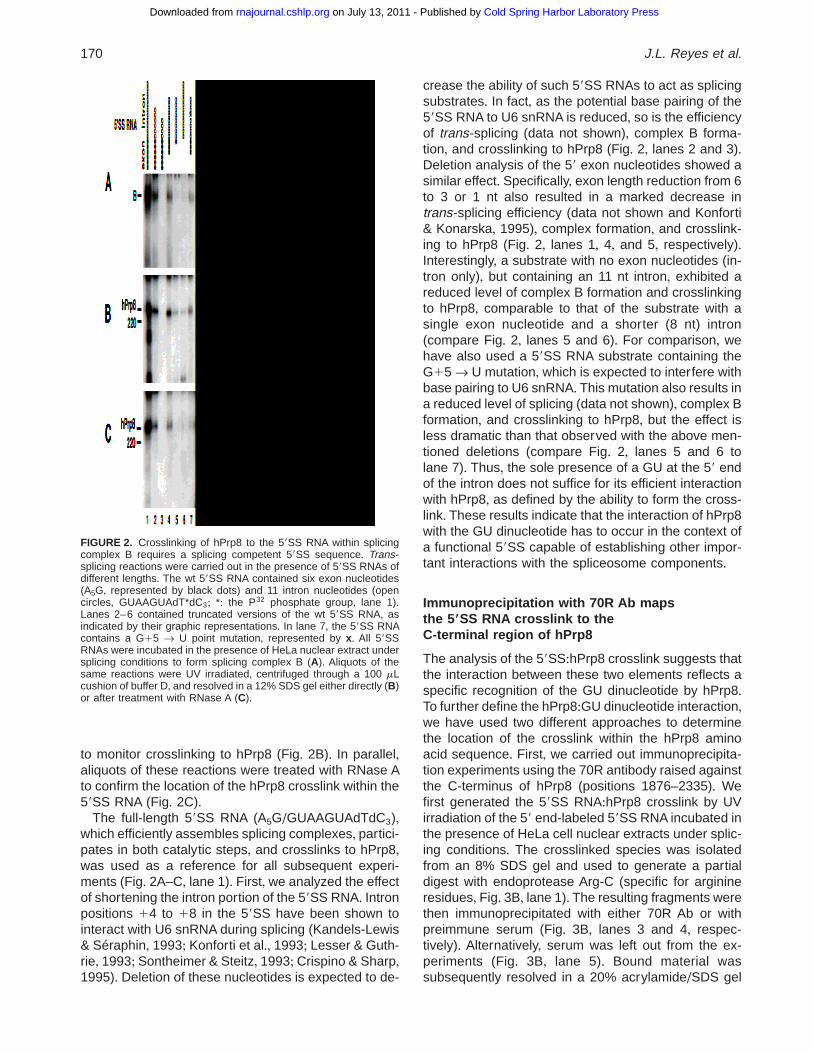
to monitor crosslinking to hPrp8 (Fig+ 2B)+ In parallel,aliquots of these reactions were treated with RNase Ato confirm the location of the hPrp8 crosslink within the59SS RNA (Fig+ 2C)+
The full-length 59SS RNA (A5G/GUAAGUAdTdC3),which efficiently assembles splicing complexes, partici-pates in both catalytic steps, and crosslinks to hPrp8,was used as a reference for all subsequent experi-ments (Fig+ 2A–C, lane 1)+ First, we analyzed the effectof shortening the intron portion of the 59SS RNA+ Intronpositions 14 to 18 in the 59SS have been shown tointeract with U6 snRNA during splicing (Kandels-Lewis& Séraphin, 1993; Konforti et al+, 1993; Lesser & Guth-rie, 1993; Sontheimer & Steitz, 1993; Crispino & Sharp,1995)+ Deletion of these nucleotides is expected to de-
crease the ability of such 59SS RNAs to act as splicingsubstrates+ In fact, as the potential base pairing of the59SS RNA to U6 snRNA is reduced, so is the efficiencyof trans-splicing (data not shown), complex B forma-tion, and crosslinking to hPrp8 (Fig+ 2, lanes 2 and 3)+Deletion analysis of the 59 exon nucleotides showed asimilar effect+ Specifically, exon length reduction from 6to 3 or 1 nt also resulted in a marked decrease intrans-splicing efficiency (data not shown and Konforti& Konarska, 1995), complex formation, and crosslink-ing to hPrp8 (Fig+ 2, lanes 1, 4, and 5, respectively)+Interestingly, a substrate with no exon nucleotides (in-tron only), but containing an 11 nt intron, exhibited areduced level of complex B formation and crosslinkingto hPrp8, comparable to that of the substrate with asingle exon nucleotide and a shorter (8 nt) intron(compare Fig+ 2, lanes 5 and 6)+ For comparison, wehave also used a 59SS RNA substrate containing theG15 r U mutation, which is expected to interfere withbase pairing to U6 snRNA+ This mutation also results ina reduced level of splicing (data not shown), complex Bformation, and crosslinking to hPrp8, but the effect isless dramatic than that observed with the above men-tioned deletions (compare Fig+ 2, lanes 5 and 6 tolane 7)+ Thus, the sole presence of a GU at the 59 endof the intron does not suffice for its efficient interactionwith hPrp8, as defined by the ability to form the cross-link+ These results indicate that the interaction of hPrp8with the GU dinucleotide has to occur in the context ofa functional 59SS capable of establishing other impor-tant interactions with the spliceosome components+
Immunoprecipitation with 70R Ab mapsthe 59SS RNA crosslink to theC-terminal region of hPrp8
The analysis of the 59SS:hPrp8 crosslink suggests thatthe interaction between these two elements reflects aspecific recognition of the GU dinucleotide by hPrp8+To further define the hPrp8:GU dinucleotide interaction,we have used two different approaches to determinethe location of the crosslink within the hPrp8 aminoacid sequence+ First, we carried out immunoprecipita-tion experiments using the 70R antibody raised againstthe C-terminus of hPrp8 (positions 1876–2335)+ Wefirst generated the 59SS RNA:hPrp8 crosslink by UVirradiation of the 59 end-labeled 59SS RNA incubated inthe presence of HeLa cell nuclear extracts under splic-ing conditions+ The crosslinked species was isolatedfrom an 8% SDS gel and used to generate a partialdigest with endoprotease Arg-C (specific for arginineresidues, Fig+ 3B, lane 1)+ The resulting fragments werethen immunoprecipitated with either 70R Ab or withpreimmune serum (Fig+ 3B, lanes 3 and 4, respec-tively)+ Alternatively, serum was left out from the ex-periments (Fig+ 3B, lane 5)+ Bound material wassubsequently resolved in a 20% acrylamide/SDS gel
FIGURE 2. Crosslinking of hPrp8 to the 59SS RNA within splicingcomplex B requires a splicing competent 59SS sequence+ Trans-splicing reactions were carried out in the presence of 59SS RNAs ofdifferent lengths+ The wt 59SS RNA contained six exon nucleotides(A5G, represented by black dots) and 11 intron nucleotides (opencircles, GUAAGUAdT*dC3; *: the P32 phosphate group, lane 1)+Lanes 2–6 contained truncated versions of the wt 59SS RNA, asindicated by their graphic representations+ In lane 7, the 59SS RNAcontains a G15 r U point mutation, represented by x+ All 59SSRNAs were incubated in the presence of HeLa nuclear extract undersplicing conditions to form splicing complex B (A)+ Aliquots of thesame reactions were UV irradiated, centrifuged through a 100 mLcushion of buffer D, and resolved in a 12% SDS gel either directly (B)or after treatment with RNase A (C)+
170 J.L. Reyes et al.
Cold Spring Harbor Laboratory Press on July 13, 2011 - Published by rnajournal.cshlp.orgDownloaded from
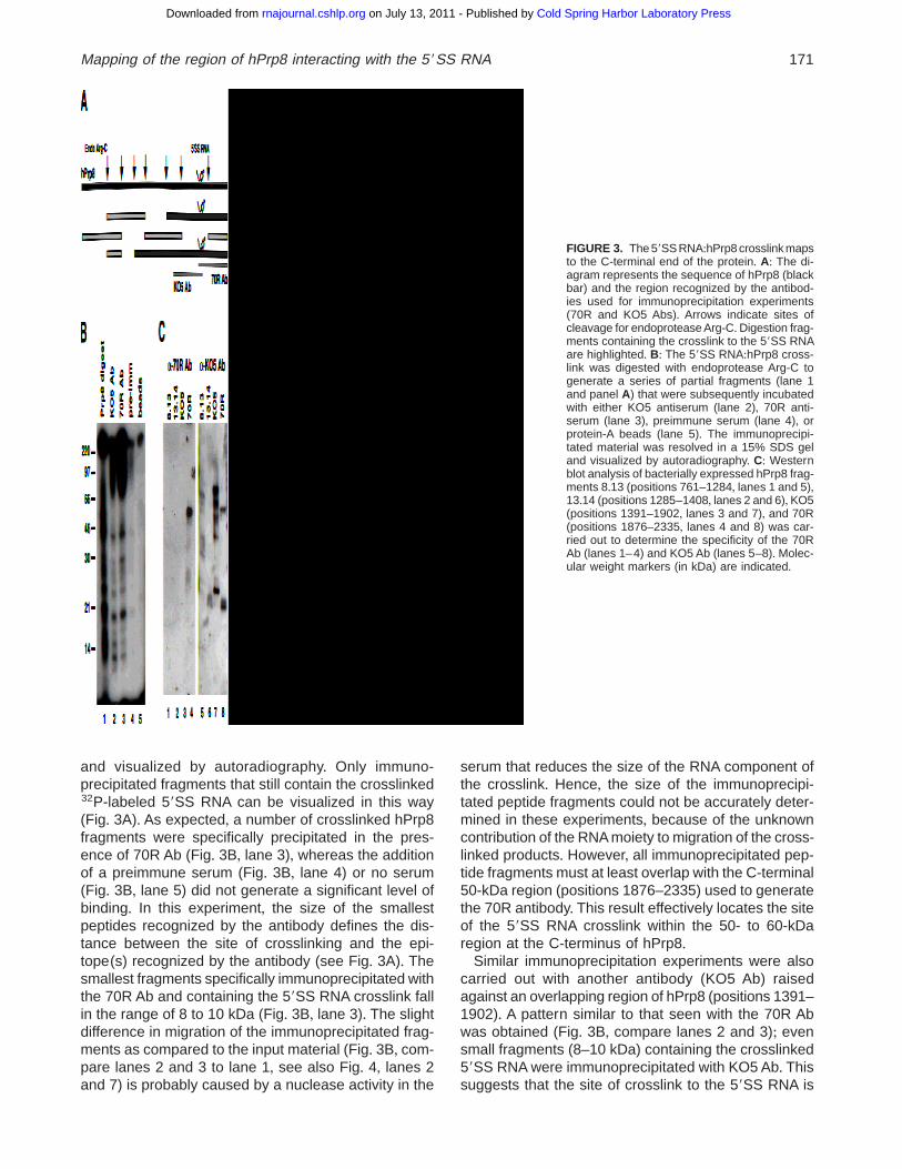
and visualized by autoradiography+ Only immuno-precipitated fragments that still contain the crosslinked32P-labeled 59SS RNA can be visualized in this way(Fig+ 3A)+ As expected, a number of crosslinked hPrp8fragments were specifically precipitated in the pres-ence of 70R Ab (Fig+ 3B, lane 3), whereas the additionof a preimmune serum (Fig+ 3B, lane 4) or no serum(Fig+ 3B, lane 5) did not generate a significant level ofbinding+ In this experiment, the size of the smallestpeptides recognized by the antibody defines the dis-tance between the site of crosslinking and the epi-tope(s) recognized by the antibody (see Fig+ 3A)+ Thesmallest fragments specifically immunoprecipitated withthe 70R Ab and containing the 59SS RNA crosslink fallin the range of 8 to 10 kDa (Fig+ 3B, lane 3)+ The slightdifference in migration of the immunoprecipitated frag-ments as compared to the input material (Fig+ 3B, com-pare lanes 2 and 3 to lane 1, see also Fig+ 4, lanes 2and 7) is probably caused by a nuclease activity in the
serum that reduces the size of the RNA component ofthe crosslink+ Hence, the size of the immunoprecipi-tated peptide fragments could not be accurately deter-mined in these experiments, because of the unknowncontribution of the RNA moiety to migration of the cross-linked products+ However, all immunoprecipitated pep-tide fragments must at least overlap with the C-terminal50-kDa region (positions 1876–2335) used to generatethe 70R antibody+ This result effectively locates the siteof the 59SS RNA crosslink within the 50- to 60-kDaregion at the C-terminus of hPrp8+
Similar immunoprecipitation experiments were alsocarried out with another antibody (KO5 Ab) raisedagainst an overlapping region of hPrp8 (positions 1391–1902)+ A pattern similar to that seen with the 70R Abwas obtained (Fig+ 3B, compare lanes 2 and 3); evensmall fragments (8–10 kDa) containing the crosslinked59SS RNA were immunoprecipitated with KO5 Ab+ Thissuggests that the site of crosslink to the 59SS RNA is
FIGURE 3. The 59SS RNA:hPrp8 crosslink mapsto the C-terminal end of the protein+ A: The di-agram represents the sequence of hPrp8 (blackbar) and the region recognized by the antibod-ies used for immunoprecipitation experiments(70R and KO5 Abs)+ Arrows indicate sites ofcleavage for endoprotease Arg-C+Digestion frag-ments containing the crosslink to the 59SS RNAare highlighted+ B: The 59SS RNA:hPrp8 cross-link was digested with endoprotease Arg-C togenerate a series of partial fragments (lane 1and panel A) that were subsequently incubatedwith either KO5 antiserum (lane 2), 70R anti-serum (lane 3), preimmune serum (lane 4), orprotein-A beads (lane 5)+ The immunoprecipi-tated material was resolved in a 15% SDS geland visualized by autoradiography+ C: Westernblot analysis of bacterially expressed hPrp8 frag-ments 8+13 (positions 761–1284, lanes 1 and 5),13+14 (positions 1285–1408, lanes 2 and 6), KO5(positions 1391–1902, lanes 3 and 7), and 70R(positions 1876–2335, lanes 4 and 8) was car-ried out to determine the specificity of the 70RAb (lanes 1–4) and KO5 Ab (lanes 5–8)+ Molec-ular weight markers (in kDa) are indicated+
Mapping of the region of hPrp8 interacting with the 59SS RNA 171
Cold Spring Harbor Laboratory Press on July 13, 2011 - Published by rnajournal.cshlp.orgDownloaded from
located at, or close to, the overlapping region recog-nized by the two antibodies (positions 1876–1902)+Con-trol Western blot analyses showed that the KO5 Abrecognizes not only the corresponding KO5 antigenpeptide, but also cross-reacts with the bacterially ex-pressed 70R peptide fragment used to generate the70R Ab (Fig+ 3C, compare lanes 7 and 8, respectively)+These results may be explained by a short stretch ofoverlapping sequence shared by the 70R and KO5 frag-ments (amino acids 1876–1902)+Alternatively, the KO5Ab may recognize a short N-terminal His-tag sequencepresent in both the 70R and KO5 peptides+ Since twoother fragments of hPrp8 (8+13, positions 761–1284,and 13+14, positions 1285–1408) also show a signifi-cantly lower, but detectable, level of reactivity with theKO5 Ab (Fig+ 3C, lanes 5 and 6), a combination of boththese interpretations may apply+ In contrast, Westernblot analysis using the 70R Ab and the same bacteriallyexpressed fragments of hPrp8 showed only specificrecognition of the 70R fragment originally used to raise
this antibody (Fig+ 3C, lane 4)+ No cross-reactivity withthe other three fragments was observed (Fig+ 3C,lanes 1–3), confirming the high specificity of the 70RAb and its suitability for mapping experiments+ Be-cause of the uncertainty regarding the specificity of theKO5 serum, we did not rely on the results obtained withthis antibody, although they are completely consistentwith the proteolytic analysis of the 59SS RNA:hPrp8crosslink shown below+ Unfortunately, all attempts toraise immunoprecipitation-competent antisera to otherregions of hPrp8 (e+g+, 8+13 and 13+14) have been un-successful to date (H+R+ Luo,M+J+Moore, unpubl+ data)+Minimally, the immunoprecipitation results indicate thatthe site of the 59SS RNA:hPrp8 crosslink is locatedwithin the C-terminus of the hPrp8 protein+ The seg-ment spanning the crosslink, a fragment of 8–10 kDa,at least overlaps with the region recognized by the 70Rantibody, between positions 1876 and the C-terminusof the protein (position 2335)+
Proteolytic analysis of the 5 9SSRNA:hPrp8 crosslink
We have extended the analysis of the 59SS RNA:hPrp8 crosslink to include a number of proteases andchemical reagents of distinct specificities+ Based onthe size of the fragments containing the 59SS RNAcrosslink and the known sequence of hPrp8 (GenBankaccession number AF092565), we could precisely lo-cate the site of crosslink within the protein sequence+
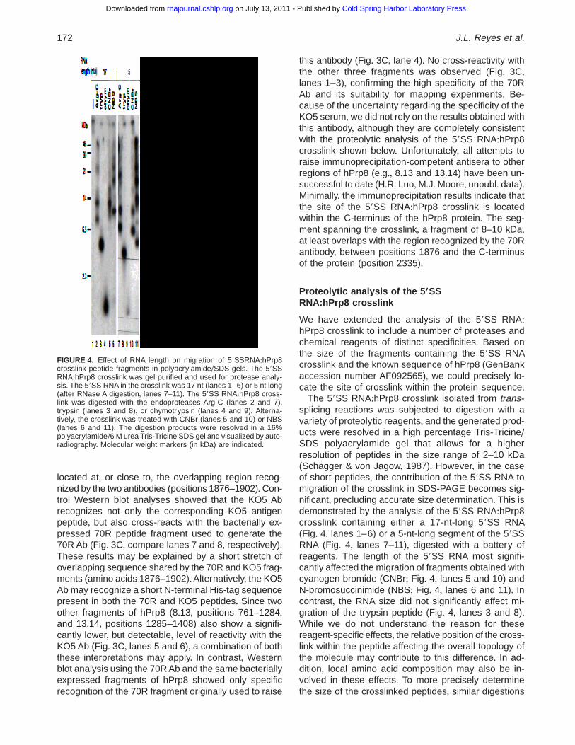
The 59SS RNA:hPrp8 crosslink isolated from trans-splicing reactions was subjected to digestion with avariety of proteolytic reagents, and the generated prod-ucts were resolved in a high percentage Tris-Tricine/SDS polyacrylamide gel that allows for a higherresolution of peptides in the size range of 2–10 kDa(Schägger & von Jagow, 1987)+ However, in the caseof short peptides, the contribution of the 59SS RNA tomigration of the crosslink in SDS-PAGE becomes sig-nificant, precluding accurate size determination+ This isdemonstrated by the analysis of the 59SS RNA:hPrp8crosslink containing either a 17-nt-long 59SS RNA(Fig+ 4, lanes 1–6) or a 5-nt-long segment of the 59SSRNA (Fig+ 4, lanes 7–11), digested with a battery ofreagents+ The length of the 59SS RNA most signifi-cantly affected the migration of fragments obtained withcyanogen bromide (CNBr; Fig+ 4, lanes 5 and 10) andN-bromosuccinimide (NBS; Fig+ 4, lanes 6 and 11)+ Incontrast, the RNA size did not significantly affect mi-gration of the trypsin peptide (Fig+ 4, lanes 3 and 8)+While we do not understand the reason for thesereagent-specific effects, the relative position of the cross-link within the peptide affecting the overall topology ofthe molecule may contribute to this difference+ In ad-dition, local amino acid composition may also be in-volved in these effects+ To more precisely determinethe size of the crosslinked peptides, similar digestions
FIGURE 4. Effect of RNA length on migration of 59SSRNA:hPrp8crosslink peptide fragments in polyacrylamide/SDS gels+ The 59SSRNA:hPrp8 crosslink was gel purified and used for protease analy-sis+ The 59SS RNA in the crosslink was 17 nt (lanes 1–6) or 5 nt long(after RNase A digestion, lanes 7–11)+ The 59SS RNA:hPrp8 cross-link was digested with the endoproteases Arg-C (lanes 2 and 7),trypsin (lanes 3 and 8), or chymotrypsin (lanes 4 and 9)+ Alterna-tively, the crosslink was treated with CNBr (lanes 5 and 10) or NBS(lanes 6 and 11)+ The digestion products were resolved in a 16%polyacrylamide/6 M urea Tris-Tricine SDS gel and visualized by auto-radiography+ Molecular weight markers (in kDa) are indicated+
172 J.L. Reyes et al.
Cold Spring Harbor Laboratory Press on July 13, 2011 - Published by rnajournal.cshlp.orgDownloaded from
were performed with the 59SS RNA:hPrp8 crosslinkcontaining a single 32P at position G11 of the 59SS(A7G/p*GUAAGUAdTdC3)+Upon digestion of the cross-link with nuclease P1, a single p*G residue remainslinked to hPrp8, thus minimizing the RNA contribu-tion to the crosslink mobility in Tris-Tricine/SDS gels+ Inall subsequent analyses, the molecular weight of thecrosslink-containing peptides generated by digestionwith a variety of proteolytic reagents was calculatedfrom crosslinks containing a single G nucleotide+ Re-markably, digestion of the 59SS RNA:hPrp8 crosslinkwith either trypsin or chymotrypsin generates single32P-labeled peptides (Fig+ 4, lanes 3 and 4), indicatinga high level of homogeneity of the crosslink with re-spect to the protein component+ Together with the pre-vious analysis that mapped the same crosslink to theGU dinucleotide within the 59SS RNA, these resultsindicate a highly site-specific, localized physical inter-action between the 59SS and a unique segment inhPrp8+
Cyanogen bromide, N-bromosuccinimide andtrypsin digestion of the 59SS RNA:hPrp8 crosslink
Figure 5A shows digestion patterns of the gel-purified59SS RNA:hPrp8 crosslink (lane 1) after treatment withtrypsin (lane 2) and chemical reagents cyanogen bro-mide (lane 3) and N-bromosuccinimide (lane 4), re-solved in a 16% Tris-Tricine/SDS gel+ As describedabove, the crosslinked peptides contain a single 32P-labeled G nucleotide, which allows one to more accu-rately estimate the peptide size+ Cyanogen bromide(CNBr) specifically cleaves proteins at the carboxy-terminus of methionine residues+ The extent of its cleav-age depends both on the conditions used and the aminoacid context around the cleavage site (Gross, 1967)+ Inthe context of the 59SS RNA:hPrp8 crosslink, we havenot been able to obtain a complete CNBr digestiongenerating a single product (Fig+ 5A, lane 3, and Fig+ 4,lane 10)+The gel-purified partial digestion products werenot further cleaved with CNBr (data not shown), pos-sibly due to modification of methionines+ The most abun-dant cleavage product corresponds to the smallestfragment of 5+1 kDa, which may represent a completedigestion product that does not include any internalmethionines+ From the amino acid sequence of hPrp8,a number of CNBr fragments can be found in the rangeof 4–6 kDa (indicated in green in Fig+ 7A)+ However,since the 5+1-kDa fragment may represent a partialCNBr digestion product, we have also consideredpeptides containing one internal methionine for furtheranalysis+ Together, 22 distinct CNBr fragments wereconsidered+
N-bromosuccinimide cleaves proteins after trypto-phan, histidine, and tyrosine residues (Shechter et al+,1976)+ As with CNBr, the sequence context affects theextent of its cleavage+ Digestion of the 59SS RNA:
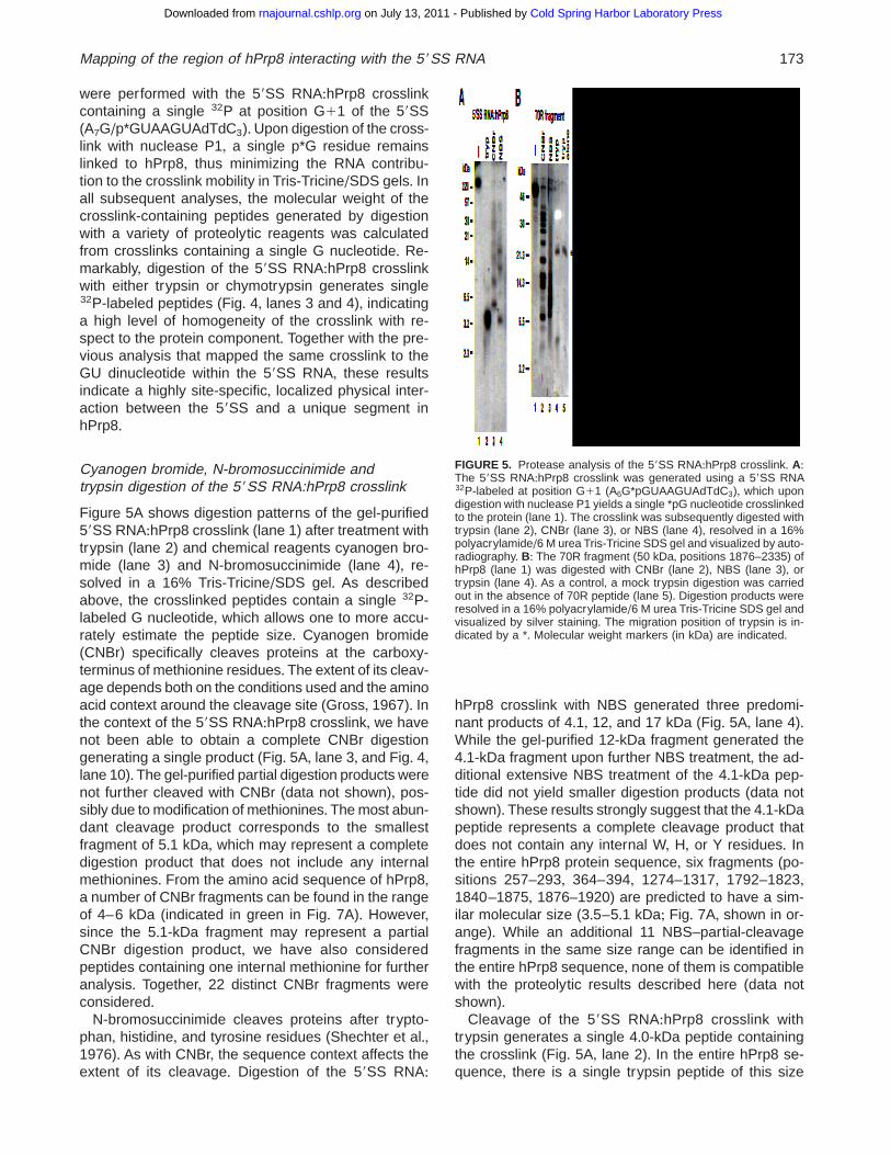
hPrp8 crosslink with NBS generated three predomi-nant products of 4+1, 12, and 17 kDa (Fig+ 5A, lane 4)+While the gel-purified 12-kDa fragment generated the4+1-kDa fragment upon further NBS treatment, the ad-ditional extensive NBS treatment of the 4+1-kDa pep-tide did not yield smaller digestion products (data notshown)+ These results strongly suggest that the 4+1-kDapeptide represents a complete cleavage product thatdoes not contain any internal W, H, or Y residues+ Inthe entire hPrp8 protein sequence, six fragments (po-sitions 257–293, 364–394, 1274–1317, 1792–1823,1840–1875, 1876–1920) are predicted to have a sim-ilar molecular size (3+5–5+1 kDa; Fig+ 7A, shown in or-ange)+ While an additional 11 NBS–partial-cleavagefragments in the same size range can be identified inthe entire hPrp8 sequence, none of them is compatiblewith the proteolytic results described here (data notshown)+
Cleavage of the 59SS RNA:hPrp8 crosslink withtrypsin generates a single 4+0-kDa peptide containingthe crosslink (Fig+ 5A, lane 2)+ In the entire hPrp8 se-quence, there is a single trypsin peptide of this size
FIGURE 5. Protease analysis of the 59SS RNA:hPrp8 crosslink+ A:The 59SS RNA:hPrp8 crosslink was generated using a 59SS RNA32P-labeled at position G11 (A6G*pGUAAGUAdTdC3), which upondigestion with nuclease P1 yields a single *pG nucleotide crosslinkedto the protein (lane 1)+ The crosslink was subsequently digested withtrypsin (lane 2), CNBr (lane 3), or NBS (lane 4), resolved in a 16%polyacrylamide/6 M urea Tris-Tricine SDS gel and visualized by auto-radiography+ B: The 70R fragment (50 kDa, positions 1876–2335) ofhPrp8 (lane 1) was digested with CNBr (lane 2), NBS (lane 3), ortrypsin (lane 4)+ As a control, a mock trypsin digestion was carriedout in the absence of 70R peptide (lane 5)+ Digestion products wereresolved in a 16% polyacrylamide/6 M urea Tris-Tricine SDS gel andvisualized by silver staining+ The migration position of trypsin is in-dicated by a *+ Molecular weight markers (in kDa) are indicated+
Mapping of the region of hPrp8 interacting with the 59SS RNA 173
Cold Spring Harbor Laboratory Press on July 13, 2011 - Published by rnajournal.cshlp.orgDownloaded from
(position 1684–1723); however, it does not overlap withany of the sequences identified with CNBr or NBS+ Thissuggests that the trypsin fragment represents a partialcleavage product that contains at least one blockedinternal trypsin site (a lysine or arginine residue)+ Aprotein may be resistant to trypsin cleavage either be-cause of particular structural features or because thecrosslinked RNA itself could block the cleavage site+ Infact, when the 4-kDa trypsin fragment was gel-purifiedand subsequently digested with a high excess of tryp-sin, a low level of a smaller, 2+0-kDa fragment contain-ing the crosslinked 59SS RNA could be detected (datanot shown)+
To independently confirm that the trypsin fragmentrepresents a partially digested peptide, we purified the70R fragment of hPrp8 (position 1876–2335) expressedin Escherichia coli as a His-tagged protein+ The gel-purified 70R fragment (Fig+ 5B, lane 1) was digested inparallel with CNBr, NBS, or trypsin, and products wereresolved in a 20% polyacrylamide/SDS gel and visu-alized by silver staining (Fig+ 5B, lanes 2–5)+ The se-quence of the 70R fragment predicts no trypsin productslarger than 3+0 kDa+ However, a single peptide with gelmobility of ;4+0 kDa was detected in this trypsin di-gestion, indicating that it must represent a partial di-gestion product (Fig+ 5B, lane 4)+ This 4-kDa peptideoriginates from the 70R fragment since it was not de-tected in the absence of the 70R substrate (Fig+ 5B,lane 5)+ Smaller digestion products generated in thisreaction could not be detected, presumably because ofthe poor staining with silver under these conditions(Fig+ 5B, lanes 2–5)+ Digestion with CNBr and NBSalso yields a partial digestion of the 70R fragment, judg-ing by the size of the detected products (Fig+ 5B,lanes 2 and 3)+ Detection of the 4-kDa trypsin peptideof the 70R fragment suggests that a similar partial pep-tide in the 59SS RNA:hPrp8 crosslink results from alocal protein sequence or structure rather than from thepresence of the crosslink at the trypsin cleavage site+Predicted trypsin fragments of hPrp8 in the range of3+1 to 5+0 kDa (a total of 38 fragments) are representedby blue bars in Figure 7A+ As with CNBr fragments,partial products are also included+
A combination of CNBr, NBS, and trypsin digestionseliminates a majority of the initial pool of the candidatesites and identifies only three possible locations con-sistent with the proteolysis results shown in this section(Fig+ 7, compare A and B)+ These sequences are lo-cated within the trypsin-generated fragments at posi-tions 228–261, 1263–1290, and 1867–1898 (Fig+ 7C)+
Chymotrypsin and Endo Glu-C protease analysis
In addition to proteolytic digestion of the full-length 59SSRNA:hPrp8 crosslink described above, we have alsoperformed secondary digestions of the selected, gel-isolated peptide fragments+ By imposing additional con-
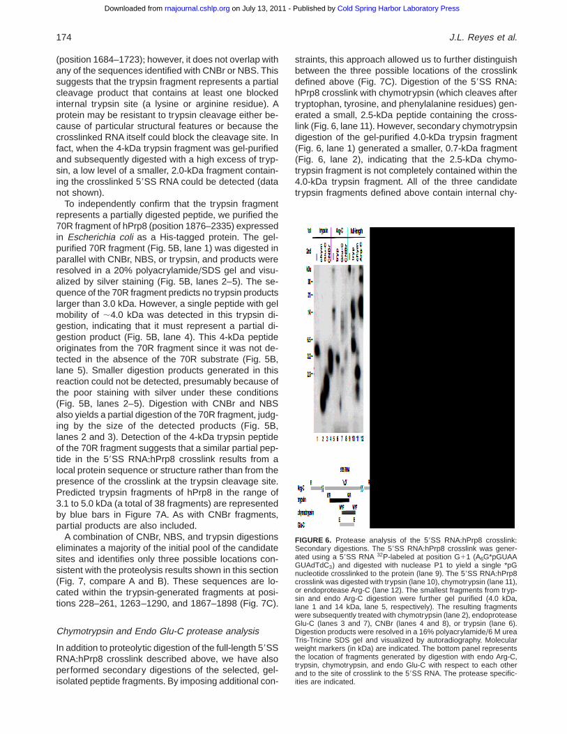
straints, this approach allowed us to further distinguishbetween the three possible locations of the crosslinkdefined above (Fig+ 7C)+ Digestion of the 59SS RNA:hPrp8 crosslink with chymotrypsin (which cleaves aftertryptophan, tyrosine, and phenylalanine residues) gen-erated a small, 2+5-kDa peptide containing the cross-link (Fig+ 6, lane 11)+However, secondary chymotrypsindigestion of the gel-purified 4+0-kDa trypsin fragment(Fig+ 6, lane 1) generated a smaller, 0+7-kDa fragment(Fig+ 6, lane 2), indicating that the 2+5-kDa chymo-trypsin fragment is not completely contained within the4+0-kDa trypsin fragment+ All of the three candidatetrypsin fragments defined above contain internal chy-
FIGURE 6. Protease analysis of the 59SS RNA:hPrp8 crosslink:Secondary digestions+ The 59SS RNA:hPrp8 crosslink was gener-ated using a 59SS RNA 32P-labeled at position G11 (A6G*pGUAAGUAdTdC3) and digested with nuclease P1 to yield a single *pGnucleotide crosslinked to the protein (lane 9)+ The 59SS RNA:hPrp8crosslink was digested with trypsin (lane 10), chymotrypsin (lane 11),or endoprotease Arg-C (lane 12)+ The smallest fragments from tryp-sin and endo Arg-C digestion were further gel purified (4+0 kDa,lane 1 and 14 kDa, lane 5, respectively)+ The resulting fragmentswere subsequently treated with chymotrypsin (lane 2), endoproteaseGlu-C (lanes 3 and 7), CNBr (lanes 4 and 8), or trypsin (lane 6)+Digestion products were resolved in a 16% polyacrylamide/6 M ureaTris-Tricine SDS gel and visualized by autoradiography+ Molecularweight markers (in kDa) are indicated+ The bottom panel representsthe location of fragments generated by digestion with endo Arg-C,trypsin, chymotrypsin, and endo Glu-C with respect to each otherand to the site of crosslink to the 59SS RNA+ The protease specific-ities are indicated+
174 J.L. Reyes et al.
Cold Spring Harbor Laboratory Press on July 13, 2011 - Published by rnajournal.cshlp.orgDownloaded from
motrypsin sites (Fig+ 7B,C)+ However, the trypsin frag-ment at position 1263–1290 overlaps with a partialchymotrypsin fragment, thus making it less likely as thecrosslink site (Fig+ 7C)+
Digestion of the 59SS:hPrp8 crosslink with endo-protease Arg-C generates a 14-kDa partial fragment(Fig+ 6, lanes 5 and 12)+ The 14-kDa Arg-C fragmentcontains the entire 4-kDa trypsin fragment, as its sec-ondary digestion with trypsin yields the same 4+0-kDapeptide (Fig+ 6, lane 6)+ When the 14-kDa Arg-C pep-tide was further digested with endoprotease Glu-C,the smallest product containing the crosslink was an;2+5-kDa peptide (Fig+ 6, lane 7)+ Under the conditionsused, Glu-C cleaves only after glutamic acid, and notaspartic acid residues (data not shown)+ The gel-purified4+0-kDa trypsin fragment digested with Glu-C gener-ates an ;1-kDa peptide, which is smaller than the
2+5-kDa Glu-C peptide obtained from digestion of theArg-C fragment (Fig+ 6, compare lanes 3 and 7)+ Thus,within the 14-kDa Arg-C fragment, the 4+0-kDa trypsinpeptide containing the crosslink must overlap with bothGlu-C and chymotrypsin fragments (Fig+ 7C)+ Inspec-tion of the three candidate locations identified by CNBr,NBS, and trypsin digestions identifies the C-terminalsite (trypsin peptide position 1867–1898) as the loca-tion of the 59SS RNA:hPrp8 crosslink+ The fact that theN-terminal trypsin fragment (position 228–261) doesnot contain any internal glutamic acid residues, andthat the second trypsin fragment (position 1869–1914)contains partial chymotrypsin and Glu-C peptides thatdo not overlap, eliminates both these sites as a possi-ble location of the crosslink (Fig+ 7C)+
In summary, the data from digestion of the 59SSRNA:hPrp8 crosslink with CNBr, NBS, and trypsin gen-
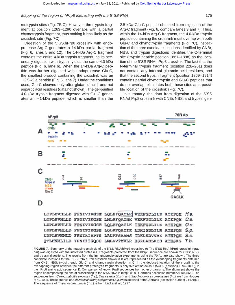
FIGURE 7. Summary of the mapping analysis of the 59SS RNA:hPrp8 crosslink+ A: The 59SS RNA:hPrp8 crosslink (graybar) was digested with the indicated proteases+ Fragments predicted from the hPrp8 sequence are shown for CNBr, NBS,and trypsin digestions+ The results from the immunoprecipitation experiments using the 70 Ab are also shown+ The threecandidate locations for the 59SS RNA:hPrp8 crosslink shown in B are represented as the overlapping fragments obtainedfrom CNBr, NBS, trypsin, endo Glu-C, and chymotrypsin digestion in C+ In the deduced location of the crosslink, theoverlapping region between the different proteolytic fragments is only five amino acids, QACLK (positions 1894–1898), inthe hPrp8 amino acid sequence+ D: Comparison of known Prp8 sequences from other organisms+ The alignment shows theregion encompassing the site of crosslinking to the 59SS RNA in hPrp8 (H+s+, GenBank accession number AF092565)+ Thesequences from Caenorhabditis elegans (C.e.), Oriza sativa (O.s.), and Saccharomyces cerevisiae (S.c.) are from Hodgeset al+, 1995+ The sequence of Schizosaccharomyces pombe (S.p.) was obtained from GenBank (accession number 2440191)+The sequence of Trypanosoma brucei (T.b.) is from Lücke et al+, 1997+
Mapping of the region of hPrp8 interacting with the 59SS RNA 175
Cold Spring Harbor Laboratory Press on July 13, 2011 - Published by rnajournal.cshlp.orgDownloaded from

erated three candidate segments that may contain thesite of crosslink (Fig+ 7B,C)+ All three segments complywith the requirements of representing overlapping pep-tides generated by different proteolytic digestions andhaving a size compatible with the estimates from thehigh resolution Tris-Tricine/SDS gels+ For the three can-didate fragments, the trypsin peptide is considered torepresent a partial digestion product with one internaltrypsin site+ Data from secondary digestions of the iso-lated trypsin peptide with chymotrypsin and endo Glu-Cproteases were used to eliminate two of the three pos-sible locations+ Only the C-terminal candidate segment(trypsin peptide positions 1867–1898) is consistent withall the proteolytic analyses and, moreover, with the im-munoprecipitation experiments (Fig+ 7A,C)+ Based onthe proteolysis results, the site of crosslink is limited bythe chymotrypsin and trypsin sites (Fig+ 7C), restrictingit to only five amino acids in the hPrp8 sequence:QACLK (positions 1894–1898)+ The achieved resolu-tion is remarkable, considering the total length of hPrp8:2335 amino acids+
While the entire sequence of Prp8, including the re-gion spanning the crosslink site, exhibits a high degreeof conservation across eukaryotes, the amino acid se-quence directly adjacent to the mapped crosslink site issignificantly less well conserved+ The human QACLKsequence (positions 1894–1898) corresponds to QAIMKin Caenorhabditis elegans, QAIIK in Schizosaccharo-myces pombe, and SAAMS (positions 1966–1970) inSaccharomyces cerevisiae (Fig+ 7D)+ It should be notedthat in S. cerevisiae, the mapped site is located in theproximity of amino acid residues implicated in poly-pyrimidine tract recognition (positions 1834 and 1960;Umen & Guthrie, 1995, 1996), and in genetic inter-action with U4 snRNA (Kuhn et al+, 1999)+ Together,these results suggest that the region of Prp8 involvedin interactions with the polypyrimidine tract and possi-bly with U4/U6 snRNAs, may also be involved in rec-ognition of the 59 splice site+
DISCUSSION
Recognition of the 59SS by base pairing to the 59 endof U1 snRNA represents one of the initial steps of splice-osome assembly+While this interaction controls the over-all selection of the 59 splice site, it does not specify theactual cleavage site (reviewed in Moore et al+, 1993;Nilsen, 1994)+ Subsequently, the 59SS:U1 snRNA base-pairing must be disrupted to allow the 59SS to interactwith Prp8, among other components of the U4/U5/U6snRNP (see Kandels-Lewis & Séraphin, 1993; Konfortiet al+, 1993; Lesser & Guthrie, 1993; Sontheimer &Steitz, 1993)+ Both human and yeast Prp8 have beenshown to crosslink to the 59SS region (Wyatt et al+,1992; Teigelkamp et al+, 1995a, 1995b; Chiara et al+,1996; Reyes et al+, 1996)+ We have previously shownthat a highly specific GU dinucleotide:hPrp8 crosslink
at the 59SS can be detected within splicing complex Bassembled in trans-splicing reactions, where the 59SSis provided as a short RNA oligonucleotide (Reyeset al+, 1996)+ The analogous 59SS:hPrp8 crosslink canalso be detected within complex B formed in cis-splicingreactions, using a unimolecular pre-mRNA substrate(Fig+ 1)+ Also the kinetics of crosslinking indicate that inboth trans- and cis-splicing, the 59SS:hPrp8 interactiontakes place early in the reaction, after formation of com-plex B, but before the appearance of splicing intermedi-ates and products+ The interaction of Prp8 with exonsequences at the 59SS is maintained beyond the firststep of splicing, as evidenced by the persistence ofcrosslinks in both yeast and mammalian systems (Wyattet al+, 1992; Teigelkamp et al+, 1995b)+ In contrast, theGU:hPrp8 interaction described here occurs withinspliceosome complex B, but crosslink formation is notdetected at later stages in the reaction (Fig+ 1)+ Thiscrosslinking profile suggests that either the GU:hPrp8interaction is disrupted at later stages of splicing, orthat an alteration in the physical environment near thecontact site may not permit UV crosslinking, even thoughthe interaction itself may be maintained+
The close contact between the 59SS and Prp8 ex-tends beyond the GU dinucleotide at the 59 end of theintron+ Introduction of relatively small acetamide groups(;3 Å) attached through the ribose backbone at posi-tions flanking the 59SS junction (from positions 22 to13) inhibits spliceosome formation, suggesting a sterichindrance in the interaction of Prp8 with the derivatized59SS RNA (Sha et al+, 1998)+ Furthermore, photoreac-tive azidophenacyl groups attached to the 59 exon (po-sitions 23, 24) form crosslinks with hPrp8 (Sha et al+,1998), consistent with earlier reports of the 59 exon:Prp8 crosslinks (Wyatt et al+, 1992; Teigelkamp et al+,1995a)+ In addition to Prp8, U2 and U6 snRNAs havealso been implicated in interactions with the GU dinu-cleotide at the 59SS (Sontheimer & Steitz, 1993; Kim &Abelson, 1996; Luukkonen & Séraphin, 1998a, 1998b)+Finally, the 59SS intron nucleotides positions 14 to 17were shown to interact with U6 snRNA (Kandels-Lewis& Séraphin, 1993; Konforti et al+, 1993; Lesser & Guth-rie, 1993; Sontheimer & Steitz, 1993) and a number ofother spliceosomal proteins (Sha et al+, 1998)+ The mul-tiplicity of factors that contact the 59SS region may ex-plain why the GU:hPrp8 crosslinking is affected by boththe exon and intron sequences flanking the 59SS junc-tion (Fig+ 2)+All these different interactions involving the59SS contribute to the overall recognition of the sub-strate, its joining with the spliceosome, and its properpositioning at the active site of the complex+
The 59SS RNA:hPrp8 crosslink maps to a smallsegment in the C-terminal region of the protein
To gain more insight into the interaction between the59SS RNA and hPrp8, we identified the region of the
176 J.L. Reyes et al.
Cold Spring Harbor Laboratory Press on July 13, 2011 - Published by rnajournal.cshlp.orgDownloaded from
protein involved in contacting the GU dinucleotide+ Inthe first approach, we carried out immunoprecipitationexperiments using antibodies raised against two over-lapping C-terminal regions of hPrp8 (70R and KO5 Abs;Fig+ 4)+ From a mixture of proteolytic fragments con-taining the crosslink, peptides as small as ;8–10 kDacould be immunoprecipitated with these antibodies, in-dicating that the site of crosslink is located within theC-terminal portion of hPrp8+ Formally, the crosslinkshould be located near the region recognized by bothantibodies, that is, positions 1876–1902+ Because ofsome cross-reactivity of the KO5 Ab, we only consid-ered the results obtained with the highly specific 70RAb, effectively defining the crosslink site to a 50–60-kDa region at the C-terminus of hPrp8+ However, theresults of the second mapping approach using a bat-tery of endoproteolytic reagents to analyze the 59SSRNA:hPrp8 crosslink were consistent with the immuno-precipitation experiments+ The crosslink peptides ob-tained from digestions with a number of proteolyticreagents constituted a single product, indicating thatthe site of crosslinking is highly homogeneous withinthe hPrp8 sequence, and allowing for the high-resolutionmapping of the crosslink+ By comparing the size of theresulting crosslink-containing peptides with the diges-tion pattern predicted from the sequence of hPrp8, wecould restrict the site of crosslink to a short, five-amino-acid stretch+
The use of just three different proteolytic reagents(CNBr, NBS, and trypsin) allowed us to identify threepotential sites of crosslinking within the entire hPrp8sequence+ Digestions of the trypsin-generated frag-ment with chymotrypsin and endoprotease Glu-C al-lowed us to eliminate two of these candidate locations+Significantly, the third location is consistent with allthe proteolysis as well as the immunoprecipitation re-sults+ The region of overlap between the different pro-teolytic fragments limits the site of crosslink to the aminoacid sequence QACLK (positions 1894–1898) at theC-terminal region of hPrp8+While it was not possible todetermine the exact amino acid that forms the photo-adduct with the GU dinucleotide at the 59SS, the cys-teine residue at position 1896 is a likely candidate+ Inmodel DNA:amino acid crosslinking systems, cysteineis one of the most reactive residues, forming a numberof different adducts with thymidine (Saito & Sugiyama,1990), although other amino acids also have some abil-ity to form crosslinks with DNA (Shetlar et al+, 1984)+Among the characterized DNA:protein complexes, athymidine:cysteine crosslink has been characterized inthe bacteriophage fd (Paradiso & Konigsberg, 1982),whereas uracil:methionine and uracil:tyrosine adductshave been described among ribosomal RNA-proteincrosslinks (Zwieb & Brimacombe, 1978; Maly et al+,1980)+ Finally, UV crosslinking of thymidine to lysineresidues has also been observed in the context of DNA:histone interactions (Saito & Sugiyama, 1990)+
Prp8 interacts with multiplespliceosomal components
The mapped 59SS:hPrp8 crosslink does not representthe sole interaction between the pre-mRNA and Prp8+Human Prp8 has been shown to crosslink to the branchsite region within splicing complexes B and C (Mac-Millan et al+, 1994) and to sequences around the 39SSwithin splicing complex C, most likely after the first andbefore the second catalytic step (Chiara et al+, 1997)+Similarly, yeast Prp8 forms crosslinks with the branchsite region and the 39 exon after the first catalytic step(Teigelkamp et al+, 1995b)+ The involvement of Prp8 inthe 39SS recognition is supported by the identificationof the yeast PRP8 allele ( prp8-101) isolated as a sup-pressor of mutations in the polypyrimidine tract thatselectively block the second step of splicing (Umen &Guthrie, 1995)+ A screen for additional mutations hasdefined two regions in the yPrp8 protein involved infidelity of the 39SS choice and polypyrimidine tract rec-ognition (Umen & Guthrie, 1996)+ A comparison of hu-man and yeast Prp8 amino acid sequences indicatesthat the polypyrimidine tract recognition domain is lo-cated in close proximity to the 59SS RNA:hPrp8 cross-link+ The prp8-101 and prp8-102 alleles implicated inpolypyrimidine tract recognition represent mutations ofE (position 1960) in yPrp8, while the 59SS crosslinkcorresponds to positions 1966–1970 of the yeast pro-tein+ This feature, together with the biochemical andgenetic data indicating similar roles for yeast and hu-man Prp8, suggests that the homologous region in yPrp8is involved in recognition of the 59SS+
In addition to the above-discussed contacts with thepre-mRNA, Prp8 is expected to interact with a numberof spliceosomal components+ An allele of PRP8 ( prp8-201) was found to suppress a cold-sensitive allele ofU4 snRNA+ The mutation in prp8-201 maps close to theregion involved in polypyrimidine tract recognition (Kuhnet al+, 1999; Umen & Guthrie, 1996)+ Prp40, a compo-nent of yeast U1 snRNP, physically interacts with theproline-rich domain at the N-terminus of yPrp8 (Abo-vich & Rosbash, 1997)+ Finally, Prp8 was shown togenetically interact with two putative helicases: DED1(Jamieson et al+, 1991) and Prp28 (Strauss & Guthrie,1991)+ Interestingly, the human homolog of Prp28 (U5-100kD) is also an integral component of the U5 snRNP(Teigelkamp et al+, 1997), suggesting that Prp8 maydirectly interact with Prp28 and possibly other U5 snRNPcomponents, including U5 snRNA+
While the crosslinking studies have confirmed a closecontact between Prp8 and pre-mRNA, Prp8 also hasbeen shown to interact with a series of other splice-osomal components throughout the reaction+ Thus, it islikely that Prp8 plays a central role in splicing, coor-dinating a series of events through interactions withseveral spliceosomal components, and perhaps par-ticipating in formation of the active site of the splice-
Mapping of the region of hPrp8 interacting with the 59SS RNA 177
Cold Spring Harbor Laboratory Press on July 13, 2011 - Published by rnajournal.cshlp.orgDownloaded from
osome+ This could be achieved either by providing abinding site for the splice sites, or possibly also bycontributing to the chemical reaction in a more directway+At present, the mechanistic details concerning thepre-mRNA splicing catalysis are not known+ The cata-lytic center is likely to involve a combination of specificRNA:RNA interactions and other contacts that rely onamino acid residues+ Because of the discussed closecontacts between Prp8, pre-mRNA, and other splice-osomal components, as well as the established RNA:RNA interactions involving U2 and U6 snRNAs, it seemslikely that Prp8, along with U2 and U6 snRNAs, par-ticipates in formation of the catalytic center+
MATERIALS AND METHODS
59SS RNA oligonucleotides
Oligonucleotides were synthesized using an Applied Biosys-tems 390 synthesizer, and phosphoramidites were from GlenResearch (Sterling, Virginia)+ The 39 end labeling of oligonu-cleotides was carried out using the Klenow fragment of E. coliDNA polymerase (Boehringer Mannheim) and a32P-dCTP(NEN) as described (Konforti & Konarska, 1995)+ The 59 endlabeling of 59SS RNAs was carried out using T4 polynucle-otide kinase (New England Biolabs) and g32P-ATP (NEN) asdescribed (Konforti & Konarska, 1995)+
39SS RNAs
In most experiments the 39SS RNA contained 83 nt of intronand 45 nt of exon and was prepared by transcription with T7RNA polymerase from a pBSAd13 plasmid (Konarska, 1989)cut with Sau3A1+ For cis-splicing experiments, the rabbitb-globin pre-mRNA was derived from the pAL4 plasmid (La-mond et al+, 1987) transcribed with T7 RNA polymerase+ Forexperiments shown in Figure 1C, the 39SS RNA transcriptcontained 145 nt of intron sequence and 54 nts of 39 exonsequence+ This RNA was subsequently ligated to the 59 end32P-labeled 59SS RNA (A5G/GUAAGUAdT) using T4 DNAligase (Boehringer Mannheim) and a bridging DNA oligonu-cleotide (Moore & Sharp, 1992)+ For Figure 1D, the b-globinpre-mRNA was transcribed from a DNA template containing20 nt of the second exon and 46 nt of the intron spanning the59 splice site region, attached to the sequences of the 39SSRNA (145 nt of the intron and 54 nt of the third exon)+
In vitro trans -splicing assays
The preparation of HeLa cell nuclear extracts and trans-splicing reactions (Konforti & Konarska, 1995) were carried outas described+ To analyze snRNP complex formation, aliquotsof the reactions were resolved in 4% polyacrylamide/50 mMTris-glycine nondenaturing gels (Konarska, 1989)+ Alterna-tively, phenol extracted RNA products were resolved in a 10%polyacrylamide/8 M urea gel+ In either case, electrophoresiswas carried out such that the unbound or unreacted 59SS RNAremained in the gel to allow for determination of complex for-mation or trans-splicing efficiency, respectively+ All gels werequantified using a Molecular Dynamics phosphorimager+
Immunoprecipitations
Polyclonal rabbit and chicken antisera were raised againstbacterially expressed His-tagged fragments of hPrp8 (70R,50-kDa protein fragment positions 1876–2335) and KO5(60-kDa fragment positions 1391–1902), respectively+ Anti-hPrp8 (70R Ab or KO5 Ab) or preimmune sera were coupledto Protein A-trisacryl beads (Pierce) in IP100 buffer (100 mMNaCl, 2 mM MgCl2, 50 mM Tris, pH 7+6, 0+5 mM DTT, 0+05%Nonidet P40) for 2 h on ice and washed twice with the samebuffer+ Samples were incubated with the antibody-coupledbeads for 3 h on ice, and rinsed twice with IP150-1M ureabuffer (150 mM NaCl, 2 mM MgCl2, 50 mM Tris, pH 7+6,0+5 mM DTT, 0+05% Nonidet P40, 1 M urea)+ The boundmaterial was resolved in a 15% polyacrylamide SDS gel+
The 8+13 (positions 761–1284), 13+14 (positions 1285–1408), KO5, and 70R peptides were expressed in E. coli asHis-tagged fusion proteins (H+ Luo, M+ Moore, unpubl+ re-sults), and additionally gel-purified from a 10% polyacryl-amide SDS gel+ Western blot analysis was carried out byblocking the membrane overnight at 4 8C in TBST buffer(10 mM Tris-HCl, pH 8+0, 50 mM NaCl, 0+1% Tween 20),followed by incubation with 70R Ab or KO5 Ab (1:5,000 dilu-tion) for 2 h at 4 8C in TBST buffer+ After extensive washes inTBS buffer (10 mM Tris-HCl, pH 8+0, 50 mM NaCl), the sec-ondary antibody (anti-rabbit IgG for 70R Ab, anti-chicken IgYfor KO5) conjugated to horseradish peroxidase was addedand incubation continued for 2 h in TBST buffer at 4 8C+ Vi-sualization of the results was performed using the Renais-sance reagents from NEN+
Proteolytic analysis of the 5 9SS RNA:hPrp8crosslink species
One hundred microliters trans-splicing reactions were UV ir-radiated for 4 3 5 min and centrifuged through a 100 mLcushion of buffer D for 30 min at 70,000 rpm in a BeckmanTL-100 ultracentrifuge+ When indicated, the resulting pelletwas further treated with RNase A (1 mg) or nuclease P1(0+5 U) for 30 min at 37 8C+ The 59SS RNA:hPrp8 crosslinkwas then purified from an 8% polyacrylamide SDS gel+ Therecovered material was dissolved directly in the digestionbuffer for each proteolytic reagent+ Incubations with endopro-tease Arg-C (1 U, Sigma) were carried out in 0+1 M sodiumacetate, pH 7+4+ To generate partial digestion products usedin Figure 3A, two parallel incubations with 0+05 U and 0+1 Uof Arg-C were used+ After digestion, fragments were directlydiluted in IP100 buffer and used for immunoprecipitation+ Un-der these conditions,Arg-C activity during the incubation withantibodies was negligible+ Digestions with trypsin (0+1 U,Sigma) were carried out in 0+1 M Tris-HCl, pH 8+0, 20 mMCaCl2, chymotrypsin (0+1 U, Sigma) in 0+1 M sodium acetate,pH 8+0, and endoprotease Glu-C (0+1 U, Sigma) in 0+1 Mammonium bicarbonate, pH 7+8+All digestions were incubatedin 10–20 mL for 2 h at 37 8C and reactions were terminatedby addition of the SDS sample buffer (2% SDS, 15 mM Tris-HCl, pH 6+8, 30% glycerol, 0+06% bromophenol blue)+
Cyanogen bromide was adjusted to 40 mg/mL and 70%formic acid in the reaction and incubation was carried outovernight at room temperature in the dark (Gross, 1967)+N-bromosuccinimide reactions contained 15 mM NBS in 40%acetic acid, 6+6 M urea (Mirfakhrai & Weiner, 1993), and in-
178 J.L. Reyes et al.
Cold Spring Harbor Laboratory Press on July 13, 2011 - Published by rnajournal.cshlp.orgDownloaded from

cubations were carried out for 1 h at room temperature+ TheCNBr and NBS treated samples were ethanol precipitated inthe presence of 10 mM Tris base+ Protease and chemicaldigestions were resolved in a Laemmli 22% polyacrylamide/SDS gel+ Alternatively, a 16% polyacrylamide/6 M urea Tris-Tricine SDS gel was used (Schägger & von Jagow, 1987)+
ACKNOWLEDGMENTS
J+L+R+ was supported by a John Calvert Eistenstein Fellow-ship+ This work was supported by the NIH grant GM49044to M+M+K+
Received September 23, 1998; returned for revisionOctober 20, 1998; revised manuscriptreceived October 30, 1998
REFERENCES
Abovich N, Rosbash M+ 1997+ Cross-intron bridging interactions inthe yeast commitment complex are conserved in mammals+ Cell89:403–412+
Chiara MD, Gozani O, Palandjian L, Reed R+ 1996+ Identification ofproteins that interact with exon sequences, splice sites, and thebranchpoint sequence during each stage of spliceosome assem-bly+ Mol Cell Biol 15:3317–3326+
Chiara MD, Palandjian L, Kramer RF, Reed R+ 1997+ Evidence thatU5 snRNP recognizes the 39 splice site for catalytic step II inmammals+ EMBO J 16:4746–4759+
Crispino JD, Sharp PA+ 1995+A U6 snRNA:pre-mRNA interaction canbe rate-limiting for U1-independent splicing+Genes & Dev 9:2314–2323+
Gross E+ 1967+ The cyanogen bromide reaction+ In: Hirs CHW, ed+Methods in enzymology+ New York:Academic Press+ pp 238–255+
Hodges PE, Jackson SP, Brown JD, Beggs JD+ 1995+ Extraordinarysequence conservation of the PRP8 splicing factor+ Yeast 11:337–342+
Jamieson DJ, Rahe B, Pringle J, Beggs JD+ 1991+ A suppressor of ayeast splicing mutation (prp8-1) encodes a putativeATP-dependentRNA helicase+ Nature 349:715–717+
Kandels-Lewis S, Séraphin B+ 1993+ Role of U6 snRNA in 59 splicesite selection+ Science 262:2035–2039+
Kim CH,Abelson J+ 1996+ Site-specific crosslinks of yeast U6 snRNAto the pre-mRNA near the 59 splice site+ RNA 2:995–1010+
Konarska MM+ 1989+ Analysis of splicing complexes and small nu-clear ribonucleoprotein particles by native gel electrophoresis+ In:Dahlberg JE, Abelson JN, eds+ Methods in enzymology+ San Di-ego, California: Academic Press+ pp 442–453+
Konforti BB, Konarska MM+ 1995+A short 59 splice site RNA oligo canparticipate in both steps of splicing in mammalian extracts+ RNA1:815–827+
Konforti BB, Koziolkiewicz MJ, Konarska MM+ 1993+ Disruption ofbase pairing between the 59 splice site and the 59 end of U1snRNA is required for spliceosome assembly+ Cell 75:863–873+
Kuhn A, Li Z, Brow DA+ 1999+ Splicing factor Prp8 governs U4/U6RNA unwinding during activation of the spliceosome+ Mol Cell+In press+
Lamond AI, Konarska MM, Sharp PA+ 1987+ A mutational analysis ofspliceosome assembly: Evidence for splice site collaboration dur-ing spliceosome formation+ Genes & Dev 1:532–543+
Lesser CF, Guthrie C+ 1993+ Mutations in U6 snRNA that alter splicesite specificity: Implications for the active site+ Science 262:1982–1988+
Lücke S, Klöckner T, Palfi Z, Boshart M, Bindereif A+ 1997+ TransmRNA splicing in trypanosomes: Cloning and analysis of a PRP8-homologous gene from Trypanosoma brucei provides evidencefor a U5-analogous RNP+ EMBO J 16:4433–4440+
Luukkonen BGM, Séraphin B+ 1998a+ Genetic interaction betweenU6 snRNA and the first intron nucleotide in Saccharomyces ce-revisiae+ RNA 4:167–180+
Luukkonen BGM, Séraphin B+ 1998b+ A role for U2/U6 helix Ib in 59splice site selection+ RNA 4:915–927+
MacMillan AM, Query CC, Allerson CR, Chen S, Verdine GL, SharpPA+ 1994+ Dynamic association of proteins with the pre-mRNAbranch region+ Genes & Dev 8:3008–3020+
Maly P, Rinke J, Ulmer E, Zwieb C, Brimacombe R+ 1980+ Preciselocalization of the site of cross-linking between protein L4 and23S ribonucleic acid induced by mild ultraviolet irradiation of Esch-erichia coli 50S ribosomal subunits+ Biochemistry 19:4179–4188+
Mirfakhrai M, Weiner AM+ 1993+ Chemical Cleveland mapping: Arapid technique for characterization of crosslinked nucleic acid–protein complexes+ Nucleic Acids Res 21:3591–3592+
Moore MJ, Query CC, Sharp PA+ 1993+ Splicing of precursors tomRNA by the spliceosome+ In: Gesteland RF, Atkins JF, eds+ TheRNA world+ Cold Spring Harbor, New York: Cold Spring HarborLaboratory Press+ pp 303–357+
Moore MJ, Sharp PA+ 1992+ Site-specific modification of pre-mRNA:The 29-hydroxyl groups at the splice sites+ Science 256:992–997+
Newman AJ+ 1997+ The role of U5 snRNP in pre-mRNA splicing+EMBO J 16:5797–5800+
Nilsen TW+ 1994+ RNA–RNA interactions in the spliceosome: Unrav-eling the ties that bind+ Cell 78:1–4+
O’Keefe RT, Norman C, Newman AJ+ 1996+ The invariant U5 snRNAloop 1 sequence is dispensable for the first catalytic step of pre-mRNA splicing in yeast+ Cell 86:679–689+
Paradiso PR, Konigsberg W+ 1982+ Photochemical cross-linking ofthe gene 5 protein+fd DNA complex from fd-infected cells+ J BiolChem 257:1462–1467+
Reyes JL, Kois P, Konforti BB, Konarska MM+ 1996+ The canonicalGU dinucleotide at the 59 splice site is recognized by p220 of theU5 snRNP within the spliceosome+ RNA 2:213–225+
Saito I, Sugiyama H+ 1990+ Photoreactions of nucleic acids and theirconstituents with amino acids and related compounds+ In: Morri-son H, ed+ Bioorganic photochemistry+ New York: J Wiley & Sons+pp 317–340+
Schägger H, von Jagow G+ 1987+ Tricine-sodium dodecyl sufate-polyacrylamide gel electrophoresis for the separation of proteinsin the range from 1 to 100 kDa+ Anal Biochem 166:368–379+
Sha M, Levy T, Kois P, Konarska MM+ 1998+ Probing of the splice-osome with site-specifically derivatized 59 splice site RNA oligo-nucleotides+ RNA 4:1069–1082+
Shechter Y, Patchornik A, Burstein Y+ 1976+ Selective chemical cleav-age of tryptophanyl peptide bonds by oxidative chlorination withN-chlorosuccinimide+ Biochemistry 15:5071–5075+
Shetlar MD, Christensen J, Hom K+ 1984+ Photochemical addition ofamino acids and peptides to DNA+ Photochem Photobiol 39:125–133+
Sontheimer EJ, Steitz JA+ 1993+ The U5 and U6 small nuclear RNAsas active site components of the spliceosome+ Science 262:1989–1996+
Strauss EJ, Guthrie C+ 1991+ A cold-sensitive mRNA splicing mutantis a member of the RNA helicase gene family+ Genes & Dev5:629–641+
Teigelkamp S, Mundt C, Achsel T, Will CL, Lührmann R+ 1997+ Thehuman U5 snRNP-specific 100-kD protein is an RS domain-containing, putative RNA helicase with significant homology tothe yeast splicing factor Prp28p+ RNA 3:1313–1326+
Teigelkamp S, Newman AJ, Beggs JD+ 1995a+ Extensive interactionsof PRP8 protein with the 59 and 39 splice sites during splicingsuggest a role in stabilization of exon alignment by U5 snRNA+EMBO J 14:2602–2612+
Teigelkamp S,Whittaker E, Beggs JD+ 1995b+ Interaction of the yeastsplicing factor PRP8 with substrate RNA during both steps ofsplicing+ Nucleic Acids Res 23:320–326+
Umen JG, Guthrie C+ 1995+ A novel role for a U5 snRNP protein in 39splice site selection+ Genes & Dev 9:855–868+
Umen JG, Guthrie C+ 1996+ Mutagenesis of the yeast gene PRP8reveals domains governing the specificity and fidelity of 39 splicesite selection+ Genetics 143:723–739+
Wyatt JR, Sontheimer EJ, Steitz JA+ 1992+ Site-specific cross-linkingof mammalian U5 snRNP to the 59 splice site before the first stepof pre-mRNA splicing+ Genes & Dev 6:2542–2553+
Zwieb C, Brimacombe R+ 1978+ RNA–protein cross-linking in Esch-erichia coli 30S ribosomal subunits:A method for the direct analy-sis of the RNA regions involved in the cross-links+ Nucleic AcidsRes 5:1189–1206+
Mapping of the region of hPrp8 interacting with the 59SS RNA 179
Cold Spring Harbor Laboratory Press on July 13, 2011 - Published by rnajournal.cshlp.orgDownloaded from