pseudomonas brassicacearum sp. nov. and pseudomonas thivervalensis sp. nov., two root-associated...
TRANSCRIPT
International Journal of Systematic and Evolutionary Microbiology (2000), 50, 9–18 Printed in Great Britain
Pseudomonas brassicacearum sp. nov. andPseudomonas thivervalensis sp. nov., tworoot-associated bacteria isolated fromBrassica napus and Arabidopsis thaliana
Wafa Achouak,1 Laurent Sutra,2 Thierry Heulin,1 Jean-Marie Meyer,3
Nathalie Fromin,1 Sylvie Degraeve,1 Richard Christen4 and Louis Gardan2
Author for correspondence: Wafa Achouak. Tel : 33 4 42 25 49 61. Fax: 33 4 42 25 66 48.e-mail : wachouak!cea.fr
1 CEA/Cadarache,DSV-DEVM, Laboratoired’Ecologie Microbienne dela Rhizosphe' re, UMR 163CNRS-CEA, F-13108 Saint-Paul-lez-Durance, France
2 Institut National de laRecherche Agronomique,Station de PathologieVe! ge! tale etPhytobacte! riologie, 49071Beaucouze! cedex, France
3 Laboratoire deMicrobiologie et deGe! ne! tique, UPRES A-7010,Institut de Botanique,67083 Strasbourg, France
4 ESA 7076 Universite! Pierreet Marie Curie et CNRS,ObservatoireOce! anologique, StationZoologique, F-06230Villefranche-sur-mer,France
Bacteria isolates phenotypically related to Pseudomonas corrugata havefrequently been isolated from the rhizosphere of Arabidopsis thaliana andBrassica napus grown on different soils. 16S rDNA (rrs) gene sequencing,DNA–DNA hybridization, biochemical characterization and siderophore typingshowed that these isolates belong to two different species that are distinctfrom other species of the genus Pseudomonas, including P. corrugata. Adescription of properties of these two new species is given based on the studyof 16 isolates. Proposed names are Pseudomonas brassicacearum (10 strainsstudied) and Pseudomonas thivervalensis (6 strains studied). The type strain ofPseudomonas brassicacearum is CFBP 11706T and that of Pseudomonasthivervalensis is CFBP 11261T.
Keywords : Pseudomonas brassicacearum sp. nov., Pseudomonas thivervalensis sp.nov., Arabidopsis thaliana, Brassica napus, polyphasic taxonomy
INTRODUCTION
The genus Pseudomonas formerly consisted of phylo-genetically unrelated groups of proteobacteria includ-ing more than 100 validly described species (Palleroni,1984; Skerman et al., 1989). rRNA–DNA hybridi-zation allowed the genus to be subdivided into fiverRNA groups I–V (Palleroni et al., 1973). Kersters etal. (1996) have summarized the recent changes in theclassification of the former Pseudomonas species,which are now classified in about 15 genera belongingto the α, β and γ subclasses of Proteobacteria. Thespecies belonging to the genus Pseudomonas sensustricto are now restricted to the rRNA similarity groupI of Palleroni (1984), which belongs to the γ subclass
.................................................................................................................................................
Abbreviations: ARDRA, amplified rDNA restriction analysis ; IEF, iso-electric focusing; pI, isoelectric values.
The GenBank/EMBL/DDBJ accession numbers for the rrs gene sequences inthis paper are AF100321–AF100323.
of Proteobacteria. The rrs gene sequences of strainsisolated from different environments suggest thattaxonomic studies might reveal new Pseudomonasspecies (Behrendt et al., 1999; Boivin-Jahns et al.,1995; Whyte et al., 1997; Wilson et al., 1996).
Pseudomonas species are frequently isolated fromdifferent ecological niches, where some may act aspathogens of animals, plants or fungi and some arebeneficial free-living soil bacteria referred to as plant-growth-promoting rhizobacteria. They are also fre-quently isolated from polluted sites containing highlevels of pollutants such as heavy metals, poly-chlorinated compounds and polyaromatic hydro-carbons.
Two groups of strains related to Pseudomonas corr-ugata were isolated from the rhizoplane of Arab-idopsis thaliana and Brassica napus growing in differentsoils. To describe the intraspecific diversity of thesepopulations and subsequently to study the molecularinteractions between A. thaliana and these bacteria, a
01057 # 2000 IUMS 9
W. Achouak and others
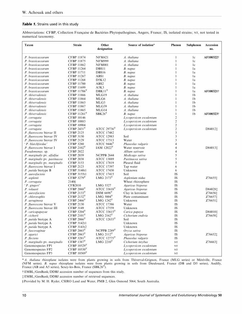
Table 1. Strains used in this study.................................................................................................................................................................................................................................................................................................................
Abbreviations: CFBP, Collection Française de Bacte! ries Phytopathoge' nes, Angers, France; IS, isolated strains ; , not tested innumerical taxonomy.
Taxon Strain Other
designation
Source of isolation* Phenon Subphenon Accession
no.
P. brassicacearum CFBP 11874 NFM421 A. thaliana 1 1c AF100322†
P. brassicacearum CFBP 11875 NFM999 A. thaliana 1 1c
P. brassicacearum CFBP 11862 NFM001 A. thaliana 1 1c
P. brassicacearum CFBP 11264 DBS11 B. napus 1 1a
P. brassicacearum CFBP 11711 DBS16 B. napus 1 1a
P. brassicacearum CFBP 11267 ABS1 B. napus 1 1a
P. brassicacearum CFBP 11268 D5K12 B. napus 1 1a
P. brassicacearum CFBP 11700 ABS2 B. napus 1 1a
P. brassicacearum CFBP 11699 A5K3 B. napus 1 1a
P. brassicacearum CFBP 11706T DBK11T B. napus 1 1a AF100321†
P. thivervalensis CFBP 11866 MLG19 A. thaliana 1 1b
P. thivervalensis CFBP 11864 MLG6 A. thaliana 1 1b
P. thivervalensis CFBP 11863 MLG3 A. thaliana 1 1b
P. thivervalensis CFBP 11867 MLG39 A. thaliana 1 1b
P. thivervalensis CFBP 11865 MLG14 A. thaliana 1 1b
P. thivervalensis CFBP 11261T SBK26T B. napus 1 1b AF100323†
P. corrugata CFBP 10146 Lycopersicon esculentum 2
P. corrugata CFBP 10883 Lycopersicon esculentum 2
P. corrugata CFBP 10904 Lycopersicon esculentum 2
P. corrugata CFBP 2431T ATCC 29736T Lycopersicon esculentum 2 D84012‡
P. fluorescens biovar II CFBP 2125 ATCC 17482 3
P. fluorescens biovar IV CFBP 3150 ATCC 12983 Soil 3
P. fluorescens biovar IV CFBP 2129 ATCC 17513 Water 3
‘P. blatchfordae ’ CFBP 3280 ATCC 9446T Phaseolus vulgaris 4
P. fluorescens biovar I CFBP 2102T IAM 12022T Water reservoir 4 D84013‡
Pseudomonas. sp. CFBP 2022 Allium sativum 4
P. marginalis pv. alfalfae CFBP 2039 NCPPB 2644 Medicago sativa 5
P. marginalis pv. pastinacea CFBP 2038 ATCC 13889 Pastinacea sativa 5
P. marginalis pv. marginalis CFBP 3153 ATCC 17819 Pleural fluid 5
P. fluorescens biovar I CFBP 2123 ATCC 17397 Tap water 6
P. putida biotype B CFBP 3140t1 ATCC 17430 Unknown 6
P. aureofaciens CFBP 3155t1 ATCC 17415 Soil IS
P. asplenii CFBP 3279T LMG 2173T Asplenium nidus IS Z76655‡
P. corrugata 2140§ Wheat rhizosphere IS
‘P. gingeri ’ CFB2810 LMG 5327 Agaricus bisporus IS
P. tolaasii CFBP 2068T ATCC 33618T Agaricus bisporus IS D84028‡
P. aureofaciens CFBP 2133T DSM 6698T Clay in kerosene IS Z76656‡
P. chlororaphis CFBP 2132T LMG 5004T Plate contaminant IS Z76657‡
P. aeruginosa CFBP 2466T LMG 1242T Unknown IS Z76651‡
P. fluorescens biovar V CFBP 2130 ATCC 17386 Water IS
P. fluorescens biovar III CFBP 3149 ATCC 17559 Unknown IS
P. caricapapayae CFBP 3204T ATCC 33615T Carica papaya IS D84010‡
P. cichorii CFBP 2101T LMG 2162T Cichorium endivia IS Z76658‡
P. putida biotype A CFBP 2066T ATCC 12633T Soil IS
P. putida biotype A CFBP 3142t1 Unknown IS
P. putida biotype A CFBP 3143t2 Unknown IS
P. fuscovaginae CFBP 2065T NCPPB 2289T Oryza sativa IS
P. agarici CFBP 2063T LMG 2112T Agaricus bisporus IS Z76652‡
P. flectens CFBP 3281T ATCC 12775T Phaseolus vulgaris IS
P. marginalis pv. marginalis CFBP 1387T LMG 2210T Cichorium intybus Z76663‡
Genomospecies FP1 CFBP 10529T Lycopersicon esculentum
Genomospecies FP2 CFBP 10530T Lycopersicon esculentum
Genomospecies FP3 CFBP 10569T Lycopersicon esculentum
* A. thaliana rhizoplane isolates were from plants growing in soils from Thiverval-Grignon, France (MLG series) or Me! re! ville, France(NFM series). B. napus rhizoplane isolates were from plants growing in soils from Dieulouard, France (DB and D5 series), Andilly,France (AB and A5 series), Sexey-les-Bois, France (SBK26T).
† EMBL}GenBank}DDBJ accession number of sequences from this study.
‡ EMBL}GenBank}DDBJ accession number of retrieved sequences.
§ Provided by M. H. Ryder, CSIRO Land and Water, PMB 2, Glen Osmond 5064, South Australia.
10 International Journal of Systematic and Evolutionary Microbiology 50
Two new root-associated Pseudomonas species
polyphasic taxonomy was necessary. Here, the desc-ription of two new Pseudomonas species is reported,for which the names Pseudomonas brassicacearum sp.nov. and Pseudomonas thivervalensis sp. nov. areproposed.
METHODS
Bacterial strains and culture conditions. In total, 53 strainswere studied (Table 1). A total of 16 strains was isolated,eight from the rhizosphere of Brassica napus cv. Brutorgrown in three soils (Andilly, Sexey-les-Bois andDieulouard,France) and eight from that of Arabidopsis thaliana ecotypeWS grown in two soils (Thiverval-Grignon and Me! re! ville,France). Plants were grown in pots and incubated in agreenhouse for 3 weeks (B. napus) or 6 weeks (A. thaliana).These bacteria were isolated from plant rhizoplane aspreviously described (Mavingui et al., 1992). A semi-selectivemedium derived from that of Gould et al. (1985) was usedfor the isolation of strains from A. thaliana rhizoplane. Thismodified medium (RCS) contained mineral solution RCV(Heulin et al., 1987) supplemented with Casamino acids (5 gl−"), sodium lauroyl sarcosinate (1±2 g l−"), sucrose (20 g l−")and agar (15 g l−"). For strains isolated from B. napusrhizoplane, mineral solution RCV supplemented with su-crose (20 g l−") and agar (15 g l−") was used. These strainsrepresented up to 10% of the total cultivable microfloraassociated with A. thaliana roots, and up to 60% of thatassociated with B. napus roots. Thirty-seven identifiedPseudomonas strains, including 18 type strains, were used asreferences in the numerical taxonomy and for DNA–DNAhybridization. Strains were grown in tryptic soy broth (TSB;Difco) medium at 28 °C. They were grown for 48 h for DNAextraction and up to OD
&*!0±5 for PCR amplification. All
the strains were maintained on tenfold diluted tryptic soyagar (TSA).
Conventional tests. The presence of oxidase, gelatinase andarginine dihydrolase activities, production of fluorescentpigment on King B medium, levan formation, acidificationof sucrose, sorbitol, erythritol and mannitol, reduction ofnitrate, hydrolysis of Tween 80 and aesculin, and thehypersensitivity reaction on tobacco leaves were tested asdescribed by Leliott et al. (1966). Pectinolytic activity wastested by the method of Prunier & Kaiser (1964) andHildebrand (1971). Utilization of lactate, ()-tartrate and(®)-tartrate was tested on the basal medium of Ayers et al.(1919) supplemented with 0±1% (w}v) organic acid sodiumsalts. The presence of DNase was tested on DNA agar(Diagnostic Pasteur). Flagella were stained by the method ofRhodes (1958).
Metabolic profiles. The oxidation of 95 carbon substrateswas determined using BIOLOG GN microplates as recom-mended by the manufacturer. Results were read with anELISA microplate reader (Dynatech, MR5000) after 24 or48 h incubation at 28 °C. Additionally, strains were studiedfor the assimilation of 99 organic substrates using Biotype100 strips (bioMe! rieux) as recommended by the manu-facturer. Results were read visually after 2, 3 and 4 dincubation at 26 °C.
Numerical taxonomy. A total of 114 characters, including theresults of 19 conventional tests, and 95 oxidation activitiesobtained with BIOLOG GN microplates were included inthe numerical taxonomy analysis. The distance matrix wascalculated using the Jaccard coefficient (Sneath & Sokal,
1973). Cluster analysis was performed using the unweightedpair group method with averages (UPGMA). At a givendistance level and for the different phenons, the amount ofinformation for each test was measured by calculating thecoefficient of diagnostic ability to determine the discrimi-nating biochemical characteristics (Descamps & Ve! ron,1981).
Determination of the rrs gene sequences. The rrs gene wasamplified as described in detail by Achouak et al. (1999). ThePCR products from three rhizoplane strains (CFBP 11706T,CFBP 11874 and CFBP 11261T) were purified on a 1% low-melting-point agarose gel. They were directly sequencedusing a protocol described by Anderson et al. (1992) and thesequencing primers as described by Achouak et al. (1999).Sequences were obtained with the Genomyx system (Beck-man).
Amplified rDNA restriction analysis (ARDRA). The amplifiedrrs genes were analysed by restriction digestion with 11restriction enzymes (AluI, CfoI, DdeI, HinfI, TaqI, HaeIII,MspI, RsaI, NdeII, NciI and ScrFI). The restriction profileswere compared with those of the type strains of relatedspecies of Pseudomonas : Pseudomonas agarici, ‘Pseudo-monas blatchfordae ’, Pseudomonas flectens, Pseudomonasfluorescens, Pseudomonas fuscovaginae, Pseudomonas mar-ginalis (data from this work, Table 1) ; and Pseudomonasaeruginosa, Pseudomonas alcaligenes, Pseudomonas aureo-faciens, Pseudomonas chlororaphis, Pseudomonas cichorii, P.corrugata, P. marginalis, Pseudomonas mendocina, Pseudo-monas pseudoalcaligenes, Pseudomonas putida, Pseudomonasstutzeri, Pseudomonas syringae, Pseudomonas tolaasii andPseudomonas viridiflava (data from Laguerre et al., 1994).
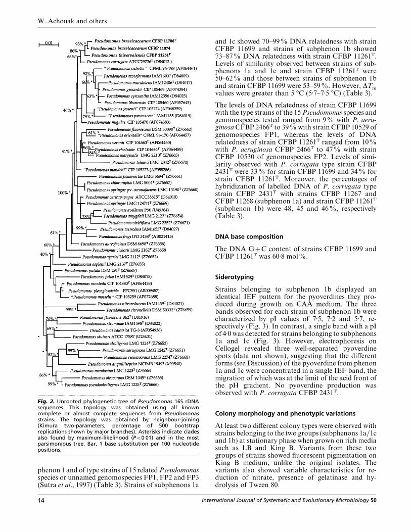
Phylogenetic analysis. The rrs gene sequences from the threerhizoplane strains (CFBP 11706T, CFBP 11874 and CFBP11261T) were aligned and analysed by comparison with adatabase of previously aligned sequences (about 10000).Three phylogenetic methods were used to assess the phylo-genetic positions of these sequences, a neighbour-joiningalgorithm according to Saitou & Nei (1987), maximum-likelihood (options QFYG, program of G. J. Olsen,University of Illinois, Urbana, IL, USA) and maximum-parsimony (PAUP 3.0s for Macintosh, heuristic search;Swofford, 1990). The robustness of each topology waschecked using the neighbour-joining method and 500 boot-strap replications (Kimura two-parameters correction).Trees were drawn using the program for Macintosh(M. Gouy, CNRS UMR 5558, Universite! Claude Bernard,Lyon, France) and MD software. Domains used toconstruct the tree shown in Fig. 2 were regions of the rrs genesequences available for all sequences : positions 49–1317corresponding to the rrs gene sequence of P. corrugata.
DNA–DNA hybridization. Extraction and purification ofDNA were performed using previously described methods(Brenner et al., 1982). Native DNAs were labelled in vitro bynick translation with tritium-labelled nucleotides (Amer-sham). The S1 nuclease–trichloroacetic acid method forhybridization has been previously described (Croza et al.,1973; Grimont et al., 1980). The reassociation temperaturewas 70 °C. DNA–DNA hybridization tests were carried outusing labelled DNA from strains CFBP 11261T, CFBP11699 and P. corrugata CFBP 2431T. The strains used inDNA–DNA hybridization are listed in Table 3.
Thermal stability of DNA reassociation. The temperature atwhich 50% of reassociated DNA became hydrolysable bynuclease S1 (T
m) was determined using the method of Croza
International Journal of Systematic and Evolutionary Microbiology 50 11
W. Achouak and others
..............................................................................................
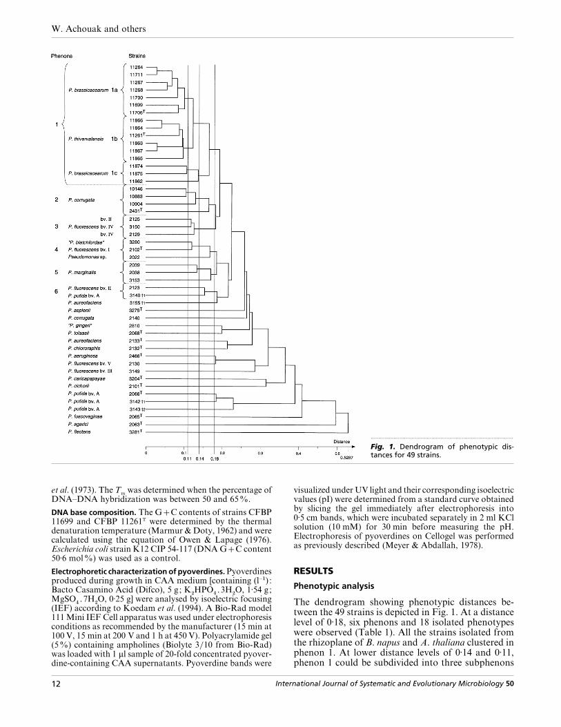
Fig. 1. Dendrogram of phenotypic dis-tances for 49 strains.
et al. (1973). The Tm
was determined when the percentage ofDNA–DNA hybridization was between 50 and 65%.
DNA base composition. The GC contents of strains CFBP11699 and CFBP 11261T were determined by the thermaldenaturation temperature (Marmur & Doty, 1962) and werecalculated using the equation of Owen & Lapage (1976).Escherichia coli strain K12 CIP 54-117 (DNA GC content50±6 mol%) was used as a control.
Electrophoretic characterization of pyoverdines. Pyoverdinesproduced during growth in CAA medium [containing (l−") :Bacto Casamino Acid (Difco), 5 g; K
#HPO
%\3H
#O, 1±54 g;
MgSO%\7H
#O, 0±25 g] were analysed by isoelectric focusing
(IEF) according to Koedam et al. (1994). A Bio-Rad model111 Mini IEF Cell apparatus was used under electrophoresisconditions as recommended by the manufacturer (15 min at100 V, 15 min at 200 V and 1 h at 450 V). Polyacrylamide gel(5%) containing ampholines (Biolyte 3}10 from Bio-Rad)was loaded with 1 µl sample of 20-fold concentrated pyover-dine-containing CAA supernatants. Pyoverdine bands were
visualized under UV light and their corresponding isoelectricvalues (pI) were determined from a standard curve obtainedby slicing the gel immediately after electrophoresis into0±5 cm bands, which were incubated separately in 2 ml KClsolution (10 mM) for 30 min before measuring the pH.Electrophoresis of pyoverdines on Cellogel was performedas previously described (Meyer & Abdallah, 1978).
RESULTS
Phenotypic analysis
The dendrogram showing phenotypic distances be-tween the 49 strains is depicted in Fig. 1. At a distancelevel of 0±18, six phenons and 18 isolated phenotypeswere observed (Table 1). All the strains isolated fromthe rhizoplane of B. napus and A. thaliana clustered inphenon 1. At lower distance levels of 0±14 and 0±11,phenon 1 could be subdivided into three subphenons
12 International Journal of Systematic and Evolutionary Microbiology 50

Two new root-associated Pseudomonas species
Table 2. Phenotypic and biochemical characters that differentiate subphenons 1a, 1b and 1c from phenons 2–6 andfrom type strains of Pseudomonas species.................................................................................................................................................................................................................................................................................................................
, Positive ; ®, negative ; , variable (number of positive strains in parentheses).
Character Phenon or subphenon no.* Type strains from CFBP collection†
1a 1b 1c 2 3 4 5 6 3279T 2068T 2133T 2132T 2466T 2101T 2066T 2065T 2063T 3281T
Production of pyoverdine ® Levan production ® (1) (1) (1) ® ® ® ® ® ® ® DNase (2) ® (1) ® ® ® ® ® ® ®Nitrate reduction (5) (2) ® ® ® ® ® ® ® ®Gelatin hydrolysis (2) ® (2) ® ® ® ® ®Assimilation of :‡
Adonitol ® ® ® ® ® (1) (1) ® ® ® ® ® ® ® ® ®Erythritol ® ® ® ® ® (2) ® ® ® ® ® ® ® ® ® ®Mannitol ® ® ® ® ® (2) ® ® ® ® ® ® ® ® ® ®Sorbitol ® ® ® ® ® ® ® ®
Oxidation of :§
p-Hydroxy phenylacetic acid ® (2) (2) ® ® ® Itaconic acid ® ® (1) ® (1) ® ® ® ® γ-Hydroxybutyric acid (1) ® ® (1) ® ® ® ® ® ® ® ® ® ®Glucuronamide ® (3) (1) (2) (1) ® ® ® ® ® ®Succinamic acid ® (3) ® ®
*Species in each phenon or subphenon: subphenon 1a, P. brassicacearum (n¯7) ; subphenon 1b, P. thivervalensis (n¯6) ; subphenon1c, P. brassicacearum (n¯3) ; phenon 2, P. corrugata (n¯4) ; phenon 3, P. fluorescens (n¯3) ; phenon 4, P. fluorescens (n¯3) ; phenon5, P. marginalis (n¯3) ; phenon 6, P. fluorescens}P. putida (n¯2).
†Pseudomonas type strains are: P. asplenii CFBP 3279T, P. tolaasii CFBP 2068T, P. aureofaciens CFBP 2133T, P. chlororaphis CFBP2132T, P. aeruginosa CFBP 2466T, P. cichorii CFBP 2101T, P. putida CFBP 2066T, P. fuscovaginae CFBP 2065T, P. agarici CFBP 2063T
and P. flectens CFBP 3281T.
‡Biotype 100 substrates.
§Biolog substrates.
1a, 1b and 1c. Phenon 1 is phenotypically related tophenon 2, which contains P. corrugata strains, and canbe differentiated by two characters : production oflevan and assimilation of sorbitol (Table 2). However,phenon 1 can be clearly differentiated from the otherphenons (3–6) and from the type strains of majorfluorescent, oxidase-positive Pseudomonas species byseveral biochemical characters (Table 2). The bio-chemical character that allows discrimination of sub-phenons 1a, 1b and 1c is the oxidation of succinamicacid. On the basis of additional results obtained withBiotype 100 strips (data not shown), none of the 99assimilation tests was found to discriminate amongsubphenons 1a, 1b and 1c.
ARDRA
The PCR products of the rrs gene of the 16 root-associated strains of phenon 1 and of the type strainsof 20 Pseudomonas species, including P. corrugata(listed in Methods) were mapped with 11 restrictionenzymes (ARDRA). The 16 root-associated strains ofphenon 1 provided exactly the same profile with therestriction enzymes tested and belonged to a singleARDRA group. They showed differences with at leasttwo restriction enzymes from the type strains of the 19Pseudomonas species analysed and only TaqI enzymecould discriminate them from the type strain of P.corrugata.
rrs gene sequence analysis
Results of phylogenetic analysis clearly showed thatthe three strains CFBP 11706T, CFBP 11261T andCFBP 11874, which belong to subphenons 1a, 1b and1c, respectively, of phenon 1 (Fig. 1), should beincluded in the true genus Pseudomonas (data notshown). To obtain a precise phylogenetic identifi-cation, analyses were thus restricted to members of thistaxon, including one sequence for each validatedPseudomonas species and the rrs gene sequence of theroot-associated strains. All three phylogenetic analyses(neighbour-joining, parsimony, maximum-likelihood)showed that the three rhizoplane strains formed arobust monophyletic unit (Fig. 2). Strains CFBP11706T, CFBP 11261T, CFBP 11874 and P. corrugataformed a clade identified by all three methods thatshowed a high bootstrap percentage (90%). rrs se-quence similarities were: 99±8% for CFBP 11706T andCFBP 11874; 99±3% for CFBP 11706T and CFBP11261T ; 99±5% for CFBP 11261T and CFBP 11874;99±1% for CFBP 11706T and P. corrugata ATCC29736T ; and 99±2% for CFBP 11261T and ATCC29736T.
DNA–DNA hybridization
Labelled DNA of strains CFBP 11261T, CFBP 11699and CFBP 2431T was hybridized to DNA of strains of
International Journal of Systematic and Evolutionary Microbiology 50 13
W. Achouak and others
.................................................................................................................................................
Fig. 2. Unrooted phylogenetic tree of Pseudomonas 16S rDNAsequences. This topology was obtained using all knowncomplete or almost complete sequences from Pseudomonasstrains. The topology was obtained by neighbour-joining(Kimura two-parameters, percentage of 500 bootstrapreplications shown by major branches). Asterisks indicate cladesalso found by maximum-likelihood (P!0±01) and in the mostparsimonious tree. Bar, 1 base substitution per 100 nucleotidepositions.
phenon 1 and of type strains of 15 related Pseudomonasspecies or unnamed genomospecies FP1, FP2 and FP3(Sutra et al., 1997) (Table 3). Strains of subphenons 1a
and 1c showed 70–99% DNA relatedness with strainCFBP 11699 and strains of subphenon 1b showed73–87% DNA relatedness with strain CFBP 11261T.Levels of similarity observed between strains of sub-phenons 1a and 1c and strain CFBP 11261T were50–62% and those between strains of subphenon 1band strain CFBP 11699 were 53–59%. However, ∆T
mvalues were greater than 5 °C (5±7–7±5 °C) (Table 3).
The levels of DNA relatedness of strain CFBP 11699with the type strains of the 15 Pseudomonas species andgenomospecies tested ranged from 9% with P. aeru-ginosa CFBP 2466T to 39% with strain CFBP 10529 ofgenomospecies FP1, whereas the levels of DNArelatedness of strain CFBP 11261T ranged from 10%with P. aeruginosa CFBP 2466T to 47% with strainCFBP 10530 of genomospecies FP2. Levels of simi-larity observed with P. corrugata type strain CFBP2431T were 33% for strain CFBP 11699 and 34% forstrain CFBP 11261T. Moreover, the percentages ofhybridization of labelled DNA of P. corrugata typestrain CFBP 2431T with strains CFBP 11267 andCFBP 11268 (subphenon 1a) and strain CFBP 11261T
(subphenon 1b) were 48, 45 and 46%, respectively(Table 3).
DNA base composition
The DNA GC content of strains CFBP 11699 andCFBP 11261T was 60±8 mol%.
Siderotyping
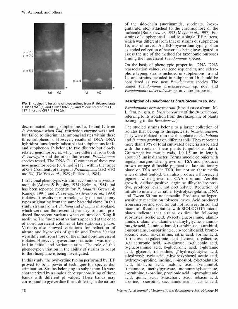
Strains belonging to subphenon 1b displayed anidentical IEF pattern for the pyoverdines they pro-duced during growth on CAA medium. The threebands observed for each strain of subphenon 1b werecharacterized by pI values of 7±5, 7±2 and 5±7, re-spectively (Fig. 3). In contrast, a single band with a pIof 4±0 was detected for strains belonging to subphenons1a and 1c (Fig. 3). However, electrophoresis onCellogel revealed three well-separated pyoverdinespots (data not shown), suggesting that the differentforms (see Discussion) of the pyoverdine from phenon1a and 1c were concentrated in a single IEF band, themigration of which was at the limit of the acid front ofthe pH gradient. No pyoverdine production wasobserved with P. corrugata CFBP 2431T.
Colony morphology and phenotypic variations
At least two different colony types were observed withstrains belonging to the two groups (subphenons 1a}1cand 1b) at stationary phase when grown on rich mediasuch as LB and King B. Variants from these twogroups of strains showed fluorescent pigmentation onKing B medium, unlike the original isolates. Thevariants also showed variable characteristics for re-duction of nitrate, presence of gelatinase and hy-drolysis of Tween 80.
14 International Journal of Systematic and Evolutionary Microbiology 50
Two new root-associated Pseudomonas species
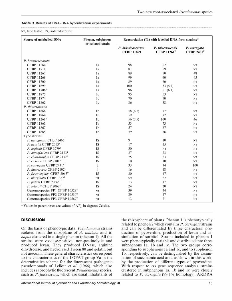
Table 3. Results of DNA–DNA hybridization experiments.................................................................................................................................................................................................................................................................................................................
, Not tested; IS, isolated strains.
Source of unlabelled DNA Phenon, subphenon
or isolated strain
Reassociation (%) with labelled DNA from strains :*
P. brassicacearum
CFBP 11699
P. thivervalensis
CFBP 11261T
P. corrugata
CFBP 2431T
P. brassicacearum
CFBP 11264 1a 98 62
CFBP 11711 1a 81 59
CFBP 11267 1a 89 50 48
CFBP 11268 1a 99 60 45
CFBP 11700 1a 89 60
CFBP 11699 1a 100 53 (5±7)
CFBP 11706T 1a 96 61 (6±1)
CFBP 11875 1c 95 53
CFBP 11874 1c 70 50
CFBP 11862 1c 86 58
P. thivervalensis
CFBP 11866 1b 58 (6±7) 77
CFBP 11864 1b 59 82
CFBP 11261T 1b 56 (7±5) 100 46
CFBP 11863 1b 53 73
CFBP 11867 1b 57 87
CFBP 11865 1b 59 86
Type strains
P. aeruginosa CFBP 2466T IS 9 10
P. agarici CFBP 2063T IS 17 15
P. asplenii CFBP 3279T IS 30
P. aureofaciens CFBP 2133T IS 27 23
P. chlororaphis CFBP 2132T IS 25 23
P. cichorii CFBP 2101T IS 10 10
P. corrugata CFBP 2431T 2 33 34 100
P. fluorescens CFBP 2102T 4 16 18
P. fuscovaginae CFBP 2065T IS 20 17
P. marginalis CFBP 1387T 22
P. putida CFBP 2066T IS 13 17
P. tolaasii CFBP 2068T IS 24 20
Genomospecies FP1 CFBP 10529T 39 44
Genomospecies FP2 CFBP 10530T 29 47
Genomospecies FP3 CFBP 10569T 13 21
*Values in parentheses are values of ∆Tm
in degrees Celsius.
DISCUSSION
On the basis of phenotypic data, Pseudomonas strainsisolated from the rhizoplane of A. thaliana and B.napus clustered in a single phenon (phenon 1). All thestrains were oxidase-positive, non-pectinolytic andproduced levan. They produced DNase, argininedihydrolase, and hydrolysed Tween 80 and gelatin butnot aesculin. These general characteristics correspondto the characteristics of the LOPAT group Va in thedeterminative scheme for the fluorescent pathogenicpseudomonads of Leliott et al. (1966), which alsoincludes saprophytic fluorescent Pseudomonas species,such as P. fluorescens, which are usual inhabitants of
the rhizosphere of plants. Phenon 1 is phenotypicallyrelated to phenon 2 which contains P. corrugata strainsand can be differentiated by three characters : pro-duction of pyoverdine, production of levan and as-similation of sorbitol. Strains included in phenon 1were phenotypically variable and distributed into threesubphenons 1a, 1b and 1c. The two groups corre-sponding to subphenons 1a and 1c, and to subphenon1b, respectively, can be distinguished by the assimi-lation of succinamic acid and, as shown in this work,by the production of different types of pyoverdine.With respect to rrs gene sequence analysis, strainsclustered in subphenons 1a, 1b and 1c were closelyrelated to P. corrugata (99±1% homology). ARDRA
International Journal of Systematic and Evolutionary Microbiology 50 15
W. Achouak and others
–
+
(a) (b) (c) (d)
pI = 7·5pI = 7·2
pI = 5·7
pI = 4
–
+
.................................................................................................................................................
Fig. 3. Isoelectric focusing of pyoverdines from P. thivervalensisCFBP 11261T (a) and CFBP 11866 (b), and P. brassicacearum CFBP11711 (c) and CFBP 11874 (d).
discriminated among subphenons 1a, 1b and 1c fromP. corrugata when TaqI restriction enzyme was used,but failed to discriminate among isolates within thesethree subphenons. However, results of DNA–DNAhybridizations clearly indicated that subphenons 1a}1cand subphenon 1b belong to two discrete but closelyrelated genomospecies, which are different from bothP. corrugata and the other fluorescent Pseudomonasspecies tested. The DNA GC contents of these twonew genomospecies (60±8 mol%) fell within the rangeof GC contents of the genus Pseudomonas (55±2–67±2mol%) (De Vos et al., 1989; Palleroni, 1984).
Intraclonal phenotypic variation is common in pseudo-monads (Adams & Pugsley, 1934; Kelman, 1954) andhas been reported recently for P. tolaasii (Grewal &Rainey, 1991) and P. corrugata (Siverio et al., 1993)isolates. It results in morphologically distinct colonytypes originating from the same bacterial clone. In thisstudy, strains fromA. thaliana andB. napus rhizoplane,which were non-fluorescent at primary isolation, pro-duced fluorescent variants when cultured on King Bmedium. The fluorescent variants appeared at the edgeof non-fluorescent colonies at late stationary phase.Variants also showed variations for reduction ofnitrate and hydrolysis of gelatin and Tween 80 thatwere different from those of the initial non-fluorescentisolates. However, pyoverdine production was ident-ical in initial and variant strains. The role of thisphenotypic variation in the ability of strains to adaptto the rhizoplane is being investigated.
In this study, the pyoverdine typing performed by IEFproved to be a powerful technique for species dis-crimination. Strains belonging to subphenon 1b werecharacterized by a single siderotype consisting of threebands with different pI values. These bands maycorrespond to pyoverdine forms differing in the nature
of the side-chain (succinamide, succinate, 2-oxo-glutarate, etc.) attached to the chromophore of themolecule (Budzikiewicz, 1993; Meyer et al., 1997). Forstrains of subphenons 1a and 1c, a single IEF pattern,which was different from that of strains of subphenon1b, was observed. An IEF–pyoverdine typing of anextended collection of bacteria is being investigated toassess the use of the method for taxonomic purposesamong the fluorescent Pseudomonas species.
On the basis of phenotypic properties, DNA–DNAreassociation values, rrs gene sequencing and sidero-phore typing, strains included in subphenons 1a and1c, and strains included in subphenon 1b should beconsidered as two new Pseudomonas species. Thenames Pseudomonas brassicacearum sp. nov. andPseudomonas thivervalensis sp. nov. are proposed.
Description of Pseudomonas brassicacearum sp. nov.
Pseudomonas brassicacearum (bras.si.ca.ce.a«rum. M.L. fem. pl. gen. n. brassicacearum of the Brassicaceae,referring to its isolation from the rhizoplane of plantsbelonging to the Brassicaceae).
The studied strains belong to a larger collection ofisolates that belong to the species P. brassicacearum.They were isolated from the rhizoplane of A. thalianaand B. napus growing on different soils. They representmore than 10% of total cultivated bacteria associatedwith the roots of these plants (unpublished data).Gram-negative motile rods, 1±0–1±5 µm in length,about 0±5 µm in diameter. Forms mucoid colonies withregular margins when grown on TSA and producesbrown–orange diffusible pigment at late stationaryphase on TSA and in TSB, but not on these mediawhen diluted tenfold. Can also produce a fluorescentpigment when grown on CAA medium. Aerobicgrowth, oxidase-positive, arginine dihydrolase-posi-tive, produces levan, not pectinolytic. Reduction ofnitrate to nitrite is variable. Hydrolyses gelatin, DNAand Tween 80 but not aesculin. Produces no hyper-sensitivity reaction on tobacco leaves. Acid producedfrom sucrose and sorbitol but not from erythritol andmannitol. Results obtained with BIOLOG GN micro-plates indicate that strains oxidize the followingsubstrates : acetic acid, N-acetylglucosamine, alanin-amide, -alanine, -alanine, -alanyl-glycine, γ-amino-butyric acid, 2-aminoethanol, -arabinose, -arabitol,-asparagine, -aspartic acid, cis-aconitic acid, bromo-succinic acid, -carnitine, citric acid, formic acid,-fructose, -galactonic acid lactone, -galactose,-galacturonic acid, α--glucose, -gluconic acid,-glucosaminic acid, -glucuronic acid, -glutamicacid, glycerol, -histidine, β-hydroxybutyric acid,γ-hydroxybutyric acid, p-hydroxyphenyl acetic acid,hydroxy--proline, inosine, m-inositol, α-ketoglutaricacid, -lactic acid, malonic acid, -mannitol,-mannose, methylpyruvate, monomethylsuccinate,-ornithine, -proline, propionic acid, -pyroglutamicacid, quinic acid, -saccharic acid, sebacic acid,-serine, -sorbitol, succinamic acid, succinic acid,
16 International Journal of Systematic and Evolutionary Microbiology 50

Two new root-associated Pseudomonas species
sucrose, -trehalose, -threonine, Tween 40, Tween 80,uridine and urocanic acid. Oxidation of 2,3-butane-diol, -α-glycerol phosphate, glycyl -glutamic acid,α-hydroxybutyric acid, α-ketobutyric acid, α-keto-valeric acid, -leucine, maltose, psicose and turanose isvariable. Oxidation of the other organic substratesincluded in BIOLOG GN microplates is negative. P.brassicacearum strains assimilate the following sub-strates (Biotype 100 strips) : N-acetyl--glucosamine,cis-aconitate, trans-aconitate, -alanine, -alanine,-α-amino-n-butyrate,-α-amino-n-valerate, -ara-binose,-arabitol, -aspartate, betaine, caprate, capry-late, citrate, ethanolamine, β--fructose, fumarate,-galactose, -galacturonate, -gluconate, -glucos-amine, α--glucose, -glucuronate, -glutamate,glutarate, -glycerate, glycerol, 2-keto--gluconate,2-oxoglutarate, -β-hydroxybutyrate, p-hydroxyben-zoate, -lactate, -lyxose, -malate, -mannose,-mannitol, mucate, myo-inositol, -proline, pro-pionate, protocatechuate, quinate, -ribose, -sac-charate, -serine, -sorbitol, succinate, sucrose,-trehalose, trigonelline, -tyrosine and -xylose.Assimilation of benzoate, ()malate, malonate,putrescine and (®)tartrate is variable. The otherorganic substrates included in Biotype 100 strips arenot assimilated. DNA GC content is 60±8 mol%.Type strain is strain DBK11T which has been depositedin the Collection Française de Bacte! ries Phytopatho-ge' nes (CFBP, Angers, France) under the numberCFBP 11706T.
Description of Pseudomonas thivervalensis sp. nov.
Pseudomonas thivervalensis (thi.ver.va.len«sis. M.L.adj. thivervalensis pertaining to Thiverval, France, theplace where most of the strains have been isolated fromthe rhizoplane of Arabidopsis thaliana).
Gram-negative motile rods, 1±0–1±5 µm in length,about 0±5 µm in diameter. Forms mucoid colonies withregular margins when grown on TSA and producesbrown–orange diffusible pigment at late stationaryphase on TSA and in TSB, but not on these mediawhen diluted tenfold. They can also produce a fluores-cent pigment when grown on CAA medium. P. thiver-valensis has the same general characteristics as P.brassicacearum. Oxidation of the following substratesis variable : -galacturonic acid, glucuronamide,-glucuronic acid, -ornithine, putrescine and -thre-onine. Unlike P. brassicacearum, P. thivervalensisstrains do not oxidize succinamic acid. P. thivervalensisstrains do not assimilate benzoate unlike 80% of P.brassicacearum strains. The oxidation and the as-similation of the other organic substrates tested withBIOLOG GN microplates and with Biotype 100 stripsare the same for strains of P. thivervalensis as for P.brassicacearum strains (see above). DNA GC con-tent is 60±8 mol%. The type strain is strain SBK26T,which has been deposited in the Collection Françaisede Bacte! ries Phytopathoge' nes (CFBP) under numberCFBP 11261T.
ACKNOWLEDGEMENTS
We thank S. Belouin for technical assistance and A. Huardfor designing the dendrogram of phenotypical distances.
REFERENCES
Achouak, W., Christen, R., Barakat, M., Martel, M. H. & Heulin, T.(1999). Burkholderia caribensis sp. nov., exopolysaccharide-producing bacteria isolated from vertisol microaggregates inMartinique. Int J Syst Bacteriol 49, 787–794.
Adams, D. B. & Pugsley, A. T. (1934). ‘Smooth-rough’ variationin Phytomonas medicagenis phaseolitica Burk. Aust J Exp BiolMed Sci 12, 193–202.
Anderson, R. D., Bao, C.-Y., Minnick, D. T., Veigel, M. & Sedwick,W. D. (1992). Optimization of double-strandedDNAsequencingfor polymerase chain reaction products. USB Editorial Com-ments 19, 39–40, 57–58.
Ayers, S. H., Rupp, P. & Johnson, W. T. (1919). A study of alkali-forming bacteria in milk. USDA Bulletin no. 782.
Behrendt, U., Ulrich, A., Schumann, P., Erler, W. & Seyfarth, W.(1999). Taxonomic study of bacteria isolated from grasses :proposal of Pseudomonas graminis sp. nov. Int J Syst Bacteriol49, 297–308.
Boivin-Jahns, V., Bianchi, A., Ruimy, R., Garcin, J., Daumas, S. &Christen, R. (1995). Comparison of phenotypical and molecularmethods for the identification of bacterial strains isolated froma deep subsurface environment. Appl Environ Microbiol 61,3400–3406.
Brenner, D. J., McWorter, A. C., Leete Knutson, J. K. & Steiger-walt, A. G. (1982). Escherichia vulneris : a new species of Entero-bacteriaceae associated with human wounds. J Clin Microbiol15, 1133–1140.
Budzikiewicz, H. (1993). Secondary metabolites from fluorescentpseudomonads. FEMS Microbiol Rev 104, 209–228.
Croza, J. M., Brenner, D. J. & Falkow, S. (1973). Use of a single-stranded-specific nuclease for analysis of bacterial and plasmiddeoxynucleic acid homo- and heteroduplex. J Bacteriol 115,904–911.
Descamps, P. & Ve! ron, M. (1981). Une me! thode de choix descaracte' res d’identification base! e sur le the!ore' me de Bayes et lamesure de l’information. Ann Inst Pasteur Microbiol 132B,157–170.
De Vos, P., Van Landshoot, A., Segers, P. & 9 other authors(1989). Genotypic relationship and taxonomic localization ofunclassified Pseudomonas and Pseudomonas-like strains bydeoxyribonucleic acid-ribosomal ribonucleic acid hybridiza-tion. Int J Syst Bacteriol 39, 35–49.
Gould, W. D., Hagedorn, C., Bardinelli, T. R. & Zablotowicz, R. M.(1985). New selective media for enumeration and recovery offluorescent Pseudomonads from various habitats. Appl EnvironMicrobiol 49, 28–32.
Grewal, S. I. S. & Rainey, P. B. (1991). Phenotypic variation ofPseudomonas putida and Pseudomonas tolaasii affects thechemotactic response to Agaricus bisporus mycelium. J GenMicrobiol 137, 2761–2768.
Grimont, P. A. D., Popoff, M. Y., Grimont F., Coynault, C. &Lemelin, M. (1980). Reproducibility and correlation study ofthree deoxynucleic acid hybridization procedures. Curr Micro-biol 4, 325–330.
Heulin, T., Guckert, A. & Balandreau, J. (1987). Stimulation ofroot exudation of rice seedlings by Azospirillum strains : carbonbudget under gnotobiotic conditions. Biol Fertil Soils 4, 9–14.
International Journal of Systematic and Evolutionary Microbiology 50 17

W. Achouak and others
Hildebrand, D. C. (1971). Pectate and pectin gels for differen-tiation of Pseudomonas sp. and other bacterial plant pathogens.Phytopathology 61, 1430–1436.
Kelman, A. (1954). The relationship of pathogenicity in Pseudo-monas solanacearum to colony appearance on a tetrazoliummedium. Phytopathology 44, 693–695.
Kersters, K., Ludwig, W., Vancanneyt, M., De Vos, P., Gillis, M. &Schleifer, K. H. (1996). Recent changes in the classification of thepseudomonads: an overview. Syst Appl Microbiol 19, 465–477.
Koedam, N., Wittouck, E., Gaballa, A., Gillis, A., Ho$ fte, M. &Cornelis, P. (1994). Detection and differentiation of microbialsiderophores by isoelectric focusing and chrome azurol Soverlay. Biometals 7, 287–291.
Laguerre, G., Rigottier-Gois, L. & Lemanceau, P. (1994). Flu-orescent Pseudomonas species categorized by using polymerasechain reaction (PCR)}restriction fragment analysis of 16SrDNA. Mol Ecol 3, 479–487.
Leliott, R. A., Billing, E. & Hayward, A. C. (1966). A determinativescheme for the fluorescent plant pathogenic pseudomonads.J Appl Bacteriol 29, 470–489.
Marmur, J. & Doty, P. (1962). Determination of the basecomposition of deoxyribonucleic acid from its thermal dena-turation temperature. J Mol Biol 5, 109–118.
Mavingui, P., Laguerre, G., Berge, O. & Heulin, T. (1992). Geneticand phenotypic diversity of Bacillus polymyxa in soil and in thewheat rhizosphere. Appl Environ Microbiol 58, 1894–1903.
Meyer, J. M. & Abdallah, M. A. (1978). The fluorescent pigmentof Pseudomonas fluorescens : biosynthesis, purification andphysicochemical properties. J Gen Microbiol 107, 319–328.
Meyer, J. M., Stintzi, A., De Vos, D., Cornelis, P., Tappe, R., Taraz,K. & Budzikiewicz, H. (1997). Use of siderophores to typepseudomonads: the three Pseudomonas aeruginosa pyoverdinesystems. Microbiology 143, 35–43.
Owen, R. J. & Lapage, S. P. (1976). The thermal denaturation ofpartly purified bacterial deoxyribonucleic acid and its taxo-nomic implications. J Appl Bacteriol 41, 335–340.
Palleroni, N. J. (1984). Genus I. Pseudomonas Migula 1894. InBergey’sManual of Systematic Bacteriology, vol. 1, pp. 141–199.Edited by N. R. Krieg & J. G. Holt. Baltimore: Williams &Wilkins.
Palleroni, N. J., Kunisawa, R., Contopoulou, R. & Doudoroff, M.(1973). Nucleic acid homologies in the genus Pseudomonas. IntJ Syst Bacteriol 23, 333–339.
Prunier, J. P. & Kaiser, P. (1964). Etude de l’activite! pectinolytiquechez des bacte! ries phytopathoge' nes et saprophytes des plantes.I. Recherche des enzymes pectinolytiques. Ann Epiphyt (Paris)15, 205–209.
Rhodes, M. E. (1958). The cytology of Pseudomonas spp. asrevealed by silver-plating staining method. J Gen Microbiol 18,639–648.
Saitou, N. & Nei, M. (1987). The Neighbor-Joining method: anew method for reconstructing phylogenetic trees. Mol BiolEvol 4, 406–425.
Siverio, F., Cambra, M., Gorris, M. T., Corso, J. & Lopez, M. M.(1993). Lipopolysaccharides as determinants of serologicalvariability in Pseudomonas corrugata. Appl Environ Microbiol59, 1805–1812.
Skerman, V. B. D., McGowan, V. & Sneath, P. H. A. (1989).Approved Lists of Bacterial Names. Washington, DC: AmericanSociety for Microbiology.
Sneath, P. H. A. & Sokal, R. R. (1973). Numerical Taxonomy:the Principles and Practice of Numerical Classification. SanFrancisco: W. H. Freeman.
Sutra, L., Siverio, F., Lopez, M. M., Hunault, G., Bollet, C. &Gardan, L. (1997). Taxonomy of Pseudomonas strains isolatedfrom tomato pith necrosis : emended description of Pseudo-monas corrugata and proposal of three unnamed fluores-cent Pseudomonas genomospecies. Int J Syst Bacteriol 47,1020–1033.
Swofford, D. (1990). : phylogenetic analysis using par-simony, version 3.0. Computer program distributed by theIllinois Natural History Survey, Champaign, Illinois, USA.
Whyte, L. G., Bourbonniere, L. & Greer, C. W. (1997). Bio-degradation of petroleum hydrocarbons by psychrophilicPseudomonas strains possessing both alkane (alk) and naph-thalene (nah) catabolic pathways. Appl Environ Microbiol 63,3719–3723.
Wilson, A. E., Moore, E. R. & Mohn, W. W. (1996). Isolationand characterization of isopimaric acid-degrading bacteriafrom a sequencing batch reactor. Appl Environ Microbiol 62,3146–3151.
18 International Journal of Systematic and Evolutionary Microbiology 50