neoscardovia arbecensis gen. nov., sp. nov., isolated from porcine slurries
TRANSCRIPT
N
CAa
b
Ic
a
ARRA
KNTBP1P
pwdmo(fbfsfahBrGsp
0h
Systematic and Applied Microbiology 35 (2012) 374– 379
Contents lists available at SciVerse ScienceDirect
Systematic and Applied Microbiology
j ourna l ho mepage: www.elsev ier .de /syapm
eoscardovia arbecensis gen. nov., sp. nov., isolated from porcine slurries
ristina García-Aljaroa,∗ , Elisenda Ballestéa , Ramon Rosselló-Mórab , Ana Cifuentesb , Michael Richterc ,nicet R. Blancha
Department of Microbiology, Faculty of Biology, University of Barcelona, Av. Diagonal 643, E-08028 Barcelona, SpainMarine Microbiology Group, Department of Ecology and Marine Resources, Institut Mediterrani d’Estudis Avanc ats (IMEDEA, CSIC-UIB), C/Miquel Marqués 21, E-07190 Esporles,
lles Balears, SpainMicrobial Genomics and Bioinformatics Research Group, Max Planck Institute for Marine Microbiology, Celsiusstrasse 1, 28359 Bremen, Germany
r t i c l e i n f o
rticle history:eceived 16 April 2012eceived in revised form 6 June 2012ccepted 8 June 2012
eywords:eoscardovia arbecensisaxonomyifidobacteriumorcine
a b s t r a c t
Three Gram-positive, anaerobic, pleomorphic strains (PG10T, PG18 and PG22), were selected among fivestrains isolated from pig slurries while searching for host specific bifidobacteria to track the source of fecalpollution in water. Analysis of the 16S rRNA gene sequence showed a maximum identity of 94% to vari-ous species of the family Bifidobacteriaceae. However, phylogenetic analyses of 16S rRNA and HSP60 genesequences revealed a closer relationship of these strains to members of the recently described Aeriscar-dovia, Parascardovia and Scardovia genera, than to other Bifidobacterium species. The names Neoscardoviagen. nov. and Neoscardovia arbecensis sp. nov. are proposed for a new genus and for the first speciesbelonging to this genus, respectively, and for which PG10T (CECT 8111T, DSM 25737T) was designated asthe type strain. This new species should be placed in the Bifidobacteriaceae family within the class Acti-
6S rRNAartial genome sequencing
nobacteria, with Aeriscardovia aeriphila being the closest relative. The prevailing cellular fatty acids wereC16:0 and C18:1ω9c, and the major polar lipids consisted of a variety of glycolipids, diphosphatidyl glyc-erol, two unidentified phospholipids, and phosphatidyl glycerol. The peptidoglycan structure was A1�meso-Dpm-direct. The GenBank accession numbers for the 16S rRNA gene and HSP60 gene sequences ofstrains PG10T, PG18 and PG22 are JF519691, JF519693, JQ767128 and JQ767130, JQ767131, JQ767133,
respectively.Microbial source tracking (MST) using host-specific bacteria is aromising tool for identifying the source of fecal contamination inater. A strong host-specificity has been described for several Bifi-
obacterium species, which has prompted the development of newethods for detecting these species [1,3,7,9,14,18,26]. The analysis
f the 16S rRNA gene with denaturing gradient gel electrophoresisDGGE) has been broadly used to study microbial diversity in dif-erent ecosystems [16,17]. A recent study analyzed the diversity ofifidobacterial populations from sewage, slurries and wastewaterrom different slaughterhouses using DGGE for identifying host-pecific bacteria as MST markers. A specific DNA band was detectedrom pig DGGE profiles, which was subsequently sequencednd used to design a DNA probe to isolate the correspondingost-specific bacteria [1]. A total of five isolates growing oneerens agar [2], namely PG10, PG18, PG19, PG22 and PG30, wereecovered by colony hybridization using a DIG-labeled probe (5′-
GGTTGTAAACCGCTTTTGATTGGGAGCAAGCG-3′) [1] from porcinelurries of a pig farm located in Arbeca (Lleida, Catalonia, Spain). Thelates were incubated at 37 ◦C for 48 h under anaerobic conditions∗ Corresponding author. Tel.: +34 934021487; fax: +34 934039047.E-mail address: [email protected] (C. García-Aljaro).
723-2020/$ – see front matter © 2012 Elsevier GmbH. All rights reserved.ttp://dx.doi.org/10.1016/j.syapm.2012.06.007
© 2012 Elsevier GmbH. All rights reserved.
(GasPak; BBL, Hampshire, United Kingdom) with CO2 atmospheregenerators (Anaerocult A; Merck, Darmstadt, Germany). The iso-lates were propagated on Columbia blood agar (CBA) (Difco, Le Pontde Claix, France) with the addition of l-cysteine hydrochloride at0.5 g L−1 (Merck, Darmstadt, Germany) and glucose at 5 g L−1, incu-bated in RCM broth at 37 ◦C, and kept frozen at −80 ◦C with 10%glycerol until further analysis.
In the present study, phylogenetic reconstruction based on anal-ysis of the 16S rRNA and HSP60 gene sequences affiliated thestrains with the family Bifidobacteriaceae (see below). However,these reconstructions could not place the isolates in any recog-nized genus of the family. Given that two of the isolates showedclonality, as revealed by the random amplification of polymorphicDNA (RAPD), the three strains showing different fingerprints wereselected for further studies. Strain PG10T was deposited in twointernational culture collections under the numbers CECT 8111T,DSM 25737T, and represents the type strain of the first speciesof the novel genus Neoscardovia gen. nov. proposed here, whichshould be placed in the Bifidobacteriaceae family within the class
Actinobacteria.For taxonomic comparisons, the type strains of representa-tive species of the genera Scardovia (Scardovia inopinata B3109T,DSM 10107T and Scardovia wiggsiae C1A 55T, DSM 22547T),

C. García-Aljaro et al. / Systematic and Applied Microbiology 35 (2012) 374– 379 375
Fig. 1. Genealogical reconstruction of the new organisms with all members of the family Bifidobacteriaceae by means of their 16S rRNA gene sequences. The tree wasreconstructed using the algorithm RAxML [23], as implemented in the ARB program package [13] with no positional filter. The reference sequences were retrieved from theL recos ers. Th
Pawwds
DmawAc1cPf33itP
ad[p3SnBoast[v(usu[st
TP release 106 database [28]. The bootstrap calculations were based on 100 randomhows a branching order that could not be resolved using different datasets and filt
arascardovia (Parascardovia denticolens B3028T, DSM 10105T)nd Aeriscardovia (Aeriscardovia aeriphila T6T, DSM 22365T)ere obtained from the DSMZ (Braunschweig, Germany) andere used as reference strains. Strains were grown on Bifi-
obacterium medium (BM) (DSMZ), except where otherwisetated.
DNA isolation was performed using the Wizard® GenomicNA Purification Kit (Promega, Barcelona, Spain) following theanufacturer’s instructions. The clonality of the isolates was
nalyzed by random amplification of polymorphic DNA (RAPD)ith the primers described in [5]: (A) 5′-CCGCAGCCAA-3′, (B) 5′-ACGCGCAAC-3′, and (C) 5′-GCGGAAATAG-3′. The PCR reactiononsisted of 0.25 �L dNTP mix (25 mM), 1 �L primer (10 �M), 2.5 �L0× Taq polymerase buffer, 1.25 U Taq polymerase and 30 ng ofhromosomal DNA used as template in a final volume of 25 �L. TheCR reaction was performed under the following conditions: 94 ◦Cor 2 min; 6 cycles of 94 ◦C for 30 s, 36 ◦C for 1 min, and 72 ◦C for 90 s;0 cycles of 94 ◦C for 20 s, 36 ◦C for 30 s, and 72 ◦C for 90 s; 72 ◦C for
min. As shown in Fig. S1, isolates PG18, PG19 and PG30 displayeddentical RAPD profiles with the three primers used, indicating thathe five isolates represented three strains, namely PG10T, PG18 andG22, which were further studied.
The 16S rRNA gene was amplified using universal primers 27Fnd 1492R [27]. Partial HSP60 gene sequences were obtained usingegenerate primers (H60F and H60R), as previously described12]. Nucleotide sequencing of the corresponding amplimers waserformed in duplicate with an automated DNA sequencer, model77 (Perkin-Elmer Applied Biosystems, Weiterstadt, Germany).equencing was carried out using the ABI PRISM Big Dye 3.1 Termi-ator Cycle Sequencing Ready Reaction Kit (Perkin Elmer, Appliediosystems, Spain). The almost complete 16S rRNA gene sequencesf the isolates were added to the LTP release 106 database [28] andligned using the ARB software package [13]. On the other hand, theequences (both nucleotide and those translated to amino acids) ofhe HSP60 dataset were aligned using the Clustal X 1.83 program25], and the alignments were improved by removing hyper-ariable positions using the available on-line program Gblockshttp://molevol.cmima.csic.es/castresana/Gblocks server.html),nder the default conditions [4]. In both cases where DNAequences were used, the tree reconstructions were performed
sing the maximum likelihood algorithm RAxML version 7.023] with the rate distribution model GTRGAMMA, and the boot-trap analyses were carried out by using 100 replicates. Also,he reconstructions were generated using all entries availablenstructed trees. GenBank accession numbers are in parentheses. The multifurcatione scale bar indicates percent of sequence divergence.
from all members of the family Bifidobacteriaceae in the LTPrelease 106 database for the 16S rRNA gene sequences andthe GenBank database for the HSP60 gene sequences. For 16SrRNA gene sequence reconstruction, different positional filters toremove phylogenetic noise were applied. The results presented inFig. 1 show a consensus tree, where the multifurcation indicatesbranch instabilities. As mentioned above, the HSP60 alignmentwas sieved previously using the Gblocks program and, in thiscase, the total alignment with 556 homologous positions wasreduced to 464 conserved positions. The resulting alignmentcontained 83% homologous positions of the original alignment.Similarly, for the protein sequences, the alignment of approxi-mately 184 homologous positions was reduced to 146 residues,and the resulting alignment represented 79% of the originaldataset. Similarities were calculated on the basis of a pair-wisecomparison (ARB software) based on the alignments used fortree reconstruction. The 16S rRNA and HSP60 gene sequencesdetermined in this study have been deposited in the GenBankdatabase under accession numbers JF519691, JF519693 andJQ767128 for the 16S rRNA genes, and JQ767130, JQ767131 andJQ767133 for the HSP60 genes, from strains PG10T, PG18 andPG22, respectively.
The almost complete sequence of the 16S rRNA gene wasobtained for all strains (1342 bp). The 16S rRNA sequence identitybetween the different strains was 99.9% with only 1 bp difference.The maximum identity to the closest relatives was 94%, includ-ing several Bifidobacterium species, such as Bifidobacterium indicumJCM 1302T and Bifidobacterium breve DSM 20213T. Phylogeneticreconstruction was performed with these sequences, together withall Bifidobacterium species and those of related genera (nucleotidesimilarity indicated in brackets): A. aeriphila T6T (93%), P. denti-colens B3028T (92%), Alloscardovia omnicolens CCUG 31649T (92%),Metascardovia criceti OMB105T (91%), Gardnerella vaginalis ATCC14018T (91%), S. inopinata B3109T (90%) and S. wiggsiae C1A 55T
(89%). The RAxML algorithm affiliated the novel species with thesame phylogenetic branch as A. aeriphila T6T, as shown in Fig. 1.Given the sequence dissimilarity of about 6% with the closest rela-tive genera, and the isolation of the branch comprising our isolates,their classification as a new genus seemed plausible. In addition,and as an alternative to 16S rRNA gene sequence analyses, phylo-
genetic reconstructions based on partial sequences of the HSP60gene have recently been shown to be an accurate tool for phyloge-netic studies [12]. In this regard, the phylogenetic reconstructionbased on the sequence of these strains with this gene showed tree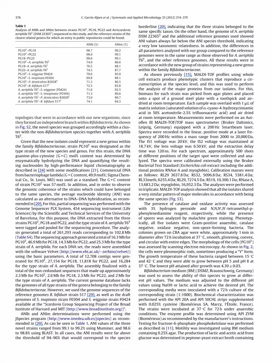
376 C. García-Aljaro et al. / Systematic and Appl
Table 1Analysis of ANIb and ANIm between strains PG10T, PG18, PG22 and Aeriscardoviaaeriphila T6T (DSM 22365T) sequenced in this study, and the reference strains of theclosest related genera for which an entry in public repositories could be found.
ANIb (%) ANIm (%)
PG10T–PG18 98.7 99.2PG10T–PG22 98.6 99.1PG18–PG22 98.6 99.1PG10T–A. aeriphila T6T 74.9 86.6PG18–A. aeriphila T6T 76.5 88.1PG22–A. aeriphila T6T 74.3 87.7PG10T–S. wiggsiae F0424 70.6 85.0PG10T–S. inopinata F0304 69.8 88.1PG10T–P. denticolens B3028T 71.3 86.5PG10T–B. bifidum S17T 72.5 83.8A. aeriphila T6T–S. wiggsiae (F0424) 71.6 92.5A. aeriphila T6T–S. innopinata (F0304) 71.3 86.6
T T
ttitT
ttgeidfiottcmGSosws(Pswueft2ttBrgaI
Jmntt
Testing for fructose-6-phosphate phosphoketolase was performed
A. aeriphila T6 –P. denticolens B3028 73.0 86.7A. aeriphila T6T–B. bifidum S17T 74.1 84.3
opologies that were in accordance with our new organisms, sincehey formed an independent branch within Bifidobacteria. As shownn Fig. S2, the novel species was grouped accordingly within a clus-er with the non-Bifidobacterium species together with A. aeriphila6T.
Given that the new isolates could represent a new genus withinhe family Bifidobacteriaceae, strain PG10T was designated as theype strain of the new species and genus. For this strain, the DNAuanine-plus-cytosine (G + C) mol% content was determined bynzymatically hydrolyzing the DNA and quantifying the result-ng nucleosides by high-performance liquid chromatography asescribed in [24] with some modifications [21]. Commercial DNArom bacteriophage lambda (G + C content, 49.9 mol%; Sigma Chem-cal Co., St. Louis, MO) was used as a standard. The G + C contentf strain PG10T was 57 mol%. In addition, and in order to observehe genomic coherence of the strains which could have belongedo the same species, the average nucleotide identity (ANI) wasalculated as an alternative to DNA–DNA hybridization, as recom-ended in [20]. For this, partial sequencing was performed with theenome Sequencer FLX System, 454 sequencing instrument (Lifeciences) by the Scientific and Technical Services of the Universityf Barcelona. For this purpose, the DNA extracted from the threetrains PG10T, PG18 and PG22, and the type strain of A. aeriphila T6T
ere tagged and pooled for the sequencing procedure. The analy-is generated a total of 261,293 reads corresponding to 102.8 MbTable S4). The sequencing approach generated a total of 16.3 Mb forG10T, 46.9 Mb for PG18, 14.3 Mb for PG22, and 25.3 Mb for the typetrain of A. aeriphila. For each DNA set, the reads were assembledith the software Velvet (http://www.ebi.ac.uk/∼zerbino/velvet/)sing the basic parameters. A total of 12,708 contigs were gen-rated for PG10T, 27,154 for PG18, 11,818 for PG22, and 16,284or the type strain of A. aeriphila. The assembly finalized with aotal of the non-redundant sequences that made up approximately.3 Mb for PG10T, 2.6 Mb for PG18, 2.3 Mb for PG22, and 2 Mb forhe type strain of A. aeriphila. The current genome database lackshe genomes of all type strains of the genera belonging to the familyifidobacteriaceae. However, we used the genome sequences of theeference genomes B. bifidum strain S17 [29], as well as the draftenomes of S. inopinata strain F0304 and S. wiggsiae strain F0424vailable at the “Scardovia Group Sequencing Project of the Broadnstitute of Harvard and MIT (http://www.broadinstitute.org/)”.
ANIb and ANIm determinations were performed using theSpecies program (http://www.imedea.uib.es/jspecies) as recom-
ended in [20]. As can be seen in Table 1, ANI values of the three
ovel strains ranged from 99.1 to 99.2% using Mummer, and 98.6o 98.8% using BLAST. In all cases, the ANI results were far abovehe threshold of 94–96% that would correspond to the speciesied Microbiology 35 (2012) 374– 379
borderline [20], indicating that the three strains belonged to thesame specific taxon. On the other hand, the genome of A. aeriphilaDSM 22365T and the additional reference genomes used showedANI values always far below the ANI species threshold, indicatinga very low taxonomic relatedness. In addition, the differences inall parameters analyzed with our group compared to the referencegenomes were in the same range as those observed for A. aeriphilaT6T, and the other reference genomes. All these results were inaccordance with the new group of strains representing a new genuswithin the family Bifidobacteriaceae.
As shown previously [15], MALDI-TOF profiles using wholecell extracts produce phenotypic clusters that reproduce a cir-cumscription at the species level, and this was used to performthe analysis of the major proteins from our isolates. For this,biomass for each strain was picked from agar plates and placedonto a spot of a ground steel plate with a toothpick and air-dried at room temperature. Each sample was overlaid with 1 �L ofmatrix solution (saturated solution of �-cyano-4-hydroxycinnamicacid in 50% acetonitrile-2.5% trifluoroacetic acid) and air driedat room temperature. Measurements were performed on an Aut-oflex III MALDI-TOF/TOF mass spectrometer (Bruker Daltonics,Leipzig, Germany) equipped with a 200 Hz Smartbeam laser.Spectra were recorded in the linear, positive mode at a laser fre-quency of 200 Hz within a mass range from 2000 to 20,000 Da.The IS1 voltage was 20 kV, the IS2 voltage was maintained at18.7 kV, the lens voltage was 6.50 kV, and the extraction delaytime was 120 ns. For each spectrum, approximately 500 shotsat different positions of the target spot were collected and ana-lyzed. The spectra were calibrated externally using the BrukerBacterial Test Standard (Escherichia coli extract including the addi-tional proteins RNAse A and myoglobin). Calibration masses wereas follows: RL29 3637.8 Da; RS32, 5096.8 Da; RS34, 5381.4 Da;RL33meth, 6255.4 Da; RL29, 7274.5 Da; RS19, 10,300.1 Da; RNase A,13,683.2 Da; myoglobin, 16,952.3 Da. The analyses were performedin triplicate. MALDI-TOF analysis showed that all the isolates shareda very similar pattern of major molecules in accordance with beingthe same species (Fig. S3).
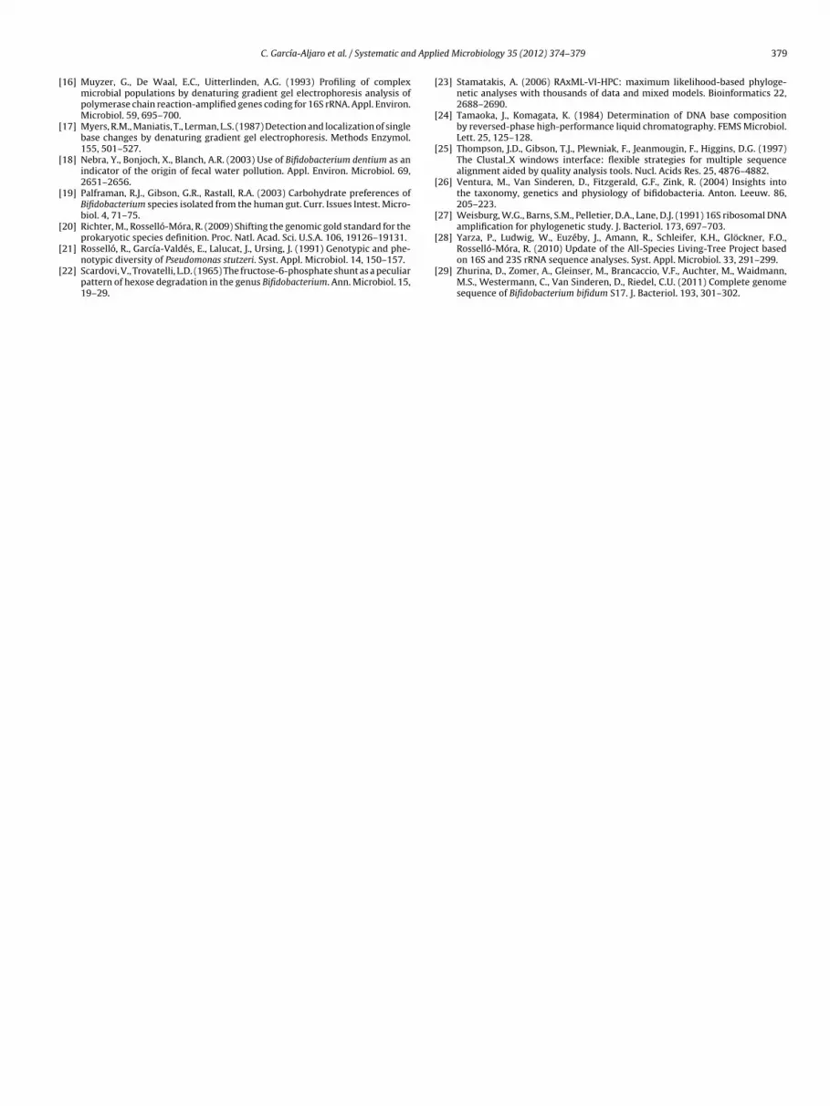
The presence of catalase and oxidase activity was assessedwith 3% hydrogen peroxide and N,N,N′,N′-tetramethyl-p-phenylenediamine reagent, respectively, while the presenceof spores was analyzed by malachite green staining. Phenotyp-ically, all five isolates were Gram-positive anaerobic, catalasenegative, oxidase negative, non-spore-forming bacteria. Thecolonies grown on CBA agar were white, approximately 1 mm indiameter after 72 h incubation at 37 ◦C, smooth, glistening, convex,and circular with entire edges. The morphology of the cells (PG10T)was assessed by scanning electron microscopy. As shown in Fig. 2,the cells were pleomorphic rods, sometimes producing a “Y” form.The growth temperature of these bacteria ranged between 15 ◦Cand 42 ◦C and they were able to grow between pH 5 and pH 8 at37 ◦C. The lowest pH attained after growth was 4.39 ± 0.03.
Bifidobacterium medium (BM) (DSMZ, Braunschweig, Germany)was used to assess the ability of this species to grow at differ-ent pH values. The medium was adjusted to the different pHvalues using NaOH or lactic acid to achieve the desired pH. Thecorresponding media were inoculated with a 72 h culture of thecorresponding strain (1:1000). Biochemical characterization wasperformed with the API 20A and API 50CHL strips supplementedwith 0.025% cysteine (Biomérieux SA, Marcy, l’Étoile, France).The strains were incubated at 37 ◦C for 72 h under anaerobicconditions. The enzyme profile was determined using API ZYM(Biomérieux) as recommended by the manufacturer’s instructions.
as described in [11]. Motility was investigated using BM mediumcontaining 0.25% agar. The production of lactic and acetic acid fromglucose was determined in peptone-yeast extract broth containing

C. García-Aljaro et al. / Systematic and Applied Microbiology 35 (2012) 374– 379 377
ia arbe
1f
6A(sFirhagmowodasl�vfAp
cSuo(Cdle(d
fpfsg
Fig. 2. Electron micrograph of Neoscardov
% glucose, as reported in [19], and the measurements were per-ormed after 5 days incubation.
Biochemical characterization included detection of fructose--phosphate phosphoketolase as well as testing with API 20A,PI 50CHL and API ZYM. The results are summarized in Table 2
detailed results for each miniaturized test can be found in theupplementary material as Table S1, Table S2 and Table S3).ructose-6-phosphate phosphoketolase is a key enzyme presentn the microorganisms belonging to the Bifidobacterium genus andelated genera, which have a characteristic metabolic pathway forexose degradation [6,22]. This enzyme was initially describeds exclusive to the Bifidobacterium genus and distinguished thisenus from other Gram-positive anaerobic bacteria that wereorphologically similar (Lactobacillus spp., Arthrobacter spp., Propi-
nibacterium spp. and Corynebacterium spp.). However, the enzymeas later detected in other genera, such as Gardnerella [10] among
thers, and therefore it cannot be exclusively linked to the Bifi-obacterium genus. All the strains produced lactic acid and aceticcid, but with a different ratio ranging from 1:10 to 1:2. All threetrains gave positive reactions in the API ZYM for leucine ary-amidase, acid phosphatase, naphthol-AS-Bi-phosphohydrolase,-and-�-galactosidase, and �-and-�-glucosidase, although withariable intensity. The members of this new group could be dif-erentiated from those of the closest relative genera Scardovia,eriscardovia and Parascardovia by the fermentation of l-fucose andotassium gluconate.
Analyses of cellular fatty acids, polar lipids and the peptidogly-an structure of strain PG10T was performed by the Identificationervice and Dr. Brian Tindall (DSMZ, Braunschweig, Germany)sing the methodology previously described [8]. The morphol-gy of the cells was studied with scanning electron microscopy20 kV). The major cellular fatty acids of strain PG10T were16:0 and C18:1ω9c, which were the same as those previouslyescribed for the genus Scardovia (Table S5). The major polar
ipids consisted of a variety of glycolipids, diphosphatidyl glyc-rol, two unidentified phospholipids, and phosphatidyl glycerolFig. S4). The peptidoglycan structure was A1� meso-Dpm-irect.
From the above results, the three strains PG10T, PG18 and PG30ormed a homogeneous group that was clearly genetically and
henotypically distinct from any other recognized genera. There-ore, we propose a novel genus Neoscardovia gen. nov. with thepecies Neoscardovia arbecensis sp. nov. as the first species of thisenus.censis PG10T: (a) 4000× and (b) 50,000×.
Description of Neoscardovia gen. nov.
Neoscardovia (Ne.o.sacr.do’via.a. Gr. pref. neo- (from. Gr. adj.neos), new; N.L. fem. n. Scardovia, a bacterial generic name; N.L.fem. n. Neoscardovia, a new Scardovia).
The cells of the genus are Gram-positive, catalase negative, oxi-dase negative, non-motile, non-spore-forming, irregular shapedrods occasionally forming a “Y” shape. Lactic and acetic acid areproduced from glucose fermentation. Fermentation of l-fucose andpotassium gluconate clearly distinguishes this genus from the clos-est related genera Scardovia, Aeriscardovia and Parascardovia. TheG + C content of the type strain is 57 mol%. The prevailing cellularfatty acids are C16:0 and C18:1ω9c, and the major polar lipids consistof a variety of glycolipids, diphosphatidyl glycerol, two unidentifiedphospholipids, and phosphatidyl glycerol. The peptidoglycan struc-ture is A1� meso-Dpm-direct. The type species of the genus is N.arbecensis, which is represented by the type strain PG10T depositedin the culture collections CECT and DSM under the culture accessionnumbers CECT 8111T and DSM 25737T, respectively.
Description of N. arbecensis sp. nov.
N. arbecensis (ar.be.cen’sis. N.L. fem. adj. arbecensis, of or belong-ing to Arbeca).
Obligate anaerobe Gram-positive, non-spore-forming bacilli.The cells are pleomorphic with occasional bifurcations, some-times arranged in a “Y” form. Colonies grown on CBA agar arewhite, approximately 1 mm in diameter after 72 h incubation at37 ◦C, smooth, glistening, convex, and circular with entire edges.The maximum temperature for growth is 42 ◦C and the mini-mum is 15 ◦C. This species can be differentiated from those ofthe related genera Aeriscardovia, Scardovia, and Parascardovia onthe basis of its ability to grow at lower temperatures, and fromthe most closely related A. aeriphila for its low tolerance tooxygen. The minimum initial pH for growth is 5.0 and the maxi-mum is 8.0. All three strains of N. arbecensis form a homogenousgroup able to ferment l-arabinose, d-ribose, d-xylose, d-galactose,d-glucose, d-fructose, salicin, d-cellobiose, d-maltose, d-lactose, d-saccharose, d-raffinose, starch, glycogen, d-turanose, l-fucose, andpotassium gluconate. All three strains have the following activi-
ties: fructose-6-phosphate-phosphoketolase, leucine arylamidase,acid phosphatase, naphthol-AS-Bi-phosphohydrolase, �-and-�-galactosidase, and �-and-�-glucosidase. Lactic and acetic acid areproduced as end products of glucose fermentation in a variable ratio
378 C. García-Aljaro et al. / Systematic and Applied Microbiology 35 (2012) 374– 379
Table 2Phenotypic characteristics distinguishing Neoscardovia arbecensis from species of closely related genera (Aeriscardovia, Scardovia, Parascardovia). All Neoscardovia arbecensisstrains fermented l-arabinose, d-ribose, d-xylose, d-galactose, d-glucose, d-fructose, salicin, d-cellobiose (+), d-maltose, d-lactose (+), d-saccharose, d-raffinose (+), starch,glycogen, l-fucose, d-turanose (+) and potassium gluconate. The phenotypic tests that differentiated the different genera are given below.
Characteristic Neoscardovia arbecensis Aeriscardovia aeriphila Scardovia sp. Parascardovia denticolens
Glycerol − − + −d-Ribose + − + +d-Xylose + + v −d-Mannose − + (+) −Methyl-�-d-glucopyranoside − (+) − −N-acetylglucosamine − − (+) −Amygdalin − (+) v −Arbutin − (+) v −d-Melibiose − + + +Inulin − (+) v (+)d-Melezitose − + (+) −Xylitol − − (+) −l-Fucose + − − −Potassium gluconate + − − −Gentiobiose − (+) v −d-Turanose (+) + v −Products of glucose fermentation: lactic:acetic (M) 1:10 to 1:2 1:2 1:1.9 to 1:2.9 1:2Peptidoglucan structure A1� meso-Dpm-direct ND A4�
l-(Lys/Orn)-(Ser/Thr)-GluND
Polar lipids Glycolipids,diphosphatidylglycerol,two unidentifiedphospholipids,phosphatidylglycerol
ND Glycolipids,diphosphatidylglycerol,one unidentifiedphospholipid and anunidentifiedphosphoglycolipid
ND
Tolerance to oxygen Low High Low LowG + C (mol%) 57 55 54–55 55
◦ a 3
+
ritsur
A
f(pUJT(aRiI0
A
fs
R
[
[
[
[
[
Temperature range for growth ( C) 15 –42
, positive; −, negative; (+) weakly positive; v, variable; ND, no data available.a Growth at the initial streak.
anging from 1:10 to 1:2. The G + C content of the type strain PG10T
s 57 mol%. Isolated from pig slurries, this species might be useful torack the source of fecal contamination of porcine origin. The typetrain has been deposited in the culture collections CECT and DSMnder the culture accession numbers CECT 8111T and DSM 25737T,espectively.
cknowledgements
We would like to thank Javier Mendez (Servei Fermentació UB)or assistance with the microscopy imaging, Rosa Maria GomilaTechnical and Scientific Services, Universitat Illes Balears) for therotein analysis and Berta Fusté (Scientific and Technical Services,niversitat de Barcelona) for the genome sequencing, as well as
ean Euzéby for his assessment in the etymology of the new taxon.his work was supported by the Xarxa de Referència en BiotecnologiaXRB), the Catalonia Government research program 2009SGR1043,nd the Spanish Government research project CGL2011-25401.RM acknowledges the funding received from the Spanish Min-
stry of Economy and Competitivity with the projects Consoliderngenio 2010 CE-CSD2007-0005, and CLG2009 12651-C02-01 and2 (all co-financed with FEDER funding).
ppendix A. Supplementary data
Supplementary data associated with this article can beound, in the online version, at http://dx.doi.org/10.1016/j.yapm.2012.06.007.
eferences
[1] Balleste, E., Blanch, A.R. (2011) Bifidobacterial diversity and the developmentof new microbial source tracking indicators. Appl. Environ. Microbiol. 77,3518–3525.
[
0–46 27–44 27–44
[2] Beerens, H. (1991) Detection of bifidobacteria by using propionic acid as aselective agent. Appl. Environ. Microbiol. 57, 2418–2419.
[3] Bonjoch, X., Balleste, E., Blanch, A.R. (2004) Multiplex PCR with 16S rRNA gene-targeted primers of Bifidobacterium spp. to identify sources of fecal pollution.Appl. Environ. Microbiol. 70, 3171–3175.
[4] Castresana, J. (2000) Selection of conserved blocks from multiple alignmentsfor their use in phylogenetic analysis. Mol. Biol. Evol. 17, 540–552.
[5] Chao, S.H., Tomii, Y., Sasamoto, M., Fujimoto, J., Tsai, Y.C., Watanabe, K. (2008)Lactobacillus capillatus sp. nov., a motile bacterium isolated from stinky tofubrine. Int. J. Syst. Evol. Microbiol. 58, 2555–2559.
[6] De Vries, W., Stouthamer, A.H. (1967) Pathway of glucose fermentation in rela-tion to the taxonomy of bifidobacteria. J. Bacteriol. 93, 574–576.
[7] Dorai-Raj, S., O’Grady, J., Colleran, E. (2009) Specificity and sensitivity evalua-tion of novel and existing Bacteroidales and Bifidobacteria-specific PCR assays onfeces and sewage samples and their application for microbial source trackingin Ireland. Water Res. 43, 4980–4988.
[8] Downes, J., Sutcliffe, I., Tanner, A.C., Wade, W.G. (2005) Prevotella marshii sp.nov. and Prevotella baroniae sp. nov., isolated from the human oral cavity. Int.J. Syst. Evol. Microbiol. 55, 1551–1555.
[9] Gavini, F., Delcenserie, V., Kopeinig, K., Pollinger, S., Beerens, H., Bonaparte, C.,Upmann, M. (2006) Bifidobacterium species isolated from animal feces and frombeef and pork meat. J. Food Prot. 69, 871–877.
10] Gavini, F., Van Esbroeck, M., Touzel, J.P., Fourment, A., Goossens, H. (1996)Detection of fructose-6-phosphate phosphoketolase (F6PPK), a key enzyme ofthe bifid-shunt, in Gardnerella vaginalis. Anaerobe 2, 191–193.
11] Holt, J.G., Sneath, P.H. 1986 Bergey’s Manual of Systematic Bacteriology:Volume 2: Gram-Positive Bacteria Other than Actinomycetes, Williams andWilkins, Baltimore, MD, p. 1423.
12] Jian, W., Zhu, L., Dong, X. (2001) New approach to phylogenetic analysis of thegenus Bifidobacterium based on partial HSP60 gene sequences. Int. J. Syst. Evol.Microbiol. 51, 1633–1638.
13] Ludwig, W., Strunk, O., Westram, R., Richter, L., Meier, H., Yadhukumar, Buch-ner, A., Lai, T., Steppi, S., Jobb, G., Forster, W., Brettske, I., Gerber, S., Ginhart,A.W., Gross, O., Grumann, S., Hermann, S., Jost, R., Konig, A., Liss, T., Lussmann,R., May, M., Nonhoff, B., Reichel, B., Strehlow, R., Stamatakis, A., Stuckmann, N.,Vilbig, A., Lenke, M., Ludwig, T., Bode, A., Schleifer, K.H. (2004) ARB: a softwareenvironment for sequence data. Nucl. Acids Res. 32, 1363–1371.
14] Lynch, P.A., Gilpin, B.J., Sinton, L.W., Savill, M.G. (2002) The detection of Bifi-dobacterium adolescentis by colony hybridization as an indicator of humanfaecal pollution. J. Appl. Microbiol. 92, 526–533.
15] Munoz, R., Lopez-Lopez, A., Urdiain, M., Moore, E.R., Rosselló-Móra, R. (2011)Evaluation of matrix-assisted laser desorption ionization-time of flight wholecell profiles for assessing the cultivable diversity of aerobic and moderatelyhalophilic prokaryotes thriving in solar saltern sediments. Syst. Appl. Microbiol.34, 69–75.
d Appl
[
[
[
[
[
[
[
[
[
[
[
[
[
C. García-Aljaro et al. / Systematic an
16] Muyzer, G., De Waal, E.C., Uitterlinden, A.G. (1993) Profiling of complexmicrobial populations by denaturing gradient gel electrophoresis analysis ofpolymerase chain reaction-amplified genes coding for 16S rRNA. Appl. Environ.Microbiol. 59, 695–700.
17] Myers, R.M., Maniatis, T., Lerman, L.S. (1987) Detection and localization of singlebase changes by denaturing gradient gel electrophoresis. Methods Enzymol.155, 501–527.
18] Nebra, Y., Bonjoch, X., Blanch, A.R. (2003) Use of Bifidobacterium dentium as anindicator of the origin of fecal water pollution. Appl. Environ. Microbiol. 69,2651–2656.
19] Palframan, R.J., Gibson, G.R., Rastall, R.A. (2003) Carbohydrate preferences ofBifidobacterium species isolated from the human gut. Curr. Issues Intest. Micro-biol. 4, 71–75.
20] Richter, M., Rosselló-Móra, R. (2009) Shifting the genomic gold standard for theprokaryotic species definition. Proc. Natl. Acad. Sci. U.S.A. 106, 19126–19131.
21] Rosselló, R., García-Valdés, E., Lalucat, J., Ursing, J. (1991) Genotypic and phe-notypic diversity of Pseudomonas stutzeri. Syst. Appl. Microbiol. 14, 150–157.
22] Scardovi, V., Trovatelli, L.D. (1965) The fructose-6-phosphate shunt as a peculiarpattern of hexose degradation in the genus Bifidobacterium. Ann. Microbiol. 15,19–29.
[
ied Microbiology 35 (2012) 374– 379 379
23] Stamatakis, A. (2006) RAxML-VI-HPC: maximum likelihood-based phyloge-netic analyses with thousands of data and mixed models. Bioinformatics 22,2688–2690.
24] Tamaoka, J., Komagata, K. (1984) Determination of DNA base compositionby reversed-phase high-performance liquid chromatography. FEMS Microbiol.Lett. 25, 125–128.
25] Thompson, J.D., Gibson, T.J., Plewniak, F., Jeanmougin, F., Higgins, D.G. (1997)The Clustal X windows interface: flexible strategies for multiple sequencealignment aided by quality analysis tools. Nucl. Acids Res. 25, 4876–4882.
26] Ventura, M., Van Sinderen, D., Fitzgerald, G.F., Zink, R. (2004) Insights intothe taxonomy, genetics and physiology of bifidobacteria. Anton. Leeuw. 86,205–223.
27] Weisburg, W.G., Barns, S.M., Pelletier, D.A., Lane, D.J. (1991) 16S ribosomal DNAamplification for phylogenetic study. J. Bacteriol. 173, 697–703.
28] Yarza, P., Ludwig, W., Euzéby, J., Amann, R., Schleifer, K.H., Glöckner, F.O.,
Rosselló-Móra, R. (2010) Update of the All-Species Living-Tree Project basedon 16S and 23S rRNA sequence analyses. Syst. Appl. Microbiol. 33, 291–299.29] Zhurina, D., Zomer, A., Gleinser, M., Brancaccio, V.F., Auchter, M., Waidmann,M.S., Westermann, C., Van Sinderen, D., Riedel, C.U. (2011) Complete genomesequence of Bifidobacterium bifidum S17. J. Bacteriol. 193, 301–302.