acinetobacter ursingii sp. nov. and acinetobacter schindleri sp. nov., isolated from human clinical...
TRANSCRIPT
Downloaded from www.microbiologyresearch.org by
IP: 54.224.135.207
On: Thu, 02 Jun 2016 09:39:58
International Journal of Systematic and Evolutionary Microbiology (2001), 51, 1891–1899 Printed in Great Britain
Acinetobacter ursingii sp. nov. andAcinetobacter schindleri sp. nov., isolated fromhuman clinical specimens
1 National Institute of PublicHealth, SC roba! rova 48,10042 Prague, CzechRepublic
2 Department of ClinicalChemistry, Microbiologyand Immunology,University Hospital, Blok A,9000 Ghent, Belgium
3 Department of MedicalMicrobiology, Malmo$University Hospital,University of Lund,S-20502 Malmo$ , Sweden
4 Department of InfectiousDiseases, Leiden UniversityMedical Center, Leiden,The Netherlands
Alexandr Nemec,1 Thierry De Baere,2 Ingela Tjernberg,3
Mario Vaneechoutte,2 Tanny J. K. van der Reijden4
and Lenie Dijkshoorn4
Author for correspondence: A. Nemec. Tel : 420 2 67 08 22 66. Fax: 420 2 72 73 04 28.e-mail : anemec!szu.cz
The taxonomic status of two recently described phenetically distinctive groupswithin the genus Acinetobacter, designated phenon 1 and phenon 2, wasinvestigated further. The study collection included 51 strains, mainly of clinicalorigin, from different European countries with properties of either phenon 1(29 strains) or phenon 2 (22 strains). DNA–DNA hybridization studies and DNApolymorphism analysis by AFLP revealed that these phenons represented twonew genomic species. Furthermore, 16S rRNA gene sequence analysis of threerepresentatives of each phenon showed that they formed two distinct lineageswithin the genus Acinetobacter. The two phenons could be distinguished fromeach other and from all hitherto-described Acinetobacter (genomic) species byspecific phenotypic features and amplified rDNA restriction analysis patterns.The names Acinetobacter ursingii sp. nov. (type strain LUH 3792T ¯NIPH 137T ¯LMG 19575T ¯CNCTC 6735T) and Acinetobacter schindleri sp. nov. (type strainLUH 5832T ¯NIPH 1034T ¯LMG 19576T ¯CNCTC 6736T) are proposed for phenon1 and phenon 2, respectively. Clinical and epidemiological data indicate that A.ursingii has the capacity to cause bloodstream infections in hospitalizedpatients.
Keywords : Acinetobacter ursingii sp. nov., Acinetobacter schindleri sp. nov.,polyphasic taxonomy
INTRODUCTION
Over the last 15 years, considerable progress has beenmade in resolving the taxonomy of the genus Acineto-bacter. The basis for the present classification wasestablished by Bouvet & Grimont (1986), with thedescription of 12 DNA–DNA hybridization groups(genomic species) within the genus. This scheme wassubsequently extended to include 10 additionalgenomic species (Tjernberg & Ursing, 1989; Bouvet &Jeanjean, 1989; Gerner-Smidt & Tjernberg, 1993).Seven genomic species have names (Acinetobactercalcoaceticus, Acinetobacter baumannii, Acinetobacterhaemolyticus, Acinetobacter junii, Acinetobacter john-
.................................................................................................................................................
Abbreviations: ARDRA, amplified rDNA restriction analysis.
The EMBL accession numbers for the 16S rRNA gene sequences of strainsLUH 3299, LUH 3792T, LUH 4763, LUH 4591, LUH 4760 and LUH 5832T arerespectively AJ275037–AJ275041 and AJ278311.
sonii, Acinetobacter lwoffii and Acinetobacter radio-resistens), while the others are designated by numbers(reviewed by Janssen et al., 1997). Another genomicspecies (‘Acinetobacter venetianus ’) comprising marineoil-degrading organisms was delineated recently (DiCello et al., 1997; Vaneechoutte et al., 1999). Never-theless, the DNA–DNA hybridization studies ofBouvet & Grimont (1986), Tjernberg & Ursing (1989)and Bouvet & Jeanjean (1989) left several strainsunclassified, which indicates that the diversity of thegenus extends beyond the described groups.
In a recent study, 45 additional unidentifiable isolateswere found among 700 clinical isolates from the CzechRepublic (Nemec et al., 2000). Two groups of isolates(designated phenon 1 and phenon 2) were delineatedamong the unidentifiable isolates, each of whichshowed distinctive phenotypic features and amplifiedrDNA restriction analysis (ARDRA) patterns. Theaim of the present study was to define the taxonomic
01914 # 2001 IUMS 1891
Downloaded from www.microbiologyresearch.org by
IP: 54.224.135.207
On: Thu, 02 Jun 2016 09:39:58
A. Nemec and others
Table 1. Strains of phenon 1 (Acinetobacter ursingii sp. nov.) and phenon 2 (Acinetobacter schindleri sp. nov.).................................................................................................................................................................................................................................................................................................................
All strains were from human specimens. CNCTC, Czech National Collection of Type Cultures, Prague, Czech Republic ; LMG,Bacteria Collection, Laboratorium voor Microbiologie Gent, Gent, Belgium; LUH and RUH, Collection L. Dijkshoorn, LeidenUniversity Medical Centre, Leiden, The Netherlands; NIPH, Collection A. Nemec, National Institute of Public Health, Prague,Czech Republic. Abbreviations: CZ, Czech Republic ; NL, The Netherlands; NO, Norway; SE, Sweden.
Strain Other strain designation(s) Reference/received from Specimen* Location and year of isolation
Phenon 1 (A. ursingii sp. nov.)
LUH 3792T NIPH 137T†¯LMG 19575T
¯CNCTC 6735T
Nemec et al. (2000) Blood (in) Praha, CZ, 1993
LUH 4582 NIPH 177† Nemec et al. (2000) Intravenous line (in) Praha, CZ, 1993
LUH 4592 NIPH 280† Nemec et al. (2000) Blood (in) Sedlc3 any, CZ, 1994
LUH 3793 NIPH 371† Nemec et al. (2000) Blood (in) Pr3 ı!bram, CZ, 1995
LUH 4613 NIPH 375† Nemec et al. (2000) Pus (in) Pr3 ı!bram, CZ, 1995
LUH 4614 NIPH 376† Nemec et al. (2000) Pus (in) Pr3 ı!bram, CZ, 1995
LUH 4618 NIPH 398† Nemec et al. (2000) Ulcer (out) Pr3 ı!bram, CZ, 1996
LUH 4622 NIPH 439† Nemec et al. (2000) Eye (out) Sedlc3 any, CZ, 1996
LUH 5767 NIPH 706† Nemec et al. (2000) Blood (in) Pr3 ı!bram, CZ, 1997
LUH 5829 NIPH 950† Nemec et al. (2000) Blood (in) Ta! bor, CZ, 1998
LUH 5830 NIPH 993† Nemec et al. (2000) Cervix (out) Pr3 ı!bram, CZ, 1998
LUH 5831 NIPH 1025† Nemec et al. (2000) Eye (out) Pr3 ı!bram, CZ, 1998
LUH 5833 NIPH 1048† Nemec et al. (2000) Blood (in) Liberec, CZ, 1998
LUH 5834 NIPH 1118† Nemec et al. (2000) Wound (out) Sedlc3 any, CZ, 1999
LUH 5835 NIPH 1120† Nemec et al. (2000) Urine (out) Pr3 ı!bram, CZ, 1999
LUH 4761 72a† Tjernberg & Ursing (1989) Urine (out) Malmo$ , SE, 1980
LUH 4762 93† Tjernberg & Ursing (1989) Blood (in) Malmo$ , SE, 1980
LUH 4763 119† Tjernberg & Ursing (1989) Urine (out) Malmo$ , SE, 1980
LUH 4766 166† Tjernberg & Ursing (1989) Wound (in) Malmo$ , SE, 1981
LUH 4768 175† Tjernberg & Ursing (1989) Wound (in) Malmo$ , SE, 1981
RUH 1501 Hairy skin (in) Rotterdam, NL, 1985
RUH 3329 Patient K† Horrevorts et al. (1995) Blood (in) Nijmegen, NL, 1990
LUH 3292† Bernards et al. (1997) Blood (in) Leiden, NL, 1995
LUH 3299† Bernards et al. (1997) Blood (in) Leiden, NL, 1995
LUH 3140 A. T. Bernards Toes (in) Enschede, NL, 1995
LUH 3059 A. T. Bernards Blood (in) Enschede, NL, 1995
LUH 3324 610}1994‡ J. G. M. Koeleman Blood Amsterdam, NL, 1994
LUH 4739 84† Bouvet & Grimont (1986) Blood Unknown
LUH 4828 1614}96‡ D. A. Caugant Abscess (out) Kristiansand, NO, 1996
Phenon 2 (A. schindleri sp. nov.)
LUH 5832T NIPH 1034T†¯LMG 19576T
¯CNCTC 6736T
Nemec et al. (2000) Urine (out) Pr3 ı!bram, CZ, 1998
LUH 4590 NIPH 228† Nemec et al. (2000) Vagina (out) Praha, CZ, 1994
LUH 4591 NIPH 257† Nemec et al. (2000) Urine (out) Hluboka! nad Vltavou,
CZ, 1993
LUH 4594 NIPH 285† Nemec et al. (2000) Throat (out) Pr3 ı!bram, CZ, 1994
LUH 4595 NIPH 286† Nemec et al. (2000) Ear (out) Pr3 ı!bram, CZ, 1994
LUH 4597 NIPH 291† Nemec et al. (2000) Nasal swab (out) Pr3 ı!bram, CZ, 1994
LUH 4598 NIPH 293† Nemec et al. (2000) Cervix (out) Sedlc3 any, CZ, 1994
LUH 4599 NIPH 296† Nemec et al. (2000) Cervix (out) Sedlc3 any, CZ, 1994
LUH 4612 NIPH 369† Nemec et al. (2000) Cervix (out) Sedlc3 any, CZ, 1994
LUH 4615 NIPH 383† Nemec et al. (2000) Nasal swab (out) Milı!n, CZ, 1996
LUH 5825 NIPH 883† Nemec et al. (2000) Urine (out) Pr3 ı!bram, CZ, 1998
LUH 5826 NIPH 900† Nemec et al. (2000) Conjunctiva (out) Sedlc3 any, CZ, 1998
LUH 5827 NIPH 904† Nemec et al. (2000) Urine (out) Pr3 ı!bram, CZ, 1998
LUH 5939 NIPH 907† Nemec et al. (2000) Nasal swab (out) Pr3 ı!bram, CZ, 1998
LUH 5828 NIPH 933† Nemec et al. (2000) Vagina (out) Pr3 ı!bram, CZ, 1998
LUH 4760 60† Tjernberg & Ursing (1989) Urine (in) Malmo$ , SE, 1980
LUH 4764 120† Tjernberg & Ursing (1989) Pleural effusion (in) Malmo$ , SE, 1980
LUH 4765 129† Tjernberg & Ursing (1989) Urine Malmo$ , SE, 1980
RUH 203† Dijkshoorn et al. (1998) Liquor (out) Rotterdam, NL, 1983
LUH 4742 594‡ P. J. M. Bouvet Skin Unknown
LUH 4743 585‡ P. J. M. Bouvet Skin Unknown
LUH 4744 586‡ P. J. M. Bouvet Skin Unknown
* If known, specimens from outpatients (out) or inpatients (in) are indicated.
†Strain designation used in a previous publication.
‡Strain designation as received.
status of these groups by a polyphasic analysis. Forthis purpose, the collection of Czech strains wasenlarged with strains from other European countriesthat showed characters similar to those of the twophenons.
METHODS
Strains. The 29 strains of phenon 1 and 22 strains of phenon2 investigated in this study are listed in Table 1. The Czechstrains (n¯ 30) were those from the previous study (Nemecet al., 2000). Additionally, 21 strains were selected from a set
1892 International Journal of Systematic and Evolutionary Microbiology 51
Downloaded from www.microbiologyresearch.org by
IP: 54.224.135.207
On: Thu, 02 Jun 2016 09:39:58
Two novel Acinetobacter species
Table 2. Biochemical characteristics of phenon 1 (A. ursingii sp. nov.) and phenon 2 (A. schindleri sp. nov.).................................................................................................................................................................................................................................................................................................................
Data are from this study and from Nemec et al. (2000). Growth on carbon sources was evaluated after 2 and 6 d of incubation., Positive for all strains ; ®, negative for all strains ; numbers are percentages of strains giving a positive reaction. All strainsutilized -lactate and acetate. None of the strains grew at 44 °C, hydrolysed gelatin, produced haemolysis on sheep-blood agar,acidified Hugh & Leifson’s medium with -glucose or utilized -4-aminobutyrate, β-alanine, -histidine, malonate, histamine, -phenylalanine, phenylacetate, laevulinate, citraconate or -leucine.
Characteristic Phenon 1 (A. ursingii sp. nov.) (n¯ 29) Phenon 2 (A. schindleri sp. nov.) (n¯ 22)
Growth at 41 °C ® *
Growth at 37 °C * Utilization of:
Citrate (Simmons) 59
Glutarate 97 95
-Aspartate 97* ®Azelate 64
-Malate * 95*
4-Hydroxybenzoate 97 64
-Tartrate ® 18
2,3-Butanediol ® 32
Ethanol 95
*Weak growth of some strains.
of about 100 Acinetobacter strains isolated by differentlaboratories that could not be identified as any of thedescribed genomic species. The 21 strains were selected fromthis set on the basis of phenotypic properties and ARDRApatterns similar to those of the phenon 1 or phenon 2 strains(Nemec et al., 2000). All 51 strains had the properties of thegenus Acinetobacter (Juni, 1984) ; i.e. they were Gram-negative, strictly aerobic, oxidase-negative, non-motilecoccobacilli and positive in the transformation assay of Juni(1972).
Phenotypic characterization. The tests described by Nemec etal. (2000) were used, with the following modifications.Carbon-source utilization tests were supplemented withthose for laevulinate, citraconate, 4-hydroxybenzoate, -tartrate, -leucine, 2,3-butanediol, ethanol and acetate. Thetest for trans-aconitate utilization was omitted since it maygive irreproducible results with some phenon 1 strains.Production of pigments was tested on glycerol-containingmedia A and B as described by King et al. (1954). All testswere performed at 30 °C unless indicated otherwise.
ARDRA. Amplified 16S rDNA was obtained by PCR andanalysed by restriction digestion with six restriction endo-nucleases (CfoI, AluI, MboI, RsaI, MspI and BfaI) asdescribed previously (Nemec et al., 2000). Interpretation ofARDRA patterns was based on the positions of thefragments of molecular size & 100 bp. The patterns werenumbered according to the scheme of Dijkshoorn et al.(1998), supplemented by Seifert et al. (1997) and Nemec et al.(2000).
AFLP fingerprinting. AFLP was performed according toKoeleman et al. (1998), with some modifications. DNA waspurified as described by Boom et al. (1990) and adapterswere as described by Vos et al. (1995). Restriction andligation were performed simultaneously at 37 °C for 3 h in a10 µl volume with 10–50 ng template DNA, 1 U EcoRI(Amersham Pharmacia Biotech), 1 U MseI (New EnglandBioLabs), 4 U T4 DNA ligase (Amersham Pharmacia
Biotech), 1¬ T4 DNA ligase buffer, 500 ng BSA, 50 mMNaCl, 2 pmol EcoRI adapters and 20 pmol MseI adapters.After incubation, the mixture was diluted with 10 mMTris}HCl, 0±1 mM EDTA (pH 8±0) to a final volume of200 µl. Five microlitres diluted mixture was added to a finalvolume of 10 µl reaction mixture containing 20 ng Cy5-labelled EcoRIA primer (Cy5-GACTGCGTACCAA-TTCa-3« ; where a is a selective A base), 60 ng MseICprimer (5«-GATGAGTCCTGAGTAAc-3« ; where c is aselective C base), 1¬ Taq polymerase buffer, 1±5 mM MgCl
#,
0±2 mM (each) dNTP and 1 U Goldstar Taq DNA poly-merase (Eurogentec). Amplification with a Progene thermo-cycler (Techne) was as follows: 2 min at 72 °C and 2 min at94 °C; one cycle of 30 s at 94 °C, 30 s at 65 °C and 60 s at72 °C; 12 cycles of 30 s at 94 °C, 30 s at a temperature of0±7 °C lower than the previous cycle, starting at 64±3 °C,followed by 60 s at 72 °C; 23 cycles of 30 s at 94 °C, 30 s at56 °C and 60 s at 72 °C; and a final cycle of 10 min at 72 °C.PCR products were mixed with 3 µl formamide containing0±5% dextran blue, heated for 5 min at 95 °C and cooled onice. Samples of 3 µl were loaded on a denaturing poly-acrylamide gel (ReproGel High Resolution; AmershamPharmacia Biotech) with 200 mm standard thermoplates.Fragment separation was performed using the ALFexpressII DNA analysis system (Amersham Pharmacia Biotech) for500 min at 55 °C and 30 W constant power with 2 s samplingintervals. The peak patterns generated were converted toTIF files, which were analysed by the BN 2.0software package (Applied Maths). Fragments in the range50–500 bp were used for cluster analysis. Pearson’s product-moment coefficient (r) was used as a measure of similarityand grouping was obtained by the unweighted pair groupaverage linked method (UPGMA).
DNA–DNA hybridization. The two-step elution procedurewas used to determine DNA–DNA relatedness (Tjernberg etal., 1989). By this method, "#&I-labelled DNA probes fromstrains LUH 3792T (phenon 1) and LUH 5832T (phenon 2)were hybridized on a filter with unlabelled DNAs of the
International Journal of Systematic and Evolutionary Microbiology 51 1893
Downloaded from www.microbiologyresearch.org by
IP: 54.224.135.207
On: Thu, 02 Jun 2016 09:39:58
A. Nemec and others
Table 3. ARDRA patterns of phenon 1 and phenon 2 strains.................................................................................................................................................................................................................................................................................................................
Data were from this study and from Nemec et al. (2000). Pattern designation according to Dijkshoorn et al. (1998) and Nemec etal. (2000). , Not determined; New, novel patterns.
Strain(s) Restriction pattern with :
CfoI AluI MboI RsaI MspI BfaI
Phenon 1 (A. ursingii sp. nov.)
LUH 3792T, LUH 3292, LUH 3299,
LUH 4592, LUH 4614, LUH 4622,
LUH 4762, LUH 4763, LUH 4766,
LUH 4828, LUH 5829, LUH 5830,
LUH 5834, RUH 1501, RUH 3329
1 4 3 5 3
LUH 3059, LUH 3793, LUH 4582, LUH 4768 1 4nw* 3 4 3
LUH 3140, LUH 5767, LUH 5835 1 4nw* 3 45 3
LUH 3324, LUH 4761 1 4nw* 13 45 3
LUH 4739 1 4 13 5 3
LUH 4618 1 4 3 45 3
LUH 5833 1 4 13 45 3
LUH 5831 1 4 3 25 3
LUH 4613 1 4† 13 25 3
Phenon 2 (A. schindleri sp. nov.)
LUH 5832T, LUH 4591, LUH 4595,
LUH 4597, LUH 4598, LUH 4599,
LUH 4615, LUH 4742, LUH 4760,
LUH 4764, LUH 5825, LUH 5827,
LUH 5828, LUH 5939, RUH 203
15 24‡ 1 2 2 10
LUH 4590, LUH 4594, LUH 4612,
LUH 4743, LUH 4744, LUH 5826
15 2 1 2 2 10
LUH 4765 5 New 1 2 2 New
*A combined AluI pattern, tentatively interpreted as the mixture of pattern 4 and a new pattern (Nemec et al., 2000; Fig. 1).
†Pattern 4 containing an additional, weak band of approximately 223 bp; this pattern is highly similar to combined AluI pattern 24(Nemec et al., 2000).
‡The band (220 bp) specific for AluI pattern 2 was diffuse in all strains (Nemec et al., 2000; Fig. 1).
phenon 1 and phenon 2 strains and reference strains of alldescribed Acinetobacter genomic species. The amount ofDNA released from the filter was measured at two tempera-tures, at 7 °C below the thermal melting midpoint of thehomologous duplex and at 100 °C. The amount of DNAreleased in the first step expressed as a percentage of the totalamount of eluted DNA at 100 °C (%DR7) was the criterionfor inclusion of strains in a species, with the intraspecies andinterspecies values for %DR7 being % 26 and & 37,respectively (Tjernberg et al., 1989). Each %DR7 valuewas calculated as a mean of at least two hybridizationexperiments.
16S rDNA sequencing and comparative analysis. A fragmentof the 16S rRNA gene (corresponding to positions 10–1507in the Escherichia coli numbering system) of three phenon 1strains (LUH 3792T, LUH 3299, LUH 4763) and threephenon 2 strains (LUH 5832T, LUH 4591, LUH 4760) wassequenced as described by Vaneechoutte et al. (2000). The16S rDNA sequences obtained for phenon 1 and phenon 2strains were compared with the sequences representing alldescribed Acinetobacter genomic species, i.e. 21 sequencesdetermined by Ibrahim et al. (1997) (EMBL accessionnumbers Z93434–Z93454) and the sequence of ‘A.
venetianus ’ strain RAG-1 (AJ295007), and the sequences ofMoraxella lacunata ATCC 17967T (AF005160) and Psychro-bacter immobilis ATCC 43116T (U39399). All steps of thecomparative sequence analysis were performed by using theGB software package (Applied Maths). Firstly, pair-wise alignment using UPGMA was carried out with a gappenalty of 100%, a unit gap cost of 20% and an ambiguitycost of 50% of the mismatch cost. Subsequently, globalalignment with P. immobilis as the outgroup was carried outon the region corresponding to positions 67–1444 of the 16SrRNA gene of E. coli, with costs as above. Finally, asimilarity matrix of the aligned sequences was constructedby global alignment homology calculation and a gap penaltyof 20%. The neighbour-joining method was used to con-struct the dendrogram based on this similarity matrix.
RESULTS AND DISCUSSION
Phenotypic characteristics
Colonies of all strains grown on nutrient agar after24 h were circular, convex, smooth and slightly opaquewith entire margins. The colonies of phenon 1 strains
1894 International Journal of Systematic and Evolutionary Microbiology 51
Downloaded from www.microbiologyresearch.org by
IP: 54.224.135.207
On: Thu, 02 Jun 2016 09:39:58
Two novel Acinetobacter species
.................................................................................................................................................................................................................................................................................................................
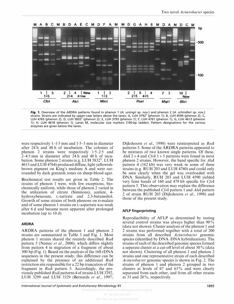
Fig. 1. Overview of the ARDRA patterns found in phenon 1 (A. ursingii sp. nov.) and phenon 2 (A. schindleri sp. nov.)strains. Strains are indicated by upper-case letters above the lanes: A, LUH 3792T (phenon 1); B, LUH 4594 (phenon 2); C,LUH 4765 (phenon 2); D, LUH 5832T (phenon 2); E, LUH 3793 (phenon 1); F, LUH 4761 (phenon 1); G, LUH 4613 (phenon1); H, LUH 4618 (phenon 1). Lanes M, molecular size markers (100-bp ladder). Pattern designations for the variousenzymes are given below the lanes.
were respectively 1–1±5 mm and 1±5–3 mm in diameterafter 24 h and 48 h of incubation. The colonies ofphenon 2 strains were respectively 1±5–2±5 and2–4±5 mm in diameter after 24 h and 48 h of incu-bation. Some phenon 2 strains (e.g. LUH 5832T, LUH4615 and LUH 4764) produced diffuse, light yellowish-brown pigment on King’s medium A and were sur-rounded by dark greenish zones on sheep-blood agar.
Biochemical test results are given in Table 2. Thestrains of phenon 1 were, with few exceptions, bio-chemically uniform, while those of phenon 2 varied inthe utilization of citrate (Simmons), azelate, 4-hydroxybenzoate, -tartrate and 2,3-butanediol.Growth of some strains of both phenons on -malateand of some phenon 1 strains on -aspartate was weakafter 6 d and became more apparent after prolongedincubation (up to 10 d).
ARDRA
ARDRA patterns of the phenon 1 and phenon 2strains are summarized in Table 3 and Fig. 1. Mostphenon 1 strains shared the recently described RsaIpattern 5 (Nemec et al., 2000), which differs slightlyfrom pattern 4 in migration of a fragment of about300 bp (Fig. 1). Based on the analysis of the 16S rDNAsequences in the present study, this difference can beexplained by the presence of an additional RsaIrestriction site responsible for a 22 bp truncation of thefragment in RsaI pattern 5. Accordingly, the pre-viously published RsaI patterns 4 of strains LUH 3292,LUH 3299 and LUH 3329 (Bernards et al., 1997;
Dijkshoorn et al., 1998) were reinterpreted as RsaIpatterns 5. Some of the ARDRA patterns appeared tobe mixtures of two known single patterns. Of these,AluI 24 and CfoI 15 patterns were found in mostphenon 2 strains. However, the band specific for AluIpattern 4 (162 kb) was very weak in some of thesestrains (e.g. RUH 203 and LUH 4760) and could onlybe seen clearly when the gel was overloaded withDNA. Similarly, RUH 203 and LUH 4590 yieldedvery faint bands of 160 and 479 kb specific for CfoIpattern 5. This observation may explain the differencebetween the published CfoI pattern 1 and AluI pattern2 of strain RUH 203 (Dijkshoorn et al., 1998) andthose of the present study.
AFLP fingerprinting
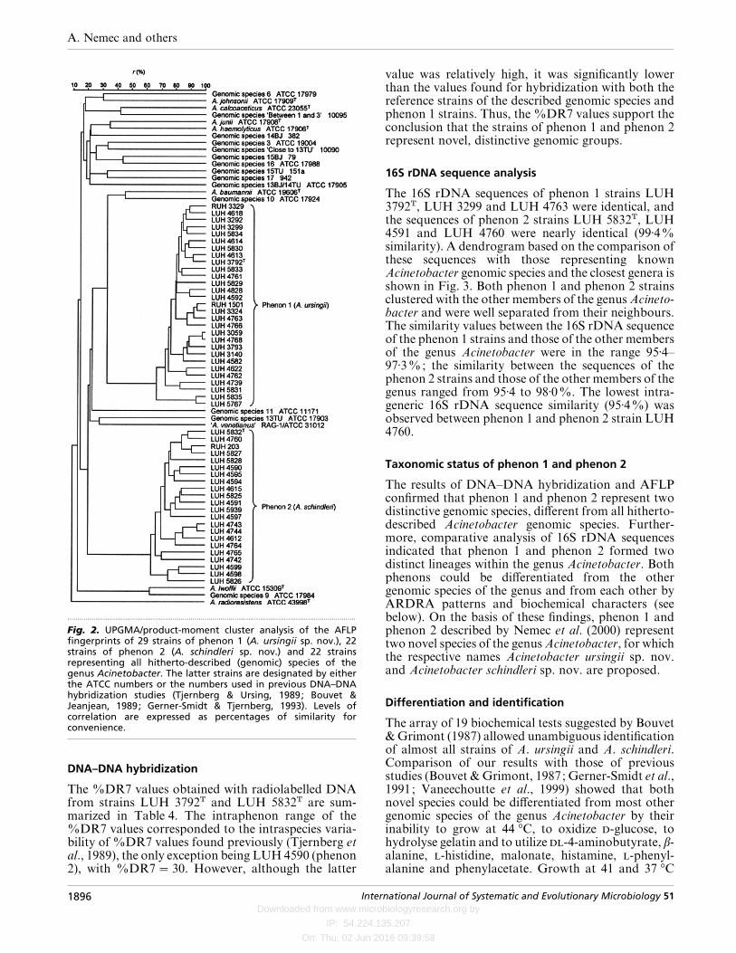
Reproducibility of AFLP as determined by testingseveral control strains was always higher than 90%(data not shown). Cluster analysis of the phenon 1 and2 strains was performed together with a total of 200strains from all described Acinetobacter genomicspecies (identified by DNA–DNA hybridization). Thestrains of each of the described genomic species formeda separate cluster at a cut-off level of about 50% (datanot shown). Clustering of all phenon 1 and phenon 2strains and one representative strain of each describedAcinetobacter genomic species is shown in Fig. 2. Thestrains of phenon 1 and phenon 2 grouped in twoclusters at levels of 67 and 63% and were clearlyseparated from each other, and from all other strainsat 33 and 20%, respectively.
International Journal of Systematic and Evolutionary Microbiology 51 1895
Downloaded from www.microbiologyresearch.org by
IP: 54.224.135.207
On: Thu, 02 Jun 2016 09:39:58
A. Nemec and others
.................................................................................................................................................
Fig. 2. UPGMA/product-moment cluster analysis of the AFLPfingerprints of 29 strains of phenon 1 (A. ursingii sp. nov.), 22strains of phenon 2 (A. schindleri sp. nov.) and 22 strainsrepresenting all hitherto-described (genomic) species of thegenus Acinetobacter. The latter strains are designated by eitherthe ATCC numbers or the numbers used in previous DNA–DNAhybridization studies (Tjernberg & Ursing, 1989; Bouvet &Jeanjean, 1989; Gerner-Smidt & Tjernberg, 1993). Levels ofcorrelation are expressed as percentages of similarity forconvenience.
DNA–DNA hybridization
The %DR7 values obtained with radiolabelled DNAfrom strains LUH 3792T and LUH 5832T are sum-marized in Table 4. The intraphenon range of the%DR7 values corresponded to the intraspecies varia-bility of %DR7 values found previously (Tjernberg etal., 1989), the only exception being LUH 4590 (phenon2), with %DR7¯ 30. However, although the latter
value was relatively high, it was significantly lowerthan the values found for hybridization with both thereference strains of the described genomic species andphenon 1 strains. Thus, the %DR7 values support theconclusion that the strains of phenon 1 and phenon 2represent novel, distinctive genomic groups.
16S rDNA sequence analysis
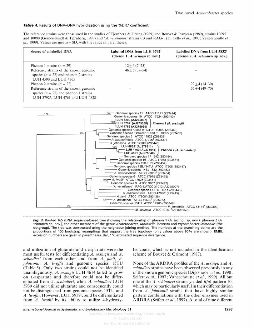
The 16S rDNA sequences of phenon 1 strains LUH3792T, LUH 3299 and LUH 4763 were identical, andthe sequences of phenon 2 strains LUH 5832T, LUH4591 and LUH 4760 were nearly identical (99±4%similarity). A dendrogram based on the comparison ofthese sequences with those representing knownAcinetobacter genomic species and the closest genera isshown in Fig. 3. Both phenon 1 and phenon 2 strainsclustered with the other members of the genus Acineto-bacter and were well separated from their neighbours.The similarity values between the 16S rDNA sequenceof the phenon 1 strains and those of the other membersof the genus Acinetobacter were in the range 95±4–97±3%; the similarity between the sequences of thephenon 2 strains and those of the other members of thegenus ranged from 95±4 to 98±0%. The lowest intra-generic 16S rDNA sequence similarity (95±4%) wasobserved between phenon 1 and phenon 2 strain LUH4760.
Taxonomic status of phenon 1 and phenon 2
The results of DNA–DNA hybridization and AFLPconfirmed that phenon 1 and phenon 2 represent twodistinctive genomic species, different from all hitherto-described Acinetobacter genomic species. Further-more, comparative analysis of 16S rDNA sequencesindicated that phenon 1 and phenon 2 formed twodistinct lineages within the genus Acinetobacter. Bothphenons could be differentiated from the othergenomic species of the genus and from each other byARDRA patterns and biochemical characters (seebelow). On the basis of these findings, phenon 1 andphenon 2 described by Nemec et al. (2000) representtwo novel species of the genus Acinetobacter, for whichthe respective names Acinetobacter ursingii sp. nov.and Acinetobacter schindleri sp. nov. are proposed.
Differentiation and identification
The array of 19 biochemical tests suggested by Bouvet& Grimont (1987) allowed unambiguous identificationof almost all strains of A. ursingii and A. schindleri.Comparison of our results with those of previousstudies (Bouvet & Grimont, 1987; Gerner-Smidt et al.,1991; Vaneechoutte et al., 1999) showed that bothnovel species could be differentiated from most othergenomic species of the genus Acinetobacter by theirinability to grow at 44 °C, to oxidize -glucose, tohydrolyse gelatin and to utilize -4-aminobutyrate, β-alanine, -histidine, malonate, histamine, -phenyl-alanine and phenylacetate. Growth at 41 and 37 °C
1896 International Journal of Systematic and Evolutionary Microbiology 51
Downloaded from www.microbiologyresearch.org by
IP: 54.224.135.207
On: Thu, 02 Jun 2016 09:39:58
Two novel Acinetobacter species
Table 4. Results of DNA–DNA hybridization using the %DR7 coefficient.................................................................................................................................................................................................................................................................................................................
The reference strains were those used in the studies of Tjernberg & Ursing (1989) and Bouvet & Jeanjean (1989), strains 10095and 10090 (Gerner-Smidt & Tjernberg, 1993) and ‘A. venetianus ’ strains C3 and RAG-1 (Di Cello et al., 1997; Vaneechoutte etal., 1999). Values are means³SD, with the range in parentheses.
Source of unlabelled DNA Labelled DNA from LUH 3792T
(phenon 1, A. ursingii sp. nov.)
Labelled DNA from LUH 5832T
(phenon 2, A. schindleri sp. nov.)
Phenon 1 strains (n¯ 29) 12³4 (7–25) –
Reference strains of the known genomic
species (n¯ 22) and phenon 2 strains
LUH 4590 and LUH 4765
46³5 (37–54) –
Phenon 2 strains (n¯ 22) – 22³4 (14–30)
Reference strains of the known genomic
species (n¯ 22) and phenon 1 strains
LUH 3792T, LUH 4761 and LUH 4828
– 57³4 (49–70)
.................................................................................................................................................................................................................................................................................................................
Fig. 3. Rooted 16S rDNA sequence-based tree showing the relationship of phenon 1 (A. ursingii sp. nov.), phenon 2 (A.schindleri sp. nov.), the other members of the genus Acinetobacter, Moraxella lacunata and Psychrobacter immobilis (theoutgroup). The tree was constructed using the neighbour-joining method. The numbers at the branching points are theproportions of 100 bootstrap resamplings that support the tree topology (only values above 90% are shown). EMBLaccession numbers are given in parentheses. Bar, 1% estimated sequence divergence.
and utilization of glutarate and -aspartate were themost useful tests for differentiating A. ursingii and A.schindleri from each other and from A. junii, A.johnsonii, A. lwoffii and genomic species 15TU(Table 5). Only two strains could not be identifiedunambiguously; A. ursingii LUH 4614 failed to growon -aspartate and therefore could not be differ-entiated from A. schindleri, while A. schindleri LUH5939 did not utilize glutarate and consequently couldnot be distinguished from genomic species 15TU andA. lwoffii. However, LUH 5939 could be differentiatedfrom A. lwoffii by its ability to utilize 4-hydroxy-
benzoate, which is not included in the identificationscheme of Bouvet & Grimont (1987).
None of the ARDRA profiles of the A. ursingii and A.schindleri strains have been observed previously in anyof the known genomic species (Dijkshoorn et al., 1998;Seifert et al., 1997; Vaneechoutte et al., 1999). All butone of the A. schindleri strains yielded BfaI pattern 10,whichmay be particularly useful in their differentiationfrom A. johnsonii strains that have highly similarpattern combinations with the other enzymes used inARDRA (Seifert et al., 1997). A total of nine different
International Journal of Systematic and Evolutionary Microbiology 51 1897
Downloaded from www.microbiologyresearch.org by
IP: 54.224.135.207
On: Thu, 02 Jun 2016 09:39:58
A. Nemec and others
Table 5. Phenotypic characters useful for discrimination of A. ursingii and A. schindleri and for their differentiationfrom phenotypically similar (genomic) species.................................................................................................................................................................................................................................................................................................................
Data for A. junii, A. johnsonii, A. lwoffii and genomic species 15TU were taken from Gerner-Smidt et al. (1991). , Positive for90–100% of strains ; ®, positive for 0–10% of strains ; , positive for 11–89% of strains.
Characteristic A. ursingii A. schindleri A. junii A. johnsonii A. lwoffii Genomic species 15TU
Growth at 41 °C ® ® ®
Growth at 37 °C ® Utilization of:
Glutarate ® ® ® ®-Aspartate ® ® ® ®
ARDRA profiles were encountered among the A.ursingii strains. In spite of this variability, severalpattern combinations may be useful for the identi-fication of A. ursingii, e.g. the combination of CfoI 1,MboI 3 or MboI 13 and MspI 3 or the combinationof RsaI 4 or RsaI 5 or RsaI 45 or RsaI 25 andMspI 3.
Clinical importance
The available clinical and epidemiological data suggestthat A. ursingii and A. schindleri differ in theirdistribution in patients. While the majority of the A.schindleri strains were isolated from non-sterile bodysites of outpatients, A. ursingii comprised mainlyclinically significant isolates from seriously ill hospital-ized patients. Almost half of the A. ursingii strains wereisolated from blood cultures and at least some of themwere recovered from patients with diagnosed bac-teraemia or septicaemia (Bernards et al., 1997; Horre-vorts et al., 1995; Nemec et al., 2000). Moreover, theidentity of typing characters that was found in twoepidemiologically related isolates (Nemec et al., 2000)indicates that A. ursingii strains have the potential tospread among patients.
Description of Acinetobacter ursingii sp. nov.
Acinetobacter ursingii (ur.sin«gi.i. N.L. gen. masc. n.ursingii in honour of Jan Ursing, the recently deceasedSwedish bacteriologist and taxonomist).
Characteristics correspond to those of the genus (Juni,1984). Colonies on nutrient agar after 24 h incubationat 30 °C are approximately 1±0–1±5 mm in diameter,circular, convex, smooth and slightly opaque withentire margins. Growth occurs at 37 °C but not at41 °C. Acid is not produced from -glucose, sheepblood is not haemolysed and gelatin is not hydrolysed.-Lactate, citrate (Simmons), azelate, -malate, etha-nol and acetate are utilized as sole sources of carbonand energy. Glutarate, -aspartate and 4-hydroxy-benzoate are utilized by most strains. -4-Amino-butyrate, β-alanine, -histidine, malonate, histamine,-phenylalanine, phenylacetate, laevulinate, citracon-ate, -tartrate, -leucine and 2,3-butanediol are notutilized.
The type strain is LUH 3792T (¯NIPH 137T¯LMG19575T¯CNCTC 6735T), isolated from blood of ahospitalized patient with endocarditis. This strainutilizes glutarate, -aspartate and 4-hydroxybenzoate.The restriction patterns of amplified 16S rDNA of thetype strain are CfoI 1, AluI 4, MboI 3, RsaI 5, MspI 3.The EMBL accession number for its 16S rDNAsequence is AJ275038.
Description of Acinetobacter schindleri sp. nov.
Acinetobacter schindleri (schin«dle.ri. N.L. gen. masc.n. schindleri in honour of Jir3 ı! Schindler, Czech micro-biologist and taxonomist).
Characteristics correspond to those of the genus (Juni,1984). Colonies on nutrient agar after 24 h incubationat 30 °C are approximately 1±5–2±5 mm in diameter,circular, convex, smooth and slightly opaque withentire margins. Growth occurs at 41 °C but not at44 °C. Acid is not produced from -glucose, sheepblood is not haemolysed and gelatin is not hydrolysed.-Lactate and acetate are utilized as sole sources ofcarbon and energy. Glutarate, -malate and ethanolare utilized by most strains. Various numbers of strainsutilize citrate (Simmons), azelate, 4-hydroxybenzoate,-tartrate and 2,3-butanediol. -4-Aminobutyrate, -aspartate, β-alanine, -histidine, malonate, histamine,-phenylalanine, phenylacetate, laevulinate, citracon-ate and -leucine are not utilized.
The type strain is LUH 5832T (¯NIPH 1034T¯LMG 19576T¯CNCTC 6736T), isolated from urineof a male outpatient with cystitis. This strain utilizescitrate (Simmons), glutarate, -malate, 4-hydroxy-benzoate and ethanol but not azelate, -tartrate or 2,4-butanediol. The restriction patterns of the amplified16S rDNA of the type strain are CfoI 15, AluI 24,MboI 1, RsaI 2, MspI 2, BfaI 10. The EMBL accessionnumber for its 16S rDNA sequence is AJ278311.
ACKNOWLEDGEMENTS
We thank Dr A. T. Bernards (Leiden University MedicalCenter), Dr P. J. M. Bouvet (Institut Pasteur, Paris), Pro-fessor D. A. Caugant (National Institute of Public Health,Oslo), Dr P. Gerner-Smidt (Statens Seruminstitut, Copen-hagen), Dr A. M. Horrevorts (University Hospital,
1898 International Journal of Systematic and Evolutionary Microbiology 51
Downloaded from www.microbiologyresearch.org by
IP: 54.224.135.207
On: Thu, 02 Jun 2016 09:39:58
Two novel Acinetobacter species
Nijmegen), Dr P. Jez) ek (General Hospital, Pr3 ı!bram) and DrJ. G. M. Koeleman (University Hospital Vrije Universiteit,Amsterdam) for generous provision of strains. We alsothank Dr H. G. Tru$ per for his help with nomenclature. Thisstudy was partially supported by research grant no.310}98}1602 of the Grant Agency of the Czech Republicawarded to A.N. Part of this work was presented at the 5thInternational Symposium on the Biology of Acinetobacter,Noordwijkerhout, The Netherlands, 2000 (abstract 22).
REFERENCES
Bernards, A. T., de Beaufort, A. J., Dijkshoorn, L. & van Boven,C. P. A. (1997). Outbreak of septicaemia in neonates caused byAcinetobacter junii investigated by amplified ribosomal DNArestriction analysis (ARDRA) and four typing methods. J HospInfect 35, 129–140.
Boom, R., Sol, C. J. A., Salimans, M. M. M., Jansen, C. L.,Wertheim-van Dillen, P. M. E. & van der Noordaa, J. (1990).Rapid and simple method for purification of nucleic acids.J Clin Microbiol 28, 495–503.
Bouvet, P. J. M. & Grimont, P. A. D. (1986). Taxonomy of thegenus Acinetobacter with the recognition of Acinetobacterbaumannii sp. nov., Acinetobacter haemolyticus sp. nov.,
Acinetobacter johnsonii sp. nov., and Acinetobacter junii sp. nov.and emended descriptions of Acinetobacter calcoaceticus andAcinetobacter lwoffii. Int J Syst Bacteriol 36, 228–240.
Bouvet, P. J. M. & Grimont, P. A. D. (1987). Identification andbiotyping of clinical isolates of Acinetobacter. Ann Inst PasteurMicrobiol 138, 569–578.
Bouvet, P. J. M. & Jeanjean, S. (1989). Delineation of newproteolytic genomic species in the genus Acinetobacter. ResMicrobiol 140, 291–299.
Di Cello, F., Pepi, M., Baldi, F. & Fani, R. (1997). Molecularcharacterization of an n-alkane-degrading bacterial communityand identification of a new species, Acinetobacter venetianus.
Res Microbiol 148, 237–249.
Dijkshoorn, L., van Harsselaar, B., Tjernberg, I., Bouvet, P. J. M. &Vaneechoutte, M. (1998). Evaluation of amplified ribosomalDNA restriction analysis for identification of Acinetobactergenomic species. Syst Appl Microbiol 21, 33–39.
Gerner-Smidt, P. & Tjernberg, I. (1993). Acinetobacter in
Denmark: II. Molecular studies of the Acinetobacter calco-aceticus–Acinetobacter baumannii complex. APMIS 101,826–832.
Gerner-Smidt, P., Tjernberg, I. & Ursing, J. (1991). Reliability ofphenotypic tests for identification of Acinetobacter species.J Clin Microbiol 29, 277–282.
Horrevorts, A., Bergman, K., Kolle! e, L., Breuker, I., Tjernberg, I. &Dijkshoorn, L. (1995). Clinical and epidemiological inves-
tigations of Acinetobacter genomospecies 3 in a neonatalintensive care unit. J Clin Microbiol 33, 1567–1572.
Ibrahim, A., Gerner-Smidt, P. & Liesack, W. (1997). Phylogeneticrelationship of the twenty-one DNA groups of the genusAcinetobacter as revealed by 16S ribosomal DNA sequenceanalysis. Int J Syst Bacteriol 47, 837–841.
Janssen, P., Maquelin, K., Coopman, R., Tjernberg, I., Bouvet, P.,Kersters, K. & Dijkshoorn, L. (1997). Discrimination of Acineto-bacter genomic species by AFLP fingerprinting. Int J SystBacteriol 47, 1179–1187.
Juni, E. (1972). Interspecies transformation of Acinetobacter :genetic evidence for a ubiquitous genus. J Bacteriol 112,917–931.
Juni, E. (1984). Genus III. Acinetobacter Brisou and Pre! vot 1954,727AL. In Bergey’s Manual of Systematic Bacteriology, vol. 1,pp. 303–307. Edited by N. R. Krieg & J. G. Holt. Baltimore:Williams & Wilkins.
King, E. O., Ward, W. K. & Raney, D. E. (1954). Two simple mediafor the demonstration of pyocyanin and fluorescein. J Lab ClinMed 44, 301–307.
Koeleman, J. G. M., Stoof, J., Biesmans, D. J., Savelkoul, P. H. &Vandenbroucke-Grauls, C. M. J. E. (1998). Comparison of ampli-fied ribosomal DNA restriction analysis, random amplifiedpolymorphic DNA analysis, and amplified fragment lengthpolymorphism fingerprinting for identification of Acinetobactergenomic species and typing of Acinetobacter baumannii. J ClinMicrobiol 36, 2522–2529.
Nemec, A., Dijkshoorn, L. & Jez) ek, P. (2000). Recognition of twonovel phenons of the genus Acinetobacter among non-glucose-acidifying isolates from human specimens. J Clin Microbiol 38,3937–3941.
Seifert, H., Dijkshoorn, L., Gerner-Smidt, P., Pelzer, N., Tjernberg,I. & Vaneechoutte, M. (1997). Distribution of Acinetobacterspecies on human skin: comparison of phenotypic and geno-typic identification methods. J Clin Microbiol 35, 2819–2825.
Tjernberg, I. & Ursing, J. (1989). Clinical strains of Acinetobacterclassified by DNA–DNA hybridization. APMIS 97, 595–605.
Tjernberg, I., Lindh, E. & Ursing, J. (1989). A quantitative bacterialdot method for DNA–DNA hybridization and its correlation tothe hydroxyapatite method. Curr Microbiol 18, 77–81.
Vaneechoutte, M., Tjernberg, I., Baldi, F., Pepi, M., Fani, R.,Sullivan, E. R., van der Toorn, J. & Dijkshoorn, L. (1999). Oil-degrading Acinetobacter strain RAG-1 and strains described as‘Acinetobacter venetianus sp. nov. ’ belong to the same genomicspecies. Res Microbiol 150, 69–73.
Vaneechoutte, M., Claeys, G., Steyaert, S., De Baere, T., Peleman,R. & Verschraegen, G. (2000). Isolation of Moraxella canis froman ulcerated metastatic lymph node. J Clin Microbiol 38,3870–3871.
Vos, P., Hogers, R., Bleeker, M. & 8 other authors (1995). AFLP:a new technique for DNA fingerprinting. Nucleic Acids Res 23,4407–4414.
International Journal of Systematic and Evolutionary Microbiology 51 1899