protein plasticity to the extreme: changing the topology of a 4-α-helical bundle with a single...
TRANSCRIPT
Protein plasticity to the extreme: changing the topology of a4-αα-helical bundle with a single amino acid substitutionNicholas M Glykos1, Gianni Cesareni2 and Michael Kokkinidis1,3*
Background: Conventional wisdom has it that two proteins sharing 98.4%sequence identity have nearly identical three-dimensional structures. Here weprovide a counter-example to this statement by showing that a single aminoacid substitution can change the topology of a homodimeric 4-α-helicalbundle protein.
Results: We have determined the high-resolution crystal structure of a4-α-helical protein with a single alanine to proline mutation in the turn region,and show that this single amino acid substitution leads to a completereorganisation of the whole molecule. The protein is converted from thecanonical left-handed all-antiparallel form, to a right-handed mixed parallel andantiparallel bundle, which to the best of our knowledge and belief represents anovel topological motif for this class of proteins.
Conclusions: The results suggest a possible new mechanism for the creationand evolution of topological motifs, show the importance of loop regions indetermining the allowable folding pathways, and illustrate the malleability ofprotein structures.
IntroductionThe Rop (repressor of primer) protein is a homodimericRNA-binding protein involved in the regulation of thecopy number of the ColE1 plasmid [1]. Rop is the para-digm of a canonical 4-α-helical bundle [2] and, as such,has been the subject of numerous investigations rangingin their approach from structural [3–6] and biochemical[7,8], to thermodynamical [9–11] and computational [12]studies. The bend region of Rop has attracted addedinterest, not least because of the ongoing debate aboutthe role of loops in the folding and stability of bundlesand proteins in general [9,13–18]. Although randommutagenesis experiments suggested that most singleamino acid substitutions in the bend region of Rop canbe tolerated by the native structure [16], one of the designed site-directed mutants — the Ala31→Pro(A31P) mutant — showed consistent and persistent devi-ations from the thermodynamical and biochemical prop-erties of the wild-type protein [7,16,18]. The crucial roleof Ala31 in the formation of the turn region of wild-typeRop was first recognised in the original structure deter-mination of the protein [3]. This residue not only hasunusual geometry — as judged from its φ,ψ angles, andthe exceptional deviation of its peptide unit from pla-narity (ω = 194.3°) — but is also unique in being the onlyamino acid that simultaneously forms hydrogen bonds toboth helices [3,18].
We designed the A31P mutant in the hope that the con-formationally constrained proline could not mimic alaninein its role as the central residue of the turn, and wouldthus result in a structure with a partly unfolded turn. Thiswould allow us to tackle a long standing, and controver-sial, issue regarding the relative contribution of turn–helixinteractions to the overall stability of the bundle[9,13,15,16].
Instead of providing an answer to this problem, the crystalstructure of the A31P mutant answered the fundamentallydifferent, and as yet unasked, question of how manymutations are needed to change the topology of a protein(which, as it happens, is a generalisation of the ‘Paracelsuschallenge’ [19,20]). In the following paragraphs wecompare the topologies and tertiary structures of the wild-type and mutant Rop proteins, describe in some detail thestructure of the turn and the mode of hydrophobic corepacking, and discuss the implications of the A31P crystalstructure with respect to the thermodynamical [18] andfunctional [7] data available for this mutant.
ResultsTopological changesA summary of the structure determination procedure forthe Rop A31P mutant is given in the Materials andmethods section. The electron-density maps in Figure 1
Addresses: 1Foundation for Research andTechnology-Hellas, Institute of Molecular Biologyand Biotechnology, PO Box 1527, 71110Heraklion, Crete, Greece, 2Department of Biology,University of Rome Tor Vergata, Via della RicercaScientifica, 00133 Rome, Italy and 3Department ofBiology, University of Crete, PO Box 2208, 71409Heraklion, Crete, Greece.
*Corresponding author.E-mail: [email protected]
Key words: bundle, fold, mutagenesis, Rop, topology
Received: 18 January 1999Revisions requested: 15 February 1999Revisions received: 1 March 1999Accepted: 4 March 1999
Published: 18 May 1999
Structure June 1999, 7:597–603http://biomednet.com/elecref/0969212600700597
© Elsevier Science Ltd ISSN 0969-2126
Research Article 597
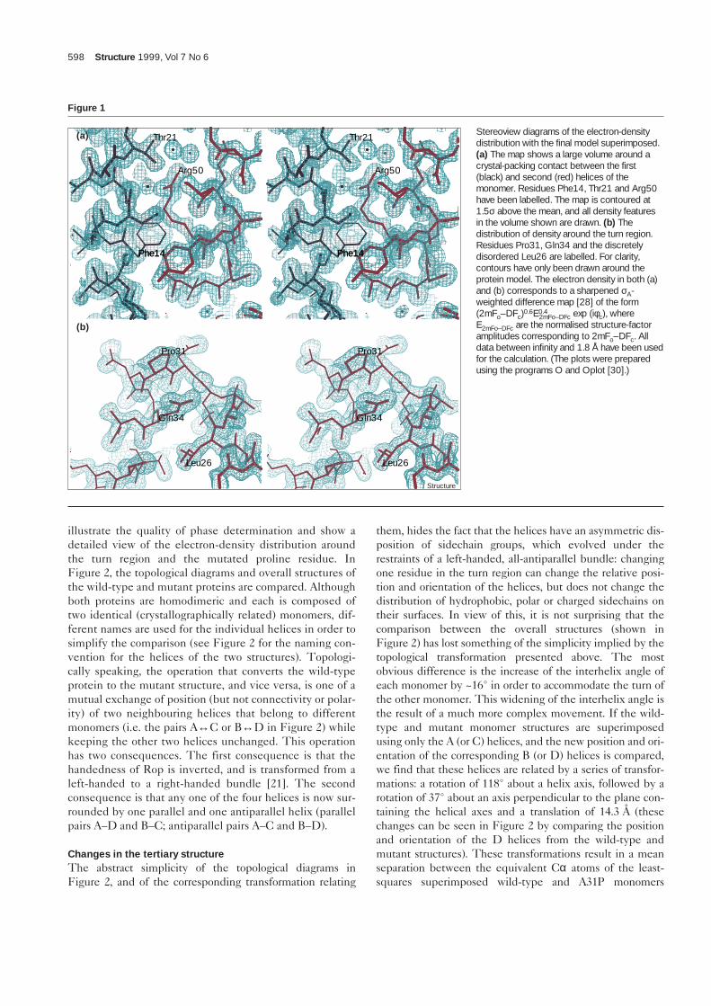
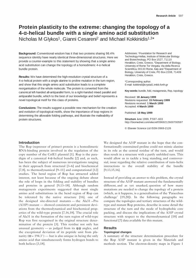
illustrate the quality of phase determination and show adetailed view of the electron-density distribution aroundthe turn region and the mutated proline residue. InFigure 2, the topological diagrams and overall structures ofthe wild-type and mutant proteins are compared. Althoughboth proteins are homodimeric and each is composed oftwo identical (crystallographically related) monomers, dif-ferent names are used for the individual helices in order tosimplify the comparison (see Figure 2 for the naming con-vention for the helices of the two structures). Topologi-cally speaking, the operation that converts the wild-typeprotein to the mutant structure, and vice versa, is one of amutual exchange of position (but not connectivity or polar-ity) of two neighbouring helices that belong to differentmonomers (i.e. the pairs A↔C or B↔D in Figure 2) whilekeeping the other two helices unchanged. This operationhas two consequences. The first consequence is that thehandedness of Rop is inverted, and is transformed from aleft-handed to a right-handed bundle [21]. The secondconsequence is that any one of the four helices is now sur-rounded by one parallel and one antiparallel helix (parallelpairs A–D and B–C; antiparallel pairs A–C and B–D).
Changes in the tertiary structureThe abstract simplicity of the topological diagrams inFigure 2, and of the corresponding transformation relating
them, hides the fact that the helices have an asymmetric dis-position of sidechain groups, which evolved under therestraints of a left-handed, all-antiparallel bundle: changingone residue in the turn region can change the relative posi-tion and orientation of the helices, but does not change thedistribution of hydrophobic, polar or charged sidechains ontheir surfaces. In view of this, it is not surprising that thecomparison between the overall structures (shown inFigure 2) has lost something of the simplicity implied by thetopological transformation presented above. The mostobvious difference is the increase of the interhelix angle ofeach monomer by ~16° in order to accommodate the turn ofthe other monomer. This widening of the interhelix angle isthe result of a much more complex movement. If the wild-type and mutant monomer structures are superimposedusing only the A (or C) helices, and the new position and ori-entation of the corresponding B (or D) helices is compared,we find that these helices are related by a series of transfor-mations: a rotation of 118° about a helix axis, followed by arotation of 37° about an axis perpendicular to the plane con-taining the helical axes and a translation of 14.3 Å (thesechanges can be seen in Figure 2 by comparing the positionand orientation of the D helices from the wild-type andmutant structures). These transformations result in a meanseparation between the equivalent Cα atoms of the least-squares superimposed wild-type and A31P monomers
598 Structure 1999, Vol 7 No 6
Figure 1
Stereoview diagrams of the electron-densitydistribution with the final model superimposed.(a) The map shows a large volume around acrystal-packing contact between the first(black) and second (red) helices of themonomer. Residues Phe14, Thr21 and Arg50have been labelled. The map is contoured at1.5σ above the mean, and all density featuresin the volume shown are drawn. (b) Thedistribution of density around the turn region.Residues Pro31, Gln34 and the discretelydisordered Leu26 are labelled. For clarity,contours have only been drawn around theprotein model. The electron density in both (a)and (b) corresponds to a sharpened σA-weighted difference map [28] of the form(2mFo–DFc)0.6E0.4
2mFo–DFc exp (iφc), whereE2mFo–DFc are the normalised structure-factoramplitudes corresponding to 2mFo–DFc. Alldata between infinity and 1.8 Å have been usedfor the calculation. (The plots were preparedusing the programs O and Oplot [30].)
Gln34 Gln34
Leu26 Leu26
Pro31 Pro31
Phe14Phe14 Phe14Phe14
Arg50 Arg50
Thr21 Thr21(a)
(b)
Structure
(residues 1–56) of ∆ = 4.55 Å, with a standard deviation (σ∆)of 5.05 Å and a maximum separation (max∆) of 9.66 Å (as cal-culated using the program LSQKAB [22]). The secondimportant difference between the overall structures of thetwo proteins is that A31P is no longer a left-handed four-stranded coiled coil. The curvature and relative orientationof the pairs of helices C–B and A–D would suggest thatA31P might be a left-handed coil of two right-handed two-stranded coiled coils. However, examination of the meanhelix–helix distances (D–B 9.5 Å, A–C 13.5 Å, A–D and C–B10.6 Å), of the orientation angles [23] (D–B –169°, A–C–173°, C–B and A–D –8°; diagonal pairs C–D and A–B+175°) and of the packing interactions between adjacenthelices, suggests that only helices D and B might indeedform a two-stranded coiled coil with a left-handed twist. Forthe other pairs of helices no conclusions can safely be drawn.
The bend and the helicesTurning our attention to the bend region and the individ-ual helices, we note that the length and location of the
turn has changed: in wild-type Rop the bend comprisesthree residues (Leu29, Asp30 and Ala31), whereas in theA31P structure the turn starts at Asn27 and finishes withAsp30. As shown in Figure 1b, the mutated proline is inthe trans isomer and is not directly involved in the turnformation. Instead, it has the role of N-cap for the secondhelix of the monomer (helices B and D; Figure 2). Twomajor geometric changes are responsible for the differentrelative position and orientation of the helices of eachmonomer in the mutant and wild-type structures: a changeof the ψ angle of Leu29 by 169°; and a change of the φ andψ angles of Asp30 by 131° and 119°, respectively.Although a comparison between the structures of the indi-vidual helices (helix A or B from wild type with helix A orB from A31P) shows less impressive differences(∆ = 0.70 Å, σ∆ = 1.16 Å, max∆ = 5.28 Å for all equivalentatoms of residues 1–26; ∆ = 1.57 Å, σ∆ = 2.16 Å,max∆ = 9.85 Å for all equivalent atoms of residues 32–56)it is still somewhat surprising to observe a standard devia-tion in the atomic positions of the order of 2 Å for two
Research Article Changing the topology of a 4-αα-helical bundle Glykos, Cesareni and Kokkinidis 599
Figure 2
Comparative schematic diagrams of thetopology and overall structure (in stereo) of(a) wild-type Rop and (b) the A31P mutant.The helices of each monomer are shown inred and yellow and the connective strands arein blue. The line thickness in the topologicaldiagrams is inversely proportional to thedistance from the viewer. The position of theintramolecular (crystallographic) dyad axis isnoted both in the topological diagrams andthe structure schematics. The orientations ofthe wild-type and mutant structures are suchthat the N-terminal helices of the redmonomers (C helices) are in exactly the sameposition and orientation in both diagrams. TheN and C termini as well as the helix namingconvention adopted are also noted. (Thestructure schematics were prepared using theprogram BOBSCRIPT [31].)
C
C
N
N
C
C
N
N
C
C
N
N
C
C
N
N
A
B
C
D
A
D
C
B
(a)
(b)
Structure
identical sequences with the same secondary structure.This, we believe, shows how the detailed atomic arrange-ment of even a relatively rigid structural element, such asan α helix, is influenced — if not determined — by itsenvironment. (It is worth noting that the observed differ-ences do not arise solely from the sidechain atoms. Whenonly the mainchain atoms are used for the least-squaressuperposition, we still observe a max∆ = 1.88 Å for the firsthelix and a max∆ = 3.37 Å for the second).
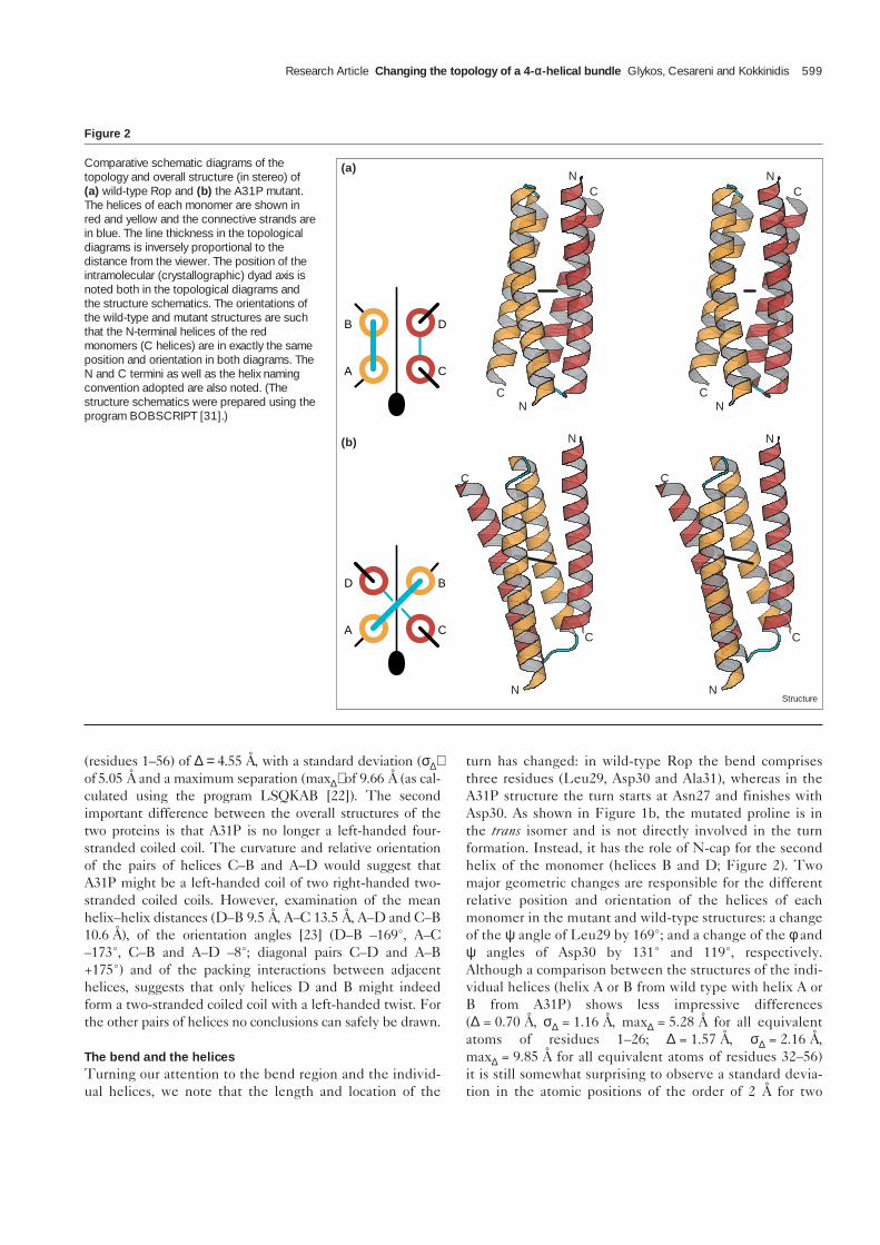
A new hydrophobic coreIn view of all these structural changes, it comes as no sur-prise that the hydrophobic core of the A31P mutant ispacked anew. The hydrophobic interactions in the centralpart of the wild-type and mutant structures are comparedin Figure 3. It is immediately obvious from this compari-son that at the atomic level the two proteins diverge somuch that it makes more sense to note similarities insteadof differences. In this spirit, we note that remnants of thelayered structure of the hydrophobic core, as described forthe wild-type protein [3], are still present in the mutantstructure. In the case of A31P, however, there are only six(instead of eight) such layers and of these the two outerones (closest to the turns) consist of clusters of only threeamino acids each, and can probably be called layers onlyfor reasons of consistency with the native protein. Further-more, the planes of these layers are highly tilted withrespect to the bundle axis (instead of being approximatelynormal to it, as is the case for the wild-type protein) and
their tilt angle increases proportionally to their separationfrom the intramolecular dyad axis. In the case of the wild-type protein, these hydrophobic layers consist exclusivelyof amino acids that occupy specific (and constant) posi-tions with respect to the heptad sequence periodicitycharacterising associating α helices, and they all have theform adad, where a and d are the generally apolar positionsof the repeat [2,3] (the other five positions of the heptadrepeat b, c, e, f and g are generally occupied by polarresidues). Although A31P shares exactly the same period-icity — or lack of it in the turn region — the distributionof amino acid types in its layers is totally different. Forexample, the two central layers closest to, and related by,the dyad axis (shown in Figure 3) have — in accordancewith the wild-type heptad assignments — the form dddd.The next two layers are even more divergent in composi-tion: each consists of five residues (Met11, Ile37, Cys38,Cys52 and Leu26) and have the form ggaaa. Finally, thetwo outermost layers comprise Ile37 (only the Cδ atoms,see below), Leu29 and Ala8 and have the form gdd. Twoother features of the A31P structure are worth noting. Thefirst is the presence of a large continuous internal cavity(with a volume [24] of ~270 Å3 for a probe with a radius of1.4 Å), which is located around the dyad axis and is sur-rounded by the first pair of hydrophobic layers (the pres-ence of the cavity can also be inferred from Figure 3b).The second feature is the large number (five permonomer) of internal hydrophobic sidechains for whichthe electron-density maps suggested the presence of at
600 Structure 1999, Vol 7 No 6
Figure 3
Comparative all-atom stereoview diagrams of acentral slice from (a) the wild-type Rop and (b)the A31P mutant structures. Theintramolecular dyad axis is parallel to the planeof the paper and the structures are oriented asshown in the topological diagrams of Figure 2.The residues forming the first hydrophobiclayer in the two structures are labelled, as areHis42 and His44. In wild-type Rop, residuesLeu22 and Leu48 belong to the secondhydrophobic layer and for clarity have not beendrawn. For residues with alternative sidechainconformations (see text), only the majorconformer is depicted. (The figure wasprepared using the program RASMOL [22].)
least one alternative conformation. Of these fivesidechains, three belong to the first hydrophobic layer andtheir mobility can be attributed to the fact that they are indirect contact with the cavity mentioned above. Of theremaining two, one is the sidechain of Ile37, whichbridges and participates in both the second and thirdhydrophobic layers and its disorder may be structurallysignificant, and the second is Leu26, which directly con-tacts Ile37 in the second hydrophobic layer.
Thermodynamic dataThe crystal structure of A31P agrees with and qualitativelyexplains the available thermodynamic and spectroscopicdata available for this mutant [18]. The observed destabili-sation of the mutant (∆∆G = 29 KJ/mole of dimer at 25°C)is explained quite adequately in terms of the reducednumber and density of the hydrophobic core packing inter-actions. The reduction of the helical content calculatedfrom the crystal structures (4%) agrees quite well with thevalue (7%) obtained from circular dichroism measurements.The interpretation — in terms of a diminished interhelicalinteraction — of the reduced ratio of the ellipticity values[Θ222nm]/[Θ208nm] and of the reduced transition enthalpy ofthe mutant [18], is in very good agreement with theincreased mean helix–helix distances. The conclusion thatthe observed thermodynamic and spectroscopic changes“... cannot be rationalized by the assumption of mere local-ized perturbations” [18] is fully supported by our results.
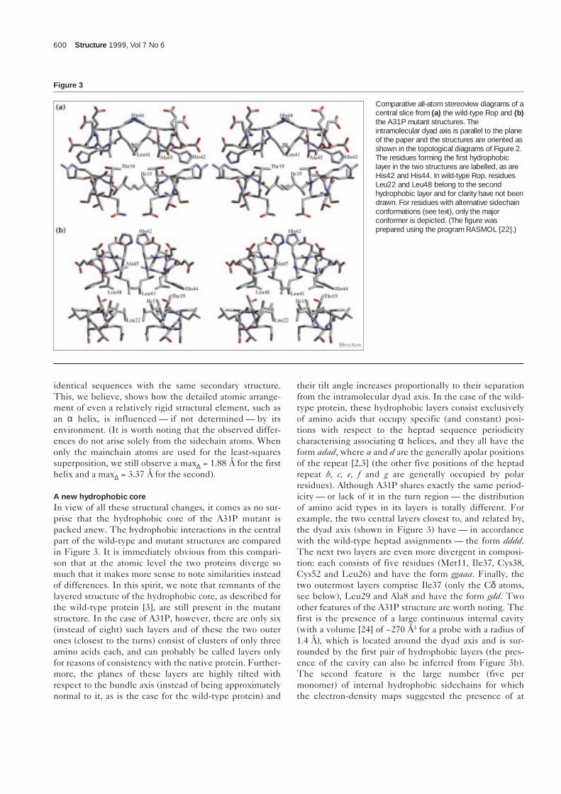
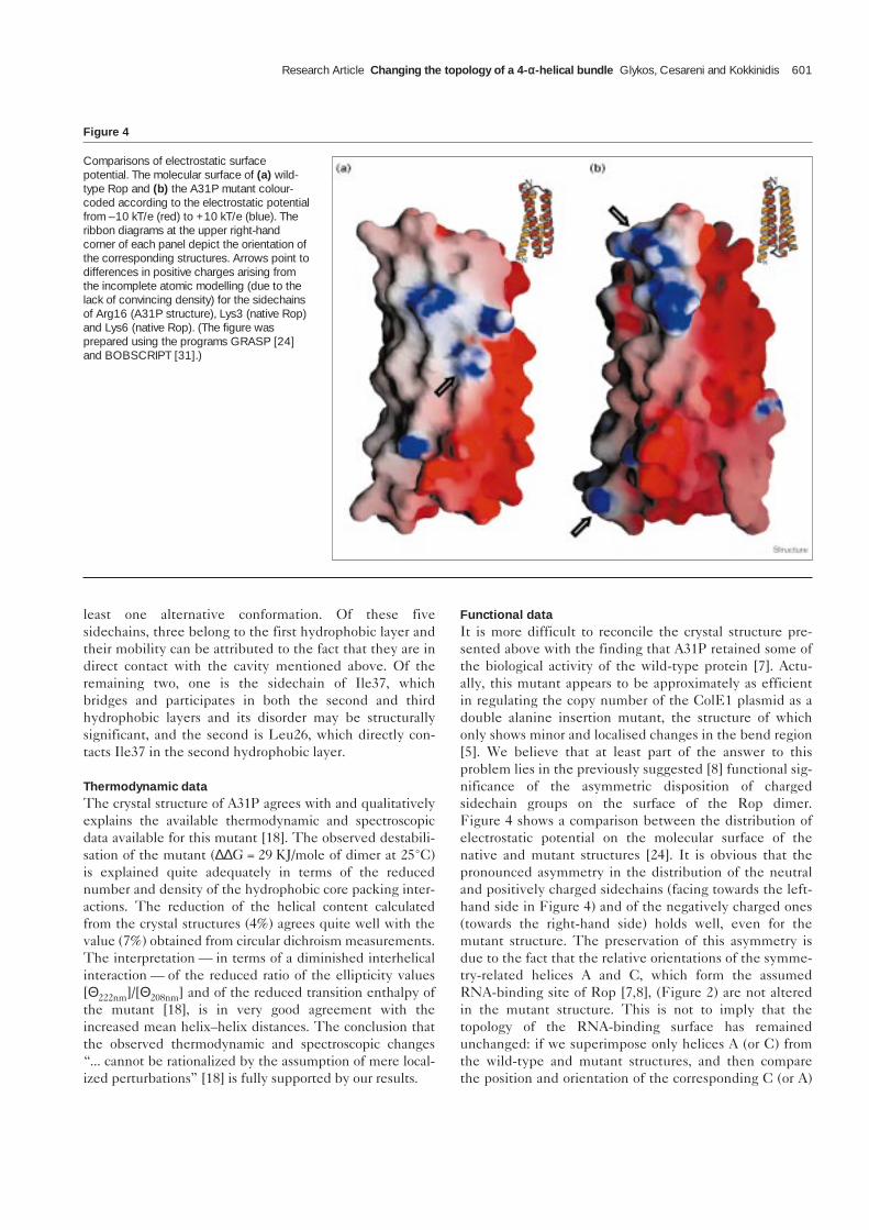
Functional dataIt is more difficult to reconcile the crystal structure pre-sented above with the finding that A31P retained some ofthe biological activity of the wild-type protein [7]. Actu-ally, this mutant appears to be approximately as efficientin regulating the copy number of the ColE1 plasmid as adouble alanine insertion mutant, the structure of whichonly shows minor and localised changes in the bend region[5]. We believe that at least part of the answer to thisproblem lies in the previously suggested [8] functional sig-nificance of the asymmetric disposition of chargedsidechain groups on the surface of the Rop dimer.Figure 4 shows a comparison between the distribution ofelectrostatic potential on the molecular surface of thenative and mutant structures [24]. It is obvious that thepronounced asymmetry in the distribution of the neutraland positively charged sidechains (facing towards the left-hand side in Figure 4) and of the negatively charged ones(towards the right-hand side) holds well, even for themutant structure. The preservation of this asymmetry isdue to the fact that the relative orientations of the symme-try-related helices A and C, which form the assumedRNA-binding site of Rop [7,8], (Figure 2) are not alteredin the mutant structure. This is not to imply that thetopology of the RNA-binding surface has remainedunchanged: if we superimpose only helices A (or C) fromthe wild-type and mutant structures, and then comparethe position and orientation of the corresponding C (or A)
Research Article Changing the topology of a 4-αα-helical bundle Glykos, Cesareni and Kokkinidis 601
Figure 4
Comparisons of electrostatic surfacepotential. The molecular surface of (a) wild-type Rop and (b) the A31P mutant colour-coded according to the electrostatic potentialfrom –10 kT/e (red) to +10 kT/e (blue). Theribbon diagrams at the upper right-handcorner of each panel depict the orientation ofthe corresponding structures. Arrows point todifferences in positive charges arising fromthe incomplete atomic modelling (due to thelack of convincing density) for the sidechainsof Arg16 (A31P structure), Lys3 (native Rop)and Lys6 (native Rop). (The figure wasprepared using the programs GRASP [24]and BOBSCRIPT [31].)
helices, we find that they are related by a rotation of ~35°followed by a translation of 4.8 Å (these changes can alsobe inferred from Figure 2). Even if the relative helix rota-tion is ignored, we find that the distance, for example,between the symmetry-related Cβ atoms of Phe14, aresidue known to be essential for RNA-binding [7,8],increases from 10.8 Å in wild type to 14.3 Å in the A31Pmutant. Given that four different Phe14 mutants (pheny-lalanine to alanine, leucine, tyrosine and tryptophan) allfailed to bind the wild-type RNA substrate [8], it is notclear how A31P, which displays such drastic structuralchanges, can still bind to it.
DiscussionIn summary, we have shown that a single amino acid sub-stitution is sufficient to change the topology of a smallprotein, leading to drastic changes both in its surface prop-erties and the packing of its hydrophobic core. Althoughthe stability of the resulting structure is significantlyreduced (compared with the wild type), the amount ofstructural differences observed between two proteinssharing 98.4% sequence identity, justifies our propositionthat “the remarkable thing is that it fold at all” [20].
The A31P crystal structure supports the view that turnsare not passive with respect to protein folding. Ourresults — and taking into account the experimentallydemonstrated insensitivity of this same protein to numer-ous other mutations in the same turn region [16] — wouldsuggest that the role of turns in protein folding is not oneof actively determining the fold, but one of activelyexcluding some of the otherwise possible folding path-ways. Clearly, when it is the major folding pathway that isbeing excluded, then whether or not the protein will foldwill depend on the existence (or otherwise) of anotherpermissible folding pathway leading to a stable molecule.This argument is valid even when inverted. It seems rea-sonable to suggest, for example, that the structure exhib-ited by the A31P mutant is also accessible by the nativeprotein — after all, the difference in terms of compositionbetween the two structures is only two atoms (the Cγ andCδ atoms of proline) and their bonds. If, in a thoughtexperiment, these two atoms were removed from theA31P structure, we would end up with a perfectly normal(with respect to its φ,ψ angles) alanine residue, and wouldhave thus returned to the wild-type Rop sequence, butfolded as observed in the A31P crystal structure. One ofthe reasons that the native molecule is not trapped in anA31P-like intermediate is because Ala31, but not Pro31, ispermissible to a folding pathway that leads to a thermo-dynamically more stable conformation.
It would appear at first sight that our results place doubtson one of the basic premises of modern molecular biology,that is, the sequence/structure/function equivalence. Wedo not think that this is the case: the creation of a new
topology places new restraints on the protein sequence,and these new restraints will eventually lead to sequencedivergence. Given enough time (and evolutionary events),the sequence of A31P, for example, would change toreflect the structural (and possibly functional) restraints ofits new topology (i.e., a right-handed, mixed parallel andantiparallel 4-α-helical bundle). As a consequence, theprotein would probably end up with a sequence having asmuch homology to the native Rop as we would presentlyexpect from two structurally similar, but different indetail, proteins.
Although tempting to suggest, we do not believe that thisabrupt way of generating a new topology corresponds to anevolutionarily frequent event. Not only does our generalexperience from mutagenesis studies shows that what weobserved is a rare occurrence, but it is also hard to imaginesuch drastic changes happening to a larger, more complexand/or non-multimeric protein. We do suggest, however,that such events may have been of some importance in theearly stages of molecular evolution.
Biological implicationsThe general experience from structural studies of mutantproteins with single amino acid substitutions is that theeffect of mutation is rather localised and minor. Wereport here an exception to this rule by showing that asingle alanine to proline substitution is sufficient forchanging the topology of a small protein. The mutationleads to drastic changes both in the surface properties ofthe protein and the packing of its hydrophobic core,while retaining some of the biological activity of the wild-type molecule. The results exemplify the complexities ofthe folding problem and show that the sequence/struc-ture/function equivalence should be treated with somecaution in the case of non-natural products. In addition,our observations suggest a possible new mechanism forthe creation and evolution of protein topologies, andunderline the importance of loop regions in determiningthe allowable folding pathways.
Materials and methodsThe expression, purification, crystallisation and preliminary crystallo-graphic characterisation of A31P has been reported previously [7,25].All crystallographic calculations were performed with the CCP4 suite ofprograms [22] and X-PLOR [26]. In summary, a 3.8 Å solvent-flattenedsingle isomorphous replacement (SIR) map (SOLOMON [22], 30%solvent content), based on a single-site platinum derivative showed theapproximate location of the helices and suggested that these may beparallel, but was otherwise uninterpretable. The structure was solvedwith a novel procedure (a detailed account of which will be publishedelsewhere) involving rigid-body simulated annealing (in X-PLOR) ofroughly positioned polyalanine models at a very high initial temperature(T0 = 10,000K) and with the geometric energy terms switched on. Theannealing procedure was iteratively repeated with successively smallerrigid bodies (down to two alanine residues per body), at successivelyhigher resolution (5, 4, 3 and 2 Å) and converged to R and Rfree values[27] of 0.403 and 0.413. These values were obtained for all databetween 8 Å and 1.8 Å from a 100% complete native data set collected
602 Structure 1999, Vol 7 No 6
on a CAD4 diffractometer (average F/σ(F) = 12.4 for all data, 2.51 forthe last resolution shell). A σA-weighted map [28] of the form(2mFo–DFAla52) exp (iφAla52) was readily interpretable in terms of theprotein sequence, and was further improved with the wARP procedure[29]. Sidechains were build using the program O [30] and the refine-ment was completed with rounds of model building in O and conjugategradient refinement in X-PLOR. The final model comprises 56 residues(Met1–Phe56) and 55 water molecules, with the terminal atoms of thesolvent-exposed sidechains of Leu9, Arg16, Asn27 and Arg55excluded due to lack of convincing density. This model has an R factorof 0.188 and an Rfree of 0.240 for all data between infinity and 1.8 Å(R = 0.149 and Rfree = 0.191 for all data with F/σ(F) > 3.0). The modelscores better than average on all of the PROCHECK [22] tests, givingan overall G factor of +0.56 with 100% of the residues in the coreRamachandran regions. The average standard deviation for mainchainbond lengths and angles is 0.007 Å and 0.909°, respectively, and the Bfactor root mean square deviation for mainchain and sidechain bonds is1.55 Å2 and 2.77 Å2, respectively.
Accession numbersThe atomic coordinates for the A31P mutant have been deposited withthe Protein Data Bank (accession code 1b6q).
References1. Polisky, B. (1988). ColE1 replication control circuitry: sense from
antisense. Cell 55, 929-932.2. Cohen, C. & Parry, D.A.D. (1990). α-Helical coiled coils and bundles:
how to design an α-helical protein. Proteins 7, 1-15.3. Banner, D.W., Kokkinidis, M. & Tsernoglou, D. (1987). Structure of the
ColE1 Rop protein at 1.7 Å resolution. J. Mol. Biol. 196, 657-675.4. Eberle, W., Pastore, A., Sander, C. & Rosch, P. (1991). The structure
of ColE1 Rop in solution. J. Biomol. NMR 1, 71-82.5. Vlassi, M., et al., & Kokkinidis, M. (1994). Restored heptad pattern
continuity does not alter the folding of a four-α-helix bundle. NatureStruct. Biol. 1, 706-716.
6. Lassalle, M.W., Hinz, H.-J., Wenzel, H., Vlassi, M., Kokkinidis, M. &Cesareni, G. (1998). Dimer-to-tetramer transformation: loop excisiondramatically alters structure and stability of the ROP four α-helixbundle protein. J. Mol. Biol. 279, 987-1000.
7. Castagnoli, L., Scarpa, M., Kokkinidis, M., Banner, D.W., Tsernoglou,D. & Cesareni, G. (1989). Genetic and structural analysis of theColE1 Rop (Rom) protein. EMBO J. 8, 621-629.
8. Predki, P.F., Nayak, L.M., Gottlieb, M.B.C. & Regan, L. (1995).Dissecting RNA–protein interactions: RNA–RNA recognition by Rop.Cell 80, 41-50.
9. Predki, P.F., Agrawal, V., Brünger, A.T. & Regan, L. (1996). Amino-acidsubstitutions in a surface turn modulate protein stability. Nat. Struct.Biol. 3, 54-58.
10. Steif, C., Hinz, H.-J. & Cesareni, G. (1995). Effects of cavity-creatingmutations on conformational stability and structure of the dimeric4-α-helical protein ROP: thermal unfolding studies. Proteins23, 83-96.
11. Munson, M., Anderson, K.S. & Regan, L. (1997). Speeding up proteinfolding: mutations that increase the rate at which Rop folds andunfolds by over four orders of magnitude. Fold. Des. 2, 77-87.
12. Kolinski, A. & Skolnick, J. (1994). Monte Carlo simulations of proteinfolding. II. Application to protein A, ROP, and crambin. Proteins18, 353-366.
13. Chou, K.-C., Maggiora, G.M. & Scheraga, H.A. (1992). Role of loop-helix interactions in stabilizing four-helix bundle proteins. Proc. NatlAcad. Sci. USA 89, 7315-7319.
14. Brunet, A.P., et al., & Hecht, M.H. (1993). The role of turns in thestructure of an α-helical protein. Nature 364, 355-358.
15. Steif, C., et al., & Kokkinidis, M. (1993). Subunit interactions provide asignificant contribution to the stability of the dimeric four-α-helical-bundle protein ROP. Biochemistry 32, 3867-3876.
16. Castagnoli, L., Vetriani, C. & Cesareni, G. (1994). Genetic andstructural analysis of the ColE1 Rop (Rom) protein. J. Mol. Biol.237, 378-387.
17. Nagi, A.D. & Regan, L. (1997). An inverse correlation between looplength and stability in a four-helix-bundle protein. Fold. Des. 2, 67-75.
18. Peters, K., Hinz, H.-J. & Cesareni, G. (1997). Introduction of a prolineresidue into position 31 of the loop of the dimeric 4-α-helical proteinROP causes a drastic destabilization. Biol. Chem. 378, 1141-1152.
19. Dalal, S., Balasubramanian, S. & Regan, L. (1997). Protein alchemy:changing β-sheet into α-helix. Nat. Struct. Biol. 4, 548-552.
20. Rose, G.D. & Creamer, T.P. (1994). Protein folding: predictingpredicting. Proteins 19, 1-3.
21. Presnell, S.R. & Cohen, F.E. (1989). Topological distribution of four-α-helix bundles. Proc. Natl Acad. Sci. USA 86, 6592-6596.
22. Collaborative Computational Project, Number 4. (1994). The CCP4suite: programs for protein crystallography. Acta Crystallogr. D50, 760-763.
23. Chou, K.-C., Maggiora, G.M., N’emethy, G. & Scheraga, H.A. (1988).Energetics of the structure of the four-α-helix bundle in proteins. Proc.Natl Acad. Sci. USA 85, 4295-4299.
24. Nicholls, A., Sharp K.A. & Honig, B. (1991). Protein folding andassociation: insights from the interfacial and thermodynamicproperties of hydrocarbons. Proteins 11, 281-296.
25. Kokkinidis, M., et al., & Hinz, H.-J. (1993). Correlation between proteinstability and crystal properties of designed ROP variants. Proteins16, 214-216.
26. Brünger, A.T. (1992). X-PLOR Version 3.1. Yale University Press, CT.27. Brünger, A.T. (1992). The free R value: a novel statistical quantity for
assessing the accuracy of crystal structures. Nature 355, 472-474.28. Read, R.J. (1986). Improved Fourier coefficients for maps using
phases from partial structures with errors. Acta Crystallogr. A42, 140-149.
29. Perrakis, A., Sixma, T.K., Wilson, K.S. & Lamzin, V.S. (1997). wARP:improvement and extension of crystallographic phases by weightedaveraging of multiple-refined dummy atomic models. Acta Crystallogr.D 53, 448-455.
30. Jones, T.A., Zou, J.Y., Cowan, S. & Kjeldgaard, M. (1991). Improvedmethods for building protein models in electron density maps and thelocation of errors in these models. Acta Crystallogr. A 47, 110-119.
31. Esnouf, R.M. (1997). An extensively modified version of MolScript thatincludes greatly enhanced colouring capabilities. J. Mol. Graph.15, 132-134.
Research Article Changing the topology of a 4-αα-helical bundle Glykos, Cesareni and Kokkinidis 603
Because Structure with Folding & Design operates a‘Continuous Publication System’ for Research Papers, thispaper has been published on the internet before being printed(accessed from http://biomednet.com/cbiology/str). Forfurther information, see the explanation on the contents page.