proliferation and th1/th2 cytokine production in human peripheral blood mononuclear cells after...
TRANSCRIPT
Research ArticleProliferation and TH1/TH2 Cytokine Production inHuman Peripheral Blood Mononuclear Cells afterTreatment with Cypermethrin and Mancozeb In Vitro
Rajesh Mandarapu,1 Rajanna Ajumeera,2
Vijayalakshmi Venkatesan,2 and Balakrishna Murthy Prakhya1
1 International Institute of Biotechnology and Toxicology, Kancheepuram District, Padappai, Tamil Nadu 601301, India2National Institute of Nutrition (ICMR), Jamai Osmania, Hyderabad, Telagana 500 007, India
Correspondence should be addressed to Rajesh Mandarapu; razesh [email protected]
Received 27 June 2014; Revised 21 August 2014; Accepted 4 September 2014; Published 18 September 2014
Academic Editor: Robert Luebke
Copyright © 2014 Rajesh Mandarapu et al. This is an open access article distributed under the Creative Commons AttributionLicense, which permits unrestricted use, distribution, and reproduction in any medium, provided the original work is properlycited.
In recent times, human cell-based assays are gaining attention in assessments of immunomodulatory effects of chemicals. In thestudy here, the possible effects of cypermethrin and mancozeb on lymphocyte proliferation and proinflammatory (tumor necrosisfactor (TNF-) 𝛼) and immunoregulatory cytokine (interferon- (IFN-) 𝛾, interleukins (IL) 2, 4, 6, and 10) formation in vitro wereinvestigated. Human peripheral blood mononuclear cells (PBMC) were isolated and exposed for 6 hr to noncytotoxic doses (0.45–30 𝜇M) of cypermethrin or mancozeb in the presence of activating rat S9 fraction. Cultures were then further incubated for 48or 72 hr in fresh medium containing phytohemagglutinin (10 𝜇g/mL) to assess, respectively, effects on cell proliferation (BrdU-ELISA method) and cytokine formation (flow cytometric bead immunoassays). Mancozeb induced dose-dependent increases inlymphocyte proliferation, inhibition of production of TNF𝛼 and the TH2 cytokines IL-6 and IL-10, and an increase in IFN𝛾 (TH1cytokine) production (at least 2-fold compared to control); mancozeb also induced inhibition of IL-4 (TH2) and stimulated IL-2(TH1) production, albeit only in dose-related manners for each. In contrast, cypermethrin exposure did not cause significant effectson proliferation or cytokine profiles. Further studies are needed to better understand the functional significance of our in vitrofindings.
1. Introduction
Pesticide-associated immune dysfunction has gained reg-ulatory and public attention in the past 20 years due tothe wide use of these agents in agriculture, industries, anddomestic purposes. Chronic exposure to pesticides increasesthe risk of immunomodulation [1–5] and the onset oflymphoid neoplasms [6] and leukemias [7]. Cypermethrinand mancozeb are widely used in agriculture, households,and industries due to their “low” toxicity in mammals andshort environmental persistence [8–11]. Earlier studies haveindicated the immunomodulatory effects such as decrease inantiovalbumin titer of blood sera, autologous rosette forma-tion of T-lymphocytes [12–14], lymphocyte transformationrate, and an increase in neutrophil phagocytosis rate [15]
from exposure to cypermethrin in animal models. Similarly,occupational exposure to mancozeb had shown significantincrease in T-cell functional response such as mitogen-induced proliferation and a decrease in TNF-alpha [16, 17].Although, this information is limited to few functional prop-erties, it clearly indicates that the proliferation and TH1/TH2cytokine production, the key indicators, play a major role inetiology of several immunological disorders.
There is a wide range of experimental protocols andguidelines that have been validated for assessing the chemicalinduced immune dysfunctions. These assays include locallymph node assay (LLNA), guinea pig maximization test(GPMT) for the dermal allergic potential, T-cell dependentantibody response (TDAR) assay for immunosuppression,and popliteal lymph node assay (PLNA) for autoimmune
Hindawi Publishing CorporationJournal of ToxicologyVolume 2014, Article ID 308286, 8 pageshttp://dx.doi.org/10.1155/2014/308286
2 Journal of Toxicology
reactions. Nevertheless, these assays are based on animalmodels and having limitations such as extrapolating the ani-mal data to humans, false positives or negatives, and severalothers in evaluating the immune system. Furthermore, the3Rs (reduce, refine, and replace) strongly recommend usingalternative approaches for evaluating the immunocompe-tence of chemicals and xenobiotics.
Proliferation and cytokine production are two key func-tions of immune cells and, as such, are important endpoints toexamine during any evaluation of immunotoxic potential ofa given xenobiotic [18–20]. Mitogen-induced proliferation oflymphocytes is often a preferable assay that correlateswith thestatus of cell-mediated immunity in a host [21] after exposureto a xenobiotic. Similarly, cytokines, as regulators of immunefunction, are sensitive indicators of immunomodulation in anexposed host [22–25].
As immune functions are mediated by several cytokines/chemokines (that, in turn, are influenced by bacterial or viralinfections, drugs, and/or exposure to environmental or work-place agents), it is necessary to analyze a panel of cytokinesto better understand effects of a given toxicant on hostimmunocompetence [26]. Evaluations of proinflammatoryand immunoregulatory agents also provide valuable informa-tion during assessments of chemical induced immunomod-ulation. In addition, because maintaining homeostasis of T-helper (TH) type 1 and TH2 cytokines is critical to immuno-competence [27, 28], deviations in levels of either of these orthe balance between these two can provide strong evidence tounderstand immunopathologies induced by chemical expo-sures [24] and also help in developing better testing regimensfor evaluating the immune system.
The present study was designed to evaluate the potentialeffects of two widely used pesticides, cypermethrin andman-cozeb, on functional properties of immune system throughlymphocyte proliferation and TH1/TH2 cytokine productionin human PBMC.
2. Materials and Methods
2.1. Chemicals. Cypermethrin (>99%) andmancozeb (>95%)were purchased from Sigma (St. Louis, MO). Stock solutionsof these chemicals were made in dimethyl sulfoxide (DMSO;Sigma) and stored at −80∘C. Stock solutions were diluted inDMSO at the desired concentrations before further dilutionin culture medium. Final culture levels of DMSO neverexceeded 0.1%.
2.2. Peripheral Blood Mononuclear Cells. Upon obtaininginformed consent, peripheral blood was collected from indi-vidual male volunteers 26–35 years of age. Selection ofvolunteers was based on the criteria listed by Indian Councilof Medical Research (ICMR-2004), briefly, only those volun-teers who are nonsmokers, are nonalcoholics, and have hadno recent history of illness as certified by a medical prac-titioner. The study was approved by the Institutional EthicsCommittee of the International Institute of Biotechnologyand Toxicology, in its meeting held on April 2, 2011.
Blood was collected into heparinized tubes and mononu-clear cells were isolated using Ficoll-Hypaque (𝜌 = 1.077 g/
mL) density gradient centrifugation at 400×g for 30min.The buffy coat containing mononuclear cells was isolated,transferred to a fresh centrifuge tube, and washed twice withPBS (using ≈3 vol of collected buffy coat each time).The finalcell pellet containing peripheral blood mononuclear cells(PBMC) was resuspended to a final level of 1-2 × 10⁶ cells/mLin RPMI 1640 medium supplemented with L-glutamine, 10%heat-inactivated fetal bovine serum, 100U penicillin/mL, and0.1mg streptomycin/mL (all Gibco, Paisley, UK).
2.3. Cytotoxicity Assessment. Preliminary cytotoxicity studieswere performed using PBMC from 2 donors/test agentto assess biovariance. PBMC (105 cells/well, 24-well plate)were exposed for 24 hr to serial doses of cypermethrin ormancozeb in the presence of a metabolic activator (i.e., ratliver S9 fraction (Moltox, Boone, NC)). Immediately beforeuse, 10% S9 mix containing 15% S9 fraction was added to thereaction medium. Based on trypan blue (Gibco, Paisley, UK)dye exclusion, doses that caused >10% of cytotoxicity wereexcluded from further analysis.
2.4. Culture Set-Up/PBMC Exposure. To assess biologicalvariance, assays were performed (in triplicate) using PBMCfrom three donors with each chemical separately. Culturesof PBMC (105 cells/well, 24-well plate) were exposed tononcytotoxic doses of cypermethrin (1.87, 3.75, 7.50, 15, or30 𝜇M) or mancozeb (0.45, 0.93, 1.87, 3.75, or 7.50 𝜇M) orto medium containing solvent (DMSO) or medium alonefor 6 hr. All cultures contain freshly prepared S9 mix. Atthe end of the exposure, the medium was removed fromeach well and fresh medium containing phytohemagglutinin(10 𝜇g PHA/mL; Gibco) mitogen was added. The cells werethen incubated at 37∘C under 5% CO
2and 95% humidity
for 48 or 72 hr to assess, respectively, cell proliferation andcytokine release.
2.5. Cell Proliferation-BrdU ELISAMethod. Cell proliferationwas measured upon completion of the 48 hr incubationperiod as noted above using a BrdU-ELISA kit (RocheDiagnostics, Mannheim, Germany) as per manufacturerinstructions. In brief, kit-provided BrdU labeling solution(20𝜇L) was added to each well and the plate was incubatedat 37∘C overnight. Thereafter, the cells were centrifuged at300×g for 10min, the labeling solution was removed, andthe plate then was dried at 60∘C for 1 hr before the cellswere fixed by addition of FixDenat (200𝜇L/well) solutionand incubation at room temperature for 15min. Antibodyconjugate (anti-BrdU-POD solution, 100 𝜇L/well) was thenadded and the plate was incubated at room temperature for90min. The cells were then washed twice with PBS (200𝜇L)and kit-provided substrate solution (100 𝜇L) was added toeach well.The plates were left at room temperature for 20minand the absorbance in eachwell was thenmeasured at 370 nmin an automated plate reader (Awareness Technology, Inc.,Palm city, FL). Sets of blank (100𝜇L culture medium alone)and control wells were included in each experiment.
2.6. TH1 and TH2 Cytokine Analysis. After the 72 hr incu-bation with PHA, culture supernatants from each well were
Journal of Toxicology 3
∗∗
∗∗∗∗∗∗
∗∗
∗∗∗∗
∗∗∗∗
∗∗
∗∗∗∗∗∗
0.00 0.45 0.93 1.87 3.75 7.500
50
100
150
200 Mancozeb
Donor-1Donor-2
Donor-3
Con
trol (
%)
Concentration (𝜇M)
(a)
Donor-1Donor-2
Donor-3
0.00 1.87 3.75 7.50 15.00 30.000
50
100
150 Cypermethrin
Con
trol (
%)
Concentration (𝜇M)
(b)
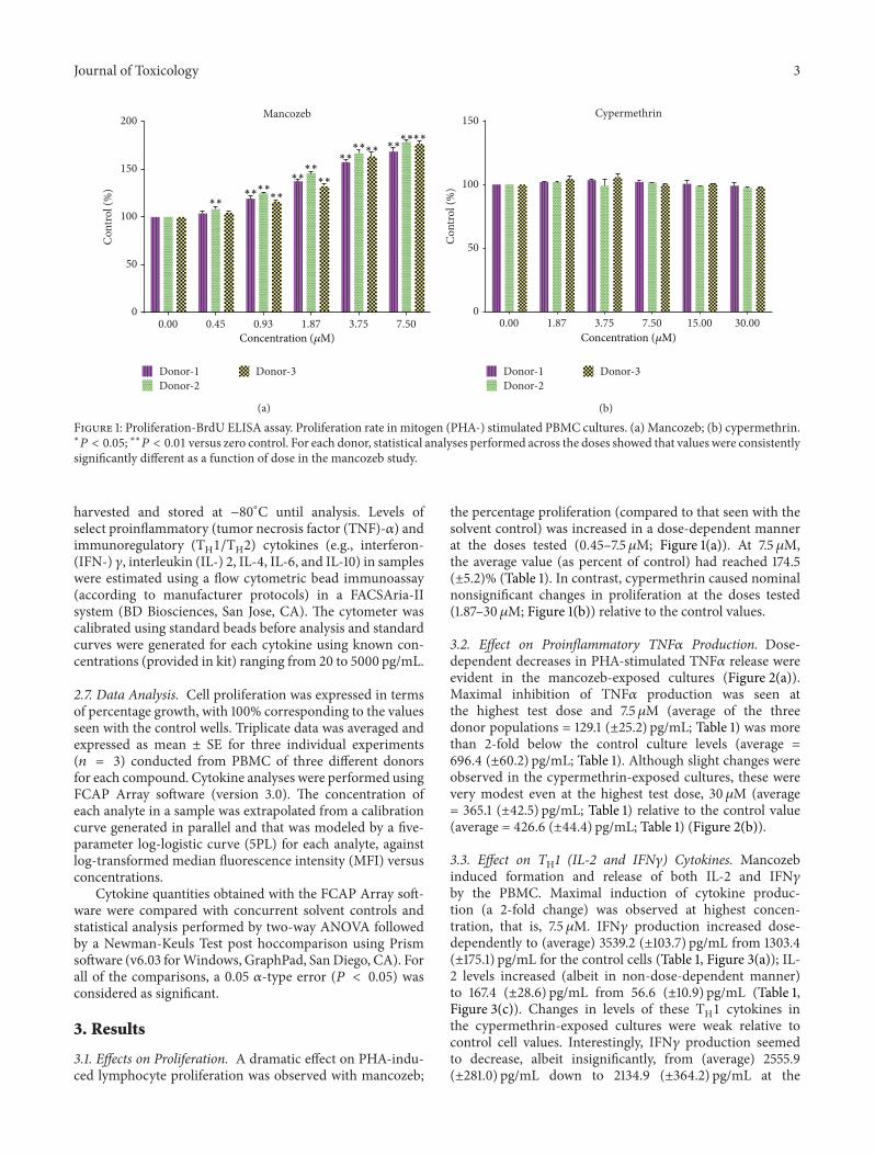
Figure 1: Proliferation-BrdU ELISA assay. Proliferation rate in mitogen (PHA-) stimulated PBMC cultures. (a) Mancozeb; (b) cypermethrin.∗
𝑃 < 0.05; ∗∗𝑃 < 0.01 versus zero control. For each donor, statistical analyses performed across the doses showed that values were consistentlysignificantly different as a function of dose in the mancozeb study.
harvested and stored at −80∘C until analysis. Levels ofselect proinflammatory (tumor necrosis factor (TNF)-𝛼) andimmunoregulatory (TH1/TH2) cytokines (e.g., interferon-(IFN-) 𝛾, interleukin (IL-) 2, IL-4, IL-6, and IL-10) in sampleswere estimated using a flow cytometric bead immunoassay(according to manufacturer protocols) in a FACSAria-IIsystem (BD Biosciences, San Jose, CA). The cytometer wascalibrated using standard beads before analysis and standardcurves were generated for each cytokine using known con-centrations (provided in kit) ranging from 20 to 5000 pg/mL.
2.7. Data Analysis. Cell proliferation was expressed in termsof percentage growth, with 100% corresponding to the valuesseen with the control wells. Triplicate data was averaged andexpressed as mean ± SE for three individual experiments(𝑛 = 3) conducted from PBMC of three different donorsfor each compound. Cytokine analyses were performed usingFCAP Array software (version 3.0). The concentration ofeach analyte in a sample was extrapolated from a calibrationcurve generated in parallel and that was modeled by a five-parameter log-logistic curve (5PL) for each analyte, againstlog-transformed median fluorescence intensity (MFI) versusconcentrations.
Cytokine quantities obtained with the FCAP Array soft-ware were compared with concurrent solvent controls andstatistical analysis performed by two-way ANOVA followedby a Newman-Keuls Test post hoccomparison using Prismsoftware (v6.03 forWindows, GraphPad, SanDiego, CA). Forall of the comparisons, a 0.05 𝛼-type error (𝑃 < 0.05) wasconsidered as significant.
3. Results
3.1. Effects on Proliferation. A dramatic effect on PHA-indu-ced lymphocyte proliferation was observed with mancozeb;
the percentage proliferation (compared to that seen with thesolvent control) was increased in a dose-dependent mannerat the doses tested (0.45–7.5𝜇M; Figure 1(a)). At 7.5 𝜇M,the average value (as percent of control) had reached 174.5(±5.2)% (Table 1). In contrast, cypermethrin caused nominalnonsignificant changes in proliferation at the doses tested(1.87–30 𝜇M; Figure 1(b)) relative to the control values.
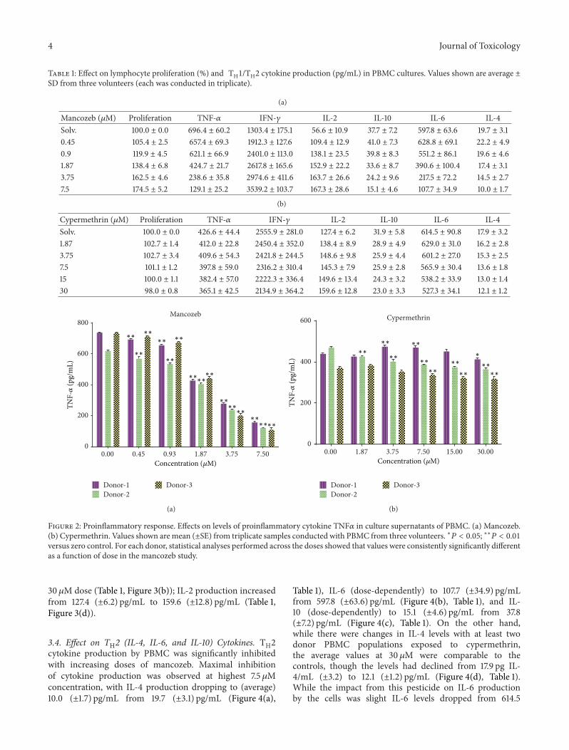
3.2. Effect on Proinflammatory TNF𝛼 Production. Dose-dependent decreases in PHA-stimulated TNF𝛼 release wereevident in the mancozeb-exposed cultures (Figure 2(a)).Maximal inhibition of TNF𝛼 production was seen atthe highest test dose and 7.5𝜇M (average of the threedonor populations = 129.1 (±25.2) pg/mL; Table 1) was morethan 2-fold below the control culture levels (average =696.4 (±60.2) pg/mL; Table 1). Although slight changes wereobserved in the cypermethrin-exposed cultures, these werevery modest even at the highest test dose, 30 𝜇M (average= 365.1 (±42.5) pg/mL; Table 1) relative to the control value(average = 426.6 (±44.4) pg/mL; Table 1) (Figure 2(b)).
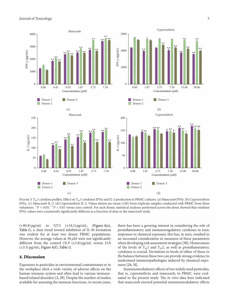
3.3. Effect on TH1 (IL-2 and IFN𝛾) Cytokines. Mancozebinduced formation and release of both IL-2 and IFN𝛾by the PBMC. Maximal induction of cytokine produc-tion (a 2-fold change) was observed at highest concen-tration, that is, 7.5 𝜇M. IFN𝛾 production increased dose-dependently to (average) 3539.2 (±103.7) pg/mL from 1303.4(±175.1) pg/mL for the control cells (Table 1, Figure 3(a)); IL-2 levels increased (albeit in non-dose-dependent manner)to 167.4 (±28.6) pg/mL from 56.6 (±10.9) pg/mL (Table 1,Figure 3(c)). Changes in levels of these TH1 cytokines inthe cypermethrin-exposed cultures were weak relative tocontrol cell values. Interestingly, IFN𝛾 production seemedto decrease, albeit insignificantly, from (average) 2555.9(±281.0) pg/mL down to 2134.9 (±364.2) pg/mL at the
4 Journal of Toxicology
Table 1: Effect on lymphocyte proliferation (%) and TH1/TH2 cytokine production (pg/mL) in PBMC cultures. Values shown are average ±SD from three volunteers (each was conducted in triplicate).
(a)
Mancozeb (𝜇M) Proliferation TNF-𝛼 IFN-𝛾 IL-2 IL-10 IL-6 IL-4Solv. 100.0 ± 0.0 696.4 ± 60.2 1303.4 ± 175.1 56.6 ± 10.9 37.7 ± 7.2 597.8 ± 63.6 19.7 ± 3.10.45 105.4 ± 2.5 657.4 ± 69.3 1912.3 ± 127.6 109.4 ± 12.9 41.0 ± 7.3 628.8 ± 69.1 22.2 ± 4.90.9 119.9 ± 4.5 621.1 ± 66.9 2401.0 ± 113.0 138.1 ± 23.5 39.8 ± 8.3 551.2 ± 86.1 19.6 ± 4.61.87 138.4 ± 6.8 424.7 ± 21.7 2617.8 ± 165.6 152.9 ± 22.2 33.6 ± 8.7 390.6 ± 100.4 17.4 ± 3.13.75 162.5 ± 4.6 238.6 ± 35.8 2974.6 ± 411.6 163.7 ± 26.6 24.2 ± 9.6 217.5 ± 72.2 14.5 ± 2.77.5 174.5 ± 5.2 129.1 ± 25.2 3539.2 ± 103.7 167.3 ± 28.6 15.1 ± 4.6 107.7 ± 34.9 10.0 ± 1.7
(b)
Cypermethrin (𝜇M) Proliferation TNF-𝛼 IFN-𝛾 IL-2 IL-10 IL-6 IL-4Solv. 100.0 ± 0.0 426.6 ± 44.4 2555.9 ± 281.0 127.4 ± 6.2 31.9 ± 5.8 614.5 ± 90.8 17.9 ± 3.21.87 102.7 ± 1.4 412.0 ± 22.8 2450.4 ± 352.0 138.4 ± 8.9 28.9 ± 4.9 629.0 ± 31.0 16.2 ± 2.83.75 102.7 ± 3.4 409.6 ± 54.3 2421.8 ± 244.5 148.6 ± 9.8 25.9 ± 4.4 601.2 ± 27.0 15.3 ± 2.57.5 101.1 ± 1.2 397.8 ± 59.0 2316.2 ± 310.4 145.3 ± 7.9 25.9 ± 2.8 565.9 ± 30.4 13.6 ± 1.815 100.0 ± 1.1 382.4 ± 57.0 2222.3 ± 336.4 149.6 ± 13.4 24.3 ± 3.2 538.2 ± 33.9 13.0 ± 1.430 98.0 ± 0.8 365.1 ± 42.5 2134.9 ± 364.2 159.6 ± 12.8 23.0 ± 3.3 527.3 ± 34.1 12.1 ± 1.2
∗∗
∗∗∗∗
∗∗∗∗∗∗
∗∗∗∗
∗∗
∗∗∗∗∗∗
∗∗
∗∗∗∗
0.00 0.45 0.93 1.87 3.75 7.500
200
400
600
800Mancozeb
Concentration (𝜇M)
TNF-𝛼
(pg/
mL)
Donor-1Donor-2
Donor-3
(a)
∗∗∗∗
∗∗ ∗∗∗∗
∗∗∗∗∗∗
∗∗ ∗∗
0.00 1.87 3.75 7.50 15.00 30.000
200
400
600 Cypermethrin
Concentration (𝜇M)
∗
TNF-𝛼
(pg/
mL)
Donor-1Donor-2
Donor-3
(b)
Figure 2: Proinflammatory response. Effects on levels of proinflammatory cytokine TNF𝛼 in culture supernatants of PBMC. (a) Mancozeb.(b) Cypermethrin. Values shown are mean (±SE) from triplicate samples conducted with PBMC from three volunteers. ∗𝑃 < 0.05; ∗∗𝑃 < 0.01versus zero control. For each donor, statistical analyses performed across the doses showed that values were consistently significantly differentas a function of dose in the mancozeb study.
30 𝜇M dose (Table 1, Figure 3(b)); IL-2 production increasedfrom 127.4 (±6.2) pg/mL to 159.6 (±12.8) pg/mL (Table 1,Figure 3(d)).
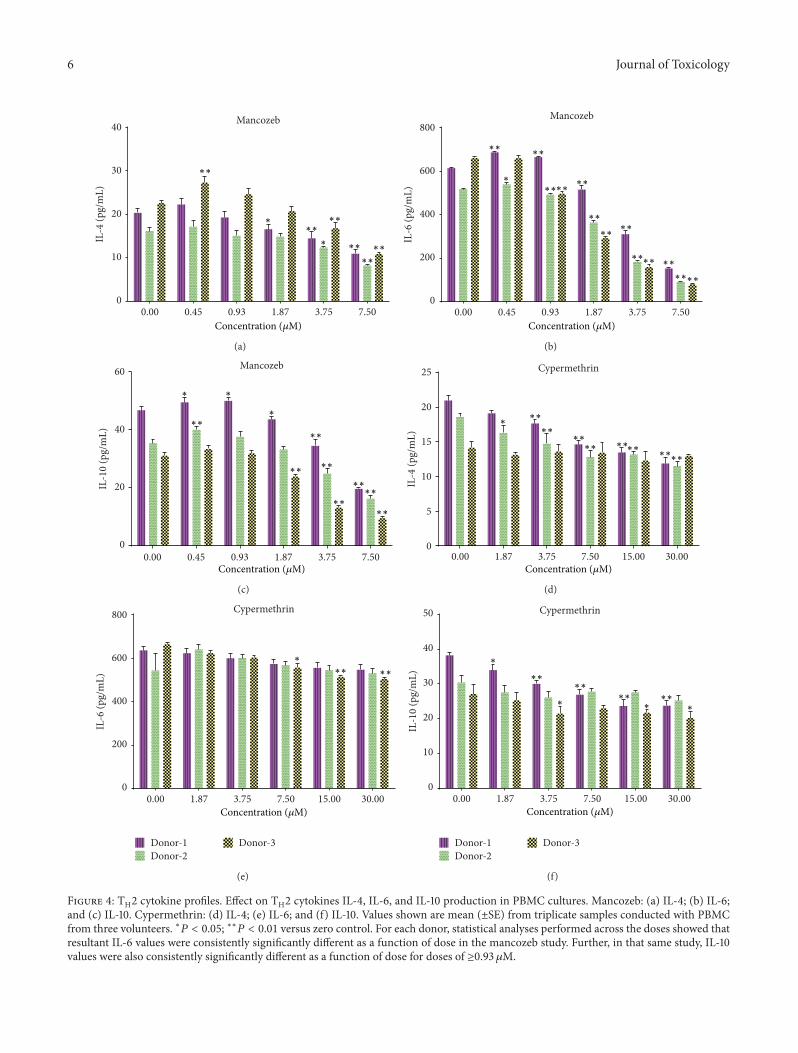
3.4. Effect on TH2 (IL-4, IL-6, and IL-10) Cytokines. TH2cytokine production by PBMC was significantly inhibitedwith increasing doses of mancozeb. Maximal inhibitionof cytokine production was observed at highest 7.5𝜇Mconcentration, with IL-4 production dropping to (average)10.0 (±1.7) pg/mL from 19.7 (±3.1) pg/mL (Figure 4(a),
Table 1), IL-6 (dose-dependently) to 107.7 (±34.9) pg/mLfrom 597.8 (±63.6) pg/mL (Figure 4(b), Table 1), and IL-10 (dose-dependently) to 15.1 (±4.6) pg/mL from 37.8(±7.2) pg/mL (Figure 4(c), Table 1). On the other hand,while there were changes in IL-4 levels with at least twodonor PBMC populations exposed to cypermethrin,the average values at 30 𝜇M were comparable to thecontrols, though the levels had declined from 17.9 pg IL-4/mL (±3.2) to 12.1 (±1.2) pg/mL (Figure 4(d), Table 1).While the impact from this pesticide on IL-6 productionby the cells was slight IL-6 levels dropped from 614.5
Journal of Toxicology 5
∗∗∗∗∗∗
∗∗
∗∗
∗∗
∗∗
∗∗
∗∗∗∗∗∗∗∗
∗∗
∗∗∗∗
0.00 0.45 0.93 1.87 3.75 7.500
1000
2000
3000
4000Mancozeb
Concentration (𝜇M)
IFN
-𝛾(p
g/m
L)
Donor-1Donor-2
Donor-3
(a)
∗∗
∗∗∗∗
∗∗
∗∗ ∗∗
∗∗
∗∗∗∗
∗∗
0.00 1.87 3.75 7.50 15.00 30.000
1000
2000
3000Cypermethrin
IFN
-𝛾(p
g/m
L)
Donor-1Donor-2
Donor-3
Concentration (𝜇M)
(b)
∗∗
∗∗∗∗
∗∗
∗∗∗∗
∗∗
∗∗
∗∗
∗∗
∗∗
∗∗
∗∗
∗∗∗∗
0.00 0.45 0.93 1.87 3.75 7.500
50
100
150
200
250Mancozeb
IL-2
(pg/
mL)
Concentration (𝜇M)
Donor-1Donor-2
Donor-3
(c)
∗∗
∗∗∗∗∗∗
∗∗∗∗
∗∗∗∗∗∗
IL-2
(pg/
mL)
0.00 1.87 3.75 7.50 15.00 30.000
50
100
150
200Cypermethrin
Concentration (𝜇M)
∗ ∗ ∗
Donor-1Donor-2
Donor-3
(d)
Figure 3: TH1 cytokine profiles. Effect on TH1 cytokines IFN𝛾 and IL-2 production in PBMC cultures. (a)Mancozeb IFN𝛾. (b) CypermethrinIFN𝛾. (c) Mancozeb IL-2. (d) Cypermethrin IL-2. Values shown are mean (±SE) from triplicate samples conducted with PBMC from threevolunteers. ∗𝑃 < 0.05; ∗∗𝑃 < 0.01 versus zero control. For each donor, statistical analyses performed across the doses showed that resultantIFN𝛾 values were consistently significantly different as a function of dose in the mancozeb study.
(±90.8) pg/mL to 527.3 (±34.1) pg/mL; (Figure 4(e),Table 1), a clear trend toward inhibition of IL-10 formationwas evident for at least two donor PBMC populations.However, the average values at 30 𝜇M were not significantlydifferent from the control (31.9 (±5.8) pg/mL versus 23.0(±3.3) pg/mL; Figure 4(f), Table 1).
4. Discussion
Exposures to pesticides as environmental contaminants or inthe workplace elicit a wide variety of adverse effects on thehuman immune system and often lead to various immune-based/related disorders [2, 29]. Despite the number of studiesavailable for assessing the immune functions, in recent years,
there has been a growing interest in considering the role ofproinflammatory and immunoregulatory cytokines in toxicresponses to chemical exposure; this has, in turn, resulted inan increased consideration in measures of these parameterswhendeveloping risk assessment strategies [30].Homeostasisof the levels of TH1 and TH2, as well as proinflammatory,cytokines is crucial. Deviations in levels of either of these orthe balance between these two can provide strong evidence tounderstand immunopathologies induced by chemical expo-sures [24, 31].
Immunomodulatory effects of twowidely used pesticides,that is, cypermethrin and mancozeb, in PBMC were eval-uated in the present study. The in vitro data here indicatedthat mancozeb exerted potential immunomodulatory effects
6 Journal of Toxicology
∗∗
∗∗
∗∗
∗∗
∗
∗∗∗
0.00 0.45 0.93 1.87 3.75 7.500
10
20
30
40 Mancozeb
IL-4
(pg/
mL)
∗∗
Concentration (𝜇M)
(a)
∗∗
∗∗
∗∗
∗∗∗∗
∗
∗∗
∗∗
∗∗
∗∗∗∗
∗∗
∗∗
∗∗
0.00 0.45 0.93 1.87 3.75 7.500
200
400
600
800Mancozeb
IL-6
(pg/
mL)
Concentration (𝜇M)
(b)
∗∗
∗∗
∗∗
∗∗
∗∗
∗∗
∗∗
∗∗
∗
∗∗
0.00 0.45 0.93 1.87 3.75 7.500
20
40
60 Mancozeb
IL-1
0 (p
g/m
L)
Concentration (𝜇M)
(c)
∗∗∗
∗∗∗∗
∗∗∗∗∗∗∗∗
∗∗
0.00 1.87 3.75 7.50 15.00 30.000
5
10
15
20
25 Cypermethrin
IL-4
(pg/
mL)
Concentration (𝜇M)
(d)
∗∗∗∗
∗
0.00 1.87 3.75 7.50 15.00 30.000
200
400
600
800 Cypermethrin
IL-6
(pg/
mL)
Concentration (𝜇M)
Donor-1Donor-2
Donor-3
(e)
∗ ∗ ∗
∗∗∗∗
∗∗∗∗
∗
0.00 1.87 3.75 7.50 15.00 30.000
10
20
30
40
50 Cypermethrin
IL-1
0 (p
g/m
L)
Concentration (𝜇M)
Donor-1Donor-2
Donor-3
(f)
Figure 4: TH2 cytokine profiles. Effect on TH2 cytokines IL-4, IL-6, and IL-10 production in PBMC cultures. Mancozeb: (a) IL-4; (b) IL-6;and (c) IL-10. Cypermethrin: (d) IL-4; (e) IL-6; and (f) IL-10. Values shown are mean (±SE) from triplicate samples conducted with PBMCfrom three volunteers. ∗𝑃 < 0.05; ∗∗𝑃 < 0.01 versus zero control. For each donor, statistical analyses performed across the doses showed thatresultant IL-6 values were consistently significantly different as a function of dose in the mancozeb study. Further, in that same study, IL-10values were also consistently significantly different as a function of dose for doses of ≥0.93 𝜇M.
Journal of Toxicology 7
characterized by (i) increased cell proliferative responses toPHA stimulation; (ii) significant reductions in PHA-inducedTNF𝛼 release; and (iii) significant alterations in TH1 and TH2cytokine profiles. These outcomes reflect the same types ofeffects ofmancozeb onmitogen-induced proliferation and onTNF𝛼 and IL-2 levels that had been reported earlier [16, 17,32, 33]. On the other hand, here, cypermethrin only causedslight changes in the cytokine profiles or onmitogen-inducedproliferation.
Numerous in vivo studies have reported dose-dependentdecreases in hematologic endpoints, such as erythrocytecounts and packed cell volumes that were accompaniedby significant changes in lymphocyte, monocyte, and totalleukocyte counts as a result of exposure to cypermethrin[34–36] or mancozeb [17, 37]. The results in the present invitro study correlated fairly well with those in vivo findingsand indicated that the mancozeb directly affected immunecells. However, cypermethrin exposure did not show anyeffect on lymphocyte proliferation and cytokine production.This clearly shows that the pesticides will act differently onimmune cells and have different effects.
The range of in vitro doses tested here was also inaccordance with those earlier in vivo studies. In those stud-ies, significant effects on hemato-/immunologic parameterswere seen with mancozeb doses that ranged from 250 to1500mg/kg BW [38] or with cypermethrin (through oralor intraperitoneal routes) at doses ranging from 25 to300mg/kg BW [34, 35]. Still, while these in vitro studiesdemonstrated effects from direct exposure of PBMC to eachpesticide, questions about relevance of the doses used (0.45–30 𝜇M of parent compounds) seem to persist. Occupationalexposure to pyrethroids and ethylenebisdithiocarbamatesoften results in increases in body burdens of the toxicantsthat are, in turn, reflected at mg levels of select metabolitesof the parent agent (for cypermethrin, 3-phenoxy-benzoate;for mancozeb, ethylene thiourea) in the blood/urine of theworkers [17, 39, 40]. As such, at even just a single mg ofeach metabolite, this would yield corresponding levels of4.7 (3-phenoxybenzoate) and 9.8 (ethylene thiourea) 𝜇M inthe human blood/urine of the exposed workers. Thus, thedoses of cypermethrin or mancozeb used in the currentexperiment were likely to have been on par with levels of theparent + metabolite found in workers routinely exposed toeither pesticide. Nevertheless, the true relationship betweencumulative quantities of metabolites produced by continuousexposures to these pesticides in workers can and shouldbe estimated by others using mathematical toxicokineticmodeling to better estimate at what doses these pesticideswould be unquestionably relevant in this type of in vitrostudies performed here.
5. Conclusions
The present study demonstrates that pesticides that are indis-criminately used might increase the risk for immunomod-ulation in an exposed host. As such, this also reflects theneed for continued evaluation of the immunotoxic poten-tials of these and other types of commonly encounteredoccupational/environmental chemicals. As the data here also
show, in vitro studies using freshly obtained human cells(i.e., PBMC) to assess agent-induced changes in endpointslike lymphocyte proliferation and TH1/TH2 cytokine pro-duction will certainly contribute to a better understandingof the effects of xenobiotics on immune function and alsoincrease the arsenal that investigators can use when design-ing/developing testing regimens to assess such effects.
Conflict of Interests
The authors declare no conflict of interests.The authors aloneare responsible for the content of this paper.
Acknowledgments
This work was supported by the International Instituteof Biotechnology And Toxicology. The authors thank themanagement of International Institute of Biotechnology AndToxicology for funding and supporting the research and thestaff for donating blood samples. This work would have notbeen possible without the moral support from Dr. SrivatsaPrakhya; special thanks are due to him.
References
[1] R. Repetto and S. Baliga, “Trends and patterns of pesticide use,”in Pesticides and the Immune System; Public Health Risks, pp. 3–8, World Resources Institute, Washington, DC, USA, 1996.
[2] T. Vial, B. Nicolas, and J. Descotes, “Clinical immunotoxicity ofpesticides,” Journal of Toxicology and Environmental Health, vol.48, no. 3, pp. 215–229, 1996.
[3] R. Stiller-Winkler,W.Hadnagy, G. Leng, E. Straube, andH. Idel,“Immunological parameters in humans exposed to pesticides inthe agricultural environment,” Toxicology Letters, vol. 107, no. 1–3, pp. 219–224, 1999.
[4] M. F. Vine, L. Stein, K. Weigle et al., “Effects on the immunesystem associated with living near a pesticide dump site,”Environmental Health Perspectives, vol. 108, no. 12, pp. 1113–1124,2000.
[5] S. Nishimoto, K. Kanda, M. Okabe, K. Akiyama, and Y. Kaki-numa, “Abnormal response induced by pesticides on mam-malian immune system,” Interdisciplinary Studies on Environ-mental Chemistry-Environmental Research in Asia, pp. 211–217,2009.
[6] M. Zakerinia, M. Namdari, and S. Amirghofran, “The rela-tionship between exposure to pesticides and the occurrence oflymphoid neoplasm,” Iranian Red Crescent Medical Journal, vol.14, no. 6, pp. 337–344, 2012.
[7] X. Ma, P. A. Buffler, R. B. Gunier et al., “Critical windowsof exposure to household pesticides and risk of childhoodleukemia,” Environmental Health Perspectives, vol. 110, no. 9, pp.955–960, 2002.
[8] W. J. Hayes and E. R. Laws, Eds., Handbook of PesticideToxicology, vol. 3 of Classes of Pesticides, Academic Press, NewYork, NY, USA, 1990.
[9] G. Lorgue and J. Lechenet, Eds., Clinical Veterinary Toxicology,Blackwell Science, London, UK, 1996.
[10] J. Descotes, “Regulatory aspects of immunotoxicity evaluation,”in Immunotoxicology of Drugs and Chemicals. An Experimen-tal and Clinical Approach. Vol. 1. Principles and Methods of
8 Journal of Toxicology
Immunotoxicology, pp. 257–268, Elsevier Science, Amsterdam,The Netherlands, 2004.
[11] H. O. Hasan, Fungicides and Their Effects on Animals, Fungi-cides, 2010.
[12] I. Desi, L. Varga, I. Dobronyi, and G. Szklenarik, “Immunotox-icological investigation of the effects of a pesticide; cyperme-thrin,” Archives of Toxicology, vol. 8, pp. 305–309, 1985.
[13] I. Desi, I. Dobronyi, and L. Varga, “Immuno-, neuro-, and gene-ral toxicologic animal studies on a synthetic pyrethroid: cyper-methrin,” Ecotoxicology and Environmental Safety, vol. 12, no. 3,pp. 220–232, 1986.
[14] WHO (World Health Organization), International Program onChemical Safety: WHO Task Group Meeting on EnvironmentalHealth Criteria for Cypermethrin . Cypermethrin, vol. 82 ofEnvironmental Health Criteria, United Nations EnvironmentProgram, the International Labor Organization, and WorldHealth Organization, Geneva, Switzerland, 1989.
[15] P. Liu, X. Song, W. Yuan et al., “Effects of cypermethrin andmethyl parathion mixtures on hormone levels and immunefunctions in Wistar rats,” Archives of Toxicology, vol. 80, no. 7,pp. 449–457, 2006.
[16] C. Colosio, W. Barcellini, M. Maroni et al., “Immunomodula-tory effects of occupational exposure to mancozeb,” Archives ofEnvironmental Health, vol. 51, no. 6, pp. 445–451, 1996.
[17] E. Corsini, S. Birindelli, S. Fustinoni et al., “Immunomodulatoryeffects of the fungicide Mancozeb in agricultural workers,”Toxicology and Applied Pharmacology, vol. 208, no. 2, pp. 178–185, 2005.
[18] L.-M. Koeper and H.-W. Vohr, “Functional assays are manda-tory for a correct prediction of immunotoxic properties ofcompounds in vitro,” Food and Chemical Toxicology, vol. 47, no.1, pp. 110–118, 2009.
[19] D. P. Lankveld, H. van Loveren, K. A. Baken, and R. J. Vande-briel, “In vitro testing for direct immunotoxicity: state of theart,”Methods in Molecular Biology, vol. 598, pp. 401–423, 2010.
[20] R. Luebke, “Immunotoxicant screening and prioritization in the21st century,” Toxicologic Pathology, vol. 40, no. 2, pp. 294–299,2012.
[21] J. Descotes, “Methods of evaluating immunotoxicity,” ExpertOpinion on Drug Metabolism and Toxicology, vol. 2, no. 2, pp.249–259, 2006.
[22] M. D. Cohen, L. B. Schook, J. J. Oppenheim, B. M. Freed, andK. E. Rodgers, “Symposium overview: alterations in cytokinereceptors by xenobiotics,” Toxicological Sciences, vol. 48, no. 2,pp. 163–169, 1999.
[23] R. V. House, “Theory and practice of cytokine assessment inimmunotoxicology,”Methods, vol. 19, no. 1, pp. 17–27, 1999.
[24] P. Duramad, I. B. Tager, andN. T.Holland, “Cytokines and otherimmunological biomarkers in children’s environmental healthstudies,” Toxicology Letters, vol. 172, no. 1-2, pp. 48–59, 2007.
[25] E. Corsini and R. V. House, “Evaluating cytokines in immuno-toxicity testing,”Methods inMolecular Biology, vol. 598, pp. 283–302, 2010.
[26] T. Ringerike, E. Ulleras, R. Volker et al., “Detection of immuno-toxicity using T-cell based cytokine reporter cell lines (“CellChip”),” Toxicology, vol. 206, no. 2, pp. 257–272, 2005.
[27] S. Romagnani, “T-cell subsets (Th1 versus Th2),” Annals ofAllergy, Asthma and Immunology, vol. 85, no. 1, pp. 9–18, 2000.
[28] T. R. Mosmann and R. L. Coffman, “TH1
and TH2
cells: diffe-rent patterns of lymphokine secretion lead to different func-tional properties,”Annual Review of Immunology, vol. 7, pp. 145–173, 1989.
[29] L. Fritschi, G. Benke, A. M. Hughes et al., “Occupationalexposure to pesticides and risk of non-Hodgkin's lymphoma,”The American Journal of Epidemiology, vol. 162, no. 9, pp. 849–857, 2005.
[30] R. Kooijman, S. Devos, and E. Hooghe-Peters, “Inhibitionof in vitro cytokine production by human peripheral bloodmononuclear cells treated with xenobiotics: implications for theprediction of general toxicity and immunotoxicity,” Toxicologyin Vitro, vol. 24, no. 6, pp. 1782–1789, 2010.
[31] L. J. Lesko and A. J. J. Atkinson, “Use of biomarkers andsurrogate endpoints in drug development and regulatory deci-sion making: criteria, validation, strategies,” Annual Review ofPharmacology and Toxicology, vol. 41, pp. 347–366, 2001.
[32] A.-H. Chung and M.-Y. Pyo, “Effects of mancozeb on theactivities of murine peritoneal macrophages in vitro and invivo,”Archives of Pharmacal Research, vol. 28, no. 1, pp. 100–105,2005.
[33] E. Corsini, B. Viviani, S. Birindelli et al., “Molecular mecha-nisms underlying mancozeb-induced inhibition of TNF-alphaproduction,” Toxicology and Applied Pharmacology, vol. 212, no.2, pp. 89–98, 2006.
[34] F. Sayim, N. U. Karabay Yavasoglu, Y. Uyanikgil, H. Aktug, A.Yavasoglu, and M. Turgut, “Neurotoxic effects of cypermethrinin Wistar rats: a haematological, biochemical and histopatho-logical study,” Journal of Health Science, vol. 51, no. 3, pp. 300–307, 2005.
[35] S. Kamal, A. M, F. Rizvi, M. Siddique, and S. Sadeeq-ur-Reha-man, “Effect of cypermethrin on clinico-haematological param-eters in rabbits,” Pakistan Veterinary Journal, vol. 27, no. 4, pp.171–175, 2007.
[36] R. R. Nair, M. J. Abraham, N. D. Nair, C. R. Lalithakunjamma,and C. M. Aravindakshan, “Hematological and biochemicalprofile in sublethal toxicity of cypermethrin in rats,” Interna-tional Journal of Biological and Medical Research, vol. 1, no. 4,pp. 211–214, 2010.
[37] R. H. Cox, “Three-month dietary toxicity study in dogs withmancozeb,” Tech. Rep.HLA417-416, Hazleton Labs, Vienna, Va,USA, 1986, Submitted to WHO by Rohm and Haas Company,Spring House, Pa, USA (as Rohm and Haas Report No. 86RC-7).
[38] P.Myoung-yun andA.Cheong, “Effects of subacute oral admin-istration of mancozeb on immunopathological parameters andsplenocyte proliferation in mice,” Environmental Health andToxicology, vol. 19, pp. 367–373, 2004.
[39] G. Leng and W. Gries, “Methods in biotechnology,” in PesticideProtocols, J. L. Martınez Vidal and A. Garrido Frenich, Eds., vol.19, Humana Press, Totowa, NJ, USA, 1999.
[40] D. Lu, D. Wang, C. Feng et al., “Urinary concentrations ofmetabolites of pyrethroid insecticides in textile workers, Eas-tern China,” Environment International, vol. 60, pp. 137–144,2013.

Submit your manuscripts athttp://www.hindawi.com
PainResearch and TreatmentHindawi Publishing Corporationhttp://www.hindawi.com Volume 2014
The Scientific World JournalHindawi Publishing Corporation http://www.hindawi.com Volume 2014
Hindawi Publishing Corporationhttp://www.hindawi.com
Volume 2014
ToxinsJournal of
VaccinesJournal of
Hindawi Publishing Corporation http://www.hindawi.com Volume 2014
Hindawi Publishing Corporationhttp://www.hindawi.com Volume 2014
AntibioticsInternational Journal of
ToxicologyJournal of
Hindawi Publishing Corporationhttp://www.hindawi.com Volume 2014
StrokeResearch and TreatmentHindawi Publishing Corporationhttp://www.hindawi.com Volume 2014
Drug DeliveryJournal of
Hindawi Publishing Corporationhttp://www.hindawi.com Volume 2014
Hindawi Publishing Corporationhttp://www.hindawi.com Volume 2014
Advances in Pharmacological Sciences
Tropical MedicineJournal of
Hindawi Publishing Corporationhttp://www.hindawi.com Volume 2014
Medicinal ChemistryInternational Journal of
Hindawi Publishing Corporationhttp://www.hindawi.com Volume 2014
AddictionJournal of
Hindawi Publishing Corporationhttp://www.hindawi.com Volume 2014
Hindawi Publishing Corporationhttp://www.hindawi.com Volume 2014
BioMed Research International
Emergency Medicine InternationalHindawi Publishing Corporationhttp://www.hindawi.com Volume 2014
Hindawi Publishing Corporationhttp://www.hindawi.com Volume 2014
Autoimmune Diseases
Hindawi Publishing Corporationhttp://www.hindawi.com Volume 2014
Anesthesiology Research and Practice
ScientificaHindawi Publishing Corporationhttp://www.hindawi.com Volume 2014
Journal of
Hindawi Publishing Corporationhttp://www.hindawi.com Volume 2014
Pharmaceutics
Hindawi Publishing Corporationhttp://www.hindawi.com Volume 2014
MEDIATORSINFLAMMATION
of