regulatory t cells control th2-dominant murine autoimmune
TRANSCRIPT
of July 21, 2022.This information is current as
Murine Autoimmune GastritisRegulatory T Cells Control Th2-Dominant
TungJessica Harakal, Claudia Rival, Hui Qiao and Kenneth S.
ol.1502344http://www.jimmunol.org/content/early/2016/06/01/jimmun
published online 3 June 2016J Immunol
MaterialSupplementary
4.DCSupplementalhttp://www.jimmunol.org/content/suppl/2016/06/01/jimmunol.150234
average*
4 weeks from acceptance to publicationFast Publication! •
Every submission reviewed by practicing scientistsNo Triage! •
from submission to initial decisionRapid Reviews! 30 days* •
Submit online. ?The JIWhy
Subscriptionhttp://jimmunol.org/subscription
is online at: The Journal of ImmunologyInformation about subscribing to
Permissionshttp://www.aai.org/About/Publications/JI/copyright.htmlSubmit copyright permission requests at:
Email Alertshttp://jimmunol.org/alertsReceive free email-alerts when new articles cite this article. Sign up at:
Print ISSN: 0022-1767 Online ISSN: 1550-6606. Immunologists, Inc. All rights reserved.Copyright © 2016 by The American Association of1451 Rockville Pike, Suite 650, Rockville, MD 20852The American Association of Immunologists, Inc.,
is published twice each month byThe Journal of Immunology
by guest on July 21, 2022http://w
ww
.jimm
unol.org/D
ownloaded from
by guest on July 21, 2022
http://ww
w.jim
munol.org/
Dow
nloaded from
by guest on July 21, 2022http://w
ww
.jimm
unol.org/D
ownloaded from
by guest on July 21, 2022
http://ww
w.jim
munol.org/
Dow
nloaded from
by guest on July 21, 2022http://w
ww
.jimm
unol.org/D
ownloaded from
by guest on July 21, 2022
http://ww
w.jim
munol.org/
Dow
nloaded from
The Journal of Immunology
Regulatory T Cells Control Th2-Dominant MurineAutoimmune Gastritis
Jessica Harakal,*,† Claudia Rival,*,†,‡ Hui Qiao,†,‡ and Kenneth S. Tung*,†,‡
Pernicious anemia and gastric carcinoma are serious sequelae of autoimmune gastritis (AIG). Our study indicates that in adult
C57BL/6-DEREG mice expressing a transgenic diphtheria toxin receptor under the Foxp3 promoter, transient regulatory T cell
(Treg) depletion results in long-lasting AIG associated with both H+K+ATPase and intrinsic factor autoantibody responses.
Although functional Tregs emerge over time during AIG occurrence, the effector T cells rapidly become less susceptible to
Treg-mediated suppression. Whereas previous studies have implicated dysregulated Th1 cell responses in AIG pathogenesis,
eosinophils have been detected in gastric biopsy specimens from patients with AIG. Indeed, AIG in DEREG mice is associated
with strong Th2 cell responses, including dominant IgG1 autoantibodies, elevated serum IgE, increased Th2 cytokine production,
and eosinophil infiltration in the stomach-draining lymph nodes. In addition, the stomachs exhibit severe mucosal and muscular
hypertrophy, parietal cell loss, mucinous epithelial cell metaplasia, and massive eosinophilic inflammation. Notably, the Th2
responses and gastritis severity are significantly ameliorated in IL-4– or eosinophil-deficient mice. Furthermore, expansion of
both Th2-promoting IFN regulatory factor 4+ programmed death ligand 2+ dendritic cells and ILT3+ rebounded Tregs was
detected after transient Treg depletion. Collectively, these data suggest that Tregs maintain physiological tolerance to clinically
relevant gastric autoantigens, and Th2 responses can be a pathogenic mechanism in AIG. The Journal of Immunology, 2016, 197:
000–000.
An essential function of the immune system is to distin-guish foreign from self-antigens, allowing for protectionfrom invading pathogens while preventing autoimmune
responses. T cell tolerance, initiated in the thymus, is maintained bythe peripheral lymphoid organs in communication with local tissueand environmental factors, such as those existing in the skin andmucosal sites (1). The cellular mechanisms include T cell deletion,anergy, and suppression by regulatory T cells (Tregs), which likelyoperate in concert to fully maintain the tolerance state. CD4+
Foxp3+ Tregs play a critical role in maintaining immune ho-meostasis and tolerance to self-antigens by suppressing effectorT cell and innate cell activities (2). The transcription factor Foxp3is required for Treg development and function (3–5). Loss-of-function mutations in Foxp3 lead to scurfy syndrome in mice
that exhibit progressive fatal multiorgan autoinflammation (6, 7)and to the immune dysregulation, polyendocrinopathy, enteropa-
thy, X-linked syndrome in patients (8, 9).Autoimmune gastritis (AIG) is a common disease of the
stomach associated with autoantibodies that target intrinsic
factor (IF), which supports vitamin B12 absorption, and the
gastric H+K+ATPase, the proton pump expressed by acid-
secreting parietal cells in gastric glands (10–15). Accordingly,
AIG patients are predisposed to the development of gastric
cancer (16–18) and pernicious anemia, the most common se-
quela of vitamin B12 deficiency, which has an estimated prev-
alence of ∼1.9% among the elderly Western population (19, 20).
The histological characterization of active human AIG includes
immune cell infiltration in the corpus and body regions of the
stomach and loss of gastric zymogenic and parietal cells (21).
Because of their strong resemblance to the human disease,
murine AIG models have been frequently used for research
on tolerance and mechanisms of organ-specific autoimmune
disease.Experimental AIG research has focused on addressing whether a
defect in tolerance mechanisms, such as Tregs, is the underpinning
of human autoimmune diseases and the rationale behind Treg-
based therapies. For many years, this question has been investi-
gated in the day 3 thymectomy (d3tx) model of BALB/c mice (22–
25). It was thought that Tregs exit the thymus after the non-Treg
T cells and should be preferentially depleted by thymectomy be-
tween neonatal days 1 and 5 (26–29). This idea was supported by
the blockade of AIG by transfer of normal Tregs soon after thy-
mectomy (22, 24, 30, 31). However, more recent studies have
yielded new findings inconsistent with this concept: 1) Tregs with
the capacity to suppress autoimmune disease were detected in the
lymph nodes and spleen before day 3 (32); 2) d3tx led to an in-
crease, rather than a reduction, of functional Treg fractions (33,
34); 3) Treg depletion by anti-CD25 Ab (PC61) in d3tx mice
greatly enhanced the AIG immunopathology (34, 35); and 4) d3tx
*Department of Microbiology, Immunology, and Cancer Biology, University of Vir-ginia, Charlottesville, VA 22908; †Beirne B. Carter Center for Immunology Re-search, University of Virginia, Charlottesville, VA 22908; and ‡Department ofPathology, University of Virginia, Charlottesville, VA 22908
ORCIDs: 0000-0003-3523-2859 (J.H.); 0000-0002-2568-5144 (H.Q.); 0000-0002-9393-0084 (K.S.T.).
Received for publication November 3, 2015. Accepted for publication April 18, 2016.
This work was supported by National Institutes of Health Grant AI41236-19. J.H.was supported by National Institutes of Health Immunology Training Grant AI07496,and C.R. was supported by National Institutes of Health Research Training in Di-gestive Diseases Grant DK007769.
Address correspondence and reprint requests to Dr. Jessica Harakal and Dr. KennethTung, University of Virginia, Box 801386, MR6 345 Crispell Drive, Charlottesville,VA 22908. E-mail addresses: [email protected] (J.H.) and [email protected] (K.S.T.)
The online version of this article contains supplemental material.
Abbreviations used in this article: AIG, autoimmune gastritis; CTLA-4, CTL-associated protein-4; d3tx, day 3 thymectomy; DC, dendritic cell; DT, diphtheriatoxin; DTR, DT receptor; GITR, glucocorticoid-induced TNF receptor; IF, intrinsicfactor; IRF4, IFN regulatory factor 4; NDLN, nondraining lymph node; PD-L2,programmed death ligand 2; SDLN, stomach-draining lymph node; Tfh, T follicularhelper; Treg, regulatory T cell; WT, wild-type.
Copyright� 2016 by The American Association of Immunologists, Inc. 0022-1767/16/$30.00
www.jimmunol.org/cgi/doi/10.4049/jimmunol.1502344
Published June 3, 2016, doi:10.4049/jimmunol.1502344 by guest on July 21, 2022
http://ww
w.jim
munol.org/
Dow
nloaded from
mice developed severe lymphopenia, and the attendant homeo-static expansion of the autoreactive effector T cell compartment,including gastritogenic T cell clones, could also contribute todisease (26, 34, 36–38).To more directly address Treg depletion without the confound-
ing lymphopenic state, recent studies have turned to geneticallymodified mouse lines expressing the diphtheria toxin (DT) receptor(DTR) under the control of a Foxp3 promoter, from which Tregscan be depleted by DT treatment. In both neonatal and adultFoxp3DTR knock-in mice, continuous DT treatment led to dra-matic expansion and activation of adaptive and innate cells, ascurfy-like phenotype, and death of unknown cause by 3–4 wk(39). Adult BALB/c Foxp3DTR mice with transient Treg depletionalso suffered from death within 4–5 wk. Moreover, despite the re-emergence of Tregs, the mice exhibited rapidly increased cytokineproduction, enhanced Ag-specific T cell activation, developmentof AIG with mononuclear cell infiltration, and parietal cell auto-antibody responses (40). These findings raise the critical questionsof whether transient Treg deficiency is sufficient to induce AIG,and why the restored Treg population fails to maintain tolerance(41).In addition to the Foxp3DTR knock-in mice, recent studies were
conducted with the DEREG (DEpletion of REGulatory T cells)mice that express a bacterial artificial chromosome containinga DTR and enhanced GFP fusion protein. Whereas DT-treatednewborn C57BL/6-DEREG mice develop scurfy-like syndrome,adult C57BL/6 and BALB/c-DEREG mice were reportedly free ofdisease after transient Treg depletion (42, 43). This lack of path-ological findings was attributed to the maintenance of tolerance bya minor population of residual DT-insensitive Tregs. Adult BALB/c-DEREG mice crossed with the Foxp3GFP mice also failed to induceAIG, but blepharitis and scurfy-like autoinflammation were detected(43). However, unlike the Foxp3DTR knock-in mice, the adultDEREG mice with transient Treg depletion did not succumb toearly fatality (42, 44).To develop an AIG model that is more suitable for detailed
mechanistic analyses, we have conducted studies using the C57BL/6-DEREG mice, kindly donated by Drs. Lahl and Sparwassar, tosystematically examine autoimmune disease development aftertransient Treg depletion. Contrary to previous findings, the study inour laboratory revealed that adult DT-treated C57BL/6-DEREGmice developed frequent and severe AIG that persisted for$16 wk.We found that after Treg depletion, the non-Treg T cells rapidlybecame less susceptible to Treg-mediated suppression. This find-ing provides a tangible explanation for the failure of the functionalrebounded Treg population to control AIG. Moreover, AIG thatdevelops in the adult DEREG model exhibited a unique Abresponse and immunopathological features indicative of a robustTh2-dominant autoimmune response dependent on IL-4 andeosinophils.
Materials and MethodsMice and Foxp3+ Treg depletion
C57BL/6-DEREG mice were kindly provided by Drs. Lahl and Sparwasser(Twin Core). A/J mice, C57BL/6 Rag12/2 mice, and C57BL/6 IL-42/2 micewere purchased from The Jackson Laboratory. C57BL/6 PHIL mice weregenerously provided by James Lee (Mayo Clinic). Rag12/2 DEREG micewere generated by crossing Rag12/2 mice with DEREG mice. IL-42/2
DEREG and DEREG PHIL mice were generated by crossing the DEREGmice with the IL-42/2 or the PHIL mice, respectively. To obtain B6AF1-DEREG mice, C57BL/6-DEREG mice were crossed with A/J mice. Todeplete Tregs, 40 mg/kg body weight of DT (Lot 322326; Calbiochem) wasinjected i.p. into 6- to 12-wk-old DEREG mice on days 0, 2, and 5. Allexperimental days were counted from day 0. As a control, we studied wild-type (WT) mice injected with DT and DEREG mice injected with PBS. Allmice were bred in house in a specific pathogen–free facility and tested
negative for fur mites, pinworm infection, and Helicobacter spp. infections.Experiments were conducted following the guidelines of the Animal Careand Use Committee of the University of Virginia.
Histopathology and AIG severity grading
Mice were euthanized from 1 to 16 wk after Treg depletion. The stomachswere weighed after the removal of gastric contents, then fixed in Bouin’ssolution, embedded in paraffin wax, and cut into 5-mm-thick sections. Thepresence of inflammation and parietal cells were examined by H&Estain, and mucin-positive cells were identified by periodic acid–Schiff/hematoxylin stain. AIG was graded as unknown samples. The AIG se-verity score was determined by the summation of the extent of inflam-mation, parietal cell loss, and mucinous cell hyperplasia. Each change wasassigned a grade of 0 (none), 1 (mild), 2 (moderate), 3 (moderate anddiffuse or severe but focal), or 4 (severe and diffuse). Because parietal cellloss and mucinous cell hyperplasia are structural changes and likely as-sociated with loss of gastric function, we multiplied their scores by a factorof 1.5. Therefore, the total AIG scores ranged from 0 to 16.
Immunofluorescence microscopy
Serum gastric autoantibodies were detected and semiquantified by indirectimmunofluorescence staining of 6-mm-thick frozen sections of normalmouse stomach prefixed in 1% periodate-lysine-paraformaldehyde. Afterthe sections were blocked with PBS containing normal goat serum and3% BSA, they were incubated with serum from control or Treg-depletedmice, followed by incubation with FITC-conjugated goat anti-mouse IgG(Jackson ImmunoResearch Laboratories), anti-mouse IgG1 (SouthernBiotech), or anti-mouse IgG2a (Southern Biotech). Staining intensity wasscored as 0 (negative) to 3 (most positive). To study IF autoantibodies,mouse sera were costained with IF Ab, a generous gift from David Alpers(Washington University, St Louis, MO), on normal stomach sections.Mouse sera staining (described above) was detected with Alexa Fluor 488–conjugated rabbit anti-mouse IgG (Jackson ImmunoResearch Laborato-ries) followed by blockade with a biotin–avidin blocking kit (VectorLaboratories) and costained with goat anti-human IF Ab followed bybiotinylated horse anti-goat IgG (Vector Laboratories) and neutralite avi-din–Texas Red (Southern Biotech). To detect eosinophils by direct im-munofluorescence, 6-mm frozen stomach sections were fixed in cold 1:1acetone:ethanol and blocked with Tris-NaCl blocking buffer (Perkin-Elmer), 3% H2O2 (EMD Chemicals), 0.1% NaN3 (ICN Biomedicals),followed by a biotin-avidin blocking kit. The sections were then stainedwith rat anti-mouse SiglecF Ab (BD Pharmingen) and biotinylated goatanti-rat IgG (Southern Biotech). Then, staining was enhanced by the biotintyramide kit according to the manufacturer’s instructions (PerkinElmer),followed by neutralite avidin–Texas Red. To detect parietal cells on thesame sections, the slides were then costained with DEREG mouse serumAb to parietal cell Ags, as described above, and mounted with Vectashieldcontaining DAPI (Vector Laboratories). Unless noted otherwise, all stainswere performed at room temperature, and the slides were examined andphotographed with a Nikon Microphot-FXA fluorescent microscope andNikon HB-10101AF Mercury Lamp and Olympus Q-Color5 camera. Al-ternatively, they were examined with a Zeiss LSM 700 confocal micro-scope or a Zeiss Axio Observer microscope with Qioptiq OptiGrid.
ELISA detection of gastric H+K+ATPase Ab and IF Ab
To detect H+K+ATPase Ab, 96-well plates were incubated with 0.5 mg/mlH+K+ATPase (Arotec Diagnostics) in 10 mM Tris-HCL (pH 8)/0.15 MNaCl/0.1% sodium desoxycholate for 16 h at 4˚C. The wells were blockedwith PBS containing 1% BSA for 1 h at 37˚C. Experimental and controlmouse sera were added to the wells at 1:50 and 1:100 dilutions in duplicateand incubated for 1 h at room temperature. The wells were then incubatedwith HRP-conjugated goat anti-mouse IgG (Southern Biotech) diluted1:2000 for 1 h at room temperature. After rigorous washing, each well wasreacted with OPD substrate solution (Sigma-Aldrich). The reaction wasterminated with 2.5N H2SO4, and absorbance at 490 nm was determinedwith a microplate reader. H+K+ATPase concentrations were expressed asarbitrary units relative to a standard curve.
To detect IFAb, 96-well plates were coated with 5mg/ml recombinant ratIF (Cloud-Clone) in 0.5M Na2HCO3 for 4 h at room temperature andblocked with PBS containing 0.05% Tween 20 overnight. Experimentaland control mouse sera were added to the wells at 1:50 dilutions for 1 h.The wells were then incubated with HRP-conjugated goat anti-mouse IgG(Southern Biotech) diluted 1:2000 for 1 h. As a positive control, a goatanti-human IF Ab (provided by David Alpers, Washington University,St. Louis, MO) was added for 1 h, followed by biotinylated horse anti-goat IgG (Vector Laboratories) diluted 1:200 for 1 h, and streptavidin-
2 Treg CELLS CONTROL Th2 AUTOIMMUNITY
by guest on July 21, 2022http://w
ww
.jimm
unol.org/D
ownloaded from
conjugated HRP (PerkinElmer Life Sciences) diluted 1:1000 for 1 h. Allincubations were performed at room temperature. After rigorous washing,each well was reacted with OPD substrate solution. The reaction wasterminated with 2.5N H2SO4, and absorbance at 490 nm was determinedwith a microplate reader.
Serum IgE determination by ELISA
The 96-well plates were coated overnight with 5 mg/ml unlabeled goat anti-mouse IgE (Southern Biotech) in PBS. After blocking and 2-h incubationwith serum samples at 1:200 and 1:400 dilutions in duplicate, the wellswere incubated with HRP-conjugated goat anti-mouse IgE (SouthernBiotech) at room temperature. After rigorous washing, each well wasreacted with OPD substrate solution, the reaction was terminated with2.5N H2SO4, and absorbance at 490 nm was determined with a microplatereader. IgE concentration (in micrograms per milliliter) was determinedfrom the standard curve of known IgE concentration.
In vivo CD4 T cell depletion, IFN-g neutralization, and IL-17neutralization
For CD4 T cell depletion, mice were injected with 100 mg anti-CD4 mAb(clone GK1.5, generated at the Lymphocyte Culture Center of Universityof Virginia) in PBS starting at the same time as the first DT injection, thenevery 3–4 d for 3 wk. Mice were euthanized at 8 wk. For IFN-g neutral-ization, mice were injected with 500 mg anti–IFN-g Ab (clone XMG1.2;BioXCell) in PBS starting at the same time as the first DT injection, thenevery 3–4 d until they were euthanized at 3 wk. Control mice were injectedwith corresponding doses of polyclonal IgG from normal rat sera (ThermoScientific). As a positive control, we used the same IFN-g Ab to block thedevelopment of neonatal autoimmune ovarian disease (45). Neonatal au-toimmune ovarian disease was induced by i.p. injection of 100 mg mAb(1G2 clone) to zona pellucida 3 peptide 336-342 on days 3 and 5 after birth(46), and anti–IFN-g Ab was injected i.p. every 3–4 d starting frompostnatal day 3. Ovarian disease was scored at 2 wk of age, as previouslydescribed (45). For IL-17 neutralization, mice were injected with 250 mganti–IL-17A Ab (clone 17F3; BioXCell) in PBS starting at the same timeas the first DT injection, then every 2–3 d until they were euthanized at3 wk. This dosing regimen was based on previous publications (47).Control mice were injected with corresponding doses of polyclonal IgGfrom normal rat sera (Thermo Scientific).
Antibiotic treatment
Mice were treated with water or antibiotics beginning 7 d prior to the firstdose of DT treatment and lasting for 2.5 wk total. Mice were subjected todaily oral gavage with 200 mL autoclaved water or autoclaved watersupplemented with neomycin (1 mg/ml; Sigma-Aldrich), vancomycin (0.5mg/ml; Sigma-Aldrich), metronidazole (1 mg/ml; Sigma-Aldrich), genta-micin (1 mg/ml; Sigma-Aldrich), and ampicillin (1 mg/ml; Sigma-Aldrich)and filtered through a 0.2-mm syringe filter, as previously described (48).WT mice treated with DT and antibiotics were used as controls. Mice wereharvested 3 wk after the first dose of DT.
Cell isolation from lymph nodes, spleen, and gastric mucosa
At euthanization, the spleen, stomach-draining lymph nodes (SDLN),axillary and brachial nondraining lymph nodes (NDLN), and/or pooled(paragastric, renal, cervical) lymph nodes were removed and prepared intosingle-cell suspensions in RPMI 1640 (Life Technologies) by mechanicaldisruption and filtration through a 70-mm cell strainer. Splenic erythrocyteswere lysed using NH4Cl buffer. The stomach infiltrating cells were isolatedas described (49). Briefly, after the stomach contents were removed byseveral washes in ice cold PBS, the stomach mucosa was injected with 20ml cold PBS containing 5% FBS, using a 5-ml syringe and 26-gaugeneedle. The stomach was gently massaged to release the infiltrating cellsthat were then filtered through a 70-mm nylon mesh, centrifuged at 400 3 gfor 5 min, counted, and stained for flow cytometry.
Flow cytometric analysis
Single-cell suspensions were stained with Abs to CD4 (RM4-5), CD45(30-F11), PD1 (RMP1-30), glucocorticoid-induced TNF receptor[(GITR) (DTA-1)], CD11b (M1/70), programmed death ligand 2 [(PD-L2)(CD273, 122)], and ILT3 (gp49 receptor, H1.1) purchased from eBio-science; and CD25 (PC61), CD69 (H1.2F3), CXCR5 (2G8), SiglecF(E50-2440), Ly6G (1A8), CD44 (IM7), CD62L (MEL-14), CD11c (HL3),and MHCII (25-9-17 and 10-3.6) purchased from BD Pharmingen. Forcell surface staining, FcgRII/III were blocked with 2.4G2 Ab, and cellswere stained for 20 min at 4˚C with Abs to the appropriate surface
markers. Dead cells were excluded by the LIVE/DEAD Red Dead CellStain Kit (Life Technologies), and samples were fixed with 2% para-formaldehyde. For intracellular staining, cells were fixed and per-meabilized with the Cytofix/Cytoperm Kit (BD Biosciences) or the Foxp3Staining Kit (eBioscience) and then stained with Abs to IFN regulatoryfactor 4 (IRF4) (3E4: eBioscience), Foxp3 (FJK-16s; eBioscience), orCTL-associated protein-4 (CTLA-4) (UC10-4F10-11; BD Pharmingen).For intracellular cytokine staining, the isolated lymph node cells wererestimulated in vitro with 50 ng/ml PMA (Sigma-Aldrich) and 2 mg/mlionomycin (Sigma-Aldrich) at 1 3 106 cells per milliliter in RPMI 1640(Life Technologies), supplemented with 10% (v/v) heat-inactivated FCS(HyClone), penicillin and streptomycin (Life Technologies), L-glutamine(Life Technologies), nonessential amino acids (Life Technologies), sodiumpyruvate (Life Technologies), HEPES (Life Technologies), and 2-ME(Sigma-Aldrich). After 2 h, 10 mg/ml brefeldin A (BD Biosciences) wasadded, and 4 h later, the cells were stained with anti-CD4 Ab. The cellswere then fixed and permeabilized using the Cytofix/Cytoperm Kit (BDBiosciences) and stained for intracellular IL-4 (11B11; eBioscience), IL-5(TRFK5; BD Pharmingen), IFN-g (XMG1.2; BD Pharmingen), or IL-17(ebioTC11-18H10.1, eBioscience). Flow cytometry data were acquired ona five-color FACScan or six-color FACSCanto I flow cytometer (BDBiosciences) using BD FACSDiva Software (Version 6.0) and analyzedwith FlowJo software (TreeStar).
In vitro CD4+CD25+ Treg suppression assay
A total of 4 3 105/ml CD4+CD252 naive T cells were purified from thelymph nodes of untreated WT mice using CD4+CD25+ MicroBeads(Miltenyi Biotec) and AutoMACS (Miltenyi Biotec) according to themanufacturer’s instructions. They were cultured with graded numbers ofCD4+CD25+ Tregs isolated from the lymph nodes of naive or Treg-depleted DEREG mice. Alternatively, 4 3 105/ml CD4+CD252 T cellswere purified from the lymph nodes of WT mice or Treg-depleted DEREGmice and were cultured with graded numbers of CD4+CD25+ Tregs iso-lated from the pooled lymph nodes of untreated WT mice. The purity ofthe isolated CD4+CD25+ cells was 91.4 6 5.1%. In each experiment, cellmixtures were cultured in the presence of 4 3 106/ml irradiated spleno-cytes as APCs and anti-CD3 mAb diluted 1:1000 (Cedarlane) in a 96-wellround-bottom plate for 80 h. Cell proliferation was assessed by [3H] thy-midine incorporation during the final 8 h of culture. Percentage of sup-pression was calculated as described previously (50).
In vivo Treg reconstitution
CD4+CD25+ Tregs were isolated from pooled lymph nodes and spleens ofWT donors. Erythrocyte-lysed spleen cells were enriched for the T cellfraction on a mouse T cell enrichment column (R&D Systems). CD4+
CD252 T cells were isolated using CD4+CD25+ Microbeads (MiltenyiBiotec) and AutoMACS (Miltenyi Biotec). Treg-depleted B6AF1-DEREGmice were injected i.v. with 2 3 106 viable Tregs, either at the time of thefirst dose of DT treatment or 7 d later. The purity of the isolated CD4+
CD25+ cells was 97.5 6 2.2%.
Statistical analyses
Statistical differences were determined by the nonparametric Mann–Whitney U test, Kruskal–Wallis test with Dunn’s posttests, or Spearmancorrelation test using GraphPad Prism Version 5.0. The one-tailed Fisherexact test was used to assess differences in disease incidences. Analyses ofin vitro suppression assays were performed after transforming the percentsuppression data to the log10 scale. Three-way ANOVAwas used, with theexperiment factor taken as a random effect. F-tests were used to comparedifferences between groups. A p value ,0.05 was considered statisticallysignificant.
ResultsCD4+ T cell–dependent AIG occurs in DEREG mice followingCD4+Foxp3+ Treg depletion
To address the role of Tregs in physiological self-tolerance, weinvestigated the emergence of spontaneous autoimmune diseasesin adult mice with Treg depletion. Depletion of ∼60% of Tregs inWT C57BL/6 or (C57BL/6 3 A/J)F1 (B6AF1) mice by anti-CD25Ab (PC61) treatment did not result in autoimmune disease (51, 52).However, AIG was observed in adult DEREG mice after ∼90% ofTregs were ablated by three DT injections (days 0, 2, and 5). A
The Journal of Immunology 3
by guest on July 21, 2022http://w
ww
.jimm
unol.org/D
ownloaded from
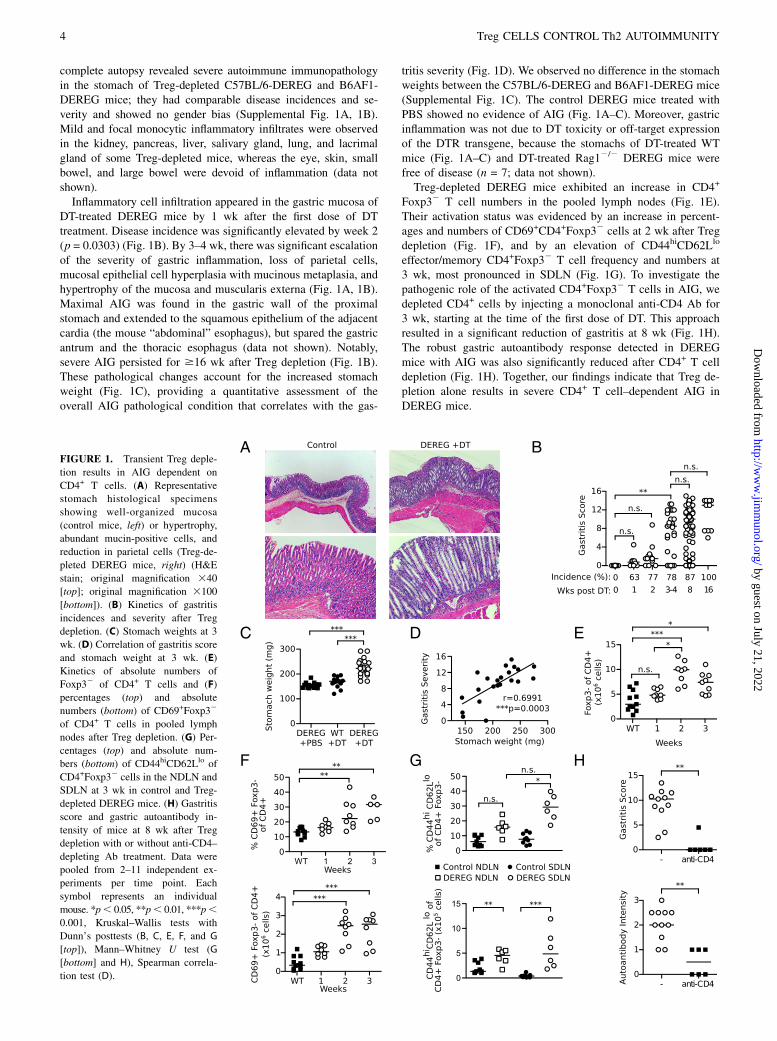
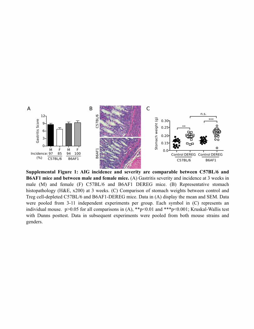
complete autopsy revealed severe autoimmune immunopathologyin the stomach of Treg-depleted C57BL/6-DEREG and B6AF1-DEREG mice; they had comparable disease incidences and se-verity and showed no gender bias (Supplemental Fig. 1A, 1B).Mild and focal monocytic inflammatory infiltrates were observedin the kidney, pancreas, liver, salivary gland, lung, and lacrimalgland of some Treg-depleted mice, whereas the eye, skin, smallbowel, and large bowel were devoid of inflammation (data notshown).Inflammatory cell infiltration appeared in the gastric mucosa of
DT-treated DEREG mice by 1 wk after the first dose of DTtreatment. Disease incidence was significantly elevated by week 2(p = 0.0303) (Fig. 1B). By 3–4 wk, there was significant escalationof the severity of gastric inflammation, loss of parietal cells,mucosal epithelial cell hyperplasia with mucinous metaplasia, andhypertrophy of the mucosa and muscularis externa (Fig. 1A, 1B).Maximal AIG was found in the gastric wall of the proximalstomach and extended to the squamous epithelium of the adjacentcardia (the mouse “abdominal” esophagus), but spared the gastricantrum and the thoracic esophagus (data not shown). Notably,severe AIG persisted for $16 wk after Treg depletion (Fig. 1B).These pathological changes account for the increased stomachweight (Fig. 1C), providing a quantitative assessment of theoverall AIG pathological condition that correlates with the gas-
tritis severity (Fig. 1D). We observed no difference in the stomachweights between the C57BL/6-DEREG and B6AF1-DEREG mice(Supplemental Fig. 1C). The control DEREG mice treated withPBS showed no evidence of AIG (Fig. 1A–C). Moreover, gastricinflammation was not due to DT toxicity or off-target expressionof the DTR transgene, because the stomachs of DT-treated WTmice (Fig. 1A–C) and DT-treated Rag12/2 DEREG mice werefree of disease (n = 7; data not shown).Treg-depleted DEREG mice exhibited an increase in CD4+
Foxp32 T cell numbers in the pooled lymph nodes (Fig. 1E).Their activation status was evidenced by an increase in percent-ages and numbers of CD69+CD4+Foxp32 cells at 2 wk after Tregdepletion (Fig. 1F), and by an elevation of CD44hiCD62Llo
effector/memory CD4+Foxp32 T cell frequency and numbers at3 wk, most pronounced in SDLN (Fig. 1G). To investigate thepathogenic role of the activated CD4+Foxp32 T cells in AIG, wedepleted CD4+ cells by injecting a monoclonal anti-CD4 Ab for3 wk, starting at the time of the first dose of DT. This approachresulted in a significant reduction of gastritis at 8 wk (Fig. 1H).The robust gastric autoantibody response detected in DEREGmice with AIG was also significantly reduced after CD4+ T celldepletion (Fig. 1H). Together, our findings indicate that Treg de-pletion alone results in severe CD4+ T cell–dependent AIG inDEREG mice.
FIGURE 1. Transient Treg deple-
tion results in AIG dependent on
CD4+ T cells. (A) Representative
stomach histological specimens
showing well-organized mucosa
(control mice, left) or hypertrophy,
abundant mucin-positive cells, and
reduction in parietal cells (Treg-de-
pleted DEREG mice, right) (H&E
stain; original magnification 340
[top]; original magnification 3100
[bottom]). (B) Kinetics of gastritis
incidences and severity after Treg
depletion. (C) Stomach weights at 3
wk. (D) Correlation of gastritis score
and stomach weight at 3 wk. (E)
Kinetics of absolute numbers of
Foxp32 of CD4+ T cells and (F)
percentages (top) and absolute
numbers (bottom) of CD69+Foxp32
of CD4+ T cells in pooled lymph
nodes after Treg depletion. (G) Per-
centages (top) and absolute num-
bers (bottom) of CD44hiCD62Llo of
CD4+Foxp32 cells in the NDLN and
SDLN at 3 wk in control and Treg-
depleted DEREG mice. (H) Gastritis
score and gastric autoantibody in-
tensity of mice at 8 wk after Treg
depletion with or without anti-CD4–
depleting Ab treatment. Data were
pooled from 2–11 independent ex-
periments per time point. Each
symbol represents an individual
mouse. *p, 0.05, **p, 0.01, ***p,0.001, Kruskal–Wallis tests with
Dunn’s posttests (B, C, E, F, and G
[top]), Mann–Whitney U test (G
[bottom] and H), Spearman correla-
tion test (D).
4 Treg CELLS CONTROL Th2 AUTOIMMUNITY
by guest on July 21, 2022http://w
ww
.jimm
unol.org/D
ownloaded from
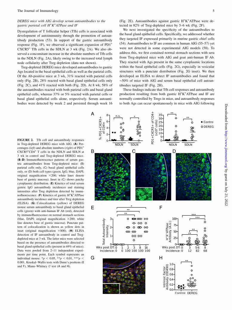
DEREG mice with AIG develop serum autoantibodies to thegastric parietal cell H+K+ATPase and IF
Dysregulation of T follicular helper (Tfh) cells is associated withdevelopment of autoimmunity through the promotion of autoan-
tibody production (53). In support of the gastric autoantibody
response (Fig. 1F), we observed a significant expansion of PD1+
CXCR5+ Tfh cells in the SDLN at 3 wk (Fig. 2A). We also ob-
served a concomitant increase in the absolute numbers of Tfh cells
in the NDLN (Fig. 2A), likely owing to the increased total lymph
node cellularity after Treg depletion (data not shown).Treg-depleted DEREG mice developed autoantibodies to gastric
Ags located in the basal epithelial cells as well as the parietal cells.
Of the Ab-positive mice at 3 wk, 31% reacted with parietal cells
only (Fig. 2B), 28% reacted with basal gland epithelial cells only
(Fig. 2C), and 41% reacted with both (Fig. 2D). At 8 wk, 58% of
the autoantibodies reacted with both parietal cells and basal gland
epithelial cells, whereas 37% or 5% reacted with parietal cells or
basal gland epithelial cells alone, respectively. Serum autoanti-
bodies were detected by week 2 and persisted through week 16
(Fig. 2E). Autoantibodies against gastric H+K+ATPase were de-tected in 82% of Treg-depleted mice by 5–6 wk (Fig. 2F).We next investigated the specificity of the autoantibodies to
the basal gland epithelial cells. Specifically, we addressed whether
they targeted IF expressed primarily in murine gastric chief cells
(54). Autoantibodies to IF are common in human AIG (55–57) yet
were not detected in some experimental AIG models (58). To
address this, we first costained normal stomach sections with sera
from Treg-depleted mice with AIG and goat anti-human IF Ab.
They reacted with Ags present in the same cytoplasmic locations
within the basal epithelial cells (Fig. 2G), especially in vesicular
structures with a punctate distribution (Fig. 2G inset). We then
developed an ELISA to detect IF autoantibodies and found that
∼50% of mice with AIG and serum basal epithelial cell autoan-
tibodies targeted IF (Fig. 2H).These findings indicate that Tfh cell responses and autoantibody
production resulting from both gastric H+K+ATPase and IF are
normally controlled by Tregs in mice, and autoantibody responses
to both Ags can occur spontaneously in mice with AIG following
FIGURE 2. Tfh cell and autoantibody responses
in Treg-depleted DEREG mice with AIG. (A) Per-
centages (left) and absolute numbers (right) of PD1+
CXCR5+CD4+ T cells in the NDLN and SDLN at
3 wk in control and Treg-depleted DEREG mice.
(B–D) Immunofluorescence patterns of serum gas-
tric autoantibodies from Treg-depleted mice: (B)
parietal cells only, (C) basal gland epithelial cells
only, or (D) both cell types (green, IgG; blue, DAPI;
original magnification 3200; white lines denote
base of gastric mucosa). Inset in (C) shows patchy
cytoplasmic distribution. (E) Kinetics of total serum
gastric IgG autoantibody incidences and staining
intensities after Treg depletion detected by immu-
nofluorescence. (F) Kinetics of gastric H+K+ATPase
autoantibody incidence and titer after Treg depletion
(ELISA). (G) Colocalization (yellow) of DEREG
mouse serum autoantibody to basal gland epithelial
cells (green) with anti-human IF Ab (red), detected
by immunofluorescence on normal stomach sections
(blue, DAPI; original magnification 3200; white
line denotes base of gastric mucosa). Punctate pat-
tern of colocalization is shown as yellow dots in
inset (original magnification 3800). (H) ELISA
detection of IF autoantibody in control and Treg-
depleted mice at 3 wk. The latter mice were selected
based on the presence of autoantibodies directed to
basal gland epithelial cells (present in 69% of mice).
Data were pooled from 2–11 independent experi-
ments per time point. Each symbol represents an
individual mouse. *p , 0.05, **p , 0.01, ***p ,0.001, Kruskal–Wallis tests with Dunn’s posttests (E
and F), Mann–Whitney U test (A and H).
The Journal of Immunology 5
by guest on July 21, 2022http://w
ww
.jimm
unol.org/D
ownloaded from
Treg depletion. The presence of these autoantibodies is consistentwith the clinical observations in patients with AIG (20).
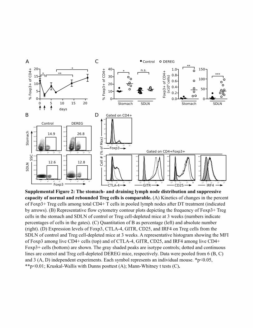
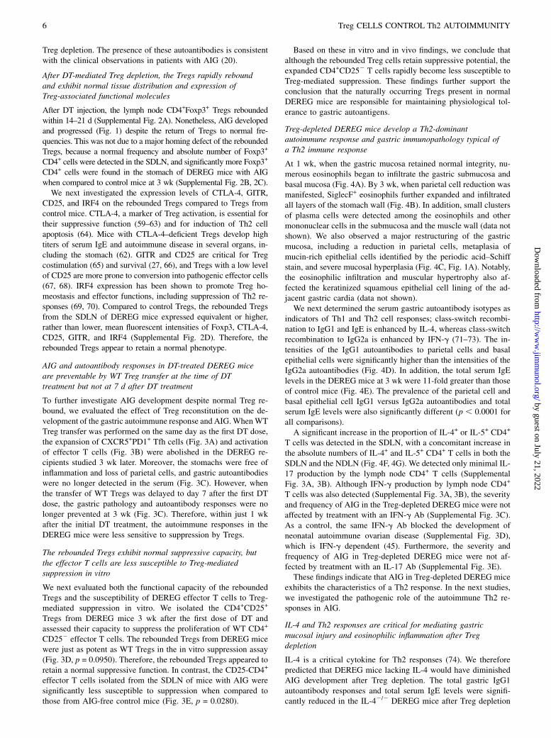
After DT-mediated Treg depletion, the Tregs rapidly reboundand exhibit normal tissue distribution and expression ofTreg-associated functional molecules
After DT injection, the lymph node CD4+Foxp3+ Tregs reboundedwithin 14–21 d (Supplemental Fig. 2A). Nonetheless, AIG developedand progressed (Fig. 1) despite the return of Tregs to normal fre-quencies. This was not due to a major homing defect of the reboundedTregs, because a normal frequency and absolute number of Foxp3+
CD4+ cells were detected in the SDLN, and significantly more Foxp3+
CD4+ cells were found in the stomach of DEREG mice with AIGwhen compared to control mice at 3 wk (Supplemental Fig. 2B, 2C).We next investigated the expression levels of CTLA-4, GITR,
CD25, and IRF4 on the rebounded Tregs compared to Tregs fromcontrol mice. CTLA-4, a marker of Treg activation, is essential fortheir suppressive function (59–63) and for induction of Th2 cellapoptosis (64). Mice with CTLA-4–deficient Tregs develop hightiters of serum IgE and autoimmune disease in several organs, in-cluding the stomach (62). GITR and CD25 are critical for Tregcostimulation (65) and survival (27, 66), and Tregs with a low levelof CD25 are more prone to conversion into pathogenic effector cells(67, 68). IRF4 expression has been shown to promote Treg ho-meostasis and effector functions, including suppression of Th2 re-sponses (69, 70). Compared to control Tregs, the rebounded Tregsfrom the SDLN of DEREG mice expressed equivalent or higher,rather than lower, mean fluorescent intensities of Foxp3, CTLA-4,CD25, GITR, and IRF4 (Supplemental Fig. 2D). Therefore, therebounded Tregs appear to retain a normal phenotype.
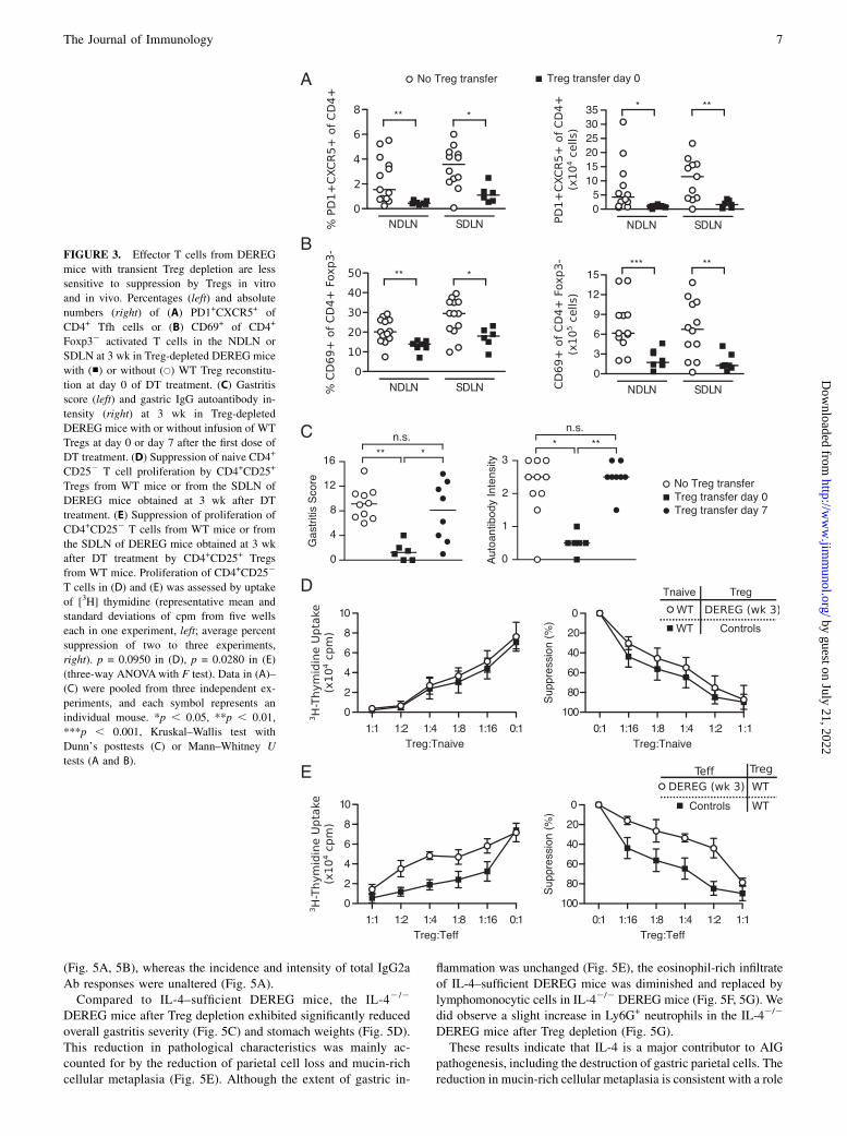
AIG and autoantibody responses in DT-treated DEREG miceare preventable by WT Treg transfer at the time of DTtreatment but not at 7 d after DT treatment
To further investigate AIG development despite normal Treg re-bound, we evaluated the effect of Treg reconstitution on the de-velopment of the gastric autoimmune response and AIG.WhenWTTreg transfer was performed on the same day as the first DT dose,the expansion of CXCR5+PD1+ Tfh cells (Fig. 3A) and activationof effector T cells (Fig. 3B) were abolished in the DEREG re-cipients studied 3 wk later. Moreover, the stomachs were free ofinflammation and loss of parietal cells, and gastric autoantibodieswere no longer detected in the serum (Fig. 3C). However, whenthe transfer of WT Tregs was delayed to day 7 after the first DTdose, the gastric pathology and autoantibody responses were nolonger prevented at 3 wk (Fig. 3C). Therefore, within just 1 wkafter the initial DT treatment, the autoimmune responses in theDEREG mice were less sensitive to suppression by Tregs.
The rebounded Tregs exhibit normal suppressive capacity, butthe effector T cells are less susceptible to Treg-mediatedsuppression in vitro
We next evaluated both the functional capacity of the reboundedTregs and the susceptibility of DEREG effector T cells to Treg-mediated suppression in vitro. We isolated the CD4+CD25+
Tregs from DEREG mice 3 wk after the first dose of DT andassessed their capacity to suppress the proliferation of WT CD4+
CD252 effector T cells. The rebounded Tregs from DEREG micewere just as potent as WT Tregs in the in vitro suppression assay(Fig. 3D, p = 0.0950). Therefore, the rebounded Tregs appeared toretain a normal suppressive function. In contrast, the CD25-CD4+
effector T cells isolated from the SDLN of mice with AIG weresignificantly less susceptible to suppression when compared tothose from AIG-free control mice (Fig. 3E, p = 0.0280).
Based on these in vitro and in vivo findings, we conclude thatalthough the rebounded Treg cells retain suppressive potential, theexpanded CD4+CD252 T cells rapidly become less susceptible toTreg-mediated suppression. These findings further support theconclusion that the naturally occurring Tregs present in normalDEREG mice are responsible for maintaining physiological tol-erance to gastric autoantigens.
Treg-depleted DEREG mice develop a Th2-dominantautoimmune response and gastric immunopathology typical ofa Th2 immune response
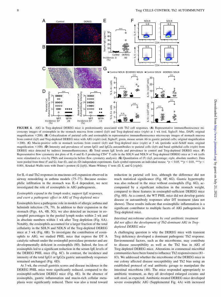
At 1 wk, when the gastric mucosa retained normal integrity, nu-merous eosinophils began to infiltrate the gastric submucosa andbasal mucosa (Fig. 4A). By 3 wk, when parietal cell reduction wasmanifested, SiglecF+ eosinophils further expanded and infiltratedall layers of the stomach wall (Fig. 4B). In addition, small clustersof plasma cells were detected among the eosinophils and othermononuclear cells in the submucosa and the muscle wall (data notshown). We also observed a major restructuring of the gastricmucosa, including a reduction in parietal cells, metaplasia ofmucin-rich epithelial cells identified by the periodic acid–Schiffstain, and severe mucosal hyperplasia (Fig. 4C, Fig. 1A). Notably,the eosinophilic infiltration and muscular hypertrophy also af-fected the keratinized squamous epithelial cell lining of the ad-jacent gastric cardia (data not shown).We next determined the serum gastric autoantibody isotypes as
indicators of Th1 and Th2 cell responses; class-switch recombi-nation to IgG1 and IgE is enhanced by IL-4, whereas class-switchrecombination to IgG2a is enhanced by IFN-g (71–73). The in-tensities of the IgG1 autoantibodies to parietal cells and basalepithelial cells were significantly higher than the intensities of theIgG2a autoantibodies (Fig. 4D). In addition, the total serum IgElevels in the DEREG mice at 3 wk were 11-fold greater than thoseof control mice (Fig. 4E). The prevalence of the parietal cell andbasal epithelial cell IgG1 versus IgG2a autoantibodies and totalserum IgE levels were also significantly different (p , 0.0001 forall comparisons).A significant increase in the proportion of IL-4+ or IL-5+ CD4+
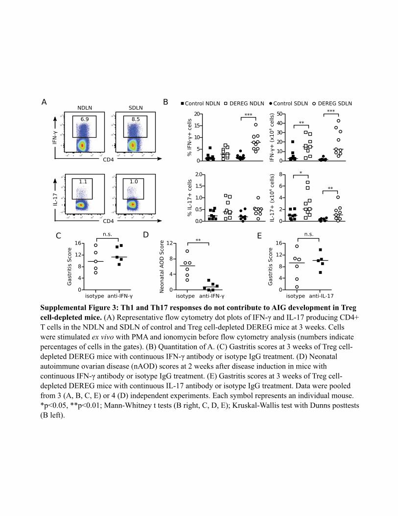
T cells was detected in the SDLN, with a concomitant increase inthe absolute numbers of IL-4+ and IL-5+ CD4+ T cells in both theSDLN and the NDLN (Fig. 4F, 4G). We detected only minimal IL-17 production by the lymph node CD4+ T cells (SupplementalFig. 3A, 3B). Although IFN-g production by lymph node CD4+
T cells was also detected (Supplemental Fig. 3A, 3B), the severityand frequency of AIG in the Treg-depleted DEREG mice were notaffected by treatment with an IFN-g Ab (Supplemental Fig. 3C).As a control, the same IFN-g Ab blocked the development ofneonatal autoimmune ovarian disease (Supplemental Fig. 3D),which is IFN-g dependent (45). Furthermore, the severity andfrequency of AIG in Treg-depleted DEREG mice were not af-fected by treatment with an IL-17 Ab (Supplemental Fig. 3E).These findings indicate that AIG in Treg-depleted DEREG mice
exhibits the characteristics of a Th2 response. In the next studies,we investigated the pathogenic role of the autoimmune Th2 re-sponses in AIG.
IL-4 and Th2 responses are critical for mediating gastricmucosal injury and eosinophilic inflammation after Tregdepletion
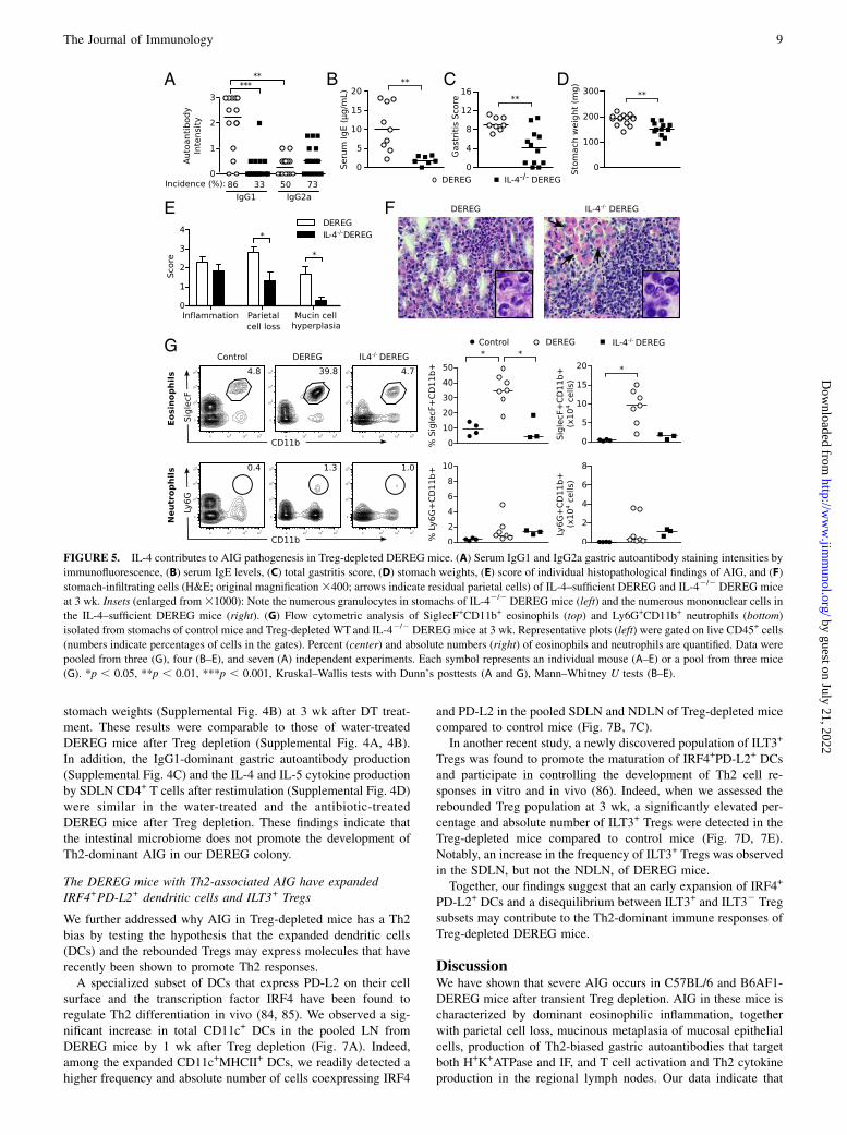
IL-4 is a critical cytokine for Th2 responses (74). We thereforepredicted that DEREG mice lacking IL-4 would have diminishedAIG development after Treg depletion. The total gastric IgG1autoantibody responses and total serum IgE levels were signifi-cantly reduced in the IL-42/2 DEREG mice after Treg depletion
6 Treg CELLS CONTROL Th2 AUTOIMMUNITY
by guest on July 21, 2022http://w
ww
.jimm
unol.org/D
ownloaded from
(Fig. 5A, 5B), whereas the incidence and intensity of total IgG2aAb responses were unaltered (Fig. 5A).Compared to IL-4–sufficient DEREG mice, the IL-42/2
DEREG mice after Treg depletion exhibited significantly reducedoverall gastritis severity (Fig. 5C) and stomach weights (Fig. 5D).This reduction in pathological characteristics was mainly ac-counted for by the reduction of parietal cell loss and mucin-richcellular metaplasia (Fig. 5E). Although the extent of gastric in-
flammation was unchanged (Fig. 5E), the eosinophil-rich infiltrateof IL-4–sufficient DEREG mice was diminished and replaced bylymphomonocytic cells in IL-42/2 DEREG mice (Fig. 5F, 5G). Wedid observe a slight increase in Ly6G+ neutrophils in the IL-42/2
DEREG mice after Treg depletion (Fig. 5G).These results indicate that IL-4 is a major contributor to AIG
pathogenesis, including the destruction of gastric parietal cells. Thereduction in mucin-rich cellular metaplasia is consistent with a role
FIGURE 3. Effector T cells from DEREG
mice with transient Treg depletion are less
sensitive to suppression by Tregs in vitro
and in vivo. Percentages (left) and absolute
numbers (right) of (A) PD1+CXCR5+ of
CD4+ Tfh cells or (B) CD69+ of CD4+
Foxp32 activated T cells in the NDLN or
SDLN at 3 wk in Treg-depleted DEREG mice
with (■) or without (s) WT Treg reconstitu-
tion at day 0 of DT treatment. (C) Gastritis
score (left) and gastric IgG autoantibody in-
tensity (right) at 3 wk in Treg-depleted
DEREG mice with or without infusion of WT
Tregs at day 0 or day 7 after the first dose of
DT treatment. (D) Suppression of naive CD4+
CD252 T cell proliferation by CD4+CD25+
Tregs from WT mice or from the SDLN of
DEREG mice obtained at 3 wk after DT
treatment. (E) Suppression of proliferation of
CD4+CD252 T cells from WT mice or from
the SDLN of DEREG mice obtained at 3 wk
after DT treatment by CD4+CD25+ Tregs
from WT mice. Proliferation of CD4+CD252
T cells in (D) and (E) was assessed by uptake
of [3H] thymidine (representative mean and
standard deviations of cpm from five wells
each in one experiment, left; average percent
suppression of two to three experiments,
right). p = 0.0950 in (D), p = 0.0280 in (E)
(three-way ANOVAwith F test). Data in (A)–
(C) were pooled from three independent ex-
periments, and each symbol represents an
individual mouse. *p , 0.05, **p , 0.01,
***p , 0.001, Kruskal–Wallis test with
Dunn’s posttests (C) or Mann–Whitney U
tests (A and B).
The Journal of Immunology 7
by guest on July 21, 2022http://w
ww
.jimm
unol.org/D
ownloaded from
for IL-4 and Th2 responses in mucinous cell expansion observed inairway remodeling in asthma models (75–77). Because eosino-philic infiltration in the stomach was IL-4 dependent, we nextinvestigated the role of eosinophils in AIG pathogenesis.
Eosinophils expand in the lymph nodes, support IgE responses,and exert a pathogenic effect in AIG of Treg-depleted mice
Eosinophils have a pathogenic role inmodels of allergic asthma andhelminth infection (78, 79). In addition to their expansion in thestomach (Figs. 4A, 4B, 5G), we also detected an increase in eo-sinophil percentages in the pooled lymph nodes within 2 wk andin absolute numbers within 1 wk after Treg depletion (Fig. 6A).Notably, the eosinophils accounted for a larger fraction of the totalcellularity in the SDLN and NDLN of the Treg-depleted DEREGmice at 3 wk (Fig. 6B). To investigate the contribution of eosin-ophils to AIG, we studied PHIL mice that express the DT Acatalytic subunit under the eosinophil peroxidase promoter and aredevelopmentally deficient in eosinophils (80). Indeed, the loss ofeosinophils led to a significant reduction in the serum IgE levels ofthe DEREG PHIL mice (Fig. 6C). In contrast, the incidence andintensity of the total IgG1 or IgG2a gastric autoantibody responsesremained unchanged (Fig. 6D).At 3 wk, the overall gastritis scores and disease incidence in the
DEREG PHIL mice were significantly reduced, compared to theeosinophil-sufficient DEREG mice (Fig. 6E). In the absence ofeosinophils, gastric inflammation and mucin-rich cellular meta-plasia were significantly reduced. There was also a trend toward
reduction in parietal cell loss, although the difference did notreach statistical significance (Fig. 6F, 6G). Gastric hypertrophywas also reduced in the mice without eosinophils (Fig. 6G), ac-companied by a significant reduction in the stomach weight,compared to these features in eosinophil-sufficient DEREG mice(Fig. 6H). As a control, the WT PHIL mice did not develop gastricdisease or autoantibody responses after DT treatment (data notshown). These results indicate that eosinophilic inflammation is asignificant contributor to multiple facets of AIG pathogenesis inTreg-depleted mice.
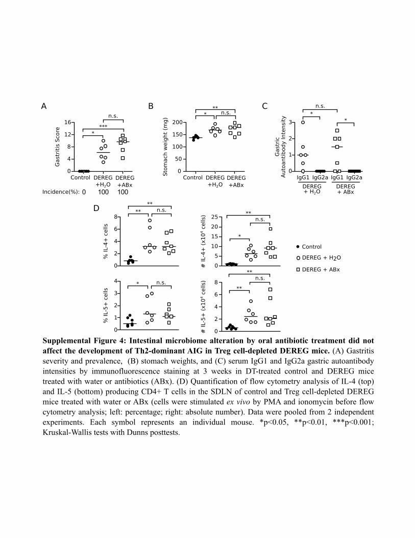
Intestinal microbiome alteration by oral antibiotic treatmentdid not affect the development of Th2-dominant AIG in Treg-depleted DEREG mice
A challenging question is why the DEREG mice with transientTreg deficiency developed a dominant pathogenic Th2 response.Environmental factors, such as the microbiome, may contributeto disease susceptibility as well as the Th2 bias in AIG ofTreg-depleted DEREG mice. Alterations in commensal microbialcommunities have been found to influence Th2 responsiveness (81–83). We addressed whether the microbiome of the DEREG mice inour colony affected disease susceptibility and Th2 bias using anestablished protocol of oral antibiotic gavage to manipulate theintestinal microbiota (48). The mice responded appropriately toantibiotic treatment, as they all developed enlarged cecums andsoft stool, as expected. Antibiotic-treated DEREG mice developedsevere eosinophilic AIG (Supplemental Fig. 4A) with increased
FIGURE 4. AIG in Treg-depleted DEREG mice is predominantly associated with Th2 cell responses. (A) Representative immunofluorescence mi-
croscopy images of eosinophils in the stomach mucosa from control (left) and Treg-depleted mice (right) at 1 wk (red, SiglecF; blue, DAPI; original
magnification 3200). (B) Colocalization of parietal cells and eosinophils in representative immunofluorescence microscopy images of stomach mucosa
from control (left) and Treg-depleted DEREG mice with AIG (right) (red, SiglecF; green, mouse serum Ab to gastric parietal cells; original magnification
3200). (C) Mucin-positive cells in stomach sections from control (left) and Treg-depleted mice (right) at 3 wk (periodic acid–Schiff stain; original
magnification 3100). (D) Intensity and prevalence of serum IgG1 and IgG2a autoantibodies to parietal cells (left) and basal epithelial cells (right) from
DEREG mice detected by indirect immunofluorescence. (E) Total serum IgE levels and prevalence in control and Treg-depleted DEREG mice. (F)
Representative flow cytometry dot plots of IL-4 and IL-5 producing CD4+ T cells in the SDLN and NDLN of Treg-depleted DEREG mice at 3 wk (cells
were stimulated ex vivo by PMA and ionomycin before flow cytometry analysis). (G) Quantitation of (F) (left, percentage; right, absolute number). Data
were pooled from three (F and G), four (E), and six (D) independent experiments. Each symbol represents an individual mouse. *p, 0.05, **p, 0.01, ***p,0.001, Kruskal–Wallis tests with Dunn’s posttest (G [left]), Mann–Whitney U tests (D, E, and G [right]).
8 Treg CELLS CONTROL Th2 AUTOIMMUNITY
by guest on July 21, 2022http://w
ww
.jimm
unol.org/D
ownloaded from
stomach weights (Supplemental Fig. 4B) at 3 wk after DT treat-ment. These results were comparable to those of water-treatedDEREG mice after Treg depletion (Supplemental Fig. 4A, 4B).In addition, the IgG1-dominant gastric autoantibody production(Supplemental Fig. 4C) and the IL-4 and IL-5 cytokine productionby SDLN CD4+ T cells after restimulation (Supplemental Fig. 4D)were similar in the water-treated and the antibiotic-treatedDEREG mice after Treg depletion. These findings indicate thatthe intestinal microbiome does not promote the development ofTh2-dominant AIG in our DEREG colony.
The DEREG mice with Th2-associated AIG have expandedIRF4+PD-L2+ dendritic cells and ILT3+ Tregs
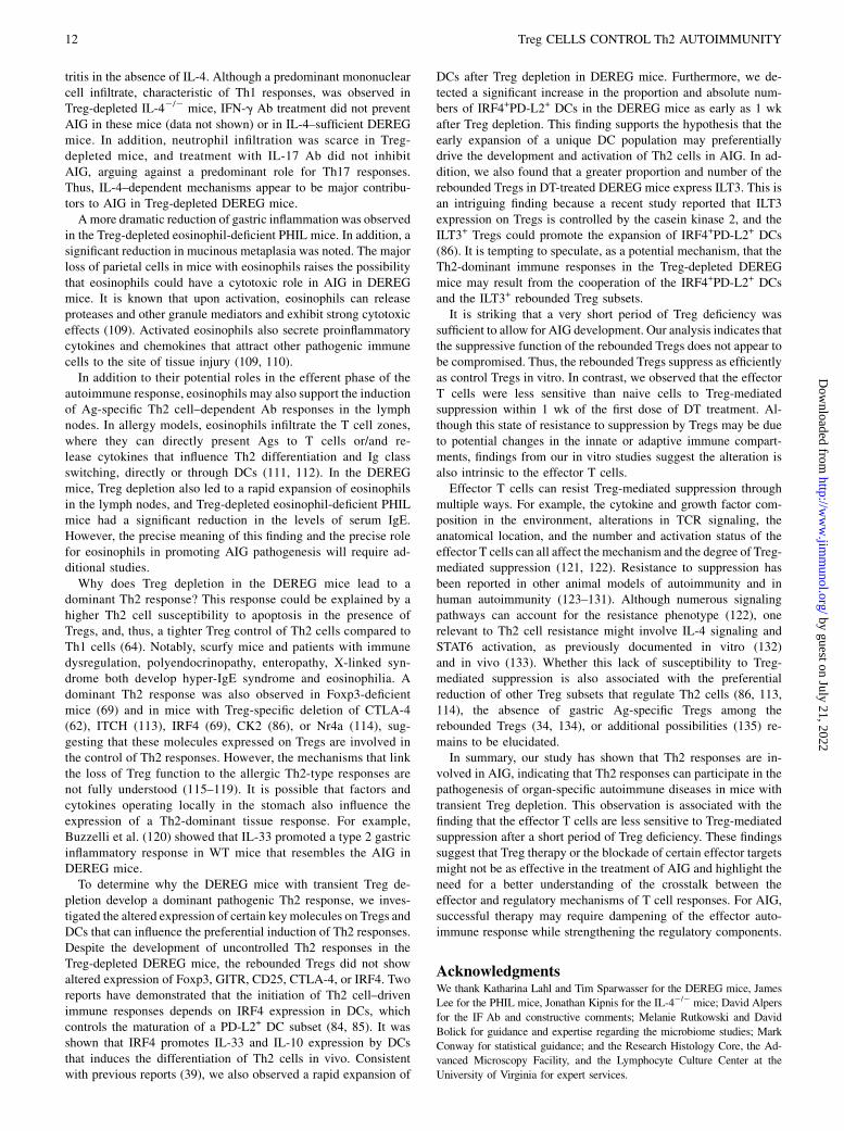
We further addressed why AIG in Treg-depleted mice has a Th2bias by testing the hypothesis that the expanded dendritic cells(DCs) and the rebounded Tregs may express molecules that haverecently been shown to promote Th2 responses.A specialized subset of DCs that express PD-L2 on their cell
surface and the transcription factor IRF4 have been found toregulate Th2 differentiation in vivo (84, 85). We observed a sig-nificant increase in total CD11c+ DCs in the pooled LN fromDEREG mice by 1 wk after Treg depletion (Fig. 7A). Indeed,among the expanded CD11c+MHCII+ DCs, we readily detected ahigher frequency and absolute number of cells coexpressing IRF4
and PD-L2 in the pooled SDLN and NDLN of Treg-depleted micecompared to control mice (Fig. 7B, 7C).In another recent study, a newly discovered population of ILT3+
Tregs was found to promote the maturation of IRF4+PD-L2+ DCsand participate in controlling the development of Th2 cell re-sponses in vitro and in vivo (86). Indeed, when we assessed therebounded Treg population at 3 wk, a significantly elevated per-centage and absolute number of ILT3+ Tregs were detected in theTreg-depleted mice compared to control mice (Fig. 7D, 7E).Notably, an increase in the frequency of ILT3+ Tregs was observedin the SDLN, but not the NDLN, of DEREG mice.Together, our findings suggest that an early expansion of IRF4+
PD-L2+ DCs and a disequilibrium between ILT3+ and ILT32 Tregsubsets may contribute to the Th2-dominant immune responses ofTreg-depleted DEREG mice.
DiscussionWe have shown that severe AIG occurs in C57BL/6 and B6AF1-DEREG mice after transient Treg depletion. AIG in these mice ischaracterized by dominant eosinophilic inflammation, togetherwith parietal cell loss, mucinous metaplasia of mucosal epithelialcells, production of Th2-biased gastric autoantibodies that targetboth H+K+ATPase and IF, and T cell activation and Th2 cytokineproduction in the regional lymph nodes. Our data indicate that
FIGURE 5. IL-4 contributes to AIG pathogenesis in Treg-depleted DEREG mice. (A) Serum IgG1 and IgG2a gastric autoantibody staining intensities by
immunofluorescence, (B) serum IgE levels, (C) total gastritis score, (D) stomach weights, (E) score of individual histopathological findings of AIG, and (F)
stomach-infiltrating cells (H&E; original magnification3400; arrows indicate residual parietal cells) of IL-4–sufficient DEREG and IL-42/2 DEREG mice
at 3 wk. Insets (enlarged from31000): Note the numerous granulocytes in stomachs of IL-42/2 DEREG mice (left) and the numerous mononuclear cells in
the IL-4–sufficient DEREG mice (right). (G) Flow cytometric analysis of SiglecF+CD11b+ eosinophils (top) and Ly6G+CD11b+ neutrophils (bottom)
isolated from stomachs of control mice and Treg-depleted WTand IL-42/2 DEREG mice at 3 wk. Representative plots (left) were gated on live CD45+ cells
(numbers indicate percentages of cells in the gates). Percent (center) and absolute numbers (right) of eosinophils and neutrophils are quantified. Data were
pooled from three (G), four (B–E), and seven (A) independent experiments. Each symbol represents an individual mouse (A–E) or a pool from three mice
(G). *p , 0.05, **p , 0.01, ***p , 0.001, Kruskal–Wallis tests with Dunn’s posttests (A and G), Mann–Whitney U tests (B–E).
The Journal of Immunology 9
by guest on July 21, 2022http://w
ww
.jimm
unol.org/D
ownloaded from

these changes are the product of the rapid loss of effector T cellsusceptibility to suppression by Tregs.Our study, along with others (40, 69), provides conclusive ev-
idence that Tregs are critical in maintaining physiological toler-ance to murine gastric autoantigens. However, our findings differfrom previous AIG studies in several important aspects. First, AIGwas not observed in adult C57BL/6 or BALB/c-DEREG miceafter a similar regimen of Treg depletion (42, 44). The differentoutcomes could be explained by diverse environmental factors aswell as differences in the microbiomes known to influence auto-immune diseases. In this regard, our mice tested negative forpinworm and fur mite infestations known to skew toward Th2responses (87, 88), and the AIG severity and Th2 phenotype werenot altered by a standard regimen of oral antibiotic treatment.Thus, the intestinal microbiota do not appear to promote thegastric-specific autoimmune responses and Th2-biased disease in
our DEREG colony. However, a role for the microbiome in sup-pression of AIG development in other mouse colonies is a pos-sibility that has not been investigated. Second, our study indicatesthat the DEREG and Foxp3DTR knock-in mice (39) exhibit dif-ferent responses and clinical outcomes after Treg depletion, al-though the nature of these differences is not fully understood.Third, similar to other AIG models induced by immunization (89)or d3tx (34), the rebounded Tregs in our model are functional.However, no immunization boost or lymphopenia is required inTreg-depleted DEREG mice for the effector T cells to resist Treg-mediated suppression.The current paradigm posits that autoimmune diseases are de-
pendent on Th1 or Th17 responses and less reliant on Th2 re-sponses, and this applies to studies on AIG (31, 90–92). Th2 cellsare generally regarded as counterinflammatory to Th1-dominantautoimmunity, with therapeutic potential (93–96). However, path-
FIGURE 6. Eosinophils contribute to AIG patho-
genesis in Treg-depleted DEREG mice. (A) Kinetics
of SiglecF+CD11b+ eosinophil expansion in pooled
lymph nodes as percentage (left) and absolute number
(right). (B) Percentage (left) and absolute number
(right) of eosinophils in the NDLN and SDLN of
control and Treg-depleted DEREG mice at 3 wk. (C)
Serum IgE levels, (D) serum IgG1 and IgG2a gastric
autoantibody staining intensities, (E) gastritis severity
and prevalence, (F) score of individual histopatho-
logical findings of AIG, (G) representative stomach
histopathological characteristics (H&E, original mag-
nification 3100), and (H) stomach weights in DEREG
mice (s) and eosinophil-deficient DEREG PHIL mice
(n) at 3 wk. Data were pooled from five (A) or seven
(B–H) independent experiments. Each symbol repre-
sents an individual mouse. *p , 0.05, **p , 0.01,
***p , 0.001, Mann–Whitney U tests (B [right], C, E,
F, and H), Kruskal–Wallis tests with Dunn’s posttest
(A, B [left], and D).
10 Treg CELLS CONTROL Th2 AUTOIMMUNITY
by guest on July 21, 2022http://w
ww
.jimm
unol.org/D
ownloaded from
ogenic Th2 responses in autoimmunity have also been sug-gested by previous studies. For example, studies on AIG havedocumented activated Th1 and Th2 autoantigen-specific T cells inthe regional lymph nodes of d3tx mice. Both the H+K+ATPase-specific Th1 and Th2 cell clones can transfer severe AIG (32), andTCR transgenic mice generated from these T cell clones sponta-neously develop AIG (97, 98). In addition, naive transgenic T cellswere able to transfer disease to immunodeficient recipients afterin vitro differentiation to Th1, Th2, and Th17 cells (91). Diseasetransfer by Th2 cells has also been documented in several otherautoimmune diseases (99–102). In this study, we have providedclear evidence for the spontaneous occurrence of a pathogenic Th2response after Treg depletion alone, without the requirement foradjuvant, lymphopenia, or use of a transgenic TCR. Our findingswill likely have clinical relevance because, despite limited data onhuman AIG, recent studies also support a possible role for Th2responses. Bettington and Brown (103) reported an increased eo-sinophil count in biopsy specimens of patients with AIG, andBedeir et al. (104) detected numerous IgG4-positive plasma B cellsin biopsy specimens of AIG patients with pernicious anemia. Thelatter is notable because human IgG4 production is associated withTh2 responses (105–107). Although it is possible that differentindividual AIG patients may exhibit different combinations of Thcell responses, our model seems appropriate for delineating acomponent of AIG in some patients.Although autoantibodies to gastric H+K+ATPase are well de-
fined (13), this study also provides documentation of autoantibody
responses to the gastric IF in experimental AIG. Autoantibodies toIF and H+K+ATPase are involved in the complications of chronicatrophic gastritis and are useful diagnostic markers of AIG (21).For example, immune-mediated depletion of parietal cells canlead to hypochlorhydria, and autoantibodies to IF, by impairingthe absorption of vitamin B12, can lead to pernicious anemia(108). Patients with AIG and pernicious anemia are reported tohave an increased risk of developing gastric cancer (16–18). In-vestigation of these important sequelae of AIG is not possible withthe current models of AIG caused by Treg depletion. In thisregard, the longevity of AIG in the DEREG mice and their sur-vival after Treg depletion may provide an opportunity for inves-tigation of these sequelae.The evidence for a Th2 type of immune response in the path-
ogenesis of AIG is supported by the following findings: 1) thesubclass of the autoantibody response, 2) the inflammatory cellinfiltrate and epithelial cell response, and 3) the cytokine pro-duction. The pathogenicity of the Th2 response is supported by thesignificant reduction in stomach disease and immune responses inIL-42/2 mice and eosinophil-deficient mice. Strikingly, the phe-notypes of these mice are not identical. The decreased disease inTreg-depleted IL-42/2 mice was mainly attributed to the reducedparietal cell loss and mucin cell hyperplasia. These changes cor-related with a reduced infiltration of eosinophils in the inflamedstomach. However, the degree of inflammation remained compa-rable to that observed in IL-4–sufficient DEREG mice, suggestingthat additional mechanisms are responsible for the residual gas-
FIGURE 7. Increase in Th2-promoting PD-L2+
IRF4+ DC subsets as well as ILT3+ Treg subsets in
transiently Treg-depleted DEREG mice. (A) Ki-
netics of the percentage (top) and absolute number
(bottom) of live, CD11c+ DCs in pooled lymph
nodes. (B) Representative flow cytometry dot plots
(numbers indicate percentages of cells in the gates)
and (C) quantitation as percentage and absolute
number of IRF4+PD-L2+ DCs among live, CD42,
CD11c+, MHCII+ cells in pooled SDLN and
NDLN of control and Treg-depleted DEREG mice
at 1 wk. (D) Representative flow cytometry dot
plots (numbers indicate percentages of cells in the
gates) and (E) quantitation as percentage (top) and
absolute number (bottom) of ILT3+ Tregs among
live, CD4+, Foxp3+ cells in the SDLN or NDLN of
control and Treg-depleted DEREG mice at 3 wk.
Data were pooled from two (B), three (C), or five
(A) independent experiments. Each symbol repre-
sents an individual mouse. *p , 0.05, **p , 0.01,
***p , 0.001, Mann–Whitney U tests (C and E
[bottom]), Kruskal–Wallis tests with Dunn’s post-
test (A and E [top]).
The Journal of Immunology 11
by guest on July 21, 2022http://w
ww
.jimm
unol.org/D
ownloaded from
tritis in the absence of IL-4. Although a predominant mononuclearcell infiltrate, characteristic of Th1 responses, was observed inTreg-depleted IL-42/2 mice, IFN-g Ab treatment did not preventAIG in these mice (data not shown) or in IL-4–sufficient DEREGmice. In addition, neutrophil infiltration was scarce in Treg-depleted mice, and treatment with IL-17 Ab did not inhibitAIG, arguing against a predominant role for Th17 responses.Thus, IL-4–dependent mechanisms appear to be major contribu-tors to AIG in Treg-depleted DEREG mice.A more dramatic reduction of gastric inflammation was observed
in the Treg-depleted eosinophil-deficient PHIL mice. In addition, asignificant reduction in mucinous metaplasia was noted. The majorloss of parietal cells in mice with eosinophils raises the possibilitythat eosinophils could have a cytotoxic role in AIG in DEREGmice. It is known that upon activation, eosinophils can releaseproteases and other granule mediators and exhibit strong cytotoxiceffects (109). Activated eosinophils also secrete proinflammatorycytokines and chemokines that attract other pathogenic immunecells to the site of tissue injury (109, 110).In addition to their potential roles in the efferent phase of the
autoimmune response, eosinophils may also support the inductionof Ag-specific Th2 cell–dependent Ab responses in the lymphnodes. In allergy models, eosinophils infiltrate the T cell zones,where they can directly present Ags to T cells or/and re-lease cytokines that influence Th2 differentiation and Ig classswitching, directly or through DCs (111, 112). In the DEREGmice, Treg depletion also led to a rapid expansion of eosinophilsin the lymph nodes, and Treg-depleted eosinophil-deficient PHILmice had a significant reduction in the levels of serum IgE.However, the precise meaning of this finding and the precise rolefor eosinophils in promoting AIG pathogenesis will require ad-ditional studies.Why does Treg depletion in the DEREG mice lead to a
dominant Th2 response? This response could be explained by ahigher Th2 cell susceptibility to apoptosis in the presence ofTregs, and, thus, a tighter Treg control of Th2 cells compared toTh1 cells (64). Notably, scurfy mice and patients with immunedysregulation, polyendocrinopathy, enteropathy, X-linked syn-drome both develop hyper-IgE syndrome and eosinophilia. Adominant Th2 response was also observed in Foxp3-deficientmice (69) and in mice with Treg-specific deletion of CTLA-4(62), ITCH (113), IRF4 (69), CK2 (86), or Nr4a (114), sug-gesting that these molecules expressed on Tregs are involved inthe control of Th2 responses. However, the mechanisms that linkthe loss of Treg function to the allergic Th2-type responses arenot fully understood (115–119). It is possible that factors andcytokines operating locally in the stomach also influence theexpression of a Th2-dominant tissue response. For example,Buzzelli et al. (120) showed that IL-33 promoted a type 2 gastricinflammatory response in WT mice that resembles the AIG inDEREG mice.To determine why the DEREG mice with transient Treg de-
pletion develop a dominant pathogenic Th2 response, we inves-tigated the altered expression of certain keymolecules on Tregs andDCs that can influence the preferential induction of Th2 responses.Despite the development of uncontrolled Th2 responses in theTreg-depleted DEREG mice, the rebounded Tregs did not showaltered expression of Foxp3, GITR, CD25, CTLA-4, or IRF4. Tworeports have demonstrated that the initiation of Th2 cell–drivenimmune responses depends on IRF4 expression in DCs, whichcontrols the maturation of a PD-L2+ DC subset (84, 85). It wasshown that IRF4 promotes IL-33 and IL-10 expression by DCsthat induces the differentiation of Th2 cells in vivo. Consistentwith previous reports (39), we also observed a rapid expansion of
DCs after Treg depletion in DEREG mice. Furthermore, we de-tected a significant increase in the proportion and absolute num-bers of IRF4+PD-L2+ DCs in the DEREG mice as early as 1 wkafter Treg depletion. This finding supports the hypothesis that theearly expansion of a unique DC population may preferentiallydrive the development and activation of Th2 cells in AIG. In ad-dition, we also found that a greater proportion and number of therebounded Tregs in DT-treated DEREG mice express ILT3. This isan intriguing finding because a recent study reported that ILT3expression on Tregs is controlled by the casein kinase 2, and theILT3+ Tregs could promote the expansion of IRF4+PD-L2+ DCs(86). It is tempting to speculate, as a potential mechanism, that theTh2-dominant immune responses in the Treg-depleted DEREGmice may result from the cooperation of the IRF4+PD-L2+ DCsand the ILT3+ rebounded Treg subsets.It is striking that a very short period of Treg deficiency was
sufficient to allow for AIG development. Our analysis indicates thatthe suppressive function of the rebounded Tregs does not appear tobe compromised. Thus, the rebounded Tregs suppress as efficientlyas control Tregs in vitro. In contrast, we observed that the effectorT cells were less sensitive than naive cells to Treg-mediatedsuppression within 1 wk of the first dose of DT treatment. Al-though this state of resistance to suppression by Tregs may be dueto potential changes in the innate or adaptive immune compart-ments, findings from our in vitro studies suggest the alteration isalso intrinsic to the effector T cells.Effector T cells can resist Treg-mediated suppression through
multiple ways. For example, the cytokine and growth factor com-position in the environment, alterations in TCR signaling, theanatomical location, and the number and activation status of theeffector T cells can all affect the mechanism and the degree of Treg-mediated suppression (121, 122). Resistance to suppression hasbeen reported in other animal models of autoimmunity and inhuman autoimmunity (123–131). Although numerous signalingpathways can account for the resistance phenotype (122), onerelevant to Th2 cell resistance might involve IL-4 signaling andSTAT6 activation, as previously documented in vitro (132)and in vivo (133). Whether this lack of susceptibility to Treg-mediated suppression is also associated with the preferentialreduction of other Treg subsets that regulate Th2 cells (86, 113,114), the absence of gastric Ag-specific Tregs among therebounded Tregs (34, 134), or additional possibilities (135) re-mains to be elucidated.In summary, our study has shown that Th2 responses are in-
volved in AIG, indicating that Th2 responses can participate in thepathogenesis of organ-specific autoimmune diseases in mice withtransient Treg depletion. This observation is associated with thefinding that the effector T cells are less sensitive to Treg-mediatedsuppression after a short period of Treg deficiency. These findingssuggest that Treg therapy or the blockade of certain effector targetsmight not be as effective in the treatment of AIG and highlight theneed for a better understanding of the crosstalk between theeffector and regulatory mechanisms of T cell responses. For AIG,successful therapy may require dampening of the effector auto-immune response while strengthening the regulatory components.
AcknowledgmentsWe thank Katharina Lahl and Tim Sparwasser for the DEREG mice, James
Lee for the PHIL mice, Jonathan Kipnis for the IL-42/2 mice; David Alpers
for the IF Ab and constructive comments; Melanie Rutkowski and David
Bolick for guidance and expertise regarding the microbiome studies; Mark
Conway for statistical guidance; and the Research Histology Core, the Ad-
vanced Microscopy Facility, and the Lymphocyte Culture Center at the
University of Virginia for expert services.
12 Treg CELLS CONTROL Th2 AUTOIMMUNITY
by guest on July 21, 2022http://w
ww
.jimm
unol.org/D
ownloaded from
DisclosuresThe authors have no financial conflicts of interest.
References1. Xing, Y., and K. A. Hogquist. 2012. T-cell tolerance: central and peripheral.
Cold Spring Harb. Perspect. Biol. 4: a006957.2. Sakaguchi, S. 2004. Naturally arising CD4+ regulatory T cells for immunologic
self-tolerance and negative control of immune responses. Annu. Rev. Immunol.22: 531–562.
3. Hori, S., T. Nomura, and S. Sakaguchi. 2003. Control of regulatory T celldevelopment by the transcription factor Foxp3. Science 299: 1057–1061.
4. Fontenot, J. D., M. A. Gavin, and A. Y. Rudensky. 2003. Foxp3 programs thedevelopment and function of CD4+CD25+ regulatory T cells. Nat. Immunol. 4:330–336.
5. Wan, Y. Y., and R. A. Flavell. 2007. Regulatory T-cell functions are subvertedand converted owing to attenuated Foxp3 expression. Nature 445: 766–770.
6. Brunkow, M. E., E. W. Jeffery, K. A. Hjerrild, B. Paeper, L. B. Clark,S. A. Yasayko, J. E. Wilkinson, D. Galas, S. F. Ziegler, and F. Ramsdell. 2001.Disruption of a new forkhead/winged-helix protein, scurfin, results in the fatallymphoproliferative disorder of the scurfy mouse. Nat. Genet. 27: 68–73.
7. Khattri, R., T. Cox, S.-A. Yasayko, and F. Ramsdell. 2003. An essential role forScurfin in CD4+CD25+ T regulatory cells. Nat. Immunol. 4: 337–342.
8. Bennett, C. L., J. Christie, F. Ramsdell, M. E. Brunkow, P. J. Ferguson,L. Whitesell, T. E. Kelly, F. T. Saulsbury, P. F. Chance, and H. D. Ochs. 2001.The immune dysregulation, polyendocrinopathy, enteropathy, X-linked syn-drome (IPEX) is caused by mutations of FOXP3. Nat. Genet. 27: 20–21.
9. Torgerson, T. R., and H. D. Ochs. 2007. Immune dysregulation, poly-endocrinopathy, enteropathy, X-linked: forkhead box protein 3 mutations and lackof regulatory T cells. J. Allergy Clin. Immunol. 120: 744–750, quiz 751–752.
10. Jeffries, G. H., D. W. Hoskins, and M. H. Sleisenger. 1962. Antibody to in-trinsic factor in serum from patients with pernicious anemia. J. Clin. Invest. 41:1106–1115.
11. Irvine, W. J., S. H. Davies, S. Teitelbaum, I. W. Delamore, and A. W. Williams.1965. The clinical and pathological significance of gastric parietal cell anti-body. Ann. N. Y. Acad. Sci. 124: 657–691.
12. Karlsson, F. A., P. Burman, L. Loof, and S. Mardh. 1988. Major parietal cellantigen in autoimmune gastritis with pernicious anemia is the acid-producingH+,K+-adenosine triphosphatase of the stomach. J. Clin. Invest. 81: 475–479.
13. Goldkorn, I., P. A. Gleeson, and B. H. Toh. 1989. Gastric parietal cell antigensof 60-90, 92, and 100-120 kDa associated with autoimmune gastritis and per-nicious anemia. Role of N-glycans in the structure and antigenicity of the 60-90-kDa component. J. Biol. Chem. 264: 18768–18774.
14. Toh, B. H., P. A. Gleeson, R. J. Simpson, R. L. Moritz, J. M. Callaghan,I. Goldkorn, C. M. Jones, T. M. Martinelli, F. T. Mu, D. C. Humphris, et al.1990. The 60- to 90-kDa parietal cell autoantigen associated with autoimmunegastritis is a beta subunit of the gastric H+/K(+)-ATPase (proton pump). Proc.Natl. Acad. Sci. USA 87: 6418–6422.
15. Jones, C. M., B. H. Toh, J. M. Pettitt, T. M. Martinelli, D. C. Humphris,J. M. Callaghan, I. Goldkorn, F. T. Mu, and P. A. Gleeson. 1991. Monoclonalantibodies specific for the core protein of the beta-subunit of the gastric protonpump (H+/K+ ATPase). An autoantigen targetted in pernicious anaemia. Eur. J.Biochem. 197: 49–59.
16. Borch, K., H. Renvall, and G. Liedberg. 1985. Gastric endocrine cell hyperplasiaand carcinoid tumors in pernicious anemia. Gastroenterology 88: 638–648.
17. Armbrecht, U., R. W. Stockbr€ugger, J. Rode, G. G. Menon, and P. B. Cotton.1990. Development of gastric dysplasia in pernicious anaemia: a clinical andendoscopic follow up study of 80 patients. Gut 31: 1105–1109.
18. Kokkola, A., S. M. Sjoblom, R. Haapiainen, P. Sipponen, P. Puolakkainen, andH. Jarvinen. 1998. The risk of gastric carcinoma and carcinoid tumours inpatients with pernicious anaemia. A prospective follow-up study. Scand. J.Gastroenterol. 33: 88–92.
19. Carmel, R. 1996. Prevalence of undiagnosed pernicious anemia in the elderly.Arch. Intern. Med. 156: 1097–1100.
20. Toh, B. H., I. R. van Driel, and P. A. Gleeson. 1997. Pernicious anemia. N.Engl. J. Med. 337: 1441–1448.
21. van Driel, I. R., E. Tu, and P. A. Gleeson. 2014. The Autoimmune Diseases. InThe Autoimmune Diseases, 5th Ed., N. R. Rose, and I. R. Machay, eds. Aca-demic Press, Cambridge, MA, p. 619–631.
22. Kojima, A., and R. T. Prehn. 1981. Genetic susceptibility to post-thymectomyautoimmune diseases in mice. Immunogenetics 14: 15–27.
23. Sakaguchi, S., K. Fukuma, K. Kuribayashi, and T. Masuda. 1985. Organ-specific autoimmune diseases induced in mice by elimination of T cell sub-set. I. Evidence for the active participation of T cells in natural self-tolerance;deficit of a T cell subset as a possible cause of autoimmune disease. J. Exp.Med. 161: 72–87.
24. Fukuma, K., S. Sakaguchi, K. Kuribayashi, W. L. Chen, R. Morishita,K. Sekita, H. Uchino, and T. Masuda. 1988. Immunologic and clinical studieson murine experimental autoimmune gastritis induced by neonatal thymectomy.Gastroenterology 94: 274–283.
25. Tung, K. S. 1994. Mechanism of self-tolerance and events leading to autoim-mune disease and autoantibody response. Clin. Immunol. Immunopathol. 73:275–282.
26. Asano, M., M. Toda, N. Sakaguchi, and S. Sakaguchi. 1996. Autoimmunedisease as a consequence of developmental abnormality of a T cell subpopu-lation. J. Exp. Med. 184: 387–396.
27. Sakaguchi, S., N. Sakaguchi, M. Asano, M. Itoh, and M. Toda. 1995. Immu-nologic self-tolerance maintained by activated T cells expressing IL-2 receptoralpha-chains (CD25). Breakdown of a single mechanism of self-tolerancecauses various autoimmune diseases. J. Immunol. 155: 1151–1164.
28. Sakaguchi, S., M. Toda, M. Asano, M. Itoh, S. S. Morse, and N. Sakaguchi.1996. T cell-mediated maintenance of natural self-tolerance: its breakdown as apossible cause of various autoimmune diseases. J. Autoimmun. 9: 211–220.
29. Itoh, M., T. Takahashi, N. Sakaguchi, Y. Kuniyasu, J. Shimizu, F. Otsuka, andS. Sakaguchi. 1999. Thymus and autoimmunity: production of CD25+CD4+naturally anergic and suppressive T cells as a key function of the thymus inmaintaining immunologic self-tolerance. J. Immunol. 162: 5317–5326.
30. Tung, K. S., S. Smith, C. Teuscher, C. Cook, and R. E. Anderson. 1987. Murineautoimmune oophoritis, epididymoorchitis, and gastritis induced by day 3thymectomy. Immunopathology. Am. J. Pathol. 126: 293–302.
31. DiPaolo, R. J., D. D. Glass, K. E. Bijwaard, and E. M. Shevach. 2005. CD4+CD25+ T cells prevent the development of organ-specific autoimmune diseaseby inhibiting the differentiation of autoreactive effector T cells. J. Immunol.175: 7135–7142.
32. Suri-Payer, E., A. Z. Amar, R. McHugh, K. Natarajan, D. H. Margulies, andE. M. Shevach. 1999. Post-thymectomy autoimmune gastritis: fine specificityand pathogenicity of anti-H/K ATPase-reactive T cells. Eur. J. Immunol. 29:669–677.
33. Dujardin, H. C., O. Burlen-Defranoux, L. Boucontet, P. Vieira, A. Cumano, andA. Bandeira. 2004. Regulatory potential and control of Foxp3 expression innewborn CD4+ T cells. Proc. Natl. Acad. Sci. USA 101: 14473–14478.
34. Samy, E. T., K. M. Wheeler, R. J. Roper, C. Teuscher, and K. S. K. Tung. 2008.Cutting edge: Autoimmune disease in day 3 thymectomized mice is activelycontrolled by endogenous disease-specific regulatory T cells. J. Immunol. 180:4366–4370.
35. Wheeler, K. M., E. T. Samy, and K. S. K. Tung. 2009. Cutting edge: normalregional lymph node enrichment of antigen-specific regulatory T cells withautoimmune disease-suppressive capacity. J. Immunol. 183: 7635–7638.
36. Gleeson, P. A., B. H. Toh, and I. R. van Driel. 1996. Organ-specific autoim-munity induced by lymphopenia. Immunol. Rev. 149: 97–125.
37. Monteiro, J. P., J. Farache, A. C. Mercadante, J. A. Mignaco, M. Bonamino, andA. Bonomo. 2008. Pathogenic effector T cell enrichment overcomes regulatoryT cell control and generates autoimmune gastritis. J. Immunol. 181: 5895–5903.
38. Datta, S., and N. Sarvetnick. 2009. Lymphocyte proliferation in immune-mediated diseases. Trends Immunol. 30: 430–438.
39. Kim, J. M., J. P. Rasmussen, and A. Y. Rudensky. 2007. Regulatory T cellsprevent catastrophic autoimmunity throughout the lifespan of mice. Nat.Immunol. 8: 191–197.
40. Nystrom, S. N., D. Bourges, S. Garry, E. M. Ross, I. R. van Driel, andP. A. Gleeson. 2014. Transient Treg-cell depletion in adult mice results in per-sistent self-reactive CD4(+) T-cell responses. Eur. J. Immunol. 44: 3621–3631.
41. Teh, C. E., and D. H. D. Gray. 2014. Can you rely on Treg cells on the rebound?Eur. J. Immunol. 44: 3504–3507.
42. Lahl, K., C. Loddenkemper, C. Drouin, J. Freyer, J. Arnason, G. Eberl,A. Hamann, H. Wagner, J. Huehn, and T. Sparwasser. 2007. Selective depletion ofFoxp3+ regulatory T cells induces a scurfy-like disease. J. Exp. Med. 204: 57–63.
43. Mayer, C. T., P. Ghorbani, A. A. K€uhl, P. St€uve, M. Hegemann, L. Berod,M. E. Gershwin, and T. Sparwasser. 2014. Few Foxp3⁺ regulatory T cells aresufficient to protect adult mice from lethal autoimmunity. Eur. J. Immunol. 44:2990–3002.
44. Berod, L., P. St€uve, F. Varela, J. Behrends, M. Swallow, F. Kruse, F. Krull,P. Ghorbani, C. T. Mayer, C. Holscher, and T. Sparwasser. 2014. Rapid reboundof the Treg compartment in DEREG mice limits the impact of Treg depletionon mycobacterial burden, but prevents autoimmunity. PLoS One 9: e102804.
45. Setiady, Y. Y., P. Pramoonjago, and K. S. K. Tung. 2004. Requirements of NKcells and proinflammatory cytokines in T cell-dependent neonatal autoimmuneovarian disease triggered by immune complex. J. Immunol. 173: 1051–1058.
46. Rival, C., E. Samy, Y. Setiady, and K. Tung. 2013. Cutting edge: Ly49C/I⁻
neonatal NK cells predispose newborns to autoimmune ovarian disease inducedby maternal autoantibody. J. Immunol. 191: 2865–2869.
47. Russell, S. E., A. M. Stefanska, M. Kubica, R. M. Horan, A. Mantovani,C. Garlanda, P. G. Fallon, and P. T. Walsh. 2013. Toll IL-1R8/single Ig IL-1-related receptor regulates psoriasiform inflammation through direct inhibitionof innate IL-17A expression by gd T cells. J. Immunol. 191: 3337–3346.
48. Hill, D. A., C. Hoffmann, M. C. Abt, Y. Du, D. Kobuley, T. J. Kirn,F. D. Bushman, and D. Artis. 2010. Metagenomic analyses reveal antibiotic-induced temporal and spatial changes in intestinal microbiota with associatedalterations in immune cell homeostasis. Mucosal Immunol. 3: 148–158.
49. Alderuccio, F., B. H. Toh, P. A. Gleeson, and I. R. van Driel. 1995. A novelmethod for isolating mononuclear cells from the stomachs of mice with ex-perimental autoimmune gastritis. Autoimmunity 21: 215–221.
50. Collison, L. W., and D. A. A. Vignali. 2011. In vitro Treg suppression assays.Methods Mol. Biol. 707: 21–37.
51. McHugh, R. S., and E. M. Shevach. 2002. Cutting edge: depletion of CD4+CD25+regulatory T cells is necessary, but not sufficient, for induction of organ-specificautoimmune disease. J. Immunol. 168: 5979–5983.
52. Wheeler, K., S. Tardif, C. Rival, B. Luu, E. Bui, R. Del Rio, C. Teuscher,T. Sparwasser, D. Hardy, and K. S. K. Tung. 2011. Regulatory T cells controltolerogenic versus autoimmune response to sperm in vasectomy. Proc. Natl.Acad. Sci. USA 108: 7511–7516.
53. Zhang, X., S. Ing, A. Fraser, M. Chen, O. Khan, J. Zakem, W. Davis, andR. Quinet. 2013. Follicular helper T cells: new insights into mechanisms ofautoimmune diseases. Ochsner J. 13: 131–139.
The Journal of Immunology 13
by guest on July 21, 2022http://w
ww
.jimm
unol.org/D
ownloaded from
54. Lorenz, R. G., and J. I. Gordon. 1993. Use of transgenic mice to study regu-lation of gene expression in the parietal cell lineage of gastric units. J. Biol.Chem. 268: 26559–26570.
55. Ungar, B., S. Whittingham, and C. M. Francis. 1967. Pernicious anaemia: in-cidence and significance of circulating antibodies to intrinsic factor and toparietal cells. Australas. Ann. Med. 16: 226–229.
56. Carmel, R. 1992. Reassessment of the relative prevalences of antibodies togastric parietal cell and to intrinsic factor in patients with pernicious anaemia:influence of patient age and race. Clin. Exp. Immunol. 89: 74–77.
57. Davidson, R. J., H. I. Atrah, and H. F. Sewell. 1989. Longitudinal study of cir-culating gastric antibodies in pernicious anaemia. J. Clin. Pathol. 42: 1092–1095.
58. Greenwood, D. L. V., and J. W. Sentry. 2007. Murine experimental autoimmunegastritis models refractive to development of intrinsic factor autoantibodies,cobalamin deficiency and pernicious anemia. Clin. Immunol. 122: 41–52.
59. Waterhouse, P., J. M. Penninger, E. Timms, A. Wakeham, A. Shahinian,K. P. Lee, C. B. Thompson, H. Griesser, and T. W. Mak. 1995. Lymphoproli-ferative disorders with early lethality in mice deficient in Ctla-4. Science 270:985–988.
60. Tivol, E. A., F. Borriello, A. N. Schweitzer, W. P. Lynch, J. A. Bluestone, andA. H. Sharpe. 1995. Loss of CTLA-4 leads to massive lymphoproliferation andfatal multiorgan tissue destruction, revealing a critical negative regulatory roleof CTLA-4. Immunity 3: 541–547.
61. Bour-Jordan, H., J. L. Grogan, Q. Tang, J. A. Auger, R. M. Locksley, andJ. A. Bluestone. 2003. CTLA-4 regulates the requirement for cytokine-inducedsignals in T(H)2 lineage commitment. Nat. Immunol. 4: 182–188.
62. Wing, K., Y. Onishi, P. Prieto-Martin, T. Yamaguchi, M. Miyara, Z. Fehervari,T. Nomura, and S. Sakaguchi. 2008. CTLA-4 control over Foxp3+ regulatoryT cell function. Science 322: 271–275.
63. Walker, L. S. K. 2013. Treg and CTLA-4: two intertwining pathways to im-mune tolerance. J. Autoimmun. 45: 49–57.
64. Tian, L., J. A. Altin, L. E. Makaroff, D. Franckaert, M. C. Cook,C. C. Goodnow, J. Dooley, and A. Liston. 2011. Foxp3⁺ regulatory T cells exertasymmetric control over murine helper responses by inducing Th2 cell apo-ptosis. Blood 118: 1845–1853.
65. Ono, M., J. Shimizu, Y. Miyachi, and S. Sakaguchi. 2006. Control of auto-immune myocarditis and multiorgan inflammation by glucocorticoid-inducedTNF receptor family-related protein(high), Foxp3-expressing CD25+ andCD25- regulatory T cells. J. Immunol. 176: 4748–4756.
66. Fontenot, J. D., J. P. Rasmussen, M. A. Gavin, and A. Y. Rudensky. 2005. Afunction for interleukin 2 in Foxp3-expressing regulatory T cells. Nat. Immunol.6: 1142–1151.
67. Komatsu, N., M. E. Mariotti-Ferrandiz, Y. Wang, B. Malissen, H. Waldmann,and S. Hori. 2009. Heterogeneity of natural Foxp3+ T cells: a committedregulatory T-cell lineage and an uncommitted minor population retainingplasticity. Proc. Natl. Acad. Sci. USA 106: 1903–1908.
68. Komatsu, N., K. Okamoto, S. Sawa, T. Nakashima, M. Oh-hora, T. Kodama,S. Tanaka, J. A. Bluestone, and H. Takayanagi. 2014. Pathogenic conversion ofFoxp3+ T cells into TH17 cells in autoimmune arthritis. Nat. Med. 20: 62–68.
69. Zheng, Y., A. Chaudhry, A. Kas, P. deRoos, J. M. Kim, T.-T. Chu, L. Corcoran,P. Treuting, U. Klein, and A. Y. Rudensky. 2009. Regulatory T-cell suppressorprogram co-opts transcription factor IRF4 to control T(H)2 responses. Nature458: 351–356.
70. Levine, A. G., A. Arvey, W. Jin, and A. Y. Rudensky. 2014. Continuous require-ment for the TCR in regulatory T cell function. Nat. Immunol. 15: 1070–1078.
71. Finkelman, F. D., I. M. Katona, T. R. Mosmann, and R. L. Coffman. 1988. IFN-gamma regulates the isotypes of Ig secreted during in vivo humoral immuneresponses. J. Immunol. 140: 1022–1027.
72. Finkelman, F. D., I. M. Katona, J. F. Urban, Jr., J. Holmes, J. Ohara, A. S. Tung,J. V. Sample, and W. E. Paul. 1988. IL-4 is required to generate and sustainin vivo IgE responses. J. Immunol. 141: 2335–2341.
73. Finkelman, F. D., J. Holmes, I. M. Katona, J. F. Urban, Jr., M. P. Beckmann,L. S. Park, K. A. Schooley, R. L. Coffman, T. R. Mosmann, and W. E. Paul.1990. Lymphokine control of in vivo immunoglobulin isotype selection. Annu.Rev. Immunol. 8: 303–333.
74. Swain, S. L., A. D. Weinberg, M. English, and G. Huston. 1990. IL-4 directs thedevelopment of Th2-like helper effectors. J. Immunol. 145: 3796–3806.
75. Gr€unig, G., M. Warnock, A. E. Wakil, R. Venkayya, F. Brombacher,D. M. Rennick, D. Sheppard, M. Mohrs, D. D. Donaldson, R. M. Locksley, andD. B. Corry. 1998. Requirement for IL-13 independently of IL-4 in experi-mental asthma. Science 282: 2261–2263.
76. Curran, D. R., and L. Cohn. 2010. Advances in mucous cell metaplasia: a plugfor mucus as a therapeutic focus in chronic airway disease. Am. J. Respir. CellMol. Biol. 42: 268–275.
77. Cohn, L., R. J. Homer, H. MacLeod, M. Mohrs, F. Brombacher, andK. Bottomly. 1999. Th2-induced airway mucus production is dependent on IL-4Ralpha, but not on eosinophils. J. Immunol. 162: 6178–6183.
78. Possa, S. S., E. A. Leick, C. M. Prado, M. A. Martins, and I. F. L. C. Tiberio.2013. Eosinophilic inflammation in allergic asthma. Front. Pharmacol. 4: 46.
79. Davoine, F., and P. Lacy. 2014. Eosinophil cytokines, chemokines, and growthfactors: emerging roles in immunity. Front. Immunol. 5: 570.
80. Lee, J. J., D. Dimina, M. P. Macias, S. I. Ochkur, M. P. McGarry,K. R. O’Neill, C. Protheroe, R. Pero, T. Nguyen, S. A. Cormier, et al. 2004.Defining a link with asthma in mice congenitally deficient in eosinophils.Science 305: 1773–1776.
81. Riiser, A. 2015. The human microbiome, asthma, and allergy. Allergy AsthmaClin. Immunol. 11: 35.
82. Wu, W., H.-P. Liu, F. Chen, H. Liu, A. T. Cao, S. Yao, M. Sun, H. L. Evans-Marin, Y. Zhao, Q. Zhao, et al. 2016. Commensal A4 bacteria inhibit intestinalTh2-cell responses through induction of dendritic cell TGF-b production. Eur.J. Immunol. doi: 10.1002/eji.201546160.
83. Josefowicz, S. Z., R. E. Niec, H. Y. Kim, P. Treuting, T. Chinen, Y. Zheng,D. T. Umetsu, and A. Y. Rudensky. 2012. Extrathymically generated regulatoryT cells control mucosal TH2 inflammation. Nature 482: 395–399.
84. Gao, Y., S. A. Nish, R. Jiang, L. Hou, P. Licona-Limon, J. S. Weinstein,H. Zhao, and R. Medzhitov. 2013. Control of T helper 2 responses by tran-scription factor IRF4-dependent dendritic cells. Immunity 39: 722–732.
85. Williams, J. W., M. Y. Tjota, B. S. Clay, B. Vander Lugt, H. S. Bandukwala,C. L. Hrusch, D. C. Decker, K. M. Blaine, B. R. Fixsen, H. Singh, et al. 2013.Transcription factor IRF4 drives dendritic cells to promote Th2 differentiation.Nat. Commun. 4: 2990.
86. Ulges, A., M. Klein, S. Reuter, B. Gerlitzki, M. Hoffmann, N. Grebe, V. Staudt,N. Stergiou, T. Bohn, T.-J. Br€uhl, et al. 2015. Protein kinase CK2 enablesregulatory T cells to suppress excessive TH2 responses in vivo. Nat. Immunol.16: 267–275.
87. Agersborg, S. S., K. M. Garza, and K. S. Tung. 2001. Intestinal parasitismterminates self tolerance and enhances neonatal induction of autoimmunedisease and memory. Eur. J. Immunol. 31: 851–859.
88. Pochanke, V., S. Hatak, H. Hengartner, R. M. Zinkernagel, and K. D. McCoy.2006. Induction of IgE and allergic-type responses in fur mite-infested mice.Eur. J. Immunol. 36: 2434–2445.
89. Scarff, K. J., J. M. Pettitt, I. R. Van Driel, P. A. Gleeson, and B. H. Toh. 1997.Immunization with gastric H+/K(+)-ATPase induces a reversible autoimmunegastritis. Immunology 92: 91–98.
90. Barrett, S. P., P. A. Gleeson, H. de Silva, B. H. Toh, and I. R. van Driel. 1996.Interferon-gamma is required during the initiation of an organ-specific auto-immune disease. Eur. J. Immunol. 26: 1652–1655.
91. Stummvoll, G. H., R. J. DiPaolo, E. N. Huter, T. S. Davidson, D. Glass,J. M. Ward, and E. M. Shevach. 2008. Th1, Th2, and Th17 effector T cell-induced autoimmune gastritis differs in pathological pattern and in suscepti-bility to suppression by regulatory T cells. J. Immunol. 181: 1908–1916.
92. Tu, E., D. K. Y. Ang, S. A. Bellingham, T. V. Hogan, M. W. L. Teng,M. J. Smyth, A. F. Hill, and I. R. van Driel. 2012. Both IFN-g and IL-17 arerequired for the development of severe autoimmune gastritis. Eur. J. Immunol.42: 2574–2583.
93. Racke, M. K., A. Bonomo, D. E. Scott, B. Cannella, A. Levine, C. S. Raine,E. M. Shevach, and M. Rocken. 1994. Cytokine-induced immune deviation as atherapy for inflammatory autoimmune disease. J. Exp. Med. 180: 1961–1966.
94. Elias, D., A. Meilin, V. Ablamunits, O. S. Birk, P. Carmi, S. Konen-Waisman,and I. R. Cohen. 1997. Hsp60 peptide therapy of NOD mouse diabetes inducesa Th2 cytokine burst and downregulates autoimmunity to various beta-cellantigens. Diabetes 46: 758–764.
95. Tisch, R., B. Wang, and D. V. Serreze. 1999. Induction of glutamic aciddecarboxylase 65-specific Th2 cells and suppression of autoimmune diabetes atlate stages of disease is epitope dependent. J. Immunol. 163: 1178–1187.
96. Raphael, I., S. Nalawade, T. N. Eagar, and T. G. Forsthuber. 2015. T cell subsetsand their signature cytokines in autoimmune and inflammatory diseases. Cy-tokine 74: 5–17.
97. McHugh, R. S., E. M. Shevach, D. H. Margulies, and K. Natarajan. 2001. AT cell receptor transgenic model of severe, spontaneous organ-specific auto-immunity. Eur. J. Immunol. 31: 2094–2103.
98. Candon, S., R. S. McHugh, G. Foucras, K. Natarajan, E. M. Shevach, andD. H. Margulies. 2004. Spontaneous organ-specific Th2-mediated autoimmu-nity in TCR transgenic mice. J. Immunol. 172: 2917–2924.
99. Lafaille, J. J., F. V. Keere, A. L. Hsu, J. L. Baron, W. Haas, C. S. Raine, andS. Tonegawa. 1997. Myelin basic protein-specific T helper 2 (Th2) cells causeexperimental autoimmune encephalomyelitis in immunodeficient hosts ratherthan protect them from the disease. J. Exp. Med. 186: 307–312.
100. Pakala, S. V., M. O. Kurrer, and J. D. Katz. 1997. T helper 2 (Th2) T cellsinduce acute pancreatitis and diabetes in immune-compromised nonobese di-abetic (NOD) mice. J. Exp. Med. 186: 299–306.
101. Lou, Y. H., K. K. Park, S. Agersborg, P. Alard, and K. S. Tung. 2000. Retar-geting T cell-mediated inflammation: a new perspective on autoantibody action.J. Immunol. 164: 5251–5257.
102. Poulin, M., and K. Haskins. 2000. Induction of diabetes in nonobese diabeticmice by Th2 T cell clones from a TCR transgenic mouse. J. Immunol. 164:3072–3078.
103. Bettington, M., and I. Brown. 2013. Autoimmune gastritis: novel clues tohistological diagnosis. Pathology 45: 145–149.
104. Bedeir, A. S., R. H. Lash, J. G. Lash, and M. B. Ray. 2010. Significant increasein IgG4+ plasma cells in gastric biopsy specimens from patients with perni-cious anaemia. J. Clin. Pathol. 63: 999–1001.
105. Lundgren, M., U. Persson, P. Larsson, C. Magnusson, C. I. Smith,L. Hammarstrom, and E. Severinson. 1989. Interleukin 4 induces synthesis ofIgE and IgG4 in human B cells. Eur. J. Immunol. 19: 1311–1315.
106. Gascan, H., J. F. Gauchat, M. G. Roncarolo, H. Yssel, H. Spits, and J. E. deVries. 1991. Human B cell clones can be induced to proliferate and to switch toIgE and IgG4 synthesis by interleukin 4 and a signal provided by activatedCD4+ T cell clones. J. Exp. Med. 173: 747–750.
107. Punnonen, J., G. Aversa, B. G. Cocks, A. N. McKenzie, S. Menon,G. Zurawski, R. de Waal Malefyt, and J. E. de Vries. 1993. Interleukin 13induces interleukin 4-independent IgG4 and IgE synthesis and CD23 expres-sion by human B cells. Proc. Natl. Acad. Sci. USA 90: 3730–3734.
14 Treg CELLS CONTROL Th2 AUTOIMMUNITY
by guest on July 21, 2022http://w
ww
.jimm
unol.org/D
ownloaded from
108. Lahner, E., and B. Annibale. 2009. Pernicious anemia: new insights from agastroenterological point of view. World J. Gastroenterol. 15: 5121–5128.
109. Fulkerson, P. C., and M. E. Rothenberg. 2013. Targeting eosinophils in allergy,inflammation and beyond. Nat. Rev. Drug Discov. 12: 117–129.
110. Jacobsen, E. A., S. I. Ochkur, R. S. Pero, A. G. Taranova, C. A. Protheroe,D. C. Colbert, N. A. Lee, and J. J. Lee. 2008. Allergic pulmonary inflammationin mice is dependent on eosinophil-induced recruitment of effector T cells. J.Exp. Med. 205: 699–710.
111. Chu, D. K., R. Jimenez-Saiz, C. P. Verschoor, T. D. Walker, S. Goncharova,A. Llop-Guevara, P. Shen, M. E. Gordon, N. G. Barra, J. D. Bassett, et al. 2014.Indigenous enteric eosinophils control DCs to initiate a primary Th2 immuneresponse in vivo. J. Exp. Med. 211: 1657–1672.
112. Jacobsen, E. A., K. R. Zellner, D. Colbert, N. A. Lee, and J. J. Lee. 2011.Eosinophils regulate dendritic cells and Th2 pulmonary immune responsesfollowing allergen provocation. J. Immunol. 187: 6059–6068.
113. Jin, H. S., Y. Park, C. Elly, and Y.-C. Liu. 2013. Itch expression by Treg cellscontrols Th2 inflammatory responses. J. Clin. Invest. 123: 4923–4934.
114. Sekiya, T., T. Kondo, T. Shichita, R. Morita, H. Ichinose, and A. Yoshimura.2015. Suppression of Th2 and Tfh immune reactions by Nr4a receptors inmature T reg cells. J. Exp. Med. 212: 1623–1640.
115. Blair, P. J., S. J. Bultman, J. C. Haas, B. T. Rouse, J. E. Wilkinson, andV. L. Godfrey. 1994. CD4+CD8- T cells are the effector cells in diseasepathogenesis in the scurfy (sf) mouse. J. Immunol. 153: 3764–3774.
116. Kanangat, S., P. Blair, R. Reddy, M. Daheshia, V. Godfrey, B. T. Rouse, andE. Wilkinson. 1996. Disease in the scurfy (sf) mouse is associated with over-expression of cytokine genes. Eur. J. Immunol. 26: 161–165.
117. Clark, L. B., M. W. Appleby, M. E. Brunkow, J. E. Wilkinson, S. F. Ziegler, andF. Ramsdell. 1999. Cellular and molecular characterization of the scurfy mousemutant. J. Immunol. 162: 2546–2554.
118. Chatila, T. A., F. Blaeser, N. Ho, H. M. Lederman, C. Voulgaropoulos,C. Helms, and A. M. Bowcock. 2000. JM2, encoding a fork head-relatedprotein, is mutated in X-linked autoimmunity-allergic disregulation syn-drome. J. Clin. Invest. 106: R75–R81.
119. Lahl, K., C. T. Mayer, T. Bopp, J. Huehn, C. Loddenkemper, G. Eberl,G. Wirnsberger, K. Dornmair, R. Geffers, E. Schmitt, et al. 2009. Nonfunctionalregulatory T cells and defective control of Th2 cytokine production in naturalscurfy mutant mice. J. Immunol. 183: 5662–5672.
120. Buzzelli, J. N., H. V. Chalinor, D. I. Pavlic, P. Sutton, T. R. Menheniott,A. S. Giraud, and L. M. Judd. 2015. IL33 is a stomach alarmin that initiates askewed Th2 response to injury and infection. C. Cell. Mol. Gastroenterol.Hepatol. 1: 203–221.e3. doi:10.1016/j.jcmgh.2014.12.003
121. Sojka, D. K., Y.-H. Huang, and D. J. Fowell. 2008. Mechanisms of regulatoryT-cell suppression—a diverse arsenal for a moving target. Immunology 124: 13–22.
122. Walker, L. S. K. 2009. Regulatory T cells overturned: the effectors fight back.Immunology 126: 466–474.
123. D’Alise, A. M., V. Auyeung, M. Feuerer, J. Nishio, J. Fontenot, C. Benoist, andD. Mathis. 2008. The defect in T-cell regulation in NOD mice is an effect on theT-cell effectors. Proc. Natl. Acad. Sci. USA 105: 19857–19862.
124. Gregori, S., N. Giarratana, S. Smiroldo, and L. Adorini. 2003. Dynamics ofpathogenic and suppressor T cells in autoimmune diabetes development. J.Immunol. 171: 4040–4047.
125. You, S., M. Belghith, S. Cobbold, M.-A. Alyanakian, C. Gouarin, S. Barriot,C. Garcia, H. Waldmann, J.-F. Bach, and L. Chatenoud. 2005. Autoimmunediabetes onset results from qualitative rather than quantitative age-dependentchanges in pathogenic T-cells. Diabetes 54: 1415–1422.
126. Korn, T., J. Reddy, W. Gao, E. Bettelli, A. Awasthi, T. R. Petersen,B. T. Backstrom, R. A. Sobel, K. W. Wucherpfennig, T. B. Strom, et al. 2007.Myelin-specific regulatory T cells accumulate in the CNS but fail to controlautoimmune inflammation. Nat. Med. 13: 423–431.
127. Monk, C. R., M. Spachidou, F. Rovis, E. Leung, M. Botto, R. I. Lechler, andO. A. Garden. 2005. MRL/Mp CD4+,CD25- T cells show reduced sensitivity tosuppression by CD4+,CD25+ regulatory T cells in vitro: a novel defect of T cellregulation in systemic lupus erythematosus. Arthritis Rheum. 52: 1180–1184.
128. Vargas-Rojas, M. I., J. C. Crispın, Y. Richaud-Patin, and J. Alcocer-Varela.2008. Quantitative and qualitative normal regulatory T cells are not capable ofinducing suppression in SLE patients due to T-cell resistance. Lupus 17: 289–294.
129. Venigalla, R. K. C., T. Tretter, S. Krienke, R. Max, V. Eckstein, N. Blank,C. Fiehn, A. D. Ho, and H.-M. Lorenz. 2008. Reduced CD4+,CD25- T cellsensitivity to the suppressive function of CD4+,CD25high,CD127-/low regu-latory T cells in patients with active systemic lupus erythematosus. ArthritisRheum. 58: 2120–2130.
130. Schneider, A., M. Rieck, S. Sanda, C. Pihoker, C. Greenbaum, andJ. H. Buckner. 2008. The effector T cells of diabetic subjects are resistant toregulation via CD4+ FOXP3+ regulatory T cells. J. Immunol. 181: 7350–7355.
131. Wehrens, E. J., G. Mijnheer, C. L. Duurland, M. Klein, J. Meerding, J. vanLoosdregt, W. de Jager, B. Sawitzki, P. J. Coffer, B. Vastert, et al. 2011.Functional human regulatory T cells fail to control autoimmune inflammationdue to PKB/c-akt hyperactivation in effector cells. Blood 118: 3538–3548.
132. Pace, L., S. Rizzo, C. Palombi, F. Brombacher, and G. Doria. 2006. Cuttingedge: IL-4-induced protection of CD4+CD25- Th cells from CD4+CD25+regulatory T cell-mediated suppression. J. Immunol. 176: 3900–3904.
133. Pillemer, B. B. L., Z. Qi, B. Melgert, T. B. Oriss, P. Ray, and A. Ray. 2009.STAT6 activation confers upon T helper cells resistance to suppression byregulatory T cells. J. Immunol. 183: 155–163.
134. Yang, S., N. Fujikado, D. Kolodin, C. Benoist, and D. Mathis. 2015. Immunetolerance. Regulatory T cells generated early in life play a distinct role inmaintaining self-tolerance. Science 348: 589–594.
135. Campbell, D. J. 2015. Control of regulatory T cell migration, function, andhomeostasis. J. Immunol. 195: 2507–2513.
The Journal of Immunology 15
by guest on July 21, 2022http://w
ww
.jimm
unol.org/D
ownloaded from