autoreactive t cell clones isolated from normal and autoimmune-susceptible mice exhibit lymphokine...
TRANSCRIPT

CLINICAL IMMUNOLOGY AND IMMUNOPATHOLOGY 57, 148-162 (1990)
Autoreactive T Cell Clones Isolated from Normal and Autoimmune-Susceptible Mice Exhibit Lymphokine Secretory
and Functional Properties of Both Thl and Th2 Cells’
V.N. KAKKANAIAH,ARUNA SETH,MITZI NAGARKATTI,AND PRAKASH ~.NAGARKATTI'
Department of Biology, Division of Microbiology and Immunology, Virginia Polytechnic Institute and State University, Blacksburg. Virginia 24061
Recent studies have suggested the existence of two mutually exclusive subpopulations of T helper (Th) cells in the murine immune system, called Thl which produces inter- leukin (IL)-2 and interferon (IFN)-?I but not IL-4 and Th2 which secretes IL-4 and IL-5 but not IL-2. Also, functionally, Thl cells generally activate the macrophages and me- diate delayed-type hypersensitivity whereas Th2 cells provide help efficiently to B cells. In the present study, we investigated the lymphokine secretory properties of two well- characterized autoreactive (self-Ia reactive) T cell clones isolated from normal DBA/2 mice and autoimmune-susceptible MRL-lpr/lpr mice. It was observed that both the au- toreactive T cell clones, following activation, produced IL-2. IL-4, and IFN-y. They induced hyper-Ia expression and cell proliferation in syngeneic B cells as well as acti- vated the macrophages to exhibit tumoristatic properties. Both clones could also induce T-T network interaction in which syngeneic naive CD4+ T cells responded directly to stimulation with autoreactive T cell clones. The T-T interaction was demonstrable in l-month-old MRL-1priIpr mice prior to the onset of the autoimmune disease but not in 6-month-old mice having lymphadenopathy and autoimmune disease. Unlike Thl and
Th2 cells which upon antigenic stimulation respond to exogenous IL-2 and IL-4, the autoreactive T cell clones responded only to IL-2 but not to IL-4. Our data suggest the existence of a unique subset of immunoregulatory CD4+ Th cells having the Iymphokine secretory and functional properties of both the murine Thl and Th2 subsets. (0 l!Ba
Academic Press, Inc.
INTRODUCTION
Recent studies using murine T helper (Th) cell clones have suggested the ex- istence of two major mutually exclusive subpopulations of Th cells. One subset, called Thl, produces interleukin (IL)-2 and interferon (1FN)q but not IL-4 after mitogenic or antigen-specific stimulation. In contrast, the second subset, called Th2, produces IL-4 and IL-5 but not IL-2 when activated (reviewed in Ref. (1)). Also, the Thl subset is believed to play a major role in inducing cell-mediated immune responses such as delayed-type hypersensitivity reaction, while Th2 cells may play a major role in humoral immunity by providing help to B cells (1). Furthermore, Thl cells use IL-2 and Th2 cells use IL-4 as autocrine growth factors in vitro. However, following antigenic stimulation, both types of clones respond to the proliferative effects of both IL-2 and IL-4 (2).
Autoreactive T cells are Th cells which respond to self-Ia antigens (reviewed in
’ This work was supported in part by NIH Grants CA45009 and CA45010. * To whom all correspondence and reprint requests should be addressed.
148 0090-1229/90 $1.50 Copyright 0 1990 by Academic Press, Inc. AlI rights of reproduction in any form reserved.
LYMPHOKINE SECRETION BY AUTOREACTIVE T CELLS 149
Ref. (3)). These cells have been shown to perform several important immunoreg- ulatory functions (3). Using cloned CD4+ autoreactive T cell clones isolated from normal mice, we and others have demonstrated that such Th clones can induce other naive CD4+ T cells to proliferate, can help in the differentiation of cytotoxic T lymphocyte (CTL) precursors, as well as induce both proliferation and differ- entiation of B cells (4-9). Recently, we also generated an autoreactive T cell clone from autoimmune MRL-lpr/lpr mice and found it to exhibit similar helper func- tions. In the present study we investigated whether these autoreactive Th clones belong to the Thl or Th2 subset. Interestingly, we observed that the autoreactive Th clones exhibited the lymphokine secretory properties of both Thl and Th2, inasmuch as they produced IL-2, IFN-?I, and IL-4. Furthermore, the autoreactive T cell clones could activate both B cells and macrophages.
MATERIALS AND METHODS
Mice
MRL-lpr/lpr and MRL- + / + mice originally procured from the Jackson Labo- ratory, Bar Harbor, Maine, were bred in our animal facilities (10). DBA/2, C3H, and Balb/C mice were purchased from National Cancer Institute, Bethesda, Maryland. BlO.K, BlO.RKDI, BlO.MBR, A.TFRl, BlO.RKB, and BlO.A(SR) strains were generously provided by Dr. Chella David (Mayo Clinic, Rochester, MN). Strain BlO.BASRl was kindly provided by Dr. D. C. Shreffler (Washington University School of Medicine, St. Louis, MO).
Cell Lines Used in the Bioassays
The IL-2/IL-4 responsive T cell line, HT-2, was kindly provided by Dr. Ellen S. Vitetta, University of Texas Southwestern Medical School, Dallas, Texas. This cell line does not respond to GM-CSF (unpublished data). HT-2 cells were main- tained in RPM1 complete medium with 5 U/ml of rIL-2. The IFN-y-sensitive B lymphoma cell line, WEHI-279, was kindly provided by Dr. C. Sidman, (Jackson Laboratory). This cell line was maintained in complete RPM1 medium. DlO.G4, a Th2 type of T cell clone, was obtained from American Type Culture Collection, Rockville, Maryland, and was maintained by stimulating with conalbumin and H-2k stimulator spleen cells (12). P815 tumor cell line was maintained by in vitro culture in complete RPM1 medium.
Antibodies, Lymphokines, and Reagents
The monoclonal antibodies 11Bll (anti-ILd), R4.6A2 (anti-IFN-y), 14.4.4 (anti- IEd), GK-1.5 (anti-CD4), 3.155 (anti-CDS), 7D4 (anti-IL-2R), and Jlld (anti- immature T cells and B cells) were obtained from American Type Culture Col- lection and were grown in vitro or in vivo as ascites and used as concentrated supernatants after precipitation with 50% ammonium sulfate and dialysis as de- scribed elsewhere (6, 7). The antibodies S4B6 (anti-IL-2) and 6B2 (anti-B220) were generous gifts from Drs. R. L. Coffman and T. R. Mosmann, DNAX Re- search Institute, Palo Alto, California. Recombinant IL-2 and IL-4 were pur-

150 KAKKANAIAH ET AL.
chased from Genzyme (Boston, MA) and were used as described elsewhere (7). Reference murine IFN-7 was kindly provided by the National Institute of Health.
Autoreactive T Cell Lines
The autoreactive T cell clone from normal DBA/2 mice designated AutoDl.4 was established as described elsewhere (6, 7). Briefly, CD4+ T cells were stim- ulated with syngeneic splenic adherant cells (SAC) enriched for macrophages and dendritic cells in primary and secondary cultures. Subsequent cultures were car- ried out using 10% purified human T cell growth factor (TCGF, Cellular Products, Inc., Buffalo, NY) and syngeneic SAC. The cell line was cloned by seeding the cells at 0.3 cells per well and the clones isolated were further characterized. AutoD1.4 is a CD4+ T cell clone which responds to syngeneic IEd molecules as characterized and reported elsewhere (6, 7). A similar autoreactive T cell clone was isolated from 4-month-old MRL-lpr/lpr mice. Since 4-month-old MRL-lpr/lpr mice have a large number of CD4-CDS- (double-negative) T cells in the periph- ery, the CD4+ T cells were purified from the lymph nodes by positive selection using panning techniques. Briefly, lymph node cells were incubated with anti-CD4 mAbs followed by panning on petriplates coated with anti-rat IgG. The adherent cells were removed by forceful pipeting and used in cultures. The autoreactive T cell clone isolated was further subcloned and designated AutoKl.4. Genetic map- ping was carried out by incubating 5 x IO5 cells with irradiated spleen cells (6 x IO’) from various strains of mice for 48 hr and the proliferation was measured by [3H]thymidine incorporation assay (6, 7). Both AutoD1.4 and AutoKl.4 clones were maintained by biweekly stimulation of 5 x 10’ T cells with 1 x lo6 syngeneic SAC in the presence of 5 U/ml of recombinant (r) IL-2 in RPM1 1640 medium supplemented with 2 mM L-glutamine, 5 x low5 M 2-mercaptoethanol, 40 &ml gentamicin sulfate, 10 mM Hepes, and 10% fetal bovine serum (GIBCO Labora- tories, Grand Island, NY). Both clones are CD3+, CD4+, Thyl.2+, Ia- and have maintained a stable phenotype and functional properties for over 2 years since their establishment.
Bioassays for IL-2, IL-$, and IFN-y
Autoreactive T cell clones (2 x lO?ml) were stimulated with Con A (2 l&ml) or with irradiated syngeneic SAC (2 x 10⁶/ml) and 24 hr later, the culture supema- tants (CS) were harvested and tested for the presence of various interleukins. To measure IL-2/IL-4, 5 x lo3 ITT-2 cells were incubated with the CS obtained from autoreactive T cell lines in the presence or absence of anti-IL-2, anti-IL-2R, or anti-IL-4 antibodies. In every experiment, appropriate controls were included consisting of HT-2 cells stimulated with rIL-2 or rIL-4 in the presence or absence of anti-IL-2R or anti-IL-4 antibodies. The IL-4 activity was also measured by its capacity to induce hyper-Ia expression on B cells (7). Briefly, purified B cells (5 x Id/we@ were cultured with 0.1 ml of CS or medium (control) in 96-well plates. After 24 hr, the B cells were stained for Ia expression by incubating B cells in the cold with 14.4.4 (anti-IEd) or with normal mouse IgG (control) foIIowed by FITC- conjugated anti-mouse IgG (Fab’), (Cappel Laboratories, Inc., CochranviIle, PA) and the cells were next analyzed on a flow cytometer (Epics V, Coulter Electron-
LYMPHOKINE SECRETION BY AUTOREACTIVE T CELLS 151
its, Hialeah, FL) as described elsewhere (7). IFN-y was detected in the culture supernatants by using an IFN-y-sensitive B lymphoma cell line, WEHI-279, as described by Reynolds et al. (11). The WEHI- cells (5 x 10³/well) were cul- tured with 0.1 ml of culture supernatants in the presence or absence of anti-IFN-y antibodies. After 24 hr, the cells were pulsed with [3H]thymidine and the radio- activity was determined 18 hr later using a liquid scintillation counter as described elsewhere (6, 7).
T-B Cell Interaction
B cells were purified from the spleen by first removing the splenic adherent cells following incubation on plastic petri plates followed by depletion of T cells using a cocktail of mAbs (anti-Thy1.2, anti-CD4, and anti-CDS) plus rabbit complement as described elsewhere (6). This treatment resulted in a preparation that consisted of ~95% Ig+ cells as judged by analysis on a cell sorter. To study the T-B interaction, varying numbers of purified B cells were cultured with irradiated autoreactive T cell clones, AutoD1.4 or AutoK1.4 for 48 hr in 0.2 ml medium, in 96-well flat bottom tissue culture plates. The cultures were pulsed with 2 @i of [3H]thymidine during the last 18 hr, harvested, and the radioactivity measured using a liquid scintillation counter.
T-T Interaction
The T-T interaction was studied as described in detail elsewhere (5,6). Briefly, CD4+ T cells from DBA/2 mice were prepared by passing the spleen cells over nylon wool columns, followed by depleting the contaminating B cells and acces- sory cells by treatment with 1: 10 dilution of anti-Ia, Jl Id, anti-B220, and anti-CD8 mAbs plus complement. Since 6-month-old MRL-lpr/lpr mice have a large number of double-negative T cells, the CD4+ T cells from MRL-lpr/lpr mice were purified by the panning method as described earlier in the methodology dealing with the establishment of autoreactive T cell lines. The purity of the CD4+ T cells was usually ~98% as determined by flow cytometric analysis. The purified CD4+ T cells were mixed with varying numbers of irradiated autoreactive T cell clones. After 48 hr, the proliferative responses of the CD4+ T cells were measured by the [3H]thymidine incorporation assay as described above.
Activation of Macrophages and Cytostasis of Tumor Cells
To study the macrophage-mediated cytostasis of tumor cells, normal resident macrophages were isolated from peritoneal exudate cells (PEC) of DBA/2 mice by repeated washing of the peritoneal cavity using cold PBS. The PEC were plated onto plastic plates and the adherent cells were removed. The adherent macro- phages (5 x lO’/well) were next incubated with 0.1 ml of culture supematants of activated autoreactive T cell clones in the presence or absence of anti-IFN-y mAb in 96-well plates at 37°C for 48 hr. The cells were treated with 20 l&ml of mito- mycin-C, washed three times, and incubated with 5 x lo4 P815 cells. The cultures were pulsed 8 hr later with 2 PCi of [‘Hlthymidine and 18 hr thereafter were harvested and radioactivity was determined using a liquid scintillation counter.
152 KAKKANAIAH ET AL.
RESULTS
Characterization of an Autoreactive T Cell Clone from MRL-lprllpr Mice
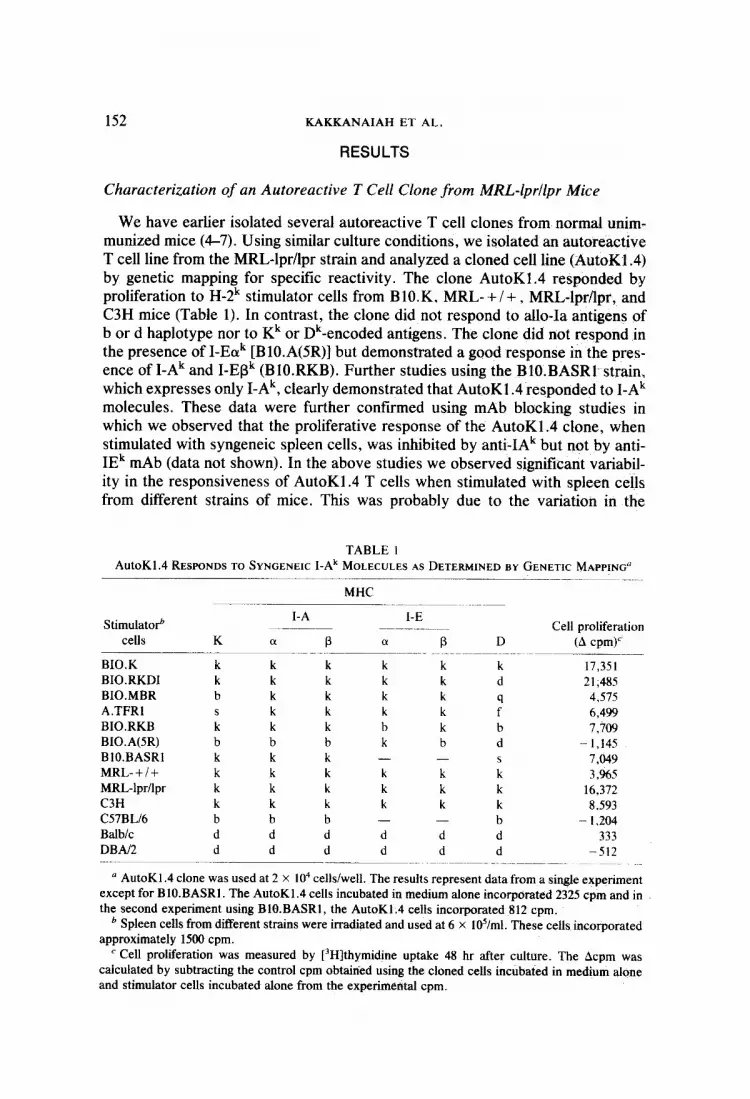
We have earlier isolated several autoreactive T cell clones from normal nnim- munized mice (4-7). Using similar culture conditions, we isolated an autoreactive T cell line from the MRL-lpr/lpr strain and analyzed a cloned cell line (AutoK1.4) by genetic mapping for specific reactivity. The clone AutoK1.4 responded by proliferation to H-2k stimulator cells from B1O.K. MRL- + /+ , MRL-lpr/lpr, and C3H mice (Table 1). In contrast, the clone did not respond to allo-Ia antigens of b or d haplotype nor to Kk or Dk-encoded antigens. The clone did not respond in the presence of I-Eok [BlO.A(SR)] but demonstrated a good response in the pres- ence of I-Ak and I-EBk (BlO.RKB). Further studies using the BlO.BASRl~ strain, which expresses only I-Ak, clearly demonstrated that AutoK1.4 responded to I-Ak molecules. These data were further confirmed using mAb blocking studies in which we observed that the proliferative response of the AutoK1.4 clone, when stimulated with syngeneic spleen cells, was inhibited by anti-IAk but not by anti- IEk mAb (data not shown). In the above studies we observed significant variabil- ity in the responsiveness of AutoK1.4 T cells when stimulated with spleen cells from different strains of mice. This was probably due to the variation in the
TABLE 1
AutoK1.4 RESPONDS TO SYNGENEIC I-Ak MOLECULES AS DETERMINED BY GENETIC MAPPING” ---____
MHC
StimulatoP I-A I-E
Cell proliferation cells K a P (I P D (A cpm)’
BI0.K k k k k k k 17,351 BIO.RKDI k k k k k d 21,485 BIO.MBR b k k k k 4.575 A.TFRl
it k k k k f” 6,499
BIO.RKB k k b k b 7,709 BIO.A(SR) b b b k b d - 1,145 BIO.BASRI k k k - - 7.049 MRL-+I+ k k k k k e 3,965 MRL-lpr/lpr k k k k k k 16,372 C3H k k k k k k 8.593 C57BLl6 b b b - - b - I.204 Balb/c d d d d d d 333 DBAl2 d d d d d d -512
a AutoKl.4 clone was used at 2 X lo4 cells/well. The results represent data from a single experiment except for BlO.BASRl. The AutoKl.4 cells incubated in medium alone incorporated 2325 cpm and in the second experiment using BlO.BASRl, the AutoKl.4 cells incorporated 812 cpm.
b Spleen cells from different strains were irradiated and used at 6 X Id/ml. These cells incorporated approximately 1580 cpm.
c Cell proliferation was measured by [3Hlthymidine uptake 48 hr after culture. The Acpm was calculated by subtracting the control cpm obtained using the cloned cells incubated in medium alone and stimulator cells incubated alone from the experimental cpm.

LYMPHOKINE SECRETION BY AUTOREACTIVE T CELLS 153
number of Ia+ cells, the density of Ia expression, or the amount of IL-l produced by the stimulators cells.
Induction of T-B and T-T Interaction by an Autoreactive T Cell Clone
We had earlier demonstrated that autoreactive T cell clones can polyclonally activate naive, resting B cells and furthermore induce a unique T-T interaction in which naive CD4+ T cells would respond directly to autoreactive T cell clones in the absence of accessory cells (4-7). We therefore investigated whether this prop- erty was restricted to clones isolated from normal DBA/2 mice or whether similar characteristics were demonstrable from the autoreactive T cell clone isolated from autoimmune-susceptible MRL-lpr/lpr mice. The data shown in Table 2 suggested that AutoKl.4 had similar properties as AutoDl.4 inasmuch as it could also poly- clonally activate B cells and induce naive CD4+ T cells to proliferate. Also, the T-T interaction induced by AutoKl.4 was inhibited by anti-CD4 but not by anti-Ia mAb (data not shown), similar to earlier studies on other autoreactive clones (5, 6). It has been shown that MRL-lpr/lpr mice develop severe lymphadenopathy and autoimmune disease at -2 months of age characterized by the accumulation of CD4-CD8- T cells (13). We therefore investigated the T-B and T-T interac- tions in these mice, before (1 months old) or after the onset (6 month old) of autoimmunity. Interestingly, CD4+ T cells purified from 6-month-old MRL-lpr/Ipr mice failed to respond to the autoreactive T cell clone, while similar cells from
TABLE 2
AUTOREACTIVE T CELL CLONES CAN INDUCE T-B AND T-T CELL INTERACTIONS
Expt.” Responders’ Stimulatorsc Cell proliferationd
(cpm k SEM)
I. CD4’ CD4’ B cells B cells -
2. CD4’ T CD4’ T CD4’ T CD4’ T -
3. B cells B cells B cells B cells
DBAl2 DBAl2 DBA/2 DBN2
MRL-lpr/lpr (1 month old) MRL-lpr/lpr (1 month old) MRL-lpr/lpr (6 months old) MRL-lpr!lpr (6 months old)
MRL-lprilpr (1 month old) MRL-lprilpr (1 month old) MRL-lpr/lpr (6 months old) MRL-lpr/lpr (6 months old)
- AutoDl.4
- AutoDl.4 AutoD 1.4 - AutoKl.4
- AutoK1.4 AutoKl.4
AutoKl.4 -
AutoKl.4
4,351 + 511 45,462 k 5,671
3,605 I 276 34,803 rt 2,767 2,660 k 389
14,854 * 960 34,249 k 2228 4.227 2 471 4,528 k 130
372 2 159 15,283 2 780 33,736 k 1090
7,692 2 103 23,871 2 655
u The data presented are representives of multiple experiments. b CD4 + T cells were used at a concentration of 2-4 x 10’ cells/well and B cells were used at 2 X IO5
cells/well. ’ The stimulator AutoDl.4 or AutoKl.4 cells were irradiated at 2000 rad and used at a concentration
of 5 x IO“ cells/well. Irradiated AutoDl.4 or AutoK1.4 cells when incubated with irradiated CD4+ T cells or B cells usually incorporated >2000 cpm.
d The T-B interaction was studied after 48 hr and T-T interaction after 72 hr incubation. The cultures were pulsed with [3H]thymidine during the last 18 hr.

154 KAKKANAIAH ET AL.
l-month-old mice exhibited strong responsiveness. The B cells from I- or 6- month-old MRL-lpr/lpr mice, in contrast, responded in a similar fashion when stimulated with the AutoK1.4 clone.
Autoreactive T Cell Clones Produce IL-2 and IL-4
We next investigated the type of lymphokine produced by the autoreactive T cell clones. The autoreactive T ceil clones, AutoDl.4 and AutoKl.4, were stim- ulated with irradiated syngeneic SAC or with Con A and 24 hr later, the super- natants were collected and tested for their capacity to induce I-IT-2 cell prolifer- ation in the absence or presence of 7D4 (anti-IL-2R mAb) or 11 .B. 11 (anti-IL-4 mAb). The data shown in Table 3 suggested that the autoreactive T cell? upon activation produced growth factors for the HT-2 cells which were partially inhib- ited by anti-IL-2R or anti-IL-4 mAbs. The anti-IL-2R or anti-IL-4 mAbs at the dilution used inhibited specifically only the rIL-2- or rIL-4-mediated proliferation of HT-2 cells, respectively. Also, DlO.G4, which is a Th2 type of T cell clone, produced following Con A activation only IL-4 but not IL-2 in confirmation-with other studies (12). The proliferation of HT-2 cells induced by culture supernatants from activated autoreactive T cell clones was also partially inhibited by anti-IL-2 (S4B6) antibodies (data not shown).
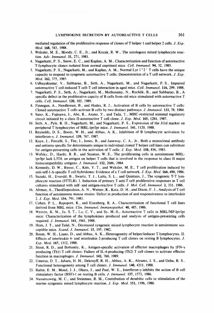
The fact that the culture supernatants from autoreactive T cells contained, IL-2 was also confirmed by using an IL-Zdependent tumor-specific cytotoxic .T cell clone, C9, developed in our laboratory. This T cell clone responds only to IL-2 but not to IL-4 and it was observed that the culture supernatants from activated AutoD1.4 or AutoKl.4 induced proliferation of this cell line which was inhibited by anti-IL-2R (data not shown). The presence of IL-4 was also confirmed by its capacity to cause hyper-Ia induction on B cells. When B cells were incubated with culture supernatants from AutoD1.4 or AutoK1.4 clones for 18-24 hr, there was an increase in the density of Ia expression as measured by flow cytometry (Fig. 1). This increase was similar to the hyper-Ia induction by rIL-4 and furthermore was inhibited following addition of anti-IL-4 antibodies during culture.
Production of IFN-y by Autoreactive T Cell Clones
WEHI-279, an IFN-y-sensitive B cell lymphoma line, was used to detect the presence of IFN-y in the culture supematants of activated autoreactive T cells. The data shown in Table 4 suggested that the autoreactive T cells produced a factor which could inhibit the proliferation of WEHI- and that mAbs to IFN-)I could reverse this inhibition significantly. As a control, the reference IFN-y could similarly inhibit the proliferation of WEHI-279, which was also reversible by mAbs to IFN-y. Together, these data suggested that both the autoreactive T cell clones produced IFN-)I.
Activation of Tumoristatic Properties of Macrophages
Thl cells have been shown to produce IFN-7 and to activate macrophages to inhibit tumor cell growth. To further substantiate whether our autoreactive T-cell clones could activate the macrophages, culture supernatants from activated AutoD1.4 and AutoK1.4 clones were tested for their capakity to induce tumori-

LYMPHOKINE SECRETION BY AUTOREACTIVE T CELLS 155
TABLE 3 DETECTIONOF IL-2 AND IL-4 INTHECULTURESUPERNATANTSOF ACTIVATEDAUTOREACTIVE
T CELLCLONES
Expt Supematanti No.” interleukinb
mAb added to culturesC
HT-2 cell proliferation
(cpm X 10-3)d
6
I
8
9
AutoDl.4
AutoKl.4
AutoDl.4
AutoKl.4
AutoDl.4
AutoKl.4
DlO.G4
rIL-2
rIL-4
- Anti-IL-2R
- Anti-IL-2R
- Anti-IL-4
- Anti-IL-4
- Anti-IL-2R Anti-IL-4
- Anti-IL-2R Anti-IL-4
- Anti-IL-2R Anti-IL-4
- Anti-IL-2R Anti-IL-4
- Anti-IL-2R Anti-IL-4
14,807 ? 698 9,766 2 973 (35)’
13.318 2 151 7,610 * 362 (43)
17,710 ? 3251 5,457 2 499 (70)
14,201 2 158 4,606 2 269 (68)
14,438 + 1012 8,238 2 634 (43) 7,652 + 516 (48)
12,002 k 524 8,306 2 104 (31) 4,012 ? 230 (67)
10,300 ” 788 13,394 ” 392 (0) 6,364 f 440 (39)
28,778 2 737 15,503 5 1610 (47) 31,618 + 1126 (0) 14,651 5 328 16,573 5 270 (0) 3,737 ? 597 (75)
n The data presented are representative of multiple experiments. b Autoreactive T cell clones, AutoD1.4 or AutoKl.4, were cultured with Con A (in Expts l-4) or
with syngeneic irradiated SAC cells (in Expts 5 and 6) for 24 hr. Antigen-specific DlO.G4 cells were cultured with Con A for 24 hr. The culture supematants were next harvested and tested for the presence of interleukins. Recombinant IL-2 and IL-4 were added at a final concentration of 5 and 25 U/ml, respectively.
’ Anti-IL-2R antibodies (7D4) and anti-IL-4 antibodies (11 .B. 11) were added al 1: 10 and 1:250 final dilution, respectively.
d HT-2 cells (5 X 10³/well) in 0.05 ml of complete medium were cultured with 0.05 ml of appropriate dilutions of the mAb and 0.1 ml of culture supematant from activated T cells or the recombinant interleukins. The plates were incubated at 37°C for 24 hr. pulsed with 2 p,Ci of [3H]thymidine, and harvested 18 hr later.
p Figures in parentheses show percentage suppression.
static activity. The data shown in Table 5 clearly demonstrated that normal mac- rophages cultured in the presence of culture supernatants from activated AutoDl.4 or AutoKI .4 cells significantly inhibited the proliferation of P815 tumor cells similar to the action of IFN-y. This effect was reversible in the presence of mAbs to IFN-y, thereby suggesting that the macrophage activation was caused by IFN-?I.
Activated Autoreactive T Cell Clones Respond Only to t-IL-2 but Not to rlL-4 Following antigen-specific activation, both Thl and Th2 clones have been

156 KAKKANAIAH ET AL.
- -
INTENSITY OF R-
FIG. 1. Enhanced Ia antigen expression on B cells following culture with the supernatants from activated autoreactive T cell clones, AutoKI.4 (A) or AutoD1.4 (B). The B cells were incubated with medium alone (b), rIL-4 (c), or with culture supematants from autoreactive T cell clones (d). In A, the B cells were incubated with culture supematant from autoreactive T cell clone plus anti-IL-4 anti- bodies (e). The B cells were stained with anti-IEd antibodies followed by FITC-conjugated anti-mouse IgG (b, c, d, e) or with normal mouse IgG followed by the FITC-conjugated second step reagent as a negative control (a).
shown to proliferate in response to exogenous IL-2 and IL-4 (2). To test this responsiveness, autoreactive T cells were cultured with irradiated syngeneic spleen cells for 48 hr and were next incubated with rIL-2 or rIL-4. The data shown in Table 6 demonstrated that both AutoD1.4 and AutoK1.4 responded only to exogenous IL-2 but not IL-4.
DlSCUSSlON
In the present study we observed that autoreactive T cell clones isolated from normal DBA/Z mice or autoimmune-susceptible MRL-lpr/lpr mice produced IL-2, IL-4, and IFN-y, thereby exhibiting the lymphokine secretory properties of both Thl and Th2 subsets. The clones were derived by limiting dilution at ~0.3 cells/ well and were also subcloned. Furthermore, the clones were stable in their phe-

LYMPHOKINE SECRETION BY AUTOREACTIVE T CELLS 157
TABLE 4 DETECTIONOF IFN-YINTHE SUPERNATANTSOF ACTIVATEDAUTOREACTIVE T CELLS
Expt Culture No. supematant”
Antibody addedb
Proliferation of WEHI- (cpm ? SE)
1 -
AutoKl.4 AutoKl.4 IFN-y IFN-y
- 2 -
AutoDl.4 AutoDl.4
- 23,479 k 600 - 1,055 f 30
Anti-IFN-y 17.748 * 177 - 1145 k 100
Anti-IFN-y 23,457 t 1931 Anti-IFN-y 25,229 t 1090
- 19,566 2 431 - 7,741 f 508
Anti-IFN-y 11,427 2 503
a Autoreactive T cells were activated using Con A, as described in Table 1. NIH reference murine IFN--r was used at a tinal concentration of 10 IU/ml.
b Anti-IFN-y mAb (R4.6A2) was used at a 1:4 final dilution of the hybridoma supematant. c WEHI- cells (5 X 10³/well) were cultured for 24 hr in the presence of the culture supematant
and in the presence or absence of mAb to IFN-y. The cultures were next pulsed with 2 @Zi of [3H]thymidine and harvested 18 hr later.
notypic and functional properties for over 2 years since their establishment (6). We confirmed the presence of IL-2, IL-4, and IFN-y in the supematants of acti- vated autoreactive T cells by two independent bioassays. The IL-2 was demon- strable using HT-2 cells whose proliferation was inhibited in the presence of
TABLE 5 CULTURE~UPERNATANTS FROMACTIVATEDAUTOREACTIVETCELL CLONES ACTIVATETHE
TUMORISTATICPOTENTIALOFTHEMACROPHAGES
Peritoneal macrophages”
Culture supematant/IFN-yb
Anti-IFN-y mAb’
P815 tumor cell proliferation (cpm f SEM)d
- - - + - - + IFN-y -
+ IFN-y + + AutoKl.4 -
+ AutoKl.4 + + AutoDl.4 - + AutoDl.4 +
182,450 k 5056 180,896 * 2479 61,784 2 1559
154,259 + 1389 84,041 2 650
162.454 k 1532 94,758 + 1522
175,245 ‘- 1322
a Peritoneal macrophages from DBA/2 mice were used at 2 x lo5 cells/well. Following activation with IFN-y or culture supematants from activated autoreactive T cell clones, the macrophages were treated with mitomycin-C, washed, and used. The macrophages incubated alone incorporated less than 1000 cpm.
b IFN--v was used at 10 IU/ml. The culture supematants from autoreactive T cell clones were collected 24 hr after stimulation with Con A.
’ Anti-IFN-y mAbs were used at a 1: 10 final dilution. d P815 tumor cells (5 x 10⁴) were incubated for 24 hr and during the last 18 hr were pulsed with
[‘Hlthymidine.

158 KAKKANAIAH ET AL.
TABLE 6 AUTOREACTIVE T CELL CLONES RESPOND TO IL-2 BUT NOT TO IL-4
Responder clone”
AutoKl.4
AutoDl.4
Interleukin
- rIL-2 (25 U/ml) rIL-4 (50 U/ml)
- rIL-2 (25 U/ml) rIL-2 (50 U/ml) rIL-4 (50 U/ml) rIL-4 (100 U/ml)
Cell proliferatio& (cpm 2 SE)
7130 t 363 34,388 -t 2014
8259 r 267 2493 2 2014
11.767 t 806 20,446 It 4725
2679 I 46 2946 f 72
a Autoreactive T cell clones were activated with irradiated syngeneic spleen cells for 48 hr, har- vested, and viable cells were purified by centrifuging over Ficoll-Hypaque. The cells (5 x 10⁴/well) were next incubated with medium or with IL-2 or IL-4.
b Cell proliferation was measured by pulsing the plates with [‘Hlthymidine after 24 hr incubation and harvested 18 hr later.
anti-IL-2R and anti-IL-2 antibodies and also using an IL-2- but not an IL- 4-dependent cytotoxic T cell line recently cloned in our laboratory. The presence of IL-4 was detected using HT-2 cells and was further confirmed by studying the hyper-Ia induction on B cells. The IFN-y was demonstrable using an IFN-y-sen- sitive B lymphoma line and by the activation of tumoristatic properties of mac- rophages. Furthermore, functionally both autoreactive T cell clones could acti- vate or induce proliferation of naive B cells, CD4+ T cells, and macrophages.
We have earlier observed that autoreactive T cell clones isolated from normal DBA/2 mice can induce the proliferation of syngeneic naive CD4+ T cells (5, 6). This response was independent of MHC molecules and did not require antigen- presenting accessory cells. The T-T interaction was regulated by IL-2 and IL-4, and when the autoreactive T cell clone was V@‘, the T-T interaction was in- hibited in the presence of anti-Vp8 mAbs (5,6) (unpublished data). Based on these observations, we speculated that the CD4+ T cells were responding directly to the idiotypic determinants on the autoreactive T cells. Similar autoreactive T cell- induced T-T network interactions have been described by others in recent years (9, 14, 15). Although the functional significance of the T-T interaction is not clear, Suzuki et al. (15) demonstrated that the autoreactive T cell clones could initiate a chain of regulatory cell interactions leading to either the stimulation or the inhi- bition of the response. Sano et al. (9) further demonstrated that an autoreactive T cell clone when administered in vivo could induce an MHC-restricted minimal regulatory circuit. This clone activated an MHC-restricted T suppressor cell which in turn suppressed heterogenous T helper cells provided that the latter had the same MHC-restricted specificity as that possessed by the autoreactive T cell clone.
MRL-lpr/lpr mice develop severe lymphadenopathy characterized by the accu- mulation of a large number of double-negative T cells (13). Such cells have been shown not to respond to polyclonal T cell activators such as Con A (16). Recently, we demonstrated that a significant proportion of these cells express Jlld, a

LYMPHOKINE SECRETION BY AUTOREACTIVE T CELLS 159
marker seen only on immature thymocytes but not on mature peripheral T cells, thereby suggesting that the double-negative cells closely resemble the CD4-CDS- thymocytes (10). We have also observed that the double-negative cells do not respond to syngeneic Ia molecules nor do they participate in the T-T interaction induced by the autoreactive T cell clones (unpublished data). Lymph node cells from young MRL-lpr/lpr mice have been shown to spontaneously pro- liferate when cultured in vitro and respond to self-Ia antigens (17, 18). Further- more, the cultured cells have been shown to be CD4+, induce syngeneic B cell proliferation and differentiation, and to produce IL-2, IL-3, and IFN-y (17, 18). However, lymph node T cell responsiveness to the self-Ia antigens decreases in aged MRL-lpr/lpr mice (19). Whether, such a decrease is due to low numbers (-8%) of CD4+ T cells and large numbers (-80%) of unresponsive double- negative T cells found in aged mice (10) or whether it is due to an intrinsic defect in the CD4+ responding T cells has not been resolved. Our data on isolation of autoreactive T cell clones from aged mice suggested that 4- to 6-month-old MRL- lpr/lpr mice may still have T cells capable of responding to self-Ia antigens and such cells may be able to polyclonally activate B cells to produce autoantibodies.
It was interesting to note that the clone that we isolated from MRL-lpr/lpr mice could also initiate a similar T-T interaction when the responding CD4+ T cells were used from l-month-old mice before the onset of the disease. Interestingly, 6-month-old mice with severe lymphadenopathy and autoimmune disease failed to demonstrate a T-T interaction. It is not clear at this juncture whether this defect is due to a decrease in numbers of CD4+ T cells capable of responding to the autoreactive T cells or whether it is due to the decreased responsiveness to the IL-2 and IL-4 produced by the autoreactive T cells. Further studies are in prog- ress to address these possibilities. However, a lack of T-T network may suggest failure to activate T suppressor cells (9) and this may lead to the unregulated production of autoantibodies in MRL-lpr/Ipr mice. We have also observed re- cently that the T-T interaction is greatly diminished in aged mice (6) which also exhibit increased susceptibility to autoimmunity.
It has been demonstrated that Th2 clones can provide excellent help to B cells due to their capacity to secrete IL-4 and IL-5 (1). The role of Thl cells in providing help to B cells is uncertain. Boom et al. (20) reported that Th2 but not Thl clones specific for rabbit y-globulin could induce polyclonal proliferation and Ig produc- tion in resting B cells in the presence of rabbit anti-mouse Ig antibodies. In contrast to the B cell activation, Stout and Bottomly (21) demonstrated that Thl but not Th2 clones could activate the macrophages to exhibit tumoristatic activity. In this context it is interesting to note that the autoreactive T cell clones provide help to B cells as well as activate the macrophages.
Since the autoreactive T cell clones could activate B cells and macrophages and also induce T-T interaction, we investigated the type of lymphokines generated by such clones. All murine long-term Th clones have been shown to secrete either IL-2 and IFN-y (Thl cells) or IL-4 and IL-5 (Th2 cells) (1). It was interesting in this context to note that both the autoreactive clones produced IL-2, IL-4, and IFN-y, thereby exhibiting the properties of both Thl and Th2 cells. This obser- vation appeared to be somewhat similar to that demonstrated with some human

160 KAKKANAIAH ET AL.
Thl clones which were found to secrete IL-2, IL-4, and IFN-y (22). IFN-r has been shown to inhibit IL-Cmediated B cell activation (23). If this was true, how do the autoreactive T cell clones activate B cells to proliferate and differentiate? One possibility is that the IFN-y produced by the autoreactive T cells may not be sufficient to inhibit the B cell activation and thus the outcome of B cell stimulation may depend on the relative amounts of IL-2, IL-4, and IFN-y secreted by the autoreactive T cells. Our data are similar to a human Th clone which, despite producing IL-2, IL-4, and IFN-y, could efficiently induce Ig synthesis by the B cells (22). Lastly, it is also possible that the type and amounts of lymphokine secreted by the autoreactive T cells may vary depending on the accessory cells involved in autoreactive T cell activation such as macrophages (4), dendritic cells (24), or B cells (7).
Recently Gajewski et al. (25) reported that when rIL-2 was used as a growth factor for T cells, the majority of the clones derived were of the Th2 type, whereas when rIL-2 plus rIFN-y was used, Thl clones were isolated. Using either proce- dure, some clones were obtained that produced IL-2, IL-4, and IFN-y. Whether such clones represented autoreactive T cells remains an interesting possibility which needs further elucidation. It should be noted that the TCGF we have employed to isolate the autoreactive T cell clones has only IL-2 and no detectable IL-4 or IFN-y activity and in spite of this we have isolated consistently autore- active T cells having mixed lymphokine-secreting properties of Thl and Th2 cells.
Thl and Th2 cells have been shown to use IL-2 or IL-4, respectively, as auto- crine growth factors in vitro. However, following antigenic stimulation, both types of clones respond to the proliferative effects of IL-2 and IL-4 (2). In this context it was interesting to note that the autoreactive T cell clones responded only to IL-2 but not to IL-4, following antigen-specific activation. These data suggested that the autoreactive T cells, although capable of secreting IL-4, do not utilize the IL-4 and use only IL-2 for growth. Whether this is due to preferential expression of IL-2R over IL-4R needs to be investigated. Further studies on the kinetics and quantitation of the lymphokines produced by autoreactive T cells may explain their unique capacity to help, suppress, and contrasuppress (26-28) an ongoing immune response in vitro. In summary, our data suggest that self-Ia reactive T cell clones isolated from unprimed mice exhibit properties that are distinct from those of antigen-specific murine Thl or Th2 cells. Whether this property is exhibited by all autoreactive T cells or only a fraction of such cells remains to be resolved by studying a large number of long-term autoreactive T cell clones. Such studies are currently in progress in our laboratory.
ACKNOWLEDGMENTS
We thank Ms. Judith McCord for excellent maintenance of the mouse colony and Mr. Robert H. Pyle for expert assistance with the flow cytometer.
REFERENCES
1. Mosmann, T. R., and Coffman, R. L.. THl and TH2 cells: Different patterns of lymphokine secretion lead to different functional properties. Annu. Rev. Zmmunol. 7, 145, 1989.
2. Femandez-Botran, R., Sanders, V. M., Mosmann, T. R., and Vitetta, E. S., Lymphokine-
LYMPHOKINE SECRETION BY AUTOREACTIVE T CELLS 161
mediated regulation of the proliferative response of clones of T helper 1 and helper 2 cells. J. Exp.
Med. 168, 543, 1988. 3. Weksler, M. E., Moody, C. E., Jr., and Kozak, R. W., The autologous mixed lymphocyte reac-
tion. Adv. Immunol. 31, 271, 1981. 4. Nagarkatti, P. S., Snow, E. C., and Kaplan, A. M., Characterization and function of autoreactive
T-lymphocyte clones isolated from normal unprimed mice. Cell. Immunol. 94, 32, 1985. 5. Nagarkatti, P. S., Nagarkatti, M., and Kaplan, A. M., Normal Lyt 1+2- T cells have the unique
capacity to respond to syngeneic autoreactive T cells: Demonstration of a T cell network. J. Exp. Med. 162, 375, 1985.
6. Udhayakumar, V., Subbarao, B., Seth, A., Nagarkatti, M., and Nagarkatti, P. S., Impaired autoreactive T cell-induced T cell-T cell interaction in aged mice. Cell. Immunol. 116, 299, 1988.
7. Nagarkatti, P. S., Seth, A., Nagarkatti, M., Muthusamy, N., Rychlik, B., and Subbarao, B., A specific defect in the proliferative capcity of B cells from old mice stimulated with autoreactive T cells. Cell. Immunol. 120, 102, 1989.
8. Finnegan, A.. Needleman, B., and Hodes, R. J.. Activation of B cells by autoreactive T cells: Cloned autoreactive T cells activate B cells by two distinct pathways. J. Zmmunol. 133, 78. 1984.
9. Sano, K., Fujisawa, I., Abe, R., Asano, Y., and Tada, T., MHC-restricted minimal regulatory circuit initiated by a class II-autoreactive T cell clone. J. Exp. Med. 165, 1284, 1987.
10. Seth. A., Pyle, R. H., Nagarkatti, M., and Nagarkatti, P. S., Expression of the Jl Id marker on peripheral T lymphocytes of MRL-lpr/lpr mice. J. Immunol. 141, 1120. 1988.
11. Reynolds, D. S., Boom, W. H., and Abbas, A. K., Inhibition of B lymphocyte activation by interferon-y. 3. Immunol. 139, 767. 1987.
12. Kaye, J., Porcelli. S., Tite, J., Jones, B., and Janeway. C. A., Jr., Both a monoclonal antibody and antisera specific for determinants unique to individual cloned T helper cell lines can substitute for antigen-presenting cells in the activation of T cells. J. Exp. Med. 158, 836, 1983.
13. Wofsky, D., Hardy. R. R., and Seaman, W. E., The proliferating cells in autoimmune MRL- 1priIpr lack L3T4. an antigen on helper T cells that is involved in the response to class II major histocompatibility antigens. J. Immunol. 132, 2686, 1984.
14. Kennedy, D. W., Russo, C., Kim, Y. T., and Weksler, M. E., T cell proliferation induced by anti-self-I-A-specific T cell hybridoma: Evidence of a T cell network. J. Exp. Med. 164,490, 1986.
15. Suzuki, H., Evavdd, B., Swartz. T. J.. Latta, S. L., and Quintans, J., The syngeneic T-T lym- phocyte reaction (STTLR). I. Induction of primary T anti-T cell proliferative responses in T cell cultures stimulated with self- and antigen-reactive T cells. J. Mol. Cell. Immunol. 2, 331, 1986.
16. Altman, A., Theofilopoulous, A. N., Weiner, R.. Katz, D. H., and Dixon, F. J., Analysis of T cell function of autoimmune mouse strains: Defect in production of and responsiveness to interleukin 2. J. Exp. Med. 154, 791, 1981.
17. Cohen, P. L., Rapoport, R., and Eisenberg, R. A., Characterization of functional T cell lines derived from MRL mice. Clin. Immunol. Immunopathol. 40, 485, 1986.
18. Weston, K. M., Ju. S. T., Lu, C. Y., and Sy, M.-S., Autoreactive T cells in MRLfMP-lprilpr mice: Characterization of the lymphokines produced and analysis of antigen-presenting cells required. J. Immunol. 141, 1941, 1988.
19. Horn, J. T., and Talal, N.. Decreased syngeneic mixed lymphocyte reaction in autoimmune sus- ceptible mice. Stand. J. Immunol. 15, 195, 1982.
20. Boom, W. H., Liano, D., and Abbas, A. K., Heterogeneity of helper/inducer T lymphocytes. II. Effects of interleukin 4- and interleukin 2-producing T cell clones on resting B lymphocytes. J. Exp. Med. 167, 1352, 1988.
21. Stout, R. D., and Bottomly, K., Antigen-specific activation of effector macrophages by IFN--y producing (Thl) T cell clones: Failure of IL-Cproducing (Th2) T cell clones to activate effector function in macrophages. J. Immunol. 142, 760, 1989.
22. Umetsu, D. T., Jabara, H. H., Dekruyff, R. H., Abbas, A. K., Abrams, J. S., and Geha, R. S.. Functional heterogeneity among T cell clones. J. Immunol. 140, 421 I, 1988.
23. Rabin, E. M., Mond, J. J., Ohara, J., and Paul, W. E., Interferon-y inhibits the action of B cell stimulatory factor (BSF)-1 on resting B cells. J. lmmunol. 137, 1573, 1986.
24. Nussenzweig, M. C., and Steinman, R. M., Contribution of dendritic cells to stimulation of the murine syngeneic mixed lymphocyte reaction. J. Exp. Med. 151, 1196, 1980.
162 KAKKANAIAH ET AL.
25. Gajewski, T. F., Joyce, J., and Fitch, F. W., Antiproliferative effect of IFN-y in immune regu- lation. III. Differential selection of Thl and Th2 murine helper T lymphocyte clones using recom- binant IL-2 and recombinant IFN-y. .Z. Zmmunol. 143, 15, 1989.
26. Clayberger, C., Dekruylf, R. H., and Cantor, H., Immunoregulatory activities of autoreactive T cells: An I-A-specific T cell clone mediates both help and suppression of antibody responses. J. Zmmunol. 132, 2237, 1984.
27. Kotani, H., Mitsuya, H., Jarrett, R. F.. Yenokida, G. G.. James, S. P., and Strober, W., An autoreactive T cell clone that can be activated to provide both helper and suppressor function. .Z. Zmmunol. 136, 1951, 1986.
28. Quintans, J., Suzuki, H., Sosman, J. A., and Shah, P. D., Immunoregulation by T cells. 1. Char- acterization of the IEk-specific Lbd self-reactive T cell clone that helps, suppresses and contra- suppresses B cell responses. J. Zmmunol. 136, 1974, 1986.
Received March 2, 1990; accepted with revision June 5, 1990