novel approaches for identifying target antigens of autoreactive human b and t cells
TRANSCRIPT

REVIEW
Novel approaches for identifying target antigensof autoreactive human B and T cells
Klaus Dornmair & Edgar Meinl & Reinhard Hohlfeld
Received: 17 April 2009 /Accepted: 13 August 2009 /Published online: 11 September 2009# Springer-Verlag 2009
Abstract Antigen-specific immune responses in multiplesclerosis have been studied for decades, but the targetantigens of the putatively autoaggressive B and T cells stillremain elusive. Here, we summarize recent strategies whichare based on the direct analysis of biopsy or autopsyspecimens from patients. Since this material is extremelyscarce, the experimental methods need to be exceptionallysensitive. We describe technologies to distinguish (auto)aggressive T cells from irrelevant bystander lymphocytesby analyzing clonal expansions in relation to the morpho-logical location of the cells in the tissue lesions. We thendiscuss approaches to clone matching α- and β-chains ofthe antigen-specific T cell receptor (TCR) molecules fromsingle T cells. This is necessary because usually, severalclones are expanded and are diluted by many irrelevantcells. The matching TCR chains from individual T cells canbe resurrected in hybridoma cells which may then be usedfor antigen searches. We discuss strategies to identifyantigens of γδ- and αβ-TCR molecules, such as biochem-ical methods, candidate antigens, human leukocyte antigenrequirements, synthetic peptide, and cDNA libraries. Thesestrategies are tailored to characterize the antigens of themembrane-anchored, low-affinity TCR molecules. Thestrategies to identify (auto) reactive B cells or immuno-
globulin (Ig) molecules are fundamentally different, be-cause Ig molecules are water-soluble and have highaffinities. We further discuss proteome-based approaches,techniques that analyze Ig-chains from single B cells, and arepertoire-based method that compares Ig-proteomes andIg-transcriptomes. The first method detects Ig antigensdirectly, whereas the latter two methods allow reconstruc-tion of Ig molecules, which can be used for antigensearches.
Keywords Autoimmunity .Multiple sclerosis .
Tcell receptor . B cells . Immunoglobulins
Introduction
The current concepts of multiple sclerosis (MS) as anautoimmune disease are mainly based on the animal model“experimental autoimmune encephalomyelitis” (EAE) [1,2]. In different EAE models, several distinct molecularlydefined antigens could be demonstrated to induce patho-genic T and/or B cell responses. However, the targetantigen(s) recognized by immune cells in the differentforms of human MS still remain speculative. In the past,most investigations of putatively pathogenic human B- andT cells relied mainly on blood as the most easily accessiblesource. Obviously, this is far from ideal, because anyrelevant cells are (a) remote from the target tissue, (b)heavily diluted with irrelevant cells, and (c) likely to havephenotypical and functional properties completely differentfrom the tissue-infiltrating cells. Most importantly, allinvestigations of isolated circulating or migrating cellsnecessarily lack crucial morphological information on howthe cells interact with the surrounding tissue. The mostdirect approach would of course be to focus on the
K. Dornmair : E. Meinl : R. HohlfeldDepartment of Neuroimmunology,Max-Planck-Institute of Neurobiology,Am Klopferspitz 18,82152 Martinsried, Germany
K. Dornmair (*) : E. Meinl :R. HohlfeldInstitute of Clinical Neuroimmunology,Ludwig Maximilians University,Klinikum Grosshadern, Marchioninistr. 15,81366 Munich, Germanye-mail: [email protected]
Semin Immunopathol (2009) 31:467–477DOI 10.1007/s00281-009-0179-y

autoimmune target tissue as the most valuable source ofinformation. This approach is far from trivial, because itdemands a set of new techniques for obtaining maximuminformation from scarce human autopsy and biopsymaterial.
The central problem of such a direct strategy is theavailability of tissue samples. For most diseases, well-preserved biopsy specimens are extremely rare. Forexample, brain or spinal cord lesions are usually notbiopsied in MS patients. The same holds true for pancreassamples of type I diabetes patients, etc. There are only fewautoimmune diseases where diagnostic biopsies are takenon a regular basis. One such example is provided by theinflammatory myopathies (IM). This situation contrastswith animal experiments, where diseases may be inducedand manipulated experimentally, and where tissue samplesmay be collected at any time point of choice, even at pre-clinical stages.
For many years, we have investigated antigen-directedimmune responses in MS and IM target tissues, that is,brain and muscle. Both in MS and IM, T cells—especiallyCD8+ cells—infiltrate the organs. It is not known howthese cells are attracted or how the local inflammatoryresponse is sustained. The target antigens are alsounknown. The B cell responses, i.e., the (auto) antibodyresponses, are somewhat better understood. This is mainlydue to the fact that soluble immunoglobulin (Ig) proteinsare easier to investigate than autoaggressive T cells wherethe autoaggressive T cell receptor (TCR) is a member of amultimeric complex of membrane proteins. During thecourse of our studies, we have developed a series oftechnologies that allow analysis of T and B cell responsesin IM and MS.
Our main long-term goal is to identify the target antigensrecognized by tissue-infiltrating T and B cells in MS andIM [3]. With very few exceptions, these target antigens are
unknown. This is unfortunate, because knowing the targetantigens would be of great help for developing diagnosticmarkers and immunotherapies. To achieve this goal, manyobstacles need to be overcome: Firstly, well-preservedtissue, especially brain biopsy material, is very rare.Secondly, putatively autoreactive T cells and B cells mustbe distinguished from bystander cells (Fig. 1). Therefore,morphological analysis followed by microdissection andsingle cell polymerase chain reaction (PCR) is required,which is, thirdly, particularly difficult owing to theextremely low RNA concentrations. Fourthly, the antigen-specific TCR and Ig molecules are both composed ofvarious genetic elements, containing hypervariable regionsand mutations. Therefore, multiplex PCR must beemployed. Fifthly, the search for antigens is not trivialbecause huge libraries must be screened. This is particularlydifficult for T cells, because the notoriously low affinities ofTCR-antigen interactions [4–7] impede straightforwardbiochemical methods.
The first step of our analysis is the identification of cellclones or Ig molecules that are expanded in the targettissue, driven by antigen recognition. Such “repertoirestudies” help us to distinguish between pathogenic andirrelevant cells or molecules. In the next step, we focus onindividual T cells and their antigen-specific receptor, or onclonally expanded antibodies. We then amplify the TCR orIg chains by PCR and express them in vitro. The trans-fectants are then used for antigen searches. In a first seriesof experiments, “best guess” candidate antigens may bescreened. Such candidates usually come from animalexperiments. A more unbiased approach is to screen cDNAexpression libraries. The cDNA libraries may be generatedfrom the affected organs, or—preferred—from the biopsyspecimen of the patient. Depending on whether B or T cellantigens are investigated, the libraries are either expressedand screened directly or must be introduced into the class-I
muscle fiber
CD8
Fig. 1 Autoaggressive T cells attack muscle fibers. In the schematicrepresentation (left) the autoaggressive T cells are shown in red. Manyother bystander T cells and T cells in blood vessels are present in thesame tissue (blue and green). On the right panel, we show animmunohistochemical stained cryosection from the muscle of a PM
patient. CD8+ T cells are stained in green. In the scheme and in thecryosection, muscle-attached single T cells and T cell clusters thatfocally invade the muscle fibers are seen (modified from [23] withpermission)
468 Semin Immunopathol (2009) 31:467–477
or class-II major histocompatibility complex (MHC) pre-sentation pathway before they are screened.
Here, we will review the current state of antigendetection efforts in MS research. Both, for B cell and forT cell antigens, several technical challenges have to beovercome. Since the experimental strategies for identifyingB and T cell antigens are quite different, we will discuss therespective approaches separately. Needless to say, thesenew techniques may also be applied to tissues from patientswith other autoimmune, neoplastic, or inflammatory dis-eases where adaptive immune responses occur.
T cell antigens
TCR repertoire in autoimmune tissue lesions
Tissue-infiltrating T cells are observed in all patients withMS or IM. In most cases, the T cell infiltrates are composedof αβ-T cells, whereas γδ-T cells are rather an exception[8–10]. In MS, CD8+ T cells usually outnumber the CD4+population [11]. In IM, it depends on the subtype of thedisease: While in inclusion body myositis and polymyosi-tis, CD8+ T cells clearly dominate, while CD4+ T cells aremore prominent in dermatomyositis [12, 13]. We haveintensively studied the αβ-TCR repertoire of infiltratingCD8+ T cells in MS brain specimens [14–16] and inmyositis muscle tissue of polymyositis and inclusion bodymyositis patients [17–19]. Using CDR3-spectratyping, wefound that in these diseases, CD8+ T cells are expanded inthe target tissues and blood, and that these expanded clonesmay persist for many years in some patients. We investi-gated the TCR repertoire in muscle samples and blood ofseveral patients with IM [19] and in brain tissue, cerebro-spinal fluid (CSF), and blood of MS patients [15]. In themyositis study, we identified expanded T cell clones inmuscle biopsy tissue of ten patients. From four patients, weisolated single, morphologically characterized T cells bylaser microdissection and analyzed the TCR β-chainsby single cell PCR. These T cells were most probablyautoaggressive, because they belonged to expanded clones,were in direct contact with their target cells, and carriedsilent nucleotide exchanges in the TCR CDR3-N-regions.This indicates that the clonal expansions were driven byparticular antigen(s). Expanded clones were not onlydetectable in muscle tissue but also in the CD8+ T cellcompartment of peripheral blood, where they persisted formany years. Persistent CD8+ T cell clones were alsodetected in MS patients [15]. We found that these CD8+clones were also present in brain tissue, CSF, and peripheralblood. By contrast, the CD4+ population was polyclonal.Strikingly, some of the CD8+ T cells that had beenidentified in brain tissue persisted in peripheral blood and
CSF for more than 7 years. These repertoire studies werethe basis of a general strategy that leads to the molecularcharacterization of αβ-TCR molecules from autoaggressiveT cell [3].
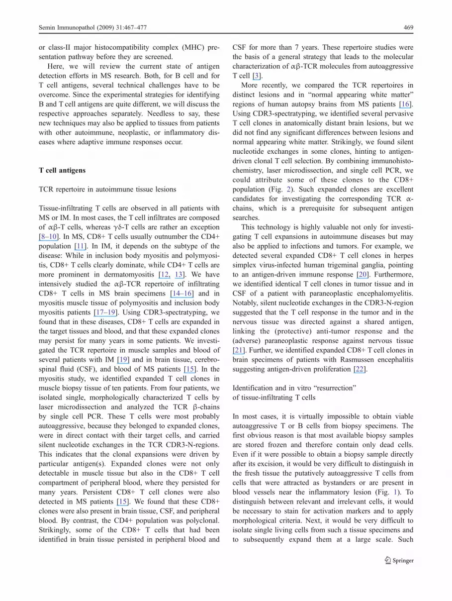
More recently, we compared the TCR repertoires indistinct lesions and in “normal appearing white matter”regions of human autopsy brains from MS patients [16].Using CDR3-spectratyping, we identified several pervasiveT cell clones in anatomically distant brain lesions, but wedid not find any significant differences between lesions andnormal appearing white matter. Strikingly, we found silentnucleotide exchanges in some clones, hinting to antigen-driven clonal T cell selection. By combining immunohisto-chemistry, laser microdissection, and single cell PCR, wecould attribute some of these clones to the CD8+population (Fig. 2). Such expanded clones are excellentcandidates for investigating the corresponding TCR α-chains, which is a prerequisite for subsequent antigensearches.
This technology is highly valuable not only for investi-gating T cell expansions in autoimmune diseases but mayalso be applied to infections and tumors. For example, wedetected several expanded CD8+ T cell clones in herpessimplex virus-infected human trigeminal ganglia, pointingto an antigen-driven immune response [20]. Furthermore,we identified identical T cell clones in tumor tissue and inCSF of a patient with paraneoplastic encephalomyelitis.Notably, silent nucleotide exchanges in the CDR3-N-regionsuggested that the T cell response in the tumor and in thenervous tissue was directed against a shared antigen,linking the (protective) anti-tumor response and the(adverse) paraneoplastic response against nervous tissue[21]. Further, we identified expanded CD8+ T cell clones inbrain specimens of patients with Rasmussen encephalitissuggesting antigen-driven proliferation [22].
Identification and in vitro “resurrection”of tissue-infiltrating T cells
In most cases, it is virtually impossible to obtain viableautoaggressive T or B cells from biopsy specimens. Thefirst obvious reason is that most available biopsy samplesare stored frozen and therefore contain only dead cells.Even if it were possible to obtain a biopsy sample directlyafter its excision, it would be very difficult to distinguish inthe fresh tissue the putatively autoaggressive T cells fromcells that were attracted as bystanders or are present inblood vessels near the inflammatory lesion (Fig. 1). Todistinguish between relevant and irrelevant cells, it wouldbe necessary to stain for activation markers and to applymorphological criteria. Next, it would be very difficult toisolate single living cells from such a tissue specimens andto subsequently expand them at a large scale. Such
Semin Immunopathol (2009) 31:467–477 469

expansion would be necessary because many millions ofcells are required to screen the huge libraries for antigendetection (see below). In summary, it will only be possibleunder exceptional circumstances to use “real” T or B cellsdirectly from tissue of patients with autoimmune diseases.The situation may be different in some tumors, where bigtissue samples are more readily available.
A feasible alternative is the recombinant expression ofthe antigen-specific receptors that were detected “at thecrime scene” in a frozen tissue sample. Based on therepertoire studies described above, we have developed anexperimental strategy allowing us to identify paired TCRα- and β-chains from single cells isolated by lasermicrodissection from cryosections (Fig. 3) [23]. Detectionof TCR α-chains has previously been difficult, becauseTCR α-chain genes contain more elements than β-chaingenes [24], and because only very few anti-α-chainantibodies are available for immunohistological staining.We distinguish putatively autoaggressive T cells fromirrelevant bystander cells by two criteria: First, we screenfor clonally expanded TCR β-chains by CDR3-spectratyping, because we assume that putatively autoag-gressive populations are expanded in situ. Second, weidentify T cells showing morphological features of autoag-gressive behavior by immunohistochemistry. Since weknow from the CDR3-spectratyping experiments whichTCR β-chains are expanded in situ, we use antibodies thatrecognize this particular Vβ-region. Candidate cells thatfulfill both criteria are isolated by laser microdissection. Toidentify matching α- and β-chains, we apply a newmultiplex PCR protocol which allows the unbiased ampli-fication of both TCR chains from single T cells isolatedfrom cryosections. The first step is to confirm that the cell
under question not only carries the correct Vβ-element butalso the correct N(D)N- and J-sequences. To this end, weamplify the TCR β-chain cDNA using clone-specificprimers based on the sequences identified by spectratyping.If such a relevant sequence can be confirmed, we detect thematching α-chain using a universal α-primer set that allowsamplification of every possible α-chain. Such a multiplexPCR is of course not as sensitive as a PCR reaction withclone-specific primers. Therefore, we re-examine all β-chain positive T cells where no α-chain has initially beendetected with the universal primer set. For this additionalscreen, we use highly efficient clone-specific primers that
Fig. 2 Some of the expanded Tcells clones in the brain tissue ofmultiple sclerosis patients be-long to the CD8+ compartment.We detected expanded T cellclones by CDR3-spectratypingand sequencing of the polymer-ase chain reaction products(upper panel). Identical sequen-ces were detected in single Tcells that were stained by anti-CD8-antibodies and isolated bylaser microdissection from cry-osections (lower panel; modi-fied from [16] with permission)
Fig. 3 Strategy for the identification of the target antigens of tissue-infiltrating autoaggressive T cells. The protocol may not only beapplied to autoimmune diseases but also to other types of inflamma-tory lesions, including tumors and transplants (modified from [23]with permission)
470 Semin Immunopathol (2009) 31:467–477

are based on the surmised α-chain sequences. Because thisPCR protocol yields only fragments of the TCR chains, thefull-length clones must next be reconstructed. Althoughlaborious, this is basically achieved by standard molecularbiology techniques. The reconstructed chains are theninserted into an expression vector and transfected into theT hybridoma cell line 58α−β− [25, 26] which lacksendogenous TCR chains but carries all CD3 moleculesand the machinery to secrete interleukine-2 after TCRstimulation. The transfected cells can then be used to searchfor the antigens. Thus, we have essentially “revived” theputatively autoaggressive T cells that were originallyobserved in “dead” tissue [23]. In the meantime, we havesuccessfully applied this technology not only to polymyo-sitis and inclusion body myositis but also to MS and otherdiseases.
So far, we have identified one γδ- and eight αβ-TCRsfrom muscle biopsy specimens of myositis patients. Westarted these studies with samples from myositis patients,because diagnostic tissue specimens were readily available.In one patient, we found four structurally closely related α-chains associated with identical β-chains. One of the α-chains was found much more often than the othersindicating a dominant expansion. Recently, we haveidentified four αβ-TCRs from the brain biopsy sample ofthe MS patient who has been studied extensively by singlecell PCR and CDR3 spectratyping [14, 15]. The TCR β-chain of the four clones is identical. The α-chains haveidentical V-regions (which code for two of the threecomplementarity determining regions (CDR1 and 2)).Therefore, five of the six CDR regions which contact theantigen are identical. Strikingly, the N-regions which codefor the CDR3-region of the four α-chains are different, buthighly homologous. This definitely excludes PCR contam-inations or cloning artifacts. In one myositis and in one MSpatient, we identified a dominant T cell clone that isaccompanied by several other subdominant clones carryingstructurally closely related TCRs. Although the clones havean ontogenetically different background, their expansionmight have been driven by a common antigen. Thisinterpretation is supported by a recent study of clonal Tcell expansions in a patient with a paraneoplastic neuro-logical disease where, again, ontogenetically different butstructurally identical TCR β-chains were observed inblood, tumor, and nervous tissue [21].
Strategies for identification of T cell target antigens
gδ-TCR antigens
The first TCR that we revived was a γδ-TCR from amuscle biopsy specimen of a polymyositis patient [8]. Theunique feature of this case was the monoclonal character of
T cell expansion [27]. This allowed us to clone both TCRchains by conventional PCR techniques. We expressed theγδ-TCR on the surface of the T hybridoma cell line58α−β− and used the transfectants for characterization ofthe unknown target antigens [28, 29]. Identification of thetarget antigens of γδ-TCRs is particularly difficult becauseγδ-TCRs are not MHC restricted. Instead, they mayrecognize the entire spectrum of chemicals, including lowmolecular weight compounds that are immobilized byproteins [30–33]. Strikingly, the affinity of γδ-TCRs totheir targets is very low. Therefore, biochemical methodssuch as immunoprecipitation or affinity chromatography,which are most efficient for detecting the ligands of Ig orreceptors (except TCRs), are not applicable. For this reason,the protein(s) that present alkyl phosphates to the mostprevalent human γδ-TCR subset, the Vγ9Vδ2 T cells, arestill not known [34]. By classical biochemical methods, wehave gathered a substantial amount of information on theproperties of the target antigen(s): it is expressed in thecytosol of the target cells, it is water-soluble, has amolecular mass greater than 8 kDa, and contains a proteinmoiety [29]. Most recently, we have gained preliminaryevidence that proteins involved in RNA translation are themolecular targets of our γδ-TCR. These proteins express acommon motif which is conserved through many speciesfrom man to bacteria. This is consistent with a molecularmimicry-like mechanism of induction of autoimmunity([35–37].
ab-TCR antigens: HLA-requirementsand candidate antigens
All αβ-TCR molecules depend on the presentation ofantigenic peptides by their relevant human leukocyteantigen (HLA) restriction molecule. When a particularTCR is cloned from a tissue sample, it is a priori notknown which of the HLA molecules of the patient isrequired for antigen presentation. For each CD8+ or CD4+T cell, any one of the six class-I or class-II molecules maybe the correct candidate. Ideally, one would therefore like touse autologous professional antigen presenting cells (APC)from the investigated patient. However, even if originaltissue or blood of the patient were available, it wouldpresumably be difficult to generate sufficient numbers ofautologous dendritic cells, macrophages, or other profes-sional APCs from such limited sources. As an alternative,Epstein–Barr virus transformed B cell line may begenerated from the patient’s blood, but such cell lines arenon-adherent, grow in big clusters, and are difficult totransfect. They are therefore not very well suited asrecipient cells for huge libraries, where a high transfectionefficiency is required. Not many alternatives remain, exceptfor determining the HLA alleles of the patients by standard
Semin Immunopathol (2009) 31:467–477 471

techniques and then co-transfecting them together withcDNA libraries into immortalized cell lines. This wasindeed the way how the first T cell antigens of tumorswere identified [38]. By contrast, in animals, where tissuesupply is virtually unlimited, things are less complicated:tissue can be fractionated by biochemical methods andpresented by autologous APCs until the antigen(s) arefinally characterized [39].
In some diseases, there are candidate antigens that maybe tested as a “best guess”. For MS, there are severalcandidates, all of which come from the animal model EAE.Examples include myelin basic protein, proteolipid protein,myelin oligodendrocyte glycoprotein, S100ß, neurofascin,neurofilament, and aquaporin-4. Some of them are availableas proteins purified from human brain or as recombinantproteins. In case of CD4+ T cells, the candidate antigensmay just be added to APCs, which take them up, digestthem in their lysosomes, and present them on their HLAclass-II molecules. In case of CD8+ T cells, things are morecomplicated: Peptides or proteins that are added to the cellsfrom the outside will reach class-I MHC molecules only ifdendritic cells are used as APCs, because dendritic cells arethe only cell type that is capable of cross-presentation [40,41]. All other cell types direct external antigens almostexclusively onto class-II MHC molecules. Dendritic cells,however, are available only in very limited amounts andcan not be expanded into high cell numbers. Thisconsiderably limits their usage. The best way to directantigens onto class-I HLA molecules is transfection of theircoding DNA into APCs. To this end, the cDNAs of thecandidate antigens need to be cloned and inserted into asuitable plasmid or viral vector, which is of courselaborious and time consuming.
ab-TCR antigens: combinatorial peptide libraries
A versatile and efficient method may be the use ofpositional scanning synthetic combinatorial peptide librar-ies (PS-SCL) [42–44]. The idea behind these libraries isthat TCR molecules recognize patterns rather than definedsequences or structures. In other words, the recognition oftarget structures is polyspecific, promiscuous, and degener-ate [45]. Therefore, it is often observed that some aminoacids of a given peptide may be substituted by chemicallysimilar amino acids. In some cases, even very differentamino acids may be substituted without loss of TCRrecognition. PS-SCL therefore contain random amino acidsin all but one position of the peptide. The one position thatis fixed is successively occupied with one defined aminoacid. Randomization is achieved by introducing all 20possible amino acids simultaneously during peptide syn-thesis. A library of 9mer peptides therefore consists of9 × 20=180 different randomized peptide pools. For
comparison, synthesis of all possible 9mer peptides wouldrequire 209 ¼ 0:5� 1012 different individual peptides.
For evaluation of TCR recognition motifs, the random-ized peptide pools are presented by appropriate APCs to theTCRs. In the example given above, 180 tests must be run.TCR activation will be observed whenever a relevant aminoacid fits to the TCR recognition motif, for example, apositive charge at position 2, an aromatic amino acid atposition 5, and small amino acids at positions 4 and 9.Several peptide pools are usually recognized, and altogeth-er, they provide the TCR recognition motif, which may thenbe searched for in databases of existing proteins [43]. Thesearch may or may not be limited to databases from par-ticular species, such as human or microbial proteins. Finally,candidate peptides are synthesized and tested individually.
ab-TCR antigens: cDNA libraries
Since the seminal studies of Van der Bruggen et al. [38], anumber of tumor antigens have been identified by screeningof cDNA libraries [46, 47]. Many studies followed theoriginal experimental concept where a cDNA library fromtumor tissue was cloned into a plasmid that contained theSV40 origin of replication. These plasmids were co-transfected together with the appropriate HLA moleculesinto COS cells, where they are amplified and they producethe putative antigens. The plasmids can be recovered frompositive APCs so that the inserts may be identified bysequence analysis. An alternative method uses a viral vectorwhich also allows plasmid recovery and sequence analysis[48]. Screening of a cDNA library has revealed autoantigensin a diabetes mouse model [49], but so far, no antigens fromhuman autoimmune diseases have been identified.
The choice of the library is critical, for which severalalternatives exist. First, tissue-specific libraries may begenerated or purchased from various suppliers. Althoughthis is the most convenient, the disadvantages are clear:These libraries do not contain any patient- or disease-specific peculiarities or polymorphisms. Further, the distri-bution of transcripts is heavily biased, and rare transcript ishighly diluted. Second, “normalization” of libraries mayovercome the latter disadvantage. Normalization means thatthe most abundant transcripts are reduced in numberthrough a series of hybridization steps. This methodrequires high amounts of cDNA to start with and istechnically far from trivial. Third, cDNA may be extractedfrom tissue of patients. Such a library would contain all thepatient- and disease-specific polymorphisms (includingviral transcripts), but clearly, such material is scarce. Afurther disadvantage is that whole biopsy tissue stillcontains many different cell types, not only the cells thatare targeted by the autoimmune attack. A fourth, technicallychallenging type of library is the subtraction library. Here,
472 Semin Immunopathol (2009) 31:467–477

disease-related transcripts are strongly overrepresented.Such libraries are generated by cloning only the cDNAsthat are differentially expressed between two tissues. Anexcess of RNA from one tissue is immobilized on a solidsurface, cDNA from another tissue is hybridized, and thenon-hybridizing transcripts are collected. They are thencloned into expression vectors which are used to transfectsuitable APCs. Subtraction libraries require high amountsof material. Although T7 promoter-based linear RNAamplification kits [50] are meanwhile available fromseveral commercial sources, there are still problemsobtaining enough full-length products. Note that for manyapplications, such as analysis by microarrays or quantitativePCR, short, incomplete sequences are often sufficient,whereas expression cloning requires full-length sequenceswith intact, in-frame translation start codons.
B cell antigens
The role of B cells in MS is increasingly appreciated [51].The search for B cell antigens may be conducted by usingserum samples or other body fluids that contain relativelyhigh amounts of (auto) antibodies. This is a big advantageas compared to investigating T cell antigens, becausecloning of Ig chains from single cells is not necessarilyrequired, although the latter approach might also be anoption. We will discuss here three different approaches toanalyze B cell (auto) antigens: The first is a proteomic-based strategy where brain tissue is fractionated, theputative antigens are separated by 2-d gel electrophoresis,and screened with patient’s sera. This resulted in theidentification of the two axoglial antigens neurofascin andcontactin-2/TAG-1 as novel targets of autoantibodies of MSpatients. Testing the functional relevance of an autoimmuneresponse against these two novel autoantigens revealed thatantibodies to neurofascin specifically target the node ofRanvier and induce an axonal injury [52], and that a Tcellular response against contactin-2 induced gray matterlesions [53]. The second approach is comparable to thestrategy for the identification of putatively autoaggressive Tcells discussed above: single B- or plasma cells are excisedfrom tissue sections, the Ig chains are analyzed, andrecombinant Ig molecules are used for antigen searches.The third strategy makes use of the virtually infinitevariability of antibodies that underwent affinity maturation:Ig-mRNA transcriptomes are established from CSF ofpatients and compared to the Ig-proteome, which isdetermined independently from the same tissue by massspectrometry. This allowed us to match the Ig-transcriptomesand -proteomes of the so-called “oligoclonal bands” (OCBs)of MS patients [54]. These experiments provided evidencethat B lineage cells are able to synthesize the OCBs and
allowed us to identify and reconstruct Ig molecules thatconstitute the OCBs.
Proteome-based approach
This strategy is based on a separation of different proteinpreparations from the brain by 2D gel electrophoresis andsubsequent identification of the proteins that are recognizedby MS patients sera with a subsequent Western blot andmass spectrometry. To follow this line, we first focused onmyelin glycoproteins as potential targets. This focusing onglycoproteins has two reasons. First, glycoproteins arequantitatively minor components, and enrichment of gly-coproteins greatly enhances the sensitivity for detection ofantibodies against them [53]. Second, glycoproteins areexpected to be largely at the outer surface and shouldtherefore be easily accessible to antibodies. With thisapproach, we could identify the two neuronal-glial antigensneurofascin and contactin-2/TAG-1 as novel targets ofautoantibodies of MS patients. Neurofascin exists in twoisoforms: NF186 is a neuronal protein concentrated inmyelinated fibers at nodes of Ranvier, and NF155 is theoligodendrocyte-specific isoform of neurofascin concentrat-ed at the paranodes. To evaluate whether circulating anti-neurofascin antibodies mediate a pathogenic effect in vivo,we co-transferred these antibodies with myelin oligoden-drocyte glycoprotein-specific encephalitogenic T cells tomimic the inflammatory pathology of MS and breach theblood–brain barrier. In this animal model, antibodies toneurofascin selectively targeted nodes of Ranvier, resultingin deposition of complement, axonal injury, and disease ex-acerbation [52]. Together, we have identified a novel mecha-nism of immune-mediated axonal injury. The relevance forthe axonal pathology in MS needs to be elaborated.
The second novel target of autoantibodies that we couldidentify with this approach is contactin-2/TAG-1 [53].Contactin-2 and its rat homologue TAG-1 (transientlyexpressed axonal glycoprotein-1) are expressed by variousneuronal populations and sequestered in the juxtaparanodaldomain of myelinated axons both at the axonal and myelinside. We found that MS patients mount both autoantibodiesand Th1/Th17 T cell responses specific for contactin-2. Thepathogenic significance of these autoimmune responses wasthen explored in EAE models in the rat. Adoptive transferof TAG-1 specific T cells induced an encephalitis charac-terized by a preferential inflammation of gray matter of thespinal cord and cortex. Co-transfer of TAG-1-specific Tcells with a myelin oligodendrocyte glycoprotein-specificmAb generated focal perivascular demyelinating lesions inthe cortex and extensive demyelination in spinal cord grayand white matter. This study identifies contactin-2 as anovel autoantigen targeted by T cells and autoantibodies inMS. Our findings suggest that a contactin-2-specific T cell
Semin Immunopathol (2009) 31:467–477 473
response contributes to the development of gray matterpathology.
B cell-based approach
Similar to the analysis of single T cells, strategies have beendeveloped to analyze antibodies produced by single cells ofthe B linage. Again, the morphology of the tissue lesion is amain criterion to discriminate between irrelevant andpotentially relevant cells, and therefore, again, multiplexsingle cell analysis is required. A number of protocols foramplification of matching heavy and light chain cDNAfrom single B- or plasma cells have been described [55–59], and many expression systems are suited to producerecombinant single-chain Fv molecules, Fab fragments, orwhole antibodies for a variety of purposes [60]. However,no antigens have so far been identified that are recognizedby antibodies detected in single B- or plasma cells isolatedfrom brain or muscle specimens [61–63]. This is particu-larly surprising because most Ig chains amplified fromtissue lesion are of the IgG1 family and contain manysomatic hypermutations. Therefore, they should bind theirantigens with relatively high affinity. This would be a majorprerequisite for employing established antigen identifica-tion techniques such as immunoprecipitation or affinitychromatography, or more recent and elaborate techniquessuch as screening of protein or lipid arrays [64–67].
B cell and immunoglobulin repertoires
Analysis of the CSF has great diagnostic value forinflammatory diseases of the central nervous system, sinceCSF samples are readily available. In particular, intrathecalantibody production is widely used in the diagnosis of MS.Since its first description in 1942 [68], it is known that CSFantibodies can be visualized as distinct bands in isoelectricfocusing gels, the OCBs. OCBs are composed of a limitednumber of Ig species. The OCB antibodies belongpredominantly to the complement-activating IgG1 family.They have undergone affinity maturation as evident fromextensive somatic hypermutation (SHM). Their pattern istypical for each individual patient, and they may persist forvery long periods of time, even during immunosuppressivetherapy. Altogether, these observations suggest an antigen-driven B cell response. Although it has long been knownthat the OCBs are synthesized intrathecally, it wasunknown whether the OCB-producing cells reside in theCSF, in the brain parenchyma, or in the ectopic B cellfollicles in the cerebral meninges. Detailed analyses of theIg transcript repertoires from CSF-resident B cells revealedclonal restrictions (“oligoclonality”) and a high degree ofSHM [59, 69–73]. Therefore, it was assumed that these Bcells might produce the OCBs, but this assumption has
never been proven. None of the previously publishedstudies attempted to link the two data sets by analyzingboth Ig proteins and transcripts in parallel.
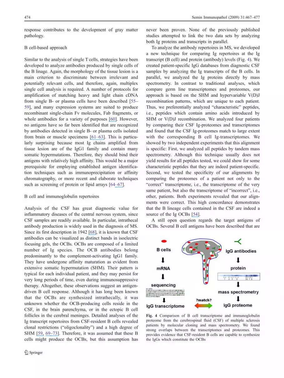
To analyze the antibody repertoires in MS, we developeda new technique for comparing Ig repertoires at the Igtranscript (B cell) and protein (antibody) levels (Fig. 4). Wecreated patient-specific IgG databases from diagnostic CSFsamples by analyzing the Ig transcripts of the B cells. Inparallel, we analyzed the Ig proteins directly by massspectrometry. In contrast to traditional analyses, whichcompare germ line transcriptomes and proteomes, ourapproach is based on the SHM and hypervariable V(D)Jrecombination patterns, which are unique to each patient.Thus, we preferentially analyzed “characteristic” peptides,i.e., peptides which contain amino acids introduced bySHM or V(D)J recombination. We analyzed four patientsby comparing their CSF Ig-proteomes and transcriptomesand found that the CSF Ig-proteomes match to large extentwith the corresponding B cell Ig-transcriptomes. Weshowed by two independent experiments that this alignmentis specific: First, we analyzed all peptides by tandem massspectrometry. Although this technique usually does notyield results for all peptides tested, we could show for somecharacteristic peptides that they are indeed patient-specific.Second, we tested the specificity of our alignments bycomparing the proteomes of a patient not only to the“correct” transcriptome, i.e., the transcriptome of the verysame patient, but also the transcriptome of “incorrect”, i.e.,other, patients. Both experiments revealed that our align-ments were correct. This high concordance demonstratesthat the B lineage cells contained in the CSF are indeed asource of the Ig OCBs [54].
A still open question regards the target antigens ofOCBs. Several B cell antigens have been described that are
Fig. 4 Comparison of B cell transcriptome and immunoglobulinproteome from the cerebrospinal fluid (CSF) of multiple sclerosispatients by molecular cloning and mass spectrometry. We foundstrong overlaps between the transcriptomes and proteomes. Thisprovides evidence that CSF-resident B cells are capable to synthesizethe IgGs which constitute the OCBs
474 Semin Immunopathol (2009) 31:467–477

recognized by (auto) antibodies from serum or CSF of MSpatients [51], but none of them could be attributed toOCBs. Interestingly, there seems to be an obvious contra-diction: On one hand, the OCB antibodies are quantitativelyexpanded Ig populations, carry extensive SHM, andpreferentially belong to the IgG1 family. This providesstrong evidence that the producing B cells had long-lastingcontact with their antigens. On the other hand, the OCB aresoluble antibodies that are present in high concentrations inthe CSF. This means that they are not absorbed by theirantigens and/or that their antigens are present in very lowconcentrations. If so, why are these antigens not removedby various antibody-dependent clearance mechanisms? Ourtechnology for analyzing OCBs may help to address thisquestion. If the paired heavy and light chains fromindividual bands could be identified, then the paired Igchains can be expressed recombinantly and used for antigensearches. Because the antibodies have undergone affinitymaturation, their affinities are presumably high. This willallow isolation of the antigens by affinity chromatographyand their analysis by mass spectrometry. However, theabovementioned correlation is technically not trivial,because the OCBs are always embedded in a pool ofpolyclonal background of many non-expanded antibodies.Therefore, such an approach would require further Igpurification by 2-D electrophoresis and high-performanceliquid chromatography. Its advantage, however, is clear: Itfocuses directly on OCB proteins. In contrast, the moretraditional analysis of antibodies produced by single B cellsdoes not reveal whether the antibodies belong to OCB.
Outlook
The new experimental tools that we described here willadvance our knowledge of the immunopathogenesis of MSand other neuroimmunological diseases. Moreover, theywill help to identify new diagnostic biomarkers, e.g., foridentifying disease subtypes and—perhaps—new pathoge-netic syndromes. Last but not least, the new techniqueshave the potential eventually to improve our therapeuticoptions, e.g., by helping to design “individualized” selec-tive immunotherapies [74].
Acknowledgements This work was supported by the Hermann andLilly Schilling Stiftung, the Deutsche Forschungsgemeinschaft throughgrants SFB571-A1 and -C3, and the sixth Framework Program of theEuropean Union NeuroproMiSe LSHM-CT-2005-018637.
References
1. Steinman L, Zamvil SS (2006) How to successfully applyanimal studies in experimental allergic encephalomyelitis to
research on multiple sclerosis. Ann Neurol 60:12–21. doi:10.1002/ana.20913
2. Wekerle H (2008) Lessons from multiple sclerosis: models,concepts, observations. Ann Rheum Dis 67:56–60. doi:10.1136/ard.2008.098020
3. Dornmair K, Goebels N, Weltzien HU, Wekerle H, Hohlfeld R(2003) T-cell-mediated autoimmunity—novel techniques to char-acterize autoreactive T-cell receptors. Am J Pathol 163:1215–1226
4. Davis SJ, Ikemizu S, Evans EJ, Fugger L, Bakker TR, Van derMerwe PA (2003) The nature of molecular recognition by T cells.Nat Immunol 4:217–224. doi:10.1038/ni0303-217
5. Krogsgaard M, Davis MM (2005) How T cells ‘see’ antigen. NatImmunol 6:239–245. doi:10.1038/ni1173
6. Rudolph MG, Stanfield RL, Wilson IA (2006) How TCRs bindMHC, and coreceptotrs. Annu Rev Immunol 24:419–466.doi:10.1146/annurev.immunol.23.021704.115658
7. Stone JD, Chervin AS, Kranz DM (2009) T-cell receptor bindingaffinities and kinetics: impact on T-cell activity and specificity.Immunology 126:165–176. doi:10.1111/j.1365-2567.2008.03015.x
8. Hohlfeld R, Engel AG, Ii K, Harper MC (1991) Polymyositismediated by T lymphocytes that express the γ/δ receptor. N EnglJ Med 324:877–881
9. Ang S-L, Seidman JG, Peterman GM, Duby AD, Benjamin D,Lee SJ, Hafler DA (1987) Functional lambda chain-associated Tcell receptors on cerebrospinal fluid-derived natural killer-like Tcell clones. J Exp Med 165:1453–1458. doi:10.1084/jem.165.5.1453
10. Wucherpfennig KW, Newcombe J, Li H, Keddy C, Cuzner ML,Hafler DA (1992) γδ T cell receptor repertoire in acute multiplesclerosis lesions. Proc Natl Acad Sci U S A 89:4588–4592.doi:10.1073/pnas.89.10.4588
11. Friese MA, Fugger L (2005) Autoreactive CD8+ T cells inmultiple sclerosis: a new target for therapy? Brain 128:1747–1763. doi:10.1093/brain/awh578
12. Dalakas MC (2006) Sporadic inclusion body myositis—diagnosis,pathogenesis and therapeutic strategies. Nat Clin Pract Neurol2:437–447. doi:10.1038/ncpneuro0261
13. Dalakas MC, Hohlfeld R (2003) Polymyositis and dermatomyo-sitis. Lancet 362:971–982. doi:10.1016/S0140-6736(03)14368-1
14. Babbe H, Roers A, Waisman A, Lassmann H, Goebels N,Hohlfeld R, Friese M, Schröder R, Deckert M, Schmidt S, RavidR, Rajewsky K (2000) Clonal expansion of CD8+ T cellsdominate the T cell infiltrate in active multiple sclerosis lesionsshown by micromanipulation and single cell polymerase chainreaction. J Exp Med 192:393–404. doi:10.1084/jem.192.3.393
15. Skulina C, Schmidt S, Dornmair K, Babbe H, Roers A, RajewskyK, Wekerle H, Hohlfeld R, Goebels N (2004) Multiple sclerosis:brain-infiltrating CD8+ T cells persist as clonal expansions in thecerebrospinal fluid and blood. Proc Natl Acad Sci U S A101:2428–2433. doi:10.1073/pnas.0308689100
16. Junker A, Ivanidze J, Malotka J, Eiglmeier I, Lassmann H,Wekerle H, Meinl E, Hohlfeld R, Dornmair K (2007) Multiplesclerosis: T-cell receptor expression in distinct brain regions. Brain130:2789–2799. doi:10.1093/brain/awm214
17. Bender A, Ernst N, Iglesias A, Dornmair K, Wekerle H, HohlfeldR (1995) T cell receptor repertoire in polymyositis: clonalexpansion of autoaggressive CD8+ T cells. J Exp Med181:1863–1868. doi:10.1084/jem.181.5.1863
18. Bender A, Behrens L, Engel AG, Hohlfeld R (1998) T-cellheterogeneity in muscle lesions of inclusion body myositis. JNeuroimmunol 84:86–91. doi:10.1016/S0165-5728(97)00246-4
19. Hofbauer M, Wiesener S, Babbe H, Roers A, Wekerle H,Dornmair K, Hohlfeld R, Goebels N (2003) Clonal tracking ofautoaggressive T cells in polymyositis by combining lasermicrodissection, single-cell PCR, and CDR3-spectratype analysis.
Semin Immunopathol (2009) 31:467–477 475

Proc Natl Acad Sci U S A 100:4090–4095. doi:10.1073/pnas.0236183100
20. Derfuss T, Segerer S, Herberger S, Sinicina I, Hüfner K, Ebelt K,Knaus H-J, Steiner I, Meinl E, Dornmair K, Arbusow V, StruppM, Brandt T, Theil D (2007) Presence of HSV-1 immediate earlygenes and clonally expanded T-cells with a memory effectorphenotype in human trigeminal ganglia. Brain Pathol 17:389–398.doi:10.1111/j.1750-3639.2007.00088.x
21. Pellkofer HL, Voltz R, Goebels N, Hohlfeld R, Dornmair K(2009) Cross-reactive T-cell receptors in tumor and paraneoplastictarget tissue. Arch Neurol 66(5):655–658
22. Schwab N, Bien CG, Waschbisch A, Becker A, Vince GH,Dornmair K, Wiendl H (2009) CD8+ T-cell clones dominate braininfiltrates in Rasmussen encephalitis and persist in the periphery.Brain 132(Pt 5):1236–1246
23. Seitz S, Schneider CK, Malotka J, Nong X, Engel AG, WekerleH, Hohlfeld R, Dornmair K (2006) Reconstitution of paired T cellreceptor α- and β-chains from microdissected single cells ofhuman inflammatory tissues. Proc Natl Acad Sci U S A 103:12057–12062. doi:10.1073/pnas.0604247103
24. Lefranc M-P, Lefranc G (2001) The T cell receptor facts book.Academic, London, UK
25. Letourneur F, Malissen B (1989) Derivation of a T cell hybridomavariant deprived of functional T cell receptor α and β chaintranscripts reveals a nonfunctional α-mRNA of BW5147 origin.Eur J Immunol 19:2269–2274. doi:10.1002/eji.1830191214
26. Blank U, Boitel B, Mège D, Ermonval M, Acuto O (1993)Analysis of tetanus toxin peptide/DR recognition by human T cellreceptors reconstituted into a murine T cell hybridoma. Eur JImmunol 23:3057–3065. doi:10.1002/eji.1830231203
27. Pluschke G, Rüegg D, Hohlfeld R, Engel AG (1992) Autoag-gressive myocytotoxic T lymphocytes expressing an unusual γ/δT cell receptor. J Exp Med 176:1785–1789. doi:10.1084/jem.176.6.1785
28. Wiendl H, Malotka J, Holzwarth B, Weltzien HU, Wekerle H,Hohlfeld R, Dornmair K (2002) An autoreactive γδ TCR derivedfrom a polymyositis lesion. J Immunol 169:515–521
29. Dornmair K, Schneider CK, Malotka J, Dechant G, Wiendl H,Hohlfeld R (2004) Antigen recognition properties of a Vγ1.3Vδ2-T-cell receptor from a rare variant of polymyositis. J Neuro-immunol 152:168–175 10.1016/j.jneuroim.2004.03.016
30. Hayday AC (2000) γδ T cells: a right time and a right place for aconserved third way of protection. Annu Rev Immunol 18:975–1026. doi:10.1146/annurev.immunol.18.1.975
31. O’Brien RL, Roark CL, Jin N, Aydintug MK, French JD, ChainJL, Wands JM, Johnston M, Born WK (2007) γδ T-cell receptors:functional correlations. Immunol Rev 215:77–88. doi:10.1111/j.1600-065X.2006.00477.x
32. Chien YH, Konigshofer Y (2007) Antigen recognition by γδ Tcells. Immunol Rev 215:46–58. doi:10.1111/j.1600-065X.2006.00470.x
33. Beetz S, Wesch D, Marischen L, Welte S, Oberg HH, Kabelitz D(2008) Innate immune functions of human γδ T cells. Immunobi-ology 213:173–182. doi:10.1016/j.imbio.2007.10.006
34. Sarikonda G, Wang H, Puan KJ, Liu XH, Lee HK, Song YC,Distefano MD, Oldfield E, Prestwich GD, Morita CT (2008)Photoaffinity antigens for human γδ T cells. J Immunol181:7738–7750
35. Fujinami RS, Oldstone MBA (1985) Amino acid homologybetween the encephalitogenic site of myelin basic protein (MBP)and virus: mechanism for autoimmunity. Science 230:1043–1046.doi:10.1126/science.2414848
36. Sospedra M, Martin R (2006) Molecular mimicry in multiplesclerosis. Autoimmunity 39:3–8. doi:10.1080/08916930500484922
37. Libbey JE, Mccoy LL, Fujinami RS (2007) Molecular mimicry inmultiple sclerosis. Int Rev Neurobiol 79:127–147
38. Van der Bruggen P, Traversari C, Chomez P, Lurquin C, De PlaenE, Van den Eynde B, Knuth A, Boon T (1991) A gene encodingan antigen recognized by cytolytic T lymphocytes on a humanmelanoma. Science 254:1643–1647. doi:10.1126/science.1840703
39. Krishnamoorthy G, Saxena A, Mars LT, Domingues HS, MenteleR, Ben-Nun A, Lassmann H, Dornmair K, Kurschus FC, LiblauR, Wekerle H (2009) Myelin specific T cells co-recognizeneuronal autoantigen in a transgenic mouse model of multiplesclerosis. Nat Med 15(6):626–632
40. Cresswell P, Ackerman AL, Giodini A, Peaper DR, Wearsch PA(2005) Mechanisms of MHC class I-restricted antigen processingand cross-presentation. Immunol Rev 207:145–157. doi:10.1111/j.0105-2896.2005.00316.x
41. Vyas JM, Van der Veen AG, Ploegh HL (2008) The knownunknowns of antigen processing and presentation. Nat RevImmunol 8:607–618. doi:10.1038/nri2368
42. Wilson DB, Wilson DH, Schroder K, Pinilla C, Blondelle S,Houghten RA, Garcia KC (2004) Specificity and degeneracy of Tcells. Mol Immunol 40:1047–1055. doi:10.1016/j.molimm.2003.11.022
43. Nino-Vasquez JJ, Allicotti G, Borras E, Wilson DB, Valmori D,Simon R, Martin R, Pinilla C (2005) A powerful combination: theuse of positional scanning libraries and biometrical analysis toidentify cross-reactive T cell epitopes. Mol Immunol 40:1063–1074. doi:10.1016/j.molimm.2003.11.005
44. Sospedra M, Martin R (2006) When T cells recognize a pattern,they might cause trouble. Curr Opin Immunol 18:697–703.doi:10.1016/j.coi.2006.09.006
45. Wucherpfennig KW, Allen PM, Celada F, Cohen IR, De Boer R,Garcia KC, Goldstein B, Greenspan R, Hafler D, Hodgkin P,Huseby ES, Krakauer DC, Nemazee D, Perelson AS, Pinilla C,Strong RK, Sercarz EE (2007) Polyspecificity of T cell and B cellreceptor recognition. Semin Immunol 19:216–224. doi:10.1016/j.smim.2007.02.012
46. Rosenberg SA (1999) A new era for cancer immunotherapy basedon the genes that encode cancer antigens. Immunity 10:281–287.doi:10.1016/S1074-7613(00)80028-X
47. Boon T, Coulie PG, Van den Eynde BJ, Van der Bruggen P (2006)Human T cell responses against melanoma. Annu Rev Immunol24:175–208. doi:10.1146/annurev.immunol.24.021605.090733
48. Smith ES, Mandokhot A, Evans EE, Mueller L, Borrello MA,Sahasrabudhe DM, Zauderer M (2001) Lethality-based selectionof recombinant genes in mammalian cells: application toidentifying tumor antigens. Nat Med 7:967–972. doi:10.1038/91017
49. Wong FS, Karttunen J, Dumont C, Wen L, Visintin I, Pilip IM,Shastri N, Pamer EG, Janeway CA (1999) Identification of anMHC class I-restricted autoantigen in type I diabetes by screeningan organ-specific cDNA library. Nat Med 5:1026–1031.doi:10.1038/12465
50. Van Gelder RN, Von Zastrow ME, Yool A, Dement WC, BarchasJD, Eberwine JH (1990) Amplified RNA synthesized from limitedquantities of heterogeneous complementary DNA. Proc Natl AcadSci U S A 87:1663–1667. doi:10.1073/pnas.87.5.1663
51. Meinl E, Krumbholz M, Hohlfeld R (2006) B lineage cells in theinflammatory CNS environment: migration, maintenance, localantibody production and therapeutic modulation. Ann Neurol59:880–892. doi:10.1002/ana.20890
52. Mathey EK, Derfuss T, Storch MK, Williams KR, Hales K,Woolley DR, Al-Hayani A, Davies SN, Rasband MN, Olsson T,Moldenhauer A, Velhin S, Hohlfeld R, Meinl E, Linington C(2007) Neurofascin as a novel target for autoantibody-mediatedaxonal injury. J Exp Med 204:2363–2372. doi:10.1084/jem.20071053
53. Derfuss T, Parikh K, Velhin S, Braun M, Mathey E, KrumbholzM, Kümpfel T, Moldenhauer A, Rader C, Sonderegger P,
476 Semin Immunopathol (2009) 31:467–477

Pöllmann W, Tiefenthaller C, Bauer J, Lassmann H, Wekerle H,Karagogeos D, Hohlfeld R, Linington C, Meinl E (2009)Contactin-2/Tag-1 directed autoimmunity is identified in multiplesclerosis patients and mediates gray matter pathology in animals.Proc Natl Acad Sci U S A 106(20):8302–8307
54. Obermeier B, Mentele R, Malotka J, Kellermann J, Wekerle H,Lottspeich F, Hohlfeld R, Dornmair K (2008) Matching ofoligoclonal Ig transcriptomes and proteomes of cerebrospinalfluid in multiple sclerosis. Nat Med 14:688–693. doi:10.1038/nm1714
55. Wang X, Stollar BD (2000) Human immunoglobulin variableregion gene analysis by single cell RT-PCR. J Immunol Meth244:217–225
56. Brezinschek H-P, Brezinschek RI, Lipsky PE (1995) Analysis ofheavy chain repertoire of human peripheral B cells using single-cell polymerase chain reaction. J Immunol 155:190–202
57. Bräuninger A, Küppers R, Spieker T, Siebert R, Strickler JG,Schleiffenbaum B, Rajewsky K, Hansmann M-L (1999) Molecularanalysis of single B cells from T cell rich B cell lymphoma showsthe derivation of the tumor cells from mutating germinal center Bcells and exemplifies means by which immunoglobulin genes aremodified in germinal center B cells. Blood 93:2679–2687
58. Wardemann H, Yurasov S, Schaefer A, Young JW, Meffre E,Nussenzweig MC (2003) Predominant autoantibody productionby early human B cell precursors. Science 301:1374–1377.doi:10.1126/science.1086907
59. Owens GP, Ritchie AM, Burgoon MP, Williamson RA, Corboy JR,Gilden DH (2003) Single-cell repertoire analysis demonstrates thatclonal expansion is a prominent feature of the B cell response inmultiple sclerosis cerebrospinal fluid. J Immunol 171:2725–2733
60. Holliger P, Hudson PJ (2005) Engineered antibody fragments andthe rise of single domains. Nat Biotechnol 23:1126–1136.doi:10.1038/nbt1142
61. Bradshaw EM, Orihuela A, McArdel SL, Salajegheh M, AmatoAA, Hafler DA, Greenberg SA, O’Connor KC (2007) A localantigen-driven humoral response is present in the inflammatorymyopathies. J Immunol 178:547–556
62. von Büdingen HC, Harrer MD, Kuenzle S, Meier M, Goebels N(2008) Clonally expanded plasma cells in the cerebrospinal fluidof MS patients produce myelin-specific antibodies. Eur J Immunol38:2014–2023. doi:10.1002/eji.200737784
63. Owens GP, Bennett JL, Lassmann H, O’Connor KC, Ritchie AM,Shearer A, Lam C, Yu XL, Birlea M, DuPree C, Williamson RA,Hafler DA, Burgoon MP, Gilden D (2009) Antibodies producedby clonally expanded plasma cells in multiple sclerosis CSF. AnnNeurol 65(6):639–649
64. Robinson WH, DiGennaro C, Hueber W, Haab BB, Kamachi M,Dean EJ, Fournel S, Fong D, Genovese MC, De Vegvar HEN,
Skriner K, Hirschberg DL, Morris RI, Muller S, Pruijn GJM, VanVenrooij WJ, Smolen JS, Brown PO, Steinman L, Utz PJ (2002)Autoantigen microarrays for multiplex characterization of autoan-tibody responses. Nat Med 8:295–301. doi:10.1038/nm0302-295
65. Kanter JL, Narayana S, Ho PP, Catz I, Warren KG, Sobel RA,Steinman L, Robinson WH (2006) Lipid microarrays identify keymediators of autoimmune brain inflammation. Nat Med 12:138–143. doi:10.1038/nm1344
66. Quintana FJ, Farez MF, Viglietta V, Iglesias AH, Merbl Y,Izquierdo G, Basso AS, Khoury SJ, Lucchinetti CF, Cohen IR,Weiner HL (2008) Antigen microarrays identify unique serumautoantibody signatures in clinical and pathologic subtypes ofmultiple sclerosis. Proc Natl Acad Sci USA 105:18889–18894.doi:10.1073/pnas.0806310105
67. Jäger D, Taverna C, Zippelius A, Knuth A (2004) Identification oftumor antigens as potential target antigens for immunotherapy byserological expression cloning. Cancer Immunol Immunother53:144–147. doi:10.1007/s00262-003-0470-z
68. Kabat EA, Moore DH, Landow H (1942) An electrophoretic studyof the protein components in cerebrospinal fluid and theirrelationship to the serum proteins. J Clin Invest 21:571–577.doi:10.1172/JCI101335
69. Qin Y, Duquette P, Zhang Y, Poole R, Antel JP (1998) Clonalexpansion and somatic hypermutation of VH genes of B cells fromcerebrospinal fluid in multiple sclerosis. J Clin Invest 102:1045–1050. doi:10.1172/JCI3568
70. Owens GP, Kraus H, Burgoon MP, Smith-Jensen T, Devlin ME,Gilden DH (1998) Restricted use of VH4 germline segments in anacute multiple sclerosis brain. Ann Neurol 43:236–243.doi:10.1002/ana.410430214
71. Baranzini SE, Jeong MC, Butunoi C, Murray RS, Bernard CCA,Oksenberg JR (1999) B cell repertoire diversity and clonalexpansion in multiple sclerosis brain lesions. J Immunol163:5133–5144
72. Colombo M, Dono M, Gazzola P, Roncella S, Valetto A,Chiorazzi N, Mancardi GL, Ferrarini M (2000) Accumulation ofclonally related B lymphocytes in the cerebrospinal fluid ofmultiple sclerosis patients. J Immunol 164:2782–2789
73. Monson NL, Brezinschek H-P, Brezinschek R, Mobley A,Vaughan GK, Frohman EM, Racke MK, Lipsky PE (2005)Receptor revision and atypical mutational characteristics inclonally expanded B cells from the cerebrospinal fluid of recentlydiagnosed multiple sclerosis patients. J Neuroimmunol 158:170–181. doi:10.1016/j.jneuroim.2004.04.022
74. Hohlfeld R, Wekerle H (2004) Autoimmune concepts of multiplesclerosis as a basis for selective immunotherapy: from pipedreams to (therapeutic) pipelines. Proc Natl Acad Sci U S A 101(Suppl 2):14599–14606. doi:10.1073/pnas.0404874101
Semin Immunopathol (2009) 31:467–477 477