probing early misfolding events in prion protein mutants by nmr spectroscopy
TRANSCRIPT
Molecules 2013, 18, 9451-9476; doi:10.3390/molecules18089451
molecules ISSN 1420-3049
www.mdpi.com/journal/molecules
Review
Probing Early Misfolding Events in Prion Protein Mutants by NMR Spectroscopy
Gabriele Giachin 1, Ivana Biljan 2, Gregor Ilc 3,4, Janez Plavec 3,4,5 and Giuseppe Legname 1,*
1 Department of Neuroscience, Scuola Internazionale Superiore di Studi Avanzati (SISSA), Via
Bonomea 265,Trieste I-34136, Italy; E-Mail: [email protected] 2 Department of Chemistry, Faculty of Science, University of Zagreb, Horvatovac 102A,
Zagreb HR-10000, Croatia; E-Mail: [email protected] 3 Slovenian NMR Centre, National Institute of Chemistry, Hajdrihova 19,
Ljubljana SI-1000, Slovenia; E-Mails: [email protected] (G.I.); [email protected] (J.P.) 4 EN-FIST Center of Excellence, Ljubljana SI-1000, Slovenia 5 Faculty of Chemistry and Chemical Technology, University of Ljubljana,
Ljubljana SI-1000, Slovenia
* Author to whom correspondence should be addressed; E-Mail: [email protected];
Tel.: +39-040-3787715; Fax: +39-040-3787702.
Received: 27 June 2013; in revised form: 1 August 2013 / Accepted: 5 August 2013 /
Published: 7 August 2013
Abstract: The post-translational conversion of the ubiquitously expressed cellular form of
the prion protein, PrPC, into its misfolded and pathogenic isoform, known as prion or PrPSc,
plays a key role in prion diseases. These maladies are denoted transmissible spongiform
encephalopathies (TSEs) and affect both humans and animals. A prerequisite for
understanding TSEs is unraveling the molecular mechanism leading to the conversion
process whereby most α-helical motifs are replaced by β-sheet secondary structures.
Importantly, most point mutations linked to inherited prion diseases are clustered in the
C-terminal domain region of PrPC and cause spontaneous conversion to PrPSc. Structural
studies with PrP variants promise new clues regarding the proposed conversion mechanism
and may help identify “hot spots” in PrPC involved in the pathogenic conversion. These
investigations may also shed light on the early structural rearrangements occurring in some
PrPC epitopes thought to be involved in modulating prion susceptibility. Here we present a
detailed overview of our solution-state NMR studies on human prion protein carrying
different pathological point mutations and the implications that such findings may have for
the future of prion research.
OPEN ACCESS
Molecules 2013, 18 9452
Keywords: prion protein; prions; genetic mutations; polymorphisms; prion diseases;
3D structure; NMR spectroscopy
1. Introduction
The physiological cellular prion protein (PrP), PrPC, is a glycosylphosphatidylinositol
(GPI)-anchored glycoprotein localized on the outer leaflet of the cellular membrane in mammalian
cells. The mature human (Hu) PrPC (HuPrP) is composed of 209 residues including a largely
unstructured N-terminal part and a globular α-helix rich C-terminal domain. Despite being highly
conserved among mammals, its physiological function has not been established with certainty.
Defining PrPC functions remains an absolute requirement for understanding transmissible spongiform
encephalopathies (TSEs), or prion diseases, which are caused by the posttranslational conversion of
PrPC into a misfolded and pathogenic isoform denoted prion or PrPSc.
The term “prion” was coined by Stanley B. Prusiner in 1982, when he introduced the heretical
notion of a small proteinaceous infectious particle as sole causal agent of TSEs in human and
animals [1]. In humans, TSEs include a heterogeneous group of invariably fatal neurodegenerative
diseases etiologically arising as sporadic, genetic or acquired. According to the recent classification,
idiopathic forms include sporadic Creutzfeldt-Jakob disease (sCJD), sporadic fatal insomnia (sFI) and
the variably protease sensitive prionopathies (VPSPr). Genetic forms comprise familial CJD (fCJD),
Gerstmann-Sträussler-Scheinker disease (GSS), fatal familial insomnia (FFI) and prion protein
cerebral amyloid angiopathy (PrP-CAA). The acquired forms are transmitted from human to human, as
iatrogenic CJD (iCJD) or Kuru, from cattle to human, and human to human, as variant CJD (vCJD) [2].
In animals, relevant TSEs are scrapie in sheep and goats, bovine spongiform encephalopathy (BSE) in
cattle, and chronic wasting disease (CWD) in deer, elk and moose [3].
From the epidemiological point of view, human TSEs are rare diseases evenly distributed
worldwide. Sporadic CJD is the most frequent form and accounts for an incidence of about 0.6–1.2 per
million per year [4]. Genetic forms account for 10%–15% of human prion diseases, whereas the
acquired forms are negligible [5].
The main focus of research in prion biology is understanding the molecular mechanisms involved in
the conformational conversion of PrPC to its pathological counterpart, PrPSc, and how prions trigger
neurotoxic signals leading to TSEs. Genetic forms of human prion diseases are linked to specific
mutations in the PrP gene (PRNP). Key evidence exists supporting the link between mutations and
spontaneous conversion of PrPC to PrPSc in the brain. Over the past few years, biological studies on
PrP pathological mutants have emerged as an invaluable tool for the comprehension of the molecular
basis of TSEs. By means of different experimental approaches, several groups explored the use of the
mutations in order to understand their effects on PrP structure and stability, elucidate the cellular
events leading to PrP accumulation and misfolding, and explain the PrPC physiological roles and its
neurotoxic potential [6–10].
In the next sections, we present an overview of our recent NMR structural studies on HuPrP
pathological mutants. We solved and analyzed the structures corresponding to the globular domain

Molecules 2013, 18 9453
of different HuPrP constructs including the wild-type (WT), the V210I and Q212P mutants
(linked to fCJD and GSS, respectively) and the E219K polymorphism. As a prelude, we first review
what is currently known on PrPC structure and functions, PrPSc structural models, as well as genetic
prion diseases.
2. Structural Features of PrPC
PRNP has been mapped in the short arm of chromosome 20 and the protein coding frame is present
within a single exon [11]. PrPC is highly expressed within the central and peripheral nervous systems,
although its content varies among distinct cell types, neurons and brain regions [12]. Expression of the
protein is developmentally regulated in different mammalian species [13–17]. The immature form of
PrPC is composed of 253 residues including an N-terminal endoplasmic reticulum (ER) signal peptide
and a C-terminal GPI-anchoring signal peptide, which are cleaved during protein maturation and
translocation to the extracellular side of the plasma membrane [18]. Similar to other GPI-anchored
proteins, PrPC is found mainly attached to low density detergent insoluble membrane domains (DRMs)
or lipid rafts [19].
Most structural information on PrPC came from bacterially expressed recombinant (rec) PrP.
Despite the lack of Asn-linked glycosylation at residues 181 and 197, recPrP is structurally equivalent
to brain-derived PrPC [20]. Atomic structures obtained by NMR techniques and X-ray crystallography
revealed that recPrP shares a very similar fold across different mammalian species [7]. The full-length
PrPC has a unique structure: while the N-terminal moiety is largely unfolded from residue 23 to 127,
the C-terminus possesses a well-defined secondary and tertiary structure (residues 128–231, Figure 1b).
The N-terminus features the octapeptide-repeat region (OR), an evolutionarily conserved motif
whose repeated units can vary among species [21]. In HuPrP, the OR consists of one nonapeptide,
PQGGGGWGQ (residues 51–59) and four repeats of sequence PHGGGWGQ (residues 60–92).
Within the four repeats, histidines and tryptophan residues were found to be essential for the high
affinity binding of copper ions and, to a lesser extent, of other divalent cations [22,23]. Additional
histidines at position 96 and 111 are involved in high affinity copper binding and form the non-OR, or
fifth, copper binding site [24,25] (Figure 1b). The positively charged segment, spanning residues 96–111,
precedes a hydrophobic sequence (residues 112–127). These two biochemically distinct segments act
in concert to control the co-translational translocation at the ER during PrPC biosynthesis [26,27].
The HuPrP C-terminal domain is composed of three α-helices (α1, α2 and α3) and two very short
β-strands which form an antiparallel β-sheet (β1 and β2). Helices α2 and α3 form the bulk of the
globular domain and are covalently bridged by a disulfide bond between Cys179 and Cys214. The
C-terminus NMR structures of WT HuPrP obtained at different pH values (4.5, 5.5 and 7) show almost
identical backbone tertiary structures with local differences in flexible regions [28–30].

Molecules 2013, 18 9454
Figure 1. (a) Secondary structure of the immature HuPrP with all the currently identified disease-associated mutations and polymorphisms
(unclassified phenotype in gray; fCJD in black; GSS in red; PrP-CAA underlined; FFI in blue; and polymorphisms in green). The mature
HuPrP consists of residues 23–231, while the N-terminal and C-terminal signal peptides are cleaved during protein maturation. In the lower
panel, the frequency of the mutations and polymorphisms along the PRNP sequence is represented by colored rectangles. (b) Cartoon of the
full-length HuPrP with the sequence-based representation of the disordered N-terminus and the structured C-terminal domain from PDB code
2LSB [28]. Up to four Cu2+ ions are bound to the OR (residues 60–92) and one Cu2+ ions is bound to the non-OR (residues 93–111).
(c) Schematic representation of the secondary elements in the C-terminal HuPrP structured domain. The stabilizing long-range interactions
were inferred from structural analysis and published data [31–33]. Residues involved in disease-influential mutations or polymorphisms are labeled.
Q217R
E200K
G127V
Q52P
Q212P/H
N173K
E211Q/N
+ 1 OP+ 2 OP+ 4 OP+ 5 OP+ 6 OP+ 7 OP+ 8 OP+ 9 OP P102L
P105L/T/VG114V
A117VG131V
S132I
- 2 OP I138M
G142S?
Y145*
R148H
Q160*D178N-M129D178N-V129
V180I
T183A
H187RT188R/A/KT193I
E196K/AF198S
D202N/GV203I/G
R208H
V210I
Y226*Y227*
Y218N M232R/TP238S
1281 23 51 60 68 76 84 92 111 131 144 154 161 164 173 194 200 228 253231
- 1 OP?M129V
N171S? E219K
N-term signal peptide
GPI-signal peptide
Disease-associated mutations
Polymorphisms
1281 23 51 60 68 76 84 92 111 131 144 154 161 164 173 194 200 228 253231
fCJD GSS FFI Unclassified Polymorphism
I215V
G54S?
A133V
D167N
V209M
23-
60-
92
111
І127
-231
α1α2
α3β1
β2
Cu2+
E196 E146
R156E207R208
V203
E200
L130
D144
S231S230G229
R228Q227Y226Y225A224Q223S222E221
E219Y218
T216I215
M213Q212E211V210V209
M206M205
D202T201
Q172N173N174F175V176H177D178
I182T183I184K185Q186H187T188V189T190T191T192T193
D181V180
T199F198
N197G195
K194
Y169E168D167M166P165R164
S170
Y163
R220
Y162 V161
Q160N159P158Y157
H155M154N153E152R151Y150Y149R148D147
Y145
S143G142F141
I139
P137
H140
I138
R136S135M134A133
Y128 M129 G131 S132G126L125G124G123
C179
K204
C214
G127
Q217
β1
β2
α1
α3
α2
Residue implicated in influential polymorphism
Residue implicated in disease-associated mutation
Hydrophobic and aromatic interactions
Salt-bridge
Disulfide bridge
N171
a
b c
PrP-CAA
Y163*
β1 α1 β2 α2 α3
V176G
Molecules 2013, 18 9455
A detailed analysis reveals that HuPrP core structure is stabilized by extensive hydrophobic,
aromatic and salt bridges networks between the β2-α2-α3 secondary elements and α3-α1 helices.
These stabilizing interactions govern the proper folding of the C-terminal domain. Importantly,
disease-related mutations are clustered almost in the folded part and may contribute to destabilize the
native HuPrP conformation (Figure 1c) [31–34].
Among different species, local sequence and structural variations are prominently localized at the
interface between the β2-α2 loop (residue 163–171) and the C-terminal part of α3-helix (residues 218–231),
providing insights into a region of potential importance for pathogenicity and barriers to TSE
transmission across species. Although this loop is highly flexible in most species, it shows a
well-defined conformation in the recPrP of Syrian hamster [35], elk [36], bank vole [37], wallaby [38],
rabbit [39] and horse [40]. In elk and bank vole recPrP, this rigidity was shown to be controlled by a
single aminoacid substitution (S170N), whereas in wallaby, rabbit and horse recPrP it correlates with
long-range interactions between residue 166 and 225. Importantly, no occurrence of TSE has been
reported in rabbit, horse or any marsupial species, suggesting that prion resistance is enciphered by the
amino acid composition of the β2-α2 loop and its stabilizing contacts with the C-terminal segment of
α3-helix [41].
The PrPC N- and C-terminal domains are often considered structurally independent and
non-interacting in prions. This view is supported by a body of evidence showing that the structured
domain plays a crucial role during the pathological conversion to prions. In fact, limited protease
K (PK) digestion of PrPSc often produces a smaller, protease-resistant segment of approximately
142 amino acids spanning residue ~90 to 231 and referred to as PrP 27–30. This molecule is sufficient
to generate infectious amyloid fibrils [42,43]. Additionally, in vitro generated amyloid fibrils
composed only of C-terminal rec murine (Mo) PrP (residues 89–230) were pathogenic and infectious
in transgenic (Tg) mice Tg9949 overexpressing MoPrP(89–230) [44]. Although the N-terminal region
is not essential for prion conversion and infectivity, in the self-association of PrPC to PrPSc it may act
as a modulator of both conversion and prion propagation [45].
Recent studies are attributing a significant role to the N-terminus in driving tertiary contacts with
the C-terminus. By means of synchrotron-based X-ray absorption fine structure (XAFS) techniques,
we showed that Q212P mutation, which is localized in α3-helix, alters the proper copper coordination
in the non-OR copper binding site. These findings imply a structural effect of C-terminal mutations on
the N-terminal part where functional domains are present [24]. Copper was shown to promote the
association of the N-terminus to α1-helix [46]. Finally, zinc binding to the OR region induces novel
electrostatic-driven contacts between this domain and the α2-α3 helices [47].
3. Defining PrPC Functions
The physiological role of PrPC is usually disregarded at the expense of its role in TSEs. However its
definition is essential to understand the molecular mechanisms leading to the disease. Common
strategies often employed to identify PrPC functions include the development of different Tg mice lines
which are knockout (KO) for PrP gene (Prnp). Despite the wide distribution of PrPC in the mammalian
central nervous system (CNS), Prnp KO mice (Prnp0/0) surprisingly display no major anatomical
developmental deficits [48]. Further evaluations revealed that Prnp0/0 mice show mild behavioral

Molecules 2013, 18 9456
phenotypes such as deficits in spatial learning and circadian rhythms [49], altered
long-term potentiation [50,51] and increased excitability of hippocampal neurons [52–55]. These
studies highlight the synaptic role of PrPC in the development of the hippocampus and are corroborated
by immunohistochemical studies on PrPC expression and localization in the hippocampus during the
development [13–17].
A neuroprotective PrPC function has been inferred from peculiar features of Prnp0/0 mice
such as increased neuronal damage after ischemic stroke [56,57], increased mortality in excitotoxic
conditions [58], enhanced susceptibility to kainate-induced seizure [59,60] and to glutamate treatment [61]
and altered N-methyl-d-aspartate (NMDA) receptor currents with greater basal excitability, increased
amplitude and slowed kinetics [62]. The observed neuroprotective roles of PrPC are consistent with the
currently accepted idea that PrPC is a copper binding protein involved in copper homeostasis. A wealth
of studies explored the functional aspects of this interaction indicating that the protein acts as a copper
transporter, a sink of copper excess or a scavenger for copper-generated free radicals [63].
Insights into a possible PrPC involvement in cell proliferation and differentiation derive from results
on embryonic hippocampal neuron cultures treated with recPrP showing an induced polarization in
synapse development and neuritogenesis [64].
Finally, putative PrPC functions are based on its numerous interacting protein molecules [65,66].
Being a GPI-anchored protein present in the DRM, PrPC interacts both in cis or in trans with a variety
of signaling molecules including, for instance, cavelolin-1, Fyn and Src tyrosines kinases [67],
neuronal cell adhesion molecules (NCAMs) [68,69], stress-inducible protein 1 [70] and, recently,
reelin [71], the NR2D subunit of the NMDA receptor [62] and the α2δ-1 subunit of the voltage gated
calcium channel [72]. Taken together these studies reinforce the idea that PrPC is a pleiotropic protein
with different functions, likely acting as a dynamic cell surface scaffolding protein for the assembly of
signaling modules.
4. Prion Conversion and PrPSc Molecular Models
PrPC is the first biological system where a polypeptide exists in at least two distinct conformations
associated with either physiological functions or disease. The central molecular event in the replication
of mammalian prions is the self-propagating conformational conversion of PrPC to the misfolded PrPSc
form. This postulate is known as the “protein-only” hypothesis [1].
Two different mechanisms of prion replication have been put forward. The “template assistance
model” proposes that PrPSc exists as a monomer that is thermodynamically more stable than PrPC,
but this favored conformer is kinetically inaccessible. In the rare event that a PrPSc is formed
spontaneously (or provided exogenously) it can template the misfolding of PrPC by direct interaction.
In this model, the critical step in the conversion is the formation of a dimer between PrPSc and PrPC,
or a partially destabilized folding intermediate of PrPC denoted as PrP*. PrPSc acts as a template that
catalyzes the refolding of PrPC to a thermodynamically more stable PrPSc conformation [73].
In the “nucleation-polymerization model” the conversion between PrPC and PrPSc is reversible,
but the PrPSc monomer is much less stable than PrPC (i.e., the equilibrium is strongly toward PrPC).
Stabilization of PrPSc occurs only upon formation of a stable oligomeric nucleus, which is
thermodynamically not favorable. However, once the nucleus has formed, additional monomeric PrPC
Molecules 2013, 18 9457
is recruited and adopts the conformation of PrPSc. The rate-limiting step in this mechanism is not the
conformational conversion itself but the nucleation step. This step, responsible for the lag phase in the
spontaneous conversion, can be bypassed and accelerated by adding preformed PrPSc seeds [74].
One of the most important challenges in prion biology is determining the structural traits of
infectious prions. Detailed information about PrPSc structure is crucial not only for understanding the
molecular basis of conversion and transmission, but also for potential pharmacological intervention.
Several spectroscopic studies revealed that the amyloid form of PrPSc is β-sheet enriched [75–80].
Numerous research groups investigated the molecular mechanism of the conversion by different
methods (reviewed in [8]). However, all the structural studies on prions are limited by the disorder
and insolubility of PrPSc and they still failed to identify its structure at the atomic level. Due to the lack
of atomistic details for PrPSc, different prion models have been proposed based on low-resolution
experimental approaches [81]. One such model is denoted as β-helical model. It is based on fiber
X-ray diffraction and imaging simulation techniques on brain-extracted Syrian hamster fibrils. It
proposes that the segment ~90–175 forms a four-stranded β-sheet core organized in a β-helical
configuration, whereas α2 and α3 helices retain their native folding [82]. By contrast, hydrogen
deuterium exchange experiments on brain-derived GPI-anchorless PrPSc showed that the region from
residue ~90 to the entire C-terminus displays slow exchange rates which are typical for a structure
consisting of a continuum of β-strands [83]. While recent X-ray diffraction data on different prion
strains (Wille, H. University of Alberta, Edmonton, Alberta, Canada, unpublished data) seem to
confirm the presence of cross-β structure consistent with the β-helical model, the latest work by
Surewicz’s group [83] indicates that other structural PrPSc models cannot be ruled out.
5. Genetic Human Prion Diseases: An Overview
Genetic forms of HuTSEs are transmitted as autosomal dominant traits and co-segregate with
missense or insertional/deletional mutations in PRNP. Over the last decades a number of CJD
surveillance reports have identified up to 58 mutations linked to human prion diseases, associated with
a heterogeneous spectrum of clinical and pathological phenotypes (Figure 1a). Missense mutations
include 44 non-synonymous codon substitutions and five non-sense (or “stop”) mutations. Insertional
or deletional mutations are exclusively localized in the OR, which normally contains one nonapeptide
and four octapeptides. The insertions identified so far include one, two and four to nine extra
octapeptides (OP), while only a deletion of two OP was reported [84]. Pathological point mutations
have been found along the entire PRNP sequence but are mainly clustered in the C-terminal structured
domain (residues 128 to 231) where about 39% of residues are implicated in disease-associated
mutations. Interestingly, almost all the mutations that have been identified so far in the globular
domain affect the hydrophobic and aromatic interactions and the salt bridges, which act in concert to
stabilize the proper α-helical PrPC fold (Figure 1c).
Pathological point mutations are classified into five clinical categories including fCJD, GSS, FFI,
PrP-CAA and unclassified phenotype (Figure 1a). Although the mutations seem defined according to a
rigid clinical classification, very often the syndromes overlap considerably. Only for a few mutations
the prion transmissibility to model animals has been proved [2], therefore many genetic human prion
diseases should be considered as proteinopathies. In general, these genetic forms show incomplete or

Molecules 2013, 18 9458
age-dependent penetrance, as frequently family members carrying a PRNP mutation do not develop
prion diseases [85].
5.1. Clinico-Pathological Features of Genetic Human Prion Diseases
The core features of fCJD include pathologically spongiform neurodegeneration, astrogliosis,
neuronal loss and amyloid plaques, and clinically progressive dementia with visual, cerebellar
and extrapyramidal signs and myoclonus [86]. Phenotypic diversity within sCJD and fCJD has been
observed and most fCJD forms share features with specific subtypes of sCJD [87]. Several
fCJD-associated mutations have been described. The most common fCJD-related mutations are
E200K, V210I, E196K and +6OP insertion, which show distinct geographic clusters with high incidence
of the disease [5,88]. Disease duration is highly variable—from weeks to less than two years [86].
FFI is caused by D178N mutation in combination with methionine at codon 129. It is characterized
by insomnia, dysautonomia and motor signs associated mainly with thalamic astrogliosis, atrophy and
spongiosis [89]. The established haplotypic relationships between D178N-M129 segregating with FFI
and D178N-V129 segregating with fCJD [90] have been questioned by patients segregating with both
FFI and fCJD phenotypes [91] or with different PRNP mutations showing a FFI-like syndrome [92].
Disease duration ranges from six to 42 months [86].
The histopathological presence of PrPSc plaque deposits, rather than the heterogeneous
clinical features, defines GSS. Several point mutations are associated with this syndrome. The
archetypal and most frequent GSS-related mutation is P102L. Other less frequently reported point
mutations are P105L, A117V, H187R and F198S, while other mutations have been described in very
few family cases [86,93].
All five non-sense mutations (Y145*, Q160*, Y163*, Y225* and Y226*) have attracted particular
interest as they are associated with PrPSc amyloid deposition in the walls of arteries, arterioles, veins
and capillaries of the CNS [93,94]. The vascular amyloid fibril deposits have been described in
other neurodegenerative diseases, including Alzheimer’s disease, and are generally defined as cerebral
amyloid angiopathy (CAA) or, in the case of PrP carrying premature stop codons, PrP-CAA [93].
Interestingly, a patient affected by Alzheimer’s disease with CAA showed PrPSc co-localization in the
same amyloid deposits [95]. The reasons of PrPSc perivascular distribution are still unknown, although
different Tg mice expressing anchorless PrP are now available [96–99]. The unique disease phenotype
observed in PrP-CAA clearly indicates that the lack of GPI-moiety greatly contributes to the formation
of PrPSc amyloid outside the cells and provides an intriguing link between prion diseases and other
neurodegenerative diseases.
For several other disease-associated mutations the clinico-pathological classification is still missing,
because published data are insufficient and they are based only on genetic tests without detailed
neuropathological and biochemical analyses. Often the new PRNP mutations have been reported in
single patients without any family history for prion diseases, and are not associated with the presence
of detectable PrPSc in the brain. Therefore the pathological role of such rare mutations remains to be
assessed. Recently new unclassified mutations have been discovered including Q52P in the OP,
D167N in the β2-α2 loop, N173K in α2-helix, V203G, V209M and Q212H in α3-helix [100,101]. Other
confounding aspects are multiple mutations affecting the same codon and segregating with completely
Molecules 2013, 18 9459
different clinico-pathological features, for instance P105L (linked to GSS) and P105T/V (unclassified
disease phenotype), D202N (GSS) and D202G (unclassified), E211Q (CJD) and E211N (GSS), Q212P
(GSS) and Q212H (unclassified).
5.2. Influential Polymorphisms in PRNP
While point mutations are responsible for genetic prion diseases, some polymorphisms in PRNP
influence the etiology and neuropathology of the disease. The polymorphic residue at codon 129
(M129V) has been studied extensively. It has been shown that M129V may affect the susceptibility to
prion diseases [102] and the incubation time of vCJD [103,104]. Methionine homozygosis at
codon 129 is considered a risk factor for sCJD and iCJD [85,100,105].
Another naturally occurring HuPrP polymorphism, E219K, was found to protect against prion
diseases. Heterozygosis at codon 219 was initially reported in the general Japanese population (allele
frequency ~6%) [106,107] and was later found also in other Asiatic populations [108,109], but very
rarely in Western Europeans [100,105]. The absence of confirmed cases of sCJD patients carrying the
E219K polymorphism in Japan or Korea suggests that E219K heterozygosis acts as a protective
factor to sCJD [110]. This polymorphism influences also the clinico-pathological features of both
genetic [111–113] and acquired [114–117] prion diseases. Polymorphism at codon 127 (G127V) has
been reported only among the natives of Papua New Guinea and is protective against Kuru [118].
Uncommon polymorphisms include the absence of 1OP [119], G54S, N171S [100] and G142S [108]
which seem to have no susceptibility or unknown effects in HuTSEs.
6. Structural Biology of HuPrP Pathological Mutants
In the previous sections we overviewed the current knowledge on mutations and polymorphisms
in PRNP and their clinico-pathological implications. Our understanding of the molecular mechanisms
whereby mutations cause genetic TSEs still remains limited. It has been proposed that in vitro
mutations may increase the likelihood of misfolding by thermodynamic destabilization of HuPrP.
While this is true for few studied mutants [120–122], mutations may also alter the protein surface
properties, triggering in turn abnormal interactions with other not yet identified cofactors [123,124]
or causing aberrant cellular trafficking and accumulation inside the cells [125].
Structural studies on HuPrP carrying pathological mutations or polymorphisms may help to
identify the regions involved in the misfolding to PrPSc. Recently, we attempted to determine the high
resolution 3D NMR structure of the HuPrP carrying the Q212P and V210I mutations [126–128] linked
to GSS and fCJD, respectively. Additionally, we solved the NMR structure of the CJD-protective
polymorphism E219K and the WT HuPrP obtained under the same experimental conditions as the
mutants [28]. Before our NMR structures, there was no clear evidence that a pathological point
mutation may cause substantial structural difference in the HuPrP fold. Indeed, the fCJD-related
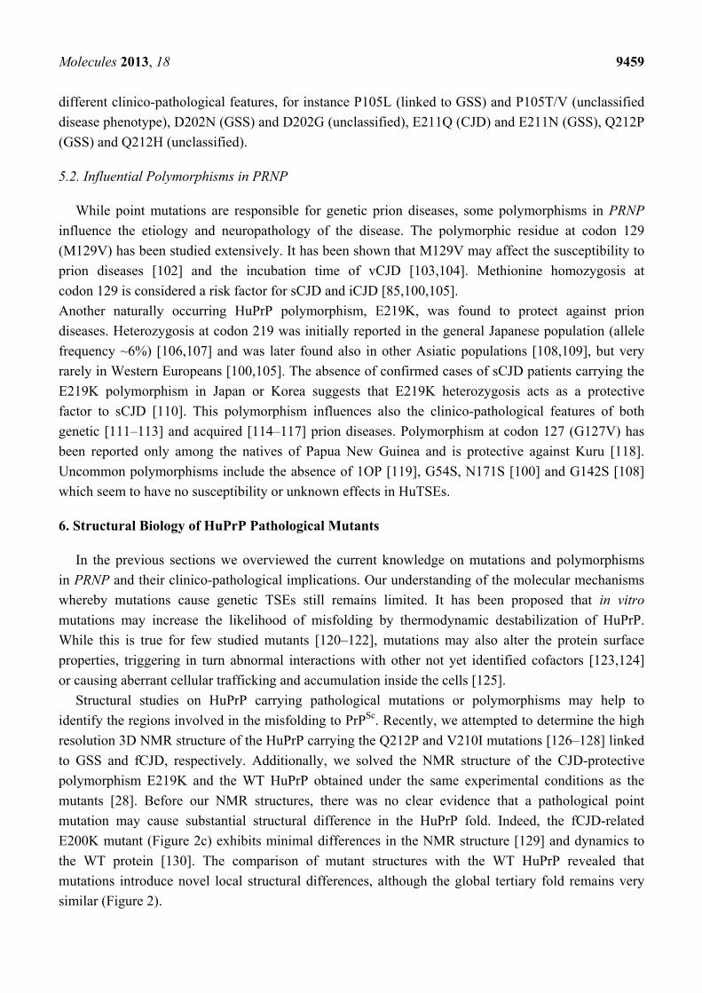
E200K mutant (Figure 2c) exhibits minimal differences in the NMR structure [129] and dynamics to
the WT protein [130]. The comparison of mutant structures with the WT HuPrP revealed that
mutations introduce novel local structural differences, although the global tertiary fold remains very
similar (Figure 2).
Molecules 2013, 18 9460
All the recHuPrP we analyzed were bacterially expressed in isotopic medium, purified from
inclusion bodies and in vitro refolded [127,128]. Proteins always adopted a globular fold as indicated
by the amide signal dispersion in the 1H-15N HSQC spectrum, providing a preliminary indication of
sample suitability for NMR experiments. The backbone assignment was achieved with standard
triple resonance experiments, including HNCA, HN(CO)CA, HNCACB and CBCA(CO)NH NMR
experiments. 1H and 13C resonances of sidechains were assigned by analyses of 3D (H)CCH-TOCSY
and 13C-edited NOESY-HSQC spectra. The large number of NOE restraints together with the
completeness of resonance assignments (>95%) allowed us to determine the structure of all HuPrP
mutants, the WT and E219K polymorphism with high resolution.
Figure 2. Cartoon representations and backbone heavy atom overlays of the different WT
and mutants HuPrP folded domains (residues 126–231): WT at pH 5.5 in red (a, PDB code
2LSB), E219K in blue (b, PDB code 2LFT), E200K in light gray (c, PDB code 1FO7),
Q212P in black (d, PDB code 2KUN), V210I at pH 5.5 in gray (e, PDB code 2LEJ) and
(f) V210I at pH 7.2 in magenta (PDB code 2LV1) superimposed with the WT at pH 7 in
green (PDB code 1HJN). The red frame on the WT (a) highlights the β2-α2 loop region.
Black arrows point at the location of each mutation.
WT, pH 5.5
α3
α2α1
β1
β2
WT, pH 5.5
E219K polymorphism, pH 5.5
WT, pH 5.5
E200K, CJD-related mutant, pH 4.6
WT, pH 5.5
Q212P, GSS-related mutant, pH 5.5
WT, pH 5.5
V210I, CJD-related mutant, pH 5.5
WT, pH 7
V210I, CJD-related mutant, pH 7.2
a b c
d e f

Molecules 2013, 18 9461
6.1. The Q212P Mutant NMR Structure
The Q212P was the first mutation to attract our interest for structural investigations. Due to its
unique sidechain features, proline is a well-known α-helix breaker. Pro212 interrupts the hydrogen
(H)-bond with Arg208, increases the helix pitch and induces local rigidity in the α3-helix. Therefore
we expected major structural rearrangements caused by this mutation, although it is associated with a
very rare form of GSS characterized by mild amyloid PrPSc deposition and reduced penetrance [131,132].
Interestingly, the NMR structure of the Q212P mutant obtained at pH 5.5 revealed only minor changes
at local level, between the segment from residue 205 to 220, but notable differences in distal regions.
In particular, α3-helix showed a broken conformation at residue 221 and 222 which led to the
formation of a novel highly flexible α-helix motif from residues 223 to 228 (Figure 2d).
Upon P212 mutation, α3-helix exhibits small rotation along the helical axis with respect to the WT
protein. A turn of α3-helix around Pro212 is altered to accommodate unfavorable steric interactions of
proline with the preceding residue Glu211. The relative orientation of α3-helix with respect to the
α2-helix changed from 51° in the WT to 33° in the Q212P mutant. As a result, several aromatic and
hydrophobic interactions at the α2-α3 interface are lost. Major structural changes are observed in the
β2-α2 loop region where the aromatic and hydrophobic clusters composed by Tyr163, Met166, Phe175,
Tyr218 and Tyr225 are changed, showing increased exposure to the solvent and longer distance
between the loop and the C-terminal part of α3-helix (Figure 3).
6.2. NMR Structures of V210I at Two Physiological pH Values
The V210I mutant is linked to fCJD, and it shows high incidence across European countries, e.g., in
Italy where geographic clusters carrying this mutation have been identified [133]. Val210 is located in
the middle of the α3-helix and it is part of tightly packed hydrophobic and aromatic residues present
at the interfaces between the α2 and α3 helices and β2 strand (Figure 1c). Therefore we expected this
mutation to promote misfolding by altering these stabilizing interactions.
We investigated the effect of V210I on HuPrP structure at two different physiological pH values.
Acidic pH conditions are characteristic of the intracellular endosomial compartments [134] where
PrPC is sequestered during its internalization. Reports using cell culture models proposed that
PrPC misfolding and accumulation may occur during endocytosis in both late and recycling
endosomes [135–137].
At pH 5.5 the Ile210 causes steric crowding in the inter-helical interface. V210I mutation induces
reorientations of several residues involved in α2–α3 hydrophobic interactions. In order to accommodate
unfavorable steric interactions with Ile210, the sidechain of Val180 changed its conformation along
Cα-Cβ bond by 180°. In addition, the mutation significantly altered the orientation of sidechains of
Val176 and Ile184, thus influencing their hydrophobic contacts with other residues. Interestingly,
as a consequence of these rearrangements the α2-helix is distorted (Figure 2e). Also in this case the
inter-helical angle changes from 51° in the WT to 23° in V210I. Another region within the globular
domain of V210I mutant that displays interesting structural features is the interface of α1 and α3
helices. Helix α1 in the mutant exhibits a reduced propensity to form tertiary hydrophobic
interactions with the protein core. The β2–α2 loop region in V210I is poorly defined, shows an altered

Molecules 2013, 18 9462
conformation and an increased distance from the α3-helix as observed also in the Q212P
mutant (Figure 3).
Figure 3. (a) Distances in Å between residues Met166 and Tyr218 within the β2-α2 loop
and of α3-helix in different WT and mutant HuPrP structures. Distances were calculated
using VMD software [138]. (b) Cartoon representations and backbone heavy atoms
overlays of WT at pH 5.5, E219K and V210I at pH 7.2. This peculiar view highlights the
different spacing observed among the HuPrP structures between the β2-α2 loop and those of
α3-helix. (c) Average values of solvent accessibility (%) for selected residues in different
WT and mutant HuPrP structures calculated using GETAREA software [139]. (d) Cartoon
representations and backbone heavy atom overlays of WT and Q212P structures. Selected
hydrophobic and aromatic residues are highlighted.
α3
α1
α2
WT, pH 5.5
E219K, pH 5.5
V210I, pH 7
Met166
a b
Met166
Phe175
Tyr162
Ile139
Met154
Pro158
c d
Molecules 2013, 18 9463
Comparison with V210I obtained at pH 7.2 showed that changing pH from 5.5 to neutral condition
barely affects the global architecture of the protein (Figure 2f). However, a careful inspection revealed
local structural differences between the two V210I structures. The most prominent pH-related changes
involve alterations in interactions within the hydrophobic core. At pH 5.5, several hydrophobic
contacts between residues at the α2–α3 interface are lost. Moreover, at neutral pH the V210I mutant
exhibits a more compact packing of hydrophobic residues at the interface of the β1–α1 loop and α1 and
α3 helices. Additional stabilizing interactions at these interfaces are provided through hydrophobic
contacts between Phe141 and Tyr145, Tyr145 and Tyr149, Tyr145 and Met209, and Met154 and
Val209, which are not present at acidic condition. In addition to electrostatic interactions that stabilize
the highly hydrophilic part of α1-helix (Asp147−Arg151 and Glu146-Lys204 salt bridges),
hydrophobic interactions are important for maintaining the stability of α1-helix and for its interactions
with other parts of the globular domain of HuPrP (Figure 1c). Taken together, the resulting
comparisons of NMR structures of V210I obtained at two pH values indicate that the separation of
the β1−α1 loop, α1-helix, and the α1−β2 loop from the α2−α3 region is a hot “spot” during the transition
from neutral to acidic pH, and it is required during the early stages of the conversion process. In
support of this view, molecular dynamics (MD) experiments highlighted the role of α1-helix in the
early steps of PrPC misfolding induced by a decrease in pH [140–142]. According to these MD studies,
lowering the pH induces a loss of contacts between α1-helix and other secondary structure elements in
the globular domain of HuPrP, as we observed in our V210I mutant structures.
6.3. NMR Structures of WT HuPrP and of HuPrP Containing E219K Protective Polymorphism
The naturally occurring HuPrP polymorphism, E219K, attracted interest because it was found to
protect against prion diseases. Heterozygosis at codon 219 was initially reported in the Japanese
population [107]. The lack of confirmed cases of sCJD patients carrying the E219K polymorphism
led to the conclusion that E219K heterozygosis acts as a protective factor against sCJD. This
characteristic, also denoted as “dominant-negative effect”, has been reported in several experimental
studies both in vivo and in vitro [123,143–147]. The structural basis responsible for the protective
effect of the E219K polymorphism has so far remained elusive. To gain insight into the structural
determinants underlying the protective influence of this naturally occurring polymorphism, we
determined its high-resolution 3D structure based on NMR datasets. Because we expected minor local
structural changes, we solved also the WT HuPrP obtained under exactly the same experimental
conditions used for mutants and E219K polymorphism (Figure 2a).
The 3D structure of WT HuPrP at pH 5.5 is very similar to the previously reported structure of
the WT protein resolved at pH 4.5 [30]. The two structures are superimposable on the backbone atoms
with an r.m.s.d. value of 1.1 Å. The most apparent differences occur within the β2-α2 loop
region referring to a different orientation of the Tyr169. At pH 4.5 this residue points toward the
interior of the protein, whereas at pH 5.5 it is exposed to solvent, thus loosing contacts with nearby
aromatic residues.
The folded domains of E219K and WT revealed that they are very similar, with backbone r.m.s.d.
of only 1.33 Å. Substitution of Glu by Lys at position 219 apparently does not induce substantial local
structural changes (Figure 2b). Nevertheless, the 3D structure of E219K pointed to rearrangements of
Molecules 2013, 18 9464
some residues in the β1-α1 loop (Phe141), α1-helix (Tyr145 and Tyr149) and α1-β2 loop (Tyr157) that
affected their mutual hydrophobic interactions with residues in α3-helix. NOE data revealed close
packing between the sidechains of several residues situated at the interface of β1-α1 loop, α1-helix,
α1-β2 loop and α3-helix. The sidechain of Phe141 is directed towards the α1-α3 inter-helical interface
engaged in π-π interactions with Tyr149 and Tyr150. These three aromatic residues form a
hydrophobic cluster. Tertiary contacts are also manifested through pronounced hydrophobic
interactions of Phe141, Tyr149 and Tyr150 with Met205, and Phe141 and Tyr150 with Val209.
Moreover, the interface of β1-α1 loop, α1-helix, α1-β2 loop and α3-helix is further stabilized through
hydrophobic interactions of Tyr157 with Met206 and Val209. The structurally poorly defined β2-α2
loop region also exhibited rearrangements involving both hydrophobic and electrostatic interactions.
Especially pronounced contacts involve Met166 and Phe175 located in the loop region and Tyr218
and Tyr225 in the α3-helix (Figure 3). Although the E219K did not induce significant local structural
changes, this polymorphism has a marked effect on the distribution of electrostatic surface potential.
Unlike the WT, large areas of positive charge are observed on the surface of the E219K. These
variations of surface electrostatic potential between the two proteins are mostly clustered around the
site of substitution, at the interface of β2-α2 loop and the C-terminal end of α3-helix, and at the
N-terminal part of α3-helix. In the WT the area around the residue at position 219 is negatively charged
or neutral, whereas in E219K the corresponding region is mostly positively charged.
In conclusion, the NMR structure of the E219K polymorphism suggests that the structural
determinants for its dominant-negative effect are enciphered by the perturbation of the surface charge,
and by subtle structural rearrangements most prominently localized in the β2-α2 loop region and at the
interface of β1-α1 loop, α1-helix, α1-β2 loop and α3-helix. Variations in the distribution of electrostatic
potential on the surface of E219K with respect to the WT are remarkable, considering that only
individuals heterozygous at codon 219 seem resistant to sCJD. Our findings suggest that different
distribution of charges in the WT and E219K may facilitate intermolecular interactions between
the two allelic variants, thus sequestering them from the early stages of the fibrillization process or
inhibiting interactions with yet unknown facilitators of prion conversion.
6.4. Common Structural Traits in HuPrP Pathological Mutants
The structural comparison of the mutants with the WT HuPrP structures allowed us to draw some
preliminary conclusions towards the identification of common regions involved in the early stage of
the conversion process to PrPSc. The observed variations are mostly clustered at the interface between
the β2-α2 loop and α3-helix. In particular, mutations alter the stabilizing interactions occurring in this
region by promoting the progressive separation of the loop from the C-terminal part of α3-helix to the
solvent. This increased distance upon mutation is clearly illustrated in Figure 3a,b. While in WT
HuPrP solved at pH 5.5 the distance between Met166 and Tyr218 is 6.4 ± 0.1 Å, in Q212P and V210I
it greatly increases up to 14.7 ± 0.4 Å. It is noteworthy that in E219K the same residues are much more
closed, thus indicating that the protective polymorphism exhibits a more compact packing at the
β2-α2 loop interface. Interestingly, the β2–α2 loop in E219K displays a tendency toward 310-helical
conformation, thus being somewhat better defined with respect to the same region in the WT and other

Molecules 2013, 18 9465
disease-related mutants. These local structural features of the polymorphism may further account for
its protective influence against sCJD.
Additionally, unlike the WT and E219K, the pathological mutants are characterized by exposure of
hydrophobic and aromatic residues to the solvent in different structural regions. At acidic pH, in
V210I the hydrophobic residues (Ile139, Met154 and Pro158) are much more solvent exposed at the
α1-α3 interface rather than the same mutant structure at pH 7.2 (Figure 3c,d). Conversely, the
Q212P showed increased exposition of the hydrophobic core in the β2-α2 loop and C-terminal part of
α3-helix (Figure 3c,d).
Several groups investigated by molecular dynamics the structural effect of different HuPrP
pathological mutants, including also V210I, E200K, Q212P and E219K [32,33,148–150]. These
studies highlight the possible role of mutations on the flexibility of the β2–α2 loop and on the
hydrophobic core organization, corroborating the NMR results.
7. Conclusions
Experimental evidence supports the hypothesis that the increased hydrophobicity of PrPC is
one of the main determinants of toxicity. Earlier studies proposed a mechanism of neurotoxicity
through a direct interaction of PrPSc, or partially unfolded intermediate states, with neuronal
membranes [151,152], supporting the hypothesis that altered membrane association of PrPC may be an
important factor during the conversion to prion [153,154]. By means of spectroscopic and cellular
techniques, several authors have shown that mild denaturation of recHuPrP leads to an increased
exposure of the hydrophobic region that facilitates internalization and neurotoxic intracellular
accumulation [155,156]. Cell treatment with recombinant HuPrP carrying disease-related mutations
induces higher intracellular retention and neurotoxicity of both native and partially denaturated
conformations [157]. Protein folding metastable intermediates are usually characterized by a
significant exposure of hydrophobic residues which facilitates intermolecular interactions [158–160].
It has been proposed that the membrane environments may alter the PrPC conformational structure.
Spectroscopic studies performed on α-helix folded full-length recHuPrP showed a direct interaction of
the protein with acidic vesicles containing acidic phosphatidylserine (POPS). The binding to POPS
seems to occur at mildly acidic pH values and it involves only the N-terminal segment, while the
globular domain retains its native conformation [161]. In further studies, the effect of different types of
lipid membranes on recPrP structure was investigated by circular dichroism and Fourier transform
infra red spectroscopy experiments [162,163]. These data showed that recPrP maintains its α-helix
conformation when interacting with lipid raft-like membranes rich in cholesterol and sphingomyelin at
pH 7. However, the binding of the protein to negatively charged lipid vesicles, such as the POPG
membranes, promotes structural changes in β-sheet enriched structures at pH 5 and, to a lesser extent,
at pH 7. Thus, the presented results clearly indicate that the preferred PrPC localization in the lipid raft
domains contributes to its stabilization in the α-helix structure. Altered PrP association with different
lipid membranes, such as those in the endocytic compartments, may contribute to destabilize the native
PrPC fold.
In conclusion, our findings on disease-associated mutants and the E219K polymorphism provide
new clues on the possible early events of HuPrP misfolding. The structural disorder of the β2–α2 loop,
Molecules 2013, 18 9466
together with the increased distance between this loop and α3-helix observed in different mutants, lead
to exposure of hydrophobic residues to solvent, thus facilitating intermolecular interactions with other
PrP or with yet unknown cellular factors and abnormal accumulation inside the cells.
Acknowledgments
This work was supported by the European Community’s Seventh Framework Programme
(FP7/2007–2013) under grant agreement n° 222887—the PRIORITY project, and from MIUR,
Ministero dell’Istruzione, dell’Università e della Ricerca (Italian Ministry for Education,
University and Research) under the programs FIRB-RBAP11FRE9, and PRIN 2008 (Meccanismi
Neurodegenerativi nelle Malattie da Prioni: Studi Conformazionali, Fisiopatologia della Proteina
Prionica e Possibili Approcci Farmacologici).
Conflict of Interest
The authors declare no conflict of interest.
References
1. Prusiner, S.B. Novel proteinaceous infectious particles cause scrapie. Science 1982, 216, 136–144.
2. Head, M.W.; Ironside, J.W. Review: Creutzfeldt-jakob disease: Prion protein type, disease
phenotype and agent strain. Neuropathol. Appl. Neurobiol. 2012, 38, 296–310.
3. Imran, M.; Mahmood, S. An overview of animal prion diseases. Virol. J. 2011, 8, 493.
4. Ladogana, A.; Puopolo, M.; Croes, E.A.; Budka, H.; Jarius, C.; Collins, S.; Klug, G.M.; Sutcliffe,
T.; Giulivi, A.; Alperovitch, A. et al. Mortality from creutzfeldt-jakob disease and related
disorders in europe, australia, and canada. Neurology 2005, 64, 1586–1591.
5. Mead, S. Prion disease genetics. Eur. J. Hum. Genet. 2006, 14, 273–281.
6. Aguzzi, A.; Falsig, J. Prion propagation, toxicity and degradation. Nat. Neurosci. 2012, 15, 936–939.
7. Legname, G.; Giachin, G.; Benetti, F. Structural studies of prion proteins and prions. In
Non-Fibrillar Amyloidogenic Protein Assemblies-Common Cytotoxins Underlying Degenerative
Diseases; Rahimi, F., Bitan, G., Eds.; Springer: Dordrecht, The Netherlands, 2012; pp. 289–317.
8. Legname, G.; Tran, T.N.L.; Giachin, G. Synthetic prions. In Prions and Prion Diseases:
New Developments; Verdier, J., Ed.; Nova Science Publishers, Inc.: Hauppauge, NY, USA, 2012;
pp. 61–83.
9. Mallucci, G.R. Prion neurodegeneration: Starts and stops at the synapse. Prion 2009, 3, 195–201.
10. Solomon, I.H.; Schepker, J.A.; Harris, D.A. Prion neurotoxicity: Insights from prion protein
mutants. Curr. Issues Mol. Biol. 2010, 12, 51–61.
11. Basler, K.; Oesch, B.; Scott, M.; Westaway, D.; Walchli, M.; Groth, D.F.; McKinley, M.P.;
Prusiner, S.B.; Weissmann, C. Scrapie and cellular prp isoforms are encoded by the same
chromosomal gene. Cell 1986, 46, 417–428.
12. Linden, R.; Martins, V.R.; Prado, M.A.; Cammarota, M.; Izquierdo, I.; Brentani, R.R.
Physiology of the prion protein. Physiol. Rev. 2008, 88, 673–728.
Molecules 2013, 18 9467
13. Benvegnu, S.; Poggiolini, I.; Legname, G. Neurodevelopmental expression and localization of
the cellular prion protein in the central nervous system of the mouse. J. Comp. Neurol. 2010, 518,
1879–1891.
14. Manson, J.; West, J.D.; Thomson, V.; McBride, P.; Kaufman, M.H.; Hope, J. The prion protein
gene: A role in mouse embryogenesis? Development 1992, 115, 117–122.
15. McKinley, M.P.; Hay, B.; Lingappa, V.R.; Lieberburg, I.; Prusiner, S.B. Developmental
expression of prion protein gene in brain. Dev. Biol. 1987, 121, 105–110.
16. Poggiolini, I.; Legname, G. Mapping the prion protein distribution in marsupials: Insights from
comparing opossum with mouse cns. PLoS One 2012, 7, e50370.
17. Sales, N.; Hassig, R.; Rodolfo, K.; Di Giamberardino, L.; Traiffort, E.; Ruat, M.; Fretier, P.;
Moya, K.L. Developmental expression of the cellular prion protein in elongating axons. Eur. J.
Neurosci. 2002, 15, 1163–1177.
18. Stahl, N.; Borchelt, D.R.; Prusiner, S.B. Differential release of cellular and scrapie prion proteins
from cellular membranes by phosphatidylinositol-specific phospholipase C. Biochemistry 1990,
29, 5405–5412.
19. Taylor, D.R.; Hooper, N.M. The prion protein and lipid rafts. Mol. Membr. Biol. 2006, 23, 89–99.
20. Hornemann, S.; Schorn, C.; Wuthrich, K. Nmr structure of the bovine prion protein isolated from
healthy calf brains. EMBO Rep. 2004, 5, 1159–1164.
21. Wopfner, F.; Weidenhofer, G.; Schneider, R.; von Brunn, A.; Gilch, S.; Schwarz, T.F.; Werner,
T.; Schatzl, H.M. Analysis of 27 mammalian and 9 avian prps reveals high conservation of
flexible regions of the prion protein. J. Mol. Biol. 1999, 289, 1163–1178.
22. Millhauser, G.L. Copper and the prion protein: Methods, structures, function, and disease. Annu
Rev. Phys. Chem. 2007, 58, 299–320.
23. Walter, E.D.; Stevens, D.J.; Visconte, M.P.; Millhauser, G.L. The prion protein is a combined
zinc and copper binding protein: Zn2+ alters the distribution of Cu2+ coordination modes. J. Am.
Chem. Soc. 2007, 129, 15440–15441.
24. D’Angelo, P.; Della Longa, S.; Arcovito, A.; Mancini, G.; Zitolo, A.; Chillemi, G.; Giachin, G.;
Legname, G.; Benetti, F. Effects of the pathological q212p mutation on human prion protein non-
octarepeat copper-binding site. Biochemistry 2012, 51, 6068–6079.
25. Hasnain, S.S.; Murphy, L.M.; Strange, R.W.; Grossmann, J.G.; Clarke, A.R.; Jackson, G.S.;
Collinge, J. Xafs study of the high-affinity copper-binding site of human prp(91–231) and its
low-resolution structure in solution. J. Mol. Biol. 2001, 311, 467–473.
26. Chakrabarti, O.; Ashok, A.; Hegde, R.S. Prion protein biosynthesis and its emerging role in
neurodegeneration. Trends. Biochem. Sci. 2009, 34, 287–295.
27. Li, A.; Christensen, H.M.; Stewart, L.R.; Roth, K.A.; Chiesa, R.; Harris, D.A. Neonatal lethality
in transgenic mice expressing prion protein with a deletion of residues 105–125. EMBO J. 2007,
26, 548–558.
28. Biljan, I.; Giachin, G.; Ilc, G.; Zhukov, I.; Plavec, J.; Legname, G. Structural basis for the
protective effect of the human prion protein carrying the dominant-negative e219k polymorphism.
Biochem. J. 2012, 446, 243–251.
29. Calzolai, L.; Zahn, R. Influence of ph on nmr structure and stability of the human prion protein
globular domain. J. Biol. Chem. 2003, 278, 35592–35596.
Molecules 2013, 18 9468
30. Zahn, R.; Liu, A.; Luhrs, T.; Riek, R.; von Schroetter, C.; Lopez Garcia, F.; Billeter, M.; Calzolai,
L.; Wider, G.; Wuthrich, K. Nmr solution structure of the human prion protein. Proc. Natl. Acad.
Sci. USA 2000, 97, 145–150.
31. Adrover, M.; Pauwels, K.; Prigent, S.; De Chiara, C.; Xu, Z.; Chapuis, C.; Pastore, A.; Rezaei, H.
Prion fibrillization is mediated by a native structural element that comprises helices h2 and h3.
J. Biol. Chem. 2010, 285, 21004–21012.
32. Rossetti, G.; Giachin, G.; Legname, G.; Carloni, P. Structural facets of disease-linked human
prion protein mutants: A molecular dynamic study. Proteins-Struct. Funct. Bioinform. 2010, 78,
3270–3280.
33. van der Kamp, M.W.; Daggett, V. Pathogenic mutations in the hydrophobic core of the human
prion protein can promote structural instability and misfolding. J. Mol. Biol. 2010, 404, 732–748.
34. Shen, L.; Ji, H.F. Mutation directional selection sheds light on prion pathogenesis. Biochem.
Biophys. Res. Commun. 2011, 410, 159–163.
35. Donne, D.G.; Viles, J.H.; Groth, D.; Mehlhorn, I.; James, T.L.; Cohen, F.E.; Prusiner, S.B.;
Wright, P.E.; Dyson, H.J. Structure of the recombinant full-length hamster prion protein
prp(29–231): The n terminus is highly flexible. Proc. Natl. Acad. Sci. USA 1997, 94, 13452–13457.
36. Gossert, A.D.; Bonjour, S.; Lysek, D.A.; Fiorito, F.; Wuthrich, K. Prion protein nmr structures of
elk and of mouse/elk hybrids. Proc. Natl. Acad. Sci. USA 2005, 102, 646–650.
37. Christen, B.; Perez, D.R.; Hornemann, S.; Wuthrich, K. Nmr structure of the bank vole prion
protein at 20 degrees c contains a structured loop of residues 165–171. J. Mol. Biol. 2008, 383,
306–312.
38. Christen, B.; Hornemann, S.; Damberger, F.F.; Wuthrich, K. Prion protein nmr structure from
tammar wallaby (macropus eugenii) shows that the beta2-alpha2 loop is modulated by
long-range sequence effects. J. Mol. Biol. 2009, 389, 833–845.
39. Wen, Y.; Li, J.; Yao, W.; Xiong, M.; Hong, J.; Peng, Y.; Xiao, G.; Lin, D. Unique structural
characteristics of the rabbit prion protein. J. Biol. Chem. 2010, 285, 31682–31693.
40. Perez, D.R.; Damberger, F.F.; Wuthrich, K. Horse prion protein nmr structure and comparisons
with related variants of the mouse prion protein. J. Mol. Biol. 2010, 400, 121–128.
41. Scouras, A.D.; Daggett, V. Disruption of the x-loop turn of the prion protein linked to scrapie
resistance. Protein Eng. Des. Sel. 2012, 25, 243–249.
42. Colby, D.W.; Prusiner, S.B. Prions. Cold Spring Harb Perspect. Biol. 2011, 3, a006833.
43. Prusiner, S.B. Prions. Proc. Natl. Acad. Sci. USA 1998, 95, 13363–13383.
44. Legname, G.; Baskakov, I.V.; Nguyen, H.O.; Riesner, D.; Cohen, F.E.; DeArmond, S.J.; Prusiner,
S.B. Synthetic mammalian prions. Science 2004, 305, 673–676.
45. Turnbaugh, J.A.; Unterberger, U.; Saa, P.; Massignan, T.; Fluharty, B.R.; Bowman, F.P.; Miller,
M.B.; Supattapone, S.; Biasini, E.; Harris, D.A. The n-terminal, polybasic region of prp(c)
dictates the efficiency of prion propagation by binding to prp(sc). J. Neurosci. 2012, 32,
8817–8830.
46. Thakur, A.K.; Srivastava, A.K.; Srinivas, V.; Chary, K.V.; Rao, C.M. Copper alters aggregation
behavior of prion protein and induces novel interactions between its n- and c-terminal regions.
J. Biol. Chem. 2011, 286, 38533–38545.
Molecules 2013, 18 9469
47. Spevacek, A.R.; Evans, E.G.; Miller, J.L.; Meyer, H.C.; Pelton, J.G.; Millhauser, G.L. Zinc
drives a tertiary fold in the prion protein with familial disease mutation sites at the interface.
Structure 2013, 21, 236–246.
48. Bueler, H.; Fischer, M.; Lang, Y.; Bluethmann, H.; Lipp, H.P.; DeArmond, S.J.; Prusiner, S.B.;
Aguet, M.; Weissmann, C. Normal development and behaviour of mice lacking the neuronal
cell-surface prp protein. Nature 1992, 356, 577–582.
49. Criado, J.R.; Sanchez-Alavez, M.; Conti, B.; Giacchino, J.L.; Wills, D.N.; Henriksen, S.J.; Race,
R.; Manson, J.C.; Chesebro, B.; Oldstone, M.B. Mice devoid of prion protein have cognitive
deficits that are rescued by reconstitution of prp in neurons. Neurobiol. Dis. 2005, 19, 255–265.
50. Maglio, L.E.; Martins, V.R.; Izquierdo, I.; Ramirez, O.A. Role of cellular prion protein on ltp
expression in aged mice. Brain Res. 2006, 1097, 11–18.
51. Maglio, L.E.; Perez, M.F.; Martins, V.R.; Brentani, R.R.; Ramirez, O.A. Hippocampal synaptic
plasticity in mice devoid of cellular prion protein. Brain Res. Mol. Brain Res. 2004, 131, 58–64.
52. Caiati, M.D.; Safiulina, V.F.; Fattorini, G.; Sivakumaran, S.; Legname, G.; Cherubini, E. Prpc
controls via protein kinase a the direction of synaptic plasticity in the immature hippocampus.
J. Neurosci. 2013, 33, 2973–2983.
53. Colling, S.B.; Collinge, J.; Jefferys, J.G. Hippocampal slices from prion protein null mice:
Disrupted Ca2+-activated K+ currents. Neurosci. Lett. 1996, 209, 49–52.
54. Fuhrmann, M.; Bittner, T.; Mitteregger, G.; Haider, N.; Moosmang, S.; Kretzschmar, H.; Herms, J.
Loss of the cellular prion protein affects the Ca2+ homeostasis in hippocampal ca1 neurons.
J. Neurochem. 2006, 98, 1876–1885.
55. Mallucci, G.R.; Ratte, S.; Asante, E.A.; Linehan, J.; Gowland, I.; Jefferys, J.G.; Collinge, J.
Post-natal knockout of prion protein alters hippocampal ca1 properties, but does not result in
neurodegeneration. EMBO J. 2002, 21, 202–210.
56. McLennan, N.F.; Brennan, P.M.; McNeill, A.; Davies, I.; Fotheringham, A.; Rennison, K.A.;
Ritchie, D.; Brannan, F.; Head, M.W.; Ironside, J.W. et al. Prion protein accumulation and
neuroprotection in hypoxic brain damage. Am. J. Pathol. 2004, 165, 227–235.
57. Spudich, A.; Frigg, R.; Kilic, E.; Kilic, U.; Oesch, B.; Raeber, A.; Bassetti, C.L.; Hermann, D.M.
Aggravation of ischemic brain injury by prion protein deficiency: Role of erk-1/-2 and stat-1.
Neurobiol. Dis. 2005, 20, 442–449.
58. Walz, R.; Amaral, O.B.; Rockenbach, I.C.; Roesler, R.; Izquierdo, I.; Cavalheiro, E.A.; Martins, V.R.;
Brentani, R.R. Increased sensitivity to seizures in mice lacking cellular prion protein. Epilepsia
1999, 40, 1679–1682.
59. Rangel, A.; Burgaya, F.; Gavin, R.; Soriano, E.; Aguzzi, A.; Del Rio, J.A. Enhanced
susceptibility of prnp-deficient mice to kainate-induced seizures, neuronal apoptosis, and death:
Role of ampa/kainate receptors. J. Neurosci. Res. 2007, 85, 2741–2755.
60. Rangel, A.; Madronal, N.; Gruart, A.; Gavin, R.; Llorens, F.; Sumoy, L.; Torres, J.M.;
Delgado-Garcia, J.M.; Del Rio, J.A. Regulation of gaba(a) and glutamate receptor expression,
synaptic facilitation and long-term potentiation in the hippocampus of prion mutant mice.
PLoS One 2009, 4, e7592.
61. Brown, D.R.; Nicholas, R.S.; Canevari, L. Lack of prion protein expression results in a neuronal
phenotype sensitive to stress. J. Neurosci. Res. 2002, 67, 211–224.
Molecules 2013, 18 9470
62. Khosravani, H.; Zhang, Y.; Tsutsui, S.; Hameed, S.; Altier, C.; Hamid, J.; Chen, L.; Villemaire, M.;
Ali, Z.; Jirik, F.R. et al. Prion protein attenuates excitotoxicity by inhibiting nmda receptors.
J. Cell. Biol. 2008, 181, 551–565.
63. Viles, J.H.; Klewpatinond, M.; Nadal, R.C. Copper and the structural biology of the prion protein.
Biochem. Soc. Trans. 2008, 36, 1288–1292.
64. Kanaani, J.; Prusiner, S.B.; Diacovo, J.; Baekkeskov, S.; Legname, G. Recombinant prion
protein induces rapid polarization and development of synapses in embryonic rat hippocampal
neurons in vitro. J. Neurochem. 2005, 95, 1373–1386.
65. Aguzzi, A.; Baumann, F.; Bremer, J. The prion’s elusive reason for being. Annu Rev. Neurosci
2008, 31, 439–477.
66. Didonna, A. Prion protein and its role in signal transduction. Cell. Mol. Biol. Lett. 2013, 18, 209–230.
67. Mouillet-Richard, S.; Ermonval, M.; Chebassier, C.; Laplanche, J.L.; Lehmann, S.; Launay, J.M.;
Kellermann, O. Signal transduction through prion protein. Science 2000, 289, 1925–1928.
68. Santuccione, A.; Sytnyk, V.; Leshchyns'ka, I.; Schachner, M. Prion protein recruits its neuronal
receptor ncam to lipid rafts to activate p59(fyn) and to enhance neurite outgrowth. J. Cell Biol.
2005, 169, 341–354.
69. Schmitt-Ulms, G.; Legname, G.; Baldwin, M.A.; Ball, H.L.; Bradon, N.; Bosque, P.J.; Crossin, K.L.;
Edelman, G.M.; DeArmond, S.J.; Cohen, F.E. et al. Binding of neural cell adhesion molecules
(n-cams) to the cellular prion protein. J. Mol. Biol. 2001, 314, 1209–1225.
70. Roffe, M.; Beraldo, F.H.; Bester, R.; Nunziante, M.; Bach, C.; Mancini, G.; Gilch, S.; Vorberg, I.;
Castilho, B.A.; Martins, V.R. et al. Prion protein interaction with stress-inducible protein 1
enhances neuronal protein synthesis via mtor. Proc. Natl. Acad. Sci. USA 2010, 107, 13147–13152.
71. Devanathan, V.; Jakovcevski, I.; Santuccione, A.; Li, S.; Lee, H.J.; Peles, E.; Leshchyns'ka, I.;
Sytnyk, V.; Schachner, M. Cellular form of prion protein inhibits reelin-mediated shedding of
caspr from the neuronal cell surface to potentiate caspr-mediated inhibition of neurite outgrowth.
J. Neurosci. 2010, 30, 9292–9305.
72. Senatore, A.; Colleoni, S.; Verderio, C.; Restelli, E.; Morini, R.; Condliffe, S.B.; Bertani, I.;
Mantovani, S.; Canovi, M.; Micotti, E. et al. Mutant prp suppresses glutamatergic
neurotransmission in cerebellar granule neurons by impairing membrane delivery of vgcc
alpha(2)delta-1 subunit. Neuron 2012, 74, 300–313.
73. Prusiner, S.B. Molecular biology of prion diseases. Science 1991, 252, 1515–1522.
74. Jarrett, J.T.; Lansbury, P.T., Jr.; Seeding “one-dimensional crystallization” of amyloid: A
pathogenic mechanism in alzheimer's disease and scrapie? Cell. 1993, 73, 1055–1058.
75. Caughey, B.; Raymond, G.J.; Bessen, R.A. Strain-dependent differences in beta-sheet
conformations of abnormal prion protein. J. Biol. Chem. 1998, 273, 32230–32235.
76. Caughey, B.W.; Dong, A.; Bhat, K.S.; Ernst, D.; Hayes, S.F.; Caughey, W.S. Secondary
structure analysis of the scrapie-associated protein prp 27–30 in water by infrared spectroscopy.
Biochemistry 1991, 30, 7672–7680.
77. Gasset, M.; Baldwin, M.A.; Fletterick, R.J.; Prusiner, S.B. Perturbation of the secondary
structure of the scrapie prion protein under conditions that alter infectivity. Proc. Natl. Acad. Sci.
USA 1993, 90, 1–5.

Molecules 2013, 18 9471
78. Goormaghtigh, E.; Cabiaux, V.; Ruysschaert, J.M. Secondary structure and dosage of soluble
and membrane proteins by attenuated total reflection fourier-transform infrared spectroscopy on
hydrated films. Eur. J. Biochem. 1990, 193, 409–420.
79. Pan, K.M.; Baldwin, M.; Nguyen, J.; Gasset, M.; Serban, A.; Groth, D.; Mehlhorn, I.; Huang, Z.;
Fletterick, R.J.; Cohen, F.E.; et al. Conversion of alpha-helices into beta-sheets features in the
formation of the scrapie prion proteins. Proc. Natl. Acad. Sci. USA 1993, 90, 10962–10966.
80. Spassov, S.; Beekes, M.; Naumann, D. Structural differences between tses strains investigated by
ft-ir spectroscopy. Biochim. Biophys. Acta 2006, 1760, 1138–1149.
81. Surewicz, W.K.; Apostol, M.I. Prion protein and its conformational conversion: A structural
perspective. Top. Curr. Chem. 2011, 305, 135–167.
82. Wille, H.; Bian, W.; McDonald, M.; Kendall, A.; Colby, D.W.; Bloch, L.; Ollesch, J.;
Borovinskiy, A.L.; Cohen, F.E.; Prusiner, S.B.; et al. Natural and synthetic prion structure from
X-ray fiber diffraction. Proc. Natl. Acad. Sci. USA 2009, 106, 16990–16995.
83. Smirnovas, V.; Baron, G.S.; Offerdahl, D.K.; Raymond, G.J.; Caughey, B.; Surewicz, W.K.
Structural organization of brain-derived mammalian prions examined by hydrogen-deuterium
exchange. Nat. Struct. Mol. Biol. 2011, 18, 504–506.
84. Croes, E.A.; Theuns, J.; Houwing-Duistermaat, J.J.; Dermaut, B.; Sleegers, K.; Roks, G.;
Van den Broeck, M.; van Harten, B.; van Swieten, J.C.; Cruts, M. et al. Octapeptide repeat
insertions in the prion protein gene and early onset dementia. J. Neurol. Neurosurg. Psychiatr.
2004, 75, 1166–1170.
85. Kovacs, G.G.; Puopolo, M.; Ladogana, A.; Pocchiari, M.; Budka, H.; Van Duijn, C.; Collins, S.J.;
Boyd, A.; Giulivi, A.; Coulthart, M.; et al. Genetic prion disease: The eurocjd experience.
Hum. Genet. 2005, 118, 166–174.
86. Legname, G.; Zanusso, G. Molecular pathogenesis of prion diseases. In Miscellanea on
Encephalopathies, Tanasescu, R., Ed.; InTech: New York, NY, USA, 2012; pp. 95–112.
87. Gambetti, P.; Kong, Q.; Zou, W.; Parchi, P.; Chen, S.G. Sporadic and familial cjd: Classification
and characterisation. Br. Med. Bull. 2003, 66, 213–239.
88. Schelzke, G.; Eigenbrod, S.; Romero, C.; Varges, D.; Breithaupt, M.; Taratuto, A.L.;
Kretzschmar, H.A.; Zerr, I. Genetic prion disease with codon 196 prnp mutation: Clinical and
pathological findings. Neurobiol. Aging. 2011, 32, 756.e1–756.e9.
89. Gambetti, P.; Parchi, P.; Petersen, R.B.; Chen, S.G.; Lugaresi, E. Fatal familial insomnia and
familial creutzfeldt-jakob disease: Clinical, pathological and molecular features. Brain Pathol.
1995, 5, 43–51.
90. Goldfarb, L.G.; Petersen, R.B.; Tabaton, M.; Brown, P.; LeBlanc, A.C.; Montagna, P.; Cortelli,
P.; Julien, J.; Vital, C.; Pendelbury, W.W.; et al. Fatal familial insomnia and familial creutzfeldt-
jakob disease: Disease phenotype determined by a DNA polymorphism. Science 1992, 258, 806–808.
91. McLean, C.A.; Storey, E.; Gardner, R.J.; Tannenberg, A.E.; Cervenakova, L.; Brown, P.
The d178n (cis-129m) “fatal familial insomnia” mutation associated with diverse
clinicopathologic phenotypes in an australian kindred. Neurology 1997, 49, 552–558.
92. Pocchiari, M.; Ladogana, A.; Petraroli, R.; Cardone, F.; D’Alessandro, M. Recent italian ffi cases.
Brain Pathol. 1998, 8, 564–566.
Molecules 2013, 18 9472
93. Revesz, T.; Holton, J.L.; Lashley, T.; Plant, G.; Frangione, B.; Rostagno, A.; Ghiso, J. Genetics
and molecular pathogenesis of sporadic and hereditary cerebral amyloid angiopathies.
Acta Neuropathol. 2009, 118, 115–130.
94. Jansen, C.; Parchi, P.; Capellari, S.; Vermeij, A.J.; Corrado, P.; Baas, F.; Strammiello, R.;
van Gool, W.A.; van Swieten, J.C.; Rozemuller, A.J. Prion protein amyloidosis with divergent
phenotype associated with two novel nonsense mutations in prnp. Acta neuropathol. 2010, 119,
189–197.
95. Paquet, C.; Privat, N.; Kaci, R.; Polivka, M.; Dupont, O.; Haik, S.; Laplanche, J.L.; Hauw, J.J.;
Gray, F. Cerebral amyloid angiopathy with co-localization of prion protein and beta-amyloid in
an 85-year-old patient with sporadic creutzfeldt-jakob disease. Acta neuropathol. 2008, 116,
567–573.
96. Mahal, S.P.; Jablonski, J.; Suponitsky-Kroyter, I.; Oelschlegel, A.M.; Herva, M.E.; Oldstone, M.;
Weissmann, C. Propagation of rml prions in mice expressing prp devoid of gpi anchor leads to
formation of a novel, stable prion strain. PLoS Pathog. 2012, 8, e1002746.
97. Rangel, A.; Race, B.; Striebel, J.; Chesebro, B. Non-amyloid and amyloid prion protein deposits
in prion-infected mice differ in blockage of interstitial brain fluid. Neuropathol. Appl. Neurobiol.
2013, 39, 217–230.
98. Raymond, G.J.; Race, B.; Hollister, J.R.; Offerdahl, D.K.; Moore, R.A.; Kodali, R.; Raymond,
L.D.; Hughson, A.G.; Rosenke, R.; Long, D.; et al. Isolation of novel synthetic prion strains by
amplification in transgenic mice coexpressing wild-type and anchorless prion proteins. J. Virol.
2012, 86, 11763–11778.
99. Stohr, J.; Watts, J.C.; Legname, G.; Oehler, A.; Lemus, A.; Nguyen, H.O.; Sussman, J.; Wille, H.;
DeArmond, S.J.; Prusiner, S.B. et al. Spontaneous generation of anchorless prions in transgenic
mice. Proc. Natl. Acad. Sci. USA 2011, 108, 21223–21228.
100. Beck, J.A.; Poulter, M.; Campbell, T.A.; Adamson, G.; Uphill, J.B.; Guerreiro, R.; Jackson, G.S.;
Stevens, J.C.; Manji, H.; Collinge, J. et al. Prnp allelic series from 19 years of prion protein gene
sequencing at the mrc prion unit. Human mutat. 2010, 31, E1551–E1563.
101. Schelzke, G.; Stoeck, K.; Eigenbrod, S.; Grasbon-Frodl, E.; Raddatz, L.M.; Ponto, C.;
Kretzschmar, H.A.; Zerr, I. Report about four novel mutations in the prion protein gene.
Dement. Geriatr. Cogn. Disord. 2013, 35, 229–237.
102. Alperovitch, A.; Zerr, I.; Pocchiari, M.; Mitrova, E.; de Pedro Cuesta, J.; Hegyi, I.; Collins, S.;
Kretzschmar, H.; van Duijn, C.; Will, R.G. Codon 129 prion protein genotype and sporadic
creutzfeldt-jakob disease. Lancet 1999, 353, 1673–1674.
103. Brandel, J.P.; Preece, M.; Brown, P.; Croes, E.; Laplanche, J.L.; Agid, Y.; Will, R.; Alperovitch, A.
Distribution of codon 129 genotype in human growth hormone-treated cjd patients in france and
the uk. Lancet 2003, 362, 128–130.
104. Cervenakova, L.; Goldfarb, L.G.; Garruto, R.; Lee, H.S.; Gajdusek, D.C.; Brown, P. Phenotype-
genotype studies in kuru: Implications for new variant creutzfeldt-jakob disease. Proc. Natl.
Acad. Sci. USA 1998, 95, 13239–13241.
105. Bishop, M.T.; Pennington, C.; Heath, C.A.; Will, R.G.; Knight, R.S. Prnp variation in uk
sporadic and variant creutzfeldt jakob disease highlights genetic risk factors and a novel non-
synonymous polymorphism. BMC Med. Genet. 2009, 10, 146.
Molecules 2013, 18 9473
106. Kitamoto, T.; Tateishi, J. Human prion diseases with variant prion protein. Philos. Trans. R. Soc.
Lond., B, Biol. Sci. 1994, 343, 391–398.
107. Shibuya, S.; Higuchi, J.; Shin, R.W.; Tateishi, J.; Kitamoto, T. Codon 219 lys allele of prnp is
not found in sporadic creutzfeldt-jakob disease. Ann. Neurol. 1998, 43, 826–828.
108. Mead, S.; Stumpf, M.P.; Whitfield, J.; Beck, J.A.; Poulter, M.; Campbell, T.; Uphill, J.B.;
Goldstein, D.; Alpers, M.; Fisher, E.M. et al. Balancing selection at the prion protein gene
consistent with prehistoric kurulike epidemics. Science 2003, 300, 640–643.
109. Soldevila, M.; Calafell, F.; Andres, A.M.; Yague, J.; Helgason, A.; Stefansson, K.; Bertranpetit, J.
Prion susceptibility and protective alleles exhibit marked geographic differences. Human Mutat.
2003, 22, 104–105.
110. Nozaki, I.; Hamaguchi, T.; Sanjo, N.; Noguchi-Shinohara, M.; Sakai, K.; Nakamura, Y.; Sato, T.;
Kitamoto, T.; Mizusawa, H.; Moriwaka, F.; et al. Prospective 10-year surveillance of human
prion diseases in Japan. Brain 2010, 133, 3043–3057.
111. Nishida, Y.; Sodeyama, N.; Toru, Y.; Toru, S.; Kitamoto, T.; Mizusawa, H. Creutzfeldt-jakob
disease with a novel insertion and codon 219 lys/lys polymorphism in prnp. Neurology 2004, 63,
1978–1979.
112. Seno, H.; Tashiro, H.; Ishino, H.; Inagaki, T.; Nagasaki, M.; Morikawa, S. New haplotype of
familial creutzfeldt-jakob disease with a codon 200 mutation and a codon 219 polymorphism of
the prion protein gene in a japanese family. Acta neuropathol. 2000, 99, 125–130.
113. Tanaka, Y.; Minematsu, K.; Moriyasu, H.; Yamaguchi, T.; Yutani, C.; Kitamoto, T.; Furukawa,
H. A japanese family with a variant of gerstmann-straussler-scheinker disease. J. Neurol.
Neurosurg. Psychiatr. 1997, 62, 454–457.
114. Hoshi, K.; Yoshino, H.; Urata, J.; Nakamura, Y.; Yanagawa, H.; Sato, T. Creutzfeldt-jakob
disease associated with cadaveric dura mater grafts in Japan. Neurology 2000, 55, 718–721.
115. Ikawa, M.; Yoneda, M.; Matsunaga, A.; Nakagawa, H.; Kazama-Suzuki, A.; Miyashita, N.;
Naiki, H.; Kitamoto, T.; Kuriyama, M. Unique clinicopathological features and prp profiles in
the first autopsied case of dura mater graft-associated creutzfeldt-jakob disease with codon 219
lysine allele observed in japanese population. J. Neurol. Sci. 2009, 285, 265–267.
116. Lukic, A.; Beck, J.; Joiner, S.; Fearnley, J.; Sturman, S.; Brandner, S.; Wadsworth, J.D.;
Collinge, J.; Mead, S. Heterozygosity at polymorphic codon 219 in variant creutzfeldt-jakob
disease. Arch. Neurol. 2010, 67, 1021–1023.
117. Yamada, M. The first japanese case of variant creutzfeldt-jakob disease showing periodic
electroencephalogram. Lancet 2006, 367, 874.
118. Mead, S.; Whitfield, J.; Poulter, M.; Shah, P.; Uphill, J.; Campbell, T.; Al-Dujaily, H.;
Hummerich, H.; Beck, J.; Mein, C.A. et al. A novel protective prion protein variant that
colocalizes with kuru exposure. N. Engl. J. Med. 2009, 361, 2056–2065.
119. Palmer, M.S.; Mahal, S.P.; Campbell, T.A.; Hill, A.F.; Sidle, K.C.; Laplanche, J.L.; Collinge, J.
Deletions in the prion protein gene are not associated with cjd. Hum. Mol. Genet. 1993, 2, 541–544.
120. Apetri, A.C.; Surewicz, K.; Surewicz, W.K. The effect of disease-associated mutations on the
folding pathway of human prion protein. J. Biol. Chem. 2004, 279, 18008–18014.

Molecules 2013, 18 9474
121. Liemann, S.; Glockshuber, R. Influence of amino acid substitutions related to inherited human
prion diseases on the thermodynamic stability of the cellular prion protein. Biochemistry 1999,
38, 3258–3267.
122. Swietnicki, W.; Petersen, R.B.; Gambetti, P.; Surewicz, W.K. Familial mutations and the
thermodynamic stability of the recombinant human prion protein. J. Biol. Chem. 1998, 273,
31048–31052.
123. Kaneko, K.; Zulianello, L.; Scott, M.; Cooper, C.M.; Wallace, A.C.; James, T.L.; Cohen, F.E.;
Prusiner, S.B. Evidence for protein x binding to a discontinuous epitope on the cellular prion
protein during scrapie prion propagation. Proc. Natl. Acad. Sci. USA 1997, 94, 10069–10074.
124. Telling, G.C.; Scott, M.; Mastrianni, J.; Gabizon, R.; Torchia, M.; Cohen, F.E.; DeArmond, S.J.;
Prusiner, S.B. Prion propagation in mice expressing human and chimeric prp transgenes
implicates the interaction of cellular prp with another protein. Cell 1995, 83, 79–90.
125. Ashok, A.; Hegde, R.S. Selective processing and metabolism of disease-causing mutant prion
proteins. PLoS Pathog. 2009, 5, e1000479.
126. Biljan, I.; Ilc, G.; Giachin, G.; Plavec, J.; Legname, G. Structural rearrangements at physiological
ph: Nuclear magnetic resonance insights from the v210i human prion protein mutant.
Biochemistry 2012, 51, 7465–7474.
127. Biljan, I.; Ilc, G.; Giachin, G.; Raspadori, A.; Zhukov, I.; Plavec, J.; Legname, G. Toward the
molecular basis of inherited prion diseases: Nmr structure of the human prion protein with v210i
mutation. J. Mol. Biol. 2011, 412, 660–673.
128. Ilc, G.; Giachin, G.; Jaremko, M.; Jaremko, L.; Benetti, F.; Plavec, J.; Zhukov, I.; Legname, G.
Nmr structure of the human prion protein with the pathological q212p mutation reveals unique
structural features. PLoS One 2010, 5, e11715.
129. Zhang, Y.; Swietnicki, W.; Zagorski, M.G.; Surewicz, W.K.; Sonnichsen, F.D. Solution structure
of the e200k variant of human prion protein. Implications for the mechanism of pathogenesis in
familial prion diseases. J. Biol. Chem. 2000, 275, 33650–33654.
130. Bae, S.H.; Legname, G.; Serban, A.; Prusiner, S.B.; Wright, P.E.; Dyson, H.J. Prion proteins
with pathogenic and protective mutations show similar structure and dynamics. Biochemistry
2009, 48, 8120–8128.
131. Piccardo, P.; Kish, S.J.; Ang, L.C.; Young, K.; Bugiani, O.; Tagliavini, F.; Dlouhy, S.R.; Ghetti, B.
Prion protein isoforms in the new variant of gerstmann-straussler-scheinker disease q212p.
J. Neuropath. Exp. Neur. 1998, 57, 885–990.
132. Young, K.; Piccardo, P.; Kish, S.J.; Ang, L.C.; Dlouhy, S.; Ghetti, B. Gerstmann-straussler-
scheinker disease (gss) with a mutation at prion protein (prp) residue 212. J. Neuropath. Exp.
Neur. 1998, 57, 369–528.
133. Ladogana, A.; Puopolo, M.; Poleggi, A.; Almonti, S.; Mellina, V.; Equestre, M.; Pocchiari, M.
High incidence of genetic human transmissible spongiform encephalopathies in italy. Neurology
2005, 64, 1592–1597.
134. Casey, J.R.; Grinstein, S.; Orlowski, J. Sensors and regulators of intracellular ph. Nat. Rev. Mol.
Cell Biol. 2010, 11, 50–61.
Molecules 2013, 18 9475
135. Godsave, S.F.; Wille, H.; Kujala, P.; Latawiec, D.; DeArmond, S.J.; Serban, A.; Prusiner, S.B.;
Peters, P.J. Cryo-immunogold electron microscopy for prions: Toward identification of a
conversion site. J. Neurosci. 2008, 28, 12489–12499.
136. Marijanovic, Z.; Caputo, A.; Campana, V.; Zurzolo, C. Identification of an intracellular site of
prion conversion. PLoS Pathog. 2009, 5, e1000426.
137. Pimpinelli, F.; Lehmann, S.; Maridonneau-Parini, I. The scrapie prion protein is present in
flotillin-1-positive vesicles in central- but not peripheral-derived neuronal cell lines. Eur. J.
Neurosci. 2005, 21, 2063–2072.
138. Humphrey, W.; Dalke, A.; Schulten, K. Vmd: Visual molecular dynamics. J. Mol. Graph. 1996,
14, 33–38.
139. Fraczkiewicz, R.; Braun, W. Exact and efficient analytical calculation of the accessible surface
areas and their gradients for macromolecules. J. Comput. Chem. 1998, 19, 319–333.
140. Alonso, D.O.; DeArmond, S.J.; Cohen, F.E.; Daggett, V. Mapping the early steps in the
ph-induced conformational conversion of the prion protein. Proc. Natl. Acad. Sci. USA 2001, 98,
2985–2989.
141. DeMarco, M.L.; Daggett, V. Molecular mechanism for low ph triggered misfolding of the human
prion protein. Biochemistry 2007, 46, 3045–3054.
142. van der Kamp, M.W.; Daggett, V. Influence of ph on the human prion protein: Insights into the
early steps of misfolding. Biophys. J. 2010, 99, 2289–2298.
143. Crozet, C.; Lin, Y.L.; Mettling, C.; Mourton-Gilles, C.; Corbeau, P.; Lehmann, S.; Perrier, V.
Inhibition of prpsc formation by lentiviral gene transfer of prp containing dominant negative
mutations. J. Cell Sci. 2004, 117, 5591–5597.
144. Geoghegan, J.C.; Miller, M.B.; Kwak, A.H.; Harris, B.T.; Supattapone, S. Trans-dominant
inhibition of prion propagation in vitro is not mediated by an accessory cofactor. PLoS Pathog.
2009, 5, e1000535.
145. Hizume, M.; Kobayashi, A.; Teruya, K.; Ohashi, H.; Ironside, J.W.; Mohri, S.; Kitamoto, T.
Human prion protein (prp) 219k is converted to prpsc but shows heterozygous inhibition in
variant creutzfeldt-jakob disease infection. J. Biol. Chem. 2009, 284, 3603–3609.
146. Lee, C.I.; Yang, Q.; Perrier, V.; Baskakov, I.V. The dominant-negative effect of the q218k
variant of the prion protein does not require protein x. Protein Sci. 2007, 16, 2166–2173.
147. Perrier, V.; Kaneko, K.; Safar, J.; Vergara, J.; Tremblay, P.; DeArmond, S.J.; Cohen, F.E.;
Prusiner, S.B.; Wallace, A.C. Dominant-negative inhibition of prion replication in transgenic
mice. Proc. Natl. Acad. Sci. USA 2002, 99, 13079–13084.
148. Chen, W.; van der Kamp, M.W.; Daggett, V. Diverse effects on the native beta-sheet of the
human prion protein due to disease-associated mutations. Biochemistry 2010, 49, 9874–9881.
149. Meli, M.; Gasset, M.; Colombo, G. Dynamic diagnosis of familial prion diseases supports the
beta2-alpha2 loop as a universal interference target. PLoS One 2011, 6, e19093.
150. Rossetti, G.; Cong, X.J.; Caliandro, R.; Legname, G.; Carloni, P. Common structural traits across
pathogenic mutants of the human prion protein and their implications for familial prion diseases.
J. Mol. Biol. 2011, 411, 700–712.
151. Lin, M.C.; Mirzabekov, T.; Kagan, B.L. Channel formation by a neurotoxic prion protein
fragment. J. Biol. Chem. 1997, 272, 44–47.
Molecules 2013, 18 9476
152. Pillot, T.; Lins, L.; Goethals, M.; Vanloo, B.; Baert, J.; Vandekerckhove, J.; Rosseneu, M.;
Brasseur, R. The 118–135 peptide of the human prion protein forms amyloid fibrils and induces
liposome fusion. J. Mol. Biol. 1997, 274, 381–393.
153. Gabizon, R.; Mckinley, M.P.; Prusiner, S.B. Purified prion proteins and scrapie infectivity
copartition into liposomes. P. Natl. Acad. Sci. USA 1987, 84, 4017–4021.
154. Lehmann, S.; Harris, D.A. A mutant prion protein displays an aberrant membrane association
when expressed in cultured-cells. J. Biol. Chem. 1995, 270, 24589–24597.
155. Chiovitti, K.; Corsaro, A.; Thellung, S.; Villa, V.; Paludi, D.; D’Arrigo, C.; Russo, C.; Perico, A.;
Ianieri, A.; Di Cola, D.; et al. Intracellular accumulation of a mild-denatured monomer of the
human prp fragment 90–231, as possible mechanism of its neurotoxic effects. J. Neurochem.
2007, 103, 2597–2609.
156. Thellung, S.; Corsaro, A.; Villa, V.; Simi, A.; Vella, S.; Pagano, A.; Florio, T. Human
prp90–231-induced cell death is associated with intracellular accumulation of insoluble and
protease-resistant macroaggregates and lysosomal dysfunction. Cell Death. Dis. 2011, 2, e138.
157. Corsaro, A.; Thellung, S.; Bucciarelli, T.; Scotti, L.; Chiovitti, K.; Villa, V.; D'Arrigo, C.; Aceto,
A.; Florio, T. High hydrophobic amino acid exposure is responsible of the neurotoxic effects
induced by e200k or d202n disease-related mutations of the human prion protein. Int. J. Biochem.
Cell Biol. 2011, 43, 372–382.
158. Gerum, C.; Schlepckow, K.; Schwalbe, H. The unfolded state of the murine prion protein and
properties of single-point mutants related to human prion diseases. J. Mol. Biol 2010, 401, 7–12.
159. Gerum, C.; Silvers, R.; Wirmer-Bartoschek, J.; Schwalbe, H. Unfolded-state structure and
dynamics influence the fibril formation of human prion protein. Angew. Chem. Int. Ed. Engl.
2009, 48, 9452–9456.
160. Kumar, J.; Sreeramulu, S.; Schmidt, T.L.; Richter, C.; Vonck, J.; Heckel, A.; Glaubitz, C.;
Schwalbe, H. Prion protein amyloid formation involves structural rearrangements in the
c-terminal domain. Chembiochem 2010, 11, 1208–1213.
161. Morillas, M.; Swietnicki, W.; Gambetti, P.; Surewicz, W.K. Membrane environment alters the
conformational structure of the recombinant human prion protein. J. Biol. Chem. 1999, 274,
36859–36865.
162. Kazlauskaite, J.; Sanghera, N.; Sylvester, I.; Venien-Bryan, C.; Pinheiro, T.J.T. Structural
changes of the prion protein in lipid membranes leading to aggregation and fibrillization.
Biochemistry 2003, 42, 3295–3304.
163. Sanghera, N.; Pinheiro, T.J.T. Binding of prion protein to lipid membranes and implications for
prion conversion. J. Mol. Biol. 2002, 315, 1241–1256.
© 2013 by the authors; licensee MDPI, Basel, Switzerland. This article is an open access article
distributed under the terms and conditions of the Creative Commons Attribution license
(http://creativecommons.org/licenses/by/3.0/).