crbl cells: establishment, characterization and susceptibility to prion infection
TRANSCRIPT
CRBL cells: Establishment, characterization and susceptibility toprion infection
Charles E. Maysa, Hae-Eun Kangb, Younghwan Kima, Sung Han Shimc, Ji-Eun Bangb, Hee-Jong Woob, Youl-Hee Choc, Jae-Beom Kimd, and Chongsuk Ryoua*
aSanders Brown Center on Aging, Department of Microbiology, Immunology and Molecular Genetics,University of Kentucky College of Medicine, Lexington, KY, U. S. A.
bLaboratory of Immunology, Seoul National University College of Veterinary Medicine, Seoul, Korea
cDepartment of Medical Genetics, Hanyang University School of Medicine, Seoul, Korea
dCaliper Life Sciences, Alameda, CA, U. S. A.
AbstractThe cerebellum is involved in complex physiological functions including motor control, sensoryperception, cognition, language, and emotion. Humans and animals with prion diseases arecharacterized clinically by ataxia, postural abnormalities and cognitive decline. Pathology in thecerebellum affected by prions includes spongiform degeneration, neuronal loss, and gliosis. Todevelop an in vitro model system for studying prion biology in cerebellar cells, we established andcharacterized an immortal cell line (CRBL) isolated from the cerebellum of mice lacking expressionof a protein involved in cell cycle arrest. The characteristics of the cells include morphologicalheterogeneity, rapid proliferation, serum responsiveness during growth, and a change in the numberof chromosomes. CRBL cells expressed both neuronal and glial cell markers as well as a considerablelevel of cellular prion protein, PrPC. Upon in vitro infection, CRBL cells exhibited selectivesusceptibility to prions isolated from different sources. These cells chronically propagated prionsfrom SMB cells. Strain-specific prion infection in CRBL cells was not due to instability of the cellline, allelic variance, or mutations in PrP gene. Molecular properties of prions derived from SMBcells were maintained in the infected CRBL cells. Our results suggest that the specific interactionbetween a prion strain and hosts determined the selective susceptibility of CRBL cells, which reflectsthe conditions in vivo. In addition to the future studies revealing cellular and molecular mechanisminvolved in prion pathogenesis, CRBL cells will contribute to the studies dealing with prion strainproperties and host susceptibilities.
Classification termsResearch report
SectionDisease-Related Neuroscience
*correspondence, Name: Chongsuk Ryou, Address: 800 Rose St. HSRB-326, Lexington, KY 40536, Phone: (859) 257 4016, Fax: (859)257 8382, E-mail: [email protected] address of H-E Kang: Sanders Brown Center on Aging, University of Kentucky, Lexington, KYPublisher's Disclaimer: This is a PDF file of an unedited manuscript that has been accepted for publication. As a service to our customerswe are providing this early version of the manuscript. The manuscript will undergo copyediting, typesetting, and review of the resultingproof before it is published in its final citable form. Please note that during the production process errors may be discovered which couldaffect the content, and all legal disclaimers that apply to the journal pertain.
NIH Public AccessAuthor ManuscriptBrain Res. Author manuscript; available in PMC 2009 May 7.
Published in final edited form as:Brain Res. 2008 May 7; 1208: 170–180.
NIH
-PA Author Manuscript
NIH
-PA Author Manuscript
NIH
-PA Author Manuscript
Keywordscerebellum; prion diseases; CRBL cells; prion susceptibility; prion strain
1. INTRODUCTIONThe cerebellum has been recognized as the structure involved in motor coordination. It is alsoknown to contribute to non-motor functions such as sensory perception, cognition and emotion(Bastian and Thach, 2002; Ghez and Thach, 2000). The cerebellum contains more neuronsthan all the other structures of the brain and retains functionally well-defined organizationwhere the networks of neural cells convey millions of bites of information related to cerebellarfunctions to and from many other regions of the brain and the spinal cord (Bastian and Thach,2002). This complex network is essential for the roles of the cerebellum in motor, sensory, andcognitive functioning. For this reason, cerebellar lesions typically exhibit deficits duringmovement execution, difficulties in maintenance of posture and balance, and dysfunctions ineye movement and speaking (Topka and Massaquoi, 2002).
Prion diseases are fatal neurodegenerative disorders caused by the proteinacious pathogen,prions (Prusiner, 1998). Prions are composed of β-sheet rich, disease-associated prion protein(PrPSc) that underwent conformational transition from α-helix rich cellular prion protein(PrPC) (Prusiner, 1998). Due to conformational changes, PrPSc becomes hydrophobic andpartially resistant to proteinase K (PK) digestion. Humans and animals with prion diseasesexhibit abnormalities in coordination of muscle movement, lack of balance, disturbance of gait,over reactive sensory perception, loss of language, dementia, and irritable demeanor (Collinge,2001). These diseases accompany pathological lesions in the brain structures including thecerebellum (DeArmond et al., 2004; Ferrer, 2002). Patients with Creutzfeld-Jakob disease(CJD) frequently exhibit accumulated prion plaques in the cortex, a widespread microvacuolarspongiform change in the molecular layer, neuronal loss in the granular and Purkinje cell layers,and gliosis in astrocytes of the cerebellum (Armstrong et al., 2001a; Armstrong et al., 2002;Armstrong et al., 2001b; Ferrer et al., 2000; Jarius et al., 2003; Schulz-Schaeffer et al., 1996).
PrPC appears to play an important physiological role for neurons in the cerebellum (Herms etal., 2000; Herms et al., 2001; Katamine et al., 1998; Laine et al., 2001; Legname et al., 2002).The major events leading to pathogenesis also occur in the cerebellum when PrPC is convertedto PrPSc (Ferrer, 2002). Despite the increasing number of studies, the mechanisms involved inboth physiology and pathology of PrP isoforms in the cerebellum is still poorly understood. Inorder to understand the cellular and molecular mechanisms involved in prion diseases, it isnecessary to investigate the roles of the cerebellar cells in a well-characterized in vitro modelsystem that mimics the conditions in vivo.
The present study describes the establishment and characterization of the immortalized mousecerebellar cells termed CRBL, and susceptibility of these cells to prions. CRBL cells will bea useful model system for the research involved in prion biology of the cerebellum.
2. RESULTS2.1. Establishment of the CRBL cells
The normal diploid cells obtained from wild type animals undergo senescence losing theirability to divide when they are cultured in vitro. To establish a cell line that mimics thephysiological conditions in the cerebellum and is continuously dividing in vitro, we obtainedcells from the cerebellum of the mice lacking expression of p53 gene. Since the p53 geneproduct arrests progression of the cell cycle when DNA damage is sensed (Harris and Levine,
Mays et al. Page 2
Brain Res. Author manuscript; available in PMC 2009 May 7.
NIH
-PA Author Manuscript
NIH
-PA Author Manuscript
NIH
-PA Author Manuscript
2005), the loss of p53 gene expression results in perpetual cellular division and occasionallycauses the spontaneous formation of tumors in the animals (Purdie et al., 1994; Tsukada et al.,1993). Thus, our strategy to immortalize the cell relies on the disruption of cell cycle control.
To screen the homozygotes for p53−/−, we genotyped mice using PCR. Use of different setsof primers in two independent PCR distinguished the heterozygotes from the homozygotes forp53−/−. Neo forward and p53 reverse primers generated nearly 800 bp amplicons from genomicDNA of both heterozygotes and homozygotes (Fig. 1, lanes 8–13). However, p53 forward andreverse primers generated amplicons of 650 bp in lengths only from the heterozygotes (Fig. 1,lanes 2, 3, and 4), but not from the homozygotes (Fig. 1, lanes 1, 5, and 6).
Cells isolated from the cerebellum of p53−/− homozygotes were cultured in vitro under thecondition described in Experimental Procedures. Because the cells are thought to expressneomycin acetyltransferase from the cassette in the transgene utilized to disrupt the expressionof p53, CRBL cells were tested to confirm whether they survive in the presence of G418. Unlikeother established cell lines, the number of CRBL cells under different concentrations of G418remained unchanged (Fig. 2). The CRBL cells survived even in the high concentrations (750µg/ml) of G418 where other cell lines do not survive.
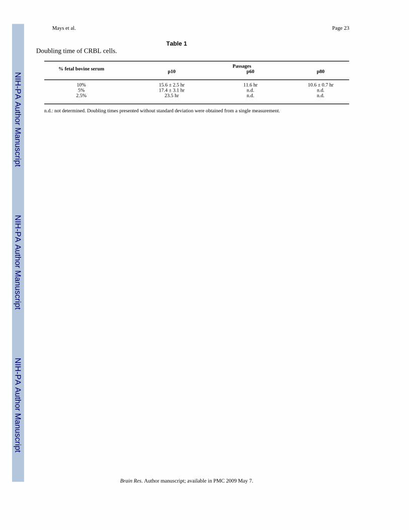
2.2. Characterization of the CRBL cellsThe CRBL cells grew immortally and were maintained for more than 80 passages. CRBL cellsproliferated rapidly during the maintenance of the culture. To examine their growth rate, cellswere grown under regular culture conditions comprised of DMEM and 10% fetal bovine serum,and their doubling times were estimated. At passage ten, CRBL cells doubled their numbersonce every 15.6 hours, while the cells at the passages sixty and eighty exhibited doubling timesof 11.6 and 10.6 hours, respectively (Table 1). The rapid growth of CRBL cells at early passageswas accelerated, but stabilized in later passages.
To investigate the serum responsiveness, CRBL cells at passage ten were maintained underdifferent serum concentrations and their growth rates compared. When cultured under reducedserum conditions (5 and 2.5 %), the growth of the CRBL cells were retarded. The doublingtime of 15.6 hours at 10 % serum concentration became extended to 17.4 hours at 5 % and 23.5hours at 2.5 % serum concentrations (Table 1).
Morphological observation revealed that CRBL cells are composed of cells in various sizes,shapes and lineages. Some cells resemble fibroblasts having spindle-like shapes, while othersappear as differentiated neurons with the various degrees of dendritic extension. In addition,cells with the typical star-shape of mature astrocytes were observed. CRBL cells grow largerand demonstrate a flattened shape at low density in culture. Such morphology became lessobvious after the cell-cell contact in a high density culture (Fig. 3).
Chromosome numbers were counted at metaphase to investigate the aneuploidic state of thecells. Cytogenetic analysis revealed that CRBL cells were maintained as hypotetraploid. Thecells had abnormal karyotypes, with the modal chromosome number varying from 66 to 78(data not shown).
Since CRBL cells appear to be composed of multiple cell types, expression of neuronal andglial cell marker proteins was examined by both Western blotting and flow cytometry. Westernblot analysis demonstrated that CRBL cells express both β III-tubulin, a neuronal markerprotein, and GFAP, a glial marker protein (Fig. 4). β III-tubulin was abundantly expressed inN2a cells, but not in C6 cells (Fig. 4, Panel A). By contrast, GFAP was expressed in C6 cells,but not in N2a cells (Fig. 4, Panel B). Expression of actin was almost equal in all cell lines.Flow cytometry was utilized to further confirm the Western blotting results and measure the
Mays et al. Page 3
Brain Res. Author manuscript; available in PMC 2009 May 7.
NIH
-PA Author Manuscript
NIH
-PA Author Manuscript
NIH
-PA Author Manuscript
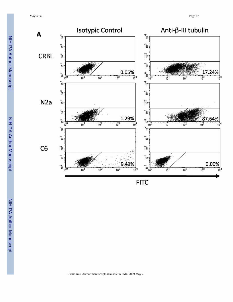
proportion of the fully differentiated neurons and glial cells within the CRBL cell population.Incubation with anti-β III-tubulin antibody followed by fluorescence measurementsdemonstrated the separation of β III-tubulin-positive cells in both CRBL and N2a cells, butnot in C6 cells (Fig. 5, Panel A). With a comparison to the isotypic controls, the percentagesof positive cells were ~ 17 % and ~ 88 % in the CRBL and N2a cells, respectively. Similaranalysis with anti-GFAP antibody resulted in GFAP-positive shifts in both CRBL and C6 cells(Fig. 5, Panel B). By comparing to isotypic controls, CRBL and C6 cells included ~ 30 % and~ 85 % of GFAP-positive cells, respectively.
2.3. PrPC expression and prion susceptibility of CRBL cellsTo investigate if CRBL cells express PrPC, the cells were analyzed by both Western blottingand flow cytometry. In Western blotting, all three different glycosylated forms of PrPC werefound in CRBL cells. Expression levels of PrPC in the cells were higher than that in NIH3T3fibroblasts, while almost comparable to that in N2a cells (Fig. 6, Panel A). N2a cells expressedquite a high level of PrPC, while RK13 cells, a negative control, showed no expression ofPrPC. However, the level of GAPDH expression in CRBL, N2a, NIH3T3, and RK13 cells wasalmost identical. Flow cytometry analysis ascertained that CRBL cells express PrPC.Approximately 62 % of the CRBL cells expressed PrPC under the experimental condition,while 86 % of N2a cells were PrPC positive (Fig. 6, Panel B).
Since CRBL cells were isolated from the cerebellum, one of the brain structures targeted byprion diseases, the susceptibility of cells to prion infection was assessed by in vitro prioninfection. CRBL cells were challenged with prion inocula prepared from cells either free of(SMB-PS and N2a) or permanently infected by prions (SMB and ScN2a). In Western blotting,PK-resistant PrPSc was propagated in the CRBL cells during the period of 4 passages followingthe inoculation with SMB cell lysate (Fig. 7, Panel A, lane 3). Interestingly, inoculation withprions from ScN2a did not yield PK-resistant PrPSc in CRBL cells (Fig. 7, Panel A, lane 5).In controls, prions from both ScN2a and SMB cells were able to infect N2a cells, where a highlevel of PrPSc was accumulated (Fig. 7, Panel A, lanes 6 and 7). The level of PrPSc in CRBLcells infected by prions from SMB cells, designated SMBinfCRBL cells, was lower than thatin N2a cells infected by the same prions (Fig. 7, Panel A, lanes 3 and 7). As expected, uninfectedCRBL cells and those exposed to inocula prepared from SMB-PS and N2a cells maintainedtheir prion-free states (Fig. 7, Panel A, lanes 1, 2 and 4).
To re-examine the inefficient transmission of ScN2a prions to CRBL, N2a and CRBL cellswere inoculated with RML prions, which were utilized in the establishment of ScN2a (Butleret al., 1988). When challenged in vitro with brain homogenate of uninfected and RML-infectedmice, PrPSc was not accumulated in the CRBL cells (Fig. 7, Panel B, lanes 1 and 2). However,the control N2a cells did propagate PrPSc when infected with RML-sick mouse brainhomogenate, but not with uninfected mouse brain homogenate (Fig. 7, Panel B, lanes 3 and4). Overall, none of the in vitro infections were shown to alter the level of PrPC (Fig. 7, PanelsA and B, upper panels). Our results showed that CRBL cells were selectively susceptible tothe prions from SMB cells, but not to the prions from ScN2a cells and RML-infected brains.
To confirm whether this differential susceptibility of CRBL cells represents a transient eventin the early stage of in vitro prion infection, the cells infected by prions from either SMB orScN2a cells were further maintained up to 15 extra passages. During this period, the low levelsof PrPSc in the early passage (Fig. 7, Panel A, lane 3) increased, and remained steady inSMBinfCRBL cells (Fig. 7, Panel C, lanes 2 and 3).This indicates that CRBL cells arepermissible for the robust replication of SMB prions and persistently infected. However, CRBLcells infected by ScN2a prions lacked PK-resistant PrPSc after the extended cell culture (Fig.7, Panel C, lanes 5 and 6), suggesting the cells were devoid of ScN2a prion replication.
Mays et al. Page 4
Brain Res. Author manuscript; available in PMC 2009 May 7.
NIH
-PA Author Manuscript
NIH
-PA Author Manuscript
NIH
-PA Author Manuscript
CRBL cells infected by SMB and ScN2a prions were further analyzed to determine whetherdifferential susceptibility was influenced by a shift of cell population from multiple to a singlecell type of either neurons or glial cells. Expression of both β III-tubulin and GFAP in CRBLcells infected by either SMB or ScN2a prions remained consistent with the levels prior toinoculation (Fig. 7, Panel D). Therefore, this indicates that selective susceptibility of CRBLcells to different prion strains was not due to the instability of CRBL cells.
In order to examine if such selective prion susceptibility of CRBL cells was caused by varianceof Prnp alleles and unexpected mutations occurred in PrP gene during the cell lineestablishment and maintenance, DNA sequences of the PrP ORF from CRBL cells in variouspassages (passages 10, 60 and 80) were compared to those from N2a, ScN2a, SMB, and SMB-PS. Determination of DNA sequences revealed that PrP ORFs from these cell lines wereidentical to each other and no mutation was found (data not shown). Moreover, DNA sequencesof the PrP ORF from the cell lines corresponded to the mouse Prnpa/a short incubation timegene (GenBank accession number: U29186; data not shown). This supports the idea thatselective susceptibility of CRBL cells to SMB prions was not due to the allelic variance ormutations in PrP gene. Rather, our data implicates that a specific host-agent interaction causedselective prion susceptibility of CRBL cells. The mechanism underneath the host-prioninteraction is unclear and requires further investigation.
2.4. Molecular properties of PrPSc and PrP processing events in SMBinfCRBL cellsSince CRBL cells were shown to propagate prions of SMB cells, it was hypothesized thatmolecular aspects of strain properties remained unaltered between SMB and SMBinfCRBLcells. The specific glycosylation banding pattern of PrPSc, a molecular signature associatedwith different prion strains, was examined. In a thorough comparison of PK-resistant PrPSc
bands from multiple Western blots, the ratio of differentially glycosylated PrPSc from bothcells was found unchanged (Fig. 8, Panel A). Furthermore, deglycosylation of PK-resistantPrPSc revealed that the size of C2, a 21 kDa C-terminal core (90–230) of PrPSc, was not differentin both cells (Fig. 8, Panel B, lanes 2, 4, 6, and 8). This indicates that conformation of PrPSc
found in both cells was identical. Our results suggest that the strain properties of SMB prionswere retained after transmission to CRBL cells. Nevertheless, examination on incubationperiods, and PrPSc distribution in the local brain regions in animals will provide a furtherunderstanding of the prion strain maintained in SMB and SMBinfCRBL cells.
Interestingly, deglycosylation of PrP revealed the unique state of proteolyzed PrP inSMBinfCRBL cells. The level of C2 fragments generated natively by the cellular machineryin SMBinfCRBL cells was found to be lower than that in SMB cells (Fig. 8, Panel B, lanes 1,3, 5, and 7). However, the level of C1, the 17 kDa C-terminal core (111–230) generated fromPrPC, was presented equally from both cell lines (Fig. 8, Panel B, lanes 5 and 7). The low levelof native C2 fragment (Fig. 8, Panel B, lanes 3 and 7) correlated with that of C2 fragmentsgenerated after PK digestion (Fig. 8, Panel B, lanes 4, and 8) and PrPSc accumulated inSMBinfCRBL cells (Fig. 7, Panel A and C).
3. DISCUSSIONEstablishment and characterization of the immortalized cells from the cerebellum of mice havemade it possible to investigate the susceptibility of cerebellar cells to prions. Since thecerebellum is largely affected by prions and PrPC appears to be involved in the physiology ofthe cerebellar cells, the importance of studying cellular and molecular events in the cerebellumunder the state of prion infection has become greater. Difficulties with in vivo models andlimitations in the current ex vivo models necessitate a new model system, which circumventsobstacles frequently associated with primary cell culture and recapitulates the conditions invivo.
Mays et al. Page 5
Brain Res. Author manuscript; available in PMC 2009 May 7.
NIH
-PA Author Manuscript
NIH
-PA Author Manuscript
NIH
-PA Author Manuscript
CRBL cells have the capacity to bypass cellular senescence, which is frequently observedduring the cultivation of primary cells. In this study, viral oncogenes were not introduced toestablish the cells proliferating immortally. Instead, CRBL cells were isolated from mice inwhich tumor suppressor gene p53 was inactivated. Lack of functional p53 in the cells causescell cycle arrest, gene amplification, and loss of growth control resulting in immortalproliferation, and increased spontaneous tumorigenesis in animals (Donehower et al., 1992;Purdie et al., 1994; Tsukada et al., 1993). The properties of CRBL cells, revealed in this study,support the features associated with inactivation of p53. CRBL cells immortally survive > 80passages without losing the ability of constant proliferation.
A majority of CRBL cells were found to have hypotetraploidy in both early and later passages.However, diploid was not common in CRBL cells in any passage of the culture. An earlierstudy demonstrated that ex vivo cell culture from p53-deficient mice were predominantlydiploid in early passages, but gradually became hypertetraploid over several passages (Tsukadaet al., 1993). Interestingly, CRBL cells divided about 4 times faster than the explanted cellsfrom p53-deficient mice. The explanted cells made about 30 cumulative doublings over a 2month period (Tsukada et al., 1993). The calculated doubling time of CRBL cells was around12 hours in our studies. These differences might be caused by different culture conditions,tissues where the cells were explanted, and age of animal where cells were obtained. Theaneuploid conditions correlate to a high rate of cell division in CRBL cells. Althoughimmortalization and aneuploidy are prerequisites for transformation, the ability of CRBL cellsto be transformed requires to be tested in animals if they induce tumors after transplantation.
Our newly established CRBL cell line more closely represents the environment found invivo. Most prion-susceptible cell lines, previously established, represent a single lineage ofcells (Solassol et al., 2003). Such cell lines have not provided the appropriate conditions tostudy the cellular interactions between different cell types, in vitro, under the prion-infectedstate. As suggested from our morphological observation and marker protein expression studies,it is likely that CRBL cells are composed of several different cell types including neurons andglial cells. Moreover, cells at various stages of differentiation such as stem cells and fullydifferentiated cells are thought to be included in the CRBL cell population. Future studiesdealing with the sub-types composing CRBL cells and their interaction during prionpathogenesis remain to be conducted.
Adequate in vitro models for studying the cellular and molecular events in prion biology mustmeet the requirement of PrPC expression and competency for prion infection. Our studies withCRBL cells showed that a considerable portion of the cell population abundantly expressedPrPC and were susceptible to prions. Interestingly, the accumulated PrPSc level inSMBinfCRBL cells was lower than that in the neuronal lineage N2a cells when both cell lineswere infected by the same SMB prions. Even after chronic infection with SMB prions,SMBinfCRBL cells maintained less PrPSc than SMB cells. It can be postulated that the abilityof CRBL cells to replicate the limited level of PrPSc results in persistent, but low PrPSc
accumulation. It is possible that not all CRBL cells are able to support the PrPSc propagation.Presumably, PrPC expressing neuronal cells within CRBL cells are considered to be the cellpopulation responsible for maintaining PrPSc propagation although a neuronal subclone ofCRBL cells has not been identified to propagate prions independently of interaction with othercell types. It is likely that CRBL cells resemble the cellular and tissue conditions of prion-infected host brains.
Our results showed that generation of native C2 fragments was extremely low in SMBinfCRBLcells, but abundant in SMB cells. This implicates that cellular machinery involved in PrPSc
processing is regulated differently in both cell lines. SMBinfCRBL cells may be a useful modelto study PrP processing events because the interaction between multiple cell types within the
Mays et al. Page 6
Brain Res. Author manuscript; available in PMC 2009 May 7.
NIH
-PA Author Manuscript
NIH
-PA Author Manuscript
NIH
-PA Author Manuscript
CRBL cells can be responsible for the orchestrated control of enzymes and inhibitors involvedin PrP processing.
Another intriguing aspect of our study is the selective susceptibility of CRBL cells to prionsobtained from different cell lines. Prions from SMB, but not from ScN2a cells were able toinfect CRBL cells. Efficient transmission of prions may depend on either the genotype of thehost or the prion strain (Bruce et al., 1976). Although CRBL cells maintain a complex geneticbackground as a result of using numerous mouse strains to generate the transgenic mice fromwhich CRBL cells were isolated (Kim et al., 2007), all mouse strains utilized retain commonPrnpa/a alleles responsible for short incubation periods upon prion infection (Carlson et al.,1986; Westaway et al., 1987). Furthermore, our DNA sequencing data showed that CRBL cellsincluded Prnpa/a gene with no mutation. Thus, our results indicate that the selectivesusceptibility of CRBL cells may be attributed to prion strain specificity in infecting host cells.
Maintenance of strain properties is an important parameter to determine the strain-specifictransmission of prions. Although it remains to be confirmed using animal systems, thesimilarities in molecular aspects of PrPSc support that the strain properties of SMB prions weremaintained in SMBinfCRBL cells. Furthermore, the observation that CRBL cells are moresusceptible to prions from SMB cells than those from ScN2a cells may reflect prion straintropism to a certain region of the brain. Regional distribution of neuropathology is a key featurethat has commonly been used to define the prion strain because strain-specific replication hasbeen shown to occur in certain subpopulations of brain cells resulting in a distinct lesion profile(Bruce et al., 1989; Hecker et al., 1992; Ridley and Baker, 1996). SMB and ScN2a prions werederived from 139A and RML prions, respectively (Birkett et al., 2001; Bosque and Prusiner,2000). Inoculation of mice with 139A prions resulted in intense vacuolation in the hippocampusand white matter of the cerebellum, while similar studies with RML prions revealed vacuolationlimited to the neocortex of the cerebrum (Carp et al., 1998; Tremblay et al., 2004). Thepathological targeting to the cerebellum by 139A, but not by RML prions correlates to selectivesusceptibility of CRBL cells to prions from SMB cells, but not from ScN2a cells or RML-infected brain homogenate. In a different scenario, it is possible that the parental Chandlerprions diverged into SMB- and ScN2a-specific prions that may have evolved to replicate inmesodermal and neuronal specific cell types, respectively, during the complex adaptationprocesses although both 139A and RML strains commonly originated from Chandler isolates(Butler et al., 1988; Chandler, 1961; Clarke and Haig, 1970).
In conclusion, our newly established CRBL cells will be a valuable tool to understand themechanisms of physiological process involved in PrPC and neuropathological events causedby prion infection as a consequence of cellular interaction among cerebellar cells. Additionally,CRBL cells represent a new cell culture model which is useful in evaluating host response todifferent prion strains and studying the mechanism of host-prion interactions.
4. EXPERIMENTAL PROCEDURES4.1. Mice and genotyping
Mice lacking expression of the p53 gene and genotyping procedures are described elsewhere(Kim et al., 2007). Although the functional assessment of focal adhesion kinase (FAK) in boththe establishment of CRBL cells and the study of their susceptibility to prions is beyond thescope of our present studies, FAK floxed p53 null (p53−/−) mice were utilized in this study.The animals used in this study have been acquired and cared for in accordance with theguidelines published in the NIH Guide for the Care and Use of Laboratory Animals.
Mays et al. Page 7
Brain Res. Author manuscript; available in PMC 2009 May 7.
NIH
-PA Author Manuscript
NIH
-PA Author Manuscript
NIH
-PA Author Manuscript
4.2. Preparation of cells from the cerebellumThe cerebella were extracted from three-month old mice. After the meningeal layer wasremoved, the cerebellum was dissected into small pieces under sterile conditions anddissociated by trypsin treatment (0.25%, Invitrogen, Carlsbad, CA) for 15 min. Followed bytrypsin deactivation with serum, brief straining (nylon mesh, 100 µm, Falcon, BD Biosciences,San Jose, CA), and centrifugation for 5 min at 1000 × g, the cells were plated in 24 well cultureplates (BD Biosciences).
4.3. Cell CultureThe newly established CRBL cells and all other cell lines used in this study were maintainedin Dulbecco’s Modified Eagle Medium (DMEM, high glucose) containing 10% fetal bovineserum (FBS), 1% glutamax, and 1% streptomycin/penicillin (Invitrogen) with 5 % CO2 andhumidity. Depending on experiments, ~ 1 – 20 × 104 cells were plated in the proper culturedishes or plates (Corning, Lowell, MA). The following established cell lines were utilized forthe study: N2a (mouse neuroblastoma, CCL-131, ATCC) (Klebe and Ruddle, 1969), ScN2a(scrapie infected N2a) (Butler et al., 1988), SMB (scrapie infected Swiss mouse neural) (Clarkeand Haig, 1970), SMB-PS (SMB cured) (Birkett et al., 2001), C6 (rat glioma, CCL-107, ATCC)(Benda et al., 1968), NIH3T3 (mouse fibroblast, CRL-1658, ATCC) (Jainchill et al., 1969),and RK13 (rabbit kidney cells, CCL-37, ATCC) (Beale et al., 1963). To induce a high levelexpression of GFAP, C6 cells were treated with 1 mM N6, 2′-O-dibutyryladenosine 3′,5′-cyclicmonophosphate sodium salt (Bt2AMP, Sigma, St. Louis, MO) for 3 days as described earlier(Takanaga et al., 2004). The Bt2AMP treated C6 cells were used in Western blotting and flowcytometry analyses. For the studies of cell growth in response to serum concentrations, theCRBL cells were maintained in DMEM with 5 % and 2.5 % FBS. For the studies of neo cassetteexpression, the cells were maintained in DMEM containing 10 % FBS and 0 –750 µg/mlGeneticin sulfate (G418, Invitrogen). The cell morphology was estimated and photographedunder a phase contrast microscope.
4.4. Cell counting and calculation of doubling timeAt given time points during experiments, cells were collected, stained with trypan blue solution(0.4 %, Sigma) and counted in hemocytometer chambers. Only live cells excluding the dyestaining were counted. The growth of the cells was monitored by estimating doubling time.Doubling time (Td) was calculated using the following formula: Td = log 2 × [(T2-T1)/(log N2− log N1)], where T2-T1 indicates the length of time between two measurements and N1 andN2 denote the numbers of cells at two points of measurement.
4.5. Cytogenetic studiesChromosome analysis of CRBL cells was performed with conventional G-banding techniques(Pathak, 1976). The exponentially growing CRBL cells were sequentially incubated with 0.1µg/ml N-deacetyl-N-methylcolchicine (Sigma) and 0.25 % sodium citrate/60 mM potassiumchloride solutions. After fixation with cold glacial acetic acid:methanol (1:3 v/v), the cells weresmeared and stained with Giemsa stains (Sigma). Chromosome numbers were counted in eachof the multiple spreads.
4.6. ImmunoblottingLysates were prepared from confluent cell cultures with lysis buffer (20 mM Tris, pH 8.0, 150mM NaCl, 0.5 % Nonidet-P 40 and 0.5 % sodium deoxycholate). After quantification of thecell lysates by BCA protein assay (Pierce, Rockford, IL), an equal amount of each cell lysate(30 µg) was separated on a 12% Tris-Glycine SDS-PAGE gel and transferred onto a PVDFmembrane (Immobilon-FL Transfer Membrane, Millipore, Billerica, MA). After blocking with5 % non-fat Carnation milk, the membrane was incubated sequentially with primary and
Mays et al. Page 8
Brain Res. Author manuscript; available in PMC 2009 May 7.
NIH
-PA Author Manuscript
NIH
-PA Author Manuscript
NIH
-PA Author Manuscript
secondary antibodies. Monoclonal primary antibodies utilized for these studies include anti-β III-tubulin (TuJ-1, R&D Systems, Mineapolis, MN), anti-GFAP (GA5, Sigma), anti-actin(ACTN05, Neomarker, Fremont, CA), and anti-GAPDH (6C5, Ambion, Austin, TX). Anti-PrP Fab fragments D13 and D18 (Inpro, South San Francisco, CA) were used for detection ofPrP. D18 was used only for the detection of C1 fragments. The peroxidase conjugatedsecondary antibodies were goat anti-mouse IgG (Pierce) and goat anti-human Fab (Pierce).Western blots were developed using ECL Plus™ Detection Reagents (Amersham Biosciences,Piscataway, NJ) and visualized after scanning in Fuji Film FLA 5000 image reader (Fuji Film,Edison, NJ).
4.7. Flow cytometryThe cells for flow cytometry analysis were prepared from cell culture by trypsinizing, repeatedwashing and passing through 70 µm cell strainer (Falcon). Approximately 1×10⁶ cells werefixed with 2% paraformaldehyde (Fisher, Pittsburgh, PA) for 30 min on ice and incubated withphosphate buffered saline (PBS) containing 0.2% saponin for 15 min at 37°C. After blockingwith antibody binding buffer (PBS containing 3% FBS and 0.05% NaN3) for 30 min on ice,cells were incubated with anti-β III-tubulin (TuJ-1, R&D Systems) or anti-GFAP (GF12–24,Chemicon, Temecula, CA) antibodies. Purified mouse IgG2a (Chemicon) was used as anisotypic control for both monoclonal primary antibodies. Cells were further incubated withFITC-conjugated goat anti-mouse IgG (Jackson Immuno Research, West Grove, PA) diluted1:200 in antibody binding buffer. For PrPC detection, anti-PrP Fab fragment D13 (Inpro) andFITC-conjugated goat antihuman IgG (1:500 dilution, Sigma) were used. Then, cells werewashed and resuspended in antibody binding buffer prior to flow cytometry analyses(FACSCalibur, BD Biosciences).
4.8. Prion infection in vitroInocula from ScN2a, N2a, SMB, and SMB-PS cells were prepared by the method previouslydescribed (Bosque and Prusiner, 2000). Briefly, confluent cells were harvested in 1 ml of PBSand lysed by repeated cycles of freezing and thawing. These lysates were then serially passedmultiple times through smaller hypodermic needles, from 16 to 26 gauges. Brain homogenates(10% w/v) from RML scrapie-infected and uninfected CD-1 mice were prepared in sterile PBSby the same manner. In vitro prion inoculation was conducted as described elsewhere (Bosqueand Prusiner, 2000) with minor modifications. CRBL and N2a cells were plated at a densityof 4×10³ cells/well in a 6-well plate and cultured in 3 ml culture media with 100 µl of inoculumfor 7 days. If the culture became confluent within the inoculation period, it was split at a 1:10dilution into a new well and supplied with new media and inoculum. After inoculation period,the cells were maintained 4 more passages in the inoculum free culture media. It is known thatall prions provided by the inoculum were washed away by dilution during the first 4 passages,leaving no detectable PrPSc in the culture (Bosque and Prusiner, 2000). When necessary, CRBLcells inoculated with prions of SMB and ScN2a cells were cultured up to 15 extra passages.For the detection of PrPSc, ~ 1 mg of each cell lysate was treated with 100 µg/ml PK (Invitrogen)for 1h at 37°C. Following PK inactivation by adding PMSF to the final concentration of 2 mM,the lysates were ultracentrifuged at 100,000 × g for 1h at 4°C in a bench top ultracentrifuge(TLX-50, Beckman, Fullerton, CA). PK-resistant PrPSc was detected by Western blottinganalysis.
4.9. Analysis of PrP sequencesN2a, ScN2a, SMB, SMB-PS cells and CRBL at passage 10, 60 and 80 were grown by themethod described above. Genomic DNA was isolated from these cells using Puregene DNApurification kit (Qiagen).The coding region for the full length mouse PrP was amplified byPCR using genomic DNA and PrP gene specific primers (forward primer: 5’-
Mays et al. Page 9
Brain Res. Author manuscript; available in PMC 2009 May 7.
NIH
-PA Author Manuscript
NIH
-PA Author Manuscript
NIH
-PA Author Manuscript
cgaattcatggcgaaccttggcta-3’ and reverse primer: 5’-ccgcggccgctcatcccacgatca-3’) under thefollowing condition: 95°C for 3 min; 40 cycles of 95°C for 30 sec/61°C for 30 sec/72°C for 1min; 72°C for 3 min. DNA sequences were determined by the fluorescent dye-terminatorsequencing method in ABI Prism™ 3730x DNA sequencers (SeqWright DNA TechnologyService, Houston, TX). The sequence data was analyzed using BLAST (National Center forBiotechnology Information).
4.10. Quantification of PrPSc glycosylation statesDensity of di-, mono-, and un-glycosylated bands of PK-resistant PrPSc in multiple Westernblots was measured using the UVP Doc-It Densitometry System (UVP, Upland, CA). Relativeratio of these bands was obtained from the densitometry of 6 (SMB cells) and 7 (SMBinfCRBLcells) independent blots of PrPSc.
4.11. Deglycosylation of PrPCell lysate with or without PK digestion was incubated in 1x denaturing buffer (0.5 % SDS,40 mM DTT) for 3 min at 100°C. Denatured cell lysate were further incubated with ~ 1000units of Peptide: N-Glycosidase F (PNGase F, NEB, Ipswich, MA) in 50 mM sodium phosphatebuffer, pH7.5, supplemented with 1 % Nonidet-P 40 for 3 h at 37°C. Deglycosylated PrP bandswere analyzed by Western blotting.
ACKNOWLEDGEMENTS
This work was partially supported by Sanders Brown Center on Aging, University of Kentucky and NIH Grant NumberP20 RR 020171 from the National Center for Research Resources. We thank William Titlow for editorial assistanceduring preparation of this manuscript. We are grateful to Glenn Telling for valuable discussion and providing celllines (SMB, SMB-PS, N2a and ScN2a) and RML prion-infected brain homogenate, to Vivek Rangnekar for NIH 3T3cell line, to Annadora Bruce-Keller for C6 cell line, to Thomas and Marilyn Getchell for anti-GFAP antibody, and toJennifer Strange and Greg Bauman of Flow Cytometry Core Facility for flow cytometry service.
REFERENCESArmstrong R, Cairns N, Lantos P. Quantification of the vacuolation (spongiform change) and prion
protein deposition in 11 patients with sporadic Creutzfeldt-Jakob disease. Acta Neuropathol 2001a;102:591–596. [PubMed: 11761719]
Armstrong R, Cairns N, Ironside J, Lantos P. The spatial patterns of prion protein deposits in cases ofvariant Creutzfeldt-Jakob disease. Acta Neuropathol 2002;104:665–669. [PubMed: 12410388]
Armstrong RA, Cairns NJ, Lantos PL. Spatial pattern of prion protein deposits in patients with sporadicCreutzfeldt-Jakob disease. Neuropathology 2001b;21:19–24. [PubMed: 11304038]
Bastian, AJ.; Thach, WT. Structure and function of the cerebellum. In: Manto, MU.; Panddolfo, M.,editors. The crebellum and its disorders. Cambridge, UK: Cambridge University Press; 2002. p. 49-66.
Beale AJ, Christofins GC, Furminger IGS. Rabbit cells susceptible to rubellar virus. Lancet 1963;2:640–641.
Benda P, Lightbody J, Sato G, Levine L, Sweet W. Differentiated rat glial cell strain in tissue culture.Science 1968;161:370–371. [PubMed: 4873531]
Birkett CR, Hennion RM, Bembridge DA, Clarke MC, Chree A, Bruce ME, Bostock CJ. Scrapie strainsmaintain biological phenotypes on propagation in a cell line in culture. EMBO J 2001;20:3351–3358.[PubMed: 11432823]
Bosque PJ, Prusiner SB. Cultured cell sublines highly susceptible to prion infection. J. Virol2000;74:4377–4386. [PubMed: 10756052]
Bruce ME, Dickinson AG, Fraser H. Cerebral amyloidosis in scrapie in the mouse: effect of agent strainand mouse genotype. Neuropathol. Appl. Neurobiol 1976;2:471–478.
Bruce ME, McBride PA, Farquhar CF. Precise targeting of the pathology of the sialoglycoprotein, PrP,and vacuolar degeneration in mouse scrapie. Neurosci. Lett 1989;102:1–6. [PubMed: 2550852]
Mays et al. Page 10
Brain Res. Author manuscript; available in PMC 2009 May 7.
NIH
-PA Author Manuscript
NIH
-PA Author Manuscript
NIH
-PA Author Manuscript
Butler DA, Scott MRD, Bockman JM, Borchelt DR, Taraboulos A, Hsiao KK, Kingsbury DT, PrusinerSB. Scrapie-infected murine neuroblastoma cells produce protease-resistant prion proteins. J. Virol1988;62:1558–1564. [PubMed: 3282080]
Carlson GA, Kingsbury DT, Goodman PA, Coleman S, Marshall ST, DeArmond S, Westaway V,Prusiner SB. Linkage of prion protein and scrapie incubation time genes. Cell 1986;46:503–511.[PubMed: 3015416]
Carp RI, Meeker H, Sersen E, Kozlowski P. Analysis of the incubation periods, induction of obesity andhistopathological changes in senescence-prone and senescence-resistant mice infected with variousscrapie strains. J. Gen. Virol 1998;79:2863–2869. [PubMed: 9820164]
Chandler RL. Encephalopathy in mice produced by inoculation with scrapie brain material. Lancet1961;1:1378–1379. [PubMed: 13692303]
Clarke MC, Haig DA. Evidence for the multiplication of scrapie agent in cell culture. Nature1970;225:100–101. [PubMed: 4983025]
Collinge J. Prion diseases of humans and animals: Their causes and molecular basis. Annu. Rev. Neurosci2001;24:519–550. [PubMed: 11283320]
DeArmond, SJ.; Ironside, JW.; Bouzamondo-Bernstein, E.; Peretz, D.; Fraser, JR. Neuropathology ofprion diseases. In: Prusiner, SB., editor. Prion Biology and Diseases. Cold Spring Harbor, NY: ColdSpring Harbor Laboratory Press; 2004. p. 777-856.
Donehower LA, Harvey M, Slagle BL, McArthur MJ, Montgomery CAJ, Butel JA, Bradley A. Micedeficient for p53 are developmentally normal but susceptible to spontaneous tumours. Nature1992;356:215–221. [PubMed: 1552940]
Ferrer I, Puig B, Blanco R, Marti E. Prion protein deposition and abnormal synaptic protein expressionin the cerebellum in Creutzfeldt-Jakob disease. Neuroscience 2000;97:715–726. [PubMed:10842016]
Ferrer I. Synaptic pathology and cell death in the cerebellum in Creutzfeldt-Jakob disease. Cerebellum2002;1:213–222. [PubMed: 12879983]
Ghez, C.; Thach, WT. The cerebellum. In: Kandel, ER.; Schwartz, JH.; Jessell, TM., editors. Principlesof neural science. New York, NY: McGraw-Hill; 2000. p. 832-852.
Harris SL, Levine AJ. The p53 pathway: positive and negative feedback loops. Oncogene 2005;24:2899–2908. [PubMed: 15838523]
Hecker R, Taraboulos A, Scott M, Pan K-M, Torchia M, Jendroska K, DeArmond SJ, Prusiner SB.Replication of distinct scrapie prion isolates is region specific in brains of transgenic mice andhamsters. Genes Dev 1992;6:1213–1228. [PubMed: 1628828]
Herms JW, Korte S, Gall S, Schneider I, Dunker S, Kretzschmar HA. Altered intracellular calciumhomeostasis in cerebellar granule cells of prion protein-deficient mice. J. Neurochem 2000;75:1487–1492. [PubMed: 10987828]
Herms JW, Tings T, Dunker S, Kretzschmar HA. Prion Protein Affects Ca2+-Activated K+ Currents inCerebellar Purkinje Cells. Neurobiol. Dis 2001;8:324–330. [PubMed: 11300727]
Jainchill JL, Aaronson SA, Todaro GJ. Murine sarcoma and leukemia viruses: Assay using clonal linesof contact-inhibited mouse cells. J. Virol 1969;4:549–553. [PubMed: 4311790]
Jarius C, Kovacs GG, Belay G, Hainfellner JA, Mitrova E, Budka H. Distinctive cerebellarimmunoreactivity for the prion protein in familial (E200K) Creutzfeldt-Jakob disease. ActaNeuropathol 2003;105:449–454. [PubMed: 12677444]
Katamine S, Nishida N, Sugimoto T, Noda T, Sakaguchi S, Shigematsu K, Kataoka Y, Nakatani A,Hasegawa S, Moriuchi R, Miyamoto T. Impaired motor coordination in mice lacking prion protein.Cell. Mol. Neurobiol 1998;18:731–742. [PubMed: 9876879]
Kim J, Leucht P, Luppen CA, Park YJ, Beggs HE, Damsky CH, Helms JA. Reconciling the roles of FAKin osteoblast differentiation, osteoclast remodeling, and bone regeneration. Bone 2007;41:38–51.
Klebe RJ, Ruddle FH. Neuroblastoma: cell culture analysis of a differentiating stem cell system. J. CellBiol 1969;43:69a.
Laine J, Marc M-E, Sy M-S, Axelrad H. Cellular and subcellular morphological localization of normalprion protein in rodent cerebellum. European J. Neurosci 2001;14:47–56. [PubMed: 11488948]
Mays et al. Page 11
Brain Res. Author manuscript; available in PMC 2009 May 7.
NIH
-PA Author Manuscript
NIH
-PA Author Manuscript
NIH
-PA Author Manuscript
Legname G, Nelken P, Guan Z, Kanyo ZF, DeArmond SJ, Prusiner SB. Prion and doppel proteins bindto granule cells of the cerebellum. Proc. Natl. Acad. Sci. USA 2002;99:16285–16290. [PubMed:12446843]
Pathak S. Chromosome banding techniques. J. Reprod. Med 1976;17:25–28. [PubMed: 59808]Prusiner SB. Prions. Proc. Natl. Acad. Sci. USA 1998;95:13363–13383. [PubMed: 9811807]Purdie CA, Harrision DJ, Peter A, Dobbie L, White S, Howie SEM, Salter DM, Bird CC, Whllie AH,
Hooper AL, Clarke AR. Tumor incidence, spectrum and ploidy in mice with a large deletion in thep53 gene. Oncogene 1994;9:603–609. [PubMed: 8290271]
Ridley RM, Baker HF. To what extent is strain variation evidence for an independent genome in the agentof the transmissible spongiform encephalopathies? Neurodegeneration 1996;5:219–231. [PubMed:8910900]
Schulz-Schaeffer WJ, Giese A, Windl O, Kretzschmar HA. Polymorphism at codon 129 of the prionprotein gene determines cerebellar pathology in Creutzfeldt-Jakob disease. Clin. Neuropathol1996;15:353–357. [PubMed: 8937783]
Solassol J, Crozet C, Lehmann S. Prion propagation in cultured cells. Br. Med. Bull 2003;66:87–97.[PubMed: 14522851]
Takanaga H, Yoshitake T, Hara S, Yamasaki C, Kunimoto M. cAMP-induced Astrocytic Differentiationof C6 Glioma Cells Is Mediated by Autocrine Interleukin-6. J. Biol. Chem 2004;279:15441–15447.[PubMed: 14754894]
Topka, H.; Massaquoi, SG. Pathophysiology of clinical cerebellar signs. In: Manto, MU.; Panddolfo, M.,editors. The cerebellum and its disorders. Cambridge, UK: Cambridge University Press; 2002. p.121-135.
Tremblay P, Ball HL, Kaneko K, Groth D, Hegde RS, Cohen FE, DeArmond SJ, Prusiner SB, Safar JG.Mutant PrPSc conformers induced by a synthetic peptide and several prion strains. J. Virol2004;78:2088–2099. [PubMed: 14747574]
Tsukada T, Tomooka Y, Takai S, Ueda Y, Nishikawa S, Yagi T, Tokunaga T, Takeda N, Suda Y, AbeS, Matsuo I, Ikawa Y, Aizawa S. Enhanced proliferative potential in culture of cells from p53-deficient mice. Oncogene 1993;8:3313–3322. [PubMed: 7504233]
Westaway D, Goodman PA, Mirenda CA, McKinley MP, Carlson GA, Prusiner SB. Distinct prionproteins in short and long scrapie incubation period mice. Cell 1987;51:651–662. [PubMed: 2890436]
AbbreviationsPrP, prion proteinPrPC, cellular prion proteinPrPSc, scrapie prion proteinCJD, Creutzfeld-Jakob diseasep53−/−, p53 nullCRBL, a cell line established from the cerebellumN2a, Neuro2aScN2a, scrapie-infected N2aSMB, scrapie-infected mouse brain cellsSMB-PS, SMB cells cured by pentosan sulfatePK, proteinase KBt2AMP, N6, 2′-O-dibutyryladenosine 3′,5′-cyclic monophosphatePMSF, phenylmethylsulphonyl fluorideGFAP, glial fibrillary acidic proteinGAPDH, glyceraldehyde-3-phosphate dehydrogenaseORF, open reading frame
Mays et al. Page 12
Brain Res. Author manuscript; available in PMC 2009 May 7.
NIH
-PA Author Manuscript
NIH
-PA Author Manuscript
NIH
-PA Author Manuscript
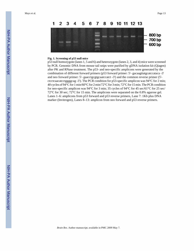
Fig. 1. Screening of p53 null micep53 null homozygote (lanes 1, 5 and 6) and heterozygote (lanes 2, 3, and 4) mice were screenedby PCR. Genomic DNA from mouse tail snips were purified by gDNA isolation kit (Qiagen)after PK and RNase treatment. The p53- and neo-specific amplicons were generated by thecombination of different forward primers (p53 forward primer: 5'- gacaagttatgcatccataca -3'and neo forward primer: 5'- gaacctgcgtgcaatccatct -3') and the common reverse primer (5'-ctcctcaacatcctggggcag -3'). The PCR condition for p53-specific amplicon was 94°C for 2 min;40 cycles of 94°C for 1 min/60°C for 2 min/72°C for 3 min; 72°C for 15 min. The PCR conditionfor neo-specific amplicon was 94°C for 3 min; 35 cycles of 94°C for 45 sec/61°C for 25 sec/72°C for 30 sec; 72°C for 15 min. The amplicons were separated on the 0.8% agarose gel.Lanes 1–6: amplicons from p53 forward and p53 reverse primers, Lane 7: 1Kb plus DNAmarker (Invitrogen), Lanes 8–13: amplicon from neo forward and p53 reverse primers.
Mays et al. Page 13
Brain Res. Author manuscript; available in PMC 2009 May 7.
NIH
-PA Author Manuscript
NIH
-PA Author Manuscript
NIH
-PA Author Manuscript
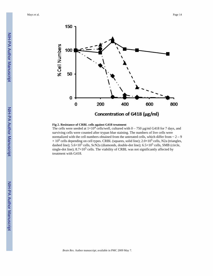
Fig 2. Resistance of CRBL cells against G418 treatmentThe cells were seeded at 1×10⁴ cells/well, cultured with 0 – 750 µg/ml G418 for 7 days, andsurviving cells were counted after trypan blue staining. The numbers of live cells werenormalized with the cell numbers obtained from the untreated cells, which differ from ~ 2 – 9× 10⁵ cells depending on cell types. CRBL (squares, solid line); 2.0×10⁵ cells, N2a (triangles,dashed line); 5.6×10⁵ cells, ScN2a (diamonds, double-dot line); 6.5×10⁵ cells, SMB (circle,single-dot line); 8.7×10⁵ cells. The viability of CRBL was not significantly affected bytreatment with G418.
Mays et al. Page 14
Brain Res. Author manuscript; available in PMC 2009 May 7.
NIH
-PA Author Manuscript
NIH
-PA Author Manuscript
NIH
-PA Author Manuscript
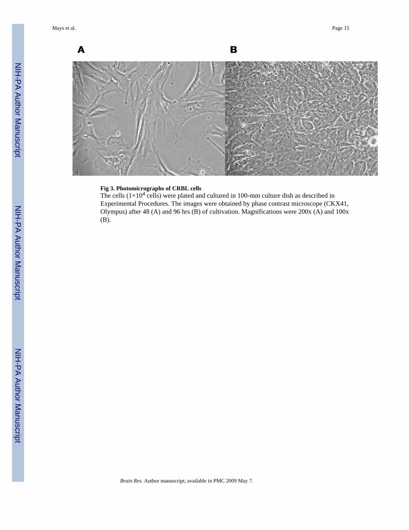
Fig 3. Photomicrographs of CRBL cellsThe cells (1×10⁴ cells) were plated and cultured in 100-mm culture dish as described inExperimental Procedures. The images were obtained by phase contrast microscope (CKX41,Olympus) after 48 (A) and 96 hrs (B) of cultivation. Magnifications were 200x (A) and 100x(B).
Mays et al. Page 15
Brain Res. Author manuscript; available in PMC 2009 May 7.
NIH
-PA Author Manuscript
NIH
-PA Author Manuscript
NIH
-PA Author Manuscript
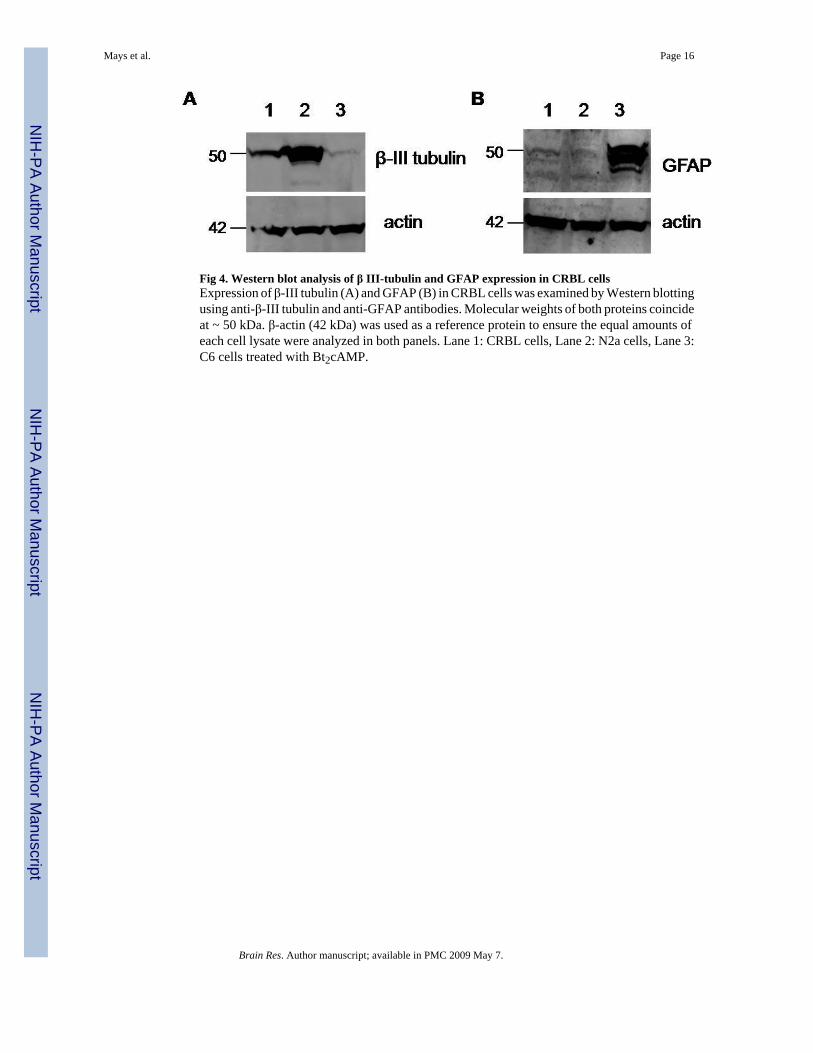
Fig 4. Western blot analysis of β III-tubulin and GFAP expression in CRBL cellsExpression of β-III tubulin (A) and GFAP (B) in CRBL cells was examined by Western blottingusing anti-β-III tubulin and anti-GFAP antibodies. Molecular weights of both proteins coincideat ~ 50 kDa. β-actin (42 kDa) was used as a reference protein to ensure the equal amounts ofeach cell lysate were analyzed in both panels. Lane 1: CRBL cells, Lane 2: N2a cells, Lane 3:C6 cells treated with Bt2cAMP.
Mays et al. Page 16
Brain Res. Author manuscript; available in PMC 2009 May 7.
NIH
-PA Author Manuscript
NIH
-PA Author Manuscript
NIH
-PA Author Manuscript
Mays et al. Page 17
Brain Res. Author manuscript; available in PMC 2009 May 7.
NIH
-PA Author Manuscript
NIH
-PA Author Manuscript
NIH
-PA Author Manuscript
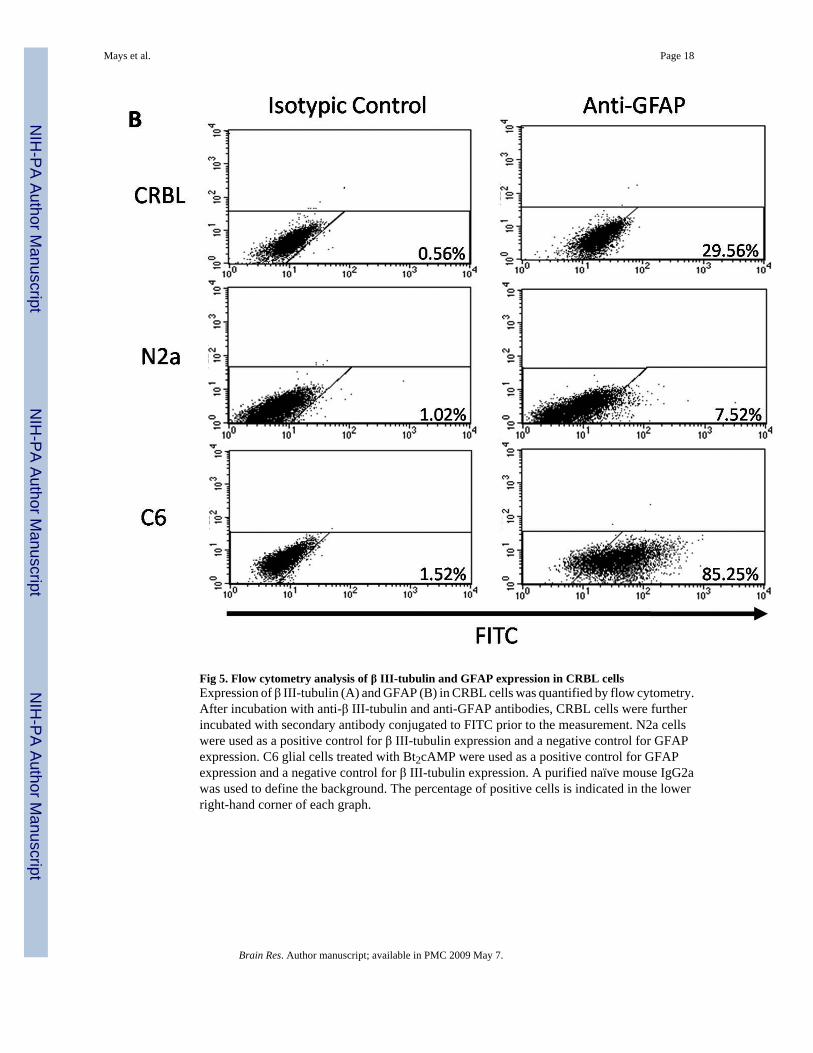
Fig 5. Flow cytometry analysis of β III-tubulin and GFAP expression in CRBL cellsExpression of β III-tubulin (A) and GFAP (B) in CRBL cells was quantified by flow cytometry.After incubation with anti-β III-tubulin and anti-GFAP antibodies, CRBL cells were furtherincubated with secondary antibody conjugated to FITC prior to the measurement. N2a cellswere used as a positive control for β III-tubulin expression and a negative control for GFAPexpression. C6 glial cells treated with Bt2cAMP were used as a positive control for GFAPexpression and a negative control for β III-tubulin expression. A purified naïve mouse IgG2awas used to define the background. The percentage of positive cells is indicated in the lowerright-hand corner of each graph.
Mays et al. Page 18
Brain Res. Author manuscript; available in PMC 2009 May 7.
NIH
-PA Author Manuscript
NIH
-PA Author Manuscript
NIH
-PA Author Manuscript
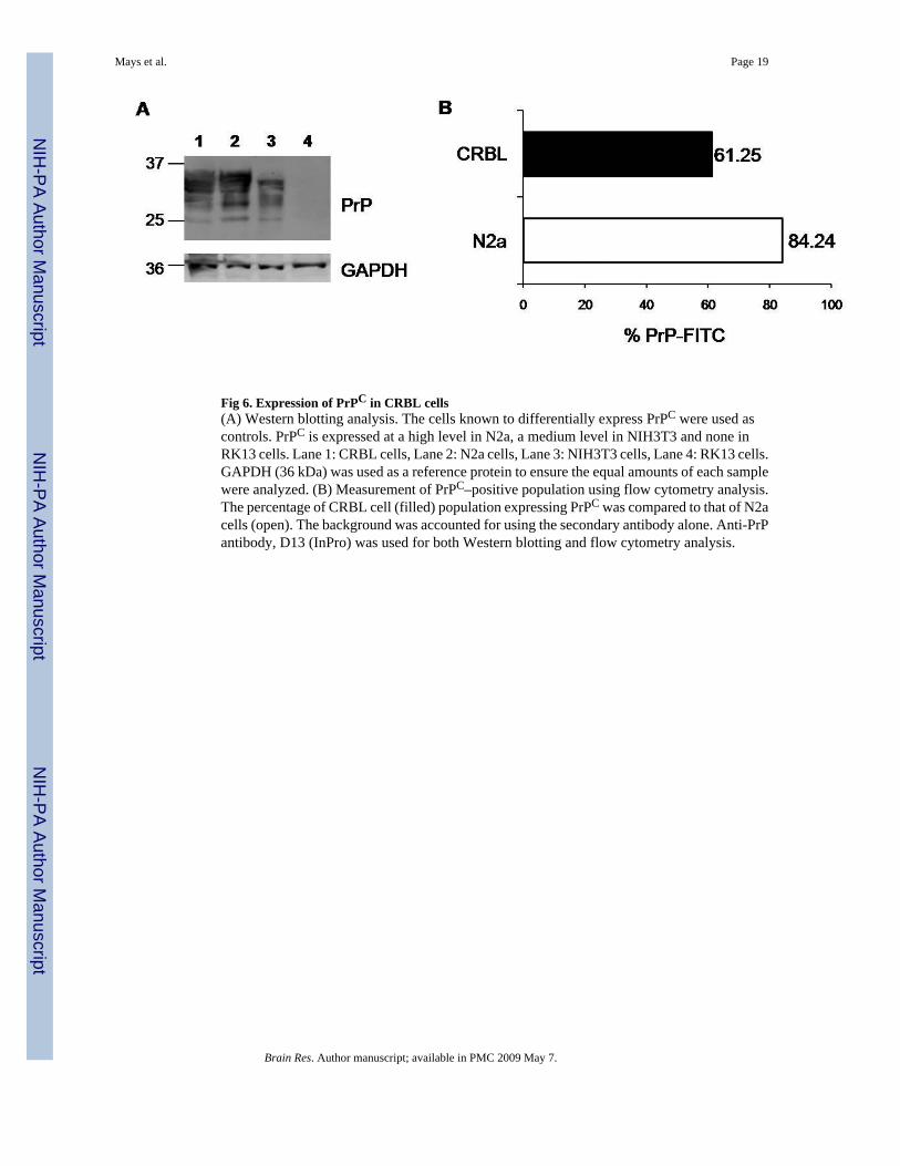
Fig 6. Expression of PrPC in CRBL cells(A) Western blotting analysis. The cells known to differentially express PrPC were used ascontrols. PrPC is expressed at a high level in N2a, a medium level in NIH3T3 and none inRK13 cells. Lane 1: CRBL cells, Lane 2: N2a cells, Lane 3: NIH3T3 cells, Lane 4: RK13 cells.GAPDH (36 kDa) was used as a reference protein to ensure the equal amounts of each samplewere analyzed. (B) Measurement of PrPC–positive population using flow cytometry analysis.The percentage of CRBL cell (filled) population expressing PrPC was compared to that of N2acells (open). The background was accounted for using the secondary antibody alone. Anti-PrPantibody, D13 (InPro) was used for both Western blotting and flow cytometry analysis.
Mays et al. Page 19
Brain Res. Author manuscript; available in PMC 2009 May 7.
NIH
-PA Author Manuscript
NIH
-PA Author Manuscript
NIH
-PA Author Manuscript
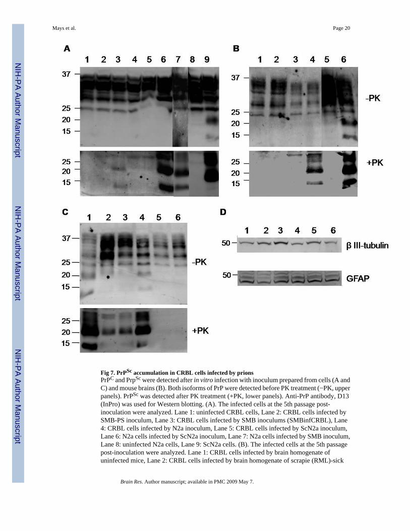
Fig 7. PrPSc accumulation in CRBL cells infected by prionsPrPC and PrpSc were detected after in vitro infection with inoculum prepared from cells (A andC) and mouse brains (B). Both isoforms of PrP were detected before PK treatment (−PK, upperpanels). PrPSc was detected after PK treatment (+PK, lower panels). Anti-PrP antibody, D13(InPro) was used for Western blotting. (A). The infected cells at the 5th passage post-inoculation were analyzed. Lane 1: uninfected CRBL cells, Lane 2: CRBL cells infected bySMB-PS inoculum, Lane 3: CRBL cells infected by SMB inoculums (SMBinfCRBL), Lane4: CRBL cells infected by N2a inoculum, Lane 5: CRBL cells infected by ScN2a inoculum,Lane 6: N2a cells infected by ScN2a inoculum, Lane 7: N2a cells infected by SMB inoculum,Lane 8: uninfected N2a cells, Lane 9: ScN2a cells. (B). The infected cells at the 5th passagepost-inoculation were analyzed. Lane 1: CRBL cells infected by brain homogenate ofuninfected mice, Lane 2: CRBL cells infected by brain homogenate of scrapie (RML)-sick
Mays et al. Page 20
Brain Res. Author manuscript; available in PMC 2009 May 7.
NIH
-PA Author Manuscript
NIH
-PA Author Manuscript
NIH
-PA Author Manuscript
mice, Lane 3: N2a cells infected by brain homogenate from uninfected mice, Lane 4: N2a cellsinfected by brain homogenate from scrapie (RML)-sick mice, Lane 5: N2a cells, Lane 6: ScN2acells. (C). The infected cells were cultured extra 15 passages post-inoculation since the initialconfirmation of PrPSc propagation shown in Panel A. Lane 1: SMB cells, Lane 2:SMBinfCRBL cells, passage 8, Lane 3: SMBinfCRBL cells, passage 15, Lane 4: ScN2a cells,Lane 5: CRBL cells infected by ScN2a inoculums, passage 7, Lane 6: CRBL cells infected byScN2a inoculums, passage 15. (D) Expression of β III-tubulin (upper panel) and GFAP (lowerpaner) in SMBinfCRBL cells. Lane 1 and 4: uninfected CRBL cells, Lane 2: SMBinfCRBLcells, passage 8, Lane 3: SMBinfCRBL cells, passage 15, Lane 5: CRBL cells infected byScN2a inoculums, passage 7, Lane 6: CRBL cells infected by ScN2a inoculums, passage 10.
Mays et al. Page 21
Brain Res. Author manuscript; available in PMC 2009 May 7.
NIH
-PA Author Manuscript
NIH
-PA Author Manuscript
NIH
-PA Author Manuscript
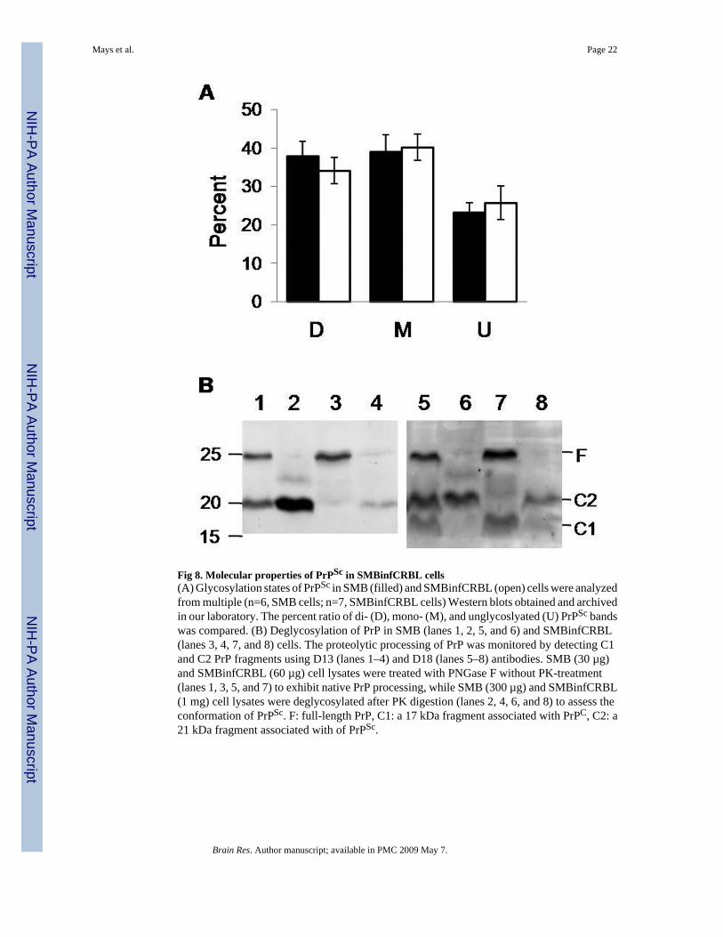
Fig 8. Molecular properties of PrPSc in SMBinfCRBL cells(A) Glycosylation states of PrPSc in SMB (filled) and SMBinfCRBL (open) cells were analyzedfrom multiple (n=6, SMB cells; n=7, SMBinfCRBL cells) Western blots obtained and archivedin our laboratory. The percent ratio of di- (D), mono- (M), and unglycoslyated (U) PrPSc bandswas compared. (B) Deglycosylation of PrP in SMB (lanes 1, 2, 5, and 6) and SMBinfCRBL(lanes 3, 4, 7, and 8) cells. The proteolytic processing of PrP was monitored by detecting C1and C2 PrP fragments using D13 (lanes 1–4) and D18 (lanes 5–8) antibodies. SMB (30 µg)and SMBinfCRBL (60 µg) cell lysates were treated with PNGase F without PK-treatment(lanes 1, 3, 5, and 7) to exhibit native PrP processing, while SMB (300 µg) and SMBinfCRBL(1 mg) cell lysates were deglycosylated after PK digestion (lanes 2, 4, 6, and 8) to assess theconformation of PrPSc. F: full-length PrP, C1: a 17 kDa fragment associated with PrPC, C2: a21 kDa fragment associated with of PrPSc.
Mays et al. Page 22
Brain Res. Author manuscript; available in PMC 2009 May 7.
NIH
-PA Author Manuscript
NIH
-PA Author Manuscript
NIH
-PA Author Manuscript
NIH
-PA Author Manuscript
NIH
-PA Author Manuscript
NIH
-PA Author Manuscript
Mays et al. Page 23
Table 1Doubling time of CRBL cells.
% fetal bovine serum Passagesp10 p60 p80
10% 15.6 ± 2.5 hr 11.6 hr 10.6 ± 0.7 hr5% 17.4 ± 3.1 hr n.d. n.d.
2.5% 23.5 hr n.d. n.d.
n.d.: not determined. Doubling times presented without standard deviation were obtained from a single measurement.
Brain Res. Author manuscript; available in PMC 2009 May 7.