primitive adult hematopoietic stem cells can function as osteoblast precursors
TRANSCRIPT
Primitive adult hematopoietic stem cells can functionas osteoblast precursorsElizabeth A. Olmsted-Davis*†‡, Zbigniew Gugala‡, Fernando Camargo*§, Francis H. Gannon¶, KathyJo Jackson*�,Kirsten Anderson Kienstra†, H. David Shine*§**††, Ronald W. Lindsey‡, Karen K. Hirschi*§, Margaret A. Goodell*†,Malcolm K. Brenner*†, and Alan R. Davis*†‡,‡‡
*Center for Cell and Gene Therapy and Departments of †Pediatrics, ‡Orthopedic Surgery, �Molecular Genetics, **Neurosurgery, ††Neuroscience, and§Molecular and Cellular Biology, Baylor College of Medicine, Houston, TX 77030; and ¶Department of Bone Pathology, Armed Forces Institute of Pathology,Washington, DC 20306
Edited by Darwin J. Prockop, Tulane University, New Orleans, LA, and approved October 28, 2003 (received for review May 16, 2003)
Osteoblasts are continually recruited from stem cell pools tomaintain bone. Although their immediate precursor is a plastic-adherent mesenchymal stem cell able to generate tissues otherthan bone, increasing evidence suggests the existence of a moreprimitive cell that can differentiate to both hematopoietic andmesenchymal cells. We show here that the ‘‘side population’’ (SP)of marrow stem cells, defined by their ability to rapidly expel aDNA-binding dye and to regenerate the hematopoietic compart-ment, can differentiate to osteoblasts through a mesenchymalintermediate. When transplanted into lethally irradiated mice,single gene-marked murine SP cells reconstituted depleted osteo-progenitor pools, such that a large proportion of the osteogeniccells in the epiphysis of long bone carried the donor SP cell marker.These findings suggest that the developmental capacity of SP cellsis not restricted to the hematopoietic lineages but extends toosteogenic differentiation. This property not only elucidates apreviously unrecognized step in osteoblast development, but alsohas intriguing implications for the use of SP cells in clinical ortho-pedics and stem cell-based disorders of bone.
Skeletal bone is unique among human tissues. It is continu-ously remodeled throughout life in a process that requires the
recruitment and proliferation of stem cells with the capacity todifferentiate to functional osteoblasts, which then deposit andmineralize extracellular bone matrix (1, 2). The identification ofan osteogenic stem cell with competency for both self-renewaland robust differentiation to bone-forming osteoblasts has beenelusive. Several studies have documented the ability of cells inwhole bone marrow to form osteoblasts in vitro (3) and in vivo(4). Pittenger et al. (3) isolated mesenchymal stem cells (MSCs)that were able to differentiate to chondrocytes, adipocytes, andosteoblasts in culture, whereas the results of serial transplanta-tion of single bone marrow-derived stem cells can be interpretedto suggest the existence of rare long-term repopulating cells thatcan regenerate not only the entire hematopoietic system, but alsoseveral different mesenchymal lineages (5, 6).
Our efforts to identify an osteogenic stem cell have focused ona side population (SP) of bone marrow cells that display stronghematopoietic reconstituting activity, as measured by competi-tive repopulation assays (7, 8). These so-called SP cells, whichcan be identified by their unique capacity to efflux fluorescentDNA-binding dye (7, 8), also have a limited capacity to differ-entiate in vivo to skeletal myocytes (9) as well as vascularendothelial cells (10), suggesting multilineage potential. To testthe candidacy of these adult stem cells as progenitors of theosteoblast lineage, we tracked the fate of gene-marked SP bothin vitro and in vivo. Donor-derived mesenchymal progenitorsdifferentiated to osteoblasts in clonogenic medium, and immu-nostaining of long-bone sections after transplantation of SP cellsinto lethally irradiated mice demonstrated an abundance ofgene-marked osteoblastic cells lining trabecular bone near thegrowth plate. These results suggest a model in which the
osteoprogenitors required for bone remodeling are recruitedfrom a pool of SP stem cells residing in bone marrow.
MethodsIsolation and Transplantation of SP Cells. SP cells were isolated fromthe bone marrow of 10 C57BL�6 CD45.2 Rosa26 mice accordingto the procedure of Goodell et al. (refs. 7 and 8; see alsowww.bcm.tmc.edu�genetherapy�goodell). Three thousand ofthese cells were injected retro-orbitally into 6- to 12-week-oldC57BL�6 CD45.1 mice that had been given 11 Gy of radiationin a split dose. After �4 weeks, the mice were bled retro-orbitally, and the percentage of hematopoietic engraftment wasdetermined by fluorescence-activated cell sorting analysis (11).
In single-cell transplantation studies, bone marrow was ob-tained from 2-month-old C57BL�6 CD45.2 Rosa26 mice. SingleSP (CD45.2� Sca-1�) cells were sorted directly into individualwells of a 96-well plate containing 150 �l of buffer by a single-celldeposition unit (Cytomation, Fort Collins, CO). Four hundredshort-term repopulating (Lin� Sca1� cKit� CD34high) cells froma CD45.1 donor were then added to each well to supporthematopoietic engraftment. The donor cells were transplantedinto lethally irradiated 6- to 8-week-old C57BL�6 CD45.1 mice,and hematopoietic contribution was measured monthly by stain-ing peripheral blood leukocytes with CD45.2FITC. Mice werekilled 14 months after the first transplantation.
Mesenchymal Progenitor Cultures. Bone marrow was isolated fromthe leg bones of mice to generate plastic adherent cells inspecialized medium [MesenCult basal medium for murine MSCssupplemented with MSC stimulatory supplement (murine), bothfrom StemCell Technologies, Vancouver]. After 3 days, theculture supernatant, together with nonattached cells was with-drawn and the medium was replaced. When the cultures reachedconfluence, the medium in some cultures was changed to�-MEM (Invitrogen) with 10% FBS containing 100 nM dexa-methasone and 50 �g�ml ascorbic acid, and the others remaineduntreated. With this procedure, an average of five bone-likenodules were identified per 107 whole bone marrow cells.Alkaline phosphatase was assayed with ELF 97 phosphatasesubstrate (Molecular Probes), which exhibits green fluorescenceafter cleavage, according to the manufacturer’s instructions.Osteocalcin staining was carried out by using goat anti-mouseosteocalcin and detected by using fluorescein. An average of fivenodules were obtained per 107 mouse whole bone marrow cells.
Immunohistochemical Analysis of Bone Sections. Six weeks aftertransplantation with either SP cells or whole bone marrow, mice
This paper was submitted directly (Track II) to the PNAS office.
Abbreviations: MSC, mesenchymal stem cell; SP, side population.
‡‡To whom correspondence should be addressed at: One Baylor Plaza, BCMN 505, Houston,TX 77030. E-mail: [email protected].
© 2003 by The National Academy of Sciences of the USA
www.pnas.org�cgi�doi�10.1073�pnas.2632959100 PNAS � December 23, 2003 � vol. 100 � no. 26 � 15877–15882
MED
ICA
LSC
IEN
CES
were killed. Hind-limb paraffin-embedded (5 �m) sections werestained with a monoclonal antibody to Escherichia coli �-galac-tosidase (Roche Applied Sciences) by using a PowerVisionHomo-Mouse IHC kit (ImmunoVision Technologies, Daly City,CA). The sections were counterstained with hematoxylin.
For double staining to detect both �-galactosidase and mouseosteocalcin, Vector Nova Red (Vector Laboratories) was sub-stituted for the standard diaminobenzidine reagent. Sectionswere coated with Clearmount (Zymed, South San Francisco,CA) after staining for �-galactosidase, examined microscopi-cally, photographed, incubated in water at 37°C to remove themounting medium, and stained with goat anti-mouse osteocalcinantibody (Biomedical Technologies, Stoughton, MA), using anABC Elite reagent (Vector Laboratories) and Vector SG (Vec-tor Laboratories). Sections were viewed microscopically to iden-tify the fields previously photographed after single staining.
ResultsCharacterization of SP and Mesenchymal Progenitor Populations. Wefirst tested whether SP cells can give rise to mesenchymal
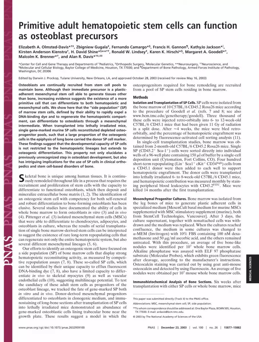
progenitors capable of differentiating to osteoblasts. IrradiatedC57BL�6 CD45.1 mice (n � 4) were transplanted with 3,000 SPcells isolated by Hoechst dye exclusion from C57BL�6 CD45.2Rosa26 mice, which express the E. coli �-galactosidase marker inall of their tissues. After 4 weeks, hematopoietic engraftment inperipheral blood exceeded 90% in all animals. Two weeks laterthe mice were killed, and bone marrow was cultured to obtainplastic-adherent cells, which were placed in osteoinductive me-dia. After 14 days, we observed bone-like nodules, approxi-mately one-half of which stained positively for �-galactosidase(Fig. 1 a and b, blue), a marker of donor SP cells, as well asalkaline phosphatase (Fig. 1 c, d, and f, green) and mineraldeposits (Fig. 1a, red-brown), indicative of an osteoblast phe-notype and function, respectively. Further, the majority of cellsin the single alkaline phosphatase-positive nodule shown in Fig.1e lacked the definitive blood cell marker CD45 (Fig. 1 g and h,expected color, red), in contrast to the CD45� phenotype ofmost cells in control cultures (not shown), which did not receiveosteoinductive media and lacked bony nodules. These results
Fig. 1. Bone-like nodules are derived from donor SP cells in cultures treatedwith osteogenic medium. Nodules were stained for both �-galactosidase (aand b, blue) and mineral deposits (a, red-brown) and costained for alkalinephosphatase, a marker of differentiated osteoblasts (c and d, green fluores-cence). A bright-field image of a typical nodule is shown in e. This nodule waslater stained for alkaline phosphatase ( f) and the CD45 surface antigen (g,red). h is a composite of f and g. (Magnification, �10.)
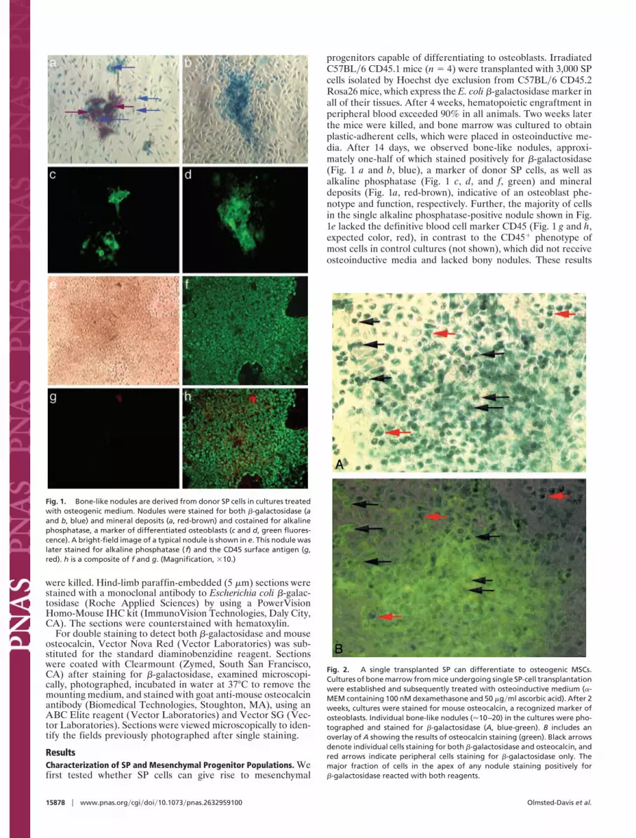
Fig. 2. A single transplanted SP can differentiate to osteogenic MSCs.Cultures of bone marrow from mice undergoing single SP-cell transplantationwere established and subsequently treated with osteoinductive medium (�-MEM containing 100 nM dexamethasone and 50 �g�ml ascorbic acid). After 2weeks, cultures were stained for mouse osteocalcin, a recognized marker ofosteoblasts. Individual bone-like nodules (�10–20) in the cultures were pho-tographed and stained for �-galactosidase (A, blue-green). B includes anoverlay of A showing the results of osteocalcin staining (green). Black arrowsdenote individual cells staining for both �-galactosidase and osteocalcin, andred arrows indicate peripheral cells staining for �-galactosidase only. Themajor fraction of cells in the apex of any nodule staining positively for�-galactosidase reacted with both reagents.
15878 � www.pnas.org�cgi�doi�10.1073�pnas.2632959100 Olmsted-Davis et al.

suggest that SP cells may possess osteogenic potential realizedthrough fibroblast intermediates.
Osteogenic Differentiation by a Single Bone Marrow-Derived SP Cell.We next performed experiments in which a single SP cell wastransplanted into recipient mice. Whole bone marrow cells fromC57BL�6 CD45.2 Rosa26 mice were stained with Hoechst 33342dye, and single CD45� Sca-1� SP cells were sorted into theindividual wells of a 96-well plate. The single SP cells weretransplanted into lethally irradiated C57BL�6 CD45.1 micetogether with short-term repopulating cells derived from thesame mouse strain, to provide committed progenitors that wouldassist in bone marrow regeneration during the early phase ofrecovery from radiation treatment (12). Approximately 14months after transplantation, whole bone marrow was isolated,and plastic-adherent cells were cultured in osteoinductive media.
Bone-like nodules that stained positively for �-galactosidaseand osteocalcin were observed in cell cultures treated withosteoinductive media (Fig. 2). The major fraction of cells in theapex of a given nodule stained positively for both markers (blackarrows), as did some cells in the periphery. Other peripheral cellsshowed only �-galactosidase activity (red arrows). Noncellularosteocalcin staining in the apex of the nodule is probablyassociated with bone matrix. This staining pattern is identical tothat described by Pockwinse et al. (13), who noted that osteo-calcin synthesis occurs postproliferatively and is therefore mostlyrestricted to cells within the apex of the nodule. Some noduleshad only a few cells at the apex that were positive for both�-galactosidase and osteocalcin, suggesting that these noduleswere in an earlier state of osteoblastic differentiation (data notshown). All bony nodules in parallel cultures of marrow from
nontransplanted C57BL�6 CD45.1 mice were positive for osteo-calcin but not �-galactosidase (data not shown). These findingsclearly demonstrate that a single SP cell can give rise torepopulating mesenchymal progenitors with the capacity todifferentiate to functional osteoblasts.
Both SP Cells and Whole Bone Marrow Generate Osteoblasts forEndochondral Bone Formation. To track the fate of SP-derivedosteoblasts in vivo and to compare their prevalence in bone withthat of osteoblasts from whole marrow, we irradiated C57BL�6CD45.1 mice and transplanted them with either 3 � 103 SP cellsor 1 � 106 unmanipulated marrow cells from C57BL�6 CD45.2Rosa26 mice (n � 3 per treatment group). NontransplantedC57BL�6 CD45.1 mice served as controls. Four weeks aftertransplantation, the percentage of hematopoietic engraftmentwas �90% in both groups.
Figs. 3 a1 and a2 (SP cells) and b1 and b2 (whole bone marrow)show representative positive results of staining osteoblasts andother osteogenic cells (e.g., bone lining cells) lining trabeculae(arrows) near the growth plate. The rim of newly formed boneapparent in Fig. 3a2 is confirmed in a3 by the decreasedbirefringence evident under polarized light. By contrast, insections of C57BL�6 CD45.1 mouse femur (no transplantation),the osteogenic cells lining trabeculae did not react with the�-galactosidase antibody (Fig. 3 c2 and c3, arrows). All sectionswere counterstained with hematoxylin. The use of horseradishperoxidase to detect �-galactosidase activity did not producebackground staining (Fig. 3 c2 and c3). Finally, analysis ofadditional donor-derived osteogenic cells revealed positivelystaining osteocytes within lacunae (red arrows) in mice trans-planted with either SP cells (Fig. 3d1a) or whole bone marrow
Fig. 3. Detection of donor SP cell-derived osteoblasts lining the trabeculae of hind-limb bone. Serial hind-limb sections from transplanted mice were stainedwith an antibody against �-galactosidase, detected with horseradish peroxidase (brown) and hematoxylin (blue). Positively staining osteogenic cells were readilyapparent in trabecular bone (arrows) whether mice were transplanted with marked SP cells (a1–a3) or whole bone marrow (b1–b3). a3 duplicates a2 usingpolarized light, and b3 shows the results with an anti-osteocalcin antibody. c2 (five sections away from the section depicted in c1) and c3 show representativebone sections from nontransplanted C57BL�6 CD45.1 mice stained with anti-�-galactosidase or an anti-human mitochondrial antigen, respectively. Althoughosteogenic cells were evident in trabecular bone, they lacked staining altogether. c1 is an adjacent section stained with hematoxylin and eosin. d1 and d2 showrepresentative bone sections of mice transplanted with either SP cells or whole bone marrow, respectively, and stained with anti-�-galactosidase antibody. Redarrows in d1a and d2 indicate the position of osteocytes staining positively for �-galactosidase. (Magnification, �40, except d1a, �100.)
Olmsted-Davis et al. PNAS � December 23, 2003 � vol. 100 � no. 26 � 15879
MED
ICA
LSC
IEN
CES

(Fig. 3d2), suggesting the ability of donor-derived osteoblasts toundergo terminal differentiation.
To confirm that the positively staining cells were indeedosteoblasts, we costained immediately adjacent serial sectionsfor the osteoblast-specific gene product osteocalcin (14, 15),which is synthesized only by late-stage osteoblasts and, in itssecreted form, serves as an integral component of bone matrix(16, 17). The results are presented in Fig. 3b3 (whole bonemarrow).
To demonstrate more rigorously the osteoblast identity ofdonor-derived cells lining trabeculae, we stained the samesection of bone with both anti-�-galactosidase and anti-mouseosteocalcin antibodies. Long-bone sections from C57BL�6CD45.1 mice (n � 4) that were lethally irradiated and repopu-lated with 3,000 SP cells from C57BL�6 CD45.2 Rosa26 micewere first stained with anti-�-galactosidase antibody (red-brown) and then with anti-mouse osteocalcin antibody (black).Fig. 4 depicts osteocalcin-positive cells (osteoblasts) lining tra-beculae near the growth plate (b), many of which also stainedpositively for the �-galactosidase marker gene (a).
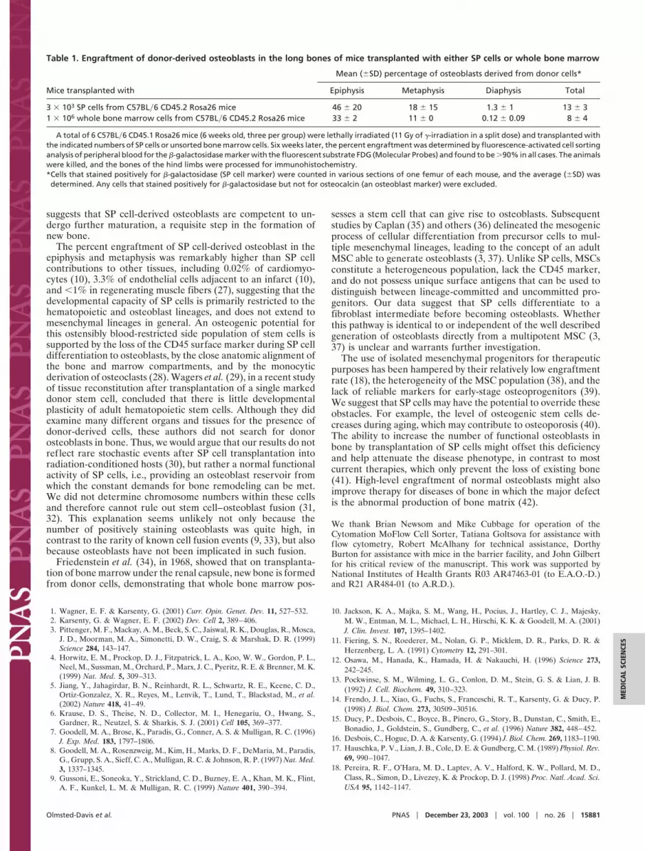
A major question in this study was whether isolated SP cellspossess the same capacity as whole bone marrow to generate newosteoblast populations. Thus, proportions of �-galactosidase-positive osteoblasts were determined in mice transplanted witheither SP cells or whole bone marrow. As shown in Table 1, asubstantial proportion of the osteoblasts were donor-derivedwhether the animals received gene-marked SP cells (13 � 3%)or whole bone marrow (8 � 4%). One explanation for these datais that the entire osteoblast regenerating power of whole bonemarrow is represented by SP cells. However, this interpretationis subject to the bias inherent in counting immunostained cells.Formal comparison of the capacity of whole bone marrow andof the SP cells it contains to produce osteoblasts in vivo wouldrequire additional large-scale experiments with MSCs them-selves, whole bone marrow depleted of MSCs, and whole bonemarrow depleted of MSCs and SP cells. The results also show(Table 1) that the vast majority of donor-derived osteoblasts(�60%) were found in the epiphysis and metaphysis; only 1.3 �1% of the osteoblasts in the diaphysis were of donor origin. Theoverall percentage of donor-derived osteoblasts was 13 � 3%.Several previous studies have reported that transplantation witheither whole bone marrow or MSCs results not only in theincorporation of donor-derived cells into bone (18), but also thatthe donor label is transferred to bone lining cells, osteoblasts,and osteocytes within bone (4, 19, 20). In contrast to the currentstudy, however, these investigations used mice with osteogenesisimperfecta (4, 18, 20), did not perform marrow ablation (21), orinjected marked cells directly into the marrow space (19).
DiscussionThese studies demonstrate that SP cells, a self-renewing popu-lation of bone marrow hematopoietic stem cells with a Lin�
Sca1� cKit� CD45� surface phenotype (22, 23), can regeneratefunctional osteoblasts. The percentage of bone-forming osteo-blasts that expressed the C57BL�6 CD45.2 Rosa26 marker of thedonor SP cells was quite high in the epiphysis and metaphysis andrepresented all of the osteogenic activity in whole bone marrow(Table 1). Most of the donor-derived osteoblasts were foundalong the margins of trabeculae of the epiphyseal and meta-physeal compartments, the most metabolically active regionduring longitudinal bone growth as well as during the remodelingof mature bone (24, 25). Jilka et al. (26) showed that osteoblastsin mouse bone have a lifespan of only 300 h, a turnover rate thatcould account, at least in part, for the high engraftment levels weobserved. Very few osteocytes in cortical bone contained thedonor marker, presumably because of the low turnover ofosteocytes compared to osteoblasts (26). Nevertheless, our de-tection of even rare donor-derived osteocytes (Fig. 3 d1a and d2)
Fig. 4. Osteocalcin analysis of bone from C57BL�6 CD45.1 mice transplantedwith marked SP cells for the presence of marked osteoblasts. Mice wereirradiated and transplanted with SP cells derived from C57BL�6 CD45.2 Rosa26mice. Paraffin-embedded sections of normal long bone were then costainedwith an anti-�-galactosidase antibody and an antibody against mouse osteo-calcin, a definitive marker of osteoblasts. A shows a representative field froma section stained with the anti-�-galactosidase antibody and Vector Nova Red(red-brown). B shows the same field after removal of the mounting mediumand staining with the antibody against mouse osteocalcin and Vector SG(black). Bone sections from nontransplanted mice were also double stainedand shown to be negative for �-galactosidase, and other murine tissues failedto react with the anti-osteocalcin antibody (not shown). (Magnification, �40.)
15880 � www.pnas.org�cgi�doi�10.1073�pnas.2632959100 Olmsted-Davis et al.
suggests that SP cell-derived osteoblasts are competent to un-dergo further maturation, a requisite step in the formation ofnew bone.
The percent engraftment of SP cell-derived osteoblast in theepiphysis and metaphysis was remarkably higher than SP cellcontributions to other tissues, including 0.02% of cardiomyo-cytes (10), 3.3% of endothelial cells adjacent to an infarct (10),and �1% in regenerating muscle fibers (27), suggesting that thedevelopmental capacity of SP cells is primarily restricted to thehematopoietic and osteoblast lineages, and does not extend tomesenchymal lineages in general. An osteogenic potential forthis ostensibly blood-restricted side population of stem cells issupported by the loss of the CD45 surface marker during SP celldifferentiation to osteoblasts, by the close anatomic alignment ofthe bone and marrow compartments, and by the monocyticderivation of osteoclasts (28). Wagers et al. (29), in a recent studyof tissue reconstitution after transplantation of a single markeddonor stem cell, concluded that there is little developmentalplasticity of adult hematopoietic stem cells. Although they didexamine many different organs and tissues for the presence ofdonor-derived cells, these authors did not search for donorosteoblasts in bone. Thus, we would argue that our results do notreflect rare stochastic events after SP cell transplantation intoradiation-conditioned hosts (30), but rather a normal functionalactivity of SP cells, i.e., providing an osteoblast reservoir fromwhich the constant demands for bone remodeling can be met.We did not determine chromosome numbers within these cellsand therefore cannot rule out stem cell–osteoblast fusion (31,32). This explanation seems unlikely not only because thenumber of positively staining osteoblasts was quite high, incontrast to the rarity of known cell fusion events (9, 33), but alsobecause osteoblasts have not been implicated in such fusion.
Friedenstein et al. (34), in 1968, showed that on transplanta-tion of bone marrow under the renal capsule, new bone is formedfrom donor cells, demonstrating that whole bone marrow pos-
sesses a stem cell that can give rise to osteoblasts. Subsequentstudies by Caplan (35) and others (36) delineated the mesogenicprocess of cellular differentiation from precursor cells to mul-tiple mesenchymal lineages, leading to the concept of an adultMSC able to generate osteoblasts (3, 37). Unlike SP cells, MSCsconstitute a heterogeneous population, lack the CD45 marker,and do not possess unique surface antigens that can be used todistinguish between lineage-committed and uncommitted pro-genitors. Our data suggest that SP cells differentiate to afibroblast intermediate before becoming osteoblasts. Whetherthis pathway is identical to or independent of the well describedgeneration of osteoblasts directly from a multipotent MSC (3,37) is unclear and warrants further investigation.
The use of isolated mesenchymal progenitors for therapeuticpurposes has been hampered by their relatively low engraftmentrate (18), the heterogeneity of the MSC population (38), and thelack of reliable markers for early-stage osteoprogenitors (39).We suggest that SP cells may have the potential to override theseobstacles. For example, the level of osteogenic stem cells de-creases during aging, which may contribute to osteoporosis (40).The ability to increase the number of functional osteoblasts inbone by transplantation of SP cells might offset this deficiencyand help attenuate the disease phenotype, in contrast to mostcurrent therapies, which only prevent the loss of existing bone(41). High-level engraftment of normal osteoblasts might alsoimprove therapy for diseases of bone in which the major defectis the abnormal production of bone matrix (42).
We thank Brian Newsom and Mike Cubbage for operation of theCytomation MoFlow Cell Sorter, Tatiana Goltsova for assistance withflow cytometry, Robert McAlhany for technical assistance, DorthyBurton for assistance with mice in the barrier facility, and John Gilbertfor his critical review of the manuscript. This work was supported byNational Institutes of Health Grants R03 AR47463-01 (to E.A.O.-D.)and R21 AR484-01 (to A.R.D.).
1. Wagner, E. F. & Karsenty, G. (2001) Curr. Opin. Genet. Dev. 11, 527–532.2. Karsenty, G. & Wagner, E. F. (2002) Dev. Cell 2, 389–406.3. Pittenger, M. F., Mackay, A. M., Beck, S. C., Jaiswal, R. K., Douglas, R., Mosca,
J. D., Moorman, M. A., Simonetti, D. W., Craig, S. & Marshak, D. R. (1999)Science 284, 143–147.
4. Horwitz, E. M., Prockop, D. J., Fitzpatrick, L. A., Koo, W. W., Gordon, P. L.,Neel, M., Sussman, M., Orchard, P., Marx, J. C., Pyeritz, R. E. & Brenner, M. K.(1999) Nat. Med. 5, 309–313.
5. Jiang, Y., Jahagirdar, B. N., Reinhardt, R. L., Schwartz, R. E., Keene, C. D.,Ortiz-Gonzalez, X. R., Reyes, M., Lenvik, T., Lund, T., Blackstad, M., et al.(2002) Nature 418, 41–49.
6. Krause, D. S., Theise, N. D., Collector, M. I., Henegariu, O., Hwang, S.,Gardner, R., Neutzel, S. & Sharkis, S. J. (2001) Cell 105, 369–377.
7. Goodell, M. A., Brose, K., Paradis, G., Conner, A. S. & Mulligan, R. C. (1996)J. Exp. Med. 183, 1797–1806.
8. Goodell, M. A., Rosenzweig, M., Kim, H., Marks, D. F., DeMaria, M., Paradis,G., Grupp, S. A., Sieff, C. A., Mulligan, R. C. & Johnson, R. P. (1997) Nat. Med.3, 1337–1345.
9. Gussoni, E., Soneoka, Y., Strickland, C. D., Buzney, E. A., Khan, M. K., Flint,A. F., Kunkel, L. M. & Mulligan, R. C. (1999) Nature 401, 390–394.
10. Jackson, K. A., Majka, S. M., Wang, H., Pocius, J., Hartley, C. J., Majesky,M. W., Entman, M. L., Michael, L. H., Hirschi, K. K. & Goodell, M. A. (2001)J. Clin. Invest. 107, 1395–1402.
11. Fiering, S. N., Roederer, M., Nolan, G. P., Micklem, D. R., Parks, D. R. &Herzenberg, L. A. (1991) Cytometry 12, 291–301.
12. Osawa, M., Hanada, K., Hamada, H. & Nakauchi, H. (1996) Science 273,242–245.
13. Pockwinse, S. M., Wilming, L. G., Conlon, D. M., Stein, G. S. & Lian, J. B.(1992) J. Cell. Biochem. 49, 310–323.
14. Frendo, J. L., Xiao, G., Fuchs, S., Franceschi, R. T., Karsenty, G. & Ducy, P.(1998) J. Biol. Chem. 273, 30509–30516.
15. Ducy, P., Desbois, C., Boyce, B., Pinero, G., Story, B., Dunstan, C., Smith, E.,Bonadio, J., Goldstein, S., Gundberg, C., et al. (1996) Nature 382, 448–452.
16. Desbois, C., Hogue, D. A. & Karsenty, G. (1994) J. Biol. Chem. 269, 1183–1190.17. Hauschka, P. V., Lian, J. B., Cole, D. E. & Gundberg, C. M. (1989) Physiol. Rev.
69, 990–1047.18. Pereira, R. F., O’Hara, M. D., Laptev, A. V., Halford, K. W., Pollard, M. D.,
Class, R., Simon, D., Livezey, K. & Prockop, D. J. (1998) Proc. Natl. Acad. Sci.USA 95, 1142–1147.
Table 1. Engraftment of donor-derived osteoblasts in the long bones of mice transplanted with either SP cells or whole bone marrow
Mice transplanted with
Mean (�SD) percentage of osteoblasts derived from donor cells*
Epiphysis Metaphysis Diaphysis Total
3 � 103 SP cells from C57BL�6 CD45.2 Rosa26 mice 46 � 20 18 � 15 1.3 � 1 13 � 31 � 106 whole bone marrow cells from C57BL�6 CD45.2 Rosa26 mice 33 � 2 11 � 0 0.12 � 0.09 8 � 4
A total of 6 C57BL�6 CD45.1 Rosa26 mice (6 weeks old, three per group) were lethally irradiated (11 Gy of �-irradiation in a split dose) and transplanted withthe indicated numbers of SP cells or unsorted bone marrow cells. Six weeks later, the percent engraftment was determined by fluorescence-activated cell sortinganalysis of peripheral blood for the �-galactosidase marker with the fluorescent substrate FDG (Molecular Probes) and found to be �90% in all cases. The animalswere killed, and the bones of the hind limbs were processed for immunohistochemistry.*Cells that stained positively for �-galactosidase (SP cell marker) were counted in various sections of one femur of each mouse, and the average (�SD) wasdetermined. Any cells that stained positively for �-galactosidase but not for osteocalcin (an osteoblast marker) were excluded.
Olmsted-Davis et al. PNAS � December 23, 2003 � vol. 100 � no. 26 � 15881
MED
ICA
LSC
IEN
CES

19. Onyia, J. E., Clapp, D. W., Long, H. & Hock, J. M. (1998) J. Bone Miner. Res.13, 20–30.
20. Horwitz, E. M., Prockop, D. J., Gordon, P. L., Koo, W. W., Fitzpatrick, L. A.,Neel, M. D., McCarville, M. E., Orchard, P. J., Pyeritz, R. E. & Brenner, M. K.(2001) Blood 97, 1227–1231.
21. Nilsson, S. K., Dooner, M. S., Weier, H. U., Frenkel, B., Lian, J. B., Stein, G. S.& Quesenberry, P. J. (1999) J. Exp. Med. 189, 729–734.
22. Hirschi, K. & Goodell, M. (2001) Differentiation 68, 186–192.23. Ramos, C. A., Venezia, T. A., Camargo, F. A. & Goodell, M. A. (2003)
BioTechniques 34, 572–591.24. Fleisch, H. (1995) Bisphosphonates in Bone Disease: From the Laboratory to the
Patient (Academic, New York).25. Steiner, E., Jergas, M. & Genant, H. K. (1996) Radiology of Osteoporosis
(Academic, New York).26. Jilka, R. L., Weinstein, R. S., Bellido, T., Parfitt, A. M. & Manolagas, S. C.
(1998) J. Bone Miner. Res. 13, 793–802.27. Ferrari, G., Cusella-De Angelis, G., Coletta, M., Paolucci, E., Stornaiuolo, A.,
Cossu, G. & Mavilio, F. (1998) Science 279, 1528–1530.28. Duong, L. T. & Rodan, G. A. (2001) Rev. Endocr. Metab. Disorders 2,
95–104.
29. Wagers, A. J., Sherwood, R. I., Christensen, J. L. & Weissman, I. L. (2002)Science 297, 2256–2259.
30. Orkin, S. H. & Zon, L. I. (2002) Nat. Immunol. 3, 323–328.31. Terada, N., Hamazaki, T., Oka, M., Hoki, M., Mastalerz, D. M., Nakano, Y.,
Meyer, E. M., Morel, L., Petersen, B. E. & Scott, E. W. (2002) Nature 416,542–545.
32. Ying, Q. L., Nichols, J., Evans, E. P. & Smith, A. G. (2002) Nature 416, 545–548.33. LaBarge, M. A. & Blau, H. M. (2002) Cell 111, 589–601.34. Friedenstein, A. J., Petrakova, K. V., Kurolesova, A. I. & Frolova, G. P. (1968)
Transplantation 6, 230–247.35. Caplan, A. I. (1994) Clin. Plast. Surg. 21, 429–435.36. Prockop, D. J. (1997) Science 276, 71–74.37. Dennis, J. E., Merriam, A., Awadallah, A., Yoo, J. U., Johnstone, B. & Caplan,
A. I. (1999) J. Bone Miner. Res. 14, 700–709.38. Phinney, D. G. (2002) J. Cell. Biochem. Suppl. 38, 7–12.39. Devine, S. M. (2002) J. Cell. Biochem. Suppl. 38, 73–79.40. Rodriguez, J. P., Garat, S., Gajardo, H., Pino, A. M. & Seitz, G. (1999) J. Cell.
Biochem. 75, 414–423.41. Rezka, A. A. & Rodan, G. A. (2003) Curr. Rheumatol. Rep. 5, 65–74.42. Mundlos, S. & Olsen, B. R. (1997) FASEB J. 11, 227–233.
15882 � www.pnas.org�cgi�doi�10.1073�pnas.2632959100 Olmsted-Davis et al.