gastroesophageal diversion for primitive alkaline gastritis r2
TRANSCRIPT
ViDEO CORNER 1
Gastroesophageal diversion for primitive alkaline gastritis
A. M. DE Simore, D. Bertero, M. Gaccone Department of Surgery and Patology • University of Turin, ltaly
Alkaline ancj/or mixed gastroesophagitis are clinical entities that only in recent years received on individu· alized pathophysiological interpretation. On the basis of their aetiology are divided into primitive and secondary types. lf there is a certain agreement on secondary types (due to gastric, esophageal, pancreatic and biliary surgery), primitive ones (with the entire stomach) are still matter of debate concerning their existence by some authors. During the lası years, based �n a strict and appropriate use of disgnostic means (pH-metry, manometr, HIDA scan, endoscopy with multiple biopsies) we noticed an increased incidence of alkaline or mixed reflux esophagitis, either primitive or secondary. Since 1982 we started treating patients with severe forms of secondary alkaline reflux esophagitis, and only recently, encouraged by good results, we started treating primitive forms (with the entire stomach) of alkaline reflux esophagitis. first we used total duodenal diversion (TDD) through a classic Roux-en-Y loop (Holt's technique), then we passed to the ffDuodenal Switchff technique (DS) as a described by de Oeester, and recently we arrived to use the gastro-esophageal diversion (GED) with inverted loops Roux-en-Y. lncostant results obtained with the formeres (TDD,DS) forced us toward the use of the latter (GED), which deserves other advantages, being effective and secondary types of alkaline reflux. The video herein shows the GED technique, after a short pathophysiological foreword, illustrating its application in a primitive alkaline reflux gastritis.
R2 limphectomy in the treatment of the early mid gastric carcinoma
S.M. Glulini, A.C. Brotto, M. Giovanetti,N. Portolani, F. BenziOepartment of Surgical Sciences - University of Brescia, ltaly
The videotape shows the operation we adopted as a standard tor the treatment of the distal and mid early gastric cancer. The gastric resection is subtotaı with the proximal resection margin on the lesser curvature 2 or 3 cm distal to the oesophago-gastric junction. The limphectomy is comprehensive of grouys 1,3,4,5,6, 7,8,9, 11 and 12, and its technique is demonstrated in the videotape. Splenectomy is considered unnecessary, considering the minimal incidence of group 1 O localizations in the early gastric cancer, and potentially depressing the immunitary response.
Cleaning of the mesenteric artery nodes is also not performed, as it is associated with an high risk of major nutritional disorders, consequent to the denervation. Nerve sparing technique tor the preservation of the celiac nerve ganglions during celiac limphectomy is illustrated.
Distal pancreatectomy with splenic conservation for benign tumors
S.M. Giulinl, U. Tambussi, A. Coniglio, D. Pinelli,G.P. Bertoloni Oepartment of Surgical Sciences-University of Brescia, ltaly
The techniques of spleen-preserving distal pancreatectomy, though logical in the pediatric patients. recently have been extensively applied in adults. The videotape shows two cases of conservative distal pancreatectomy performed tor benign tumors. The first patient a 28 year-old woman, was admitted to our hospital tor a large mass of the pancreatic tail found during ultrasonographic exam. CT-scan confirmed the presence of an 8x1 O cm tumor. Following left subcostal incision and widely opening of the gastrocolic omentum, the mass is visualized: the tip of the distal part of the pancreas is readily identified and mobilized in the hilum of the spleen, so the dissection may begin at the tip and proceed in a prograde direction. After tumor removal the pancreatic remmant is sutured with interrupted not readsorbable stitches. Histology shows the tumor to be a cystic and solid papillary neoplasia. The second patient, a 67 year-old woman, was admitted in our hospital tor relapsing abdominal colies. Ultrasound and CT-scan showed a small tumor (2 cm) between the body and tail of the pancreas. The abdomen is opened through a bilateral subcostal incision and a generous extent of the gastrocolic omentum is divided between clamps. The tumor is located in the body and does not appear on the surface of the pancreas; then the peritoneum is incised along the inferior border and the posteior surtace of the pancreas is exposed by blunt dissection. Silastic loops are placed around the splenic artery and the pancreas. The dissection of the splenic vein progresses from the proximal to the distal part of the pancreas; multiple small branches of the vein are identified and ligated til the splenic hilum. following pancreas transection and good haemostasis of the stump retrograde dissection of the splenic artery is continued towards the spleen dividing several branches of the artery. At the end of the arterial dissection the body and the tail of the pnacreas are completely freed and removed. The pancreatic stump is sutured with interrupted not readsorbable stitehes and covered with viable omentum. The frozen sections show the tumor to be a microcystic adenoma.
15
Left hepate�tomy for giant hemangioma of the liver
S.M. Giulini, N. Portolani, L. Congiotti,U. Tambussl, S. Bonardelli, F. BenziDepartmanı of Surgical Sciences-University of Brescia, ltaly
The videotape illustrates the case of a female 59 years old patient, affected by a giant cavernous hepatic hemangioma (30x8 cm) who underw�nt . iefthepatectomy. The iesion, almos_t as_ympt?,:natıc, fılleda big part of the abdominai cavıty, ıts orıgrn from the left liver, but they couldn't define precisely its right limits and particularly the eventual involvement of segements V and VIII. At operation the iesion results to occupate the whole iV segment; the left iobe is also completely involved, segments V and VIII presents diffuse microhemangiomatous alt�rations. The hepatic peduncle is prepared; left hepatıc artery and partal vein are tied and severed, with preservation of the branches of the caudatic lobe. The suprahepatic and infrahepatic vena cava are prepared. The ieft hepatic vein is interrupted and sutured. The hepatic peduncle is clamped. Left hepatectomy with minimal resection of segment V and VIII is performed by digitoclasy without blood loss requiring transfusion.
Hepatic segmentectomy for focal nodular hyperplasia
F. Castro Sousa, L, Manso, E. Martires, E. Granjo,O. Matos, E. Pinheiro3th Surgical Department-Coimbra University Hospital-3049Coimbra, Portugal
Correct diagnosis of nodular lesions of the liver is sometimes difficult or even impossible. A 31 years old woman was hospitalized twice with a growing symptomatic lesion of the right lobe of the liver. Ecography, scanner, RMN and even biopsy were_notable to establish a precise diagnosis. The patıent was operated and an hepatic bisegmentectomy (VI and VII) has been carried out. The interest of the u�e of an ultrasonic dissector and argon electrocautery rn hepatic resection is shown in the video. Pathologic examination of the specimen confirmed the macroscopic diagnosis of focal nodular hyperplasia. Two years after the operation the patient remains assymptomatic with no imagiologic signs of recurrence.
Prosthetic H-graft portacaval shunt
S.M. Giulini, N� Portolani, A. Pouche, A. Lazzarinl,F. BenziDepartment of Surgical Sciences-University of Brescia. ltaly
The tape shows an operation of an H portacaval shunt, performed by the interposition of a very short segment of prosthesis, in a male cirrhotic patient which presented recurrent bleeding from oesophageal varices, already treated in the past by sclero-
16
therapy. After a limited isolation the infraepatic ven� cava is partially clamped and an armed 12 mm calıber PTFE graft is anastomosed in a termino-lateral way to its anterior face. The tube is shortened as necessary to joint it without traction or kinking to the posterior face of partal vein (less than 1 cm in this case). After declamping the partal pressure, which was 30 mmHg before, falls to 16 mm. At 14 months after the operation the patient is in good conditions free from bleeding or encephalopathy. The color Ooppler duplex scanning shows the patency of the graft and a residual partal flow to the liver. Compared to the classical direct portacaeval anastomosıs the shunt presented here presents a definite and stable caliber and offers a betler technical situation in case of an eventual liver tranplantation. Compared to the prosthetic shunt of Sarfeh it presents a lower risk of thrombosis being shorter and more direct so presenting the need to sacrifice the major partal collaterals (left gastric vein, gastroepiploic vein ete.) as described by the original technique to increase the flow through the graft and long term patency.
ViDEO CORNER il
Placement of a single device for adjuvant hepatic arterial infusion chemotherapy after reimplantation of a right aberrant hepatic artery on the left hepatic artery
S.M. Giulini, N. Portolani, G. Pulcini, L. Taglietti,M. RonconiDepartment of Surgical Sciences, Univ. of Brescia, ltaly
Colorectal hepatic metastases are a well accepted indication tor liver resection. Unfortunately, only 20-30 % will be cured after surgery alone. The value of adjuvant therapy after "curative" liver resection is not well established: some recent reports seem to suggest the value of hepatic arterial infusion chemotherapy, evantually associated to a systemic one, to �revent recurrence in the liver remnant. The atypıcal distribution of the vascular tree may complicate the placement of the arterial catheter. The presence of a right hepatic artery arising from the superior mes�nteric artery usually requires two porth-catheter devıces, the first positioned as usual into the gastroduodenal artery lor the left hepatic lobe, the second tangentially inserted in the right hepatic artery. This procedure is expensive, it increases the risk of vascular thrombosis besides the possibility of a reflux in the intestinal vessels. in this tape we present an alternative solution with the reconstruction of a "normal" hepatic vascular tree. With this aim, we severed the right hepatic artery arising from_ the _ superior me�enteric artery in the retropancreatıc sıte. The proxımal stump is tied, the distal one is anastomized in a termino-lateral fashion on the left hepatic artery with interrupted stitches. So we are able to use a single porth-catheter device placed in the usu�I man�er into the gastroduodenal artery. Postoperatrve angıo-
graphy, performed by direct puncture · of the porth, shows the perfusion of the hepatic artery, the recounstructed hepatic biforcation with the right and left hepatic artery and then, in the same time and with the sama intensity, of the intraepatic vascular tree of both hepatic lobes.
Transcatheter arterial embolization STAE in hepathocellular carclnoma (HCC)
A. Gaetlnl, M. Grosso, F. Spalluto, M. Bertolino,F. Pı:ıdrazzlnl, O. Bertero, M. GlacconeDepartment of General Surgery and Radiology University ofTurin, ltaly
We want to present our experience of 139 trans�atheter arterial embolizations in IICC performed ın 4 years at the institute of Radiology of Turin. After an angiographical study of celiac trunk and of the upper mesenteric artery, a selective catheterization of the hepatic artery is performed to introduce the cY1ostatic emulsion (Doxorubicin 20-30 mg) and m.d.c. liposolubic (Lipiodol 6-10 mi) that shows a particular tropism tor hypervascularized neoplastic tissues fixing itself absac ali on the HCC nodes and so acting as carrier tor chemotherapeutic substance. Thus a selective embolization by fibrin foam (Spongostan) is performed in ordar to add an ischemic effect to the pharmacological one. The 139 patients have been divided into 3 stages according to the Osida classification: 66 stage 1, 64 stage 11, 9 stage 111. The pecentages of survival of patients to stage I are 100 % as 3 months, 85 % at 5 months, 59 % of 12 months, 28 % at 24 months, 12 % at 36 monts. in stage 86 % at 3 months, 67 % at 6 months. 49 % at 12 months, 15 % of 24 months, 3 % at 36 months, in stage 111 78 % at 3 months, 33 % at 6 months, 33 % at 2 months. According to our experience we have found that surgical therapy, when feasible achieves the besi results at medium and short term, but that TAE achieves undoubted results in patients which are no longer operable (therefore in these subjects it has to be applied because the surviv and the general conditions improve considerably) good results are also reported using the method before and during the operation, providing the surgeon of useful vascular map and allowing him to operate in excellent conditions in some cases previously considered inoperable Furthermore T AE has to be applied in patients carriers of HCC proposed tor transplantation, because it _reduces thespeed of increase of the neoplasm durıng the long time of waiting before the operation.
Spontaneous pneumothorax treated by thoracoscopy
A. Baltasar, F. Arlandis, E. Marcote,R. Martinez, C. Serra, LA. CipagautaCid 61. 03800 Alcoy. Spain
Spontaneous pneumothorax is a medical condition treated often by simple underwater drainage. Sometimes surgical intervention is necessary. in the 70's a full posterolateral thoracocotomy was done. in the 80's an axillary minimal thoracotomy was the most common approach. With the new thoracoscopic tech· niques a minimally invasive intervention is required. A patient is presented in whom a right axillary operation was done two years earlier tor spontaneous pneumothorax. Nowadays, the patient was admitte� with a left spontaneous pneumothorax and an atypıcal lung resection was done, and the patient was discharged two days later.
Surgical treatment in the Crohn's disease stricturoplasty
A. Gaetini, M DE Slmone, M. Glaccone, D. BerteroDepartmanı of Surgery and Pathology Unıversıty of Turinltaly
in the videotape are discussed some indications about the adoption of stricturoplasties ın the Crohn disease therapy. Therefore dıfferent methods are illustrated in detail using even some animated cartoons. Different methodologies tor ileal and colonic stricturoplasties are shown. in particular the Heineke - Mikulicz's technique, the Judd's technique and theFinney·s technique are like the omonymous op�rations tor piloroplasty. Moschel Walske's technıque is the same ol the one adopted tor treating ureteral stenosıs. Every technique has some exact indications and allows to treat Crohn's intestinal stenosıs saving the most of bowel. Patients: from 1984 to 1992 we have performed 82 stricturoplasties on of 25 patients. it has respectively been performed_ astricturoplasty at the duodenal level, and 63 at the ıejuno-ileal level and 2 at the colon. A Mickulicz's plastic has been performed at the duodenum: at the ileo 43 Mikulicz, 16 Judd and 4 Maschel have been performed; in the colon it has been performed one Maschel and one Judd. We have noticed neither complications after the operation nor mortality in ali these cases. We have not recorded relapses in the "follow up" (range 2-8 yr.) in the stricturopiasty level. We hava reoperated 6 patients (19 %) some years later since the first operation: in 4 cases because of the restarting of the dısease in other sites; in 2 other cases it was the appearance of a pseudo-tumor showed up in the recess of the Finney stricturoplasty. Therefore we can considar our experience positively and we believe the conditions that have allowed this success have to be attributed to the correct surgical choice and surgical indications: a good nutritional preparation, disease in a cronity phase, good intestinal preparation, and again correct surgical choise.
17
Radical resection ofa giant advanced ACTH-secreting timic carcinoid
S.M. Glulinl, U. Tambussi, G. Galvani, A. PoucheDepartment of Surgical Sciences University of Brescialtaly
The patient, a 34-year-old lady is admitted tor menstrual irregularity, hirsutism, emotional lability and obesity tor about six months. Plasma ACTH and cortisol are elevated. A chest x ray shows a large irregular mass in the left anterior mediastinum. Ct scanning shows the large tumour, measuring 10x15 cm in size with irregular margins surrounding the mediastinal vessels, without signs of infiltration. After median sternotomy careful dissection of pleural reflexion and soft retrosternal tissue is performed. For a better check of vessel connections the pericardium is opened. The next step is freeing the mass from perivasaı tissue after ligation and section of fibrous connections. The mass lies behind the left innominate vein that is sectioned between clamps. The tumour invades the proximal segment of the vein that must then be resected. The stumps are secured by non readsorbable monofilament suture. The mass is freed from the innominate artery, left common carotid artery and left subclavian artery which are adherent but not invaded by the tumour. lnstead the invaded vags nerve is ligated and resected. After blunt dissection from the posterior layer the mass is excised with a large portion of left mediastinal pleural reflexion that appeared to be invaded. The last view of the surgical field shows the aortic arch with the supraaortich arteries completely freed and the lung. A no 28 chest tube in the mediastinum and a separate pleural drainage are positioned. Microscopic examination of the tumour shows a timic carcinoid with prominent vascular and perineural invasion. No other therapy is performed and the patient is free of disease four months atter surgery.
Recto - duplication
A. Gaetlni, M. GiacconeOepartment of Surgery and Patology University of Turinltaly
The videotape presents two clinical cases of patients affected with recto-duplication. The clinical interest is not only the rarity of this pathology but the whole of clinical relevant problems which reach from the identification of lesion to her anatomical definition and problem of surgical therapy. After explaining the embriologycal origin of duplication, we report tor every case the radiological and clinical doucumentation, that allows the correct diagnosis. Then we explain the operation of exeresis of duplication, that's ditferent in two patients tor some technical details requested by seat and mass of malformation, and by necessity of to restore the layer of elevator muscles, in one patient.
18
Eversion endarterectomy and reimplantation of the internal carotid artery tor proximal stenosis and distal kinking
S.M. Giulini, S. Bonardelli, N. Portolani,R. Maffels, F. Nodari, A. Vinco, M. Bertoli*,G. Guarneri**, G. Tomasoni ...Department of General Surgery - University of Brescia, ltaly• Chair of Radiology - Unıversity of Brescia, ltaly•• Service of Neurophysiopatology Spedali Civili of Brescia, ltaly••• Oepartment of Anaesthesiology and lntensive Care Unit- University of Brescia, ltaly
A 70-year-old male patient presented a left TIA and theretore he was studied with echo-color Doppler and digital substraction angiography which showed a high grade stenosis and distal kinking of the left internal carotid artery (ICA). So. the patient underwent the surgical correction ot these lesions which required an extensive dissection of the hypoglossus nerve, very close to base of the cranium, and of carotid arteries, with the cutting of the dygastric muscle. After intravenous heparinization, and commonexternal carotid damping. the back pressure was 70 mmHg. The good tollerance to carotid occlusion was confirmed by a continuous monitoring of SEPPs during the whole procedure. Firstly, an eversion endoarterectomy of ICA was performed through the transverse transection of its origin at the bifurcation. The arteriotomy was prolonged longitudinally towards the distal common carotid artery (CCA) along the lateral margin ot the vessel at the aim to make a new proximal end tor ICA. Therefore, after the completion of the eversion endoarterectomy of the ICA, the native origin of the ICA was closed by continous suture and the artery was reimplanted more proximally on the CCA arteriotomy to suppress the distal kinking. lmmediate Doppler spectral analysis and postoperative angiography demonstrate the good morphological and functional result of the reconstruction.
Embolization and resection of carotid body tumor
S.M. Giulini, S. Bonardelli, G.A.M. Tiberio,L. Cangiotti, P. Re, M. Belloni, R. Maroldi*,B. Guarneri**, R. Favero***Department of General Surgery . university of Brescia, ltaly• Chair _of Radiology - University of Brescia, ıtaly•• Servıce of Neurophysiopathology - Spedali Civili of Brescia, ltaly ••• Departmanı of Anaesthesiology and lntensive Care Unit · University of Brescia, ltaly
A 29 years old woman presented a right laterocervical mass and therefore she underwent MAi of the neck which showed a right oval carotid body tumor, sized 3.8 cm with cranial end near to the body of the 2nd cervical vertebra. Tumor was vascularized by collateral vessels of the right carotid arteries and the internal carotid artery was laterally dislocated
and coiled. To recude the tumor vascularization, during a digital substraction angiography, the selective embolization was performed in the nutritive vessels originating from the external carotid artery. The day after, the patient undergoes operation. The access is a longitudinal laterocervical incision. Firstly the lateral surface of the carotid body tumor is exposed, and, proximally the ı::ommon carotid artery is dissected and surrounded with vessel-loop. A careful dissection of vagus and hypoglossus nerves and of the distal internal and external carotid arteries and the accurate dissection of the external surface of the tumor are completed, first along the cervical and then in the subavventitial plane detected just proximamlly to the carotid bifurcation. Hemostasis is achieved with coagulation or division between suture of the nutritive vessels, and so en-bloc resection of the body tumor is performed. Hystological study shows a capsulated alveolar paraganglioma with jalinoid perivascular sclerosis and the patient is disease free two years after operation.
PANEL ABSTRACTS
Esophagectomy for cancer: Videothoracoscopic approach
S. Bona, R. Rosati, U. Fumagalli, M. Montorsi,A. Peracchia1st. di Chlrurgia Generale e Oncologia Chirurgical Osp. Policlinico, Via Sforza 35-20122 Milan, ltaly
lncreasing experience in minimally-invasive surgery of the esophagus prompted us to perform thoracoscopic esophageal dissection during esophagectomy for cancer. Since january 1991, thoracoscopic esophagectomy was attempted in 16 patients with T1 -T2 tumor at risk tor thoracotomy otherwise candidate to transhiatal esophagectomy. After dividing the azygos vein by means of an ENDO-GIA or ENDOTA stapler 5TM}. the esophagus was mobilized together with the periesophageal mediastinal tissue and with periesophageal, paratracheal and subcarenal lymphnodes; it was then divided at its upper third with an ENDO-GIA (TM} in order to ease dissection. The procedure was completed through laparotomy and cervicotomy. No operative mortality was recorded. Average duration of the thoracoscopic procedure was 125 min. Postoperative complications occurred in 3 patients (2 bleeding and 1 mycobacterial pneumonia}. Two patients showed tumour recurrence at 6 and 18 months. Thoracoscopic esophagectomy reduces postoperative pain but requires longer selective lung exclusion compared to thoracotomy; therefore, postoperative pulmonary function requires further evaluation. Dissection of the esophagus under direct vision allows lower morbidity compared to blunt transhiatal esophagectomy. Presently, in our opinion, thoracoscopic mobilization of the esophagus is indicated in high risk patients with subcarinal T1-T3 tumors. The extent of lymphadenectomy is under evaluation through macroscopic and pathological sampling.
Laparoscopic hernia repair of 415 cases using the "Dudai Butterfly" with or without mesh according to hernia type
M. Dudai, O. Avrutis, S.N. AdlerThe Reichmann Department of Surgery, Bikur Cholim Hospital 5 Strauss st. P.O. Box 492 Jerusalem. lsrael
The purpose of this study is to demonstrate that groin hernicts of different types can be managed by an adjusted surgical approach. Because small hemias (Nyhus classification Type 1+11) are marked by a minor defect in the internal ring with normal pelvic floor strength and normal shutter mechanism ot the inguinal canal, we choose to use the laparoscopic versus the anterior approach thereby not damaging the intact abdominal wall. We use a small Laparoscopic procedure for those small Hernias. The hernia sac is inverted, the peritoneum is opened and dissected behind the internal ring. A tension free closure of the defect is achieved by using the Dudai Butterfly (DB). Because big hernias (Type 111 and iV} are marked not only by a large defect but also by pelvic floor weakness, we demanded a big t-aparoscopic repair of two layer. The first layer is a DB placed in the defect. The second layer is a wide mesh covering the pelvic floor. The sac undergoes "ring excision" as appropriate. Wide dissection ot the pelvic floor and division of inferior epigastric vessels resulting in wide free margings ot the hernia detect. Wide deperitonealization of the lower flap from the cord and the blood vessels. Closure of the defect by DB and closing of the pelvic floor using mesh affixed with staples excluding the "Neurovascular triangle". The use of the DB provides tor tension free closure and support of the defect resulting in reducing the chance of recurrence. The placement of the DB is achieved with ease. DB also stimulates growth of fibroblasts. The subsequent placement of mesh gives additional strength to the entire pelvic floor by receiving homogenous support from the entire area including the hernia defect closed by the 08. Results: Total of hernia repair 415, No. of patients 306, Bil. Hernia 109, Hernia Types 1:12, 11:116, 111:242, IV:45, Post. op. Narcotics o. Post. op. Analgetics 234, Post. op. stay 1.09 days, full recovery 3.8 days, Subcut. Hematoma 9, Wound lnfection 1, Entrapment of LFCN 1, Bladder lnjury 1, Recurrence 3 (0.7 %}. Ali our complications occured in the first 50 cases. We conclude that we adjust the Laparoscopic repair to the Hernia type. Large hernias require a large laparoscopic repair which is superior to the anterior approach. We suggest to consider for the reapir of small hernias a small laparoscopic repapir which does not affect the integrity of the anterior abdominal wall.
19
Laparoscopic Vagotomy in 38 cases, 30 month follow up. Comparing of posterior truncal anterior highly selective to complete highly selective vagotomy
M. Dudal, O. Avrutis, J. Mesholam, 5.N. AdlerThe Reichmann Departmanı of Surgeıy, Bikur Cholim Hospital 5 Strauss st. P.0. Box 492 Jerusalem, lsrael
Before the laparoscopic (lap) era we performed highly selective posterior and anterior vagotomies (HSV). With the introduction of laparoscopy we performed Lap posterior Truncal Anterior HSV (LpTaHSV) according to Zucker and Bailey. Our first 1 O operations were successfuly performed according to the lpTaHSV technique. With experience our lap technique improved. lnitial operation time was 4 hours, the duration of the later operations was approximately 2 hours. No significant complications were encoun� tered. Patients were discharged 3-4 days following surgery and were fully recovered 6 days later. Following surgery a 72 % reduction in acid output was observed. Endoscopy six weeks later revealed complete ulcer healing in ali instances. 5/1 o patients reported mild to moderate symptoms of delayed gastric emptying which improved with time. One other patient had severe symptoms of delayed gastric emptying unresponsive to repeated balloon dilatations of the pylorus and ultimately required pyloroplasty. Because of these complications we tempted to perform Lap. HSV (LHSV) according to the technique used by us in open operations. We successfully operated our following 28 patients according to LHSV, in a 6 step technique developed by us. Operation time tor
LHSV is a 1/4 hour lenger then tor LpTaHSV, app. 2 1/4 hours. No significant complications were encountered. Some analgesics were required postoperatively. Patients were discharged from the hospital 2-3 days after surgery. There were no complaints of postmeal epigastric fullness, delayed gastric emptying and diarrhea. Acid output following surgery reduced by 81 % and at endoscopy six weeks later ali ulcers had healed. in 30 month tollow up ali the patients keep doing well except one patient from the LpTaHSV group that recurre. in conclusion A) it is feasable to perform LHSV. B) in our experience this procedure appears superior to LpTaHSV and probably also to open HSV. C) We suggest to consider LHSV as the treatment of choice of patients with chronic duodenal ulcer disease as an alternative to chronic H2 receptor antagonist therapy taking into consideration cost, side effects, effectiveness and compliance.
20
Prospective study of laparoscopic proximal gastric vagotomy
G.B. Cadiere, R. Verroken, J. Bruyns, J. Himpens, D. Urbain, A. RajanDepartment of G.I. Surgeıy, Popital Universitaire SaintPierre. 322 rue Haute, 1000 Brussels, Belgium
Patients: Between April, 1992 and December, 1993, 55 consecutive patients, 45 males and 1 O females. median age 39 years (range 19-65) underwent proximal gastric vagotomy (PGV) without pyloroplasty, by strictly laparoscopic means. Ali patients were ASA 1 or il. Two patients had had previous gastric surgery (one pyloroplasty and one raphy of a perforated ulcer). Ten others had had lower abdominal procedures. Fourthy three patients were operated on electively, because of chronic ulcer disease (CUD) (n=30) or because of gasttoesophageal reflux disease (GERD, n=13) associated with CUD. Twelve patients were operated in emergency tor perforated ulcer. Thirty one patients underwent Nissen fundoplication during the same procedure; 12 patients underwent associated gastric ulcer repair and a cholecystectomy. Method: The procedure consisted of selective severance of all neurovascular bundles originating from Latarjet's nerve and going to the lesser gastric curvature, cephalad of the crow's toot. care being taken not to injure the main trunk. Dissection included the last 8 cm of esophagus in ali cases. Results: One major postoperative complication (lesser curve necrosis) required a second look laparotomy on the ninth postoperative day. Operative time was on average 172 minutes (range 90-270). Postoperative st�y was 2 days in the elective group and 7 days in the emergency group. This difference was caused by the iV antibiot�repay and prolanged gastric suction in the perforated ulcer at the lesser curvature, probably also due to lesser curve ischaemia. Seven patients were staged as Visick ili, because of epigastric pain (n=2), gas bloating (n=3), heartburn (n=1 ), or diarrhea (n=1 ). There were no gastroscopically proven recurrences of duodenal ulceration. Conclusion: Laparoscopic PGV is feasible and carries a low morbidity. it can systematically be associated with the laparoscopic treatment of perforated ulcer. Complications on long term are the same as known tor PGV, particularly if associated at fundoplication.
Laparoscopic assisted sigmoid resection for malignant disease
M. Morino, C. Garrone, V. Festa, C. Migliettalstituto Di Clinical Chirurgiı;a, Generale. ltalıa
Laparoscopic assisted colon resections tor colorectal diseases had recently developed thanks to the success of laparoscopic cholecystectomy. The video shows a laparoscopic assisted sigmoid resection tor
an adenocarcinoma located at 15 centimeters from the anal marge. After identification, ligature and section of the sigmoid vessels, the bowel was divided 4
to 5 centimeters distal to the lesion with an Endo GIA stapling device. A 5 centimeters right-lower transverse incision was made tor bowel extraction and introduction of the stapler envil. After reinstauration of pneumoperitoneum, the colorectal continuity was restaured by a Knight-Griffen procedure performed transanally. Postoperative course was uneventful and the patient was dismissed on 7th postoperative day. We apply this procedure tor colorectal cancers non protruding the serosa or as a palliative procedure in non resectable metastatic disease.
Laparoscopic totally preperitoneal inguinal hernioplasty
J. Himpens, G.B. Cadiere, J. BruynsDepartment of G.I. Surgery, Hôpital Universitaire SaintPierre, 322 rue Haute, 1000 Brussels, Belgium
The entirely preperitoneal approach tor laparoscopic inguinal hernioplasty (TPP) is getting wide acceptance. This video demonstrates the operative strategy, with successive dissection of Coopers ligament, the epigastric vessels and the psoas muscle. finally, the hernia sac is dissected with bimanuaı technique. The sac is transected and not ligated. A 1 O by 15 cm polyester mesh strengthened by a radiopaque Nitinol frame is then inserted and placed over the three potential hernia orifices. The prosthesis is not stapled. Between 1.5.1993 and 1.8.1993, 31 hernias in 21 patients were treated with this technique. AII the hernias were primary. The mean age was 48 years. There were 20 males and 1 female. Fifteen hernias were indirect, 2 were pantaloon hernias, 13 were direct and 1 was femoral. Mean operating time was 35k minutes per hernia. The mean postoperative stay was 1 day. Postoperative analgesia could simply be assured with paracetamol. Postoperative analgesia could simply be assured with paracetamol. Postoperative working incapacity was 4 days on average tor independent and 4 weeks tor salaried people. There was no morbidity nor mortality. More specifically, there were neither neuralgias nor postoperative obstructions. No recurrences were recorded except in the very first patient where postoperative X ray revealed erroneous too medial a placement of the prosthesis, followed by recurrent indirect hernia. in conclusion, this method appears promising by its relative ease and by the fewer postoperative complications as compared to the transabdominal laparoscopic hernioplasty (TAPP) and is now our preferred one, despite our extensive experience (>300 cases) with TAPP.
Lung cancer in high risk patients conservative treatment with V .A. T .s.
H. Hoyo S. Nestor, D. Walter, G. Miguel, and G.PedroBuenos Aires, Argentina
During the period March-91 to March-94, six patients having lung cancer were operated on by video assisted thoracoscopic surgery (VATS) (over 69 operations tor lung cancer) at the Durand General Hospital, Bs. As., Argentina. Two lobectomies and four segmentectomies were made by this method, in patients having modezate to severe respiratory incapacity. Owing to that, the proceedings were all conservative. in five of the six cases it was necessary to widen one of the hales (up to 5 cm) in order to take off the specimer. AII the patients had a good postoper"ative course, with scarce or absent pain. There was a localized posoperative ampiema in one case, which healed by punction. lnternatior average was of 5 days. We considerate that V.A.T.S. may be an excellert proceeding in those patients having functional respiratory results con traindicating conventional thoracic surgery.
21
FORUM 1
Visual laser ablation of the prostate
S. T.ellaloğlu, A. Kadıoğlu, T. Erdoğru, M. Usta,C. Ozsoy, H. Ander, M. Tunç, M. Akıncı, T. Koçak1st. Univ. lstanbul Faculty of Medicine Oepartment of Urology, lstanbul, Turkey
Transurethral resection (TUR) is the most widely used method in the surgical treatment of benign prostate hyperplasia (BPH) especially tor those less than 60 grams. However 18% morbidity rate of TUR have been observed in well documented studies. Transurethral laer ablation of the prostata (TULAP) have been suggested as an alternative procedure in order to decrease the high morbidity rate of TUR. in our study, efficacy of TULAP was investigated in 18 patients with BPH by. using Nd: YAG (1060 nm wave) and ultraline probe with lateral out-put. AII pa· tients whose age range between 44 and 80 (mean age: 65.5±8.06) were evaluated by American Urolog· ic Association (AUA) symptom score, uroflowmetry, prostate volume by measuring transrectal ultrasonography and prostate specific antigen density, preoperatively and by AUA symptom score, uroflowmetry in postoperative 3rd 6th weeks and 3rd month as shown in the table. During this procedure local anesthesia was used in nine patients and general anesthesia in the remaining nine. Mean follow up period is 6.16±3.86 (2· 12) weeks of this still ongoing study .and all patients will have been definitely evaluated at the end of 3rd month postoperatively.
Preoperatlve range (avg.)
Postoperative range (avg.)
AUA smptom score 5-13 (10.69±2.35) 2·9 (5.22±229)
Uroflowmetry maximum flow average flow
9-20 (14.38±77) 0· 16 (6.29±5, 15) 5-12 (8.28±2.33)0-11 (3.78±3.12)
Transurethral "Sidefiring" laser application using endocamera in BPH
S. Küpeli, E. Özdiler, K. AydosDept. of Urology, School of Med. Univ. of Ankara, Turkey
There are some alternative treatment modalities in the surgical management of BPH. Recently some different instruments are being used in the transurethral laser treatment of BPH. in our cases we used "Sidefiring" type ultraline laser probe. Volume of the prostate (s; 50 gr) was another important selection criteria in our study group. Number of the patients was limited because of our initial experience in this field and observation of our effectivity in the beginning. AII of the patients were treated with the help of endocamera together with suprapubic cystofix application. Depending on the volume of the prostate 20.000-69.000
22
joule energy was applied with Nd-YAG laser. Our results indicated that transurethral "Sidefiring" laser application under the guide of endocamera gives the opportunity of wider insight which consequently results in lower bleeding incidence and shorter operation time. Hence we believe that this new treatment modality may be an efficient alternative in the surgical management of BPH. However, larger number of patients are needed to observe the possible early and late complications of the procedure.
The use of a second generation removable stent (Prostacoil) in prostatic obstructions
O. Yachia, M. Beyar, I.A. ArıdoğanOept. of Urology Hillel Yaffe Med. Ct, Hadera, lsrael
During the last decade various intraurethral stenting devices entered the urologic armamentarium, taking the place of indwelling catheters. Because of the migration and occlusion of the fixed and narrow caliber first generation stents, large caliber, self-expanding either permanent or temporary stents were developed tor use in prostatic obstructions. The video describes the use of the ProstaCoil which is a selfexpanding and self-retaining, long term but removable stent and the results obtained with more than 100 patients.
Treatment of recurring strictures along the urethra using a new temporary coil stent: urocoil, urocoil-s, urocoil-Twin
O. Yachia, M. Beyar, I.A. ArıdoğanOept. of Urology Hillel Yaffe Med. Ct, Hadera, lsrael
A new self-expanding and self-retaining long-term but temporary stent was inserted to patients who had recurrent strictures necessitating frequent dilatation and/or urethrotomy. Strictures of 0.5 to 11 cm in various parts of the urethra were treated with this device which comes in 3 different configurations tor use in strictures in different localizations: UroCoil: in post-bulbar strictures as distal as the urethral meatus; UroCoil-s: in bulbomembranous strictures; UroCoil-Twin: in the combined strictures of the prostatic and bulbomembranous urethra.
This device holds the stenosed part of the urethra open tor logn periods, allowing complete healing of the incised stricture without becoming incorporated into the urethral wall even after 12 months. After this period they can be removed without surgery using simple manipulations. The video describes the use of these stents and the results obtained with 75 patients after removal of the stent with more than 1 year mean follow-up.
lncision venous patch technique in some rare type of Peyronie's disease
A. Kadıoğlu, T. Erdoğru, S. Tellaloğluısı. Univ. lstanbul Faculty of Medicine, Departmanı of Urology, lstanbul, Turkey
Peyronie's disease (induratio penis plastica) is caused by fibrosis of the tunica albuginea and is characterized by penile curvature, dispareunia and lump formation at the side of induration. The incidence of Peyronie's disease is 1 % among general population but only a few of them require surgical treatment for penile curvature. On the other hand 10% of the patients with penile deformity shows spontaneous regression. The direction of the deviation is usually to the dorsal side, to the righVleft side or toward combination of the previously mentioned directions. Penile vascular system of six patients, who have ventral penile curvature (n:1), dorsal penile curvature (n:3) and hour-glass deformity (n:2), were evaluated functionally by color Ooppler ultrasonography. Four patients with normal penile vascular system underwent lnsicion Venous Patch -IVP- procedure. The other two impotent patients, with dorsal penile curvature, implantation of penile prost�esis was combined with this technique. in four patıents with dorsal or ventral penile curvature deep dorsal vein and in two patients with hour-glass deformity deep dorsal vein and saphenous vein was used as a venous patch after incision of the plaque. On the follow-up (9-11 months, mean 10.20±0.74 months) complete success were achieved functionally and anatomically in the patients who had IVP procedure. in the remaining two patients penile deformity was corrected completely by the combined techniques.
Treatment of penile curvature in children using the incisional corporoplasty
D. Yachia, I.A. ArıdoğanDept. of Urology, Hillel Yafte Med. Ct, Hadera, lsrael
Many children with congenital penile curvature without hypospadias are seen in our "Penile Oeformation Cllnic". After obtaining excellent results with the surgical technique modifıed by the senior author in adult cases, this technique was used for straightening pediatric penile curvatures. Differing from the classical Nesbit Corporaplasty, this technique is based on the Heineke-Mikulicz principle in which longitudinal incisions are done to the tunica albuginea of the corpora cavernosa and then closed horisontally for shortening the convex part of the curvature. in this technique since no parts of the tunica albuginea are removed no mobilization of the corpus spongiosum or the neurovascular bundle is needed. The danger of injury to these structures is minimal even when working on a penis of a small child. This technique was used in 16 children aged 3-9 during the lası 2 years with results as satisfactory as in adults. The video describes the surgical technique step-by-step.
Our practice in Mentor lnflatable Mark il penile prosthesis implantation
E. Özdiler, S. Küpeli, K. Aydos, N. Arıkan,K.SarıcaDept. of Urology, School of Med. Univ. of Ankara, Turkey
Surgical treatment of erectile impotence was advanced significantly by the inflatable penile prosthesis introduced in 1973. Since then, there have been numerous report on refinements in device design, surgical implantation technique and over-all success in terms of patients satisfaction and device mechanics. The inflatable penile prosthesis has the advantage of a normal appearing penis in both the erect and flaccid state that under complete patient control. Herein, we presented our experience with the implantation technique tor the Mentor lnflatable Mark il Penile Prosthesis and emphasised the key points regarding the surgical technique resulting from the extensive clinical trials and the authors' personal observation. Because the mentor Mark il inflatable penile prosthesis does not have an abdominal reservior and there is no need to cut tubing or place connectors because these implants come preassembled and sterile, it provides a particularly attractive alternative to the three-component prosthesis.
Subureteral polytetrafluoroethylene injection in the treatment of vesicoureteral reflux
H. Ander, O. Ziylan, T. Esen, i. Nane, T. Alp,S. Tellaloğlu ... ısı. Medical Faculty, Department ot Urology, and Dıvısıon ofPediatric Urology, lstanbul, Turkey
Between May 1991 and April 1994, sixty two patients vesicoureteral reflux were treated by subureteral polytetrafluoroethylene injection technique in our instutition. Out of 62, 46 patients with adequate follow0up data and period were included in this study. There were 29 femaıe and 17 maıe patients. Mean age at operation was 6 years. Unilateral and bilateral reflux existed in 24 and 22 patients respectively. Of the 68 refluxing unit, 61 were dilating reflux. Associated disorders or anomalies were encountered in more than 50 % of the patients. These were bladder cancer in 1, diabetes insipidus in 1, horse shoe k(dney in 1, disfunctional voiding syndrome in 9, menıngomyelocele in 5, neurogenic bladder due to spinal cord tumor in 1 , posterior uretheral valve in 3, complete ureteral duplication in 1, and sacral agenesis with neurogenic bladder in 1 patient. Nine patients had end stage renal disease awaiting renal transplantation. Two patients had already undergone renal transplantation. Other 5 patients had a history of 2 more previous unsuccessful open antireflux interventions. Average follow-up period was 12.6 months. lncrease upper tract dilatation was observed in 1 patient. However it resolved spontaneously in a week.
23
No deterioration in renal functions was observed. Average hospitalisation period was 1.6 days postoperatively. Semptomatic urinary tract infection was seen in 60 % of the patients before injection, despite antibiotic proflaxis. However, this figure was 22 % postoperatively, with cessation of antibiotic proflaxis within 3 months after surgery. Relapse of vesicoureteral reflux was observed in 22 % of the patients. This relapse rate is significantly high comparing with the results of the studies on open surgical correction of primary vesicoureteral reflux. High presence of predisposing conditions to vesicoureteral reflux, and history of previous unsuccessful surgery in our series, should be taken into consideration. Open surgical techniques also carry high complication rates in secondary vesicoureteral reflux. Subureteral injection of polyetetrafluoroethylene is a good alternative in the treatment of vesicoureteral reflux particularly in the presence of predisposing factors and in renal transplantation candidates having reflux, with is minimal invasiveness, and its reproducibility.
lncisionless 4-point suspension of the bladder neck and urethra for urethral incompetence
O. Yachia
Dept. of Urology Hillel Yaffe Med. Ct, Hadera, lsrael
Cases of urethraı incompetence, which is the result of intrinsic syphincter dysfuntion alone or in combination with urethral hypermobility cannot be treated successfully by bladder-neck suspension which restores the anatomic position of the bladder-neck. These cases can be treated by increasing the coaptation of the urethra and also by increasing the sphincteric function, using surgery which elevates and supports the proximal two-thirds of the urethra and the bladder-neck. This can be obtained by sling operations using synthetic or autologous materials. Encouraged by the satisfactory results obtained with our "lncisionless Bladder-Neck Suspension with Balanced T ension Sutures", a procedure based on this technique, using 4 non-absorbable elastic suspension sutures was used in 16 patients with urethral incompetence. With a follow-up of 6-18 months (mean 10 months) only 1 case showed a partial recurrence. The video describes this easy to perform procedure.
FORUM 2
Free intestinal graft for urethral reconstruction
J. Patrlclo, L. Silveira, F. FalcaoDept. of Surgery il. Hospitals da Universidade de Coimbra,Portugal
A 18-year-old boy presented multiple lacerations involving perineum and genitalia, urethral avulsion, partial loss of penis and total loss of scrotum and testis. The patient voided through a perineal urethrosto-
24
my. Recipient vessels tor a micrusurgical transfer were not locally available. On a first operative time an arteriovenous fistula was created between femoral vessels by means of a long saphenous vein loop. Ten days after a segment of jejunum tor urethral reconstruction was transferred to the new recipient vessels which were the result from the saphenous loop graft section and were located in the inguinal area. At the same operative time the penis and the perineum were reconstructed with a musculocutaneous gracilis graft. Six years after urethral reconstruction the penile aesthetic result is acceptable and the skin correspondant to the gracilis flap is sensible. The patient voids well.
FORUM 3
Pain after laparoscopic cholecystectomy effect of local anesthesia
A.M. Saraç, A.Ö. Aktan, C. Yeğen, R. YalınMarmara Univ. School of Medicine Dept. of General Surgery, lstanbul, Turi<ey
Although postoperative pain in laparoscopic cholecystectomy (LC) seen much less than open surgery it increases postoperative morbidity and complications. The aim of our study whether local anaesthetic infiltration could decrease postoperative pain so decrease morbidity. 50 patients undergoing LC randomized to receive either local anesthetic (bupivacaine) infiltration and 0.9% NaCI infiltration. 25 patients received 3 mi 0.5% bupivacaine infiltration to the port sites at the time of desufflation. The other 25 received 0.9% NaCI to the same sites. in the postoperative period intensity of pain was measured by "0-1 O Numeric Pain Distress Scale". in the 1.-3.-5.-7.-12. hours patients were asked to record or teli their pain intensity. Meperidine HCI 1 mg/kg im. were given to the patients whose yain intensity greater than 5. The number of doses were recorded and pain scores and doses between groups compared with student T test. in the 1. postoperative hour only 7 patients required analgesia while in control group 19 patients required analgesia. As the scores compared patients felt lower pain intensity in the 1 . and 3rd hours so they required lower analgesia than control group patients. As the pain in LC localized in trocar wound sites local infiltration anesthesia decreases this pain in the early postoperative period.
Laparoscopic cholecystectomy during pregnancy
T. Onghena, L. Vereecken, P. Wallaert, H. Van derDoncktA.Z. St Vincentlus, Ghent, Belgium
Laparoscopic cholecystectomy has proven to be superior to open cholecystectomy. The patients experience less pain, fewer incisional problems, no or shorter ileus and shorter hospital stay. Pregnancy
'."'as considered as a contraindication tor laparoscopıc cholecystectomy in the beginning of the experience. The authors present two cases of laparoscopic cholecystectomy in pregnant women, 15 and 30 weeks of gestation, with smyptomatic gallbladder stones. No complications with the mother or the foetus occured, and patients had a quick postoperative reco�ery._ One patients has now a healthy baby andone ıs stıll pregnant at this moment. This procedure can be safely accomplished during pregnancy, and should be the procedure of choice if conservative management fails.
Laparoscopic cholecystectomy in the presence of cardiopulmonary disease
A. Zorluoğlu, T. Yılmazlar, N. Korun, H. ÖzgüçGeneral Surgery Department of Uludağ Univ. Med. Faculty,Bursa, Tur1<ey
From November 1992 to April 1994 (18 months) we �erformed laparoscopic cholecystectomy in 55 patıents with smptomatic cholelilhiasis associated with cardiac or pulmonary disease at the General Surgery Department of Uludağ University Medicaı Faculty. Median_age was 56 (42-82) 24 patients were with hypertensıon, 18 were compansated cardiac failure, one of previous myocardial infarction, one of mitral v�lve disease, s�ven chronic obstructive pulmonary dısease (two patıents had more than one associated disease), one patient underwent open heart operation (postoperative tifteen day). Patients with pulmonary disease preoperatively evaluated with pulmonary f�nction tests and intraoperatively managed accordıng to the. measurements of arterial blood gaslevels and end-tıdal C02 values. Patients with clinical evidence of cardiac disease preoperatively evaluated by a cardiologist. We performed successful laparoscopic cholecystectomy in our patient with dose intraoperative monitorisation and observation of hypercarbia and rapid treatment of arrythmia, bradycardia and hypertension based on hypercarbia. As an intraoperative complication of hypercarbia hypertension was seen in 25 patients, ventricular arrythmia in thre�. t9:chycardia in four, bradycardia in one, hypotensıon ın one. At early postoperative period hypertension developed in two patients and subendocardial ischemia in one, bradycardia in one. Mortality did not occur. Wh�n hypercardia occur intraoperatively, the pne�moperıtoneum should be evacuated rapidly, approprıate medication administered and then the pneumoperitoneum slowly reestablished atter stabilization. As a conclusion, patients with associated cardiopulmonary disease should be evaluated by a cardiologist. anesthesiologist, pneumologist and if there is n? contrendication for general anesthesia, laparoscopıc cholecystectomy could be performed safely.
Reasons for conversion to laparotomy during laparoscopic cholecystectomy
B.M. Güllüoğlu, A.Ö. Aktan, R. İnceoğluC. Yeğen, R. YalınMarmara Univ. School of Medicine Depı. of General Sur gery, lstanbul. Turkey
Betwe�n August 1992 and January 1994, 146 laparoscopıc cholecystectomies were performed in General Surgery D�partment of Marmara University Hospital. Conversıon to laparotomy was required in 15 (10.3%) cases. Theres 15 patients comprised seven men and eight women of mean age of 56.5. in nine �atients adhesions and unclear anatomy, in three patıents ch?lecystitis,. in one patient "pack of stone" gall�ladder, ın one patıent pneumothorax and in one patıent uncontrolled hemorrhage at trocar site were the indications for conversion to open procedure. Neither intraabdominal hemorrhage nor common bile duct injury were among indications. Prior to operation ultrasonographic examination of all acute cholecystitis cases revealed distended gallbladder with oedematous thick wall. And of chronic diseased cases revealed thick gall bladder wall in five cases, contracted gali bladder in one case and "pack of stone" gallbladder ine one case. General indications for conversion to an open procedure in laparoscopic cholecystectomy are frequently adhesions, unclear anatomy and increased gall bladder wall thickness due to cholecystitis. Most of these situations can be d�tected by abdominal ultrasonography prior ıo operatıon.
Haemmorage during laparoscopic cholecystectomy and management
H. Tao�fik, P. Papadothomakos, J. Taslopoulos,T. Segdıtsas, A. Bersis, G. Katsaros, K. Galli1st. Surgical Clinic "Hygeıa· Hospital, Athens, Greece
Since 1990, 230 cases had been operated tor laparoscopic cholecystectomy. 13 cases tor empyema, 34 cases tor acute cholecystitis, 2 cases tor gall bladder reptures, 1 O cases tor hydropas (2 of them with impacted cystic duct big stone) 12 cases with bile duct stones (_ERCP with stones removal was plreviouslydone) mıld Hge. was due to many causes as abdominal wall vessels laseration with trocars, trauma of omentum, liver bed or abdominal wall by different instruments or diathermy. Massive Hge. was due to tra�ma or improper clamping of cystic artery specially ın abnormal gall bladder vascularisation. 4 cases were diviated to open surgery. 2 uncontroled Hge. of cystic artery, 1 with massive bleeding of liver bed in cirrotic patient and 1 due to sysfunction of the camera. Of course, these 4 cases were included in the first 40 cases.
25
Complications of laparoscopic cholecystectomy treated by CT-guided percutaneous drainage
L. Laufer, L. Lupu, Y. levl, A. Ovnat, Y. Barki,Y. HertzanuDept. ol Radiology, Surgery A and Surgery C, Soroka Medical Center, Bengurion Univ. of the·Negev, Beer-Sheva, lsrael
Laparoscopic cholecystectomy is the modern alternative to conventional surgery. Advantages of this procedure include: shorter hospitalization, quicker recovery, minimal scar and reduced expenditure. The overall complication rate after laparoscopic cholecystectomy is lower than in conventional surgery. Reported complications include: 1. lntrahepatic or extrahepatic bile duct injury, bile leak and possible biloma or bile ascities, 2. lntra-abdominal bleeding, 3. Liver gali bladder and bowel laceration, 4. lntra-abdominal infection: primary abscess, infected biloma or hematoma. This procedure has been performed in Soroka Medical Center for the pası three years. 13 patients with loculated abdominal fluid collection (abcess, biloma, infected hematoma) were detected by ultrasound or computed tomography studies. AII these patients were treated successfully by CT-guided percutaneous procedure.
Complications in laparoscopic cholecystectomy
M.N. Al SayedK. Fahd Spc. Hosp., Burajdah, Saudi Arabia
Since the first successful laparoscopic cholecystectomy done by Moret 1987, the procedure gain wide popularity and become in six years the procedure of choice tor cholecystectomy. The new procedure is not, however without problems, in particular the apparent increase in damage to extra hepatic biliary system, retained CBD stones, visceral and vascular injuries. Reviewing 333 cases in KFSH-Burajdah and comparing it with the results in the world literature, suggested that complications of the new procedure are related to lack of experience on the side of surgeons excessive use of thermal agents during the procedure, non availability of laparoscopic diagnostic and therapeutic facilities tor CBD stone management at the beginning of laparoscopic era The majority of retained CBD-stones are diagnosed and treated by ERCP and papillotomy, while visceral and vascular injuries caused by verres needle and trocare can be avoided by experience, using schilded trocars and the readiness to change to open pneumoperitoneum (Hasson-technique). Bile duct injury is the most feared and serious complication of the new procedure. Reviewing the available videotapes, pathology and management of the original operation retrospectively proved that immediate or early detection of bile duct disruption and its management by Roux-Y hepatico Jejunostomy is the treatment of choice in this
26
complication. The most critical concern rose by this study is the prevention. Proper training of surgeons, liberal conversion to open cholecystectomy whenever difficulties arise, proper technique of dissection of calots triangle, using caution by interpreting cholangiograms, never using cautary or elips blindly and making use of new visual facilities will decrease the incidence of complications in laparoscopic cholecystectomy.
Pulmonary embolism and laparoscopic surgery
A. Aren, Ş. Özsoy, T. Özpaçacı, S. ErolSSK Okmeydanı Hospital, istanbul, Turkey
lntraoperative venous statis may increase the risk tor preoperative deep wein thrombosis and pulmonary embolism. The examination of femoral veins suggest, that abdominal insufllation cases venous statis during laparoscopic cholesistectomies. 15 patients undergoing pulmonary pertusion sintigraphy pre-and postoperatifly were analysed to determine if abdominal insuflation during laparoscopic cholecystectomy causes pulmonary embolism. AII analysis was performed with Single Pholon Emission Computed Tomography (SPECT) in Nuclear Medicine Clinic of İstanbul Okmeydanı Hospital. Pulmonary sintigraphy was normal in 8 cases (53.3 %) and the presence of pulmonary embolism was highly probable in 6 cases (40 %) and lowly probable in 1 cases (6.6 %). AII cases were asymptomatic only one case had a deep vein thrombosis which was shown by duplex scanner. The same patient had a low probability sintigraphy tor presence of pulmonary embolism. Of 750 cases which were operated laparoscopic in the years 1990-1994 in Okmeydanı Hospital, we have only 1 case of pulmonary embolism which was treated with anticoagulants successfully. The early results of these study show that the rate of asymptomatic pulmonary embolism in patients after laparoscopic cholecystectomy is high. Laparoscopic cholecystectomy may increase the risk of pulmonary embolism. Measures shown to reduce intraoperative venous statis, such as pneumatic compressive stockigs may benefit patients undergoing these procedurs.
FORUM 4
Laparoscopic nephrectomy
T. Onghena, D. Maes, L. Vereecken, P. WallaertA.Z. St Vincentlus, Ghent, Belgium
The authors demonstrate a laparoscopic left nephrectomy. A 23-year old woman with a chronic ureteropelvic junction obstruction and frequent infections, resulting in a non-functional left kidney, is operated. An embolisation of the left renal artery is performed the day prior to the operation. The position is an incomplete right lateral decubitus with a pillow. Four trocars are placed, one umbilical, one in the anteior axillary line and two in the midclavicular line. First the
left colon is mobilised, then the ureter is isolated. The fower pole and hilus of the kidney are dissected. The renal vessels are ligated and cut. Finaly, after the ureter is secured and divided, the upper pole is freed, saving the adrenal gland. The organ is removed after placing it in a bag. Patient recovered wonderfully, and left hospital on the IVth postoperative day.
Retroperitoneoscopic nephrectomy
M. Berberoğlu, H. Başar, F. Ercan, O. Hamamcı,A. Korkmaz6th Surgiacal Unit, 1st Urological Unit, Ankara Numune Hospital, Turkey
Retroperitoneoscopic nephrectomy is a rece_ntly introduced technique which avoids the undesıred effects of pneumoperitoneum and transperitoneal approach. in this video we are p�esentign M'.o cases of laparoscopic nephrectomy usıng retroperıtone_al approach both patients were female and had � dıag�osis of hypoplastic kidney. POB Balloon Dıstensıon System was inserted of the incising the skin, muscles and the Gerota fascia with the patiens under general anaesthesia in a lateral decubitis position. Following the balloon distension, a blunt up troc_ar was placed through this incision tor the laparoscopıc. Two additional 5 mm ports were placed. The ureter was identified and dissected proximally to the renal hulus. Kidney was dissected from perinephritic tissue. The ureter clipped and cut. Hilar vessels were ligated in the first case and clipped in the second case. The artery and then the veins were divided. The kidney was mobilised from the remaining attachments and removed from the retroperitoneum through the enlarged 1 O mm thoracic site. Laparoscopiy is a minimally invasive surgical. technique thathas had a significant impact on urologıcal surgery ıı:ıebelieve that with requirements in technique and ınstrumentation it may evenually we applied widely.
Laparoscopic marsupialization of simple renal cysts: a definitive solution
T. Esen, M. Akıncı, L. Avtan, C. Avqı, A. İğe!Oepartment of Urology, Medical Faculty lstanbul, lstanbul,Turkey
in the last year a total of 5 patients with simple renal cysts refractory to aspiration and ;iClerother�py underwent transperitoneal laparoscpoıc exploratıon and marsupialization in our department. The average patient age was 40 (22-55) and there were 3 males and 2 females. in the lası two patients a specially designed ultrasonic probe w�s us� to identify t�e cy�t before incising the posterıor perıtoneum. Patıent ın the lateral decubitus position the cyst were first punctured under laparoscpic control and then the cyst wall was excised circumferentially. The average operative duration was 40 min. There were no intra-or postoperative complications and all patients were
discharged the following day. The video deli�eates the operative details of this effective method ın the treatment of symptomatic, large and otherwise treatment-refractory renal cysts.
The rationale of laparoscopic staging lymphadenectomy in prostatic cancer
T. Esen, M. Tunç, F. Özcan, M. Akıncı, V. Özmen,A. iğciOepartments of Urology and Surgery. Medical Faculty of lstanbul, Turkey
A total of 6 patients (age 60-66) with bioptically proven adenocarcinoma of the prostc)te underwent transperitoneal laparoscopic staging lymphadenectomy. The selection criteria were PSA 20 ng/dl, Gleason score 5 stage B1 disease, 3 had B2 and 1 patient had clinical stage C disease. A modified technique was used startin on the side of maximum tumor burden palpated. Frozen section showed positive nodes on 2 patients and the procedure was stopped. in the remaining 4 patients with negative node� radi�al retropubic prostatectomy was performed ın 2 ın the same session. 1 patient with stage C disease and anotherone with spinal deformity preventing proper positioning tor open operation received ra�iation. The average operative time was 2 hours, no ıntra-or postoperative complications were observed. Avg. hospital stay was 2 Haysz. We believe that laparoscopic staging lymphadenectomy should be preserved to all patients with a minimum chance of 4� % of having positive nodes. Video shows the technıcal details.
Sutureless intestinal anastomosis in urologic surgery
O. Yachia, N. ErlichOept. of Urology Hillel Yaffe Med. Ct, Hadera, lsraeı
Urological procedures using parts of the gastrointestinal tract tor performing a diversion or a bladder substitution are time consuming procedures. During the last year we used a biofragmentable anastomosis rign (BAR) to perform ileo-ileal, colo-colic and �olosigmoid anastomoses. The anastomoses obtaıned with the BAR were functional end-to-end, completely inverted ones allowing us to give liquid diet 4-5 days after the surgery when bowel peristalsis started. The devices fragmented within 2-3 weeks and pa_ssed o�tin the stool. This passage was followed radıographıcally. Creating the anastomosis with this ?evice was easy to learn and its use shortened the tıme of su�gery at least 30 minutes. No fistula or other complıcations related to the device were noted in our patients. The video describes the use of the BAR during the creation of a substitution bladder using a segment of the sigmoid colon.
27
Reinforced bladder auto- augmentation (a new technique)
o. YachiaDept. of Urology Hillel Yaffe Med. Ct, Hadera, lsrael
in order to prevent the possible short and long-term complications caused by the parts of the gastrointestinal tract (GI) used for bladder augmentation, multiple detrusorotomies or partial detrusorectomy leaving the bladder mucosa intacı were not successful because of subsequent fibrosis development. For avoiding the use of any part of the GI tract in any form, a new technique was developed for augmenting the bladder capacity. Two thirds of the detrusor was resected from the anterior hemisphere of the bladder. The mucosa which was left intacı was reinforced by covering it with a layer of stretchable absorbable mesh (dexon mesh). At the end of the procedure the bladder was left full tor 48-72 hours to prevent shrinkage of the augmented part. During the last 2 years, 4 patients with low compliance high pressure bladder were treated using this technique. With a follow-up of 6-24 months the results obtained were found to be very satisfactory, inGluding low intravesical pressures, almost normal voiding patterns and bladder emptying. The video describes the procedure and the results obtained in 2 of the patients after 6 and 12 months.
Videoscopic extraperitoneal illac lmyphadenectomy
K. Karayalçın, F. Ercan, O. Hamamcı,M. Berberoğlu, H. Başar6th Surgiacal Unit, 1 st Urological Unit, Ankara Numune Hospital, Turl<ey
Between October 1993 and March 1994, 6 videoscopic extraperitoneal iliac lymphadenetomy were performed (4 bladder, 2 prostate cancer). AII patients were male with a mean age of 63.2 years. Preperitoneal space was created by using the preperitonel balloon distention system. Mean operative time was 95 minutes (range 75 to 135). No perioperative or postoperative complication occured. Average number of disseceted lymph nodes were 6 (range 4-9). Extraperitoneal approach has some adventages such as avoiding peritoneal entry, less C02 ensulflation, wide exposure of pelvic area. This procedure appears to be as effective as operative approach in detecting positive nodes in staging blodder and prostate cancer while decreasing both convalescence and postoperative pain.
28
FORUM 5
Laparoscopically assisted vaginal hysterectomy (L.A.V.H.) cases peri ormed in Bakırköy maternity hospital
T.B. Arsan, M. Benhabib, i. Çelebi, M. Üstün SSK Bakırköy Doğumevi Kadın ve Çocuk Hastalıkları Hastanesi, İstanbul, Turkey
Eleven L.A.V.H. cases were performed at the Endoscopic Surgery Unit in Bakırköy Maternity Hospital since January 1994. Histopathological results of the cases were 7 myoma uteri, 4 endometrial hyperplasia. The patients stayed in the hospital lor 5 days in average. A single dose prophylactic antibiotic was performed to each patient, postoperatively. Foley catheter was left in the bladder lor 5 days. in one case, a vesicovaginal fistula occured postoperatively as a complication.
Gynecologic operative laparoscopy cases. performed in Bakırköy maternity hospital
M. Behnabib, B. Arsan, i. Çelebi, M. ÜstünSSK Bakırköy Doğumevi Kadın ve Çocuk Hastalıkları Hastanesi, İstanbul, Turkey
Eleven Laparoscopic assisted vagina! hysterectomy, 1 O ovarian cyst, 2 eotopic pregnancy, 5 myomectomy, 6 ooforectomy, 2 salpengectomy, 4 ovarian wedge resection, 2 adhesiolysis, 1 salpingostomy cases were performed and 12 polycystic over syndrom cases cauterised laparoscopically, since January 1994. Histopathological results of all cases were benign. The average duration of stay at the hospital was between 2 and 5 days. A single prophylactic dose of antibiotic was performed to each patient postoperatively. Foley catheter was left in the bladder lor 5 days in L.A.V.H. cases and tor 1 day in other cases. A vesicovaginal fistula occured in one L.A.V.H. case and pelvic infection occured in onemyomectomy case.
Video-laparoscopic surgery cases at Zeynep Kamil women's and children hospital
µ. Kuyumcuoğlu, H. Görgen, A. Karateke, 1. Yücesoy, M.N. DelikaraZeynep Kamil Women·s and Children Hospital, İstanbul, Turl<ey
We presented 28 cases of video-laparocopic surgery which has been initiated in January, 1994 in Zeynep Kamil Women's and Children Hospital. The diagnosis of the cases as follows: ·Ectopic pregnancy :6•ovarian cysts :8 *Endometriosis :6 •rubai sıerilization :5
·Polycystic ovary :1·rranslocated IUD :1•pyosalpinges :1
Mean preoperative and postoperative periods of patients were 2.11 days and 2.56 days, respectively. Avarege operation duration was 38.33 minutes. There wasn't any operative complications during the procedures. in this study we evaluated our laparoscopic surgery cases. P.S.: Presentation of two cases will be by video.
IUD translocation: Management with laparoscopic surgery under cystscopic guidence
U. Kuyumcuoğlu, S. Türkmen, A. Karateke,H. Görgen, O.Z. Artuğ, M.N. DelikaraZeynep Kamil Women's and Children Hospital, lstanbul,Turkey
The incidence of translocated IUD's is one in 1000-2000 procedures. There are several difficulties in localizing and diagnosing the intrauterine device (IUD) complicated with uterine perforation. We presented a case of old Copper-lUD translocation where localized in the vesicocervical region. This case was managed by videolaparoscopic surgery with the guidence of cystoscopy.
The richter's procedure spinal fixation tor treatment of vaginal prolapse
P. Mouly, M. Soulie, N. Roques, P. Plante,F. PontonnierHospital de Rengueil - CHV. 31054 Toulouse, France
The different times of the technique of spinofixation according to Richter are shown on a patient with a genito-urinary prolapse: - Locating the vaginal section the colpotomy median 2 cm in front of the urethral "meatus" finishing at the "vulvar fourchet". Detachment of the two vagina! parts and mobilization after the dissection of the bladder, the "Douglas Pouch" and the rectum. Partitioning of the Douglas by a pouch. - The pressing of the bladder with RAZ type cervico-suspension (the threads will be tied at the end of the operation). - The previous colpotomy opening of the "fascia recti" breaking down of the rectovaginal attachments and exposition of the sacrosciatic ligament in the pararectal fossa. The passage of the needle in the ligament which is 1 cm from the sciatic spine marked by the finger is very delicate because of the proximity of the neurovascular pedicle. Two threads are thus placed on either side then, on the vaginal section (Cardionly no 1, round needle), Anterior colpoperineorraphy and partially posterior. Tıghtening of the threads of spinafixation. - Myorraphy of the levators plasty of the rectovaginal attachments and complement o fthe posterior perineorraphy. Tight "mechage" and a vesical probe. This operation permits the surgical correction of the three stops of the female pelvis, away from any important static disturbances. it has the benefit of being realised only by a vaginal route, but it requires an excellent exposition and mastery of the anatomy of the pelvis in order to avoid hemorrhagic and neurological problems of the puddendal pedicle.
FORUM 6
Laparoscopic Nissen Fundoplication
G.B. Cadiere, J. Bruyns, J. Himpens, R. Verroken Department of GI Surgery, Hospital Universitaire Saint-Pierre, 322 rue Haute, 1000 Brussels, Belgium
Laparoscopic surgery has been applied to treat gastro-esophageal reflux disease (GERD) by Nissen Fundoplication (NF). Patients: 156 patients (100 males 56 females) aged 17-77 years (median 46.5 years) not responding to conservative treatment of GERD. One hundred and twenty six patients presented heartburn accompanied by oesophagitis grade il or ili as diagnosed by endoscopy. in 22 patients heartburn was accompanied by grade I oesophagitis, and in 8 patients an atypical pain was accompanied by an oesophagitis grade il. in these cases manometry and PH metry linked the symptoms to GERD. The duration of the medical treatment ranged from 6 to 180 months, (median 24 months). The ASA operative risks were as follows: 137 ASA 1, 17 ASA il, 2 ASA ili. Sixty eight patients had had previous abdominal surgery. Method: Five trocars are needed for the operation. The phrenoesophageal ligament and the lesser omentum are incised with a coagulation hook. The posterior wall of the esophagus is dissected from right to left until the left crus is seen. The short vessels are isolated and clipped, hereby permitting mobilisation of the greater curvature. A fundic wrap of at least 5 cm is created with laparocopic suturing and intracorporeal knotting technique. Results: There were four peroperative complications: one gastric perforation, two pleural perforations and one liver laceration. There were three conversions to laparotomy: one because of a defective needle holder and two because of left liver lobe hypertrophy. The hospital stay was 2 to 14 days (median 2 days). Morbidity included one wrap necrosis, two pulmonary complications, one small bowel perforation and one intrathorassic migration of the stomach with obstruction. One recurrent heartburn has been observed in the short time follow-up (4-897 days; median 347). Mild dysphagia during the first three months after the operation occurred in 12 patients. There were two patients with dysphagie, lasting more than three months. There was one case of gas bloating, one case of clip migration into the esophagus. Endoscopy was performed in 70 patients on the third postoperative months. A normal mucosa was seen in 64 patients, 5 patients presented an asymptomatic grade I esophagitis and one patient presented a grade il esophagitis. in 11 patients esophagoscopy one year after operation showed normal mucosa. Two patients presented an incisional hernia on a trocar site on the linea alba. Conclusion: Nissen's fundoplication is feasible by laparoscopy. This technique improves the patient's comfort and shortens the hospital stay. Further evaluation and longterm follow-up are warranted.
29
FORUM 7
Laparoscopic transcholedochotomic approach to common bile duct stones
G. Navarra, S. Occhionorelli, N. Fabbri,P. Carcoforo, E. Pozza, 1. DoniniDepartmenı of Surgery-University of Ferrara, ltaly
The experience acquired in the last years and the continuing advances in laparoscopic surgery provided increased impetus tor using it for the management of the common bile duct stones (CBDS). our diagnostic and therapeutic approach to coledocholithiasis is reported and the tecqinique of laparoscopic transcholedochotomic approach is shown. lf a pre-operative suspect of common bile duct stones is present we perform an ERCP. it this is confirmed follows an EPT with clearance of the calculi. in selected cases, during laparoscopic cholecistectomy, transcystic intraoperative cholangiography is performed using the Olsen cholangiogram clamp and a 3.5 Fr urological catheter. When the cholangiography shows CBDS or is uncertain we perform a choledochoscopic transcystic examination. lf the diagnosis is confirmed a choladochoscopic trancystic approach is attampted using Dormia Basket. lf this approach fails a laparoscopic choledochotomy is performed and stones are removed with balloon catheter and Dormia basket. Biliary drainage, with a T tube, is estabilished in all patients. in this way we performed 6 transcholedochotomic clearance of the common bile duct. No mortality or morbidity has been registered in the postoperative course. Particularly, no iatrogenic lesions of the biliary tract have been observed.
Surgical treatment of choledocal cyst
F.J. Oliveira Servico Cirurgia il Hospital Universidae Coimbra, Portugal
Biliary cysts are a fairly infrequent pathology in occidental countries: the majority of cases are encountered in Japanase satistics. This congenital disorder is revealed especially during the first decade of life and only in 25 % of the cases in adulthood. The high percentage of malignant degeneration conditions surgical treatment leading to prefer complete cyst resection. The authors present the surgical treatment of a choledochal cyst (type I of Todani Classification) in an adult patient.
Surgical treatment of type I choledochal cyst with emphasis to antireflux technique
T. Benhidjeb, H. Mau, 8. Rudolph, U. SpecthSurgical Clinic, University Hospital Charite Schumannstr.20/21, 10098 Berfin, Germany
Cystic dilatation of the biliary tract is a rare anomaly
30
in the western world. The aim of treatment is first to free the patient of all symptoms, and secondly to remove the possibility of malignant degeneration and complications such a suppurative cholangitis, lithiasis, pancreatitis, cirrhosis, portal hypertension, and intrahepatic abcesses. The prefered treatment is the complete, early excision of the cyst with construction of a Roux-en-Y-Hepaticojejunostomy. To avoid cholangitis due to reflux of bacteria laden intestinal contens into the biliary tree via the jejunal loop, varying operations utilizing valved intestinal conduits have been proposed. We would like to present a video showing our method of antireflux technique. After excision of the choledochal cyst. a segment of jejunum is isolated 40 cm distal to the ligament of Treitz with its vascular pedicle intact. The distal segment of the Roux-jejunal loop is invaginated and the common hepatic duct stuck into the resulting jejunal lumen. The invaginated bowel (hepaticojejunostomy) is then fıxed to the liver undersurface. This method has been used in 3 patients (2 girls and 1 boy) with Type 1 choledochal cyst (Todani classification) aged at operation respectively 16 weeks, 7 months, and 1 O years.
Laparoscopic common bile duct exploration
S. Doğan, U. SungurtekinEşrefpaşa Hospital, lzmir, Turkey
As surgeons have gained experience with laparoscopic cholecystectomy, employment of this modality as a therapeatic measure has broadened. To evaluate the laparoscopic heartment of common bile duct calculi as .an alternative method to open surgery, we have presented our first cholecystocholedocholithiasis case here. The case was 39-year-old woman has been operated by using laparoscopic technique. Her postoperative course was unevenful she required only oral analgesia and was discharged 48 hours after the operation. She was reviewed 2 weeks after being discharged, T-tube cholangiography was normal and has been removed. Repeated liver function tests at this time were completely normal. it can be said that this medhod is an alternative to open surgery and must be considered in future.
Technique of laparoscopic cholangiography and removal of common duct stones
F. Kurz, M. AufschnaiterKH der Barmherzigen Schwesıern. Linz, Austria
Preoperative evaluation of potential laparoscopic cholecystectomy patients should include sonographic visualization of the biliary tree to assess common duct size and evaluation of liver function to assess possible obstructive changes. Despite careful preoperative determinations patients may found to have common duct stones only on intraoperative cholan-
giography. Renouncing the intravenous cholangiography intraoperatively the cholangiograhpy was performed. The self developed technique consists of percutaneous direct punction of the cystic duct using a special plastic coated cannula. The authors report 900 cases of laparoscopic cholecystectomies. Unexpected common duct stones were found in 45 patients by IOC. in 31 patients laparoscopic stranscystic stone removalu was successful.
Should cholangiography be routine to avoid from Bile duct injury in laporoscopic cholecystectomy?
A. Zorluoğlu, T. Yılmazlar, H. Özgöç, i. TaşdelenGeneral Surgery Department of Uludağ University MedicalFaculty, Bursa, Turkey
Between November 1992 to April 1994 {18 months) we reviewed 246 patients that are underwent laparoscopic cholecystectomy at the general Surgery Oepartment of Uludağ University Medical Faculty. Male to female ratio was 71/175. Median age was 50 (range trom 1 � to 82). Cholecystectomy was performed to 209 (85 %) patients because of chronic calculose cholecystitis, 17 (7 %) of acute calculose cholecystitis, eight (3 %) of galibladder polip, seven (3 %) of asymptomatic galistone, 1 (0.5 %) of galibladder tumor, one of cholelithiasis-choledecholithiasis and one of pancreas tumor. 14 patients converted to open cholecystectomy because of intraoperative hemorrhage, difficult dissection, intraabdominal severe adhesions, choledocholithiasis, and synchronous colon tumor. During the same period 40 open cholecystectomy in patients who have high bilirubine, alkaline phosphatase levels or previous jaundice history were performed and 22 of them converted into open cholecystectomy-common bile duct exploration. During the laparoscopic cholecystectomy, perforation of gallbiadder developed in 50 patients, hemorrhage in nine (three of them converted to open cholecystectomy), cystic injury in five, insuflation to retroperitoneum in one, colon injury in one, falling of stone in to abdomen in twelve and cardiac complication in 31 patients. Laparoscopic cholecystectomy can be done safely by; 1. Maximal cephalic traction of gallbladder, 2. Lateral and inferior traction of Hartmann's pouch,by pulling it away from the liver, 3. Meticulcos dissection of Callot's triangle in lateral and medial directionas close as to the gallbladder, 4. Obtaining clear visualisation of both limbs of the elips, 5. Avoiding electrocauterisation as possible as in Callot's triangle, 6.Pertorming selective cholangiography, it biliary tractanatomy is not clear, 7. it there is any doubt aboutsafety of operation. it should be converted to opencholecystectomy. As a conclusion; it patients withhigh bilirubin or ALP levels or history of jaundiceeradicated before operation, there is no need to routine operative cholangiography tor avoiding bile ductinjury.
Laparoscopic common bile duct exploration
Y. Tekant, O. Bilge, K. Acarlı, A. Aydın, A. Emre,O. ArıoğulHepatobiliary Surgery Unit, Univ. of lstanbul, Turkey
This videofilm demonstrates the laparoscopic technique of common bile duct (CBD) exploration and stone removal in a patient with cholecystocholedocholithiasis in whom preoperative endoscopic sphincterotomy and stone extraction had failed due to intradiverticular location of the papilla of Vater. Under general anaesthesia, access was achieved through five laparoscopic ports. The cystic artery was clipped and cut and the gallbladder was retracted cephalad from the rundus and traction applied on the CBD tor
betler visualization and easier access. Choledochotomy was then made using a needle tip electrocautery and scissors and the cBD explored with a choledochoscope. A single CBD stone, 1.5 cm in diameter, was found to be located distally which was extracted with the help of a basket. Clearence was then checked endoscopically and a 12 French T-tube was inserted into the CBD. Choledochotomy incision was then sutured intracorporeally with interrupted sutures using 4/0 POS. The cystic duct was then clipped and the gallbladder removed in the usual laparoscopic way before the proximal end of the T-tube was extracted through the subcostal trocar port. The duration of the operation was 180 minutes. Postoperative recovery was uneventful and rapid and the patient was discharged on the fourth day after the operation. The T-tube was taken out on the 10th day following a normal cholangiogram. Laparoscopic CBD exploration is a viable alternative tor the treatment or cBD calculi. it requires experience in biliary surgery and laparoscopic techniques.
Laparoscopic cholangiography by direct canulation
L. Avtan, C. AvcıGeneral Surgery Department of lstanbul University Medical Faculty, Turkey
Peroperatory cholangiography has great importance in video laparoscopic cholecystectomy patients with pathologic factors in their history (cholangitis ·and icterus), in biochemistry (bilirubine and transaminase elevation) or ultrasound findings (dilated bile ducts) and also in cases when bile ducts aren't visible enough during laparoscopy as we all know. in these cases perop. cholangiography is autstanding to varify the biliary pathology, bile duct anatomy and possible anomalies, especially in laparoscopic cases where direct palpation is impossible. Some perform and advice perop. cholangiography during laparoscopic cholecystectomy in rutine and others defend the idea of using this technique when it's necessary. Because of its disadvantages such as being timeloosing and requiring some special equipment we prefer to perform cholangiography during laparos-
31
copic cholecystectomy by direct canulation. 12 of our laparoscopic cholecystectomy patients underw�nt cholangiography by direct percutaneous canulatıon of the cystic duct with no problem in bile leekage and satisfaying results. We believe that this technique is simple with no requirements of special equipment and effectionate, reliable time saving process.
FORUM 8
Dual port cholecystectomy (Gasless laparoscopic management)
M. Berberoğlu, F. Ercan, O. Hamamcı,K. Karayalçın, A. Korkmaz6.th Surgical Univ. Ankara Numune Hospital, Turkey
Laparoscopic cholecystectomy is continely performed with C02 insuflation using 4 trocars. C02 insuflation has many adverse hymodynamic effects and is assocrated with postoperative shoulder pain. We performed gaslesslaparoscopic cholecystectomy in 7 selected patients using laparolift with 2 trocars to avoid these complications. Mean operative time was 56 minutes. There was no morbidity and mortality. Dual port cholecystectomy can easily be preferred in selected cases with minimal cost. The operative tecnique is presented in the video tape.
Gasless laparoscopic cholecystectomy (Using conventional instruments)
F. Ercan. A. Korkmaz, M. Berberoğlu,O. Hamamcı, K. Karayalçın6.th Surgical Unit, Ankara Numune Hospital, Turkey
Gasless Laparoscopic Cholecystectomy technique offers the advantage of avoiding undesired effects of pneumoperitoneum and has a potential of using conventional instruments by which the cost of operation can be minimised. in this video, we are presenting our experience in using conventional instruments during gasless laparoscopic cholecystectomy.
Our laparoscopic cholecystectomy experiences
A. Demir, Ş. Taner, C. Atalay, B. Baydar, S. SarıGeneral Surgery Oepartment, Ankara-Turkey
80 laparoscopic cholecystectomy cases, operated in our elinle between November 1992-April 1994 are presented. This is our clinic's fırst experience with this technique. The number of female and male patients were 74 and 6 respectively. The youngest patient was 20 years old and the oldest patient 75
� years old. Ultrasonographic examination of the abdomen revealed 76 chronic cholecystitis, 3 acute cholecystitis and 1 gallbladder polyp. Fourteen of our patients had operations related to lower abdominal or pelvic regions. Laparoscopic cholecystectomy was performed through 4 cannulas which were placed above umbilicus, in the right subcostal region on mid-
32
clavicular and anterior axillary lines and below xiphoid on the midline. Although intraoperative and postoperative morbidity occured, no mortality was recorded. For today we believe that laparoscopic cholecystectomy is an alternative method to open cholecystectomy.
Laparoscopic cholecystectomy performed in a patient with situs inversus
A. Demir, C. Atalay, A.Ş. Taner, S. SarıDepartment of General Surgery, Ankara Hospital, Ankara, Turkey
A 64-year old woman, who had a known situs inversus of the abdominal organs without any congenital abnormalities, presented with cholelitiasis. After the routine laboratory investigations, it was shown that she did not have any diseased organ in her abdomen, except the gallbladder containing multiple gallstones 4-5 mm in size. The intra- and extrahepatic biliary tract was normal. Although it was technically difficult, a laparoscopic cholecystectomy was succesfully performed.
Laparoscopic cholecystectomy: two years experince
S. Doğan, U. Sungurtekin, G. Çiftdemir,P. PalandüzEşrefpaşa Hospital, lzmir, Turkey
After explosion in laparoscopic cholecystectomy, it has been a "New Gold Standart" tor the treatment of cholelithiasis. We presented our two years experience here. total of 400 cases were performed between May 1992 and March 1994. 308 of the cases were female and 92 were male. The overall conversion rate to open chotecystectomy was 1.5 % (6 patients). Total complication rate was 3.75 % (15 patients). Mortality rate was nil. The intraduction of laparoscopic cholecystectomy has resulted in an increased frequency of laparoscopic surgery without an increase in surgicat mortality.
Our results of laparoscopic cholecystectomy
K. Mümtaz, E. İsmet, Ö. Vasfi7.th Surgical Clinic Numune Hospital. Ankara, Turkey
Between October 1993-February 1994, Laparoscopic Cholecystectomy was performed in 42 women, 4 men (mean age: 51.09) in our clinic. in six patients, the diagnosis was acute cholecystitis. in three cases, we performed choledochoscopy and cholangiography. in eighl cases during operation, gallbladder pe�foration occured. in one case, small gallstone remaıned in the abdomen. following gallbladder perforation. in one case, we return to open cholecystectomy. Wound infectionwas detected in three cases. Total complication rate 6.5 %, no mortality is seen.
'Xeta Atnerican MedicaJ Systems* Penile Prosthesis AMs MalleabJe 600 AMs MaJieabJe 600 MAMs DynaflexAMS7oocx
AMS7oocXM AMs 70o Ultrex AMs 700 Ultrex Plus* AMs Uroluıne • AMs OptıJume • AMs 800 Asp Sphfucter*AMSEntrac
Dr. OsYPk;ı CnıbH • Rotacs c• Rot.ıcs p
ı flfQ . T bbi Aygıtlar 1 •�onık 1 ETA E eıur. . lat ihracat M""hendislık ltha
. "ted Şirketı. u . Ticaret lımı Sanayı ve . cMedlha Eldem Mtôdüttulı . Dr
kar• - .
20 Kızılay An Sokak 44 IJ33 � 50 • 135 � ('(;hO lr 111: (312)
2) 43518 19-Tu
M«liha f1d<·m Fıuc:(Jl
Müdürlüğü: Or. Ankara Böl�•PO l\ızılay-Ankar;JS
32 77 Sol.ak 44/3 • 1 •�: (31 J) Ttt·IJ 12) 4)5 32.:........., Kısıldı Cad.
İtıt-.ı aıııı:-',okok No 5.81190 Haluk 1'lrl<sı>,!_111bul Alnınl:r.ıde.
/0 t8 oo (·1 hal,) llf. (216) 3 50 fax:(216) 34177 jl(l· 1356 !;okak 1/5 • .1161pMlldllrlll �ı;;0 Als•nc•\��: <2J2H6l 15 59 TIU2321 463 49
HASTANE OEZENFEKSIYONU
Endoskop dezenfeksiyonu Otomatik hijyen sistemleri Yüzey dezenfeksiyonu
Optimum maliyetle en yüksek seviyede hijyenik
güvenlik sağlayan komple ürün ve sistem programı.
Hastanelerde ve araştırma
laboratuvarlarında uzun yıllar süren
araştırmalardan sonra seçkin
biyokimyacılar ve uzmanlar, uluslararası
kabul edilmiş metotlar kullanarak hijyen
sistemleri üzerinde özel çalışmalar, eğitici
ve geniş kapsamlı tartışmalardan sonra
Sacti-med serisinin; hastane
dezenfeksiyonu için ideal ve komple bir
program olduğu konusunda ortak karara
varmışlardır.
Sacti-med serisi hastanelerde;
ALETLER: Isıyla sterilize edilemeyen endoskoplann ve hassas aletlerin,
ultrasonik ve soğuk sterilizasyon
uygulanan cerrahi aletlerin,
YÜZEYLER: Yüksek risk taşıyan
bölümlerdeki duvarlar, yerler ve tüm yüzeylerin, dezenfeksiyonu için güvenilir
mamüllerdir.
Genel Müdürlük Lever Temizlik Maddeleri Sanayi ve Ticaret A.Ş. Büyükdere Cad. Maya Akar Center No: 100-102 B Blok Kat: 26 80280 Esentepe-İSTANBUL Tel: (212) 275 45 00 Fax: (212) 211 28 26
Lever Endu.striyel �HtflENiiiiiii
FORUM9
Giant exulceration of breast cancer recurrence - resective surgery and reconstruction of the defect
H. Piza-Katzer, R. Roka, M. Hermann . Department of plastic and reconstructive surgery, KH L�ınz and department of surgery, Kaiserin Elisabeth Hospıtal, Austria
We show a 35 years old patient with a giant breast cancer recurrence. The exulceration had a dimension of about 20 cm in diameter. We show the complete resection of the tumor including the pectoralis major muscle. The defect measuring 30 cm could_ �e covered with a pedicled musculus rectus abdomınıs flap. The tension on the graft could be reduced by arelaxing incision on the contralateral breast.
Breast reconstruction by tissue expansion and symmetrisation
C. Zeybek, J.P. Chavoin, J.L. Grolleau,M. Costagliola . . SeNice de Chirurgie Plastique et Reparatnce - Hôpıtal de RANGUEIL 1, Avenue Jean Poulhes 31054 - Toulouse Cedex - France
After mammectomy tor breast cancer, the measures of reconstruction are different considering the condition of local skin. Whether the skin has not been damaged after radiotherapy, the .best and easiest technique is prosthetic reconstructıon_ preceded of a time of progressive cutaneous expansıon. The re�onstruction in two time is associated to a symmetrısation mammaplasty of the opposite breast using the "remaining breast" technique with fo�edrawing and L shaped scar. Finally, the last touch ıs to restore the nipple-areola complex using graft or local flap (lor the nipple) and dermopigmentation (for the areola). The Authers report the different technical stages of this kind of reconstruction and present the results which can be obtained.
Prefabricated antebrachial flap for nasal pyramid reconstruction
A. Luis Silveira, A. Ferreira, Durate andG. SarabandoOept of Surgery 1. Hospitals da Universidade de Coi.!Tlbra Coimbra, Portugal
The most recent advance for reconstructive microsurgery are the prefabricated flaps because where traditional methods (axial flaps) fail they assure the repair of complex lesions of the face. The Autho�s present a technique already essayed by Baudet ın 1991 with sucess. A 62-year-old male who attacked himself with a hunting-gun presented jaw and nasal pyramid loss. He underwent se�eral s.urgeries �or madible, jaw and lips reconstructıon - rıb and axıal
flaps were used respectively. B�cause th.ese w_erelacking a prefabricated antebrachıal flap wıth aurıc�lar pavilion cartilage was preferred tor nasal �yramıd reconstruction. Difeerent steps of the surgerıes are showed. in the first operative time the flap was lifted on the upper right limb, the shape was adjusted by transferrin some auricular cartilage to the septum and an arterio-venous fistula with saphanous long loop was built once there were no good receiver vessels close to the lesion. Fourteen days after a second operative time was performed. The receiver bed was prepared, the flap was transposed and micr�vascular anastomoses were accomplished. Two sılastic tubes (7 mm) have repermeabilized the nostrils and were left there for a perior of five months. No post-operative complications were occurred. The result we present (at the fifth month) is not final although the patient considers it �atisfactory .. Ot�ertechniques are discussed lor maıor nose proıectıon purpose.
Total parotidectomy with facial nerve dissection
L. Silveira, J. Patricio, A. Velez, A. Ouarte,G. Sarabando and M. MendesOepartment of Surgery I and il, Hospitais da Universidadede Coimbra. 3000 Coimbra, Portugal
Some benign cancers of the deep lobe of the parotid gland demand lor total pa.roti�ectomy wit� preservation of the facia! nerve maınly ın young patıents. A 18 year-old boy underwent surgery tor mixed cancer of the deep lobe. The Authors pres�nt the surgicaı. technique, the identification of the facıal tru�k, the dıssection of its branches under operative mıcroscope and the plasty of the residual cavity with (partial) musc�lar pedicle flap of sternocleidomastoid after to!al excı· sion of the gland. No definitive nervous lesıon had ocurred under this technique (12). in two cases there were paresias of the frontal and ehin branches with complete recovery at the 5th and 4th month respectively. The cosmetic results are good.
Omental free flap transposition to the forearm
J. Patrico, J. Botelho, L. Sllvera, F. Falcao,L.J.RaposoDepartment of Surgery il. Hospitais da Universidade de Coimbra, Portugal
in oncological surgery the omentum reveals itself .as being an effective method were other reconstructıve tehniques fail. When there are a need of nob_le structures preservation mainly in extensive and ır�egular lesions associated to distrophic -and infected tıssues, the omentum seems to be the method of choice lor its dimension, circulatory characteristics. noninfective character, good answer to cutaneous gr�fts and plastic qualities. A patient with early destructıonof noble tissues of the anterior torearm and back of the hand due to anti neoplastic agents with pre-
33
sumed indication for amputation is presented. The surgical approach consisted of free grafts revascularization through micro anastomosis. With a follow-up of eight years the pain have disappeared and the patient had ecovered her professional capacity.
FORUM10
Right hepatectomy and lymphoadenectomy of hepatic pedicle for metastasis of colorectal carcinoma
M. Morino, C. Garrone, V. Festa, C. MigllettaClinica Chirurgica I University of Turin, Torino, ııaly
The video shows a right hepatectomy (following Couinaud's classification) for a metastatic lesion from colorectal cancer. Vascular control is obtained by the Bismuth's combined technique. lntraoperative US is made to certify the absence of left hepatic involvement. A complete hepatic pedicle lymphadenectomy is performed; right hepatic pedicle is identified and clamped, without ligature. Then the liver is opened along the main scissural line and the portal elements are located and transected inside the parenchyma. The dissection is continued and the hepatic right vein is ligated inside the liver. This technique has and advantage of a selective control of the vessels before the liver transection and of dividing the vessels inside the parenchyma, thus avoiding anatomical abnormalities. We are currently evaluating the role of pedicle's lymphoadenectomy lor staging and prognosis of hepatic malignancies.
Surgical technique in liver resection
M. Rees, R. Borrowdale Baslngstoke District Hospital, England
Until recently, major liver resections were daunting procedures with considerable blood loss and significant post-operative morbidity and mortality. Technical refinements and betler understanding of hepatobiliary anatomy mean that liver resection can be performed safely. The videotape presentation ·surgical Technique in Lever Resection· demonstrates a standard right hepatic lobectomy for colorectal cancer metastases. The video specifically illustrates the anatomic details and highlights the surgical techniques employed that allow major hepatic resections to be performed with minimal blood loss or morbidity. Technical refinements and better, understanding of hepatobiliary anatomy mean that liver resection can be performed safely. The videotape presentation "Surgical Technique in Liver Resection· demonstrates a standard right hepatic lobectomy for colorectal cancer metastases. The video specifically illustrates the anatomic details and highlights the surgical techniques employed that allow major hepatic resections to be performed with minimal blood loss or morbidity.
34
Right hepatectomy for rectal metastases
F. Castro Sousa, A. Millheiro, J. Alegrio,E. Pinheiro, J. Agular3th Surgical Oepartmenı Coımbra Unıversıty Hospıtal3049 Coimbra, Portugal
Surgical resection is the only curative treatment lor hepatic metastasis of colorectal cancer. in a 72 years old patient an hepatic deposit was diagnosed seven months afte an anterior resection for a rectal carcinoma (pT3NO). The patient was treated by a right hepatectomy with previous control of the right hepatic vein and the right bronches of hepatic artery and portal vein. As may be seen in the video an ultrasonic dissector was used and the complications were detected during and after the operation. Ten months after the operation there is no clinical or imagiologic evidence of relapse.
Echoguided exercises of centro hepatic tumors
D. Valleix, 8. Descottes, M. Sodit, Ph. Thognon,G. Breuille Department of surgery B Chu Limoges, France
The authors report two cases of echoguided exercises of centro-hepatic tumors. Peroperative ultrasonography permits the localızation of the tumor thanks to a needle in the first case of a centrohepatic adenoma. in the second case, peroperative ultrasonography permits to define the safety margin during a resectıon of a centrohepatic metastasis. The procedure is able to avoid an important parenchymatous sacrifice in case of tumors wich would need an extended hepatectomy.
Surgical resection of a Klatskin tumour
F. Castro Sousa, L. Manso, O. Andril, E. Pinheiro,J.G. Aguiar3th Surgical Department Coımbra Unıversity Hospııaı3049 Coimbra, Portugal
in the western world Klatskin's tumours are normally diagnosed very late and in our experıence (30 cases) the ressecability rate is very low (32 %). A Klatskin tumor was diagnosed in a jaundiced 64 years old patient, with the help of imagiologic methods: ecography, scanner and ERCP. The patient was treated with resection of the common bile duct including the right and left hepatic ducts as will be seen in the vid· eo; a triple bilioenteric anastomosis using a Roux en Y loop was used lor biliary drainage. No complications were detected in the post-operative period and, three years after, the patıent remains wıthout jaundice and free of relapse.
Our attitude in the treatment of the bleeding partal hypertension
S. MoralesSevilla, Spain
Once the different hemodinamic studies have been carried out, we can determine those patients that should be treated and their prognostic tactors. Sclerotherapy is the correct treatment in emergency cases of hemorrhage. We consider that the elective treatment of this cases are: Distal Spleno-Renal (Warren), which does not decrease the blood flow to the liver. Calibrated Porto-Cava, which preserves the blood flow to the liver. lntrahepatic Porto-Cava (transparietohepatic and trans]ugular) with calibrated prosthesis. Liven transplantation is used in cases with specific indications.
Hemodinamic study of patientswith partal hypertension: Our sistematic
S. Morales
Sevilla, Spain
Our sistematic of study of these patients is: Determ_ine the presenc_e _ of esophagic varices (EV); study�ıth Nuclear Medıcıne (NM); and study of the sistemıc and pulmonary hemodynamics (SPH). EV are detected by radiographic study with contrast and/or high diğestive endoscopy. The hepatic blood flow is determined by NM. The hepatic hemodynamics is studied by HP, which determines the caliber and pressures of the vessels. The SPH shows the hyperdynami? stage of th� patients and/or pulmonary hypertensıon. Conclusıons: We can determine which patients must be treated and their prognostic factors.
FORUM 11
Minimally invasive video-assisted thoracic surgery our experience
A. Sortin(, M. Santini, S. Occtionorelll, G. Navarra,A. Sartorı, E. Pozza, 1. DoniniDepartment of Surgery University of Ferrara ( ltaly)
!he Aut�ors repo� their video-assisted thoracoscopıc experıence durıng the late 2 years. The caseseries consists of 75 patients (53 males and 22 females), mean age 6� years. The operations performed were: 40 atypıcal pulmonary resections (30 cases of spontaneous Pneumotorax due to blebs rupture and 10 cases of periferal nodules), 7 sympathe�tomy tor hyperhydrosis, 15 pleural bipsies tor effusıons or neopfasms, 2 tymectomies tor timomas 2 biopsie� of mediastinal finphoadenomegaly, 9 cas
1
es �f stagıng _tor hung ca��er. We have no intraoperatıve mortalıty nor morbılıty. Pleuric drainage was feft for s�me days (�verage 5 days) Discbarge from the Hospıtal was achıved at sixth postoperative day.
Pericardic window by thoracoscopy
A. Baltasar, FACTS, F. Arlandis, E. MarcoteR. Martinez, C. Serra, LA. CipagautaAlcoy, Spain
A patient with severe pericardial effusion due to diffuse carcinomatosis and congestive heart failure is presented. By Ecocardio the large liquid pericarditis and enlarged suprahepatic veins were shown. By thoracoscopy pleural effusion was detected and aspirated. A puncture into the pericardium released 600 cc. of bloody fluid. Enlargement of the opening,creating a large pericardiaf window, decompresedthe fonle pericardium and the heart failure improved.
Thoracoscopy for chest wall Tumors
T. Lerut, W. Coosemans, D. Van Daemdonck,P. DefeynDepartment General Thoracic Surgery, Cathofic University Hospitals, Leuven, Belgium
Two chest wall tumors (two intercostal nerve neurino�as) were resected by VATS. Tpib ;oncerune ib quıte casy to perform as these tumors are refatively easy t� localize and to revect with a small segment of the ıntercostal nerve on each side of the tumor. Benign chest. wall tumors usually are quite easy to identify and to dissect due to excellent exposure off�red by the magnification of the camera. Five patıents underwent a thoracoscopic sympathectomy tor neurovascular disorders of the upper extremity. in ali patıents, the porcedures were performed without much difficulty and without comptication. Recovery was uneventful and, until now, evatuation of the functi�nal results is excellent in all 5 patients. Compared wıth open surgery, VATS offers· much better visualization of the sympathectic chain and its branches due to magnification on the screen. This is in our opinion, the method of choice in the surgic�t treat�ent of. neurovascular disorders of the upper extremıty and ın the treatment of hyperhidrosis.
Thoracoscopic thymectomy
T. Lerut, W. Coosemans, D. Van DaemdonckP. Deleyn Department General Thoracic Surgery, Catholic University Hospitals. Leuven. Belgium
in a pat!ent with myasthenia gravis, a thoracoscopic exploratıon was attempted through a left-sided anterior thoracoscopic ap�roach. The thymus was performed. Oıı the lef! sıde, all surrounding fatty tissu and lym�h nodes of the upper anterior mediastinum d_own to the lung hilus were removed. On the rightsıde, the surrounding fatty tissue was removed astar as P?Ssible. ':4, comple.te resection of the thymus iscertaınly feasıble by usıng VATS. Avoidance of an often disturbing (lor the patient) scar at the level of the manubrium sterni is certainly a benefit. This benefit, however, has to be weighed against the possible dis-
35
advantage of prolonged one-lung ventilation. Wheter thymectomy by VATS will become a valuable alternative to the transhoracic approach in terms o controlling the myasthenia gravis remains to be investigated with carefully randomized prospective studies.
Videothoracoscopy evaluation of 26 cases applied in our hospital
1. Doğusoy, T. Okay, M. Yıldırım, M. Yaşaroğlu,A. Kanca, E. Erenlstanbul Cardiovascular Thoracic Surgery Center, Turkey
Videothoracospy techniques which have gained great access to thoracic procedures around the world in the lası 4 years, have been performed on 26 consecutive patient in our clinic with succes. Thise porcedures include (1 ). Resection of apical bullae and pleurodesis in 8 patients; (2) wedge resections of pulmonary nodules in 4 patients (3) excision of a mediastinal tumor in 2 patients (4) excision of the pericardium and drainage of the pericardial sac in 2 patients (5) thoracic symphatectomy forraynoud disease in 1 patient (6) diagnostic thoracoscopy tor the diagnosis and treatment of the pleural disease in 7 patients (7) wedge resections tor the diagnosis of interstitial lung disease in 2 cases. Average operation time was 2.1 hours, average time tor removal of chest tube was 3.4 days and average postoperative hospital stay duration was 4.8 days. Postoperative pain was minimal in all patients. We belive, in selected cases VATS is a very good method. As the patients had very little pain morbidity rates were very low.
Neurilemoma resection by thoracoscopy
A. Baltasar FACS, E. Marcote, F. Arlandis,R. Martinez, C. Serra, L.A. CipagautaCid 61 Alcoy-Spain
A 58 years old patient was addmited to the hospital, and a large (3.5 cm) posterior mediastinal mass was detected by chest x-rays and CAT scan. By thoracoscopy with 3 ports, resection was undertaken. Complete dissection of the mass was achieved. A bleeding intercostal vessel was controled by electrocoagulation and clipaged. The patient recovered and was send home in two d&ys.
Giant bullae emphisema, treatment by V.A.T.S.
H.HoyoCarlos Durand General Hospital Bs. As. Argentina
Giant bullae emphisema is a pathology giving a severe evolutive restrictive respiratory insufficiency. At the Carlos Ourard general Hospital, Bs. As. Arcentina 4 video assisted thoracoendoscopic surgery (VATS) were made in patients whose pathologies la-
36
cated at the superior lobes. in three cases it ocured in the right lung and in the other in the left. Preoperative evaluation was made by DAT and respiratory functional examinations (The four patients had a moderate to severe restrictivedeficit). By VATS bullae resections were made with mechanical endosia 3.0 or 6.0 sutures. Taking off of the specimens was made by 1 O mm. holes. Patients had scarce to absent posoperative pain. There was a posoperative lack of exparsion, which was treated with a prolongued tube. The posoperative respiratory functional examination showed better results than the preoperative ones.
Surgical and technical failures in thoracoscopic surgery
H. Hauck, D. Janakiev, N. Pridun, R. Roka,M. HermannDepartment of Thoracic Surgery. Pulmologisches Zentrum der Stadı Wien; Surgical Departmanı of Kaisein Elisabeth Hospital, Vienna, Austria
in this video, we show a summary of surgical and technical problems in thoracoscopic surgery: the section of an intercostal artery during biopsy of a severe pleuritis resulting in a massive bleeding requiring urgeni thoracotomy (thoracotomy not shown). We also show the breakdown of technical instruments in the thorax; the ENDO-GIA: knife fails to cut, loss of magazin in the thorax, insufficiant placement of the staples on the azygos vein during thoracoscopic esophagectomy. A TROKAR breaks to pieces, the piece can be successfully removed from inside the thorax.
FORUM 12
Laparoscopic drainage in hydatid cysts of liver
A. Sağlam, Ö. Şakrak, N. Akyürek, N. Bengisu,O. Banlı, M. ŞahinErciyes Universty Medical Faculty, Kayseri, Turkey
There are various surgical technigues tor the treatment of liver hydatid disease. in general the cystic cavity is evacuated and after this evacuation a series of surgical interventions could be tried to promote healing of the residual cystic cavity and to prevent bile fistula formation. We have developed novel tool tor the evacuation of liver hydatid cysts. The tool has been successfully tried on open surgical operations with outstanding results since 1991. lts is a sucker with a rotating blade inside, never blocked due to this rotating blade which driven by an electrical motor via a flexible cord. it opens a tight hole on the wall of cyst, and then, grounds and sucks out the cystic contents. While the evacuations processes, scolocidal �gent is_ also continuously given into the cyst throughıts specıal duct. We also found, after evacuation of the hydatid contents, applying negative pressure into the cavity, makes vacum capitonage prevents bile fistula formations and also accelerates healing of the
residual cystic cavity Now we have collaborated, our new sucker, vacium capitonage, and laparoscopic surgical tednigues to gather. in 6 patients il liver hydatid cysty hav ebeen treated using this laparoscopic technigue. Conversion to laparotomy was necessary in one casek because of bleeding. Postoperative CT examinations displayed marked shrinking or obliteration of the residual cystic cavities.
Hydatid cysts of the liver
F. Ercan, M. Berberoğlu, O. Hamamcı,K. Karayalçın, A. Korkmaz6.th Surgical Unit. Ankara Numune Hospital-Turkey
This video shows the gasless laparoscopic treatment of 3 cases of hydatid livar cyst. First case was a 30 year old woman who has a 6 cm eysi in !he left lobe second case was a woman ageal 18 with 1 O cm cystin the right lobe and the third case was 27 year old mata with 15 cm cyst in the left lobe of the liver. Under general anesthesia laparoscopy was performed through an supra-umblical incision with the help of the Laparolift. Further access to the peritoneum was gained by inserting a second 1 O mm port. The cyst content was aspirated and hypertonic saline was injected into the cyst cavity. The roof of the cyst was incised by diatermy and cyst content were (gemioative membrane) transferred in to a endobag and evaenated through the umblical port. A foley catheter was inserted in to the cyst cavity through the lateral port as a drain. Laparoscopic management of hydatic "disease reduces the morbidity and hospital stay, provides excellent postoperative comfort and avoids long abdominal incisions.
Laparoscopic management of hepatic hydatid disease
A. S. Taner, A. Demir, B. Baydar, S. Sarı Department of General Surgery, Ankara Hospital, Ankara, Turkey
Although several operative procedures have been developed tor the management of hydatid disease of the liver tor many years, the great potential of morbidity of these procedures hava been a major challenge for the attending surgeons. With the recent introduction of laparoscopic procedures into the field of general surgery, a new concept in the management of this disease offering several advantages has began to come out. We hava management two patients with hydatid disease of the livar laparoscopically; the first patient undergoing a laparoscopically aided externat drainage with removet of the parasitic content. in the second patient nearly total perycystectomy was pertormed to a cyst measuring 17x20 centimeters with exophitic localisation in the right anterior lobe of the liver. in our two patients we have seen that laparoscopic partical cystoperycystectomy with removel of the parasitic content, and external drainage can be performed safely in appropriately selected cases with the advantages of less postoperative
pain, short hospital stay, quick return to work, mora aestetic result and decreased morbidity.
Video laparoscopic treatment of liver hydatid cysts with partial cystectomy and omentoplasty: A report of two cases
O. Yücel, M. Talu, S. Özdede, S. Ünalmışer, T.Müftüoğlu Haydarpaşa Numune Hospital, lstanbul, Turkey
There are a veriety of surgical treatment modalities available tor the treatment of the liver hydatid disease, each with its own advantage and disadvantages. Partial cystectomy and omentoplasty, being one of the prefered surgical methods, have been performed on two selected cases of liver hydatid cyst using a video-laparoscopic approach. Patients showed an uneventful recovery and were discharged by the 3rd and 5th postoperative days respectively. in this video presentation we show the technical details of the operations.
Laparoscopic treatment of a splenic cyst
L. Vereecken, T. Onghena, C. Van Loon,P. WallaertA.Z. St. Vincentius, Ghent. Belgium
A case of a laparoscopic approach of an epithetial splenic cyst in a 14-year otd girl is described. Patient was placed in a classical lithotomy position. Beside the camera in the umbilicus !here were three more working channels, subxyhoidal left, midepigastric and lef! hypochondrium. After punctioning the cyst, the latter was unroofed in her fibrotic part. A large fenestration was performed. No attempt to pack the defect by means ot an omentoplasty was made. A close suction drainage was removed on POD il and after an uneventfull postoperative course patient was discharged on POD iV.
Laparoscopic splenectomy
G.B. Cadiere, J. Bruyns, R. Verroken, J. Himpens, N. ClumeckDepartment of G.I. Surgery, Hôpital Universitaire SaintPierre, 322 rue Hauıe, 1000 Brussels. Belgium
Splenectomy by laparoscopy was attempted in 17 patients, including 2 children (11 and 16 years). Two adult patients had to be converted due to intraoperative bleeding. in our 15 successfull cases. The strategy of the splenectomy procedure was always identical as is explained in this video. The spleen is first dissected at its lower pole with severance of the splenocolic ligament. The transected ligamentous attachements of ıtıe spleen are then grasped and the spleen can be lifted upwards. The anterior peritoneal leaf of the gastrosplenic ligament is incised the
37
splenophrenic ligament is severed and the short gastric vessels are isolated, clipped and cut. The hilus can now clearly be seen. Artery and vein are isolated and ligated with intracorporeal knotting technique. Finally, the lası posterior attachements of the splenorenal ligament are taken down ad the spleen is put in a plastic bağ, which is partially pulled out through one of the trocar openings. The spleen is then mechanically crushed and removed piecemeal. Usually a drain is left in the left upper quadrant. This technique is attractive because of the low morbidity (O in our series), the short in hospital time (3. days on the average) and the advantageous esthetical results.
Laparoscopic splenectomy
A. Sağlam, Ö. Şakrak, N. Akyürek, E. Karagözler,Z. Çetinkaya, O. UsluErciyes University, School of Medicine Kayseri, Turkey
The recent popularity of videolaparoscopic cholecystectomy had led to the adaptation of this technique to many routine surgical procedures. We are presenteng our initial two experiences of laparoscopic splenectomy. Case One: (YM 651035) 24 years old female patient, diagnosed Hodgkin lymphoma. A staging laparotomy was indicated. She was positioned in lithotomy position. General anesthesia with endotracheal intubation was used. A pnemoperitoneum is established with carbon dioxide. Four 1 O mm and one 5 mm ırocar was inserted. Only hemoclips and prolene sutures had been used tor vascular ligations. After freeing the spleen from other attachments it is inserted into a plastic bag. Then it was brought out after 4 cm enlarging the umblical incision. She was discharged at third postoperative day without·any problems. Case Two: (A.K. 664437) 21 years old female has diagnosed intractable idiopathic thrombocytopenic purpura. Four 1 O mm trocars was used. Operation was performed on as in the former one. After inserting the smpleen in a plastic bag the open end of the bag is brought out through the umblical port. The spleen is smashed in the bag with a smaal ring forceps and it was sucked out using a special sucker that we used for hydatid disease.
FORUM 13
Cervico-mediastinal lymphnodedissection in differentiated thyroid carcinoma
H.D. Roeher, P.E. Goretzki, D. Simon, J. Witte Departmanı of Surgery, Heinrich-Heine-Universiıaı OOsseldorf, Germany
Papillary and C-cell-carcinoma of the thyrpid are typically presenting with regional metastatic spread to oervical lymphnodes almost at any stage of the primary tumor, while in follicular carcinoma this is the case only in advanced primary tumor disease. Cervical and mediastinal metastatic lymphnode spread of differentiated thyroid carcinoma may also occur over
38
years of follow up after radical primary treatment. The indication for systematic cervical and also mediastinal lymphnode dissection is justified in advanced primary disease at the first surgical event and also as recurrent surgery with considerable hope tor cure. The video shows the meticulous technique of cervical and mediastinal lymphnode dissection and also gives statistics and results of own patients including morbidity and survival.
Recurrence of follicular and anaplastic thyroid carcinoma
M. Hermann, R. RokaSurgical department, Kaiserin Elisabeth Spital, Vienna, Austria
in this video, we present a giant follicular thyroid carcinoma of 1900 grams, which could be radically removed in 1988. The 50 years old patient refused ali follow-up treatment such as radioiodine treatment or chemotherapy, and she appeared 5 years later, in 1993; with a loca! recurrence invading the upper part of the sternum and extensive lymph node metasıasis. We also show the removal of !his recurrence including partial resection of the sternum and neckdissection. A microscopical radical operation was possible. in conlrast to this, we also present a second case of a 49 years old patient suffering from an anaplastic thyroid carcinoma with an inoperable loca! recurrence only 1 O weeks after first surgery. This video will emphasize, that the operability and the survival time in thyroid carcinoma is very much depending on the histology of the tumor.
Primary hyperparathyroidism in recurrent nodular goiter
R. Roka. M. HermannSurgical department, Kaiserin Elısabeth Spital, Vienna, Austria
We present the surgical procedure in a patient with a parathyroid adenoma located at the right upper pole of the thyroid. Because of a previous operation due to a nodular goiter, we used the lateral access ıo the parathyroid space, between the sternohyoid and the sternocleidomastoid muscle. Alt 4 parathyroids (one of them with an ectopic localization in the thymus gland), their blood supply, and the laryngeal recurrent nerves are dissected clearly. The parathyroid adenoma on the residual upper right pole of the recurrent thyroid is resected. We also show the removal of the recurrent goiter.
Visual evoqued responses (VER) and hypophyseal surgery
JJ. Haffmann Paris, Fransa
Visual evoqued responses (VER) or visual evoqued potentials are and Hypophyseaı surgery recorded during hypophyseal-surgery after stimulation with
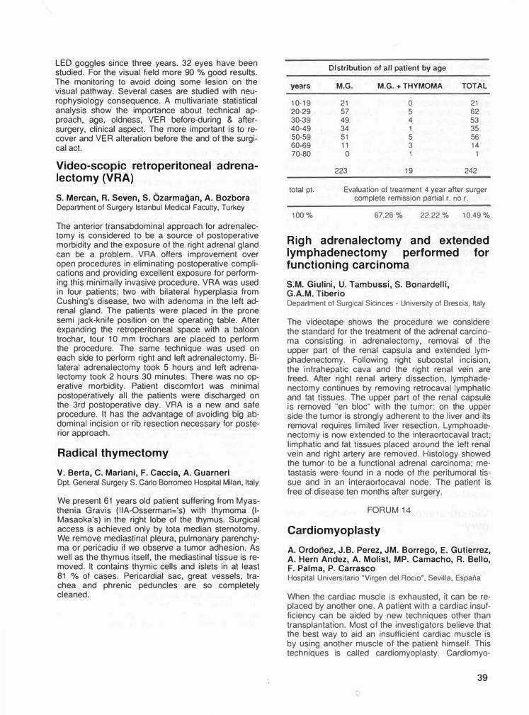
LED goggles since three years. 32 eyes have been studied. For the visual field more 90 % good results. The monitoring to avoid doing some lesion on the visual pathway. Several cases are studied with neurophysiology consequence. A multivariate statistical analysis show the importance about technical approach, age, oldness, VER before-during & aftersurgery, clinical aspect. The more important is to recover and VER alteration before the and of the surgical act.
Video-scopic retroperitoneal adrenalectomy (VRA)
S. Mercan, R. Seven, S. Özarmağan, A. BozboraDepartment of Surgery lstanbul Medical Faculty, Turkey
The anterior transabdominal approach tor adrenalectomy is considered to be a source of postoperative morbidity and the exposure of the right adrenal gland can be a problem. VRA offers improvement over open procedures in eliminating postoperative complications and providing excellent exposure tor performing this minimally invasive procedure. VRA was used in four patients; two with bilateral hyperplasia from Cushing's disease, two with adenoma in the left adrenal gland. The patients were placed in the prone semi jack-knife position on the operating table. After expanding the retroperitoneal space with a baloon trochar, tour 1 O mm trochars are placed to perform the procedure. The same technique was used on each side to perform right and left adrenalectomy. Bilateral adrenalectomy took 5 hours and left adrenalectomy took 2 hours 30 minutes. There was no operative morbidity. Patient discomfort was minimal postoperatively all the patients were discharged on the 3rd postoperative day. VRA is a new and safe procedure. it has the advantage of avoiding big abdominal incision or rib resection necessary tor posterior approach.
Radical thymectomy
V. Berta, C. Mariani, F. Caccia, A. GuarneriOpt. General Surgery S. Carfo Borromeo Hospital Milan, ltaly
We present 61 years old patient suffering from Myasthenia Gravis (IIA-Osserman='s) with thymoma (1-Masaoka's) in the right lobe of the thymus. Surgical aocess is achieved only by tota median sternotomy. We remove mediastinal pleura, pulmonary parenchyma or pericadiu if we observe a tumor adhesion. As well as the thymus itself, the mediastinal tissue is removed. it contains thymic cells and islets in at least 81 % of cases. Pericardial sac, great vessels, trachea and phrenic peduncles are so completely cleaned.
Dlstrlbution of all patient by age
years M.G. M.G. + THYMOMA TOTAL
10-19 21 o 21 20-29 57 5 62 30-39 49 4 53 40-49 34 1 35 50-59 51 5 56 60-69 11 3 14 70-80 o 1 1
223 19 242
total pt. Evaluation of treaıment 4 year aher surger complete remission partial r. no r.
100% 67.28 % 22.22% 10.49 %
Righ adrenalectomy and extended lymphadenectomy performed for functioning carcinoma
S.M. Giulini, U. Tambussi, S. Bonardelli,G.A.M. TiberioOepartment of Surgical Sicinces - University of Brescia, ltaly
The videotape shows the procedure we considere the standard tor the treatment of the adrenal carcinoma consisting in adrenalectomy, removal of the upper part of the renal capsula and extended lymphadenectomy. Following right subcostal incision, the infrahepatic cava and the right renal vein are freed. After right renal artery dissection, lymphadenectomy continues by removing retrocaval lymphatic and tat tissues. The upper part of the renal capsule is removed "en bloc" with the tumor: on the upper side the tumor is strongly adherent to the liver and its removal requires limited liver resection. Lymphoadenectomy is now extended to the interaortocaval tract; limphatic and fat tissues placed around the left renal vein and right artery are removed. Histology showed the tumor to be a functional adrenal carcinoma; metastasis were tound in a node of the peritumoral tissue and in an interaortocaval node. The patient is free of disease ten months after surgery.
FORUM 14
Cardiomyoplasty
A. Ordoıiez, J.B. Perez, JM. Borrego, E. Gutierrez,A. Hern Andez, A. Molist, MP. Camacho, R. Bello,F. Palma, P. CarrascoHospital Universitario "Virgen del Rocio", Sevilla, Espana
When the cardiac muscle is exhausted, it can be replaced by another one. A patient with a cardiac insufficiency can be aided by new techniques other than transplantation. Most of the investigators believe that the best way to ald an insutticient cardiac muscle is by using another muscle of the patient himself. This techniques is called cardiomyoplasty. Cardiomyo-
39
plasty is the technique by which a transformed skeletal muscle is moved into the thorax to wrapp the heart and help it in its work. The search of a muscle able to aid the diseased heart has been a topic of discussion for years. Our research group has as its main goal the development of complex techniques of training of the skeletal muscle to transform to trans· form in into a tireless muscle, resistant to fatigue and functionally similar to the myocardium. The transformation of the skeletal muscle into a cardiac muscle is feasible thanks to an electrostimulation protocol. The surgical technique begings with the dissection of the muscle. We use the latissimus dorsi muscle due to its proximity to the heart. Cardiomyoplastia is a surgical technique with an important future in the management of the patient with advance cardiac impairment. Besides the surgical methodology, we present an intracellular technique that allows the de· tection of the optimal time of transformation of a trainned squeltal muscle to function without getting tired like the cardiac muscle. Due to the few donors and to the severe selection criteria to be a candidate tor a cardiac transplantation, the cardiomyoplasty of· fers a valid surgical alternative and a hope of life to a lot of patients with endstage cardiac disease.
Extraperitoneal endoscopic lumbar sympathectomy
P. Hourlay, B. Topal, G. Vangertruyden,F. Trimpeneers, J. HendriickxSalvator Hospital, Hasselt-Belgium
The objective is to evaluate a technique which combines the advantages of minimal invasive surgery with those of the extraperitoneal approach. in the period between May 1993 up to September 1993, 8 pa· tients presented with causalgia of the left leg due to a previous trauma. AII these patients underwent an extraperitoneal endoscopic lumbar sympathectomy on the left side. By improving the technique, the operative time decreased from 21 O minutes to less than 40 min. The technique was feasable and effective in all cases. No complications occured. This technique seems to be successful for lumbar sympathectomy as well as for other retroperitoneal procedures.
Videothoracoscopy in cardiac surgery
A. Ordonez, J.B. Perez, J. Borrego, E. Gutierrez,A. Hernandez, A. Molist, MP. Camacho, A. Bravo,R. BelloHospital Universitario "Virgen del Ricio", Sevilla, Espana
There is some pericardiac disease that may be to handle through Thoracocscopic without thoracotomy. The more important is "The pericardial window" in chronic effusive pericarditis. Our research group has as its main goal the development of this technique tor training of the surgeon team. This video-film show the surgical technique of a ppleuropericardial window
40
and a pericardial biopsy by trough videothroracoscopic. The Videothoracoscopic is a surgical tech· nique with an important future in the management of the patient with cronic pericardial effusion and / or need of the pericardial biopsy.
Videothoracoscopic sympatectomy: A method of choice
Y. Yörük, R. Mehmet, S. KöseTrakya Tıp Fakültesi, Edirne, Turkey
Eleven videothoracoscopic thoracic sympatectomies were performed in eight patients. The indication for surgery was neurovascular disorders in 3, Raynaud disease in 2 patients and hyperhydrosis in one with bilateral reguirement, Burgery disease in 2. Median duration of operation was 35 min. (range 25-60). T2, T3, T4 ganglia were excided in operatios. There was no operative colication necessitating open thoracoto· my. No postoperative complication was observed. Five, patients were discharged in the second day and the others were in third. We think that videotho· racoscopic sympatectomy must be the method of obaice because of better visualisation and magnification with very less morbidity according to open surgery.
Thoracoscopic upperthoracic sympathectomy for 23 palmar hyperhidrosis - our experience
1. Levy, A. Ariche, G. Sebbag, J. KhoddaBen Gurion University, Soroka Hospital, Beer Shave, İsrael
Palmar Hyperhidrosis is excessive sweating beyond physiological needs in the palm without recognized etiology. Though benign affection, it is anoying to most patients. Currently the best treatment tor this condition is upper thoracic sympathectomy via many different approaches. Among those, the videothoracoscopic approach has been recomanded as a minimally invasive procedure. We repport our one year experience with transaxillary endoscopic sympathectomy in 99 patients with palmar hyperhidrosis. The standart video-laparoscopy was used in a transaxillary approach to perform the sympathectomy the mean operative time of this intervention was 12 min and mean hospital stay was 32 hours. The results in term of warm and dry hands were excellent. Only one case of transitory horner syndrome was noted. Transaxillary thoracoscopc sympahectomy tor pal· mar hyperhidrosis is relatively simple and ettective procedure wich necessitate standart laparoscopic instruments. The adventages are, short recovery time and hospitalisation along with excellent functionnal and cosmetic results. We are convinced that thoracoscopic sympathectomy is the procedure of choice tor the treament of palmar hyperhidrosis.
Superior-septal approach to the mitral valve
C. Alhan, M. İdiz, E, Demiray, i. Kayacıoğlu,M. Ateş, S. Tarcanısıanbul Cardiovascular and Thoracic Surgery Cenıer, Turl<ey
Superior-septal approach to the mitral valve has been used in 11 patients. AII of these cases underwent mitral valve replacement (MVR) and De Vega annuloplasty (OVA) procedure. it two of these patients an additional aortic valve replacement was carried on and one patient required left atrial thrombectomy. Overall mean aortic cross clamp time was 62.7±7.4 min., being 52±2.8 min. tor isolated MVR+DVA procedures. Mortality rate was O% and there were no bleeding complications. 11 patients with atrial fibrillation preoperatively had atrial fibrilla· tion postoperatively, except one who still is with NSR, 2 moths after operation. Superior-septal approach is an effective alternative way in reoperations, in patients wih small left atrium and it procedures requiring concomittent right atriotomy. Moreover, if further investigations prove this technique to be safe in respect to atrial disrhythmias, it may be used routinely in mitral valve operations.
Biliary lithiasis, AAA and renal artery stenosis: Endoscopic/surgical combined treatment
G. Battistl, lu Giacomelli, G. Viceconte,A. Brescia, F. Stio, A. Michelettl, M. De Vita,L. Leone111 Dept. of Surgery, University "La Sapienza·. Rome, ııaly
The Authors report a case of an aneurism of the abdominal aorta associated with biliary tract lithiasis in a gastroresected patient. The need to simultaneously treat both the diseases is stressed. An technique which involves endoscopy during open surgery in order to prevent contamination is illustrated. The technique used together with other manoeuvres are part of a methodology which aims to broaden the indications to the treatment of abdominal diseases associated with vascular diseases, limiting the risk of infection of the prosthesis.
Eversion carotid endoarterectomy and carotid to carotid crossover bypass graft for occlusion of the innominate artery and severe stenosis of the left carotid axis
S.M. Giulini, L. Cangiotti, S. Bonardelll, F. Benzi,G. Pulcini, L. Taglietti, M. Bertoli •,G. Tomasoni ••, G. De Maria •••Departmanı of General Surgery • University of Brescia, ltaly • Chair of Radiology • Universiıy of Brescia, ltaty•• Departmanı of Anesthesiology and tnıensive Cara UnitUniversity of Brescia, ltaly00 Service of Neurophysiopathology Spedali Civili Di Brescia, ltaly
A 69 years old male patient presented occlusion of the innominate artery with right vertebral to subclavian and common carotid artery steal syndrome; severe stenosis of the left carotid arteries; unstable angina pectoris. Therefore, it became necessary to correct the epiaortic lesions before the patient underwent aortocoronary by-pass owing to the risk of acute thrombosis of cerebral vessels during E.C.C. and also to improve the perfusion of the right internal mammary artery which could be used tor myocardial revascularization. The operation begins with the bilateral exposure of the carotid axis through a left longitudinal and a low transverse cervical incision. On the left, an external carotid shunting is apposed between two longutudinal arteriotomies, the proximal one on the common carotid artery and the distal one on the carotid bifurcation. The section of the common carotid artery allows the eversion endoarterectomy of the whole vessel, white an usual internal carotid endoarterectomy is completed through the distal arteriotomy. The proximal longitudinal common carotid arteriotomy is utilized to perform the donor end-to-side anastomosis of a carotid-to-carotid cross over PTFE by-pass grafting. The previously sectioned left common carotid artery is reconstructed with an end-to-end interrupted 6-0 suture and the internal carotid arteriotomy is closed with a continuous 6-0 suture while the shunt is divided and removed.The graft is clamped at its origin. The flow in the feftcarotid axis is restored. The graft is placed in a tunnerl obtained with finger dissection below the sternocleidomastoid muscle and beneath the thyroid muscles, and ttıe recipient end-to-side anastomosisbetween the graft and the right common carotid artery is performed. Finally the flow is restored in the right carotid arteries. lntraoperative Doppler spectralanalysis and postoperative angiographic controlsshow the success of the operation, with a bilateralnormal perfusion.
41
FORUM 15
Surgical aspects of abdominal tuberculosis (15 cases)
F. Oerbel, J. Sakhrl, Y. Dahmane, A. Ben Ali,R. Ben Amara, M. Ben Yahia, ı. Hachicha,R. Letaief, M. Kechida, S. El A)mi,R.B. Hadj HamidaService de chirurgie digestive, Sahloul, Tunisia
The tuberculosis in abdominal localisation is very rare, is treatment is medical once the diagnosis is established. From 1983 to 1993, 15 patients with a mean age 32 (7-64 years} were operated tor acute peritonitis (5), intestinal obstruction (3), and tor abdominal mass (7). in 5 cases, the peritonitis was due to intestinal perforation and in one case to a mesenteric lymph node perforation. in the other cases the tuberculosis was peritoneal (9) and hepatic (2). The operation consisted in intestinal resection in 3 cases and multiple biopsies in ali the cases. The treatment of abdominal tuberculosis is mainly medical, however some patients are first seen by surgeons tor complication such a perforation of the bowel.
Laparoscopic treatment of perforated gastric ulcer
G.B. Cadiere, J. Bruyns, R. Verroken, J. Himpens, A. RajanDepartment ol G.I. Surgery, Hôpital Universltalre SaintPierre. 322 rue Haute, 100 Brussels, Belgium
The records of the last fourty consecutive patients with perforated gastroduodenal ulcer disease treated at our department were reviewed. Twelve patients had a history of peptic disease. Six patients had had previous abdominal surgery. lnitial treatment was conseıvative in one patient, by lmaparotomy in three patients and laparoscopic in the remaining thirty six patients. The conseıvatively treated patients had a big ulcer at the lesser cuıvature. lmmediate laparotmy was performed oce because of septic shock due to a posteriorly localised ulcer and twice because of surgeon's inexperience. Thirty six patients treated laparoscopically were so immediately in 31 and after peritoneal lavage via a Veress needle in 5 cases. The course of the laparoscopic inteıvention was uneventful in 33 procedures. in 19 patients simple ulcer closure was performed; in 14 an asociated denervation procedure was performed. in three cases conversion to laparotomy was necessary: twice because of an ulcer located at the posterior site of the gastroduodenal junction and once because of a giant ulcer at the anterior site of the bulbus. Postoperative morbidity consisted of a Douglas abcess in one patient, a bronchopneumonia in one and a prolonged ileus in one patint. There was one enterocutaneous fistula. There was no other abdominal wall morbidity. Mean postoperative hospital stay was 7 days. Conclusion: Laparoscopic closure of perforated gastroduodenal ulcers is feasable, even it associated at an eventual
42
denervation procedure. Experience in advanced laparoscopic procedures is necessary and conversio advocated in cases of posteriorly localised ulcers. lnfluence on patient discomfort is less obvious then lor other laparoscopic procedures because of the emergency character of the inteıvention.
Diagnostic laparoscopy
H. Taouik, P. Papadothomakos, J. Taslopoulos,T. Segditsas, A. Bersis, G. Katsaros, K. GalliAthens, Greece 1 st. Surgical Clinic "Hygenia" Hospital
Since 1990 sixty cases were diagnosed in our clinic laparoscopicaly. Of these sixty, 18 were males and 42 females in which the main reason lor laparoscopy was to exclude gynaecological pathology. During diagnostic laparoscopy, il surgery was indicated, this was carried out through the laparoscope. Such surgery included, appendicectomy division of adhensions, ovarian eysi and uterine polyp removat, lymph node and liver biopsy. During diagnostic laparoscopy when indicated open laparotomy was carried out. in 6 such cases 4 were tor ovarian and 1 adenocarcinoma of the coecum. The advantages of the method are: 1. Short stay in the hospital (6·36 hours). 2. Early return to social activity), 3. Excellent cosmetic results
The laparoscopic second look
H. Kais, S. Slutzki, Z. Halpern, M. Negrl, A. HavelyDepartmanı of Surgery ·s·, Assal Harofeh Medical Center,Afliliated to Sackler Faculty ol Medicine, Tel AvivUniversity, lsrael
A second look laparotomy after 12-72 hours is the safest and most accepted procedure after bowel resection tor ischemic bowel disease. This defines the integrity of the bowel anastomosis, any extension of vascular thrombosis, and the presence of more segments of ischemic bowel. in order to avoid a second operation, we used laparoscopic minimal invasive techniques in three patients. One with acute arterial embolism, and two with acute venous thrombosis. A laparoscopic trachar as inserted, covered with a sterile glove, and left in the right lower quadrant of the abdomen following the resection of ischemic bowel. Under general anesthesia, 24 to 72 hours after the primary operation, the abdomen was insufflated with C02 gas via the trochar valve and the remaining bowel, and anastomotic integrity assessed by laparoscopy. Using this technique, a second laparotmy may be avoided.
Comparison of laparoscopic conventional cholecystectomy treatment of acute cholecystitis
F. Atalay, V. Kırımlıoğlu, S. Bostanoğlu,A. Keskin, H. GündoğduAdvanced Specialist Hospital, Gi-Surgey Clinic,Turkey
and for
Ankara,
in the ıast 3 years the records of the 150 patients undergoing cholecystectomy (75 laparoscopic, 75 conventional) tor acute cholecystitis were evaluated in the Gastrointestinal Tract Surgery Clinic of the Advanced Specialist Hospital, Ankara. These two groups are compared with respect to their peroperative postoperative early and late morbidity and mortality. The results are evaluated accordng to student's t test. There is not any statistical significant differences between two groups with respect to per operative morbidity and mortality {p>0.05). Post operative early and late morbidity differs in both groups which is higher in the conventional chotecystectomy group (p<0.05). As a result, in the hands of experienced surgeons there is not any statisticaly significant difference with respect to perop. morbidity between open and laparoscopic cholecystectomy groups. There is a statisticaly signifıcant differences in favour of open cholecystectomy group with respect to postoperative early and late morbidity and mortality and hospital stay.
Laparoscopic primary suture and omental patching in peptic ulcer perforation
S. Gürer, M. Gürel, A. Demirkıranlnönü Ünuversity Faculty of Medicine Department ofGeneral Surgery, Malatya. Turkey
Diagnostic or therapeutic laparoscopic interventions are being widely used in acute abdomen cases. We present a case in which laparoscopic primary suture and omental patching was performed due to a periorated peptic ulcer. An 84 year old female patient who had complaints of peptic ulcer disease tor 15 years we admitted to our hospital with a history of vomiting, severe abdominal pain and distention lasting tor 3 days. On her initial physical examination, abdominal tenderness and distention was found. Her plain abdominal films revealed no pathological finding. On her endoscopic examination, a perforated ulcer on the anterior wall ot duodenal bulb was found. · Plain abdominal film taken after endoscopic examination revealed fire air in the subdiaphragmatic space. With these findings an operation was planned. A 1 O mm trocar from umbilicus and three additional 5 mm trocars were inserted. On the exploration of a 3 mm perforation on the anterior wall of duodenal bulb, about 1 1. of gastric content within the abdominal cavity and peritonitis were found. After the primary closure of the perforation with three 2/0· silk sutures, an omental patch was tled on the suture line. The ab-
dominal cavity was than irrigated with % 0.9 NaCI solution. No complications occured after the operation. Her gastrointestinal modility was normal on the second postoperative day and oral intake was started.
FORUM 16
Laparoscopic atypic gastrectomy far leiomyoma
J. MacielR. Antonio Candido, 320-20 4200 Porto. Portugal
We present a video from a 72 years otd man who entered in the emergency room of St. Jonh's Hospital of the University ot Oporto-Portugal in September 1992 with hematemeses and melena. At the entrance, the hemogram showed a 6.8 gril hemoglobin and the endoscopy revealed an utcerated and bleeding pseudopolypoid mass of the anterior face of the stomach. The patient had been controled only with medical treatment. Ten days after, an endoscopy and ecoendoscopy were performed and some biopsys were laken. The endoscopy showed no differences except that there was no hemorrhage. in the endoultrassonography we could see a well delimited tumor, 6 cm diameter, at the muscular layer of the gastric wall. Histologically the biopsys were compatible with leyomioma and no malignant signs were detected. A la· paroscopic atypic gastric ressection was intended and successefuly performed using the endo-GIA devices. The procedure took about 45 min and the patient had recovered well and went home on the 50 postoperative day. Histologically the specimen had confirmed a leyomioma without malignity. On the follow-up after one and three months, and at the year the patient was assymptomatic. The endoscopy at the three months showed no tumor recurrence.
Lymphadenectomy in gastric cancer
J.M. Mendes de Almedialnstituto Portugues de Oncologia de Francisco Gentil Clinica Oncologica 111. Rua Prof. Uma Basto. Usbon, Portugal
Since 1980 we proposed that every patients operated with a curative intent would have a standardized lymphadenectomy associated to a sub-total or total gastrectomy. From January 1980 to December 1991, we operated 295 patients with a gastric carcinoma. in 166 cases (56.3 %) surgery was performed with curative intent. in 93 patients (56 %) a sub-total gastrectomy was performed and in 73 cases (44 %) a total gastrectomy. in all the cases an "R-2" type lymphadenectomy was used. Global morbidity and mortality rates of these two operations were statistically different. Global 5 year survival estimate for the whole series is 61.3 %. Univariate and multivariate analysis according to T-N (TNM classification), the humber of positive nodes resected. and the relation positive/resected nodes, reveled statistically different outcomes. This kind of quantitative classification allowed identification of high risk groups irrespective of
43
site of nodal involvement. The video shows a total gastrectomy with lymphadenectomy in a fat man, demonstrative of the difficulties in this cases.
Abdominal surgical approaches in carcinoma of the cardia
J.M. Mendes de Almeidalnstituto Portugues de Oncologia de Francisco Gentil ClinicaOncologica 111. Rua Prof. Uma Basta, 1093 Usbon. Portugal
The surgical approaches and the limits of the resection on the esophagus and the stomach, are a matter of continuous discussion. Another point of discussion is the extension of the lympadenectomy. On the video we can see a patient with an adenocarcinoma of the cardia, type 111, on Siewert classification, submited to a sub-total gastrectomy, 30 years before. The surgical approach is a median laparotomy with resection of the xiphoid and linear incision of the diaphragm. The lymphadenectomy include the fields 1, 2 and part of the 3, in the Japonese classification. The dissection is in bloc, on the retroperitoneal space from the coeliac tronc just to the mediastrium and than in the mediastinum just to the carina, including all the pre-aortic tissues. The reconstruction is done by esophago-jejunostomy Roux on Y with long loop and with stappler.
Adjustable silicone gastric banding by laparoscopic approach
M. Morino, M. Toppino, C. Garrone, F. Morinolstituto di Clinica Chirurgica Generale, Torino, ltaly
The adjustable silicone gastric band for morbid obesity consists of an inflatable baloon jointed to a self sealing reservoire by a thin tubing, allowing an accurate intraoperative calibration and an easy postoperative correction. Subsequently we choose to apply the same device by laparoscopic approach. The surgical procedure is carried out at a 14 mmHg C02 pressure. Five trocars, 2 of 5 mm diameter, 2 of 1 O mm and 1 of 20 mm, are introduced into the upper abdomen. After retrogastric dissection using a Roticular Endograsper (USSC), the band, filled with 0.8 mi of saline, is introduced through the 20 mm trocar. The inflatable band is then positioned around the stomach and finally calibrated. The procedure is completed by subfascial positioning of the reservoir connected to the band cetheter. Postoperative course is uneventful and characterised by the various advantages of laparoscopic surgery: minimum pain and respiratory distress, immediate mobilisation and prompt recovery.
44
Laparoscopic continent gastrostomy
C. Georgeac, J.P. Arnaud, C. Casa, A. ManuntaCHU Angers - Chirurgie Visceraıe - Angers 19033, France
lntroduction: A simple laparoscopic technique is described to create a tube gastrostomy obviating the need for open surgery in the patients with complete oesophagus obstruction. Operating: The patients lies supine on the operating table with his legs apart ... 2 to 3 liters of carbondioxide are insufflated into the peritoneal cavity ... 4 trocars are inserted: - a 1 O mm trocar is introduced in the umbilical region for insertion of le laparoscope - 2 other 1 O mm trocars are inserted in the right and left upper quadrants for the passage of endoscopic 8abcock forceps. - a 12 mm trocar is introduced 4 cm to the right of the umbilicus to allow insertion of an endo-GIA 60 stapler. A fold of gastric wass is elevated with the two 8abcock forceps and a gastric tube is faschioned with the endo-GIA 60 stapler. The pneumoperitoneum is evacuated and the gastric quadrant trocar. The exteriorised end of the tube is opened and sutured to the skin of the abdominal wall. An inwelling catheter is inserted into the stoma. Results: 5 patients have been operated with this technique. The postoperative period has been uneventful in all cases and all patients could be discharged from the surgical ward by the third post-operative day. Conclusion: in advanced lesions which do not allow the passage of the endoscope, a laparoscopic tube gastrostomy can be performed avoiding open surgery in these debilitated patients.
Gastroesophageal diversion after gastric resection
A. Gaetini, M. De Simone, M. Giaccone, R. lulianiDepartmenı of Surgery and Patology University of Turin,ltaly
The problem of alkaline gastroesophageal reflux after gastric surgery {81, 82, Gastroenteroanastomosis, Piloraplasty, Cardiomiotomy) is presented on videotape. The gastroesophageal diversion technique is showed begilming from 8illroth 2 gastric resection. The method in an interesting variation or rather it is a modification of classic method of duodenal diversion with the Y ansa of Roux. The operation consists in transmposing the jaws of Y in such away as to put in derivation or diversion the esophagus or stomach as to the main axis of bowel which drains the biliopancreatic secretions. As to the classic diversion, you have to performe one more jejunojejunal anastomosis, but you obtain the not indifferent benefit of avoiding any biliopancreatic reflux on stomach and duodenum and benefit avoiding Roux ansa syndrome because the ansa which drains the stomach is anastomosed in end to side direction and it opposes the refluxreascended. it is possible to use an autonomus ansa of only 35-40 cm rather than 70 cm as weluas it occurs with classic diversion.
FORUM 17
Use of the urologic resectoscope in colorectal obstruction and palliation in poor risk patients
C. Georgeac, J.P. ArnaudCHU Angers - Chirurgie Viscerale - Angeers 49033 France
Palliative treatment tor carcinoma of the rectum was not rare. Reasons to defer surgical treatment are the presence of metastatic disease, refusal to accept colostomy or unacceptable medicaf risk. Traditional methods of palliation include proximal diverting cofostomy, electrofulguration, and laser therapy. The use of a "rectum resector", similar to a cystoscope was reported. Preoperative preparation includes a thorough mechanical and antibiotic intestinal "prep" whenever possible. The patient is placed in the dorsal lithotomy position; the standard urologic resectoscope is then inserted into the rectum using the obturator. After the instrument is placed under vision through the constricting lesion, the resection is begun in much the same manner as a transuretheral resection of the prostate. The object of the resection is to debulk the tumor and restore a capacious lumen to the rectum. The tumor is resected flat to the adjacent normal rectal wall. Resection is carried out circumferentially around the lumen until all obstructing tumor is removed. The irrigant carries the chips and blood into the colon and allows excellent visualization without anal dilatation. A few chips are gathered tor pathologic examination. Using this technique, a colostomy was avoided in 15 patients no mortality or morbidity were observed. The average operative time was 35 minutes and the average hospital stay was 4 days. Palliation of constipation, bleeding and tenesmus has been good for periods ranging from three to six months. This procedure does not require anaesthesia, surgical assistants, new equipment or prolonged operative time. in selective instances, this transanal resection of rectal carcinomas offers excellent palliation.
Laparoscopic assited right hemicolectomy far malignant disease
M. Morino, C. Garrone, V. Festa, C. Migliettalstituto Di Clinica Chirurgica Generale, Torino, ltaly
The success of cholecystectomy has led to the development of other laparoscopic procedures, including colon resections. This video shows a laparoscopic assisted right hemicolectomy for a Duke's A adenocarcinoma of the ascending colon. Four trocars were located tor retraction, mobilization and transection of the bowel. lleocolic vessels were identified, ligated and resected. Terminal ileum right and transverse colon were taken out through an upper quadrant transverse 5 cm incision. The colon and the ileum were divided extracorporeally and a termino-
lateral mechanical anastomosis was performed. The bowel was reintegrated in the abdomen and mesocolon reconstructed. Postoperative course was uneventful and the patient dismissed on 8th postoperative day. We perform this procedure tor adenocarcinomas of right colon non protruding the serosa, tor cancers with metastatic irresectable diffusion and tor benign disease not endoscopically resectables.
Videoassisted reversal of Hartmann's proce dure without pneumoperitoneum
G. Navarra, A. Sortini, S. Occhionorelli,M, Santinl, A, Sartori, E, Pozza, 1, DoniniOepartment of Surgery - University of Ferrara - ltaly
More than 60 % ot patients, who underwent Hartmann procedure, refuse to undergo reversal. We suggest a minimally invasive approach that can offer some advantages: fess surgical trauma and postoperative pain, shorter postoperative ifeus and hospital stay. Reversal is carried out with the patient in LloydOavies position. After the colostomy and the descending colon mobilisation, a pursestring suture is performed and the anvil of the CEEA P is inserted in the proximal colon and temporaneously abandoned in the peritoneal cavity. The telescope is then inserted at the periumbelical site through a 1 O mm trocar tube. Laparoscopic devices are put into the peritoneum through the previous laparotomy in order to dissect the rectal stump, avoiding the need of pneumoperitoneum. The small bowel is mobilized out of the pelvis exposing the pouch ot Douglas. The CCEA P is inserted trans anally and advanced. The stapler pressure against the apex of the rectal stump makes easier to recognize the site of the previous linear stapling suture. A special endoknife, patented by the Authors, helps the CEEA P trocar to punch the anterior wall of the rectal stump. Stapler is assembled under direct vision and a end to side anastomosis pertormed. lntegrity of the anastomosis is checked by rectal instillation of dye. Four male patients, average age 64 yors, underwent laparoscopic assisted reversal of Hartmann's procedure. AII operations have been completed as planned in abouth 150'. Patient's mobilization was nearly immediate and post operative pain almost absent. Peristaltis began after 36-48 hours and spontaneous canalization three of four days after surgery. The drain was removed on day 4. We didn't observe mortality nor morbiditiy. The patients were discharged on day 6. We conclude that this new laparoscopic procedure may lead to shorter hospital stay and increased patients acceptance preserving the same safety of the traditional open procedure.
45
Laparoscopic sigmoid colon resection
M. Ertem, S. Ergüney, M. Özcan, A.E. KoçI.U. Cerrahpaşa Tıp Fak. Genel Cerrahi Anabilim Dalı,lstanbul, Turkey
Laparoscopic procedures are well accepted through out the world. Laparoscopic colon resection is a relatively recent technic. it is safe, �ersatile a.nd it �ffers a very smooth postoperative perıod . lts unıqu.e dı�adventage is its high cost and longer operatıve time which it takes. We present in this videotape an elective sigmoid colon resection we performed in a 45 years old man tor recurrent sigmoid colon volvulus. The operative time was 200 minutes. The colorectal anastomosis was made intraabdominally with an EEA 31 stapling device introduce per anus. The postoperative course was uneventful and the patient was discharged on the 4th postoperative day.
FORUM 18
The effect of ıncreased ıntraabdominal pressure on deep venous thrombosis
Ö. Günal, V. Yavuz, T. Biren, A.Ö. Aktan Marmara Univ . Medical Faculty Dept. of Surgery and Radiology, lstanbul, Turkey
The effect of increased intraabdominal pressure due to C02 insufflation during laparoscopic cholecystectomy (LC), on the lower extremity deep venous system was investigated prospectively. Thirteen patients undergoing open and 30 patients undergoing LC were evaluated. During LC the intraabdominal pressure was increased to 14 mmHg. With color doppler ultrasonography, thrombus and flow in the deep veins were investigated preoperatively, and postoperatively on the eighth hour, first and seventh days in both the open and LC patients. in eight patients undergoing LC and three patients undergoing LC and three patients undergoing open cholecystectomy centralv enous pressure (CVP) was monitored during the surgical procedure. With color doppler ultrasonography no thrombus or change in flow pattem was demonstrated in the deep veins postoperatively in neither the open or LC groups. The difference between the CVP values before and after C02 insufflation was not significant (p<0.05) in the LC patients. it is concluded that increased intraabdominal pressure during LC does not increase the risk of lower extremity deep venous thrombosis.
46
Does C02 pneumoperitoneum effect bacteraemia in experimental e. coli peritonitis?
H. Özgüç, A. Zorluoğlu, T. Yılmazlar, S. Gedikoğl�General Surgery and Microbiology Department of UludagUniversity Medical Faculty, Bursa, Turkey
T o perform pneumoperitoneum to the patterts with peritonitis is a question. Oiaphragmatic stomatas become patent when intraabdominal pressure increases. Theretore 14 mmHg C02 pneumoperitoneum may increase bacteriemia in peritonitis. We examined the effect of pneumoperitoneum on bacteriemia in animal model of peritonitis. 24 rabbits divided into three groups. 1 oa CFU (colony forming units) E.Coli obtanied from human infection at Uludağ University Medical Faculty Microbiology Departments Laboratory) injected intraperitonealy to group one (n:8). An identical bacterial innoculum injected lor two hours tollowed by midine laparotomy tor one hour to group two (n:8). An identical bacterial innoculum injected lor two hours tollowed by a 1 4 mmHg C02 pneumoperitoneum lor one hour. At all groups, growth values (GV) measured in from blood samples at the third an sixth hour with employed Bactec NR 730 system. This system is used tor the measurement of C02 levels producted by the bacterias. Third hour of innoculum mean GV measured 147.5 in group one. 150.5 in group two and 154.8 in group three. Sixth hour of innocutum GV measured 129.0 in group one, 107.2 in group two, 107.3 in group three. in comparison of ali groups, there was no significant difference at the results of growth values at third hour (t:0.227, t:0.081, t:0.127, p>0.05) and sixth hour (t:0.500, t:0.471, t:0.003, p>0.05) and sixth hour (t:-0.500, t:-0.471, t:-0.003 p>0.05). As a result; 14 mmHg C02 pneumoperitoneum does not increase bacteraemia at experimental E.Coli peritonitis in rabbits when compared with laparatomy and control groups 14 mmHg C02 pneumoperitoneum could be done safe· ly at peritonitis.
Effects of duration of C02 insufflation on blood gases during laparoscopic cholecystectomy
A.Ş. Taner, C. Atalay, A. Demir, B. Baydar, S. SarıDepartment of General Surgery, Ankara Hospital,Ankara-Turkey
Effects of duration of C02 insufflation on blood gases during laparoscopic cholecystectomy were evaluated in 20 patients. The patients were divided into 3 groups according to the duration of operation. Arterial pC02, p02, pH and HC03 were measured preop· eratively, intraoperatively and postoperatively. The results obtained in each group were compared with each other and with the control group and no significant difference was found.
A simple way for intracorporeal knotting during laparoscopy
Ş. Doğan, U. Sungurtekin, Ü. Şungurtekin Eşref Paşa Hosp, Ege Univ. Hosp., lzmir, Turkey
Using laparoscopy in advanced minimal invasive surgery has been necessitating the developments of new surgical technques. Even in experienced hands watching the screen and working on two dimension may cause problems during intracoproreal knotting there have been some problems that arised from int�aabdominal maneuvers during intracoproreal knottıng. IJ'!e descrıbed an easy way tor intraabdominal knotting. Apart from being easy, this technique has also some advantages like preventing gas leakage from the port, spending less suture material.
The changes in acid-base and blood gas values in laparatomy and laparoscopic ınterventions
O. Doğru, M. Karaayyaz, M. Doğanay, E. Göçmen,N.A. Kama 4th Surgical Clinic, Numune Hospital, Ankara, Turkey
Laparoscopic interventions are widely used today. �hether pneumoperitoneum made by C02 insufflatıon creates further problems, differing from laparato· my, has been subject of many studies. in this study open and laparoscopic interventions were compared rega�ding the ac(d-base balance and blood gas values ın preoperatıve, peroperative and postoperative periods, between January, 1993-July, 1993. Each of the groups consisted of 30 patients and in both the values were in normal limits in preoperative pe'riod. Th�re was atendency toward acute respiratory alka· l?sıs due to hyperventilation in both groups peroperatıvely but pH was never in abnormal limits and no statistically significant difference was observed (p>0.05). The only difference between two groups was observed in P02 values in peroperative and recovery period. P02 was significantly lower in lapar?SC?f>Y gro�p (p<0.05). There was no statistically sıgnıfıcant dıfference after postoperative 24 hours. f1:s a result, it can be said that laparoscopic interventıons that require C02 insufflation are as safe and reliable as laparatomy except tor pulmonary insufficiency cases in which postoperative hyposia can develop.
Physiological and metabolic responses of the organism to open and laparoscopic cholecystectomy
V. Kırımlıoğlu, M. Akoğlu, S. Ünver, N. Tatlıcan���Advanced Specialist Hosp. Ankara, Turkey
'.his was a prospective study performed on 25 patıents who underwent open cholecystectomy (OC)
an� laparoscopic cholecrsıectomy (LC). As physiologıca_l responces: �espıratory function tests (Vitalrapacıty, f?rced expıratuar volume in is and 3 speak flow) arterıel blood gasses and hemodynomic measurements (PAP, CUP, PCWP, CO, CI, MAP, HR, PAP) are evaluated. As metabolic reponces. Acutephase reactants (CRP, ESR, C3) hepatic function tesis (ALT, AST, TB, AP) are measured finaly the analganic requinement of LC an OC groups are evaluated. The statistical comparisons of both LC an OC groups are evaluated. As a result we found that: The d�torieration of the OC was statisticaly significant �>0.02. Same results are in found in respect of arterıel blood gasses, measured by Astrup tecniqne. Oth· er perameters of the physiological and metabolic rasposes didn't give any statistical significancek in the post op. period. The analganic requirement of the LC group differences was less than the OC group. which was statisticaly significant p<0.002.
Metabolic and endocrine changes after open and laparoscopic cholecystectomy
N. Akyürek, E. Sözüer, F. KuşErciyes Univ. Med. Fac. Kayseri, Turkey
Laparos�opic cholecystectomy, initially performed in France ın 1987, has become a popular operation, has many advantages. We have compared metabolic and endocrine changes after laparoscopic cholecystectomy (n:20), with those after open cholecystectomy (n:20). Venous blood samples were laken �efore operation, just after and 24h later postoperat�vely. Blood levels of glucose, cortisole, adrenocortıcotrophic hormone (ACTH) and beta endorphine were analysed. The durations of operation amount of hemocrahagie, mean hospital stay wer� significantly shorter in th� laparoscopic cholecystectomy group (p<� .. 05). Paın scores and analgesic usagewere sıgnıfıcantly decreased in the laparoscopic cholecystectomy group (p<0.05). However the results were not significantly different between the two groups (p>0.05), the metabolic and acute phase responses (such as hyperthermia, plasma cortisol, AC'.H and glucose levels) were less after laparoscopıc cholecystectomy compared with open cholecystectomy. Beta endorphine concentrations were less i� the open cholecystectomy group than laparoscopıc cholecystectomy group after surgery.
New standards in laparoscopic surgery
K. Yüce, M. L. PolatDörtyol State Hospital. Hatay. Turkey
Fifty o�e v!?eo-endoscopic operations were performe� ın Dortyoı State Hospital. Fifty of them were cholesıstectomıes and one of them was a primary suture tor duoden�I ulcer perforation. The patients were separated ın two groups: in the first group
47
(n:20) the operations were completed with 4 trocars in 16 patients and 3 trocars in 4 patients. lntraabdominal C02 pressure was 12-14 mmHg. The mean operation time was 75 minutes. Two of them were shifted to open laparatomy. in the second group (n:30) 4 patients had acute cholesistitis and the operations were completed with 3 trocars. Additional procedures (hernia repair, appendectomy) were performed in 2 patients. C02 pressure was 8 mmHg and mean operation time was 53 minutes. it was concluded that 3 trocars were enough and C02 pressure had to be 8 mmHg.
FORUM19
Laparoscopic truncular vagotomy and pyloromyotomy
F. Castro Sousa, L. Manso, A. Milheiro, E. Pinheiro, J. Aguiar3rd Surgical Department - Coimbra University HospitaJ3049 Coimbra, Portugal
The best method tor laparoscopic treatment of duodenal ulcer is still a matter of controversy. A 34 years old patient with a duodenal ulcer (associated with a bulb deformation) was treated by laparoscopic troncular vagotomy and piloromyotomy. A four trochars approach was used and no complications were detected in the post-operative period. A ninety percent reduction of the gastric secretion was observed three months after the operation. One and a halt years after the procedure the patient remains in the Visick grade 1.
Posterior truncal & anterior highly selective vagotomy
A. Alponat, A. Kaya, SS Ngoi, O. Özban, C. YavuzElmadağ State Hospital. Elmadağ-Ankara
The authors demonstrate a laparoscopic posterior truncal and anterior highly selective vagotomy. A 42 year-old man with intractable duodenal ulcer pain tor last three years despite the regular anti-ulcer therapy, was operated. Gastroduodenoscopic examination prior to operation showed active ulcer and deformation of duodenum due to chronic ulcer disease. A nasogastric tube and urinary catheter were placed before starting operation. lntra-abdominal pressure was maintained at 14 mmHg. Five trochars were inserted, first 3 cm above the umblicus, one on the left rectus muscle at the same level of first trocar, one just right to xyphoid, one on right and one on the left mid-axillary line. First, lesser sac was entered and once posterior vagal trunk was identified between right crus of diaphragma and postero-lateral surface of esophagus, it was dissected free from the other structures, clipped and cut. in the second step, "crow's toot", innervating antrum and pylorus, was identified and all neurovascular bundels above the heel of "crow's toot" entering the stomach on the lesser curvature side were dissected free, clipped
48
and cut. By this procedure, anterior Laterjet and its antral and pyloric branches (crow's foot) were preserved and rest of the stomach was denervated. Patient recovered very well and discharged on the tourth postoperative day. Now, a month later he has been symptom free and his active ulcer is healed on endoscpy.
Laparoscopic bilateral truncal vagotomy and gastrojejunostomy
A. Alponat, A. Kaya, N. Yavuz, F. Bulut, A. ÇangaElmadağ State Hospital, Elmadağ-Ankara
The authors demonstrate a laparoscopic bilateral truncal vagotomy and gastrojejunostomy. A 64 year old man with recurrent symptoms of gastric autlet obstruction due to chronic duodenal ulcer disease. was operated. Preoperative radiologic and gastroduodenoscopic investigations showed moderate obstruction of gastric autlet and deformation of bulbus of duodenum. Nasogastric aspiration and parenteral alimentation was started three days before operation. Five trocars were inserted. Flrst 3 cm above the umblicus tor laparoscope, one on the left rectus muscle at the same level of first trocar, one just right to xyphoid, one on the left and one on the right mid-axillary line. Firstly, the lesser sac was entered through pars flaccida over the caudate lobe of liver. Once the posterior and anterior vagal trunks were identified between the right diaphragmatic crus and posterolateral aspect o osephagus and between left diaphragmatic crus and anterolateral aspect of esophagus respectively, they were dissected free from the other structures, clipped and cut. in the second step, a jejunal loop 20 cm distal to Treitz was pulled up and hold together with anterior wall of the most dependent part of stomach by an endobabcock. Two small holes were created both on the jejunum and stomach to be able to insert the legs of endo-GIA. A 6 cm long stoma created by liring endo-GIA. Lateral opening was closed by endo-T A. The stomach was filled 300 mi saline with metilen blue to check any leakage. Postoperative period was uneventful and patient was discharged on the fifth postoperative day.
Laparoscopic posterior truncal anterior highly selective vagotomy
A. Sağlam, ö. Şakrak, Z. Çetinkaya, O. BanlıErciyes Üniversity, School of Medicine, Kayseri-Turkey
Proximal gastric vagotomy is a physiological operation tor peptic ulcer. Vagal denervation of parietal cells results in a decrease at a rate of 70-80 percent in basa! acid output, and 50-60 percent in maximal acid output. Since antrum and pylor was not denervated, any derangement in function of gastric emptying does not occur. Overall morbidity is lower than 0.03 percent. in experienced hands, recerrence rate of ulcer is less than 1 O percent. We had performed laparoscopic posterior truncal and anterior highly se-
lective vagotomy in a 44 years aged man who had incurable chronic duodenal ulcus, tor ten years. Under general anaesthesia, following peritoneal insufflation 5 trochars tor video-camera, retractor, babcock clamp, endograsper and dissector were inserted. Preserving the antral and pyloric vagal branches, only the fundus and corpus branches of anterior vagus was cut as tor PGV. Truncal vagotomy was performed tor posterior vagus. The patient was dischanged without any complication at postoperative sixth day.
Total truncal vagotomy by thoracoscopy
M. Aufschnaiter, F. KurzKH der Barmherzigen Schwester-Linz, Austria
More experience in thoracoscopic procedures lead to further technique like the thoracoscopic total truncal vagotomy as it will be presented in our video.
FORUM 20
Total esophagectomy for a rare giant tumor
N. Gürkan, Y. Onaran,1st. Tıp Fak. Genel Cerrahi Anabilim Dalı, Turkey
Here we reported a 72 year old female patient. Her coas progressive dysfagia tor solid tood since 3 months. Physical examination and laboratory tesis revealed no pathological finding. Barium meal graphy demonstrated extramural impression on the esophagus extending from cervical region to cardia. Computerized tomography revealed a mass originating from posterior wall of the esophagus at the cervival level. The size of the mass enlarged at lower levels and reached a diameter of 6 centimeters at the thoracal part of the esophagus. The tissue planes between the mass and the prevertebral fascia and atrium were not clear suggesting possible invasion to these structures. At the endoscopy, mucosa was intact but there was a submucosal mass extending from cervical esophagus to cardia. Preoperative diagnosis was leiomyosarcoma of the esophagus. She underwent right thoracotomy and total esophagectomy and faringoastrostomy. Histopathological examination revealed undifferentiated lymphoma.
Reconstruction of the cervical esophagus with free jejunal graft by AVCl's procedure
�ı
A_vcı, L. Avtan, S. Tezelman I.U. 1st. Tıp Fak. Genel Cerrahi Anabilim Dalı, istanbulTurkey
The reconstruction of the cervical esophagus and hypopharyax by using a jejunal autograft is one of the most popular method. The identical calibry, satisfac-
tory peristaltism and anatomic vascular properties of this segment are the reasons tor to choose of jejunal loop in the reconstruction of the cervical esophagus. The revascularization of the jejunal loop transplanted to the neck is an important point of the operation. The development of the microsgical techniques has a positive role. in general, two microvascular anastomosis tor graft revascularisation in sufficient. But since tree years, we sistematically use tour microvascular anastomosis (two arteries, two veines} tor reduces the risk of graft necrosis and promotes the healing of the anastomoses. in our service, we employed this technique on twenty two cervical esophageal and hypopharyngeal malignnancies cases with very satisifying results. This film shows our method conducted to a patient with cervical esophageal cancer.
Gastroepiploic pedicle flap for cervical oesophageal reconstruction
J. Patriclo, J. Ganho, J.L. Pereira, L. Silveira,A. Bernardes, F. Martinho, H. BaptistaDept. of Surgery il. Hospitais da Universidade de Coimbra,Coimbra, Portugal
Some types of tissues of digestive tract as stomach, jejunum, ileum and colon can be used lor hypopharynx and cervical oesophagus reconstruction, as lree grafts. These methods are experimented and fiables, but circumstances exist, as atherosclerosis or radic lesions, where revascularization becarnes difficult. Based on a cadaveric study we use a gastric tube from the greater curvature nurrished by the left gastroepiploic vessels and transposed to the neck as a pre sternal pedicle flap. The authors have no references registered from this technique in literature. under this technique one female and three male (aged 29, 72, 74 and 76 respectively} were treated. They showed cervical oesophageal stenosis tor tumoral relapse and radic lesion of the neck. Al! of them underwent laryngectomy and radical cervical lymphadenectomy 15 months ago lor adenoid cystic carcinoma in the first case; in the others left three, four years and 13 months ago for larynx adenocarcinoma. Ali of them were submitted to complementary radiotherapy. A surgical excision (palliative} of the tumour mass and the stenosed oesophagus was performed. The last one was replaced by gastric flap from the greater curvature with 7-3 cm mean lenght and 2 cm diameter with the correspondent omentum. This gastroepiploic flap was used as a pedicle one and bloodsupplied by the left gastroepiploic vessels. These were left in a left paramedian presternalk incision. in ali of them a safety suplemmentary distal venous anastomosis was performed. No per or post operative complications were observed chiefly partial neorosis flaps. They al! began with a well tolerated oral food on the 11th day and anti-H2 therapy. The above mentioned female and the second male died on the 4th and 5th months; the third patient died �n the 11 month respectively by relapse and tumoral
49
dissemination. With a follow-up of 13 months the lası patient is stili alive and swallows with no dysphagy. Considering the general condition of the patients and the palliative character of these surgeries, despite the residual anaesthetical deformation and the need of taking anti-H2 this technique presents its advantages: it is not dependent from the arterial anastomosis; it has a shorter operative time; it reveals itself easier to execute than free flaps. Despite this is a small series we conclude that this method is an effective and fiable one and is indicated for palliative surgery in cervical oesophageal reconstruction.
FORUM 21
Surgical treatment of perianorectal fistulae in Crohn's disease
A. Gaetini • M. DE Simone - M. GiaoconeDepartment of Surgery and Patology - University ofTurin, ltaly
The perianorectal fistulae which arise in Crohn's disease, represent an important therapeutic problem which receives little attention in the literature. The frequency of perianal fistulae is variable, ranging from 14 % in cases with disease affecting the ileon and 100 % when the rectum is involved. The aetiopathogenesis of the perianal fistulae in Crohn's disease is stil! under discussion, but we share Hughes opinion which differentiates the fistulae produced at the site of disease from those caused by the irritant action of the diarrhoea during the course of the disease. lndeed, the fact that the perianal manifestations of the disease may be the first sign to indicate a reactivation of Crohn's disease at intestinal level seems to have been confirmed. Nevertheless, no agreement yet exists in the literature as to the therapy indicated in the diverse situations created by perianal Crohn's disease. Due to these doubts, we find !hat no clinical classification of the perianal fistulae inCrohn's disease exists, which would serve as aguide to the choice of therapy. in this video we propose a clinical classification through which our surgical procedure is revelaed.
Surgical technical problems on colorectal polyposis
A. Gaetini, M. Glaccone, M. DE Simone, R. LulianiDepartmenı of Surgery and Patology - University of Turinltaly
The showed videotape deals with some surgical technical problems about coloproctectomy with ileus pouch-rectoanal anastomosis. Problems, !hat are peculiar to this operation can be resumed in the following five points: 1. The extension you have to do at the exeresis.2. The rescue of nerve and hypogastric superior andinferior plexus.3. The preparation of ileus pounch.4. The type of ileus pounch - anal anastomosis.
50
Some surgical sequences and some schematic drawing are presented to showing surgical problems and technical solutions that you can use in order to make easier the operation either in the case of colorectal polyposis or 180.
Endorectal approach of rectocele using the linear cutter stapler
L. Bresler, J.M. Tortuyaux, P.J. Courbey,P. BoisselChirurgie C, C.H.U. Nancy-Brabo is, 54511-Vandoeuvre,France
Rectocele presents as a protrusion of the anterior wall of the rectum into the vagina. in some cases, rectocele is associated with outlet obstruction. Surgical treatment is, in these cases mandatory. We describe here a new technique of endorectal repair of rectocele using a stapling device. The patient is placed in the jack knife position. Endorectal exposure is obtained with retractors and the rectocele is pulled in the cana! anal. Excision of the rectocele is then performed with 2 or 3 application of the linear cutter device. The suture line is then renforced with a 2/0 vicryl running stitch. This procedure mixes a transverse suture of the rectovaginal septum and a resection of the mucosal excess. Eight patients have been operated between 1988 and 1990 after a full proctological and physiological assessement. No severe postoperative complication occured. With a 3 year follow-up, 4 patients are symptome free, 3 are improved and 1 has no change in symptomes. Our experience with these 8 cases suggests that endo anal repair of rectocele using the linear cutter stapler is both a simple and effective mean of dealing with outlet obstruction when associated with a rectocele.
Laparoscopic appendectomy and adhesiolysis in the material of the Elisabeth Hosp
G. David, A. BodnarElisabeth Hosp. Dep. Surg. Budapest. Hungary
We like to interpretate on video our pract ic es on the field of laparoscopic appendectomy and adhaesiolysis, wich seem to be safe and applicable methods.
Laparoscopic appendectomy
O. Hamamcı, M. Berberoğlu, F. Ercan,K. Karayalçın, A. Korkmaz6.th Surgical Unit. Ankara Numune Hospital-Turkey
in this video, we are presenting a case of laparoscopic appendectomy who underwent drainage of a appendieular absces three months ago. Laparoscopic appendectomy can be salely performed even in the presence of adhesions with minimal patients discomfort.
FORUM 22
Reparation of postoperative abdominal wall hernia. Three types of use of prolene mesh
C.F. Escalante, A. lngelmo Setien, S. RevueltaUniversity Valdecilla Hospital. Laredo Hospital. Cantabria,Spain
A film with three reparations of incisional hernia is presented: 1. Unchtenstein's technique performed with previsceral mesh in medial incisional hernia.2. Modified Albanese's technique with pre-peritonealmesh edge dettect fixed.3. A double previsceral mesh which covers great deffect of abdominal wall, suturing two standard great size pieces.
Laparoscopic preperitoneal extraperitoneal prosthetic repair of inguinal hernias with balloon dissector
S. Revuelta-Alvarez, C.F. Escalante, F. Hemanz,A. GutierrezReci Cross Hospital, Torrelavega, University Valdecilla Hospital, Santander Cantabria-Spain
in recent years, operative laparoscopiy in· general surgery has emerged as a viable alternative to traditional open techniques. Because of the success reported by Stoppa and his colleagues with the preperitoneal prosthetic repair tor inguinal hernias, this technique has been adapted tor use laparoscopically. A large anatomic working space is created between the abdominal wall and peritoneum using the balloon disector which is expanded with saline solution. This space is then insuttlated and the propilene mesh can be positioned over the hernia defect while the peritoneum remains completely intact. The extraperitoneal approach avoids the risk of adhesions and complications associated with entry into the peritoneum. The use of balloon dissector into the preperitoneal space reduces operating time with a wider dissection.
Extraperitoneal endoscopic inguinal hernia repair
P. Hourlay, B. Topal, G. Vangertruyden,F. Trimpeneers, J. HendrickxSalvator Hospital, Hasselt, Belgium
The objective of the extraperitoneal endoscopic approach and inguinal hernia repair is to combine the well-known advantages of minimal invasive surgery with the proven results obtained by the Stoppa technique. in the period between October '92 up to March'94, a total of 301 patients (mean age: 56y) were treated tor hernia repair. AII patients were operated on by the extraperitoneal endoscopic technique,
irrespective of whether they had a direct, an indirect or a recurrent hernia. 220 patients underwent a bilateral repair. 138 patients presented previous surgery in the lower abdomen, including 55 recurrent inguinal hernias. 563 polypropylene meshes were placed preperitoneally. The mean operating time was 45 minutes tor unilateral and 57' tor bilateral repair. Special new instrumentation has been developed and is used by the authors. Up until March'94, no peroperative or major postoperative complications occured; no recurrence has been seen. in conclusion, this technique is safe. it combines the well-known advantages of the use of a mesh with those of minimal invasive surgery. Most important however is that the peritoneal cavity is not opened.
Laparoscopic hernia repair
E. M. Sözüer, N. Akyürek, Z. YılmazErciyes University, Medical Faculty Surgical Dept., Kayserı, Tuıi<ey
Between April 1993 and April 1994 1 O patients with inguinal hernias were operated on laparoscopically. There were 9 male and 1 female patients. Their overall mean age was 52 years, range 15 to 71: Th�rewere 6 indirect 3 direct and one recurrent dırect ınguinal hernias. This study has been performed to evaluate the salety and efficacy of laparoscopic hernia repair. Patients were excluded lor whom general anesthesia had a high risk or who had incarcereted or strangulated hernias. The average duration of operation was 70 minutes. Complication occured in one patient, but there were no recurrences during a m�dian follow-up 6 months (4-6 months). The medıan postoperative hospital stay was 2.5 days and lor return to normal activity 5 days. Although long-term follow-up has not yet available, laparoscopic inguinal hernia repair is an effective operation with low morbidity. Long-term follow-up is needed to determine the durability of the repair.
Extraperitoneal laparoscopic hernia repair
A. Korkmaz, M. Berberoğlu, O. Hamamcı,F. Ercan, K. Karayalçın6.lh Surgical Unit, Ankara Numune Hospital-Turkey
The transperitoneal approach tor laparoscopic hernia repair is expensive relatively invasive and requir�spneumoperitoneum. We performed 12 laparoscopıc hernia repairs (7 unilateral indirect, 2 bilateral direct, 1 bilateral indirect. 1 femoral, 1 recurrent direct) using. Preperitoneal Balloon Oistension System. Hernial sac was dissected and returned to the abdominal cavity or splitted by Zigg technique. And a 1 Ox20 cm Marlex mesh was placed over the defect without and sutures or staples. AII patients were discharged on the second postoperative day. Extraperitoneal apprach limits the C02 insulflation and protects the intraabdominal organs. Therefore, it is a sale, easy and rapid technique.
51
Laparoscopic inguinal hernia repair
M. Gürel, S. Gürer, M. Şarelnönü University Faculty of Medicine Departmanı of General Surgery, Malatya-Turkey
During one year period between February 1993 and February 1994 laparoscopic hernia repair was performed in 21 cases. There were 17 male and 4 female patients with an average age of 39 (20-65). The hernia type was indirect hernia in 16, direct hernia in 3 and recurrent hernia in 2 cases. Two of our patients had bilateral hernia tor a total of 23 hernia. We used transabdominal approach in 20 and extraperitoneal in one case. A large mesh was placed in 18 hernias, 3 were managed by internal ring closure and 2 hernia were repaired by a two-level repair; Dudai butterfly+mesh. Additional laparoscopic procedures were performed in 3 cases consisting of bilateral varicocele ligation, cholecystectomy and tubal sterilisation. Complications were seen in 5 cases; 2 hydroceles, 2 nerve traumas and 1 scrotal edema. We ha�e seen no r�currences during this short follow up perıod. We beheve that laparoscopic hernia repair should be done on an experimental basis in selected centers.
Gilbert's hernia repair (sutureless prosthetic repair)
M. De Simone, T. Lubrano, E.L. Murix,M. Glaccone, A. Gaetlnl, A.I. GllbertDepartmanı of Surgery and Palhology, University of Turin, ltaly
The Gilbert's technique tor hernia repair is a prosthetic Hernia repair technique. it is a Sutureless repair, in which no suture is used except for the one of the externatk obtique aponeurosis. The operation is performed under local anesthetic block of Lidocaine and Bupivicaine. After skin incision the external obtique aponeurosis is opened. The cremasteric fascia is opened and its medial flap excised. The internal spermatic fascia is also opened. The hernia sac is isolated. After reduction of the peritoneal sac a Prolenea plug !s insert� th:ough the internal ri�g. and the posterıor wall ıs reınforced with a second swatch of Prolenea Mesh. Both swatches of mesh are held i� places by the body's intemal hydrostatic torces. Beıng sutureless no tension is placed on any lay.er, no holes have done on muscles that are intact.T�ıs pro�edure has been used in 1152 inguinal hernıa repaırs. The results have been satisfactory: 3.6 % of complications and 0.26 % of recurrences.
52
FORUM 23
Laparoscopic vagotomy and antrectomy with a Billroth il anastomosis
F.N.Schutte Sunward Park Hospital, Johannesburg. South Africa
Laparoscopic surgeons have eagerly awaited the development and availability of the powered endo GIA 60 and powered TA 60 (United States Surgical Corporation, Connecticut) stapling devices. Recently, (1993) three laparoscopic vagotomy and antrectomies with a Billroth il anastomosis were performed tor the definitive treatment of refractory peptic ulcers. P�eop�rati�e assessrı:ent included gastroscopies wıth bıopsıes and barıum meal. No peri-operative mortalities or morbidities occured and all patients experienced minimal post operative discomfort. Normal fe�ing. resumed w.ith!n 2 days with patients finallybeıng dıscharged wıthın 4 days. Gastrographin swallo� "".as routinely p�rformed post operatively. As famıharıty and experıence with the instruments has grown, average operating time has been reduced to 85 minutes. The excellent results observed thus far have led . me to believe that the laparoscopic approach wıll become the procedure of choice ın the hands of experienced laparoscopic surgeons. As our experience in this field increases with the ongoing development of laparoscopic instruments, the future P:Ospects tor laparoscopic surgery are infinite. The vıdeo provides a detailed visual on the procedure.
Laparoscopic Billroth I a Billroth il operations
B. Ablassmaier, U. Stelnhilper, W.D. BandlT. Ziehen, H. FöckerspergerDepartement of Surgery, Munich-Pasing Hospital, Steınerweg 5, 81241 Munich, Germany
Lecture and Video-Presentation: Between October 1993 and February 1994, we performed three distal s�omach resection operations laparoscopically (twice Bıllroth II anastomosis, once Billroth I anastomosis combined with truncal vagotomie). The first patient, 83_years ?ld, presented a stenosing, bleeding, prepylorıc, mahgnant Non-Hodgkin Lymphoma. The second patient, 82 years old, reszented an ulcerated non-malignant Leiomyoblastoma of the antrum. Du� to the histological type of the tumors a radical lymphadenectomy was not performed. The third patient p�esented a penetrating, prepyloric ulcer combined wıth an almost complete stenosis of the pylorus. in all three cases, it was possible to follow the principles of conventional open surgery. The partially resected stomach (two thirds) was removed in a lap sac through a 3.5 cm intraumbilical incision. The anastom�ses were all stapled intracorporally, no intraabdomınal complications occured. However the first patient died on the 21th postoperative day
0trom
cardio-pulmonal failure, the second and third patient were discharged feeling well on 11 th/12th postoperative day.
Laparoscopic Billroth-11 gastrectomy
P. Goh, Y. Tekant, SS, Ngoi, CK, Kum, J. lsaacOept. of Surgery, NAtional Univ. Hosp., Singapore
Totally intraabdominal laparoscopic Billroth-11 gas· trectomy was pertormed on eight patients. Ali were male with a mean age of 56 years. lndication tor sur· gery was benign gastric ulcers not healing with medi· cation or complicated by bleeding in 7 cases and combined duodenal and gastric ulcer in one case. The procedure was performed through five ports. The distal two thirds of the greater curvature was mobilized using sharp dissection supplemented with cautery. Large vessels were secured with endoclips before being divided. The duodenum was transected with a single application of the Endo-GIA {Autosuture, USSC). Resection of the stomach and subsequent gastrojejunostomy was achieved with EndoGIA staplers. The stomach was removed by extending one of the ports to 2.5 cm. The mean duration of the procedure was 240 minutes {range 150 mins to 280 mins). One case was converted to open surgery because of technical difficulty. Postoperative recovery was rapid with minimal morbidity. The average hospital stay was six days {range four to twelve days). One case developed partial gastric outlet obstruction due to anastomotic edema which resolved with conservative treatment. Ali the other patients experienced a dramatic postoperative recovery with immediate mobilization and enteric feeding on the third postoperative day. Laparoscopic gastrectomy is a feasible operation for benign gastric ulcers. However, more data on its safety and more ergonomic instruments to facilitate the procedure are needed before it can be recommended as a method of choice.
Laparoscopic vagotomy and gastroenterostomy
S. Mercan, R. Seven, S. Özarmağan, D. BudakOepartment of Surgery. lstanbul Med. Faculty, Turl<ey
The aim of this presentation is to assess feasibility and safety of vagotomy and gastroenterostomy under laparoscopy.
The patient {70M) who had been suffering abdominal pain and nausea and been diagnosed to have peptic ulcer complicated with pyloric stenosis. Technically 5 ports were utilized with the camera placed at umbilical port. The left lobe of the liver was mobilized and gastro-esophageal jnuction was dissected. The anterior and posterior vagus was found and divided. A loop of the proximal jejunum 20-25 cm away from treitz of ligament was brought up antecolically to the anterior stomach. Two stay sutures were used to anchor the stomach to the jejunum at the site of the
proposed gastrojejunostomy. A side to side gastrojejunostomy was fashioned with an endo-GIA (6 cm). The defect in the stomach and jejunum was closed by intracorporal suturing with propilene 2/0. The operation lasted 3.5 hours and patients recovered uneventfully and were discharged on the 4th postoperative day.
Laparoscopic highly selective vagotomy and laparoscopic pyloroplasty
B. Ablassmaier, U. Steinhilper, W.D. Bandl,T. Ziehen, H. FöckerspergerDepartment of Surgery, Munich-Pasing Hospital, Steınerweg 5, 81 241 Munich, Germany
Lecture and Video-Presentation: From May 1993 we performed 13 highly selective Vagotomies laparoscopically. Ther selected tor this procedure presented a 2 to 21 year history of unsuccesful medical treatment {in 3 patients complicated by recurrent ul?erbleeding, in 2 patients by duodenal ulcer perforatıon; in one of these two patients the perforated ulcer was treated by suturing laparoscopically three month prior to the vagotomy). in one patient the vagotomy was combined with a laparoscopic pyloroplasty {Holle). in all cases, it was possible to follow exactly the principles of conventional open surgery for the anterior and posterior vagus-nerve.
FORUM 24
Prosthesic material in abdominal surgery
S. MoralesCallc Genova, 41010, Sevilla. Spain
There are some situations in abdominal surgery, in which the organic tissues do not ofter !he proper guarantees, and some other times in which we just want to increase the biological resistance of them. Several prosthesic materials have been used to solve these problems. in this film, we are going to expose some of these procedures, only based on our experience. We use prosthesic material in Gastrointestinal Surgery; to correct hiatal hernia by pexia with round ligament, when this one does not exist. To protect sutures to avoid dehiscences, using "Biological glue" or intraluminal material which will be released later, and in colonic surgery, by using blodegradable rings of polyglycolic acid. Surgery ot solid organs: to suture parenchyma or to protect them with hemostatic material, to use absorbable mesh in broken organs and to place capsulas of infusion to treaı tumors. Billio-Pancreatic surgery: to protec sutures and to place stents in billio-digestive anastomosis. Digestive vascular Surgery: prosthesis of PTFE in vascular derivations in bleeding Portal Hypertension, or intrahepatic prosthesis in this pathology. Surgery of the abdomi· nal wall; different prosthesis to treat incisional hernia.
53
Laparoscopic surgery; prosthesic material to treat inguinal hernia and also in incisional hernia. We make a sumary of the different application of prothesis in abdominal hernia.
The primary arthroscopy in injuries of the ankle joint
O. Wolf-RüdigerDep. for General Surgery, Traumatology and EndoscopicSurgery Sana-Krankenhaus, Krankenhaustrasse 42 50354
Hürth/Cologne Germany
The film shows the techniques of primary arthrosco
PY of the ankle joint the indication is the modern standart likewise in knee arthroscopy. lndication for arthroscopy is established on hemarthros, exclusion of accompanied lesions of osteochondral fractures and draduation of outerligament ruptues. The ultrasonic is an essential part in diagnostics and "held" radiograms are no longer necessary because the sprained ankle can be demonstrated on videomonitor. We perform the arthroscopy in local or general anaesthesia and prefer a 5 mm arthroscope with 300 telescope and usually without distractor. The findings and surgical interventions are filmed entirely tor documentation. 89 % of all patients presented a hemarthros with different forms of outerligament ruptures, 32 % showed additional osteochondral fractures and loosened flakes. 9 % out of this group had to be operated transarthroscopically or by arthrotomy by subluxation of the foot more than 200. The primary arthroscopy permit a direct and graduated operative or conventional treatment.
Functional endoscopic sinus surgery for the treatment of chronic sinusitis
K. Değer, N. Keleş, E. Yücellstanbul University, Faculty of Medicine, Dept. of ORL.,Turkey
Functional Endoscopic Sinus Surgery is based on the principle that, most infections of the larger sinuses are rhinogenic and are commonly caused by untreated anterior ethmoid disease. The relief of the obstruction caused by the anterior ethmoids, will provide adequate ventilation and drainage of the larger sinuses and will permi! the resolution of the disease in the maxillary and frontal sinuses. The combination of coronal plane computed tomography and nasal endoscopy has proved to be excellent tor
the evaluation of the lateral nasal wall. Here two cases; first being an ethmoidofrontal mucopyocel and the second being a chronic bacterial sinusitis treated by FESS are presented. Messerklinger technique that is an anterior to posterior approach and accepts the anterior ethmoids as the target area is used.
54
Laparoscopic management of peritoneal dialysis catheters
H. Kais, J. Welssgarten, A. HalevyDepartments of Surgery ·s· and Nephrology, Assal Harofehmedical Center, Affiliated to Sackler Faculty of Mediclne,Tel Aviv University, lsrael
Despite the increased use of the Tenckhoff catheter tor peritoneal dialysis and the standardization of surgical techniques, this device is still associated with a significant number of complications. The most frequent mechanical complication is outflow obstruction which necessitates catheter removaı or replacement. Conservative attempts at catheter salvage are usually unsuccessful when outflow obstruction occurs. in our series, we used laparoscopic techniques tor replacement or redirection and adhesiolysis of a nonfunctional catheter in 4 patients. A follow-up of 6 months showed functioning catheters in all patients. A few reports have described the use of laparoscopy in the management of Tenckhoff catheters in small groups of patients. We conclude that this technique is useful in managing patients with a nonfunctional Tenckhoff catheter.
Extraction of two surgical sponges with laparoscopic intervention (two cases)
F. Atalay, V. K.ı.rımlıoğlu, S. Bostanoğlu,A. Keskin, K. OcalAdvanced Specialist Hospital Gastrointestinal Tract SurgeryClinic
Laparoscopic surgical intervention became the choice of procedure tor most patients with gallbladder disease and tor the disease of the abdominal and thoracal organs since its first application in 1987.
Since March 1992, we have been performing laparoscopic interventions tor diagnosis and the treatment of the symptomatic gallbladder disease. Since that time 2 surgical retained foreign bodies (surical sponges) were diagnosed and operated upon successfuly. The first patient had cholecystectomy tor 2 years ago and now operated on because of cystic stump syndrome. The second patient had a celiotomy because of abdominal trauma and now operated on because of symptomatic gallbladdler disease. A foreign body (Surgical sponge) is tound too. This op. is recorded which will be shown.