ectopicmsx2overexpression inhibits andmsx2antisense stimulates calvarial osteoblast differentiation
TRANSCRIPT
A
poapdss
Developmental Biology 209, 298–307 (1999)Article ID dbio.1999.9258, available online at http://www.idealibrary.com on
Ectopic Msx2 Overexpression Inhibits andMsx2 Antisense Stimulates CalvarialOsteoblast Differentiation
Milan Dodig,*,1 Tade Tadic,*,1 Mark S. Kronenberg,*,1 Sanja Dacic,*,1
Yi-Hsin Liu,†,2 Rob Maxson,† David W. Rowe,*,1 andlexander C. Lichtler*,1,3
*Department of Pediatrics, University of Connecticut Health Center, Farmington,Connecticut 06030; and †Department of Biochemistry and Molecular Biology,Institute for Genetic Medicine, Kenneth R. Norris Cancer Hospitaland Institute, Los Angeles, California 90033
Msx2 is believed to play a role in regulating bone development, particularly in sutures of cranial bone. In this study weinvestigated the effects of retroviral-mediated overexpression of Msx2 mRNA, in both sense and antisense orientations, onprimary cultured chick calvarial osteoblasts. Unregulated overexpression of sense mRNA produced high levels of Msx2rotein throughout the culture period, preventing the expected fall as the cells differentiate. The continued high expressionf Msx2 prevented osteoblastic differentiation and mineralization of the extracellular matrix. In contrast, expression ofntisense Msx2 RNA decreased proliferation and accelerated differentiation. In other studies, we showed that the Msx2romoter was widely expressed during the proliferative phase of mouse calvarial osteoblast cultures but was preferentiallyownregulated in osteoblastic nodules. These results support a model in which Msx2 prevents differentiation andtimulates proliferation of cells at the extreme ends of the osteogenic fronts of the calvariae, facilitating expansion of thekull and closure of the suture. © 1999 Academic Press
Key Words: Msx2; osteoblast; differentiation; homeodomain; bone; craniosynostosis.
Mie1nsbvaRetmotef
INTRODUCTION
Proper development of the skull requires precise controlof proliferation and differentiation of osteoblast precursorsto balance the need to allow growth to accommodate theexpanding brain with the requirement for timely ossifica-tion. In these studies we have examined the role of thehomeodomain-containing protein Msx2 in this process.
1 Present address: Department of Genetics and DevelopmentalBiology, University of Connecticut Health Center, Farmington, CT06030.
2 Present address: Center for Craniofacial Molecular Biology,USC School of Dentistry, Los Angeles, CA.
3 To whom correspondence and reprint requests should be ad-dressed at the Department of Genetics and Developmental Biology,MC1515, University of Connecticut Health Center, 263 Farming-ton Avenue, Farmington, CT 06030. Fax: (860) 679-1047. E-mail:
298
sx2 is part of the Drosophila msh gene family, whichncludes vertebrate Msx1, 2, and 3. While Msx3 is primarilyxpressed in the central nervous system (Shimeld et al.,996; Wang et al., 1996), Msx1 and 2 are both expressed inumerous tissues at many stages of development (David-on, 1995). Several studies suggest that Msx2 plays a role inone development. MSX2 is expressed and regulated byitamin D3 in human osteoblasts (Hodgkinson et al., 1993)nd inhibits the cotransfected type I collagen promoter inOS 17/2.8 osteosarcoma cells (Dodig et al., 1996; Towlert al., 1994). Msx2 inhibits the osteocalcin promoter inransfected MC3T3E1 cells, but stimulates the same pro-oter in ROS 17/2.8 cells (Towler et al., 1994). Msx2 and
steocalcin are expressed in a reciprocal pattern duringooth development (Bidder et al., 1998). Msx2 is stronglyxpressed in cells at the extreme ends of the osteogenicronts of the calvarial sutures, and a gain-of-function muta-
ion of MSX2 causes Boston-type craniosynostosis (Jabs et0012-1606/99 $30.00Copyright © 1999 by Academic Press
All rights of reproduction in any form reserved.

F2dFuw
ac2
299Msx2 Inhibits Osteoblast Differentiation
al., 1993; Ma et al., 1996), a condition in which cranialsutures close prematurely. Overexpression of mutant orwild-type Msx2 driven either by the CMV promoter or bythe mouse Msx2 promoter also causes craniosynostosis (Liuet al., 1995). In more recent studies, it was shown thatoverexpression of Msx2 driven by the mouse Msx2 pro-moter caused an increased number of proliferative osteo-genic cells in the sutures of transgenic mice (Liu et al.,1999). The studies described to date suggest an importantrole for Msx2 in skull development; however, because Msx2is expressed in both osteogenic cells and cells in the duramater adjacent to the suture (Kim et al., 1998), it is possiblethat Msx2 causes the release of factors from the dura materwhich affect the osteoblastic cells of the suture. Thus webelieve that it is important to determine the direct effect ofMsx2 on osteoblastic cells.
To examine the effect of Msx2 on osteoblasts, we usedthe differentiating primary chicken calvarial osteoblast(cCOB) culture system, which provides a highly manipu-lable model of osteoblast development. Primary cCOBcultures show a pattern of bone marker expression andextracellular matrix maturation which is very similar tonormal bone development (Gerstenfeld et al., 1987). Exog-enous genes can be stably integrated in essentially all thecells of a culture by infection with replication-competentretroviral vectors (Petropoulos and Hughes, 1991). In thesestudies, we showed that ectopic overexpression of Msx2inhibited calvarial osteoblast differentiation in vitro. Incontrast, antisense Msx2 RNA inhibited proliferation andcauses premature differentiation of these cells. We alsoexamined expression of a transgenic Msx2 promoter-drivenb-galactosidase construct in cultured calvarial osteoblasts,and we found that the transgene was specifically downregu-lated in differentiating osteoblasts. These results suggestthat one of the normal biological roles of Msx2 is to inhibitdifferentiation and stimulate proliferation in preosteoblas-tic cells in the osteogenic front of the sutures, therebyfacilitating expansion of the skull bones and closure of thesutures.
MATERIALS AND METHODS
Primary Chick Calvarial Osteoblast Culture
Chick calvarial osteoblasts were isolated from 15-day-old chickembryonal calvariae as described in Gerstenfeld et al. (1987) by foursequential 15-min digestions in 0.05% trypsin (GIBCO, GrandIsland, NY) and 0.1% collagenase P (Boehringer Mannheim) at 37°Con a rocking platform. Fractions 2–4 were collected and plated at5000 cells/cm2 in six-well culture plates in DMEM containing 20%CS. The medium was changed to DMEM containing 10% FCS4 h later. After the cells became confluent, differentiation me-ium (BGJb, Fitton–Jackson modification; GIBCO; containing 10%CS, 50 mg/ml ascorbic acid, and 5 mM b-glycerophosphate) wassed to maintain the cells for the duration of the experiment. Cells
ere harvested for analysis at different stages of differentiation.Copyright © 1999 by Academic Press. All right
Primary Mouse Calvarial Osteoblast Culture andb-Galactosidase Staining
Primary mouse calvarial cells were isolated as described inDodig et al. (1996), using a modification of the method of Wong andCohn (1974). Briefly, calvariae from 6- to 8-day-old transgenic micecontaining the Msx2 promoter driving the Escherichia coli LacZgene (Liu et al., 1994) were dissected free of sutures and adherenttissue and then digested with trypsin–collagenase as described forchick calvaria. Five fractions were collected; the first two werediscarded and fractions 3–5 were pooled, plated at 104 cells/cm2,nd cultured in DMEM with 10% fetal bovine serum. After theells reached confluence, the medium was changed to aMEM with5 mg/ml ascorbate and 5 mM b-glycerophosphate to allow differ-
entiation. b-Galactosidase staining was carried out using a protocolgiven to us by Dr. Alexandra Joyner. This protocol is essentially asdescribed in Logan et al. (1993), with the exception that 0.1 MNaPO4, pH 7.3, was used instead of phosphate-buffered saline, and0.1% Na deoxycholate was used in the stain and wash buffers.
Retroviral Vectors and Infection of Chick PrimaryOsteoblast Cultures
Full-length chicken Msx2 cDNA was cloned into the ClaI site ofthe RCASBP(A) helper-independent retroviral vector (Petropoulosand Hughes, 1991) in the sense and antisense orientation. As acontrol virus we used an RCASBP(A) vector without Msx2 cDNA.Vectors were transfected into producer cells (chicken embryonalfibroblasts (CEF)) using the calcium phosphate method. CEF con-ditioned medium with high reverse transcriptase activity, mea-sured by the procedure described in Petropoulos and Hughes (1991),and a titer estimated at 1–3 3 108 infectious particles per milliliter(Ferrari et al., 1998) was collected and stored at 270°C until use.cCOBs were infected with 0.5 ml of conditioned medium contain-ing RCASBP(A)-Msx2, RCAS-Msx2 As, RCAS, or no virus (mock)on 3 successive days beginning with the first day after the cellswere plated.
Assessment of Mineralization
Mineralization of cultures was determined by von Kossa staining(Page, 1982).
Immunostaining for Msx2
Cells were fixed in ice-cold 95% ethanol for 1 h, followed by 1 hin ice-cold 100% methanol and overnight postfixation in 95%ethanol. Cells were then treated with 100% ethanol:glacial aceticacid (95:5) for 5 min and then dehydrated with four changes ofice-cold 100% ethanol. Cells were washed with phosphate-bufferedsaline three times for 5 min each and incubated with a polyclonalrabbit antibody against chicken Msx2 produced and characterizedas previously described (Ferrari et al., 1998). The cells were thenincubated with cy3-conjugated goat anti-rabbit antibody (JacksonImmunoResearch Laboratories, West Grove, PA). Fluorescentblack and white 403 images were photographed using KodakTekpan ASA 100 film and a Nikon Optiphot microscope with arhodamine filter and digitized using a slide scanner. Color 43images were photographed using a Diagnostic Instruments Spotcooled CCD digital camera. For both types of image, all cell types
were photographed using the same exposure conditions. Both typess of reproduction in any form reserved.
bta
ifclf1bl
ctcMet
300 Dodig et al.
of image were arranged and labeled and intensity levels adjustedusing Adobe PhotoShop. All adjustments were made on all imagesof a group simultaneously, so that no changes in relative intensitiesof different images were introduced. Images were printed using aKodak dye sublimation printer.
Isolation and Analysis of RNA
RNA was isolated using TRI reagent (Molecular Research Cen-ter, Inc.) (Chomczinski, 1993). Ten micrograms of total RNA wasseparated on a 1% agarose 1.1 M formaldehyde gel and transferredto nylon membrane (Extra Strength Nytran; Schleicher & Schuell).Probes for chick Msx2 (Coelho et al., 1991), osteocalcin (Neuge-auer et al., 1995), osteopontin (Rafidi et al., 1994), bone sialopro-ein (BSP) (Yang et al., 1995), and alkaline phosphatase (Crawford etl., 1995) were full-length cDNAs cloned into the EcoRI site of pBS
(Stratagene, La Jolla, CA). The probe for chick a1(I) collagen wassolated from the clone pcg54 (Finer et al., 1987). The cDNAragments were gel purified using SpinBind DNA purificationolumns (FMC BioProducts, Rockland, ME) and random primerabeled (Feinberg and Vogelstein, 1983). Hybridization was per-ormed in 50% formamide and 63 SSPE at 42°C (Sambrook et al.,989). Hybridization to 18S RNA probe or inspection of ethidiumromide-stained filters was used to evaluate loading of differentanes.
Assessment of Cell Proliferation
Cell number of 5-day-old cultures was determined by trypsiniz-ing the cells from the plate and counting in a hemacytometer usingthree independent samplings per plate.
For quantitation of dividing cells, 4-day-old cultures were incu-bated in 20 mM BrdU (Sigma) overnight at 37°C. BrdU-positive cellswere visualized using a protocol derived from published methods(Gray, 1985). Cells were fixed with 10% formaldehyde, permeabil-ized and denatured with 2 M HCl in 0.1% Triton X-100 for 30 minat 37°C, and washed with 0.1 M sodium borate, pH 8.5. Cells wereincubated with monoclonal anti-BrdU antibody (Sigma) diluted1:1000 in PBA for 90 min, followed by incubation with TRITC-conjugated anti-mouse IgG (Sigma). After excitation at 546 nm
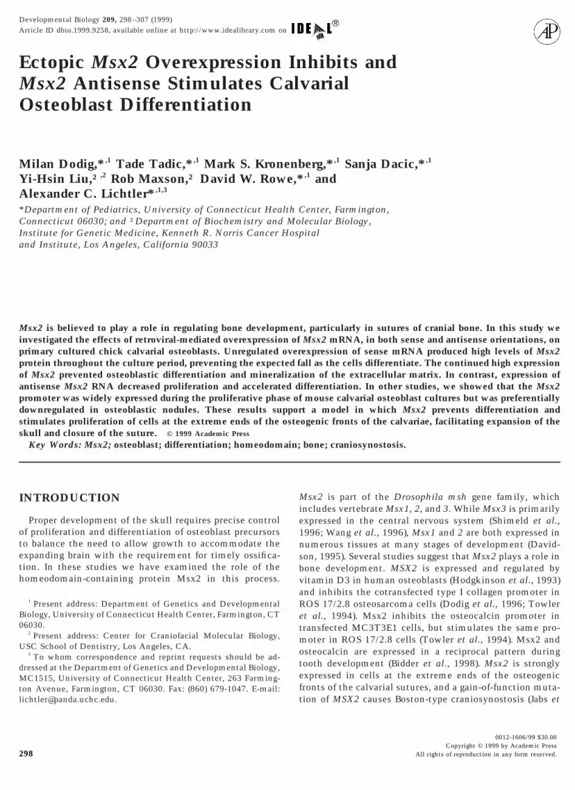
FIG. 1. Msx2 mRNA expression during chick calvaria osteoblastdifferentiation. Numbers indicate days after plating. Cultures beginto differentiate after day 7. Hybridization to 18S RNA is shown as
a loading control.Copyright © 1999 by Academic Press. All right
cells with incorporated BrdU were visualized, photographed, andcounted in 10 random fields using a fluorescence microscope (ZeissAxiovert 135) at 103 magnification.
DNA content of 5-day-old cultures was measured by extractingDNA using the TRI reagent (Molecular Research Center, Inc.)(Chomczinski, 1993) and measuring the absorbance at 260 nm.
RESULTS
To study the role of Msx2 in osteoblast development, weused the chick calvarial osteoblast culture system. North-ern blot analysis reveals that Msx2 expression in differen-tiating cCOB is highest in early cultures (days 7 and 12),while in the more differentiated cultures (days 17–27) Msx2mRNA levels are significantly reduced (Fig. 1). The increaseseen between day 7 and day 12 was not always seen; how-ever, the decrease seen in later cultures was very reproduc-ible. This pattern corresponds to that seen in differentiatingmurine calvarial osteoblast cultures.
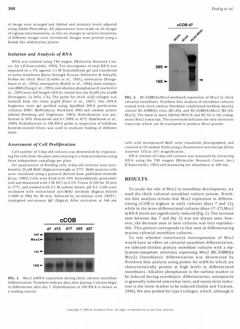
To test whether constitutive overexpression of Msx2would have an effect on calvarial osteoblast differentiation,we infected chicken primary osteoblast cultures with a rep-lication-competent retrovirus expressing Msx2 (RCASBP(A)-Msx2). Osteoblastic differentiation was determined byNorthern blot analysis using probes for mRNAs which arecharacteristically present at high levels in differentiatedosteoblasts. Alkaline phosphatase is the earliest marker tobe induced during osteoblastic differentiation; osteopontinis generally induced somewhat later, and osteocalcin induc-tion is the latest marker to be induced (Aubin and Turksen,
FIG. 2. RCASBP(A)-Msx2-mediated expression of Msx2 in chickalvarial osteoblasts. Northern blot analysis of osteoblast culturesreated with chick embryo fibroblast conditioned medium (mock),ontrol RCASBP(A) virus (RCAS), and RCASBP(A)-Msx2 (RCAS-sx2). The band in lanes labeled MOCK and RCAS is the endog-
nous Msx2 transcript. The arrowhead indicates the only retrovirusranscript which can be translated to produce Msx2 protein.
1996). We also probed for type I collagen, which, although it
s of reproduction in any form reserved.

e1deetadfmRtdtt
v
301Msx2 Inhibits Osteoblast Differentiation
is expressed in osteoblast precursors, is induced to highlevels during differentiation. Although some reports indi-cate that type I collagen expression is highest in the earlyproliferative phase of osteoblast culture (Owen et al., 1990),our studies have found that this marker is highest inpost-proliferative differentiating cultures (Dodig et al., 1996and unpublished results). Control experiments demon-strated that exposure of cCOB cultures to chick embryofibroblast conditioned medium (mock infection) or toRCASBP(A) virus had no effect on differentiation (data notshown). Msx2 overexpression in infected cCOB cultureswas examined by Northern blot analysis of Msx2 mRNA(Fig. 2). The arrowhead indicates the spliced retroviral RNAwhich is the only RNA species which can be translated toproduce Msx2 protein. The larger RNAs have upstreamreading frames which encode the viral gag, pol, and envproteins and thus cannot translate the Msx2 cistron (Petro-poulos and Hughes, 1991). In addition, immunostaining
FIG. 3. Immunohistochemistry detecting Msx2 protein inRCASBP(A)-Msx2-infected (A, D), control (B, E), and RCASBP(A)-Msx2 antisense-infected (C, F) chick calvarial osteoblasts. A–C areimages taken with a 403 objective of cells replated at low densityto allow visualization of individual cells. D–E are images takenwith a 43 objective of primary cells at high density to allowvisualization of overall intensity of staining. Phase-contrast visu-alization of these cultures indicated that the three types of cellswere at similar densities.
using polyclonal Msx2 antibodies indicated that most if not
Copyright © 1999 by Academic Press. All right
all of the cells in the RCASBP(A)-Msx2 infected cultures(Figs. 3A and 3D) had strongly staining nuclei, in contrast tothe control cultures (Figs. 3B and 3E), which were morelightly stained, reflecting the moderate expression of endog-enous Msx2. The proliferation and appearance of undiffer-ntiated 5-day-old cultures was not affected by Msx2 (Tableand data not shown). To determine the effect of Msx2 on
ifferentiation into mature osteoblasts, we examined thexpression of osteoblastic markers in the cultures by North-rn blot analysis. Expression of type I collagen and os-eopontin was decreased (Fig. 4A), and alkaline phosphatasend osteocalcin in the 28-day-old cultures were barelyetectable (Fig. 4B), compared with control cultures in-ected with RCASBP(A) or mock-infected controls. The
o s t d r a m a t i c m o r p h o l o g i c a l e f f e c t o fCASBP(A)-Msx2 infection was the absence of mineraliza-
ion of the extracellular matrix, while mineralization wasemonstrated by both mock and RCASBP(A) control cul-ures (Fig. 4C). These experiments were repeated threeimes with very reproducible results in each case.
In a second series of experiments we engineered a retro-irus expressing Msx2 RNA in the antisense orientation
(RCASBP(A)-Msx2As). Immunostaining with Msx2 anti-bodies demonstrated decreased levels of Msx2 protein inantisense-treated cultures compared to controls (compareFigs. 3C and 3F to Figs. 3B and 3E) Differentiation ofantisense-infected cultures was accelerated as shown byearlier expression of bone markers. Type I collagen mRNAin Msx2 antisense-infected cultures was increased com-pared to RCASBP(A)-infected cultures on day 7, indicatingthat the Msx2 antisense has accelerated the normal differ-entiation of these cultures. By day 12 collagen mRNA levelswere similar in controls and antisense-treated cultures (Fig.5A). This is consistent with our observation that endoge-nous levels of Msx2 are decreasing during this time period,
TABLE 1Effect of Msx2 Sense and Antisense on Cell Proliferation
RCAS Msx2As Msx2
Cell numberExpt 1 38.7 6 0.7 (3) 29.3 6 1.4 (3)* 38.0 6 0.6 (3)Expt 2 19.3 6 2.0 (3) 15.3 6 1.2 (3)* 20.3 6 0.3 (3)Expt 3 35.0 6 1.7 (3) 26.3 6 0.3 (3)* 35.0 6 1.5 (3)
DNA contentExpt 1 11.8 mg 8.5 mg 11.5 mgExpt 2 16.3 mg 9.5 mg 13.8 mg
BrdU incorporation 94.1 6 7.4 (10) 72.0 6 2.7 (10)* 94.1 6 7.4 (10)
Note. Cell number and BrdU incorporation are shown as themean 6 standard error of the mean, with the number of replicatesin parentheses. DNA content was determined by quantitation ofDNA in 5 or 6 35-mm dishes, then calculation of the amount ofDNA per dish. All assays were done on 5-day-old cultures.
* Values significantly different from RCAS values at P , 0.05.
s of reproduction in any form reserved.

imC2niet
efsiNirt
302 Dodig et al.
allowing increased expression of type I collagen, which is arelatively early marker of differentiation. OsteopontinmRNA was slightly induced on day 7 and was stronglyinduced on day 12, when very little endogenous mRNA waspresent in the control cultures. Alkaline phosphatase wasinduced relative to control on days 7 and 12. Osteocalcinwas strongly induced on day 12 of culture, when the controlcultures showed very minor induction of this mRNA.Mineralization of the extracellular matrix was also stronglyinduced compared to control in day 12 cultures (Fig. 5C).These observations, which were consistently seen in threeseparate experiments, support the conclusion of the Msx2overexpression studies, suggesting that endogenous Msx2inhibits osteoblastic differentiation during the early prolif-erative phase of in vitro osteoblast differentiation.
Msx2 antisense-infected cultures appeared to be lessproliferative than control cultures. Since proliferation is
FIG. 4. Effect of Msx2 on osteoblast markers in 28-day osteoblastEffect of Msx2 on alkaline phosphatase (AP) and osteocalcin (OC).Black spots are areas of mineralization. Labels are as in Fig. 3 capt
often inversely correlated with differentiation, we exam- g
Copyright © 1999 by Academic Press. All right
ned three indices of proliferation in three separate experi-ents comparing Msx2 antisense and control cultures.ultures infected with RCASBP(A)-Msx2As demonstrated5–30% fewer cells, less DNA per plate, and a reducedumber of BrdU-positive cells compared with culturesnfected with control virus (Table 1), indicating that endog-nous Msx2 stimulates proliferation in calvarial osteoblas-ic cells.
Although previous studies showed that Msx2 mRNAxpression decreases during mouse calvarial osteoblast dif-erentiation (Dodig et al., 1996), Msx2 mRNA levels weretill substantial at a time when the cultures were undergo-ng differentiation. It could not be determined using theseorthern blot studies whether Msx2 decreased in all cells
n the culture at the same rate or if Msx2 decreases moreapidly in osteoblastic nodules than in surrounding nonos-eoblastic cells. To address this question, we used trans-
res. (A) Effect of Msx2 on Col1a1 and osteopontin (OP) mRNA. (B)ffect of Msx2 on mineralization as revealed by von Kossa staining.Msx2 a and b are duplicate experiments.
cultu(C) Eion.
enic mice containing 5.3 kb of the Msx2 gene 59 flanking
s of reproduction in any form reserved.

fo
303Msx2 Inhibits Osteoblast Differentiation
sequence driving b-galactosidase. Previous studies (Liu etal., 1994; 1999; and our unpublished observations) haveshown that this transgene is expressed in the calvarialsutures in a pattern similar to that of the endogenous gene.Calvarial osteoblasts from these mice were cultured underconditions which allow in vitro differentiation and stainedor b-galactosidase at various stages. Although sutures were
removed from the calvariae, so that most of the plated cellswould not have expressed Msx2 in vivo, by day 1 afterplating most of the cells were beginning to express thetransgene (Fig. 6A), and by day 4 almost all cells expressed
FIG. 5. Effect of Msx2 antisense (Msx2As) on osteoblast differenCol1a1 and osteopontin (OP) mRNA levels. (B) Effect of Msx2 on athe blot shown in A, so the 18S normalization applies here as well.Black spots are areas of mineralization.
b-galactosidase (Fig. 6B). By day 11, when osteoblastic
Copyright © 1999 by Academic Press. All right
nodules had begun to form, b-galactosidase was restrictedto cells between the nodules (Fig. 6C). By day 16b-galactosidase was restricted to the cells immediatelysurrounding the nodules (Fig. 6D). This pattern of expres-sion is consistent with the hypothesis that Msx2 inhibitssteoblastic differentiation.
DISCUSSION
In these studies we examined the role of Msx2 in skull
n after 7 (d7) or 12 (d12) days in culture. (A) Effect of Msx2As one phosphatase (AP) and osteocalcin (OC). B is a rehybridization offfect of Msx2 on mineralization as revealed by von Kossa staining.
tiatiolkalin(C) E
bone development. In order to directly assess the function
s of reproduction in any form reserved.

eostogsmtsM
dit
i
o
cMeosccoszcnt
t
304 Dodig et al.
of Msx2 in this process, we forced continued expression ofMsx2 in calvarial osteoblasts beyond the stage at which thendogenous gene is normally downregulated. We found thatsteoblastic differentiation was strongly inhibited. Anti-ense inhibition of Msx2 expression stimulated differentia-ion and decreased proliferation, further supporting the rolef endogenous Msx2 in inhibiting differentiation and sug-esting that Msx2 also stimulates proliferation. Overexpres-ion of Msx2 did not appear to stimulate proliferation. Thisay be because the endogenous levels of Msx2 present in
he cultures coupled with serum growth factors may havetimulated maximal proliferation, such that higher levels ofsx2 were not able to stimulate increased proliferation.Previous studies had indicated that Msx2 expression
ecreases as calvarial osteoblast cultures differentiate. Tonvestigate this observation in greater detail, we usedransgenic mice containing an Msx2 promoter-driven
b-galactosidase transgene. We found that this transgenes expressed in calvarial cells cultured in vitro, despite
the fact that the sutures and outer cells of the perios-teum, which contain the vast majority of Msx2-expressing cells in the calvaria (Jabs et al., 1993), wereremoved before isolation of the cells which were platedfor the experiments. This observation is consistent withprevious studies showing that endogenous Msx2 mRNAis expressed in cultured mouse calvarial cells but not incalvariae with the sutures and part of the periosteumremoved (Dodig et al., 1996). The induction of Msx2 incells which did not express Msx2 in vivo suggests thepossibility that all calvarial osteoblasts may be capable of
FIG. 6. Msx2 promoter-driven b-galactosidase transgene expres-sion during calvarial osteoblast development. Calvarial osteoblastsfrom 6- to 8-day-old transgenic mice containing 25.2/lacZ werecultured for 1, 4, 11, and 16 days (A, B, C, and D, respectively) andthen stained for b-galactosidase activity. Arrows in C and D pointo osteoblastic nodules.
expressing Msx2 in vivo and that a specific signal induces
Copyright © 1999 by Academic Press. All right
Msx2 in the cells in the suture area. Alternatively Msx2expression may be constitutive in calvarial osteoblastprecursors, but may be suppressed in cells outside thesuture. The second possibility is consistent with the factthat Msx2 is expressed in migrating cranial neural crestcells, which include the precursors of calvarial osteo-blasts. It appears that the conditions allowing expressionof Msx2 are mimicked by in vitro culture. Analysis ofb-galactosidase expression during in vitro differentiation
f these cultures indicated that the Msx2 transgene wasdownregulated first in the osteoblastic nodules and onlylater in the internodular cells. This observation furthersupports the hypothesis that Msx2 inhibits osteoblasticdifferentiation.
Our results, combined with previous studies on Msx2,can be used to construct a model for the role of Msx2 inalvarial bone development and to explain why increasedsx2 function causes craniosynostosis (Fig. 7). Msx2 is
xpressed primarily in cells at the extreme end of thesteogenic front of the suture and in cells in between theutures. There is decreased or absent expression in theells flanking the front. We believe that Msx2 expressiononstrains the committed preosteoblastic cells in thesteogenic front in an undifferentiated stage and probablytimulates proliferation to some degree, maintaining aone of rapid lateral bone growth at the edge of thealvariae (Fig. 7A). Downregulation of Msx2 expression isecessary for the progression of these cells further intohe osteoblastic lineage. The Msx2 gain-of-function mu-
tation found in Boston-type craniosynostosis (Jabs et al.,1993), or increased expression of normal Msx2 in trans-genic mice (Liu et al., 1995), may lead to an expansion ofthe zone of undifferentiated, proliferating cells whichmake up part of the osteogenic front (Fig.7B). The in-crease in the size and growth rate of this group of cellsdisrupts the synchronization between calvarial bonegrowth and expansion of the brain, which is needed tomaintain the proper distance between the skull bones,therefore the osteogenic ridges from neighboring calvar-ial bones meet prematurely (Fig. 7C). In studies on thecalvariae of Msx2-overexpressing transgenic mice, it wasfound that the osteogenic fronts of mice expressing Msx2from the Msx2 promoter show enhanced growth and in-creased BrdU labeling compared to those of control mice,consistent with our hypothesis (Liu et al., 1999). Wehypothesize that in the human the juncture of two os-teogenic fronts triggers downregulation of Msx2, allow-ing osteoblastic differentiation and final closure of thesuture. In the mouse, fusion of the sutures does not occurin 4-month-old adult mice (Zimmerman et al., 1998),therefore Msx2 may not be downregulated. Mice express-ing Msx2 driven by the CMV promoter had normal ossi-fied bone (Liu et al., 1995). Based on the results of the
current studies, we would predict that this would nots of reproduction in any form reserved.

acmltheW
wt
nd t
305Msx2 Inhibits Osteoblast Differentiation
occur if the Msx2 gene was expressed in mature osteo-blasts.
The role that we have proposed for Msx2 in modulatingcell division and differentiation of specific preosteoblasticcells is consistent with the effects of the “selector” homeo-proteins on Drosophila development, which include modu-lation of rates of cell division and differentiation intospecific cell types (Biggin and McGinnis, 1997).
Winograd et al. (1997) have analyzed transgenic micecontaining the human MSX2 gene and have found anextremely severe phenotype characterized by perinatal le-
FIG. 7. Model of the role of Msx2 in normal skull development a
thality and multiple craniofacial malformations, often char- (
Copyright © 1999 by Academic Press. All right
cterized by loss of skeletal structures. This apparent dis-repancy with the results of Liu et al. (1995) may be becauseultiple integrated copies of the entire human MSX2 gene
ocus produced much higher levels of MSX2 protein thanhe transgene used in the studies of Liu et al., or MSX2 mayave been expressed ectopically. The pattern and levels ofxpression of the MSX2 protein were not assessed byinograd et al., 1997.Our model predicts that inactivation of the Msx2 geneould cause delayed closure of the cranial sutures. Al-
hough an Msx2 gene knockout mouse has been described
he role of its overexpression in craniosynostosis.
Maas and Bei, 1997), the time of closure of the cranial
s of reproduction in any form reserved.

B
F
F
F
K
L
L
L
L
M
M
N
O
P
P
306 Dodig et al.
sutures was not reported. It will be of interest to examinesuture closure in further studies of this mouse.
ACKNOWLEDGMENTS
We thank Dr. R. Kosher, C. Dealy, and D. Ferrari for providing uswith the Msx2 antibody and for the immunohistochemistry proto-col. We also thank Dr. Stephen Hughes for giving us theRCASBP(A) vector and for many helpful suggestions concerning itsuse. This work was supported by the following grants from theNational Institutes of Health: AR29983 (A.C.L.), HD22610 (R.Rosher and A.C.L.), and AR38933 (A.C.L. and D.W.R.).
REFERENCES
Aubin, J. E., and Turksen, K. (1996). Monoclonal antibodies as toolsfor studying the osteoblast lineage. Microsc. Res. Tech. 33,128–140.
idder, M., Latifi, T., and Towler, D. A. (1998). Reciprocal tem-porospatial patterns of Msx2 and osteocalcin gene expressionduring murine odontogenesis. J. Bone Miner. Res. 13, 609 –619.
Biggin, M. D., and McGinnis, W. (1997). Regulation of segmenta-tion and segmental identity by Drosophila homeoproteins: Therole of DNA binding in functional activity and specificity.Development 124, 4425–4433.
Chomczinski, P. (1993). A reagent for the single-step simultaneousisolation of RNA, DNA and proteins from cell and tissuesamples. BioTechniques 15, 532–535.
Coelho, C. N. D., Sumoy, L., Rodgers, B. J., Davidson, D. R., Hill,R. E., Upholt, W. B., and Kosher, R. A. (1991). Expression of thechicken homeobox-containing gene GHox-8 during embryonicchick limb development. Mech. Dev. 34, 143–154.
Crawford, K., Millan, J. L., Weissig, H., Goetinck, P. F., and Binette,F. (1995). Tissue-nonspecific alkaline phosphatase participates inthe establishment and growth of feather germs in embryonicchick skin cultures. Dev. Dyn. 204, 48–56.
Davidson, D. (1995). The function and evolution of Msx genes:Pointers and paradoxes. Trends Genet. 11, 405–411.
Dodig, M., Kronenberg, M. S., Bedalov, A., Kream, B. E., Grono-wicz, G., Clark, S. H., Mack, K., Liu, Y., Maxon, R., Pan, Z. Z.,Upholt, W. B., Rowe, D. W., and Lichtler, A. C. (1996). Identifi-cation of a TAAT-containing motif required for high levelexpression of the COL1A1 promoter in differentiated osteoblastsof transgenic mice. J. Biol. Chem. 271, 16422–16429.
einberg, A. P., and Vogelstein, B. (1983). A technique for radiola-beling DNA restriction endonuclease fragments to high specificactivity. Anal. Biochem. 132, 6–13.
errari, D., Lichtler, A. C., Pan, Z. Z., Dealy, C. N., Upholt, W. B.,and Kosher, R. A. (1998). Ectopic expression of Msx-2 in posteriorlimb bud mesoderm impairs limb morphogenesis while inducingBMP-4 expression, inhibiting cell proliferation, and promotingapoptosis. Dev. Biol. 197, 12–24.
iner, M. H., Doty, P., and Boedtker, H. (1987). Construction andcharacterization of cDNA clones encoding the 59 end of thechicken pro alpha 1(I) collagen mRNA. Gene 56, 71–78.
Gerstenfeld, L. C., Chipman, S. D., Glowacki, J., and Lian, J. B.(1987). Expression of differentiated function by mineralizing
cultures of chicken osteoblasts. Dev. Biol. 122, 49–60.Copyright © 1999 by Academic Press. All right
Gray, J. (1985). Monoclonal antibodies against bromodeoxyuridine.Cytometry 6(6). [Special issue]
Hodgkinson, J. E., Davidson, C. L., Beresford, J., and Sharpe, P. T.(1993). Expression of a human homeobox-containing gene isregulated by 1,25(OH)2D3 in bone cells. Biochim. Biophys. Acta1174, 11–16.
Jabs, E. W., Muller, U., Li, X., Ma, L., Luo, W., Haworth, I. S.,Klisak, I., Sparkes, R., Warman, M. L., Mulliken, J. B., Snead,M. L., and Maxson, R. (1993). A mutation in the homeodomain ofthe human MSX2 gene in a family affected with autosomaldominant craniosynostosis. Cell 75, 443–450.
im, H. J., Rice, D. P. C., Kettunen, P. J., and Thesleff, I. (1998).Fgf-, Bmp- and Shh-mediated signalling pathways in the regula-tion of cranial suture morphogenesis and calvarial bone develop-ment. Development 125, 1241–1251.
iu, Y. H., Kunda, R., Wu, L., Luo, W., Ignelzi, M. A., Snead, M. L.,and Maxson, R. E. (1995). Premature closure and ectopic cranialbone in mice expressing Msx2 transgenes in the developing skull.Proc. Natl. Acad. Sci. USA 92, 6137–6141.
iu, Y. H., Ma, L., Wu, L. Y., Luo, W., Kundra, R., Sangiorgi, F.,Snead, M. L., and Maxson, R. (1994). Regulation of the Msx2homeobox gene during mouse embryogenesis: A transgene with439 bp of 59 flanking sequence is expressed exclusively in theapical ectodermal ridge of the developing limb. Mech. Dev. 48,187–197.
iu, Y. H., Tang, Z., Kundu, R. K., Wu, L., Luo, W., Zhu, Z.,Sangiorgi, F., Snead, M. L., and Maxson, R. E., Jr. (1999). Msx2gene dosage influences the number of proliferative osteogeniccells in growth centers of the developing murine skull: A possiblemechanism for msx2-mediated craniosynostosis in humans.Dev. Biol. 205, 260–274.
ogan, C., Khoo, W. K., Cado, D., and Joyner, A. L. (1993). Twoenhancer regions in the mouse En-2 locus direct expression tothe mid/hindbrain region and mandibular myoblasts. Develop-ment 117, 905–916.a, L., Golden, S., Wu, L., and Maxson, R. (1996). The molecularbasis of Boston-type craniosynostosis: The Pro148–His mutationin the N-terminal arm of the MSX2 homeodomain stabilizesDNA binding without altering nucleotide sequence preferences.Hum. Mol. Genet. 5, 1915–1920.aas, R., and Bei, M. (1997). The genetic control of early toothdevelopment. Crit. Rev. Oral Biol. Med. 8, 4–39.eugebauer, B. M., Moore, M. A., Broess, M., Gerstenfeld, L. C.,and Hauschka, V. (1995). Characterization of structural se-quences in the chicken osteocalcin gene: Expression of osteocal-cin by maturing osteoblasts and by hypertrophic chondrocytes invitro. J. Bone Miner. Res. 10, 157–163.wen, T. A., Aronow, M., Shalhoub, V., Barone, L. M., Wilming, L.,Tassinari, M. S., Kennedy, M. B., Pockwinse, S., Lian, J. B., andStein, G. S. (1990). Progressive development of the rat osteoblas-tic phenotype in vitro: Reciprocal relationships in expression ofgenes associated with osteoblast proliferation and differentiationduring formation of the bone extracellular matrix. J. Cell.Physiol. 143, 420–430.
age, K. M. (1982). Bone and the preparation of bone sections. In“Theory and Practice of Histological Techniques” (J. D. Bancroftand A. Stevens, Eds.), pp. 324–325. Churchill Livingston, Edin-burgh.
etropoulos, C. J., and Hughes, S. H. (1991). Replication-competentretrovirus vectors for the transfer and expression of gene cas-
settes in avian cells. J. Virol. 65, 3728–3737.s of reproduction in any form reserved.

W
W
Y
Z
307Msx2 Inhibits Osteoblast Differentiation
Rafidi, K., Moore, M. A., Simkina, I., Gerstenfeld, L. C., andJohnson, E. (1994). Characterization of the chicken osteopontin-encoding gene. Gene 140, 163–169.
Sambrook, J., Fritsch, E. F., and Maniatis, T. (1989). “MolecularCloning: A Laboratory Manual.” Cold Spring Harbor LaboratoryPress, Cold Spring Harbor, NY.
Shimeld, S., McKay, I. J., and Sharpe, P. T. (1996). The murinehomeobox gene Msx-3 shows highly restricted expression in thedeveloping neural tube. Mech. Dev. 55, 201–210.
Towler, D. A., Rutledge, S. J., and Rodan, G. A. (1994). Msx-2/Hox8.1: A transcriptional regulator of the rat osteocalcin promoter.Mol. Endocrinol. 8, 1484–1493.
Wang, W., Chen, X., Xu, H., and Lufkin, T. (1996). Msx3: A novelmurine homologue of the Drosophila msh homeobox gene re-stricted to the dorsal embryonic nervous system. Mech. Dev. 58,203–215.inograd, J., Reilly, M. P., Roe, R., Lutz, J., Laughner, E., Xu, X.,Hu, L., Asakura, T., vander Kolk, C., Strandberg, J. D., and
Copyright © 1999 by Academic Press. All right
Semenza, G. L. (1997). Perinatal lethality and multiple craniofa-cial malformations in MSX2 transgenic mice. Hum. Mol. Genet.6, 369–379.ong, G. L., and Cohn, D. V. (1974). Separation of parathyroidhormone and calcitonin-sensitive cells from non-responsivebone cells. Nature 252, 713–315.ang, R., Gotoh, Y., Moore, M. A., Rafidi, K., and Gerstenfeld,L. C. (1995). Characterization of an avian bone sialoprotein(BSP) cDNA: Comparisons to mammalian BSP and identifica-tion of conserved structural domains. J. Bone Miner. Res. 10,632– 640.immerman, B., Moegelin, A., de Souza, P., and Bier, J. (1998).Morphology of the development of the sagittal suture of mice.Anat. Embryol. (Berlin) 197, 155–165.
Received for publication November 25, 1998Revised February 26, 1999
Accepted February 26, 1999
s of reproduction in any form reserved.