arterial po2 stimulates intimal hyperplasia and serum stimulates inward eutrophic remodeling in...
TRANSCRIPT
Biomech Model MechanobiolDOI 10.1007/s10237-010-0224-8
ORIGINAL PAPER
Arterial pO2 stimulates intimal hyperplasia and serum stimulatesinward eutrophic remodeling in porcine saphenous veins culturedex vivo
Binata Joddar · Rebecca J. G. Shaffer ·Rashmeet K. Reen · Keith J. Gooch
Received: 23 December 2009 / Accepted: 3 May 2010© The Author(s) 2010. This article is published with open access at Springerlink.com
Abstract Ex vivo culture of arteries and veins is an estab-lished tool for investigating mechanically induced remod-eling. Porcine saphenous veins (PSV) cultured ex vivowith a venous mechanical environment, serum-supplementedcell-culture medium and standard cell-culture conditions(5% CO2 and 95% balance air ∼140 mmHg pO2) developintimal hyperplasia (IH), increased cellular proliferation,decreased compliance and exhibit inward eutrophic remod-eling thereby suggesting that nonmechanical factors stim-ulate some changes observed ex vivo. Herein we explore
Electronic supplementary material The online version of thisarticle (doi:10.1007/s10237-010-0224-8) contains supplementarymaterial, which is available to authorized users.
B. Joddar · K. J. Gooch (B)Department of Biomedical Engineering, Ohio State University,290 Bevis Hall, 1080 Carmack Road, Columbus, OH 43210, USAe-mail: [email protected]
B. Joddar · R. K. Reen · K. J. GoochDavis Heart & Lung Research Institute, Ohio State University,Columbus, OH 43210, USA
R. J. G. Shaffer · K. J. GoochDepartment of Bioengineering, The Institute for Medicineand Engineering, 1142 Vagelos Research Laboratory,University of Pennsylvania, 3340 Smith Walk,Philadelphia, PA 19104, USA
R. K. ReenDepartment of Surgery, Ohio State University,Columbus, OH 43210, USA
Present Address:R. J. G. ShafferNovo Nordisk Inc., 100 College Rd. W,Princeton, NJ 08540-6658, USA
the contribution of exposure to greater than venous pO2 andserum to these changes in cultured veins. Removing serumfrom culture medium did not inhibit development of IH,but did reduce cellular proliferation and inward eutrophicremodeling. In contrast, veins perfused using reduced pO2(75 mmHg) showed reduced IH. Among the statically cul-tured vessels, veins cultured at arterial pO2 (95 mmHg) andabove showed IH as well as increase in proliferation and ves-sel weight compared to fresh veins; veins cultured at venouspO2 did not. Taken together, these data suggest that exposureof SV to arterial pO2 stimulates IH and cellular prolifera-tion independent of changes in the mechanical environment,which might provide insight into the etiology of IH in SVused as arterial grafts.
Keywords Saphenous vein · Intimal hyperplasia ·Eutrophic remodeling · Ex-vivo culture system
1 Introduction
Under the appropriate culture conditions, conduit blood ves-sels cultured ex vivo remain viable and vasoactive for days(Porter et al. 1996b; Clerin et al. 2003; Gusic et al. 2005a,b).A primary benefit of these ex-vivo models is that they affordmuch better control and monitoring of the mechanical andchemical environments than possible with vessels in vivowhile allowing the study of whole-vessel behavior not cap-tured in cell culture. Capitalizing on these strengths of the ex-vivo culture systems, investigators have used them to studythe independent contribution of mechanical factors (e.g.,flow, pressure, and pulsatility) (Clerin et al. 2003; Han et al.2003; Davis et al. 2005; Gusic et al. 2005a,b; Han et al. 2006;Gleasonm et al. 2007; Wayman et al. 2008; Yao et al. 2009) ondifferent aspects of vascular remodeling and to understand
123
B. Joddar et al.
how multiple mechanical factors (e.g., pressure and axialloading) interactively regulate vascular remodeling (Nicholet al. 2005, 2009; Lawrence and Gooch 2009a,b). Manyaspects of mechanically induced remodeling of blood vesselsoriginally observed in vivo have also been observed in ves-sels cultured ex vivo including pressure-induced hypertrophy(Gusic et al. 2005a,b), axial loading-induced growth/remod-eling (Clerin et al. 2003; Nichol et al. 2005, 2009; Gleasonmet al. 2007; Lawrence and Gooch 2009a,b), and flow-inducedchanges in luminal area (Nichol et al. 2005, 2009).
While much of the observed remodeling can be attributedto mechanically induced changes, some changes in the cul-tured vessel’s dimension, composition, activity and mechan-ical properties appear to occur independently of changesin the mechanical environment. For example, vessels cul-tured ex vivo with mechanical environments similar to theirnative in vivo environments exhibit hypertrophy (Clerin et al.2003; Gusic et al. 2005a,b), lengthen (Nichol et al. 2005,2009; Gleasonm et al. 2007; Lawrence and Gooch 2009a,b),eutrophically remodel (Gusic et al. 2005a,b), develop inti-mal hyperplasia (Gusic et al. 2005a,b), as well as alter theirextracellular matrix content (Gusic et al. 2005a,b), matrixmetalloproteinase activity (Nichol et al. 2009) and biome-chanical properties (Clerin et al. 2003; Gusic et al. 2005a,b;Lawrence and Gooch 2009a,b). There are at least three gen-eral explanations for these changes. First, it is possible thatthe ex-vivo culture environment fails to adequately replicateimportant aspects of the native mechanical environment andthese differences stimulate the observed changes. Second,processes associated with the harvest of the vessel such assurgical trauma including disruption of the vasa vasorumor denervation might account for the observed remodeling.Here we conducted experiments to explore a third possi-bility—i.e., non-mechanical aspects of the ex-vivo culturesystem might account for some of the observed changesin cultured vessels. Saphenous veins (SV) are attractivevessels to use for these studies because (1) they are wellstudied in various ex-vivo culture systems (Porter et al.1996a,b, 1998a,b, 1999, 2001, 2002; Gusic et al. 2005a,b),(2) they exhibit pronounced changes in culture which donot appear to be due to the mechanical environment (Porteret al. 1996b, 1999), and (3) their remodeling is an impor-tant component of both the success and failure of veingrafts used in coronary artery bypass grafting and periph-eral revascularization (Bourassa 1991; Motwani and Topol1998).
SV grafts rapidly develop intimal hyperplasia (IH) after afew weeks of grafting into the arterial circulation after cor-onary artery bypass grafting (CABG) (Motwani and Topol1998). Although this initial IH does not significantly impedeblood flow, it does provide the foundation for subsequent ath-erosclerosis and thrombosis (Motwani and Topol 1998). Thusinhibition of this initial and rapid IH is an attractive target
for improving vein graft performance. It is often suggestedthat this IH may be a pathological response to abrupt changein the hemodynamic environment as the vein goes from therelatively mild venous mechanical environment to the arte-rial environment with its increased circumferential tension,cyclic stretching, and high flow-induced stresses caused bythe higher blood pressure and flow velocities (Dobrin PBand Endean 1989; Motwani and Topol 1998). To investigatethe stimulus for IH further, a number of studies by othersand our laboratory have employed porcine SV (PSV). Wehave previously reported that perfused PSV develop signifi-cant IH during ex-vivo culture using 5% CO2/ 95% balanceair (∼ 140 mmHg pO2 or normal cell culture atmospheres)and serum-containing medium (Gusic et al. 2005a,b). Collec-tively data from our prior work (Gusic et al. 2005a,b) and oth-ers (Porter et al. 1996a,b, 2001) suggest that the developmentof this observed IH in cultured PSV does not require expo-sure to an environment mimicking in vivo arterial mechanicalconditions. In contrast, arterial levels of mechanical forcesreduced the observed IH (Porter et al. 1996b; Gusic et al.2005a,b). In addition, PSV cultured ex vivo under venousmechanical conditions exhibit inward eutrophic remodelingand reduced compliance (Porter et al. 1996b; Gusic et al.2005a,b).
Based on these observations from SV cultured ex vivo,we speculated that non-mechanical factor(s) modulate thedevelopment of IH in PSV cultured ex vivo. Serum containspotent mitogens for cultured vascular SMC, such as plate-let-derived growth factor, which is suspected to be involvedin atherosclerosis (George et al. 1996). In addition to suchbiochemical factors, aspects of the chemical environmenttypically used for ex-vivo culture might stimulate IH. Cul-tured PSV are typically exposed to a dramatic increase inoxygen from venous levels (pO2 = 40 mmHg) to that of5% CO2 balanced with air (pO2 = 140 mmHg). In con-trast to PSV, porcine arteries cultured ex vivo, which experi-ence a smaller increase in pO2, do not develop IH in culture(Clerin et al. 2002; Gusic et al. 2005a,b). Therefore, in thisstudy, we sought to explore the role serum factors and ele-vated oxygen levels play in the changes in observed in PSVcultured ex vivo. This knowledge will aid in the designbetter ex-vivo culture systems and the interpretation of datafrom ex-vivo studies. While we are focusing on features of theex-vivo environment that might induce remodeling in PSV,it should be noted that human SV (HSV) used as coronaryartery bypass grafts in vivo are likely exposed to elevatedplatelet-derived growth factor (PDGF, due to platelet adhe-sion and thrombus formation) (George et al. 1996; Wanget al. 2005) and are exposed to elevated pO2 due to expo-sure to arterial blood with a pO2 of 95 mmHg. Thus resultsfrom these ex-vivo studies with PSV may provide insightinto factors stimulating IH in HSV grafted into the arterialcirculation (Schachner et al. 2006).
123

Arterial pO2 stimulates intimal hyperplasia
2 Materials and methods
2.1 Vessel harvest and preparation
All animal work was reviewed and approved by the Insti-tutional Animal Care and Use Committees (IACUC) at theUniversity of Pennsylvania and at the Ohio State University.PSV and femoral arteries were harvested aseptically frompigs (Sus scrofa). After harvesting, all vessels were washedin HEPES buffer (Invitrogen, Carlsbad, CA) and transportedback to the laboratory in a gas-impermeable container with∼100 cc of Dulbecco’s modified Eagle’s medium pre-equili-brated with the desired gas mixture, representing either highor low pO2 levels and immediately set up for culture. Thetime between vessel harvests to culture setup was typicallyless than 1 h.
2.2 Ex-vivo perfusion
The ex-vivo perfusion system has been previously described(Clerin et al. 2003; Gusic et al. 2005a,b; Nichol et al. 2005,2009; Lawrence and Gooch 2009a,b) and is similar to thoseused by others (Porter et al. 1996b; Han et al. 2003, 2006;Davis et al. 2005; Gleasonm et al. 2007; Wayman et al. 2008;Yao et al. 2009). PSV isolated from 25–35 kg pigs were cul-tured in low-glucose Dulbecco’s modified Eagle’s mediumsupplemented with 10% fetal bovine serum (Gibco, CA),100 μg/mL penicillin, 100 μg/mL streptomycin, 0.25 μg/mLamphotericin B, and 25 mM HEPES (all from InvitrogenCorp., Rockville, MD). A subset of vessels was perfused withserum-free medium, which contained 2% of bovine serumalbumin (Sigma, MO).
Culture medium was typically equilibrated with a gas mix-ture consisting of 5% CO2 / 95% air resulting in a pO2 of∼140 mmHg, which is somewhat higher than ∼95 mmHgfound in the arterial circulation and much higher than the∼40 mmHg found in the venous circulation. Perfusion condi-tions consisted of steady, venous levels of flow (10 mL/ min)and a pressure (25 mmHg) that was slightly greater than typ-ical in vivo venous pressures. To test the response of PSVto lower oxygen environments, PSV were perfused withmedium bubbled with a 10% CO2/90% N2 gas mixture. Thisresulted in a pO2 of ∼75 mmHg representing an intermedi-ate between typical arterial and venous concentrations. Theoxygen tension achieved was roughly half of that in typ-ical, 5% CO2/ air studies (∼140 mmHg); but greater thanin-vivo venous conditions (∼40 mmHg). PSV perfused exvivo with serum-free culture medium or with serum-con-taining culture medium at a reduced pO2 were cultured atthe same time as other PSV used to study the effects ofmechanical factors on IH (Gusic et al. 2005a,b). Thus, selectdata from these published studies provide appropriate control
groups to compare the results presented here. All perfusionexperiments were run for 7 days, which we have previouslyshown to be long enough for changes in PSV dimensionsand mechanical properties to occur (Gusic et al. 2005a,b).At least five vessels were subjected to each experimentalcondition.
2.3 Histological analysis
At the completion of experiments, vein length and mass weremeasured, and sections of veins were fixed at zero pressurein either 70% ethanol or formalin overnight, dehydrated,embedded in paraffin, cut into 5 μm thick sections, andmounted on glass slides. Elastin staining (Accustain, Sigma,MO) was used according to manufacturer’s instructions, andthe intimal and medial areas of vein cross-sections, whichwere delineated by the external and internal elastic lamina,were measured using Scion Image (Scion, MD). Proliferat-ing cells were stained with monoclonal mouse PC10 antibodyrecognizing proliferating cell nuclear antigen/HRP (PCNA,1:1, DAKO). To account for the cell type which accounted forthe IH, both SMC and EC were counterstained with markersidentifying each cell type. SMC were identified with an anti-body against the SMC lineage marker myosin heavy chain(SM-MHC), and EC were identified by antibodies againstCD31. In addition the in situ Cell Death Detection, PODkit (TUNEL, Roche, IN) was used as directed. TUNEL andPCNA stained sections were counterstained with DAPI (Vec-tor Labs, CA).
2.4 Wet weight, dry weight and collagen assay
Vessel segments (∼2–3 cm long) were weighed to providewet weight, lyophilized, and weighed to provide dry weightand stored at −20◦C until processing. Frozen vessel seg-ments were digested with papain (25 mg/ml papain, 10 mmcysteine in buffered solution, both from Sigma, MO) at 60◦Cfor 16 h. Collagen content was determined using a modifiedversion of the protocol of Woessner previously adapted forthis tissue (Woessner 1961; Gusic et al. 2005a,b).
2.5 Vein Mechanics
At the completion of the one-week ex-vivo perfusion cul-ture, transverse sections of the vein were taken for his-tology and the remaining vein segment was subjected tomechanical testing. As described previously (Gusic et al.2005a,b), pressure and outer-diameter measurements ofveins were recorded using a modified protocol of Coxet al. (1978). The medium in the vessel chamber wasreplaced with phosphate-buffered saline (PBS). Calcium-free Hanks’ balanced salt solution (HBSS) from a second
123

B. Joddar et al.
reservoir was diverted to the vein by means of three-way stopcocks and a second peristaltic pump. A pres-sure head of approximately 80 mmHg was generated andthe vein was allowed to incubate for 1 h at this transmu-ral pressure. At the end of this period, the vein was pre-conditioned by subjecting it to 10 continuous cycles ofinflation and deflation at a rate of 2 mmHg/s between 10and 100 mmHg, and then three inflation responses wererecorded at 1 mmHg/s between 10 and 100 mmHg. The veinwas then pressurized again to 80 mmHg and norepineph-rine (NE) (10−4 M, Sigma, MO) was added to the ves-sel bath and the vein was allowed to constrict for 2 min,after which the pressure was lowered to 10 mmHg andmaintained at this level until no changes in diameter wereobserved. The vessel bath was drained, rinsed, and replacedwith calcium-free PBS, supplemented with 2 mM ethyleneglycol-bis(B-aminoethyl ether)-N,N,N′,N′-tetraacetic acid(EGTA, Sigma, MO). The vein was again pressurized to80 mmHg and was allowed to incubate at this pressure for30 min. At the end of this period, the vein was precondi-tioned and then three inflation responses were recorded at1 mmHg/s between 10 and 100 mmHg. At the end of the test-ing, veins were removed from the vessel chamber, blottedlightly with gauze, measured and weighed.
A pseudo-elastic model for an incompressible, homoge-neous, cylindrical, isotropic vein wall, with a Poisson’s ratioof 0.5, was assumed to look at the mean behavior of thevein wall in order to compare changes that may occur withremodeling. The inner radius, Ri , of the vein was calculatedusing the measured outer radius, the vein mass, length and anassumed density of 1.06 g/cm3. Wall thickness, h, was calcu-lated as (Ro −Ri ). Luminal area was calculated at a pressureof 10 mmHg under calcium-free conditions. The compliancewas calculated as the change in luminal area induced by atransmural pressure change (�P) of 20 mmHg, and the dis-tensibility was calculated as Distensibility = 2�Ri
(Ri �P).
Laplace’s law for a thin-walled vessel was employed toestimate the circumferential stress σ = P Ri/h in the veinwall, as veins typically display relatively thin walls. Mea-surements of thickness to radius ratios for fresh saphenousveins averaged ∼0.1, while measurements from culturedveins ranged from 0.1 to 0.2.
Circumferential strain (ε) was calculated as the change ininternal radius from that measured at 10 mmHg or
ε = (Ri − Ri(10 mmHg))/R i(10 mmHg).
2.6 Ex-vivo static culture
While the perfused system is optimal to study mechanicalfactors (Clerin et al. 2002; Gusic et al. 2005a,b; Nichol et al.2005; Lawrence and Gooch 2009a,b) it is a complex sys-
tem and cumbersome to operate. To conduct extensive stud-ies on the role of pO2 in a timely manner, we adopted amuch simpler non-perfused SV organ culture system widelyused by others (Soyombo et al. 1990; Porter et al. 1996b,1998a,b, 1999, 2001, 2002; Jeremy et al. 1997; Corpatauxet al. 2005; Mekontso-Dessap et al. 2006). Porcine ves-sels from pigs 60–65 kg weight were cultured inside sterile100- mm plastic Petri dishes (Nalgene Nunc, Fisher Scien-tific), pre-coated with ∼7 mm of Sylgard 184 resin (DowCorning Corporation, USA). Intact PSV selected for culture,were cleaned from adherent tissues, opened longitudinallyand pinned with 26-gauge 5/8”–stainless-steel needles to theSylgard with the luminal surface facing up. PSV were cul-tured in culture medium as described earlier pre-equilibratedwith the desired gas mixture. PSV were cultured at 37◦Cin Petri dishes, placed on an orbital shaker at 30 RPM andhoused inside O2-, CO2-, N2-and humidity-controlled incu-bator (NUAIRE 4950). Veins were exposed to a pO2 of 140and 75 mmHg, to match the perfused conditions as well asarterial pO2 (95 mmHg) and venous pO2 (40 mmHg). Por-cine femoral arteries were included as control vessels forculture at arterial pO2 (95 mmHg) since arteries experiencethe same pO2 level in vivo. Spent medium was replaced every2 days with fresh medium pre-equilibrated with the desiredpO2. Samples of spent culture medium equilibrated with thedesired pO2 were sampled every 2 days in a blood-gas ana-lyzer (Rapid lab, Siemens Healthcare, USA) for determina-tion of pCO2, pO2 and pH. After 14 days of static culture,PSV were fixed at zero pressure overnight in 70% ethanol orformalin and processed as required for histological and otheranalyses.
2.7 Analysis of 4-Hydroxynonenal (4-HNE)by immunostaining and Western Blotting
In addition to the other histological analysis performedas described earlier, PSV cultured statically were alsoanalyzed for the lipid peroxidation marker, 4-Hydroxy-nonenal (4-HNE). 4-HNE immunostaining was performedon paraffin-tissue sections to semi-quantitatively comparethe extent of lipid peroxidation in vessel sections usingpolyclonal antibodies recognizing 4-HNE adducts (BethylLabs, Montgomery, TX). Previously frozen tissue fromfreshly isolated and cultured HSV was homogenized andlysed for Western blot analysis using 4-HNE polyclonalantibodies to detect and quantify levels of 4-HNE pro-duced in experimental samples and in controls (Axxora,San Diego, CA).
2.8 Data analysis
All data are reported as means ± SD. Differences from freshand cultured veins in both perfused and statically cultured
123

Arterial pO2 stimulates intimal hyperplasia
A
C
E
F
D
B
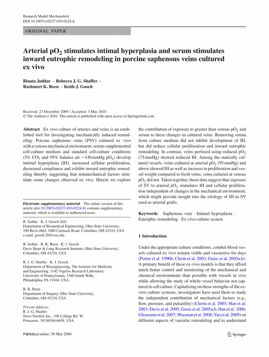
Fig. 1 Freshly isolated and cultured PSV after culture under nativevenous mechanical conditions in a perfused system ex-vivo a, cPressure-diameter relationships of fresh and perfused veins mechan-ically tested with (a) and without (c) calcium. SD of vessel diameteracross all groups was 13 ± 5%. b, d Wall stress versus strain mechan-ically tested with (b) and without (d) calcium. Error bars representSD. e Luminal area calculated from pressure-diameter curves above inthe presence of calcium at a pressure of 10 mmHg. Error bars repre-sent SD. π denotes values published Gusic et al. (2005a,b) and have
been reported for comparison. f Vasoactivity and normal distension forfresh and perfused veins. Black bars represent the measured changein external diameter from a pressure of 80–10 mmHg after stimula-tion with norepinephrine (vasoactive response). Grey bars represent themeasured change in external diameter at the same pressure change asa passive response. *denotes data statistically different from fresh ves-sels (p < 0.05) where indicated. n = 7 for fresh, and n = 5 for PSVcultured at high pO2 with or without no serum, and PSV cultured at lowpO2
vessels were tested using the Dunnett’s Test used as a post hocanalysis. Specifically in the statically cultured vessels groupdifferences among groups were tested using Dunnett’s with
a one-way ANOVA. A value of p < 0.05 was consideredstatistically significant. For each experiment or conditionn ≥ 6 PSV were used except as noted otherwise.
123
B. Joddar et al.
Table 1 Intimal, medial and total wall areas, cell proliferation and cell death index for freshly isolated and cultured PSV in a perfused flow systemex vivo
Intimal area (mm2) Medial area (mm2) Total wall area (mm2) Cell proliferation index (%) Cell death index (%) TUNEL
Freshly isolatedSV n = 7
0.06 ± 0.01π 1.08 ± 0.05π 1.16 ± 0.05π Intima: 3.5±1.8π
Media: 1.1±0.1πIntima:0π
Media: 0π
Cultured with highpO 2(140 mmHg) n = 5
0.16 ± 0.02∗π 1.27 ± 0.04π 1.48 ± 0.03π Intima: 25.2±16.3∗π
Media: 6.9 ± 1.6∗πIntima:4.2 ± 2.9π
Media: 1.8 ± 0.8π
Cultured with noserum at highpO2 (140 mmHg)n = 5
0.14 ± 0.05 1.40 ± 0.21∗ 1.59 ± 0.21∗ Intima: 3.1 ± 1.7Media: 1.0±0.5#
Intima: 3.1 ± 2.3Media: 0.5 ± 0.2
Cultured with lowpO2 (75 mmHg)n = 5
0.08 ± 0.03 1.17 ± 0.02 1.29 ± 0.02 Intima: 8.4 ± 3.5Media: 0.9±0.9#
Intima:14.3 ± 3.7∗Media: 6.4 ± 4.3
All data are shown as mean ± SD. *indicates p < 0.05 relative to fresh veins, and # indicates p < 0.05 relative to veins cultured at 140 mmHgwith fresh data excluded in the analysis. π denotes values that that been previously published [Gusic et al. (2005a,b)] and have been reported hereonly for comparison
3 Results
3.1 Perfused vessels
3.1.1 Pressure-diameter curves and biomechanics
Pressure versus outer diameter relationships revealed thatall groups of cultured PSV had smaller outer diametersthan freshly isolated PSV (Fig. 1a). The decrease in outerdiameter was not solely due to vasoconstriction, since whentested with calcium-free medium, which should relax theveins, cultured PSV still exhibited smaller outer diametersthan freshly isolated PSV (Fig. 1c). Comparing pressure-diameter curves in the presence and absence of calciumrevealed that freshly isolated PSV as well as PSV culturedunder high pO2 with serum had little basal tone (Fig. 1a,c). In contrast, veins cultured under low pO2 with serumor high pO2 without serum exhibited greater basal tone(Fig. 1a, c).
When tested in the presence of calcium, the stress–straincurves for all cultured vessels were similar to or rightwardshifted (Fig. 1b) from freshly isolated veins. Since differentgroups of vessels had different amounts of basal tone andsince the amount of basal tone affects the reference diameterfor strain calculations, stress-strain curves were calculatedfrom pressure-diameter curves generated in the absence ofcalcium (Fig. 1d). Removing calcium from the medium usedfor biomechanics testing had the greatest impact on the stress-strain curves for the groups of veins that had the greatest basaltone (i.e., no serum group or low pO2 group). Luminal andwall areas were calculated from the outer diameter measuredat a pressure of 10 mmHg (calcium free) as well as the wetweight and length of the PSV. The luminal areas for all groupsof cultured PSV were smaller than freshly isolated PSV. PSVcultured with serum and high pO2lost more than 10 mm2
or two thirds of their luminal area (Fig. 1e). This dramaticdecrease in luminal areas was associated with only a modestincrease (less than 0.5 mm2) in wall area (Table 1) suggestingthat the decrease in luminal area was due to inward eutro-phic remodeling. All groups of PSV contracted in responseto norepinephrine with no statistically significant differencesbetween the groups (Fig. 1f).
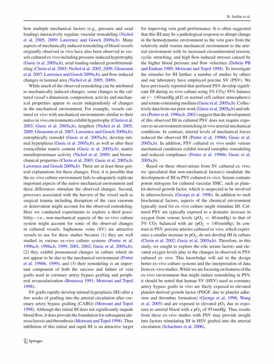
Cultured PSV were less compliant than freshly isolatedveins, especially at pressures less than 30 mmHg (Fig. 2a, c).In contrast, distensibility was more similar among all groupsof veins, especially under calcium-free conditions (Fig. 2b,d). The fact that ex-vivo culture had greater effects on compli-ance than distensibility suggests that effects on compliancewere largely due to changes in the veins’ dimensions—espe-cially the decrease in inner diameter—and not changes intheir material properties.
3.1.2 Histological analysis and collagen content
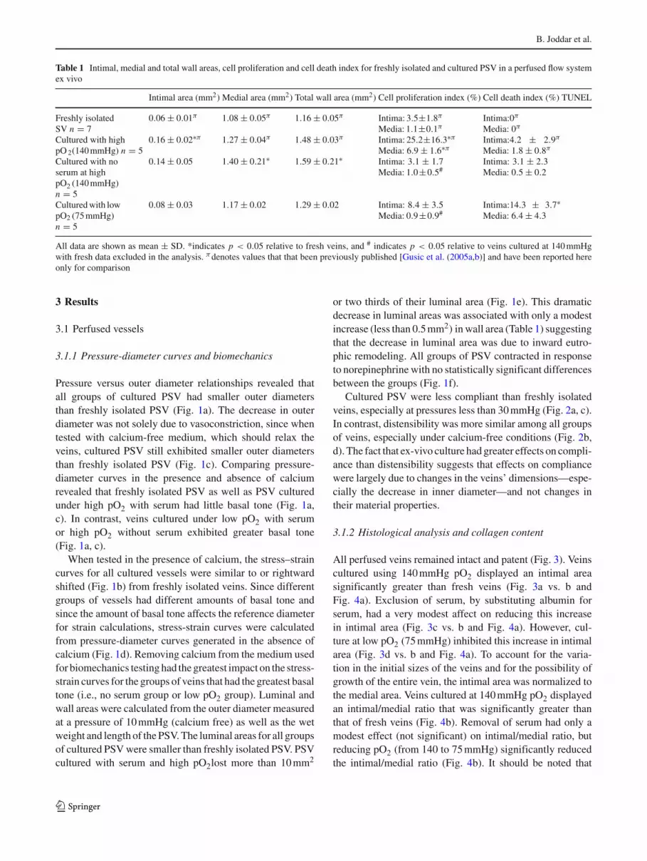
All perfused veins remained intact and patent (Fig. 3). Veinscultured using 140 mmHg pO2 displayed an intimal areasignificantly greater than fresh veins (Fig. 3a vs. b andFig. 4a). Exclusion of serum, by substituting albumin forserum, had a very modest affect on reducing this increasein intimal area (Fig. 3c vs. b and Fig. 4a). However, cul-ture at low pO2 (75 mmHg) inhibited this increase in intimalarea (Fig. 3d vs. b and Fig. 4a). To account for the varia-tion in the initial sizes of the veins and for the possibility ofgrowth of the entire vein, the intimal area was normalized tothe medial area. Veins cultured at 140 mmHg pO2 displayedan intimal/medial ratio that was significantly greater thanthat of fresh veins (Fig. 4b). Removal of serum had only amodest effect (not significant) on intimal/medial ratio, butreducing pO2 (from 140 to 75 mmHg) significantly reducedthe intimal/medial ratio (Fig. 4b). It should be noted that
123
Arterial pO2 stimulates intimal hyperplasia
A
B
DC
Fig. 2 Freshly isolated and cultured PSV after culture under nativevenous mechanical conditions in a perfused system ex-vivo a–dCompliance and distensibility for fresh veins (solid line) and veinsperfused under different conditions tested either with and without cal-cium. π denotes values published [Gusic et al. (2005a,b)] and have been
reported for comparison. *denotes data statistically different from freshvessels (p < 0.05). Error bars represent SD in all graphs. n = 7 forfresh, and n = 5 for PSV cultured at high pO2 with or without no serum,and PSV cultured at low pO2
Porcine SV cultured in a perfused flow system ex vivo using native venous mechanical conditions
Fresh porcine SV high pO2 high pO2 (no serum) low pO2
A DCB
IM
IM
IM
I
M
Fig. 3 Freshly isolated and cultured porcine saphenous veins after cul-ture under native venous mechanical conditions in a perfused system exvivo, stained using Elastin stain (Accustain). (I) stands for intima and
(M) stands for media. Scale bars depict 100μm. n = 7 for fresh, andn = 5 for PSV cultured at high pO2 with or without no serum, and PSVcultured at low pO2
tissues shrink during processing for histology so the intimaand medial areas underestimate the areas in the vessels beforeprocessing. Relative to freshly isolated veins, cell prolifer-ation was elevated 8-fold in the intima, and 7-fold in the
media in veins cultured at 140 mmHg pO2 (Table 1). Cultureat 75 mmHg pO2 did stimulate cell proliferation 2-fold in theintima in the vessel intima relative to fresh veins which wasnot statistically significant (Table 1). Culture at high pO2,
123

B. Joddar et al.
A
B
Fig. 4 Quantification of intimal and medial areas for fresh and cul-tured veins which were perfused under venous mechanical conditionsat variable pO2, as indicated. a Quantification of intimal area (square.mm). b Ratio of intimal to medial area. *denotes statistically significant(p < 0.05) compared to other unmarked groups. n = 7 for fresh, andn = 5 for PSV cultured at high pO2 with or without no serum, and PSVcultured at low pO2
140 mmHg without serum did not stimulate cell proliferationin the intima or media compared to fresh vessels (Table 1).When counter stained with antibodies for smooth musclemyosin heavy chain (MHC), 80 ± 15 % of the proliferatingcells in the neointimal region, and 65 ± 10 % in the mediastained positively (Supplementary Fig. 1). None of these pro-liferating cells reacted with the antibody for endothelial cellmarker (CD31) (images not shown). Cultured veins displayedlow and similar numbers of TUNEL stained cells (Table 1)except for veins cultured at 75 mmHg pO2 which show sig-nificantly larger number of TUNEL positive cells in both theintima and the medial layers. While perfused veins did not
significantly increase their mass relative to freshly isolatedveins, all perfused veins displayed significant increases intheir collagen content (Table 2). All the perfused vesselsshowed inward eutrophic remodeling which lead to overallreduction in lumen area. To accommodate for this decreasein lumen area, the wall thickness was increased. When ana-lyzed, all cultured vessels showed decrease in circumferentiallength and increase in wall thickness. From the fresh vessel,a part of which was cut and perfused for culture ex vivo;the circumferential length decreased from 300 ± 20 μm to200 ±10 μm increasing the wall thickness from 100 ± 5 μmto 150 ± 20 μm when measured in at least eight vessels. Thishowever did not affect the resultant trends in total wall areasin cultured vessels calculated from histological analysis asshown earlier (Table 1), or the wet weights of the vesselsnormalized to their respective axial lengths (Table 2).
3.2 Non-perfused vessels
3.2.1 Chemical culture conditions
By placing the cultured veins in a pO2-controlled incuba-tor, the pO2 of the culture medium could be varied fromvenous (∼40 mmHg) to near atmospheric levels (140 mmHg)(Table 3). These changes in culture medium pO2 wereachieved without changing pCO2 or pH (Table 3).
3.2.2 Intimal and medial dimensions
PSV (Fig. 5 a–e) and femoral arteries (Fig. 5f, g) culturedin the non-perfused culture system for 2 weeks maintainedviability as evidenced by the maintenance of normal cellularstructure in the vessel wall and low rates of TUNEL staining(Table 4). As sometimes seen in perfused vessels exhibitingIH, in static veins IH was sometimes associated with disrup-tion of the IEL and invasion of the underlying medial cells(Fig. 5d, e). On the contrary porcine femoral arteries culturedat arterial pO2 did not show any evidence of IH (Fig. 5g)when compared to their fresh vessel controls (Fig. 5f). PSVcultured at pO2 levels from 75–140 mmHg exhibit dose-dependant increase in intimal and medial areas (Fig. 6). PSVcultured at venous pO2 of 40 mmHg did not exhibit increasedintimal or medial areas (Fig. 6). The increases in medial areain statically cultured PSV are greater than the change inmedial area observed with ex vivo perfused vessels at thecorresponding pO2.
3.2.3 Cell indices and dry weight
Proliferation was elevated in veins cultured at 75–140 mmHgpO2 compared to freshly harvested PSV (Fig. 7). Cell
123

Arterial pO2 stimulates intimal hyperplasia
Table 2 Wall thickness, wet weights and collagen contents for freshly isolated and cultured veins in a perfused flow system ex vivo
Wall thickness (mm) Wet weight per length (mg/ mm) % Collagen per wet weight
Freshly isolated SV n = 7 2.5 ± 0.5 4.8 ± 0.4π 7.7 ± 0.3π
Cultured with high pO2 (140 mmHg) n = 5 3.0 ± 1.0 5.1 ± 0.2π 10.3 ± 1.3π∗
Cultured with no serum at high pO2 (140 mmHg) n = 5 3.5 ± 1.0 5.3 ± 0.7 10.2 ± 1.1∗
Cultured with low pO2 (75 mmHg) n = 5 4.0 ± 1.0∗ 4.7 ± 0.2 12.3 ± 0.3∗
All data are shown as mean ± SD* indicates p < 0.05 relative to fresh veins. π denotes values that that been previously published [Gusic et al. (2005a,b)] and have been reportedhere only for comparison
Table 3 Maintenance of pH, pO2 and pCO2 in a static ex-vivo culture system
Set points pH pO2 (mmHg) pCO2 (mmHg)
Standard cell culture atmosphere (140 mmHg) 7.34 150 ± 8 36.6 ± 0.8
Arterial (95 mmHg) 7.35 104 ± 5 35.8 ± 0.6
Venous (40 mmHg) 7.36 42 ± 4 34.6 ± 0.8
n = 6 for all groups shown
Freshly isolated PSV at 40 mm Hg pO2 at 75 mm Hg po2 at 95 mm Hg pO2 at 140 mm Hg pO2
PSV statically cultured
Porcine femoral arterycultured at 95 mm HgpO2
A B C D
I
I
MMMM
II
E
I
M
Freshly isolated porcine femoral artery
F G
I
M
I
M
Fig. 5 Freshly isolated and statically cultured PSV (a–e) and femoral arteries (f, g) stained using Elastin stain (Accustain). (i) stands for intima and(m) stands for media. The veins and arteries were statically cultured at variable pO2 in a static ex-vivo culture system. Scale bars denote 100μm
proliferation was elevated in the intima and media by4-fold and 8-fold, 14-fold and 18-fold, and 22-fold and24-fold respectively in PSV cultured at 75, 95 and 140 mmHgpO2 respectively. All PSV cultured with increased pO2
showed low and similar percentage of TUNEL positive nuclei(Table 4). Quantification of DAPI-stained nuclei revealedthat culture at higher pO2 increased cell density by 2-fold inboth the intima and media relative to freshly isolated vessels
123

B. Joddar et al.
Table 4 Cell death index (TUNEL) and DAPI (cell counts) for freshly isolated and cultured veins in a static culture system ex-vivo
Cell death index (%) DAPI (No./ mm2)
Freshly isolated SV 0 Intima: 50 ± 10 Media: 60 ± 14
Cultured at 40 mmHg pO2 2.2 ± 0.4 Intima: 40 ± 17 Media: 60 ± 15
Cultured at 75 mmHg pO2 2.2 ± 0.2 Intima: 60 ± 17 Media: 70 ± 15
Cultured at 95 mmHg pO2 2.4 ± 0.1 Intima: 80 ± 10∗ Media: 90 ± 10∗
Cultured at 140 mmHg pO2 2.8 ± 0.3 Intima: 95 ± 15∗ Media: 140 ± 12∗
All data are shown as mean ± SEM* indicates p < 0.05 relative to fresh veins. n = 6 for all groups shown
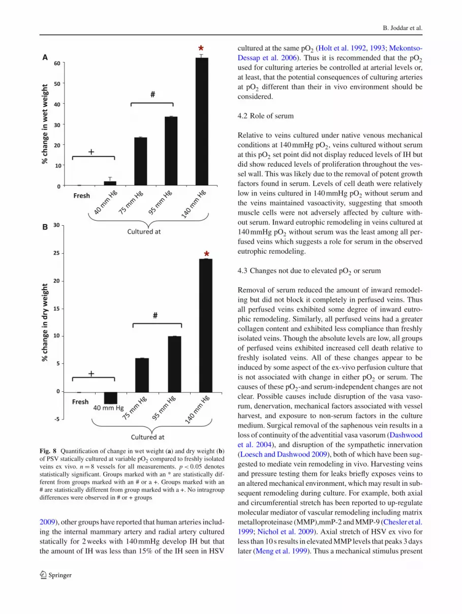
(Table 4). In contrast, the numbers of DAPI-stained nuclei inthe intima and in the media of vessels cultured at venous pO2were comparable to freshly isolated vessels (Table 4). Theobserved increases in intimal and medial area for PSV cul-tured at 75–140 mmHg pO2 were associated with an increasein wet and dry weight of the tissue (Fig. 8).
3.2.4 4-HNE adduct formation
PSV cultured at 140 mmHg pO2 showed a greater intensityof 4-HNE immunostaining and western blot band intensitywhen compared to fresh PSV or PSV cultured at 40 mmHgpO2 (Fig. 9). The protein adduct intensity of 4-HNE wasundetectable in fresh PSV (Fig. 9).
4 Discussion
Saphenous veins cultured ex vivo with mechanical conditionsintended to mimic their native environment rapidly developIH, alter their biomechanical properties, and exhibit inwardeutrophic remodeling (Gusic et al. 2005a,b). It is difficult tounderstand these changes in the cultured vessels as resultingsolely from mechanically induced remodeling, which led usto consider other aspects of the ex-vivo culture environmentthat might stimulate these changes.
4.1 Role of elevated pO2
The oxygen tension typically used in tissue culture is signif-icantly greater than venous pO2. We hypothesized that expo-sure to this elevated pO2 stimulated some of the changesobserved in cultured veins. Consistent with this hypothesis,relative to PSV culture with 140 mmHg, PSV cultured undernative venous mechanical conditions for 7 days with loweroxygen tensions exhibited reduced IH, reduced proliferationin the intima and media, and less disruption of the IEL. PSVcultured statically for 2 weeks at venous pO2 were indistin-guishable from freshly isolated PSV with respect to theseparameters. The dramatic impact that exposure to elevatedpO2 had on IH in PSV was not expected since it is widelybelieved that aspects of the mechanical environment are the
primary stimuli for IH in SV used as grafts in the arterial cir-culation (Dobrin PB and Endean 1989; Motwani and Topol1998; Grabellus et al. 2007). There are other studies, how-ever, that also suggest that exposure to an arterial mechanicalenvironment is not a primary cause of IH in SV. Porter et al.reported that relative to static conditions, HSV cultured exvivo with a combination of venous levels of pressure andshear stress developed less IH; arterial levels of pressureand shear stress further reduced IH (Porter et al. 1996b).Similarly, using perfused PSV, we reported similar observa-tions to Porter et al. We further explored the role of specificflow-induced shear stress, which is greater in the arterialcirculation than the venous circulation. Across five sets ofmechanical conditions, there was a monotonic decrease inIH with increasing flow-induced shear stress (Gusic et al.2005a,b). These ex vivo observations of elevated averageshear stresses inhibiting IH in PSV are consistent with theclinical observation that HSV used for CABG have bet-ter patency when installed in areas with good distal out-flow (Motwani and Topol 1998) as well as the notion thatsteady and pulsatile shear stress is athero-protective forarteries (Davies 1995). Relative to freshly isolated PSV, cel-lular proliferation was elevated in the media of PSV cul-tured under native venous mechanical conditions with serumand at 140 mmHg pO2. In PSV perfused for 7 days undervenous mechanical conditions, the increase in proliferationwas accompanied by only an 18% increase in wall area,which was reduced to an 8% increase by culturing with a pO2of 75 mmHg (Table 1). The increased cell death observed inPSV due to culture at low levels of pO2 (75 mmHg), mayhave potentially offset the elevated cellular proliferation andresulted in reduced IH. However, in statically cultured veins,decreasing pO2 inhibited IH without increasing cell death.In PSV cultured statically for 2 weeks at 140 mmHg pO2, theincrease in medial area was much greater ∼150% (Fig. 6b).It is possible that the greater increase in medial area withthe statically perfused veins is partially due to the longer cul-ture duration. We chose to statically culture veins for 2 weeksinstead of the 1 week we used for the perfused veins, to facili-tate comparison of our results to a number of reports by otherswith vessels cultured statically for 2 weeks (Soyombo et al.1990; Masood et al. 1997; Mekontso-Dessap et al. 2006).
123

Arterial pO2 stimulates intimal hyperplasia
*
#
A
*
#
B
Fig. 6 Quantification of (a) intimal and (b) medial areas from PSVeither freshly isolated or cultured in a static system ex-vivo. Culturedveins were maintained at variable pO2 (140, 95, 75 and 40 mmHg) asshown above. n = 8 vessels for all area measurements. p < 0.05 denotesstatistically significant. Groups marked with an *are statistically differ-ent from groups marked with an #. There were no intragroup differencesin both groups
In addition, the larger increase in wall area was likely par-tially due to increased water content. This was because theincrease in dry weight of the tissue was a more modest 30%.
#
*
Fig. 7 Quantification of cell proliferation or mitotic index from por-cine saphenous veins either freshly isolated or cultured in a static systemex-vivo. Cultured veins were maintained at variable pO2 (140, 95, 75and 40 mmHg) as shown above. n = 8 vessels for all measurements.p < 0.05 denotes statistically significant. Groups marked with an* are statistically different from groups marked with an #. There were nointragroup differences in groups marked with an #. In groups markedwith an * cell proliferation in the media in PSV cultured at 95 and140 mmHg pO2 both were statistically different from cell proliferationin the media of vessels cultured at 75 mmHg pO2. groups. Cell prolifer-ation in the intima when compared in between PSV cultured at 95 and140 mmHg pO2 were not statistically different
In addition to affecting cell proliferation, intimal and insome cases medial area, veins cultured at reduced pO2 hadnoticeably different stress-strain curves both in the presenceand absence of calcium relative to freshly isolated veins orthose cultured in the presence of high pO2 and serum. Rel-ative to veins cultured with higher pO2 and serum, thosecultured with lower pO2 and serum exhibited greater basaltone as evidenced by the shift in the pressure-diameter andstress-strain curves for these vessels upon the removal of cal-cium. Even in the presence of higher pO2, veins cultured inthe absence of serum exhibited basal tone indicating a non-linear interaction between pO2 and serum.
Arteries are typically cultured ex vivo with a pO2 of140 mmHg, which is greater than the 95 mmHg of arterialblood. While this increase in pO2 for cultured arteries is notas great as that for cultured veins, it is possible the exposureto elevated pO2 might have some effects on cultured arteries.While we do not see IH in pig carotid arteries perfused ex vivofor 1 week at 140 mmHg (Clerin et al. 2002, 2003; Lawrenceet al. 2003; Lawrence and Gooch 2009a,b; Nichol et al. 2005,
123
B. Joddar et al.
*
*
#
#
A
B
+
+
Fig. 8 Quantification of change in wet weight (a) and dry weight (b)of PSV statically cultured at variable pO2 compared to freshly isolatedveins ex vivo. n = 8 vessels for all measurements. p < 0.05 denotesstatistically significant. Groups marked with an * are statistically dif-ferent from groups marked with an # or a +. Groups marked with an# are statistically different from group marked with a +. No intragroupdifferences were observed in # or + groups
2009), other groups have reported that human arteries includ-ing the internal mammary artery and radial artery culturedstatically for 2 weeks with 140 mmHg develop IH but thatthe amount of IH was less than 15% of the IH seen in HSV
cultured at the same pO2 (Holt et al. 1992, 1993; Mekontso-Dessap et al. 2006). Thus it is recommended that the pO2used for culturing arteries be controlled at arterial levels or,at least, that the potential consequences of culturing arteriesat pO2 different than their in vivo environment should beconsidered.
4.2 Role of serum
Relative to veins cultured under native venous mechanicalconditions at 140 mmHg pO2, veins cultured without serumat this pO2 set point did not display reduced levels of IH butdid show reduced levels of proliferation throughout the ves-sel wall. This was likely due to the removal of potent growthfactors found in serum. Levels of cell death were relativelylow in veins cultured in 140 mmHg pO2 without serum andthe veins maintained vasoactivity, suggesting that smoothmuscle cells were not adversely affected by culture with-out serum. Inward eutrophic remodeling in veins cultured at140 mmHg pO2 without serum was the least among all per-fused veins which suggests a role for serum in the observedeutrophic remodeling.
4.3 Changes not due to elevated pO2 or serum
Removal of serum reduced the amount of inward remodel-ing but did not block it completely in perfused veins. Thusall perfused veins exhibited some degree of inward eutro-phic remodeling. Similarly, all perfused veins had a greatercollagen content and exhibited less compliance than freshlyisolated veins. Though the absolute levels are low, all groupsof perfused veins exhibited increased cell death relative tofreshly isolated veins. All of these changes appear to beinduced by some aspect of the ex-vivo perfusion culture thatis not associated with change in either pO2 or serum. Thecauses of these pO2-and serum-independent changes are notclear. Possible causes include disruption of the vasa vaso-rum, denervation, mechanical factors associated with vesselharvest, and exposure to non-serum factors in the culturemedium. Surgical removal of the saphenous vein results in aloss of continuity of the adventitial vasa vasorum (Dashwoodet al. 2004), and disruption of the sympathetic innervation(Loesch and Dashwood 2009), both of which have been sug-gested to mediate vein remodeling in vivo. Harvesting veinsand pressure testing them for leaks briefly exposes veins toan altered mechanical environment, which may result in sub-sequent remodeling during culture. For example, both axialand circumferential stretch has been reported to up-regulatemolecular mediator of vascular remodeling including matrixmetalloproteinase (MMP),mmP-2 and MMP-9 (Chesler et al.1999; Nichol et al. 2009). Axial stretch of HSV ex vivo forless than 10 s results in elevated MMP levels that peaks 3 dayslater (Meng et al. 1999). Thus a mechanical stimulus present
123

Arterial pO2 stimulates intimal hyperplasia
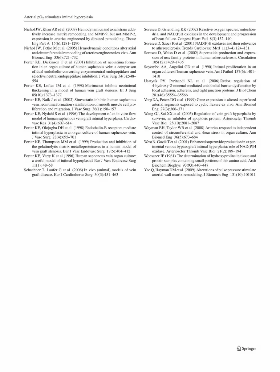
∗∗Sample
At 140 mm Hg pO2 At 40 mm Hg pO24-HNE Michael adducts formed in PSV cultured at 140 mm Hg pO2
Media
A B C
Fig. 9 Quantification of 4-HNE adduct intensity. Expression of 4-HNEnormalized with actin in PSV either freshly isolated or cultured at vari-able pO2. a Western blot bands for 4-HNE protein adducts varying from25–48 KDa. Samples marked with ‘140’ indicate porcine SV culturedat 140 mmHg, and ‘40’ indicate those cultured at 40 mmHg. Freshlyisolated PSV are indicated by ‘fresh’. b Expression of 4-HNE bandintensities normalized to β-actin, n = 8 for all groups. Normalized
4-HNE adduct band intensities of PSV cultured at 140 mmHg pO2 aremarked with *and are significantly different from the other group cul-tured at 40 mmHg pO2. In the freshly isolated PSV no bands for 4-HNEwere detected, and so no quantification for this group is included onthe graph shown. c Immunostained 4-HNE Michael adducts formed inPSV cultured at 140 mmHg pO2
during vessel harvest or preparation might manifest itself asremodeling days later. Finally, it is possible that biochem-ical factors other than those in serum might stimulate theobserved changes. It is possible that the observed changes inthe cultured vessels that do not initially appear to be due tochanges in the mechanical environment are actually due toour inability to adequately reproduce the in vivo mechanicalenvironment in the ex-vivo perfusion system. For example,though we attempted to reproduce/control many aspects ofmechanical environment (e.g., flow, pressure, axial stretchratio) to match venous conditions in vivo, it is possible thatrelatively small differences between the in vivo and ex vivovalues of these parameters might be responsible for someforms of the remodeling observed. It is possible that addi-tional mechanical factors that are not present or well con-trolled in our ex-vivo system might stimulate remodeling.Two examples of such factors that have been suggested tostimulate vascular remodeling include flexure, (i.e., bendingof blood vessels often due to tissue motion) (Vorp et al. 1999)as well as tethering or compressive forces of the tissue actingon the adventitia (Jackson et al. 2002).
4.4 Role of pO2-induced IH and potential relevanceto vein graft failure following CABG
The observation that veins cultured ex vivo remodel inde-pendent of changes in the pO2, presence of serum, or well-defined changes in the mechanical environment suggests thatcaution should be taken when interpreting data from suchsystems. Despite this limitation, ex-vivo culture of vesselsremain an attractive tool to study vascular remodeling, espe-cially as it allows for isolation and control of variables thatwould be very difficult to accomplish in vivo. pO2 and the
mechanical environment are examples of variables that aredifficult to decouple in vivo. HSV used for CABG or periph-eral revascularization are exposed to both arterial pO2 andhigher pressures and flows associated with the arterial cir-culation. By controlling pO2 independent of the mechanicalenvironments, exposure of veins to arterial pO2 was shown tostimulate IH and increase SMC proliferation across a varietyof mechanical environments. Though the molecular mech-anisms relating an increase in pO2and IH are not known,increased oxidative stress due to the former could be onepossible mechanism. While pO2is not a widely recognizedcontributor to IH, the role of oxidative stress has often beendiscussed in the context of a number of vascular diseasesincluding atherosclerosis and restenosis (Griendling et al.2000; Lassegue et al. 2001; Jeremy et al. 2002; Sorescu et al.2001, 2002a; Sorescu and Griendling 2002). Due to oxida-tive stress, superoxide production is elevated in porcine andhuman vein grafts, relative to arterial grafts (West et al. 2001).Therefore, we cultured porcine femoral arteries alongsidePSV at the same level of pO2 (arterial pO2 ∼95 mmHg),based upon the hypothesis that an arterial pO2 environmentwould not impose oxidative stress on the artery but only onthe PSV which was removed from a low venous pO2 andplaced within an arterial pO2 environment. Oxidative stresscan lead to many biochemical perturbations, including pro-tein or DNA damage (Jaimes et al. 2001) or lipid peroxidation(Usatyuk et al. 2006). Lipid peroxidation products such as4-HNE can in turn lead to activation of downstream signal-ing pathways such as ERK, JNK, and p38 MAPK (Usatyuket al. 2006), which are also involved in cellular prolifera-tion and migration (Nelson et al. 1998). In our study, theenhanced intensity of the 4-HNE adducts formed in porcineSV cultured at 140 mmHg pO2 compared to the other groups
123

B. Joddar et al.
implicate that oxygen sensitive pathways such as MAPKmight be involved in this pO2-induced intimal hyperplasiaevident in porcine SV cultured at higher than venous pO2.Further these 4-HNE adducts were mostly located in theneointimal and medial region of the cultured PSV. Since thisregion is mostly made of SMC, it can be inferred that thispO2 -induced IH involves redox-dependant 4-HNE pathwayswhich regulate smooth muscle cell proliferation (Kakishitaand Hattori 2001). Our findings therefore not only suggest asignificant role for elevated pO2 in the pathological remod-eling of veins ex vivo, but also in the development of IH inHSV grafted into the arterial circulation in vivo.
Acknowledgments This work was supported by AHA (grants0555538U and 0655323B) to KJG.
Open Access This article is distributed under the terms of the CreativeCommons Attribution Noncommercial License which permits anynoncommercial use, distribution, and reproduction in any medium,provided the original author(s) and source are credited.
References
Bourassa MG (1991) Fate of venous grafts: the past, the present and thefuture. J Am Coll Cardiol 17(5):1081–1083
Chesler NC, Ku DN et al (1999) Transmural pressure induces matrix-degrading activity in porcine arteries ex vivo. Am J Physiol HeartCirc Physiol 277(5):H2002–H2009
Clerin V, Gusic RJ et al (2002) Mechanical environment, donor age,and presence of endothelium interact to modulate porcine arteryviability ex vivo. Ann Biomed Eng 30(9):1117–1127
Clerin V, Nichol JW et al (2003) Tissue engineering of arteriesby directed remodeling of intact arterial segments. Tissue Eng9(3):461–472
Corpataux JM, Naik J et al (2005) A comparison of six statins on thedevelopment of intimal hyperplasia in a human vein culture model.Eur J Vasc Endovasc Surg 29(2):177–181
Cox RH, Bockus Institute PP (1978) Comparison of carotid arterymechanics in the rat, rabbit, and dog. Am J Physiol Heart CircPhysiol 234(3):H280–H288
Dashwood MR, Anand R et al (2004) Hypothesis: a potential role forthe vasa vasorum in the maintenance of vein graft patency. Angi-ology 55(4):385–395
Davies PF (1995) Flow-mediated endothelial mechanotransduction.Physiol Rev 75(3):519–560
Davis NP, Han HC et al (2005) Sustained axial loading lengthens arter-ies in organ culture. Ann Biomed Eng 33(7):867–877
Dobrin PB, Endean ED (1989) Mechanical factors predisposing to inti-mal hyperplasia and medial thickening in autogenous vein grafts.Surgery 105(3):393–400
George SJ, Williams A et al (1996) An essential role for platelet-derived growth factor in neointima formation in human saphenousvein in vitro. Atherosclerosis 120(1–2):227–240
Gleason RL, Wilson E et al (2007) Biaxial biomechanical adaptationsof mouse carotid arteries cultured at altered axial extension. J Bio-mech 40(4):766–776
Grabellus F, Worm K et al (2007) Induction of the matrix metallo-proteinase-2 activation system in arteries by tensile stress.Involvement of the p38 MAP-kinase pathway. Pathol Res Pract203(3):135–143
Griendling KK, Sorescu D et al (2000) NAD(P)H oxidase: role in car-diovascular biology and disease. Circ Res 86(5):494–501
Gusic RJ, Myung R et al (2005) Shear stress and pressuremodulate saphenous vein remodeling ex vivo. J Biomech 38(9):1760–1769
Gusic RJ, Petko M et al (2005) Mechanical properties of native and exvivo remodeled porcine saphenous veins. J Biomech 38(9):1770–1779
Han HC, Ku DN et al (2003) Arterial wall adaptation under elevatedlongitudinal stretch in organ culture. Ann Biomed Eng 31(4):403–411
Han HC, Marita S et al (2006) Changes of opening angle in hyperten-sive and hypotensive arteries in 3-day organ culture. J Biomech39(13):2410–2418
Holt CM, Francis SE et al (1993) Comparison of response to injuryin organ culture of human saphenous vein and internal mammaryartery. Ann Thorac Surg 55(6):1522–1528
Holt CM, Francis SE et al (1992) Intimal proliferation in an organculture of human internal mammary artery. Cardiovasc Res26(12):1189–1194
Jackson ZS, Gotlieb AI et al (2002) Wall tissue remodeling regulateslongitudinal tension in arteries. Circ Res 90(8):918–925
Jaimes EA, Sweeney C et al (2001) Effects of the reactive oxygenspecies hydrogen peroxide and hypochlorite on endothelial nitricoxide production. Hypertension 38(4):877–883
Jeremy JY, Birkett SD et al (1997) The influence of surgicalpreparation on cyclic nucleotide synthesis in an organ cultureof human saphenous vein. Eur J Vasc Endovasc Surg 13(1):72–78
Jeremy JY, Yim AP et al (2002) Oxidative stress, nitric oxide, andvascular disease. J Card Surg 17(4):324–327
Kakishita H, Hattori Y (2001) Vascular smooth muscle cell activationand growth by 4-hydroxynonenal. Life Sci 69(6):689–697
Lassegue B, Sorescu D et al (2001) Novel gp91(phox) homologuesin vascular smooth muscle cells : nox1 mediates angiotensinII-induced superoxide formation and redox-sensitive signalingpathways. Circ Res 88(9):888–894
Lawrence AR, Gooch KJ (2009) Differences in transmural pressure andaxial loading ex vivo affect arterial remodeling and material prop-erties. J Biomech Eng 131(10):101009
Lawrence AR, Gooch KJ (2009) Transmural pressure and axial load-ing interactively regulate arterial remodeling ex vivo. Am J PhysiolHeart Circ Physiol 297(1):H475–H484
Lawrence AR, Gusic RJ et al (2003) Noninvasive determination ofperfused artery dimensions ex vivo using a pressure-diameter rela-tionship. Biorheology 40(5):523–529
Loesch A, Dashwood MR (2009) On the sympathetic innervation of thehuman greater saphenous vein: relevance to clinical practice. CurrVasc Pharmacol 7(1):58–67
Masood I, Porter KE et al (1997) Endothelin-1 is a mediator of intimalhyperplasia in organ culture of human saphenous vein. Br J Surg84(4):499–503
Mekontso-Dessap A, Kirsch M et al (2006) Vascular-wall remodelingof 3 human bypass vessels: organ culture and smooth muscle cellproperties. J Thorac Cardiovasc Surg 131(3):651–658
Meng X, Mavromatis K et al (1999) Mechanical stretching of humansaphenous vein grafts induces expression and activation of matrix-degrading enzymes associated with vascular tissue injury andrepair. Exp Mol Pathol 66(3):227–237
Motwani JG, Topol EJ (1998) Aortocoronary saphenous vein graftdisease: pathogenesis, predisposition, and prevention. Circulation97(9):916–931
Nelson PR, Yamamura S et al (1998) Smooth muscle cell migration andproliferation are mediated by distinct phases of activation of theintracellular messenger mitogen-activated protein kinase. J VascSurg 27(1):117–125
123
Arterial pO2 stimulates intimal hyperplasia
Nichol JW, Khan AR et al (2009) Hemodynamics and axial strain addi-tively increase matrix remodeling and MMP-9, but not MMP-2,expression in arteries engineered by directed remodeling. TissueEng Part A 15(6):1281–1290
Nichol JW, Petko M et al (2005) Hemodynamic conditions alter axialand circumferential remodeling of arteries engineered ex vivo. AnnBiomed Eng 33(6):721–732
Porter KE, Dickinson T et al (2001) Inhibition of neointima forma-tion in an organ culture of human saphenous vein: a comparisonof dual endothelin-converting enzyme/neutral endopeptidase andselective neutral endopeptidase inhibition. J Vasc Surg 34(3):548–554
Porter KE, Loftus IM et al (1998) Marimastat inhibits neointimalthickening in a model of human vein graft stenosis. Br J Surg85(10):1373–1377
Porter KE, Naik J et al (2002) Simvastatin inhibits human saphenousvein neointima formation via inhibition of smooth muscle cell pro-liferation and migration. J Vasc Surg 36(1):150–157
Porter KE, Nydahl S et al (1996) The development of an in vitro flowmodel of human saphenous vein graft intimal hyperplasia. Cardio-vasc Res 31(4):607–614
Porter KE, Olojugba DH et al (1998) Endothelin-B receptors mediateintimal hyperplasia in an organ culture of human saphenous vein.J Vasc Surg 28(4):695–701
Porter KE, Thompson MM et al (1999) Production and inhibition ofthe gelatinolytic matrix metalloproteinases in a human model ofvein graft stenosis. Eur J Vasc Endovasc Surg 17(5):404–412
Porter KE, Varty K et al (1996) Human saphenous vein organ culture:a useful model of intimal hyperplasia? Eur J Vasc Endovasc Surg11(1): 48–58
Schachner T, Laufer G et al (2006) In vivo (animal) models of veingraft disease. Eur J Cardiothorac Surg 30(3):451–463
Sorescu D, Griendling KK (2002) Reactive oxygen species, mitochon-dria, and NAD(P)H oxidases in the development and progressionof heart failure. Congest Heart Fail 8(3):132–140
Sorescu D, Szocs K et al (2001) NAD(P)H oxidases and their relevanceto atherosclerosis. Trends Cardiovasc Med 11(3–4):124–131
Sorescu D, Weiss D et al (2002) Superoxide production and expres-sion of nox family proteins in human atherosclerosis. Circulation105(12):1429–1435
Soyombo AA, Angelini GD et al (1990) Intimal proliferation in anorgan culture of human saphenous vein. Am J Pathol 137(6):1401–1410
Usatyuk PV, Parinandi NL et al (2006) Redox regulation of4-hydroxy-2-nonenal-mediated endothelial barrier dysfunction byfocal adhesion, adherens, and tight junction proteins. J Biol Chem281(46):35554–35566
Vorp DA, Peters DG et al (1999) Gene expression is altered in perfusedarterial segments exposed to cyclic flexure ex vivo. Ann BiomedEng 27(3):366–371
Wang GJ, Sui XX et al (2005) Regulation of vein graft hyperplasia bysurvivin, an inhibitor of apoptosis protein. Arterioscler ThrombVasc Biol 25(10):2081–2087
Wayman BH, Taylor WR et al (2008) Arteries respond to independentcontrol of circumferential and shear stress in organ culture. AnnBiomed Eng 36(5):673–684
West N, Guzik T et al (2001) Enhanced superoxide production in exper-imental venous bypass graft intimal hyperplasia: role of NAD(P)Hoxidase. Arterioscler Thromb Vasc Biol 21(2):189–194
Woessner JF (1961) The determination of hydroxyproline in tissue andprotein samples containing small portions of this amino acid. ArchBiochem Biophys 93(93):440–447
Yao Q, Hayman DM et al (2009) Alterations of pulse pressure stimulatearterial wall matrix remodeling. J Biomech Eng 131(10):101011
123