prevalence and clinical implications of incidentally detected
TRANSCRIPT
medicina
Article
Prevalence and Clinical Implications of IncidentallyDetected Parotid Lesions as Blind Spot on Brain MRI:A Single-Center Experience
In-Chul Nam 1, Hye-Jin Baek 1,2,* , Kyeong-Hwa Ryu 1, Jin-Il Moon 1, Eun Cho 1 , Hyo-Jung An 3, Seokho Yoon 4
and Jiyeon Baik 5
�����������������
Citation: Nam, I.-C.; Baek, H.-J.;
Ryu, K.-H.; Moon, J.-I.; Cho, E.;
An, H.-J.; Yoon, S.; Baik, J. Prevalence
and Clinical Implications of
Incidentally Detected Parotid Lesions
as Blind Spot on Brain MRI: A
Single-Center Experience. Medicina
2021, 57, 836. https://doi.org/
10.3390/medicina57080836
Academic Editor: Eng Ooi
Received: 15 June 2021
Accepted: 16 August 2021
Published: 18 August 2021
Publisher’s Note: MDPI stays neutral
with regard to jurisdictional claims in
published maps and institutional affil-
iations.
Copyright: © 2021 by the authors.
Licensee MDPI, Basel, Switzerland.
This article is an open access article
distributed under the terms and
conditions of the Creative Commons
Attribution (CC BY) license (https://
creativecommons.org/licenses/by/
4.0/).
1 Department of Radiology, Gyeongsang National University School of Medicine and Gyeongsang NationalUniversity Changwon Hospital, Changwon 51472, Korea; [email protected] (I.-C.N.);[email protected] (K.-H.R.); [email protected] (J.-I.M.); [email protected] (E.C.)
2 Institute of Health Sciences, Gyeongsang National University School of Medicine, Jinju 52727, Korea3 Department of Pathology, Gyeongsang National University School of Medicine and Gyeongsang National
University Changwon Hospital, Changwon 51472, Korea; [email protected] Department of Nuclear Medicine and Molecular Imaging, Gyeongsang National University School of
Medicine and Gyeongsang National University Changwon Hospital, Changwon 51472, Korea;[email protected]
5 Department of Radiology, Haeundae Paik Hospital, Inje University College of Medicine, Busan 48108, Korea;[email protected]
* Correspondence: [email protected]
Abstract: Background and objective: This study was conducted to assess the prevalence and clinicalimplications of parotid lesions detected incidentally during brain magnetic resonance imaging (MRI)examination. Materials and Methods: Between February 2016 and February 2021, we identified 86 le-sions in the brain MRI reports of 84 patients that contained the words “parotid gland” or “PG”. Ofthese, we finally included 49 lesions involving 45 patients following histopathological confirma-tion. Results: Based on the laboratory, radiological or histopathological findings, the prevalence ofincidental parotid lesions was low (1.2%). Among the 45 study patients, 41 (91.1%) had unilaterallesions, and the majority of the lesions were located in the superficial lobe (40/49, 81.6%). Themean size of the parotid lesions was 1.3 cm ± 0.4 cm (range, 0.5 cm–2.8 cm). Of these, 46 parotidlesions (93.9%) were benign, whereas the remaining three lesions were malignant (6.1%). Conclusions:Despite the low prevalence and incidence of malignancy associated with incidental parotid lesionsdetected on brain MRI, the clinical implications are potentially significant. Therefore, clinical aware-ness and appropriate imaging work-up of these lesions are important for accurate diagnosis andtimely management.
Keywords: incidental parotid lesions; parotid gland; parotid neoplasm; brain; magnetic resonanceimaging
1. Introduction
Radiologists frequently encounter incidental findings in all imaging modalities duringroutine clinical practice [1–3]. Over the years, the prevalence of incidental findings hasincreased because of the increased demand for accurate diagnostic imaging, as well as rapidtechnical improvements and diagnostic quality of imaging modalities in the radiologydepartment [4–6]. In particular, the number of brain magnetic resonance imagings (MRIs)for evaluating various intracranial diseases has dramatically increased. Thus, radiologistsmay increasingly encounter asymptomatic incidental parotid lesions within the scan range.However, the management of incidental lesions detected on brain MRI is limited by alack of knowledge regarding their clinical significance, partial visualization of lesions, andnon-availability of dedicated additional sequences for the incidental lesions [7].
Medicina 2021, 57, 836. https://doi.org/10.3390/medicina57080836 https://www.mdpi.com/journal/medicina
Medicina 2021, 57, 836 2 of 10
The parotid gland is the largest salivary gland and is located in the parotid spacenear the ear [8]. A wide variety of lesions are associated with the parotid gland, suchas congenital anomalies, inflammatory or infectious processes, and benign or malignantneoplasms [9]. Although the majority of the parotid lesions are benign [10], incidentalparotid lesions may require additional diagnostic work-ups to exclude malignancies andfacilitate appropriate management. A few imaging studies have reported the presenceof unexpected asymptomatic parotid lesions; however, these studies mainly focused onparotid lesions detected via 18-F fluorodeoxyglucose (FDG) positron emission tomography(PET) or PET–computed tomography (CT) [11–15]. In contrast, the clinical significance ofincidental parotid lesions detected via other more commonly utilized imaging modalitiesremains unclear [16]. Furthermore, the frequency of the incidental findings in previousstudies has varied according to the imaging modality used and the area of interest in-vestigated [11–16]. Therefore, the purpose of our retrospective study was to assess theprevalence and clinical implications of incidental parotid lesions as potential blind spotsdetected during brain MRI examination.
2. Materials and Methods2.1. Patients
The Institutional Review Board of Gyeongsang National University Changwon Hospi-tal approved the study (No.: GNUCH 2021-03-012) on 25 March 2021. However, no patientapproval or informed consent was required due to the retrospective nature of the study.
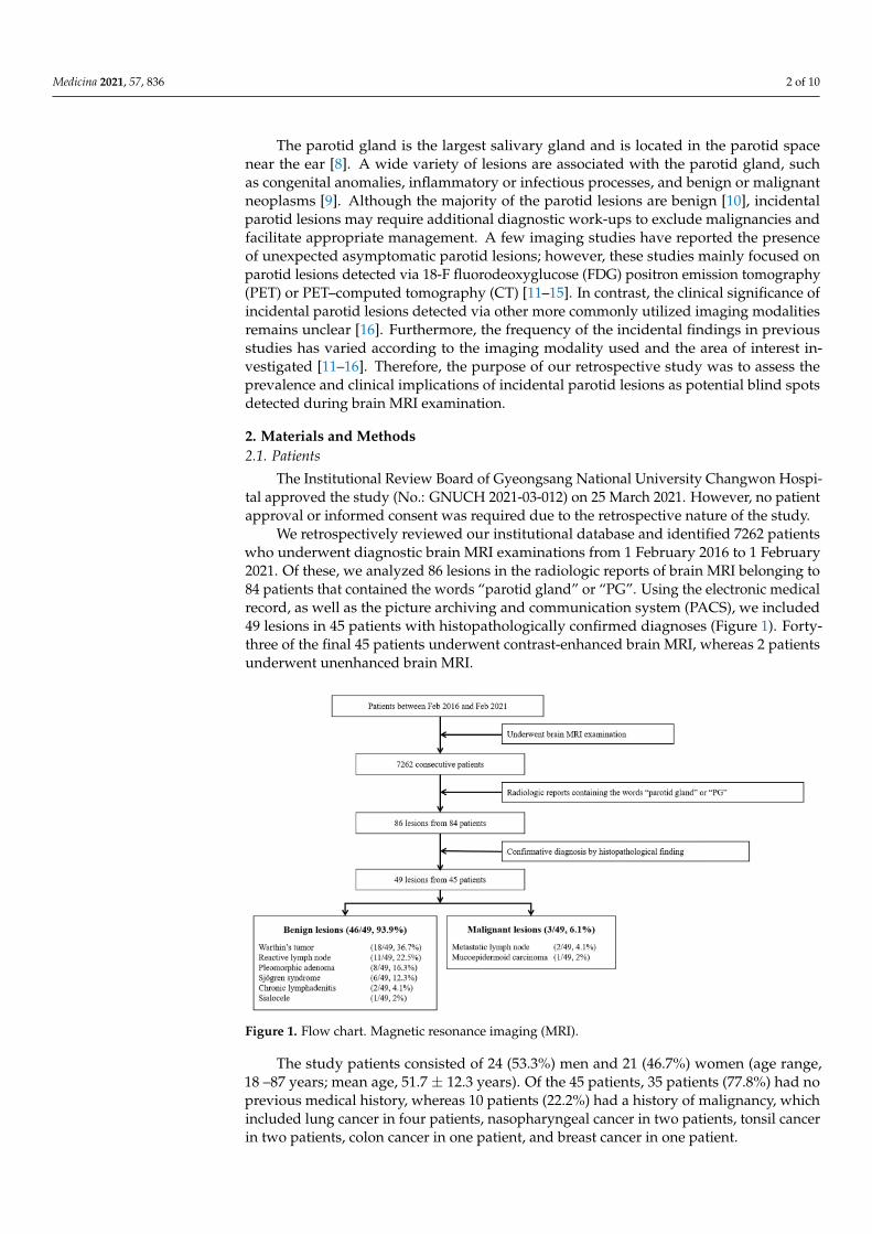
We retrospectively reviewed our institutional database and identified 7262 patientswho underwent diagnostic brain MRI examinations from 1 February 2016 to 1 February2021. Of these, we analyzed 86 lesions in the radiologic reports of brain MRI belonging to84 patients that contained the words “parotid gland” or “PG”. Using the electronic medicalrecord, as well as the picture archiving and communication system (PACS), we included49 lesions in 45 patients with histopathologically confirmed diagnoses (Figure 1). Forty-three of the final 45 patients underwent contrast-enhanced brain MRI, whereas 2 patientsunderwent unenhanced brain MRI.
Figure 1. Flow chart. Magnetic resonance imaging (MRI).
The study patients consisted of 24 (53.3%) men and 21 (46.7%) women (age range,18 –87 years; mean age, 51.7 ± 12.3 years). Of the 45 patients, 35 patients (77.8%) had noprevious medical history, whereas 10 patients (22.2%) had a history of malignancy, whichincluded lung cancer in four patients, nasopharyngeal cancer in two patients, tonsil cancerin two patients, colon cancer in one patient, and breast cancer in one patient.
Medicina 2021, 57, 836 3 of 10
2.2. Brain MRI Parameters
MRI was performed using two 3T MR scanners. Our conventional brain MRI protocolincluded the following sequences: axial T2-weighted fast spin-echo imaging with the Dixontechnique, axial T1-weighted fluid attenuation inversion recovery imaging (T1-FLAIR),axial T2-weighted fluid attenuation inversion recovery imaging (T2-FLAIR), diffusion-weighted imaging, and susceptibility-weighted imaging with or without contrast-enhanced3D T1-weighted gradient-echo imaging. The examination of 25 patients was performed ona 3T MR scanner with a 48-channel head coil (Signa™ Architect; GE Healthcare, Waukesha,WI, USA), whereas the other 20 patients underwent brain MRI using another 3T MR scannerwith a 32-channel head coil (Ingenia 3.0 CX; Philips Healthcare, Best, The Netherlands).
2.3. Assessment of Incidental Parotid Lesions: Image Analyses
The brain MRIs were reviewed by an attending neuroradiologist (H.J.B) with 11 yearsof experience evaluating incidental parotid lesions within the scan range. The reader as-sessed the digital PACS for the lesion size, location (superficial lobe/deep lobe), bilaterality,multiplicity, contrast enhancement, and additional imaging studies (i.e., neck ultrasound(US), neck CT, PET–CT, or salivary gland scintigraphy).
A clinical lecturer with one year of experience in interventional radiology (I.C.N)reviewed and categorized the parotid lesions according to the final diagnosis based onclinical and laboratory findings as well as histopathological analyses using electronicmedical records.
2.4. Statistical Analysis
The prevalence of incidental parotid lesions and individual disease entities was an-alyzed statistically. Continuous variables were expressed as the mean ± standard devia-tion (SD).
3. Results
Among the total 86 parotid lesions detected on brain MRI incidentally, 49 lesionswere confirmed via US-guided fine-needle aspiration biopsy (31/49, 63.3%) or US-guidedcore needle biopsy (18/49, 36.7%) in 45 patients, including four patients who underwentUS-guided core needle biopsy for bilateral parotid lesions. In the study, 28 patients (62.2%)underwent superficial parotidectomies.
Of the 49 lesions, 47 (95.9%) were only detected on contrast-enhanced 3D T1-weightedimages. All 49 lesions were evaluated in subsequent imaging studies, including neckUS (49/49, 100%), neck CT (18/49, 36.7%), PET–CT (9/49, 18.4%), and salivary glandscintigraphy (6/49, 12.2%).
Of the 45 patients, only four patients (8.9%) carried bilateral parotid lesions. However,the lesions of only two of these four patients were identified on brain MRI because of thelimited scan range and head tilting during the scan. Bilateral parotid lesions were confirmedin the other two patients via other imaging studies (US and salivary scintigraphy).
In addition, the majority of the parotid lesions (40/49, 81.6%) were found in thesuperficial lobe. Five of the remaining nine lesions (5/49, 10.2%) were located in the deeplobe, whereas four lesions (4/49, 8.2%) were identified in both superficial and deep lobes.The mean size of the incidental parotid lesions was 1.3 ± 0.4 cm with a range of 0.5–2.8 cm;17 lesions were less than 1 cm (34.7%), 25 lesions were between 1 and 2 cm (51%), and7 lesions (14.3%) were larger than 2 cm.
The final diagnosis based on histopathological reports, as well as clinical and labora-tory findings, indicated that 46 parotid lesions (93.9%) were benign and only three (6.1%)were malignant (Table 1). Of the 46 benign parotid lesions, 40 (40/46, 87%) were focallesions manifesting as nodules or masses, whereas the remaining 6 (13%) were diffuseglandular lesions. The most common diagnosis of the 40 focal lesions was Warthin tumor(18/49, 36.7%), followed by reactive lymph node (11/49, 22.5%), and pleomorphic adenoma(8/49, 16.3%) (Figures 2 and 3). However, only one of the patients with a Warthin tumor
Medicina 2021, 57, 836 4 of 10
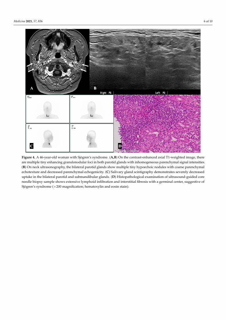
showed bilaterality. The six diffuse glandular lesions with bilateral involvement in threepatients were associated with Sjögren’s syndrome characterized by decreased salivaryexcretion in the bilateral parotid and submandibular glands in salivary gland scintigraphy,as well as positive test results for anti-SS-A or anti-SS-B antibodies (Figure 4).
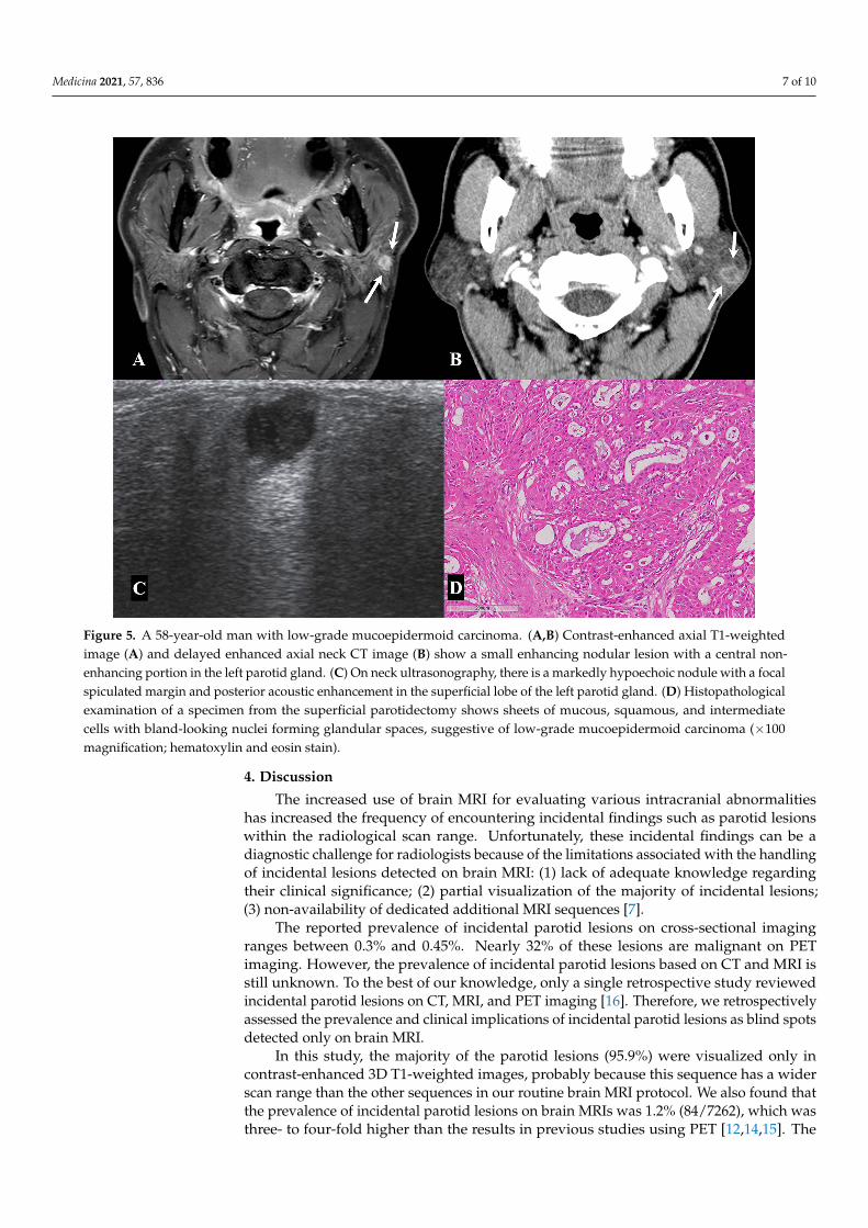
The three malignant parotid lesions comprised metastatic lymph nodes (2/49, 4.1%)and mucoepidermoid carcinoma (1/49, 2%) (Figure 5).
Table 1. Final diagnosis of 47 incidental parotid lesions based on histopathologic results.
Histopathology (n = 49) Number of Lesions (%)
Benign Lesion (n = 46)Warthin tumor 18/49 (36.7%)
Reactive lymph node 11/49 (22.5%)Pleomorphic adenoma 8/49 (16.3%)
Sjögren’s syndrome 6/49 (12.3%)Chronic lymphadenitis 2/49 (4.1%)
Sialocele 1/49 (2 %)Malignant lesion (n = 3)Metastatic lymph node 2/49 (4.1%)
Mucoepidermoid carcinoma 1/49 (2 %)
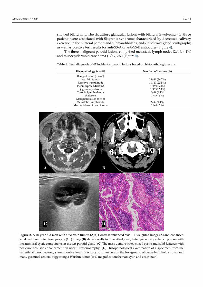
Figure 2. A 48-year-old man with a Warthin tumor. (A,B) Contrast-enhanced axial T1-weighted image (A) and enhancedaxial neck computed tomography (CT) image (B) show a well-circumscribed, oval, heterogeneously enhancing mass withintratumoral cystic components in the left parotid gland. (C) The mass demonstrates mixed cystic and solid features withposterior acoustic enhancement on neck ultrasonography. (D) Histopathological examination of a specimen from thesuperficial parotidectomy shows double layers of oncocytic tumor cells in the background of dense lymphoid stroma andmany germinal centers, suggesting a Warthin tumor (×40 magnification; hematoxylin and eosin stain).
Medicina 2021, 57, 836 5 of 10
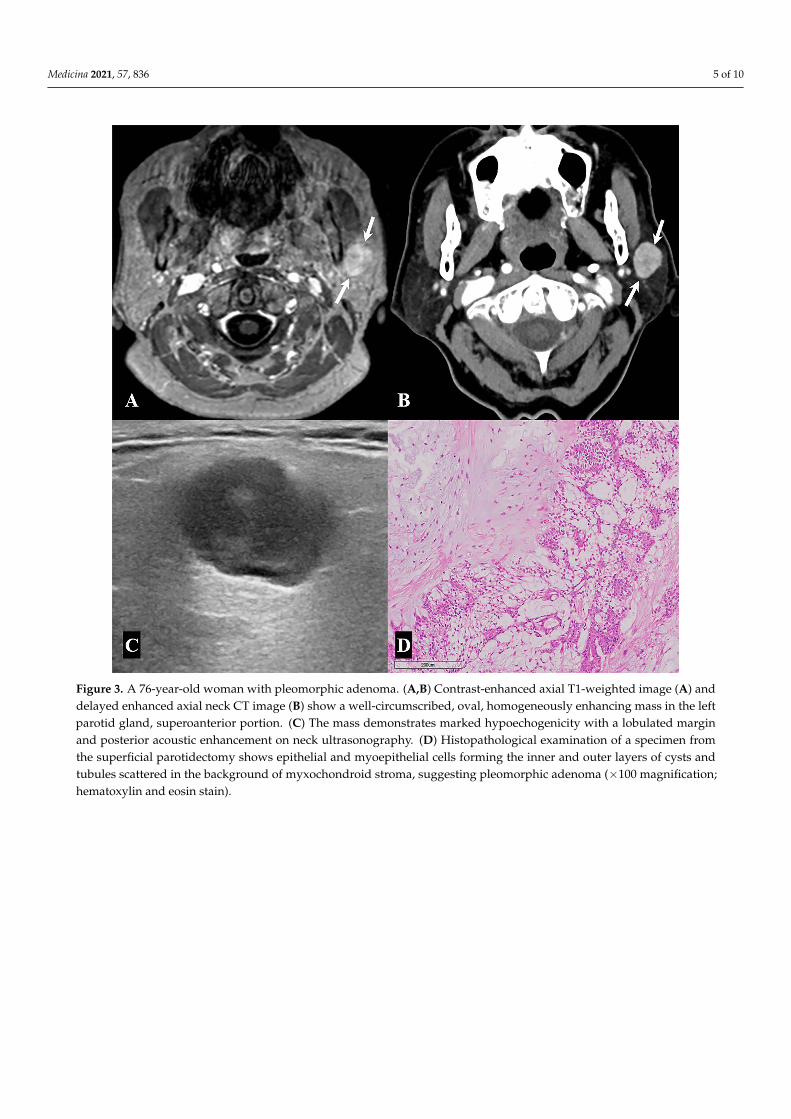
Figure 3. A 76-year-old woman with pleomorphic adenoma. (A,B) Contrast-enhanced axial T1-weighted image (A) anddelayed enhanced axial neck CT image (B) show a well-circumscribed, oval, homogeneously enhancing mass in the leftparotid gland, superoanterior portion. (C) The mass demonstrates marked hypoechogenicity with a lobulated marginand posterior acoustic enhancement on neck ultrasonography. (D) Histopathological examination of a specimen fromthe superficial parotidectomy shows epithelial and myoepithelial cells forming the inner and outer layers of cysts andtubules scattered in the background of myxochondroid stroma, suggesting pleomorphic adenoma (×100 magnification;hematoxylin and eosin stain).
Medicina 2021, 57, 836 6 of 10
Figure 4. A 46-year-old woman with Sjögren’s syndrome. (A,B) On the contrast-enhanced axial T1-weighted image, thereare multiple tiny enhancing granulonodular foci in both parotid glands with inhomogeneous parenchymal signal intensities.(B) On neck ultrasonography, the bilateral parotid glands show multiple tiny hypoechoic nodules with coarse parenchymalechotexture and decreased parenchymal echogenicity. (C) Salivary gland scintigraphy demonstrates severely decreaseduptake in the bilateral parotid and submandibular glands. (D) Histopathological examination of ultrasound-guided coreneedle biopsy sample shows extensive lymphoid infiltration and interstitial fibrosis with a germinal center, suggestive ofSjögren’s syndrome (×200 magnification; hematoxylin and eosin stain).
Medicina 2021, 57, 836 7 of 10
Figure 5. A 58-year-old man with low-grade mucoepidermoid carcinoma. (A,B) Contrast-enhanced axial T1-weightedimage (A) and delayed enhanced axial neck CT image (B) show a small enhancing nodular lesion with a central non-enhancing portion in the left parotid gland. (C) On neck ultrasonography, there is a markedly hypoechoic nodule with a focalspiculated margin and posterior acoustic enhancement in the superficial lobe of the left parotid gland. (D) Histopathologicalexamination of a specimen from the superficial parotidectomy shows sheets of mucous, squamous, and intermediatecells with bland-looking nuclei forming glandular spaces, suggestive of low-grade mucoepidermoid carcinoma (×100magnification; hematoxylin and eosin stain).
4. Discussion
The increased use of brain MRI for evaluating various intracranial abnormalitieshas increased the frequency of encountering incidental findings such as parotid lesionswithin the radiological scan range. Unfortunately, these incidental findings can be adiagnostic challenge for radiologists because of the limitations associated with the handlingof incidental lesions detected on brain MRI: (1) lack of adequate knowledge regardingtheir clinical significance; (2) partial visualization of the majority of incidental lesions;(3) non-availability of dedicated additional MRI sequences [7].
The reported prevalence of incidental parotid lesions on cross-sectional imagingranges between 0.3% and 0.45%. Nearly 32% of these lesions are malignant on PETimaging. However, the prevalence of incidental parotid lesions based on CT and MRI isstill unknown. To the best of our knowledge, only a single retrospective study reviewedincidental parotid lesions on CT, MRI, and PET imaging [16]. Therefore, we retrospectivelyassessed the prevalence and clinical implications of incidental parotid lesions as blind spotsdetected only on brain MRI.
In this study, the majority of the parotid lesions (95.9%) were visualized only incontrast-enhanced 3D T1-weighted images, probably because this sequence has a widerscan range than the other sequences in our routine brain MRI protocol. We also found thatthe prevalence of incidental parotid lesions on brain MRIs was 1.2% (84/7262), which wasthree- to four-fold higher than the results in previous studies using PET [12,14,15]. The

Medicina 2021, 57, 836 8 of 10
reason for this discrepancy is unclear. However, it appears to be related to the intrinsicdifferences between the two imaging modalities of brain MRI and PET or PET–CT.
Parotid lesions without FDG uptake are undetectable in PET or PET–CT, which arefunctional imaging techniques used to detect tissue metabolism representing the degree ofglucose utilization using a glucose analog as an indicator [17]. By contrast, parotid lesionsare more easily detected using brain MRI if the lesion is within the scan range because MRIis an anatomically oriented cross-sectional imaging modality. In addition, PET and PET–CTused to detect these lesions are limited by poor resolution and the partial volume effect.Furthermore, PET or PET–CT is usually performed during the initial staging work-up orfollow-up of cancer patients [12]. Therefore, the clinical indications are limited comparedto brain MRI.
In the current study, the average size of the lesions was 1.3 cm, similar to the resultsof a previous study, suggesting that the small size of parotid lesions is a possible causeof asymptomatic conditions [16]. In addition, most of the parotid lesions (81.6%) werelocated in the superficial lobe, which is consistent with the results of previous studiesthat described the imaging characteristics of parotid lesions [18–20]. However, only fourpatients had bilateral parotid lesions, which is inconsistent with previous studies, whichreported up to 25% [16,18–20]. The reason for this discrepancy is unclear, but it is probablyrelated to the differences in the number of enrolled study patients and the histopathologyof the lesions. We also found that the lesions were undetectable on brain MRI due tothe limited scan range and inadequate scan position, such as head tilting, limiting theevaluation of bilaterality or multiplicity. Therefore, a subsequent imaging work-up isneeded to determine the clinical significance of incidental parotid lesions.
Our results suggest that the majority of the parotid lesions (93.9%) were benign, andthe most common histopathology was Warthin tumors (36.7%), followed by reactive lymphnodes (22.5%) and pleomorphic adenomas (16.3%). These findings were inconsistent withprevious studies regarding symptomatic parotid tumors, which reported pleomorphicadenoma as the most common tumor in the parotid gland [21,22]. However, they areconsistent with recent studies suggesting that Warthin tumors were the most frequentbenign lesion in asymptomatic parotid lesions [16,23]. Al-Balas et al. [16] reported thatmore than 50% of the lesions were Warthin tumors, followed by pleomorphic tumors.Bothe et al. [23] found a similar preponderance of Warthin tumors, constituting 83% of theasymptomatic parotid lesions. Additionally, the difference in the frequency of Warthintumors between this study and previous studies may be related to the differences in thenumber of enrolled study patients. However, the findings of our study and previousstudies cannot reflect data in real-world practice because of the selection bias related tothe clinical characteristics of the study patients (i.e., age, sex or smoking status) and theuse of PET or PET–CT. In particular, it is well known that Warthin tumors often revealincreased FDG uptake that is often equivalent to the uptake level by malignant tumors, andthe previous studies using PET or PET–CT demonstrated a higher prevalence of Warthintumors as incidentalomas in the parotid gland [24–27].
We found that the overall incidence of parotid malignancy was 6.1%, including metastaticlymph nodes (4.1%) and primary salivary gland tumors (2%). These results are consistentwith previous studies that reported an incidence in the range of 3.1–7.9% for asymptomatic orincidentally detected parotid lesions [16,28,29]. We also found that the risk of malignancy inincidental parotid lesions was substantially lower in patients without a history of malignancy(1/35, 2.9%) than in patients with underlying malignancy (2/10, 20%).
The study limitations should be considered when interpreting the results. First, thisstudy was retrospectively designed, which suggests selection bias. We included studypatients by reviewing the radiologic reports. Therefore, it is difficult to completely excludethe possibility of parotid lesions on brain MRI missed by radiologists during the initialimaging assessment. Considering this issue, the actual prevalence of incidental parotidlesions on brain MRI might be higher than the results of the current study. Second, the
Medicina 2021, 57, 836 9 of 10
sample size was small. Third, routine brain MRI and MR protocols vary according to theinstitution, and these factors can affect the prevalence of incidental parotid lesions.
Despite these limitations, our study may be useful in understanding incidental parotidlesions detected on brain MRI. Additional imaging studies and histopathological analysesare needed to demonstrate their prevalence and the associated risk of malignancy. There-fore, further studies with a larger sample size and different brain MRI protocols and MRsystems can facilitate the validation of our findings and propose a lesion-specific diagnosticworkflow using rational and cost-effective strategies for the management of incidentallydetected parotid lesions on brain MRI.
5. Conclusions
In conclusion, we found a low prevalence of incidental parotid lesions on brain MRI.These lesions also showed a low risk of malignancy. However, the clinical significance ofthese lesions should not be overlooked in daily clinical practice, and appropriate imagingwork-up is invaluable in evaluating incidental parotid lesions and avoiding unnecessarybiopsies. Therefore, clinical awareness and meticulous image inspection are importantfor accurate diagnosis and timely management of incidental parotid lesions detected onbrain MRI. In addition, our observations underscore the need for a subsequent studywith a larger patient population to investigate the clinical implications and managementof the incidental findings beyond the intracranial space based on ethical concerns andhealthcare policy.
Author Contributions: Conceptualization: H.-J.B., S.Y.; methodology: I.-C.N., H.-J.B., K.-H.R.,H.-J.A.; formal analysis and investigation: I.-C.N., H.-J.B., J.-I.M., E.C.; writing—original draft prepa-ration: I.-C.N., H.-J.B.; writing—review and editing: I.-C.N., H.-J.B., J.B.; resources: not applicable;supervision: H.-J.B. All authors read and agreed to the published version of the manuscript.
Funding: This research received no external funding.
Institutional Review Board Statement: The study was conducted according to the guidelines ofthe Declaration of Helsinki, and approved by the Institutional Review Board of Gyeongsang Na-tional University Changwon Hospital (protocol code: GNUCH 2021-03-012 and date of approval:25 March 2021).
Informed Consent Statement: Patient consent was waived due to the retrospective nature of thestudy that did not alter the patient’s management and clinical outcomes.
Data Availability Statement: The anonymized data that support the results of this study are availableand shared on reasonable request by any qualified investigator.
Acknowledgments: We would like to thank our colleagues in the MRI section, Seong Jin Kim,Myungwook Lee, and Jae Hyun Lim, for their efforts in ensuring that all MR images were obtainedoptimally, and we also thank Tae Byeong Kim for his help in strengthening our work.
Conflicts of Interest: The authors declare that they have no competing interest.
References1. Freda, P.U.; Beckers, A.M.; Katznelson, L.; Molitch, M.E.; Montori, V.M.; Post, K.D.; Vance, M.L. Pituitary incidentaloma: An
endocrine society clinical practice guideline. J. Clin. Endocrinol. Metab. 2011, 96, 894–904. [CrossRef] [PubMed]2. The Incidentally Discovered Adrenal Mass|NEJM. Available online: https://www.nejm.org/doi/pdf/10.1056/NEJMcp065470
(accessed on 6 April 2021).3. Hitzeman, N.; Cotton, E. Incidentalomas: Initial management. Am. Fam. Physician 2014, 90, 784–789. [PubMed]4. Morris, Z.; Whiteley, W.N.; Longstreth, W.T.; Weber, F.; Lee, Y.-C.; Tsushima, Y.; Alphs, H.; Ladd, S.C.; Warlow, C.; Wardlaw, J.M.;
et al. Incidental findings on brain magnetic resonance imaging: Systematic review and meta-analysis. BMJ 2009, 339, b3016.[CrossRef] [PubMed]
5. Flor, N.; Di Leo, G.; Squarza, S.A.C.; Tresoldi, S.; Rulli, E.; Cornalba, G.; Sardanelli, F. Malignant incidental extracardiac findingson cardiac CT: Systematic review and meta-analysis. AJR Am. J. Roentgenol. 2013, 201, 555–564. [CrossRef] [PubMed]
6. Xiong, T.; Richardson, M.; Woodroffe, R.; Halligan, S.; Morton, D.; Lilford, R.J. Incidental lesions found on CT colonography:Their nature and frequency. Br. J. Radiol. 2005, 78, 22–29. [CrossRef] [PubMed]
Medicina 2021, 57, 836 10 of 10
7. Makdissi, J.; Pawar, R.R.; Radon, M.; Holmes, S.B. Incidental findings on MRI of the temporomandibular joint. DentomaxillofacialRadiol 2013, 42, 20130175. [CrossRef]
8. Holsinger, F.C.; Bui, D.T. Anatomy, function, and evaluation of the salivary glands. In Salivary Gland Disorders; Myers, E.N., Ferris,R.L., Eds.; Springer: Berlin/Heidelberg, Germany, 2007; pp. 1–16. ISBN 978-3-540-47072-4.
9. Lennon, P.; Silvera, V.M.; Perez-Atayde, A.; Cunningham, M.J.; Rahbar, R. Disorders and tumors of the salivary glands in children.Otolaryngol. Clin. N. Am. 2015, 48, 153–173. [CrossRef]
10. Pinkston, J.A.; Cole, P. Incidence rates of salivary gland tumors: Results from a population-based study. Otolaryngol. Neck Surg.1999, 120, 834–840. [CrossRef]
11. Basu, S.; Houseni, M.; Alavi, A. Significance of incidental fluorodeoxyglucose uptake in the parotid glands and its impact onpatient management. Nucl. Med. Commun. 2008, 29, 367–373. [CrossRef]
12. Lee, S.K.; Rho, B.H.; Won, K.S. Parotid incidentaloma identified by combined 18F-fluorodeoxyglucose whole-body positronemission tomography and computed tomography: Findings at grayscale and power doppler ultrasonography and ultrasound-guided fine-needle aspiration biopsy or core-needle biopsy. Eur. Radiol. 2009, 19, 2268–2274. [CrossRef]
13. Mabray, M.C.; Behr, S.C.; Naeger, D.M.; Flavell, R.R.; Glastonbury, C.M. Predictors of pathologic outcome of focal FDG uptake inthe parotid gland identified on whole-body FDG PET imaging. Clin. Imaging 2015, 39, 1073–1079. [CrossRef]
14. Makis, W.; Ciarallo, A.; Gotra, A. Clinical significance of parotid gland incidentalomas on (18)F-FDG PET/CT. Clin. Imaging 2015,39, 667–671. [CrossRef]
15. Wang, H.-C.; Zuo, C.-T.; Hua, F.-C.; Huang, Z.-M.; Tan, H.-B.; Zhao, J.; Guan, Y.-H. Efficacy of conventional whole-body 18F-FDGPET/CT in the incidental findings of parotid masses. Ann. Nucl. Med. 2010, 24, 571–577. [CrossRef] [PubMed]
16. Al-Balas, H.; Metwalli, Z.A.; Eberson, S.; Sada, D.M. Clinicopathological features of incidental parotid lesions. Head Face Med.2021, 17, 10. [CrossRef]
17. Haberkorn, U.; Strauss, L.G.; Reisser, C.; Haag, D.; Dimitrakopoulou, A.; Ziegler, S.; Oberdorfer, F.; Rudat, V.; van Kaick, G.Glucose uptake, perfusion, and cell proliferation in head and neck tumors: Relation of positron emission tomography to flowcytometry. J. Nucl. Med. 1991, 32, 1548–1555.
18. Joe, V.Q.; Westesson, P.L. Tumors of the Parotid Gland: MR imaging characteristics of various histologic types. AJR Am. J.Roentgenol. 1994, 163, 433–438. [CrossRef]
19. Stoia, S.; Băciut, , G.; Lenghel, M.; Badea, R.; Csutak, C.; Rusu, G.M.; Băciut, , M.; Tamas, T.; Bot,an, E.; Armencea, G.; et al. Cross-sectional imaging and cytologic investigations in the preoperative diagnosis of parotid gland tumors—An updated literaturereview. Bosn. J. Basic Med. Sci. 2021, 21, 19–32. [CrossRef]
20. Mantsopoulos, K.; Tschaikowsky, N.; Goncalves, M.; Mueller, S.K.; Iro, H. Evaluation of preoperative ultrasonography in the dif-ferentiation between superficial and deep parotid gland tumors. Ultrasound Med. Biol. 2020, 46, 2099–2103. [CrossRef] [PubMed]
21. Spiro, R.H. Salivary neoplasms: Overview of a 35-year experience with 2807 patients. Head Neck Surg. 1986, 8, 177–184. [CrossRef]22. Bradley, P.J.; McGurk, M. Incidence of salivary gland neoplasms in a defined UK population. Br. J. Oral Maxillofac. Surg. 2013, 51,
399–403. [CrossRef]23. Bothe, C.; Fernandez, A.; Garcia, J.; Lopez, M.; León, X.; Quer, M.; Lop, J. Parotid incidentaloma identified by positron
emission/computed tomography: When to consider diagnoses other than warthin tumor. Int. Arch. Otorhinolaryngol. 2015, 19,112–115. [CrossRef] [PubMed]
24. Keyes, J.W., Jr.; Karkness, B.A.; Greven, K.M.; Williams, D.W., III; Watson, N.E., Jr.; McGuirt, W.F. Salivary gland tumors:Pretherapy evaluation with PET. Radiology 1994, 192, 99–102. [CrossRef]
25. Okamura, T.; Kawabe, J.; Koyama, K.; Ochi, H.; Yamada, R.; Sakamoto, H.; Matsuda, M.; Ohashi, Y.; Nakai, Y. Fluorine-18fluorodeoxyglucose positron emission tomography imaging of parotid mass lesions. Acta Otolaryngol. Suppl. 1998, 538, 209–213.[CrossRef] [PubMed]
26. Horiuchi, M.; Yasuda, S.; Shohtsu, A.; Ide, M. Four cases of Warthin′s tumor of the parotid gland detected with FDG PET. Ann.Nucl. Med. 1998, 12, 47–50. [CrossRef] [PubMed]
27. Uchida, Y.; Minoshima, S.; Kawata, T.; Motoori, K.; Nakano, K.; Kazama, T.; Uno, T.; Okamoto, Y.; Ito, H. Diagnostic value of FDGPET and salivary gland scintigraphy for parotid tumors. Clin. Nucl. Med. 2005, 30, 170–176. [CrossRef]
28. Stein, A.P.; Britt, C.J.; Saha, S.; McCulloch, T.M.; Wieland, A.M.; Harari, P.M.; Hartig, G.K. Patient and tumor characteristicspredictive of primary parotid gland malignancy: A 20-year experience at the University of Wisconsin. Am. J. Otolaryngol. 2015,36, 429–434. [CrossRef]
29. Stodulski, D.; Mikaszewski, B.; Stankiewicz, C. Signs and symptoms of parotid gland carcinoma and their prognostic value. Int. J.Oral Maxillofac. Surg. 2012, 41, 801–806. [CrossRef]