prevalence of abnormalities in knees detected by mri in adults without knee osteoarthritis:...
TRANSCRIPT

Prevalence of abnormalities in knees detected by MRIin adults without knee osteoarthritis: population basedobservational study (FraminghamOsteoarthritis Study)
OPEN ACCESS
Ali Guermazi professor of radiology 1, Jingbo Niu research assistant professor of medicine 2, DaichiHayashi research assistant professor of radiology 1, Frank W Roemer associate professor ofradiology13, Martin Englund associate professor, epidemiologist24, Tuhina Neogi associate professorof medicine and epidemiology2, Piran Aliabadi professor of radiology5, Christine E McLennan projectmanager 6, David T Felson professor of medicine and epidemiology 2
1Department of Radiology, Boston University School of Medicine, FGHBuilding, 820 Harrison Avenue, Boston, MA 02118, USA; 2Clinical EpidemiologyResearch and Training Unit, Boston University School of Medicine, Boston; 3Klinikum Augsburg, Department of Radiology, Augsburg, Germany;4Lund University, Clinical Sciences Lund, Department of Orthopaedics, Lund, Sweden; 5Brigham and Women’s Hospital, Harvard Medical School,Department of Radiology, Boston, MA 02115; 6OptumInsight Life Sciences, Waltham, MA 02451
AbstractObjective To examine use of magnetic resonance imaging (MRI) ofknees with no radiographic evidence of osteoarthritis to determine theprevalence of structural lesions associated with osteoarthritis and theirrelation to age, sex, and obesity.
Design Population based observational study.
Setting Community cohort in Framingham, MA, United States(Framingham osteoarthritis study).
Participants 710 people aged >50 who had no radiographic evidenceof knee osteoarthritis (Kellgren-Lawrence grade 0) and who underwentMRI of the knee.
Main outcomemeasuresPrevalence of MRI findings that are suggestiveof knee osteoarthritis (osteophytes, cartilage damage, bone marrowlesions, subchondral cysts, meniscal lesions, synovitis, attrition, andligamentous lesions) in all participants and after stratification by age,sex, body mass index (BMI), and the presence or absence of knee pain.Pain was assessed by three different questions and also by WOMACquestionnaire.
Results Of the 710 participants, 393 (55%) were women, 660 (93%)were white, and 206 (29%) had knee pain in the past month. The meanage was 62.3 years and mean BMI was 27.9. Prevalence of “anyabnormality” was 89% (631/710) overall. Osteophytes were the mostcommon abnormality among all participants (74%, 524/710), followedby cartilage damage (69%, 492/710) and bone marrow lesions (52%,371/710). The higher the age, the higher the prevalence of all types ofabnormalities detectable by MRI. There were no significant differencesin the prevalence of any of the features between BMI groups. The
prevalence of at least one type of pathology (“any abnormality”) washigh in both painful (90-97%, depending on pain definition) and painless(86-88%) knees.
ConclusionsMRI shows lesions in the tibiofemoral joint in most middleaged and elderly people in whom knee radiographs do not show anyfeatures of osteoarthritis, regardless of pain.
IntroductionAgeing of the population and increasing obesity contribute tomorbidity worldwide. Osteoarthritis is the most prevalentmedically treated arthritic condition worldwide (for example,3532 per 100 000 people in the United States).1 2 Diagnosis ofosteoarthritis is made on the basis of clinical examination orradiography. Population based longitudinal studies in the US3
and the United Kingdom4 showed the lifetime risk of kneeosteoarthritis increases with age,3with the risk highest in obesepeople.3 4 Other prevalence surveys showed that radiographicosteoarthritis of the knee is common in middle aged and olderadults.5 6
Although many publications have reported structural changesin people with radiographic knee osteoarthritis, few data areavailable regarding what structural changes are present in kneeswithout any radiographic features of osteoarthritis. About halfof people with knee pain have no radiographic osteoarthritis.In clinical practice, it is unclear how to investigate and managesuch people and whether additional imaging with magneticresonance imaging would be of clinical value. Such data can becollected only in population based studies as people with normal
Correspondence to: A Guermazi [email protected]
No commercial reuse: See rights and reprints http://www.bmj.com/permissions Subscribe: http://www.bmj.com/subscribe
BMJ 2012;345:e5339 doi: 10.1136/bmj.e5339 (Published 29 August 2012) Page 1 of 13
Research
RESEARCH
knees are not usually enrolled into clinical studies or undergofurther imaging evaluation. Radiography can show osteophytes,bony outgrowths at the joint margin, and narrowing of the jointspace, but it cannot visualise soft tissue pathology.7 In contrast,MRI can visualise various tissues that are clinically relevantand have an important role in regard to structural progressionnot seen on radiography. MRI can also show incidental findingsin otherwise asymptomatic people.8 9 In the knee,MRI visualisesmost components of the joint, including articular cartilage,menisci, intra-articular ligaments, synovium, bone marrow,subchondral cysts, and other periarticular and intra-articularlesions that are not detectable by radiography.10
We used MRI to evaluate the presence of structural changes inknees that were free from radiographic tibiofemoralosteoarthritis. We focused on the tibiofemoral joint, whichincludes numerous bony and soft tissue structures that can beevaluated by MRI. We evaluated the prevalence of cartilagedamage, meniscal lesions, osteophytes, subchondral cysts, bonemarrow lesions, ligamentous lesions, attrition, and synovitis onMRI in participants of the Framingham Osteoarthritis Studywho had radiographically normal tibiofemoral knee joints. Wealso assessed whether the prevalence of these features differedaccording to age, sex, body mass index (BMI), or knee pain.
MethodsStudy design and participantsThe Framingham Community cohort was recruited from theFramingham, MA, census tract data for the year 2000 andrandom digit telephone dialling. All participants were examinedbetween 2002 and 2005. This study cohort is distinct from theFraminghamHeart Study and the FraminghamOffspring Studycohorts. Participants were not selected on the basis of havingknee or other joint problems, and potential participants werenot told that knees were a focus of the study.Eligible participants were aged at least 50 and ambulatory (theuse of assistive devices such as canes and walker was permitted),with no plans to move out of the area for at least five years toaccommodate the possibility of longitudinal follow-up. Weexcluded those with a history of bilateral total knee replacement,rheumatoid arthritis, dementia, or terminal cancer and thosewho had contraindications to MRI. Of 2582 people aged 50 orolder and living in Framinghamwhowere contacted by randomdigit dialling, 1830 expressed interest in participating in thestudy.8 Of those, 39 were lost to contact, 194 were ineligiblefor the study, and 558 declined to participate. Consequently,1039were examined, 993 underwentMRI, and 992 had readablescans (one knee per participant, right knee preferred; left kneeif right knee not available (fig 1⇓).
Knee radiography and gradingParticipants underwent weight bearing posteroanterior kneeradiography with the fixed-flexion protocol.11 Onemusculoskeletal radiologist, who was blinded to the MRIfindings and clinical data, graded radiographs using theKellgren-Lawrence grading system (intraobserver κ 0.83).12 13
Because we wanted to focus on “normal” tibiofemoral kneejoints (Kellgren-Lawrence grade 0), we excluded 253participants with radiographic tibiofemoral osteoarthritis(Kellgren-Lawrence grade 2 or above), doubtful or equivocalfindings of radiographic evidence of tibiofemoral osteoarthritis(Kellgren-Lawrence grade 1), or missing radiographs orradiographic readings. Finally, we excluded 30 participantsbecause of unreadable or poor quality MRIs. This resulted in
710 radiographically “normal” tibiofemoral knee joints beingincluded in the final sample for analysis (fig 1⇓).
MRI grading of osteoarthritis featuresMRI was done with a 1.5 Tesla scanner (Siemens MedicalSystems, Erlangen, Germany) with a phased array knee coil.Images from four pulse sequences were used in the assessmentof osteoarthritis features: axial, sagittal and coronal fat saturated,proton density weighted, turbo spin echo images (repetitiontime 3610 msec; echo time 40 msec; slice thickness 3.5 mm;interslice gap 0 mm; echo train length 7; field of view 140 mm× 140mm;matrix 256 × 256) and sagittal T1 weighted spin echoimages without fat saturation (repetition time 475 msec; echotime 24msec; slice thickness 3.5 mm; interslice gap 0 mm; fieldof view 140 mm×140 mm; matrix 256×256).MRI scans were read by two trained and experiencedmusculoskeletal radiologists (who did not read the radiographs)using a standardised and validated method called the wholeorgan magnetic resonance imaging score (WORMS).14 Theyrecorded the presence or absence of the specific features(described below) related to osteoarthritis that were included inour assessment of the tibiofemoral joint (that is, tibial plateausand the central weight bearing and posterior portions of femoralcondyles). In the WORMS system, the tibiofemoral joint issubdivided into 10 different subregions for scoring of eachfeature. Readings from all subregions were amalgamated withinthe knee.14 Agreement between observers (κ statistic) for thedetection of the MRI features was as follows: cartilage damage0.89; meniscal lesions 0.71; osteophytes 0.73; ligamentouslesions 0.49; bone marrow lesions 0.85; subchondral cysts 0.57;and synovitis 0.63. The relatively low value of κ for ligamentouslesions was because few knees had ligamentous lesions in thereliability sample.Cartilage damage was considered present if there was a smallfocal loss less than 1 cm in greatest width or areas of diffusepartial or full thickness loss (WORMS grade ≥2). In this studywe did not consider intrachondral signal alterations (WORMSgrade 1), which are thought to occur before cartilage damagedevelops14 but are of unknown clinical importance, to representcartilage damage.Meniscal lesions (WORMS grade ≥1) included displaced ornon-displaced meniscal tears or evidence of previous surgery(including repair and partial or complete resection) and completemaceration or destruction (that is, loss of normal contour andsignal homogeneity within the meniscus) within the anteriorand posterior horns and the body of the medial and lateralmenisci.14
Osteophytes were considered present if there were bonyprojections that form along different margins of the tibiofemoraljoint of the knee (WORMS grade ≥2). Tiny bony spurs thatwere equivocal on visual evaluation (that is, “lipping,”WORMSgrade ≥1) were not considered as osteophytes.Ligamentous abnormalities were defined as the presence of acompletely torn anterior or posterior cruciate ligament, or a tornor thickened medial or lateral collateral ligament (WORMSgrade ≥1).Bone marrow lesions—Subchondral bone marrow lesions, alsoknown as “bone marrow edema-like lesions,”15were consideredpresent if there are non-cystic subchondral areas of ill definedhigh signal on proton density weighted MR images with fatsignal suppression (WORMS grade ≥1).Subchondral cystswere identified as areas of markedly increasedsignal intensity in the subarticular bone with sharply defined
No commercial reuse: See rights and reprints http://www.bmj.com/permissions Subscribe: http://www.bmj.com/subscribe
BMJ 2012;345:e5339 doi: 10.1136/bmj.e5339 (Published 29 August 2012) Page 2 of 13
RESEARCH

rounded margins and no evidence of internal marrow tissue ortrabecular bone on the fat saturated proton density weightedimages (WORMS grade ≥1).Synovitis was considered present if the synovial cavity wasdistended and filled with fluid (high signal intensity on fatsaturated proton density weighted images), representing synovialthickening and joint effusion (WORMS grade ≥1).14
Attrition—Flattening or depression of the articular surfaces ofthe tibia or femur was termed bone attrition, and any degree ofdeviation from the normal bony contour was consideredabnormal (WORMS grade ≥1).
Additional analysis with a more stringentdefinition of “abnormality”Currently there is no concrete definition of what is “abnormal”in terms of MRI findings in the knee, and the use of differentcut off points for the definition of “abnormality” might producedifferent results. We also examined a more stringent definitionof lesions detected by MRI, which included cartilage damageand osteophytes=WORMS grade ≥3; all other lesions=grade≥2.
Assessment of weight, height, and painWe measured the participants’ weight when they were notwearing shoes with the use of a balance beam scale andmeasured height with a stadiometer. At the clinic visit allparticipants were asked about knee symptomswith the followingquestion: “In the past month, have you had any pain, aching, orstiffness in your knee?” (for this study, we focused on pain inthe knee with MRI reading). Additionally, we assessed kneepain in three more ways. Participants responded to the questions,“Did you have knee pain lasting at least a month in the pastyear?” and “Do you have knee pain on most days?” A positiveresponse to these questions was considered to indicate thepresence of knee pain. Each participant was also asked to fillout the Western Ontario McMaster University arthritis index(WOMAC) questionnaire, and any score ≥1 in the pain subscalein the knee was considered to indicate the presence of knee pain.For WOMAC pain, we restricted our analysis to participantswho had Kellgren-Lawrence grade 0 knees bilaterally as theWOMAC questionnaire was person based and not knee based.
Statistical analysisWe calculated the prevalence of the aforementionedosteoarthritis features on MRI and stratified the data accordingto sex, age group (sixth decade, seventh decade, and older),BMI (<25, ≥25-<30, ≥30), and the presence of pain. We usedχ2 tests to assess the presence of significant differences betweenmen and women, and among different age and BMI groups. Forcartilage and bone marrow lesions, results were stratifiedaccording to the medial and lateral tibiofemoral compartmentsof the knee. All statistical analyses were performed with SASfor Windows, version 9.1. Results were considered to besignificant when a two tailed P<0.05.
ResultsCharacteristics of study sampleOf the 710 participants, 393 (55%) were women, 660 (93%)were white, and 206 (29%) had painful knees. The mean agewas 62.3 (range 51-89), and the mean BMI was 27.9 (range16.6-50.6) (table 1⇓).
Prevalence of bony and soft tissueabnormalities onMRI with standard definitionOverall, 631 (89%) knees had at least one type of abnormality(fig 2⇓, table 2⇓). The three most common findings wereosteophytes, cartilage damage, and bone marrow lesions. In thelocation specific analysis, cartilage damage was more prevalentin the medial tibiofemoral compartment (33% (95% confidenceinterval 30% to 37%), 235/710) than in the lateral tibiofemoralcompartment (20% (17% to 23%), 141/710). Likewise, therewere more bone marrow lesions in the medial (19% (16% to22%), 133/710) than in the lateral tibiofemoral compartment(12% (10% to 16%), 87/710).Table 2 summarises the prevalence of eachMRI feature overalland in men and women⇓. The prevalence of meniscal lesionswas significantly higher in men than in women (110/317 (35%)v 57/393 (15%); P<0.001). No other features were significantlydifferent between men and women. There were no significantdifferences in the prevalence of any of the features betweenBMI groups (table 2). The prevalence of all features was withinabout 7% among all BMI groups.Older age groups had more abnormalities of all types. Of theparticipants in their sixth decade, 86% (271/316) had featuresof osteoarthritis. The rate increased to 91% (227/249) in theseventh decade and 92% (133/145) in the oldest age group.Specific types of abnormalities (cartilage damage, meniscallesions, osteophytes, subchondral cysts) also increased witheach decade (table 3⇓ and fig 3⇓). The prevalence ofligamentous lesions, bonemarrow lesions, attrition, and synovialthickening and joint effusion was also higher in older agegroups, but the differences between groups were not significant.The prevalence of attrition (38% v 30%; P=0.04), bone marrowlesions (59% v 50%; P=0.03), and subchondral cysts (31% v23%; P=0.04) was higher in participants with painful knees thanthose without pain (table 2). The prevalences for the otherfeatures were within about 4% of one another among painfuland painless knees with no significant differences (table 2).Indeed, the prevalence of at least one type of MRI detectedpathology (“any abnormality”) was high in both painful (91%)and painless (88%) knees (table 2⇓). Regardless of the definitionof pain used, MRI detected abnormalities were highly prevalentin people with (90-97%) andwithout (86-88%) knee pain.Whilethe prevalence of MRI abnormalities was not significantlydifferent in those with versus those without knee pain for mostdefinitions of pain we tested, the prevalence of “any MRIabnormality” was higher in those withWOMAC pain comparedwith those without pain (P=0.002). Even so, the prevalence ofany MRI abnormality was as high as 86% in those withoutWOMAC pain.
Prevalence of bony and soft tissueabnormalities on MRI with more stringentdefinitionWhenwe used themore stringent definition ofMRI abnormality,overall the prevalence of MRI detected lesions dropped asexpected (table 4⇓), to 14% for osteophytes, 44% for cartilagedamage, 16% for bone marrow lesions, 4% for synovitis, 10%for attrition, 5% for subchondral cysts, 8% for meniscal lesions,and 2% for ligamentous lesions. The prevalence of anyabnormality, however, remained high (53%, 373/710). Exceptfor bone attrition, painful knees did not differ from those withoutpain in terms of the prevalence of specific features (table 4⇓).Regardless of the definition of pain used, any abnormality waspresent in 57-70% of participants with pain and about half ofthose without pain (table 5⇓). There were significant differences
No commercial reuse: See rights and reprints http://www.bmj.com/permissions Subscribe: http://www.bmj.com/subscribe
BMJ 2012;345:e5339 doi: 10.1136/bmj.e5339 (Published 29 August 2012) Page 3 of 13
RESEARCH

between groups with and without pain in three out of fourdefinitions of pain, and the largest difference was seen withWOMACpain (15%, P<0.001). Prevalence of “any abnormality”in those without WOMAC pain, however, was still high (48%).
DiscussionWe found that MRI detected features of osteoarthritis are highlyprevalent in the tibiofemoral joint of knees that did not haveany radiographic features of osteoarthritis in participants bothwith and without knee pain. Nearly 90% of our participants hadat least one feature of osteoarthritis on MRI. Osteophytes werethe most common, followed by cartilage damage and bonemarrow lesions. In general, the older the age group, the higherthe prevalence of features of osteoarthritis, although differencesamong age groups were not significant for synovitis and effusionand of borderline significance for ligamentous lesions and bonemarrow lesions. Only meniscal lesions were more prevalent inmen than women. No significant differences were observed forany type of lesions by BMI.
Strengths and limitationsThis population based study documented the high prevalenceof MRI features suggestive of knee osteoarthritis in peoplewithout radiographic osteoarthritis. We included only kneesthat were definitely lacking any radiographic features that couldindicate the presence of osteoarthritic changes(Kellgren-Lawrence grade 0) to ensure our analysis is specific.Although Kellgren-Lawrence grade 1 knees also do not qualifyfor having radiographic osteoarthritis, a “doubtful” bonyabnormality is present and one could argue such equivocalfindings are difficult to interpret.
LimitationsOur sample was primarily (although not exclusively) white,reflecting the population of Framingham, MA. The number ofpeople from other racial or ethnic groups was too small forcomparisons. Our prevalence estimates cannot be generalisedto adults younger than 50. In particular, meniscal lesions inyoung active otherwise healthy adults are more likely to becaused by trauma than the degenerative process seen in middleaged and older people. We had no arthroscopic correlation ofour MRI findings. Ideally, intra-articular pathology (that is,cartilage, meniscus, and ligaments) should be confirmed bydirect visualisation during arthroscopy. Arthroscopy, however,is neither feasible nor ethical in large scale population basedstudies. Furthermore, arthroscopy cannot visualise some of theMRI findings that are indicative of the osteoarthritis diseaseprocess such as subchondral bone marrow lesions. Nearly allthe knees in our sample were right knees (with only five leftknees). A comparison of 99 people with both right and left kneeMRIs in this sample, however, showed no difference in findings,and which knee is studied is therefore unlikely to affect ouroverall outcome. We did not include the evaluation ofradiographic patellofemoral joint pathology in this study becausewe used the posteroanterior radiograph to classify thetibiofemoral joint of the knee using Kellgren-Lawrence grading.We dealt with this fact by including only subregions of the kneethat correspond to the tibiofemoral joint for MRI analysis.Because we focused on knees with clearly normal radiographicappearance (Kellgren-Lawrence grade 0), we excludedKellgren-Lawrence grade 1 knees. One might argue that suchknees are also without radiographic osteoarthritis and warrantinclusion in our analysis. Inclusion of the 39 Kellgren-Lawrencegrade 1 knees in our sample (total 749 knees) did not alter the
demographic characteristics of the participants or analyticalresults for all aspects of the study. There are many ways todefine pain, and it is not possible to include all different painassessment tools available to date in a single study.We selectedthe WOMAC pain subscale because it has been validated andis widely used.16 17
Our results raise additional questions. More detailed analysisevaluating the factors that could contribute to the differencesseen in men and women and the osteoarthritis features in thedifferent age groups would be of interest. Also, comparison ofthe prevalence of these findings in those with and withoutradiographic osteoarthritis would tackle the question of whetherosteoarthritis is an inevitable consequence of ageing.
Comparison with previous studiesOf our findings, the most notable is that 74% of the knees hadosteophytes. As a bony abnormality should be clearly visibleon radiograph, we did not expect the prevalence to be this high.Presumably, because the MRI assessment used three imagingplanes, it could detect osteophytes that were hidden by theoverlapping femur or tibia on posteroanterior view radiographs.This is a substantial problem as the presence of definiteosteophytes defines the diagnosis of radiographic osteoarthritis.12Thus far, epidemiological or clinical studies of kneeosteoarthritis depend largely on the radiographic definition ofosteoarthritis.18 19 As radiography fails to detect such a largeproportion of osteophytes, there could be misclassification ofa large number of potentially eligible people in kneeosteoarthritis studies and underestimation of the true prevalenceof this condition.20
Although cartilage itself is aneural and is unlikely to be a directcause of knee pain, cartilage damage is associated with changein bone marrow lesions,21 high BMI, meniscal damage, andsynovitis or effusion.22Cartilage thickness has traditionally beenassessed by its surrogate marker—the radiographic width of thejoint space of the tibiofemoral joint. Narrowing of the jointspace, however, can result not only from cartilage damage butalso frommeniscal lesions.23 It has been shown that radiographyis less sensitive than MRI for detection of cartilage loss.7 Thus,it is not surprising to find a high prevalence of cartilage damageon MRI in the knees of middle aged and older people withoutradiographic joint space narrowing.The presence and extent of bone marrow lesions and synovialthickening/effusion can be appreciated only on MRI. Theselesions have been associated with pain in knees withosteoarthritis.24 25 Furthermore, in people at high risk ofdeveloping osteoarthritis, bone marrow lesions in asymptomaticknees with no radiographic osteoarthritis at baseline predictdevelopment of pain 15 months later.26 We also found anassociation of bonemarrow lesions with knee pain among peoplewithout radiographic osteoarthritis.A high prevalence of incidental meniscal findings on MRI inparticipants of the Framingham Osteoarthritis Study has beenreported previously.8 One or more meniscal tears was presentin 32% (41/127) of knees with symptoms, 23% (146/548) ofknees without symptoms, and 24% (187/775) overall when therewas no or equivocal radiographic evidence of osteoarthritis.Although the results were similar to the present study, they arenot identical because Englund and colleagues included kneeswith Kellgren-Lawrence grade 0 and 1,8 whereas we focusedon Kellgren-Lawrence grade 0.We saw fewer incidental ligamentous lesions than any otherfeature. This could be because the semiquantitative scoringsystem we used only scores a complete tear as a lesion, and
No commercial reuse: See rights and reprints http://www.bmj.com/permissions Subscribe: http://www.bmj.com/subscribe
BMJ 2012;345:e5339 doi: 10.1136/bmj.e5339 (Published 29 August 2012) Page 4 of 13
RESEARCH

partial tears are given a score of zero. Imaging diagnosis ofpartial ligamentous tears on MRI can be difficult. The role ofintra-articular and periarticular ligaments of the knee inpredicting structural progression of knee osteoarthritis remainsunclear. Disruption of the integrity of these ligaments, however,will probably cause alterations in knee kinematics.A recent systematic review reported that bone marrow lesionsand effusion/synovitis were associated with knee pain.27 In ourstudy, however, these lesions were not significantly moreprevalent in participants who had knee pain than in thosewithout, with both definitions of MRI abnormality. Thisdiscrepancy is probably because the systematic review includedonly studies involving mostly people with radiographic kneeosteoarthritis. Thus, the conclusion of the systematic review isnot applicable to the present study.
Clinical implicationsOur findings indicate that the prevalence of MRI detectedosteoarthritis features increases with age in the absence ofradiographic features of osteoarthritis.We have shown thatMRIis more sensitive than radiographs to changes in bone and softtissue that are considered features of osteoarthritis,28 29Our datashowed that the prevalence of these MRI detected features ishigh irrespective of the knee pain status. When we comparedthe prevalence of MRI abnormalities in knees in people withand without pain, there were two trends. Firstly, and mostimportantly, the prevalence ofMRI findings was extremely highin those without pain, suggesting that usingMRI as a diagnostictest for people with normal knee radiographs in this age groupwould have poor specificity. Secondly, the prevalence offindings was modestly higher in those with pain than in thosewithout, with the difference sometimes reaching significance.These differences, however, were not particularlyinformative—for example, the highest prevalence of MRIabnormalities was actually in those with mild pain rather thanmoderate or severe pain.Thus,MRI features suggestive of osteoarthritis in people withoutradiographic osteoarthritis are commonly seen in those with orwithout knee pain, implying thatMRI alone is not diagnosticallyuseful to discriminate between people with and without pain inthe context of knee osteoarthritis. MRI might still play animportant diagnostic role, especially in younger people, in whomother reasons for knee pain should be considered such asinflammatory arthritides, insufficiency fractures, or spontaneousosteonecrosis. Nonetheless, in all likelihood, MRI features ofosteoarthritis will be found regardless of the source of the pain.Our study also highlights the limitations of conventionalradiography to detect a large number of abnormalities relatedto osteoarthritis in the knee.20
As high BMI is a known risk factor for both incident kneeosteoarthritis and for progression of knee osteoarthritis30 31 weexpected to see higher prevalence of MRI features in obesepeople compared with non-obese people. We did not find highBMI to be associated with higher prevalence of MRI featuresoverall compared with low BMI, but rather that these MRIabnormalities were equally highly prevalent in all BMI groups.We speculate that BMI is important for progression of laterstages of osteoarthritis, but potentially age is a much morerelevant trigger of early stages of osteoarthritis.Although there is thought to be only a modest correlationbetween clinical symptoms and radiographic tibiofemoralosteoarthritis,32 recent work has highlighted an associationbetween structural osteoarthritis pathology and knee pain.33 34
It is important for the clinical community to recognise that
findings that would be interpreted as abnormal and suggestiveof disease are in fact present in most knees without any pain,even when different definitions of pain are used. That meansthat the clinical significance of these MRI findings isquestionable. The same message has been reported forradiographic findings in patients with low back pain (similarhighly prevalent abnormalities were seen in those without lowback pain), and this led to discouraging radiographic evaluationsin those with low back pain.35
ConclusionsChanges indicative of osteoarthritis are commonly present inthe knees of most people aged 50 and over who have noradiographic evidence of tibiofemoral osteoarthritis.Osteophytes, cartilage damage, and bone marrow lesions areespecially common amongmiddle aged and older people. Thesefeatures are common in knees with pain and in those that arepainless and can potentially represent pre-radiographic or earlystage osteoarthritis. A longitudinal study is needed to determinewhat proportion of people without radiographic osteoarthritisbut withMRI abnormalities subsequently develop radiographicosteoarthritis.
Contributors: AG, JN, DH, and DTF conceived and designed the study.AG, JN, FWR, PA, CEM, and DTF collected the data. AG, DH, FWR,ME, TN, and DTF reviewed the literature. AG, JN, DH, FWR, ME, TN,and DTF directed the analyses, which were carried out by JN. All authorsparticipated in the discussion and interpretation of the results. AG andDH organised the writing and wrote the initial drafts. All authors criticallyrevised the manuscript for intellectual content and approved the finalversions. AG and DTF are guarantors.Funding: This study was funded by the National Institutes of Health(AG18393 and AR47785) and the Arthritis Foundation. The funders hadno role in study design, data collection and analysis, decision to publish,or preparation of the manuscript. The researchers work independentlyof their funders.Competing interests: All authors have completed the ICMJE uniformdisclosure form at www.icmje.org/coi_disclosure.pdf (available onrequest from the corresponding author) and declare: AG is the presidentof Boston Imaging Core Lab (BICL), LLC, and a consultant to MerckSerono, Stryker, Genzyme, AstraZeneca, and Novartis; FWR a vicepresident and shareholder of BICL and is a consultant to Merck Seronoand National Institute of Health; ME is funded by the Swedish ResearchCouncil, the Greta and Johan Kock Foundation, King Gustaf V 80-yearBirthday Foundation, and the Faculty of Medicine, Lund University,Sweden; TN is supported by NIAMS AR055127 and the ArthritisFoundation Arthritis Investigator Award.Ethical approval: This study was approved by the institutional reviewboard of Boston University Medical Centre (protocol number H-22674),and written informed consent was obtained from all participants.Data sharing: No additional data available.
1 Sacks JJ, Luo YH, Helmick CG. Prevalence of specific types of arthritis and other rheumaticconditions in the ambulatory health care system in the United States, 2001-2005. ArthritisCare Res (Hoboken) 2010;62:460-4.
2 Bedson J, Jordan K, Croft P. The prevalence and history of knee osteoarthritis in generalpractice: a case-control study. Fam Pract 2005;22:103-8.
3 Murphy L, Schwartz TA, Helmick CG, Renner JB, Tudor G, Koch G, et al. Lifetime risk ofsymptomatic knee osteoarthritis. Arthritis Rheum 2008;59:1207-13.
4 Wills AK, Black S, Cooper R, Coppack RJ, Hardy R, Martin KR, et al. Life course bodymass index and risk of knee osteoarthritis at the age of 53 years: evidence from the 1946British birth cohort study. Ann Rheum Dis 2012;71:655-60.
5 Felson DT, Zhang Y. An update on the epidemiology of knee and hip osteoarthritis witha view to prevention. Arthritis Rheum 1998;41:1343-55.
6 Lawrence RC, Felson DT, Helmick CG, Arnold LM, Choi H, Deyo RA, et al. Estimates ofthe prevalence of arthritis and other rheumatic conditions in the United States. Part II.Arthritis Rheum 2008;58:26-35.
7 Amin S, LaValley MP, Guermazi A, Grigoryan M, Hunter DJ, Clancy M, et al. Therelationship between cartilage loss on magnetic resonance imaging and radiographicprogression in men and women with knee osteoarthritis. Arthritis Rheum 2005;52:3152-9.
No commercial reuse: See rights and reprints http://www.bmj.com/permissions Subscribe: http://www.bmj.com/subscribe
BMJ 2012;345:e5339 doi: 10.1136/bmj.e5339 (Published 29 August 2012) Page 5 of 13
RESEARCH
What is already known on this topic
MRI can detect features suggestive of knee osteoarthritis that cannot be visualised on conventional radiography, which is insensitive tomany findingsIn roughly half of people with knee pain, radiography shows no abnormalities
What this study adds
Changes indicative of osteoarthritis are commonly present in the knees of most people aged 50 and over who have no radiographicevidence of tibiofemoral osteoarthritisMRI detected findings of osteoarthritis are common in people with and without knee pain, suggesting that the clinical significance of MRIfindings in such knees is not clear
8 EnglundM, Guermazi A, Gale D, Hunter DJ, Aliabadi P, ClancyM, et al. Incidental meniscalfindings on kneeMRI in middle-aged and elderly persons.NEngl J Med 2008;359:1108-15.
9 Vernooij MW, Ikram MA, Tanghe HL, Vincent AJ, Hofman A, Krestin GP, et al. Incidentalfindings on brain MRI in the general population. N Engl J Med 2007;357:1821-8.
10 Guermazi A, Roemer FW, Hayashi D. Imaging of osteoarthritis: update from a radiologicalperspective. Curr Opin Rheumatol 2011;23:484-91.
11 Kothari M, Guermazi A, von Ingersleben G, Miaux Y, Sieffert M, Block JE, et al.Fixed-flexion radiography of the knee provides reproducible joint space widthmeasurements in osteoarthritis. Eur Radiol 2004;14:1568-73.
12 Kellgren JH, Lawrence JS. Radiological assessment of osteo-arthrosis. Ann Rheum Dis1957;16:494-502.
13 Felson DT, Naimark A, Anderson J, Kazis L, Castelli W, Meenan RF. The prevalence ofknee osteoarthritis in the elderly. The Framingham Osteoarthritis Study. Arthritis Rheum1987;30:914-8.
14 Peterfy CG, Guermazi A, Zaim S, Tirman PF, Miaux Y, White D, et al. Whole-organmagnetic resonance imaging score (WORMS) of the knee in osteoarthritis.OsteoarthritisCartilage 2004;12:177-90.
15 Roemer FW, Frobell R, Hunter DJ, CremaMD, Fischer W, Bohndorf K, et al. MRI-detectedsubchondral bone marrow signal alterations of the knee joint: terminology, imagingappearance, relevance and radiological differential diagnosis. Osteoarthritis Cartilage2009;17:1115-31.
16 Theiler R, Sangha O, Schaeren S, Michel BA, Tyndall A, Dick W, et al. Superiorresponsiveness of the pain and function sections of the Western Ontario and McMasterUniversities Osteoarthritis Index (WOMAC) as compared to the Lequesne-AlgofunctionalIndex in patients with osteoarthritis of the lower extremities. Osteoarthritis Cartilage1999;7:515-9.
17 Avasthi S, Sanghi D, Singh A, Kumar A, Kumar S, Misra A, et al. Significance of clinicalparameters and role of clinical scoring systems in predicting severity of primaryosteoarthritis knee. Int J Orthoped Surg 2009;13:1.
18 Felson DT, Gale DR, Elon Gale M, Niu J, Hunter DJ, Goggins J, et al. Osteophytes andprogression of knee osteoarthritis. Rheumatology (Oxford) 2005;44:100-4.
19 Guermazi A, Hunter DJ, Li L, Benichou O, Eckstein F, Kwoh CK, et al. Different thresholdsfor detecting osteophytes and joint space narrowing exist between the site investigatorsand the centralized reader in a multicenter knee osteoarthritis study-data from theOsteoarthritis Initiative. Skeletal Radiol 2012;41:179-86.
20 Sanghi D, Avasthi S, Mishra A, Singh A, Agarwal S, Srivastava RN. Is radiology adeterminant of pain, stiffness, and functional disability in knee osteoarthritis? Across-sectional study. J Orthop Sci 2011;16:719-25.
21 Roemer FW, Guermazi A, Javaid MK, Lynch JA, Niu J, Zhang Y, et al. Change inMRI-detected subchondral bone marrow lesions is associated with cartilage loss: theMOST Study. A longitudinal multicentre study of knee osteoarthritis. Ann Rheum Dis2009;68:1461-5.
22 Roemer FW, Zhang Y, Niu J, Lynch JA, Crema MD, Marra MD, et al. Tibiofemoral jointosteoarthritis: risk factors for MR-depicted fast cartilage loss over a 30-month period inthe multicenter osteoarthritis study. Radiology 2009;252:772-80.
23 Hunter DJ, Zhang YQ, Tu X, Lavalley M, Niu JB, Amin S, et al. Change in joint spacewidth: hyaline articular cartilage loss or alteration in meniscus? Arthritis Rheum2006;54:2488-95.
24 Felson DT, Chaisson CE, Hill CL, Totterman SM, Gale ME, al. SKe. The association ofbone marrow lesions with pain in knee osteoarthritis. Ann Intern Med 2001;134:541-9.
25 Hill CL, Gale DG, Chaisson CE, Skinner K, Kazis L, Gale ME, et al. Knee effusions,popliteal cysts, and synovial thickening: association with knee pain in osteoarthritis. JRheumatol 2001;28:1330-7.
26 Javaid MK, Lynch JA, Tolstykh I, Guermazi A, Roemer F, Aliabadi P, et al. Pre-radiographicMRI findings are associated with onset of knee symptoms: the most study. OsteoarthritisCartilage 2010;18:323-8.
27 Yusuf E, Kortekaas MC, Watt I, Huizinga TW, Kloppenburg M. Do knee abnormalitiesvisualised on MRI explain knee pain in knee osteoarthritis? A systematic review. AnnRheum Dis 2011;70:60-7.
28 Peterfy CG, Gold G, Eckstein F, Cicuttini F, Dardzinski B, Stevens R. MRI protocols forwhole-organ assessment of the knee in osteoarthritis. Osteoarthritis Cartilage2006;14(suppl A):A95-111.
29 Conaghan PG, Felson D, Gold G, Lohmander S, Totterman S, Altman R. MRI andnon-cartilaginous structures in knee osteoarthritis.Osteoarthritis Cartilage 2006;14(supplA):A87-94.
30 Neogi T, Zhang Y. Osteoarthritis prevention. Curr Opin Rheumatol 2011;23:185-91.31 Yusuf E, Bijsterbosch J, Slagboom PE, Rosendaal FR, Huizinga TW, Kloppenburg M.
Body mass index and alignment and their interaction as risk factors for progression ofknees with radiographic signs of osteoarthritis.Osteoarthritis Cartilage 2011;19:1117-22.
32 Szebenyi B, Hollander AP, Dieppe P, Quilty B, Duddy J, Clarke S, et al. Associationsbetween pain, function, and radiographic features in osteoarthritis of the knee. ArthritisRheum 2006;54:230-5.
33 Zhang Y, Nevitt M, Niu J, Lewis C, Torner J, Guermazi A, et al. Fluctuation of knee painand changes in bone marrow lesions, effusions, and synovitis on magnetic resonanceimaging. Arthritis Rheum 2011;63:691-9.
34 Neogi T, Felson D, Niu J, Nevitt M, Lewis CE, Aliabadi P, et al. Association betweenradiographic features of knee osteoarthritis and pain: results from two cohort studies.BMJ 2009;339:b2844.
35 Chou R, Fu R, Carrino JA, Deyo RA. Imaging strategies for low-back pain: systematicreview and meta-analysis. Lancet 2009;373:463-72.
Accepted: 23 July 2012
Cite this as: BMJ 2012;345:e5339This is an open-access article distributed under the terms of the Creative CommonsAttribution Non-commercial License, which permits use, distribution, and reproduction inany medium, provided the original work is properly cited, the use is non commercial andis otherwise in compliance with the license. See: http://creativecommons.org/licenses/by-nc/2.0/ and http://creativecommons.org/licenses/by-nc/2.0/legalcode.
No commercial reuse: See rights and reprints http://www.bmj.com/permissions Subscribe: http://www.bmj.com/subscribe
BMJ 2012;345:e5339 doi: 10.1136/bmj.e5339 (Published 29 August 2012) Page 6 of 13
RESEARCH

Tables
Table 1| Characteristics of participants without knee abnormalities. Figures are numbers (percentage) unless stated otherwise
Data (n=710)Characteristic
Age (years):
62.3 (8.4)Mean (SD)
(51-89)Range
316 (45)≥50-<60
249 (35)≥60-<70
145 (20)≥70
Body mass index (BMI):
27.9 (5.1)Mean (SD)
(16.6-50.6)Range
222 (31)<25.0
278 (39)25-29.9
204 (29)≥30
393 (55)Women
660 (93)White
206 (29)Knee pain present
No commercial reuse: See rights and reprints http://www.bmj.com/permissions Subscribe: http://www.bmj.com/subscribe
BMJ 2012;345:e5339 doi: 10.1136/bmj.e5339 (Published 29 August 2012) Page 7 of 13
RESEARCH
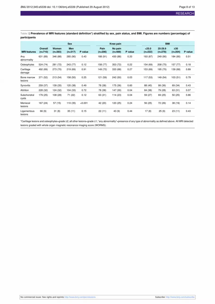
Table 2| Prevalence of MRI features (standard definition*) stratified by sex, pain status, and BMI. Figures are numbers (percentage) ofparticipants
BMIKnee painSex
Overall(n=710)MRI features P value
≥30(n=204)
25-29.9(n=278)
<25.0(n=222)P value
No pain(n=489)
Pain(n=206)P value
Men(n=317)
Women(n=393)
0.51184 (90)249 (90)193 (87)0.20430 (88)188 (91)0.43285 (90)346 (88)631 (89)Anyabnormality
0.18157 (77)208 (75)154 (69)0.22353 (72)158 (77)0.12243 (77)281 (72)524 (74)Osteophytes
0.89139 (68)195 (70)153 (69)0.27333 (68)149 (72)0.91219 (69)273 (70)492 (69)Cartilagedamage
0.79103 (51)149 (54)117 (53)0.03242 (50)121 (59)0.25158 (50)213 (54)371 (52)Bone marrowlesions
0.4369 (34)99 (36)88 (40)0.60175 (36)78 (38)0.49120 (38)139 (35)259 (37)Synovitis
0.0763 (31)79 (28)84 (38)0.04147 (30)78 (38)0.72104 (33)124 (32)228 (32)Attrition
0.8650 (25)69 (25)59 (27)0.04114 (23)63 (31)0.1271 (22)108 (28)179 (25)Subchondralcysts
0.1438 (19)72 (26)56 (25)0.24120 (25)42 (20)<0.001110 (35)57 (15)167 (24)Meniscallesions
0.4323 (11)25 (9)17 (8)0.4443 (9)22 (11)0.1535 (11)31 (8)66 (9)Ligamentouslesions
*Cartilage lesions and osteophytes=grade ≥2; all other lesions=grade ≥1; “any abnormality”=presence of any type of abnormality as defined above. All MRI detectedlesions graded with whole organ magnetic resonance imaging score (WORMS).
No commercial reuse: See rights and reprints http://www.bmj.com/permissions Subscribe: http://www.bmj.com/subscribe
BMJ 2012;345:e5339 doi: 10.1136/bmj.e5339 (Published 29 August 2012) Page 8 of 13
RESEARCH

Table 3| Prevalence of MRI features (standard definition*) stratified by age group. Figures are numbers (percentage) of participants
Age group (years)
MRI features P value≥70 (n=145)≥60-<70 (n=249)≥50-<60 (n=316)
0.00780 (116/145)78 (193/249)68 (215/316)Osteophytes
<0.00180 (116/145)74 (183/249)61 (193/316)Cartilage damage
0.0658 (84/145)55 (137/249)48 (150/316)Bone marrow lesions
0.1041 (60/145)39 (97/249)32 (102/316)Synovial thickening and joint effusion
0.2035 (51/145)35 (86/249)29 (91/316)Attrition
0.00432 (47/145)29 (71/249)19 (61/316)Subchondral cysts
<0.00136 (52/145)24 (60/249)17 (55/316)Meniscal lesions
0.0614 (20/145)10 (24/249)7 (22/316)Ligamentous lesions
*Cartilage lesions and osteophytes=grade ≥2; all other lesions=grade ≥1.
No commercial reuse: See rights and reprints http://www.bmj.com/permissions Subscribe: http://www.bmj.com/subscribe
BMJ 2012;345:e5339 doi: 10.1136/bmj.e5339 (Published 29 August 2012) Page 9 of 13
RESEARCH
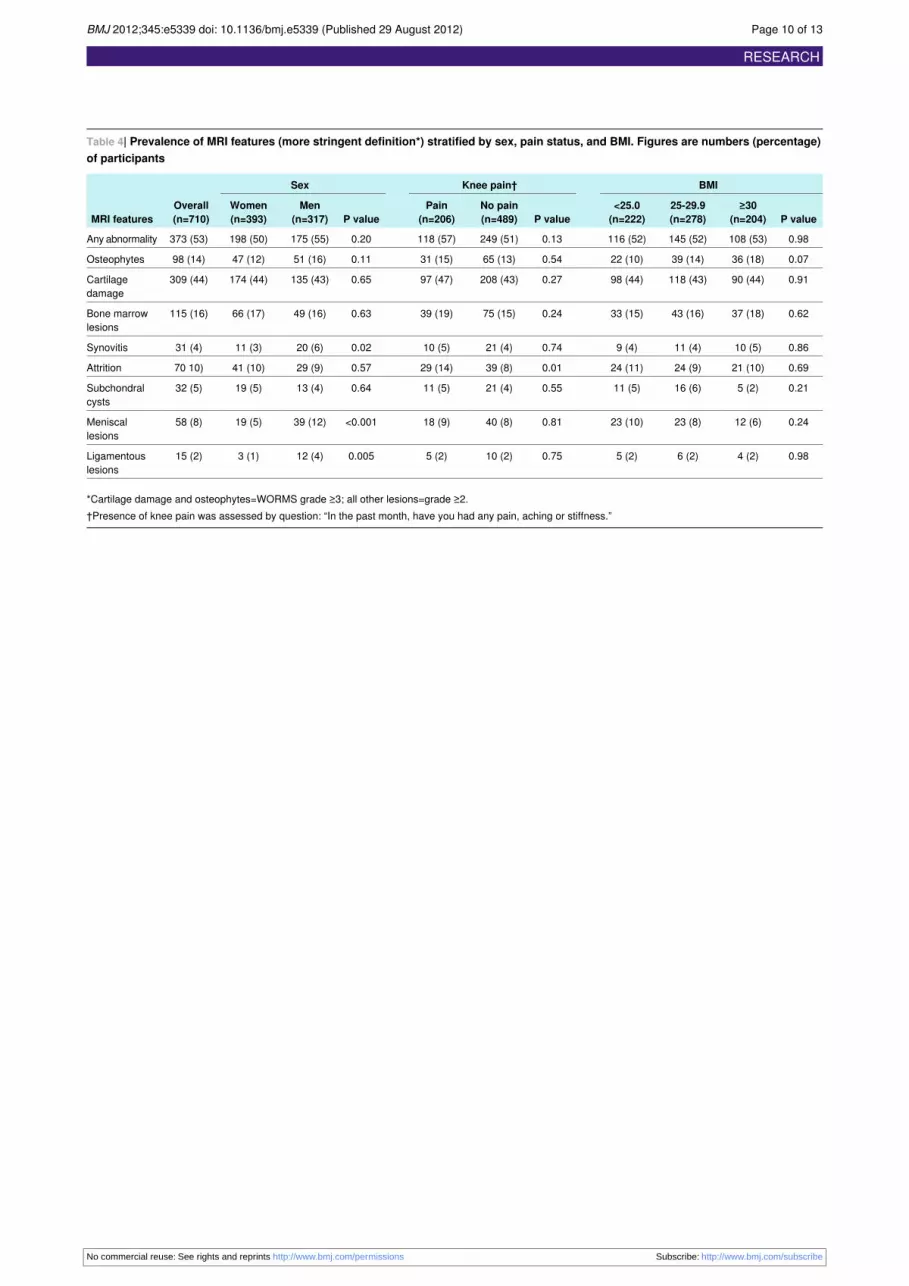
Table 4| Prevalence of MRI features (more stringent definition*) stratified by sex, pain status, and BMI. Figures are numbers (percentage)of participants
BMIKnee pain†Sex
Overall(n=710)MRI features P value
≥30(n=204)
25-29.9(n=278)
<25.0(n=222)P value
No pain(n=489)
Pain(n=206)P value
Men(n=317)
Women(n=393)
0.98108 (53)145 (52)116 (52)0.13249 (51)118 (57)0.20175 (55)198 (50)373 (53)Any abnormality
0.0736 (18)39 (14)22 (10)0.5465 (13)31 (15)0.1151 (16)47 (12)98 (14)Osteophytes
0.9190 (44)118 (43)98 (44)0.27208 (43)97 (47)0.65135 (43)174 (44)309 (44)Cartilagedamage
0.6237 (18)43 (16)33 (15)0.2475 (15)39 (19)0.6349 (16)66 (17)115 (16)Bone marrowlesions
0.8610 (5)11 (4)9 (4)0.7421 (4)10 (5)0.0220 (6)11 (3)31 (4)Synovitis
0.6921 (10)24 (9)24 (11)0.0139 (8)29 (14)0.5729 (9)41 (10)70 10)Attrition
0.215 (2)16 (6)11 (5)0.5521 (4)11 (5)0.6413 (4)19 (5)32 (5)Subchondralcysts
0.2412 (6)23 (8)23 (10)0.8140 (8)18 (9)<0.00139 (12)19 (5)58 (8)Meniscallesions
0.984 (2)6 (2)5 (2)0.7510 (2)5 (2)0.00512 (4)3 (1)15 (2)Ligamentouslesions
*Cartilage damage and osteophytes=WORMS grade ≥3; all other lesions=grade ≥2.†Presence of knee pain was assessed by question: “In the past month, have you had any pain, aching or stiffness.”
No commercial reuse: See rights and reprints http://www.bmj.com/permissions Subscribe: http://www.bmj.com/subscribe
BMJ 2012;345:e5339 doi: 10.1136/bmj.e5339 (Published 29 August 2012) Page 10 of 13
RESEARCH

Table 5| Prevalence of “any abnormality” on MRI stratified by pain status with standard and more stringent definitions of pain. Figures arenumbers (percentage) of participants
P value for differenceNo painMild painPain presentPain definition*
Presence of knee pain in past month†:
0.20430/489 (88)—188/206 (91)Standard definition
0.13249/489 (51)—118/206 (57)More stringent definition
Knee pain‡ lasting at least a month in past year†:
0.08474/541 (88)54/56 (96)90/98 (92)Standard definition
0.01272/541 (50)39/56 (70)57/98 (58)More stringent definition
Knee pain‡ on most days†:
0.12516/586 (88)58/60 (97)45/50 (90)Standard definition
0.02297/586 (51)41/60 (68)29/50 (58)More stringent definition
Score ≥1 in any WOMAC pain questions in person with Kellgren Lawrence grade 0 knees bilaterally§:
0.002411/476 (86)—219/233 (94)Standard definition
<0.001226/476 (48)—146/233 (63)More stringent definition
*Standard definition includes cartilage lesions and osteophytes=grade ≥2; all other lesions=grade ≥1; any abnormality=presence of any type of abnormality asdefined above. More stringent definition includes cartilage damage and osteophytes=WORMS grade ≥3; all other lesions=grade ≥2.†Knee level pain assessment.‡Moderate/severe pain.§Person level pain assessment.
No commercial reuse: See rights and reprints http://www.bmj.com/permissions Subscribe: http://www.bmj.com/subscribe
BMJ 2012;345:e5339 doi: 10.1136/bmj.e5339 (Published 29 August 2012) Page 11 of 13
RESEARCH
Figures
Fig 1 Selection process of knees included in present study
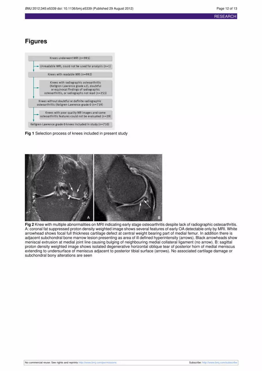
Fig 2 Knee with multiple abnormalities on MRI indicating early stage osteoarthritis despite lack of radiographic osteoarthritis.A: coronal fat suppressed proton density weighted image shows several features of early OA detectable only by MRI. Whitearrowhead shows focal full thickness cartilage defect at central weight bearing part of medial femur. In addition there isadjacent subchondral bone marrow lesion presenting as area of ill defined hyperintensity (arrows). Black arrowheads showmeniscal extrusion at medial joint line causing bulging of neighbouring medial collateral ligament (no arrow). B: sagittalproton density weighted image shows isolated degenerative horizontal oblique tear of posterior horn of medial meniscusextending to undersurface of meniscus adjacent to posterior tibial surface (arrows). No associated cartilage damage orsubchondral bony alterations are seen
No commercial reuse: See rights and reprints http://www.bmj.com/permissions Subscribe: http://www.bmj.com/subscribe
BMJ 2012;345:e5339 doi: 10.1136/bmj.e5339 (Published 29 August 2012) Page 12 of 13
RESEARCH

Fig 3 Prevalence of osteoarthritis features on MRI in knees without radiographic osteoarthritis stratified by age group withstandard and more stringent definitions of MRI abnormalities
No commercial reuse: See rights and reprints http://www.bmj.com/permissions Subscribe: http://www.bmj.com/subscribe
BMJ 2012;345:e5339 doi: 10.1136/bmj.e5339 (Published 29 August 2012) Page 13 of 13
RESEARCH