prenatal endocrine-disrupting chemical pollutant exposure
TRANSCRIPT
© The Author(s) 2020. Published by Oxford University Press on behalf of the European Society of Human Reproduction and Embryology. All rights reserved.For permissions, please e-mail: [email protected].
Human Reproduction Update, Vol.26, No.2, pp. 214–246, 2020
Advance Access Publication on February 28, 2020 doi:10.1093/humupd/dmz044
Hypothetical roadmap towardsendometriosis: prenatalendocrine-disrupting chemicalpollutant exposure, anogenitaldistance, gut-genital microbiota andsubclinical infectionsPilar García-Peñarrubia 1,*, Antonio J. Ruiz-Alcaraz1,María Martínez-Esparza1, Pilar Marín2, andFrancisco Machado-Linde3
1Departamento de Bioquímica, Biología Molecular (B) e Inmunología. Facultad de Medicina, IMIB and Regional Campus of InternationalExcellence “Campus Mare Nostrum”, Universidad de Murcia, 30100 Murcia, Spain 2Servicio de Ginecología y Obstetricia, Hospital ClínicoUniversitario Virgen de la Arrixaca, IMIB, Murcia, Spain 3Servicio de Ginecología y Obstetricia, Hospital Clínico Universitario Reina Sof ía,CARM, Murcia, Spain
*Correspondence address. Department of Biochemistry, Molecular Biology B and Immunology. School of Medicine, IMIB and RegionalCampus of International Excellence “Campus Mare Nostrum”. University of Murcia. 30100 - Murcia. Spain. E-mail: [email protected]
https://orcid.org/0000-0002-2922-4804
Submitted on April 17, 2019; resubmitted on November 8, 2019; editorial decision on November 15, 2019
TABLE OF CONTENTS. . . . . . . . . . . . . . . . . . . . . . . . . . . . . . . . . . . . . . . . . . . . . . . . . . . . . . . . . . . . . . . . . . . . . . . . . . . . . . . . . . . . . . . . . . . . . . . . . . . . . . . . . . . . . . . . . . . . . . . . . . . . . . . . . . . . . . .
• Introduction• Methods
Bibliography search strategyInclusion and exclusion criteriaSelection procedure
• Female genital tract mucosal immune microenvironment• Endometriosis is considered as an immune-mediated chronic inflammatory pathology• Endocrine disrupting chemicals and anogenital distance• Cervicovaginal microbiota• Dysbiosis of the female genital microbiome• Upper genital tract microbiota• Potential role played by subclinical infections of the female genital tract in the development of endometriosis• Oestrogens and microbiota• Proposed model of endometriosis development• Compatibility of this hypothesis with previous theories• Future directions in endometriosis research
BACKGROUND: Endometriosis is a gynaecological hormone-dependent disorder that is defined by histological lesions generated by thegrowth of endometrial-like tissue out of the uterus cavity, most commonly engrafted within the peritoneal cavity, although these lesions canalso be located in distant organs. Endometriosis affects ∼10% of women of reproductive age, frequently producing severe and, sometimes,incapacitating symptoms, including chronic pelvic pain, dysmenorrhea and dyspareunia, among others. Furthermore, endometriosis causes
.
Dow
nloaded from https://academ
ic.oup.com/hum
upd/article/26/2/214/5765414 by guest on 30 June 2022
Hypothetical roadmap towards endometriosis 215
infertility in ∼30% of affected women. Despite intense research on the mechanisms involved in the initial development and later progressionof endometriosis, many questions remain unanswered and its aetiology remains unknown. Recent studies have demonstrated the critical roleplayed by the relationship between the microbiome and mucosal immunology in preventing sexually transmitted diseases (HIV), infertility andseveral gynaecologic diseases.
OBJECTIVE AND RATIONALE: In this review, we sought to respond to the main research question related to the aetiology ofendometriosis. We provide a model pointing out several risk factors that could explain the development of endometriosis. The hypothesisarises from bringing together current findings from large distinct areas, linking high prenatal exposure to environmental endocrine-disruptingchemicals with a short anogenital distance, female genital tract contamination with the faecal microbiota and the active role of genital subclinicalmicrobial infections in the development and clinical progression of endometriosis.
SEARCH METHODS: We performed a search of the scientific literature published until 2019 in the PubMed database. The searchstrategy included the following keywords in various combinations: endometriosis, anogenital distance, chemical pollutants, endocrine-disruptingchemicals, prenatal exposure to endocrine-disrupting chemicals, the microbiome of the female reproductive tract, microbiota and genital tract,bacterial vaginosis, endometritis, oestrogens and microbiota and microbiota–immune system interactions.
OUTCOMES: On searching the corresponding bibliography, we found frequent associations between environmental endocrine-disruptingchemicals and endometriosis risk. Likewise, recent evidence and hypotheses have suggested the active role of genital subclinical microbialinfections in the development and clinical progression of endometriosis. Hence, we can envisage a direct relationship between higher prenatalexposure to oestrogens or estrogenic endocrine-disrupting compounds (phthalates, bisphenols, organochlorine pesticides and others) anda shorter anogenital distance, which could favour frequent postnatal episodes of faecal microbiota contamination of the vulva and vagina,producing cervicovaginal microbiota dysbiosis. This relationship would disrupt local antimicrobial defences, subverting the homeostasis stateand inducing a subclinical inflammatory response that could evolve into a sustained immune dysregulation, closing the vicious cycle responsiblefor the development of endometriosis.
WIDER IMPLICATIONS: Determining the aetiology of endometriosis is a challenging issue. Posing a new hypothesis on this subject providesthe initial tool necessary to design future experimental, clinical and epidemiological research that could allow for a better understanding of theorigin of this disease. Furthermore, advances in the understanding of its aetiology would allow the identification of new therapeutics andpreventive actions.
Key words: endometriosis / anogenital distance / chemical pollutants / prenatal exposure to endocrine-disrupting chemicals / femalegenital microbiome / microbiota-immune system interactions
IntroductionEndometriosis is a common gynaecological hormone-dependent dis-order, defined by histological lesions generated by the growth ofendometrial-like tissue (glands and endometrial stroma) out of theuterine cavity, most commonly engrafted within the peritoneal cavity(i.e. in the peritoneum wall, ovaries, rectosigmoid colon and bladder)although it can also be located in distant organs, such as the lung, liver,pleura, diaphragm, eye and brain. Different types of endometriosisinclude superficial peritoneal endometriosis, ovarian endometriomas,deep infiltrating endometriosis (DIE) and extragenital endometriosis(Vercellini et al., 2014; Zondervan et al., 2018).
Endometriosis has a remarkable impact on women’s healthcare andquality of life, because it usually causes severe symptoms, includingchronic pelvic pain, dysmenorrhea and dyspareunia, among others(Jones et al., 2004; Denny and Mann, 2007; Vercellini et al., 2014;Schliep et al., 2015; Aerts et al., 2018; Zondervan et al., 2018; As-Sanieet al., 2019). It is estimated that endometriosis affects approximately176–200 million women worldwide, more than 10% of women ofreproductive age (As-Sanie et al., 2019), which can reach up to 40% ofwomen aged 18–44 years and undergoing pelvic surgery (Buck Louiset al., 2011; Peterson et al., 2013). The annual incidence rate, from1987 to 1999, in US women aged >15 years was 187 per 100 000,which peaked at ages 25–34 years (380–417 per 100 000) (reviewedby Shafrir et al., 2018), was 350 per 100 000 in Germany (from 2004–2008) (Abbas et al., 2012) and was 108 per 100 000 in Swedish womenaged 15–50 years (Gao, Allebeck, et al., 2019). However, from a
.
.
.
.
.
.
.
.
.
.
.
.
.
.
.
.
.
.
.
.
.
.
.
.
.
.
.
.
.
.
.
.
.
.
.
.
.
.
.
.
.
.
.
.
.
.
.
.
.
.
.
.
.
.
.
.
.
.
population perspective, these numbers could be higher because notall affected women seek medical care or are diagnosed (Vercelliniet al., 2014; Zondervan et al., 2018). The diagnosis of this pathologyis difficult and most frequently requires invasive techniques, such aslaparoscopic surgery for macroscopic observation and/or histologicalconfirmation by biopsy (Vercellini et al., 2014; Zondervan et al., 2018;As-Sanie et al., 2019). The annual medical cost of endometriosis in theUSA was estimated to reach $22 billion in the year 2002 (Simoenset al., 2007) and $69.4 billion in 2010 (Simoens et al., 2012), dueto its prevalence, the diagnostic techniques used and the insufficientavailability of effective treatments (As-Sanie et al., 2019).
Endometriosis is not caused by transformed cells; therefore, it isconsidered a benign disease. Nevertheless, endometriosis shares com-mon characteristics with neoplasia, such as uncontrolled cell growth,invasion of adjacent tissues, defective apoptotic ability, a sustainedlocal inflammatory response and altered angiogenesis, and it has beenrelated to an increased risk of developing ovarian cancer (Wei et al.,2011; Pearce et al., 2012; Kim et al., 2014; Kim, et al., 2015; Machado-Linde et al., 2015).
Although the aetiology of endometriosis remains largely unknown, itseems to have a multi-causal origin. Some hypotheses about the originand pathogenesis of this disease include the following: adhesion andgrowth of endometrial tissue fragments after migration from the uterustoward the peritoneal cavity by a retrograde menstruation process(Sampson, 1927); coelomic metaplasia (peritoneal coelomic epitheliumdifferentiates into endometrial-like cells); neonatal lymphatic or vascu-lar metastatic spread of somatic stem cells and peritoneal implantation
Dow
nloaded from https://academ
ic.oup.com/hum
upd/article/26/2/214/5765414 by guest on 30 June 2022
216 García-Peñarrubia et al.
that remains dormant until menarche; and abnormal embryonic rem-nant Müllerian ducts (from which the upper vagina, uterus and fallopiantubes develop) (Sourial et al., 2014; Asghari et al., 2018). Nevertheless,none of these theories has definitively demonstrated an absolute causalassociation with endometriosis (Vercellini et al., 2014). For example,although retrograde menstruation is a frequent event, only 10% ofwomen develop endometriosis; thus, other concurrent mechanismsneed to be involved in its aetiology. In this sense, individual predis-position factors of genetic (Montgomery et al., 2008; Borghese et al.,2017; Matalliotakis et al., 2017; Fung and Montgomery, 2018) andenvironmental origin (Hunt et al., 2016; Smarr et al., 2016; Cano-Sancho et al., 2019; Wen et al., 2019), together with a deficient oraberrant immune response (Asghari et al., 2018; Riccio et al., 2018;Symons et al., 2018; Zhang, De Carolis, et al., 2018), would allow thepersistence and growth of endometrial tissue outside the uterus.
Many epidemiological studies have explored potential risk factorsassociated with endometriosis. As reviewed by Parasar et al. (2017),Parazzini et al. (2017) and Shafrir et al. (2018), consistent results haveshown that higher parity and a higher body mass index (BMI) areinversely associated with endometriosis, while heavy menstrual cycles,early age at menarche and a lower BMI (Hediger et al., 2005) are asso-ciated with a higher risk. Infertility (Peterson et al., 2013; Gao, Allebeck,et al., 2019), lower overall adiposity and adipose tissues concentratedbelow the waist (Backonja et al., 2017) have also been reported ashigh risk factors. Additionally, inconsistent results have been reportedon the association of endometriosis with alcohol and caffeine intake,smoking and physical activity (Heilier et al., 2007; Matalliotakis et al.,2008; Parasar et al., 2017; Parazzini et al., 2017; Saha et al., 2017; Shafriret al., 2018; Hemmert et al., 2019).
Presently, there is growing evidence linking endometriosis risk withhigh levels of endocrine-disrupting chemicals (EDCs) in women.This evidence has been reviewed and weighted by several authors,highlighting current knowledge gaps and future directions relative toendometriosis studies in adulthood (Guo et al., 2009; Hunt et al.,2016; Smarr et al., 2016; Cano-Sancho et al., 2019; Wen et al.,2019). Industrial and household activity, agrochemical businesses andhuman pharmacological consumption produce a large environmentalaccumulation of EDCs, especially in the sewage network and,ultimately, in surface water, as is the case for alkylphenols (fromdetergents and surfactants), organochlorines (from insecticides),phthalates and bisphenol A (BPA), octylphenol and nonylphenol(from plasticisers), flame retardants, heavy metals (cadmium used inbatteries), dioxins and several compounds contained in cosmetics, suchas benzophenones and natural oestrogens and contraceptives, whichare detected in surface water in ranges of ng/L to μg/L (Rahman et al.,2009; Adeel et al., 2017). Furthermore, it should be noted that humansare, in fact, exposed to a mixture of EDCs and the additive, synergistic,inhibitory and overall cumulative effects of multiple EDCs have notbeen empirically delineated (Gore et al., 2015). Furthermore, there isa high concern about the growing experimental evidence that supportsthe theory of the ‘foetal basis of adult disease’ (Barker, 1990), whichpostulates that prenatal environmental exposure may be associatedwith the development of future diseases. Accordingly, estrogenic oranti-androgenic EDCs could induce significant alterations in prenataldevelopment and the early post-natal periods of life, even at verylow doses of exposure, and would be linked to the developmentof many chronic diseases (Street et al., 2018), for example, breast
.
.
.
.
.
.
.
.
.
.
.
.
.
.
.
.
.
.
.
.
.
.
.
.
.
.
.
.
.
.
.
.
.
.
.
.
.
.
.
.
.
.
.
.
.
.
.
.
.
.
.
.
.
.
.
.
.
.
.
.
.
.
.
.
.
.
.
.
.
.
.
.
.
.
.
.
.
.
.
.
.
.
.
.
.
.
.
.
.
.
.
.
.
.
.
.
.
.
.
.
.
.
.
.
.
.
.
.
.
.
.
.
.
.
.
.
.
.
.
.
.
.
.
.
.
.
cancer (Soto et al., 2013; Paulose et al., 2015; Scsukova et al., 2016),diabetes (Howard, 2019), obesity (Braun, 2017), neurodevelopment(Braun, 2017), allergies (Berger et al., 2019; Quirós-Alcalá et al., 2019),endometriosis (Upson et al., 2015), uterine fibroids (Katz et al., 2016)and several reproductive alterations (Sifakis et al., 2017) includingduring early puberty (Harley et al., 2019; Lee et al., 2019), amongothers (Paulose et al., 2015; Prusinski et al., 2016; Rashtian et al.,2019). Hence, epidemiological studies of endometriosis have focusedon risk factors directly or indirectly associated with prenatal exposures,e.g. maternal smoking (Vannuccini et al., 2016; Gao, Scott, et al.,2019), low birth weight (Missmer et al., 2004; Borghese et al., 2015;Shafrir et al., 2018; Gao, Scott, et al., 2019) and prematurity (Upsonet al., 2015; Vannuccini et al., 2016), which have been associated withan increased endometriosis risk, although a lower risk associationwith maternal smoking (Buck Louis et al., 2007) or no associationwith others prenatal factors has also been reported (Somiglianaet al., 2011; Wolff et al., 2013). Those observations, and others thatrevealed the presence of ectopic endometrium in human foetuses(Signorile et al., 2012) as well as a prenatal-induced endometriosis-likephenotype in mice (Signorile et al., 2010; Koike et al., 2013), supportthis theory (Wei et al., 2016). The first historical human evidencewas the increased risk of endometriosis found in the daughters ofwomen who were administered diethylstilboestrol (DES) (Missmeret al., 2004; Upson et al., 2015), a potent synthetic oestrogen drugwith endocrine-disrupting effects that was prescribed between 1947and 1971 to millions of women worldwide to prevent miscarriage(Reed and Fenton, 2013; Wei et al., 2016). More recently, a shorterfemale anogenital distance (AGD), an indirect and reliable biomarkerof the prenatal hormonal milieu, including EDCs (Callegari et al.,1987; Thankamony et al., 2009, 2016; Liu et al., 2014; Barrett et al.,2018; Schwartz et al., 2019), has been significantly associated withendometriosis (endometriomas, DIE or both) (Mendiola et al., 2016),suggesting that certain in utero hormones or EDCs may cause femalegenital tract (FGT) alterations that could be relevant to developendometriosis. To our best knowledge, there is still no epidemiologicaldata linking those variables.
Finally, the explosive growth of knowledge about the human micro-biome has led to the emergence of a new hypothesis posing aninfectious origin of endometriosis. Accordingly, it could be promotedby certain, but yet undetermined, alterations of the healthy microbiota(gut, oral and/or FGT), which have recently been associated withendometriosis risk, likely by disrupting the microbiota-immune systemtolerance, thus causing a subclinical inflammatory state that couldallow endometriosis development (Kavoussi et al., 2009; Laschke andMenger, 2016; Lin et al., 2016; Khan et al., 2018; Tai et al., 2018;Thomas et al., 2018).
Herein, we have aimed to merge rather large and speculativedistinct areas into an aetiological roadmap to endometriosis basedon emerging epidemiological and experimental evidence. Hence,we hypothesise that there may be a direct relationship betweenhigh prenatal exposure to environmental and/or pharmaceuticalEDCs (phthalates, bisphenols, organochlorine pesticides, dioxin-likecompounds and others) with a shorter AGD or other FGT alterations,which could favour frequent postnatal episodes of faecal microbiotacontamination of the vulva and vagina, producing cervicovaginal (CV)microbial dysbiosis that sustains subclinical inflammation processes anda high risk of developing endometriosis.
Dow
nloaded from https://academ
ic.oup.com/hum
upd/article/26/2/214/5765414 by guest on 30 June 2022
Hypothetical roadmap towards endometriosis 217
Methods
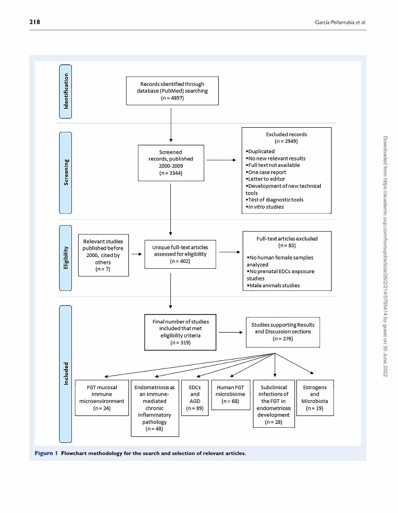
Bibliography search strategyA flowchart of the search strategy and selection of articles includedin this work is shown in Fig. 1. We conducted a systematic searchin PubMed. The search strategy included keywords related to thescientific literature that focused on studies containing research data thatrelated endometriosis to the following: immune system alterations,the female AGD, prenatal risk factors, prenatal exposure to EDCs,prenatal exposure to EDCs and the AGD, postnatal exposure toEDCs, intestinal and FGT microbiota, dysbiosis of the FGT, bacterialvaginosis, endometritis, estrobolome, FGT subclinical infections,periodontitis and microbiota–immune system interactions. Specifically,the combinations of terms used for the literature search were thefollowing (with ‘n’ being the number of publications found for eachcombination of terms): ‘Endometriosis and oestrogens’ (n = 1618),‘Endometriosis and immune system’ (n = 989), ‘Endometriosis andrisk factors’ (n = 1499), ‘Endometriosis and prenatal risk factors’(n = 21), ‘Anogenital distance and endometriosis’ (n = 6), ‘Endocrinedisruptors and endometriosis’ (n = 46), ‘BPA and endometriosis’(n = 20), ‘Diethylstilboestrol and endometriosis’ (n = 82), ‘Dioxin andendometriosis’ (n = 136), ‘Hexachlorobenzene and endometriosis’(n = 4), ‘Organochlorines and endometriosis’ (n = 68), ‘Pesticides andendometriosis’ (n = 47), ‘Phthalates and endometriosis’ (n = 24), ‘EDCand human prenatal exposure’ (n = 80), ‘Endocrine disruptors andAGD’ (n = 45), ‘Prenatal bisphenol A and AGD’ (n = 13), ‘Prenataldioxins and AGD’ (n = 2), ‘Prenatal diethylstilboestrol and AGD’(n = 3), ‘Prenatal hexachlorobenzene and AGD’ (n = 0), ‘Prenatalorganochlorines and AGD’ (n = 9), ‘Prenatal pesticides and AGD’(n = 16), ‘Prenatal phthalates and AGD’ (n = 14), ‘Estrobolome’(n = 5), ‘EDCs and microbiome’ (n = 19), ‘Female genital tract micro-biome and endometriosis’ (n = 5), ‘Female genital tract microbiomeand dysbiosis’ (n = 99), ‘Microbiome and endometriosis’ (n = 17) and‘Periodontitis and endometriosis’ (n = 10).
Inclusion and exclusion criteriaThe articles eligible for inclusion herein were those available online,written in English and published from 2000 until 2019 (20 years),although relevant related articles from earlier dates were also included.Given the amplitude of literature relative to certain areas, recentrelevant reviews were selected to present the conceptual frameworkof our hypothesis. Additionally, reviews containing the meta-analysisof epidemiological studies conducted in women related to these areaswere included. Consequently, some of the references analysed inselected reviews and meta-analyses were not cited here becausetheir results were already implicitly considered. Other reviews orpublications without new relevant results were excluded, as well asarticles for which only the abstract was available but not the fulltext. We also excluded single case reports, letters to editors andpapers displaying the development of new technical tools or testingof diagnostic and therapeutic tools. The population of interest wasprincipally women, although some studies on animal models relatedto EDCs and the AGD, EDCs and microbiota–immune system inter-actions were also selected, given the absence of published resultscorresponding to long-term human studies on these issues. Hence,most articles based solely on data from male animal models were
.
.
.
.
.
.
.
.
.
.
.
.
.
.
.
.
.
.
.
.
.
.
.
.
.
.
.
.
.
.
.
.
.
.
.
.
.
.
.
.
.
.
.
.
.
.
.
.
.
.
.
.
.
.
.
.
.
.
.
.
.
.
.
.
.
.
.
.
.
.
.
.
.
.
.
.
.
.
.
.
.
.
.
.
.
.
.
.
.
.
.
.
.
.
.
.
.
.
.
.
.
.
.
.
.
.
.
.
.
.
.
.
.
.
.
.
.
.
.
.
.
.
.
.
excluded. The same reason applied to the exclusion of most in vitrostudies.
Selection procedureTwo authors (P.G.P. and A.J.R.A.) read the titles and abstractsindependently to check the eligibility. The first search yielded 4897results, with 3344 papers published in the selected period of time(2000–2019). After screening the titles and abstracts, 402 uniquefull-text articles (including 7 articles published before 2000) thatmet the eligibility criteria were downloaded and 83 articles report-ing only male animal results, as well as others in which nei-ther human female samples nor prenatal EDCs exposure wereinvestigated, were also excluded. After reading the full text, thefinal number of eligible articles selected was 319. Next, thosearticles were used to establish the conceptual framework pre-sented in the ‘Introduction’ section, as well as to present, weighand discuss in the ‘Results and Discussion’ section the evidencerelated to the ‘Proposed model of endometriosis development’and ‘Future direction in endometriosis research’ sections. Thus, thearticles selected for this purpose were those that investigated theassociation between a high prenatal exposure to EDCs with alterationsof the female AGD and those that investigated the association ofendometriosis with the following: FGT immune dysregulation, EDCs,the AGD and alterations of oral, intestinal and genital microbiota.
Female Genital Tract MucosalImmune MicroenvironmentRecent studies of the FGT mucosal immune system have demonstratedthe critical role that it plays in maintaining the balance between defenceagainst infections and tolerance to microbiota as well as to both ‘non-self ’ sperm and semi-allogeneic foetuses (Black et al., 2000 and reviewsby Pepe et al., 2018 and Zhou et al., 2018).
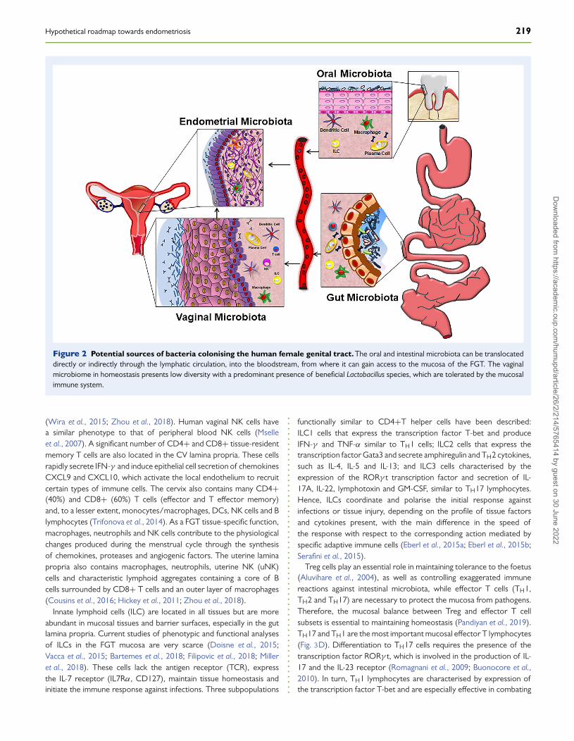
Different types of immune cells are scattered throughout themucosal lamina propria, such as antigen-presenting cells (APCs)(macrophages, dendritic cells (DC) and B lymphocytes), innatelymphoid cells (ILC), NK cells, T cells (CD4+ and CD8+) andantibody-secreting plasma cells, among others (Fig. 2) (Hickey et al.,2011; Zhou et al., 2018).
Four types of myeloid-derived APCs have been identified in the CVmucosa, namely CV-Langerhans cells (located in the CV epitheliumin homeostasis), CD14− DC, CD14+ DC and CD14+ macrophages(located in the lamina propria) (Duluc et al., 2013). Mucosal DCsplay an important role in maintaining microbiota tolerance becauseof their ability to detect, process and present microbial antigens toT lymphocytes (Anahtar et al., 2015). In this regard, CV-Langerhanscells and CD14− DCs have gene expression profiles associated withtolerogenic or TH2-inducing activity. In turn, B lymphocytes specificallyrecognise microbial antigens and differentiate into plasma cellsthat secrete IgM, IgA and IgG; the latter seems to be more abundantthan IgA at this location (Crowley-Nowick et al., 1995; Hickey et al.,2011; Zhou et al., 2018). IgG and IgA play crucial roles in mucosalimmunity by neutralising the growth of microorganisms, therebypreventing adhesion and invasion of pathogens. Notably, the pro-duction of IgG and IgA in the FGT seems to be hormonally regulated
Dow
nloaded from https://academ
ic.oup.com/hum
upd/article/26/2/214/5765414 by guest on 30 June 2022
218 García-Peñarrubia et al.
Figure 1 Flowchart methodology for the search and selection of relevant articles.
Dow
nloaded from https://academ
ic.oup.com/hum
upd/article/26/2/214/5765414 by guest on 30 June 2022
Hypothetical roadmap towards endometriosis 219
Figure 2 Potential sources of bacteria colonising the human female genital tract. The oral and intestinal microbiota can be translocateddirectly or indirectly through the lymphatic circulation, into the bloodstream, from where it can gain access to the mucosa of the FGT. The vaginalmicrobiome in homeostasis presents low diversity with a predominant presence of beneficial Lactobacillus species, which are tolerated by the mucosalimmune system.
(Wira et al., 2015; Zhou et al., 2018). Human vaginal NK cells havea similar phenotype to that of peripheral blood NK cells (Mselleet al., 2007). A significant number of CD4+ and CD8+ tissue-residentmemory T cells are also located in the CV lamina propria. These cellsrapidly secrete IFN-γ and induce epithelial cell secretion of chemokinesCXCL9 and CXCL10, which activate the local endothelium to recruitcertain types of immune cells. The cervix also contains many CD4+(40%) and CD8+ (60%) T cells (effector and T effector memory)and, to a lesser extent, monocytes/macrophages, DCs, NK cells and Blymphocytes (Trifonova et al., 2014). As a FGT tissue-specific function,macrophages, neutrophils and NK cells contribute to the physiologicalchanges produced during the menstrual cycle through the synthesisof chemokines, proteases and angiogenic factors. The uterine laminapropria also contains macrophages, neutrophils, uterine NK (uNK)cells and characteristic lymphoid aggregates containing a core of Bcells surrounded by CD8+ T cells and an outer layer of macrophages(Cousins et al., 2016; Hickey et al., 2011; Zhou et al., 2018).
Innate lymphoid cells (ILC) are located in all tissues but are moreabundant in mucosal tissues and barrier surfaces, especially in the gutlamina propria. Current studies of phenotypic and functional analysesof ILCs in the FGT mucosa are very scarce (Doisne et al., 2015;Vacca et al., 2015; Bartemes et al., 2018; Filipovic et al., 2018; Milleret al., 2018). These cells lack the antigen receptor (TCR), expressthe IL-7 receptor (IL7Rα, CD127), maintain tissue homeostasis andinitiate the immune response against infections. Three subpopulations
.
.
.
.
.
.
.
.
.
.
.
.
.
.
.
.
.
.
.
.
.
.
.
.
.
.
.
.
.
.
.
.
.
.
.
.
.
.
.
.
.
.
.
.
.
.
.
.
.
.
.
.
.
.
functionally similar to CD4+T helper cells have been described:ILC1 cells that express the transcription factor T-bet and produceIFN-γ and TNF-α similar to TH1 cells; ILC2 cells that express thetranscription factor Gata3 and secrete amphiregulin and TH2 cytokines,such as IL-4, IL-5 and IL-13; and ILC3 cells characterised by theexpression of the RORγ t transcription factor and secretion of IL-17A, IL-22, lymphotoxin and GM-CSF, similar to TH17 lymphocytes.Hence, ILCs coordinate and polarise the initial response againstinfections or tissue injury, depending on the profile of tissue factorsand cytokines present, with the main difference in the speed ofthe response with respect to the corresponding action mediated byspecific adaptive immune cells (Eberl et al., 2015a; Eberl et al., 2015b;Serafini et al., 2015).
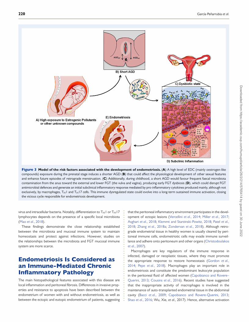
Treg cells play an essential role in maintaining tolerance to the foetus(Aluvihare et al., 2004), as well as controlling exaggerated immunereactions against intestinal microbiota, while effector T cells (TH1,TH2 and TH17) are necessary to protect the mucosa from pathogens.Therefore, the mucosal balance between Treg and effector T cellsubsets is essential to maintaining homeostasis (Pandiyan et al., 2019).TH17 and TH1 are the most important mucosal effector T lymphocytes(Fig. 3D). Differentiation to TH17 cells requires the presence of thetranscription factor RORγ t, which is involved in the production of IL-17 and the IL-23 receptor (Romagnani et al., 2009; Buonocore et al.,2010). In turn, TH1 lymphocytes are characterised by expression ofthe transcription factor T-bet and are especially effective in combating
Dow
nloaded from https://academ
ic.oup.com/hum
upd/article/26/2/214/5765414 by guest on 30 June 2022
220 García-Peñarrubia et al.
Figure 3 Model of the risk factors associated with the development of endometriosis. (A) A high level of EDC (mainly oestrogen-likecompounds) exposure during the prenatal stage induces a shorter AGD (B) that could affect the physiological development of other sexual featuresand enhance future episodes of retrograde menstruation. (C) Additionally, during childhood, a short AGD would favour frequent faecal microbiotacontamination from the anus toward the external and lower FGT (the vulva and vagina), producing early FGT dysbiosis (D), which could disrupt FGTantimicrobial defences and generate an initial subclinical inflammatory response mediated by pro-inflammatory cytokines produced mainly, although notexclusively, by macrophages, TH1 and TH17 cells. This immune dysregulated state could evolve into a long-term sustained immune activation, closingthe vicious cycle responsible for endometriosis development.
virus and intracellular bacteria. Notably, differentiation to TH1 or TH17lymphocytes depends on the presence of a specific local microbiota(Mao et al., 2018).
These findings demonstrate the close relationship establishedbetween the microbiota and mucosal immune system to maintainhomeostasis and protect against infections. However, studies onthe relationships between the microbiota and FGT mucosal immunesystem are more scarce.
Endometriosis Is Considered asan Immune-Mediated ChronicInflammatory PathologyThe main histopathological features associated with this disease arelocal inflammation and peritoneal fibrosis. Differences in invasive prop-erties and resistance to apoptosis have been described between theendometrium of women with and without endometriosis, as well asbetween the ectopic and eutopic endometrium of patients, suggesting
.
.
.
.
.
.
.
.
.
.
.
.
.
.
.
.
.
.
.
.
.
.
.
.
.
.
.
.
.
.
.
.
.
.
.
.
.
.
.
.
.
that the peritoneal inflammatory environment participates in the devel-opment of ectopic lesions (Vercellini et al., 2014; Miller et al., 2017;Asghari et al., 2018; Klemmt and Starzinski-Powitz, 2018; Patel et al.,2018; Zhang et al., 2018a; Zondervan et al., 2018). Although retro-grade endometrial tissue in healthy women is usually cleared by peri-toneal immune cells, endometriotic cells may evade immune surveil-lance and adhere onto peritoneum and other organs (Christodoulakoset al., 2007).
Macrophages are key regulators of the immune response ininfected, damaged or neoplastic tissues, where they must promotethe appropriate response to restore homeostasis (Gordon et al.,2014; Pepe et al., 2018). Macrophages play an important role inendometriosis and constitute the predominant leukocyte populationin the peritoneal fluid of affected women (Capobianco and Rovere–Querini, 2013; Cousins et al., 2016). Recent studies have suggestedthat the inappropriate activity of macrophages is involved in themaintenance of auto-transplanted endometrial tissue in the abdominalcavity (Bacci et al., 2009; Capobianco and Rovere-Querini, 2013;Shao et al., 2016; Wu, Xie, et al., 2017). Hence, alternative activation
Dow
nloaded from https://academ
ic.oup.com/hum
upd/article/26/2/214/5765414 by guest on 30 June 2022
Hypothetical roadmap towards endometriosis 221
of macrophages towards a tissue-repair phenotype (M2) would allowthe survival, neovascularisation and growth of endometriotic lesions(Bacci et al., 2009; Capobianco and Rovere-Querini, 2013; Wu, Xie,et al., 2017; Duan et al., 2018; Sun et al., 2019). Additionally, otherauthors have described classical macrophage polarisation (M1) in theeutopic endometrium of patients with endometriosis with respectto healthy individuals (Takebayashi et al., 2015). Analysis of themolecular mechanisms that mediate the activation and polarisationof macrophages is necessary to understand the pathophysiologyof endometriosis and allow the development of new medicaltreatments. Furthermore, dysfunctional NK cells (Elkabets et al., 2010;Kang et al., 2014; Yu et al., 2016), which express higher levels ofinhibitory receptors (KIR) (Wu et al., 2000) and a lower number ofactivating receptors (KAR), down-regulate the phagocytic activity ofmacrophages and expression of scavenger receptors (Chuang et al.,2009) and induce Treg lymphocytes, whose inhibitory activity allowsendometrial cells to escape from peritoneal immune surveillance(Christodoulakos et al., 2007; Gogacz et al., 2014; de Barros et al.,2017; Wang et al., 2017; Hanada et al., 2018). Additionally, the rapidincrease in peritoneal myeloid-derived suppressor cells (MDSCs) in thepresence of endometrium could be involved in the depressed immuneresponse (Elkabets et al., 2010; Zhang et al., 2018b). Consequently,there is an increase in soluble mediators secreted by macrophagesand other cells (Hassa et al., 2009; Mu et al., 2018; Nothnick andAlali, 2016; Rakhila et al., 2016), including TGF-β (responsible forpost-surgical adhesions) (Choi et al., 2017; Hanada et al., 2018), IL-1β (induces COX-2 and VEGF expression, increasing angiogenesis)(McLaren, 2000; Huang et al., 2013), IL-8 (increases proliferation,migration, angiogenesis and survival of migrated endometrial cells)(Ulukus et al., 2009; Sikora et al., 2017), PGE2 (induces oestrogensynthesis in collaboration with IL-4, inhibits apoptosis and increasesthe production of FGF-9) (Urata et al., 2013), MIF (increases theexpression of VEGF, IL-8 and MCP-1 in endometriotic cells, inducesangiogenesis and is a tissue remodelling agent) (Veillat et al., 2010),MCP-1 (increases monocyte recruitment) (Ulukus et al., 2009; Li et al.,2012; Gou et al., 2019), sICAM-1 (Kuessel et al., 2017), IL-6 (Li et al.,2017) (inhibits cytotoxicity mediated by NK cells (Kang et al., 2014)), IL-27 (triggers the secretion of IL-10 by TH17 cells inducing the promotionof endometriosis) (Chang et al., 2017) and IL-32 (Lee et al., 2018),among others (Nothnick and Alali, 2016; Mu et al., 2018). Throughthe study of transcriptomes, the STAT proteins, SMAD transcriptionfactors and Akt and MEK/ERK signalling pathways have been identifiedas central signalling regulators of the pathophysiological processesthat lead to the development of endometriosis (Li et al., 2012, 2019;Matsuzaki and Darcha, 2015 and reviews by Aznaurova et al., 2014;McKinnon et al., 2016; Patel et al., 2018; Riccio et al., 2018).
Endocrine-Disrupting Chemicalsand Anogenital DistanceAs mentioned above, numerous cohort, cross-sectional and case–control studies conducted in patients with endometriosis have soughtthe association between EDC levels in women and endometriosis.Although some inconsistent results have been obtained, as new studiesaccumulate, evidence of an EDC–endometriosis association obtainedin systematic reviews and meta-analyses of the available studies is
.
.
.
.
.
.
.
.
.
.
.
.
.
.
.
.
.
.
.
.
.
.
.
.
.
.
.
.
.
.
.
.
.
.
.
.
.
.
.
.
.
.
.
.
.
.
.
.
.
.
.
.
.
.
.
.
.
.
.
.
.
.
.
.
.
.
.
.
.
.
.
.
.
.
.
.
.
.
.
.
.
.
.
.
.
.
.
.
.
.
.
.
.
.
.
.
.
.
.
.
.
.
.
.
.
.
.
.
.
.
.
.
.
.
.
.
.
.
.
.
.
.
.
.
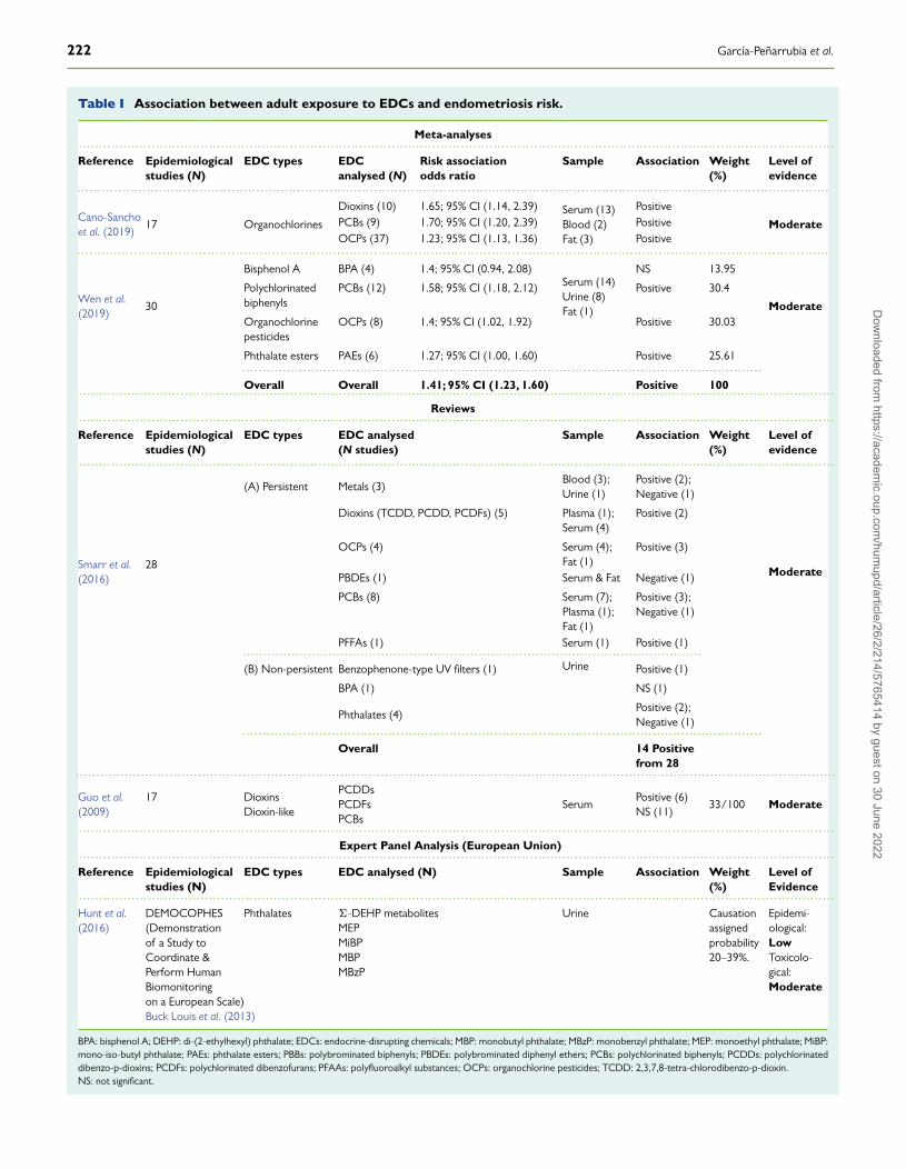
increasing, as shown in Table I. Early on, Guo et al. (2009) reviewed themolecular biology and weighted the evidence on 17 epidemiologicalstudies of the association between dioxins and endometriosis, con-cluding that no significant evidence supported a link between dioxinand endometriosis. More recently, Smarr et al. (2016) summarisedthe weight of evidence on the relationships between endometriosisand several persistent and non-persistent EDCs, concluding that theexisting evidence supported a possible relationship between manyEDCs and endometriosis, except for BPA and polybrominated diphenylethers (PBDEs). Furthermore, they discussed the key methodologicalchallenges that preclude a more complete understanding of the litera-ture, followed by suggestions to answer critical data gaps. An analysisof the association between phthalates and endometriosis conductedby an expert panel in the European Union rated the strength ofepidemiological and toxicological evidence as ‘low’ and ‘moderate’,respectively (Hunt et al., 2016). Recently, Cano-Sancho et al. (2019)reported a significant but ‘moderate evidence’ of association betweenorganochlorines and endometriosis by a meta-analysis of 17 studies,and Wen et al. (2019) also analysed and weighted the evidence of 30studies conducted on EDCs and endometriosis (4 with BPA, 12 withPCBs, 8 with organochlorine pesticides (OCPs) and 6 with phthalates)and reported a significant association with all of them, except with BPA.
This association could likely be produced by the suggested potentialof EDCs to modify the attachment and proliferation of endometrial(Kim et al., 2015) and endothelial cells (Bredhult et al., 2007), toincrease reactive oxygen species generation and decrease expressionof antioxidant enzymes (Cho et al., 2015), as well as to disrupt inflam-matory and endocrine responses, as described from in vitro and murineexperimental studies (Huang et al., 2017), as well as to modify theinvasion parameters in endometriotic lesions in a rat model, increasingthe expression of COX-2, VEGF and TNF-α (Chiappini et al., 2019).
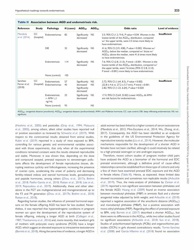
Recently, a short anofourchettal distance (AGDAF) in adults has beenstatistically associated with adult endometriosis (endometriomas, DIEor both) (Mendiola et al., 2016). Consequently, it has been proposedas a useful risk marker for clinical and epidemiology studies (Sánchez–Ferrer et al., 2017, 2019) (Table II). Hence, although the current levelof evidence is insufficient, given the small number of studies and samplesize, it will be of great interest to uncover the mechanisms underlyingthe association between the AGD and endometriosis.
The AGD is a sexual dimorphic anatomical characteristic, defined asthe length from the anus to the genitals. In many species of mammals,including humans, the AGD is almost twice as long in males than infemales both in childhood (Thankamony et al., 2009) and throughoutlife (Salazar-Martinez et al., 2004; Barrett et al., 2014; Kim, Lee, et al.,2014; Thankamony et al., 2016; Priskorn et al., 2018; Schwartz et al.,2019).
Studies in animal models have shown that the AGD at birth isdirectly related to intra-uterine hormonal exposure (Wolf et al., 2002;Dean et al., 2012). Thus, a higher content of androgens results ina longer AGD, while higher oestrogen or anti-androgen exposure isassociated with a shorter AGD (Hotchkiss et al., 2007; Dean andSharpe, 2013; Schwartz et al., 2019). Association with a shorter AGDin female animals has been reported for phthalates: di-ethylhexyl-phthalate (DEHP) (Brehm et al., 2018), a mixture (Zhou et al., 2017a;Repouskou et al., 2019), a mix of phthalates and alkylphenols (Patiño–García et al., 2018), BPA (Christiansen et al., 2014), butylparaben(Boberg et al., 2016), nonylphenol (Takagi et al., 2004), benzophenone
Dow
nloaded from https://academ
ic.oup.com/hum
upd/article/26/2/214/5765414 by guest on 30 June 2022
222 García-Peñarrubia et al.
Table I Association between adult exposure to EDCs and endometriosis risk.
Meta-analyses..........................................................................................................................................................................................Reference Epidemiological
studies (N)EDC types EDC
analysed (N)Risk associationodds ratio
Sample Association Weight(%)
Level ofevidence
.........................................................................................................................................................................................
Cano-Sanchoet al. (2019)
17 Organochlorines
Dioxins (10) 1.65; 95% CI (1.14, 2.39) Serum (13)Blood (2)Fat (3)
Positive
ModeratePCBs (9) 1.70; 95% CI (1.20, 2.39) PositiveOCPs (37) 1.23; 95% CI (1.13, 1.36) Positive
.........................................................................................................................................................................................
Wen et al.(2019)
30
Bisphenol A BPA (4) 1.4; 95% CI (0.94, 2.08)Serum (14)Urine (8)Fat (1)
NS 13.95
Moderate
Polychlorinatedbiphenyls
PCBs (12) 1.58; 95% CI (1.18, 2.12) Positive 30.4
Organochlorinepesticides
OCPs (8) 1.4; 95% CI (1.02, 1.92) Positive 30.03
Phthalate esters PAEs (6) 1.27; 95% CI (1.00, 1.60) Positive 25.61................................................................................................................................Overall Overall 1.41; 95% CI (1.23, 1.60) Positive 100..........................................................................................................................................................................................
Reviews..........................................................................................................................................................................................Reference Epidemiological
studies (N)EDC types EDC analysed
(N studies)Sample Association Weight
(%)Level ofevidence
..........................................................................................................................................................................................
Smarr et al.(2016)
28
(A) Persistent Metals (3)Blood (3);Urine (1)
Positive (2);Negative (1)
Moderate
Dioxins (TCDD, PCDD, PCDFs) (5) Plasma (1);Serum (4)
Positive (2)
OCPs (4) Serum (4);Fat (1)
Positive (3)
PBDEs (1) Serum & Fat Negative (1)
PCBs (8) Serum (7);Plasma (1);Fat (1)
Positive (3);Negative (1)
PFFAs (1) Serum (1) Positive (1).................................................................................................................(B) Non-persistent Benzophenone-type UV filters (1) Urine Positive (1)
BPA (1) NS (1)
Phthalates (4)Positive (2);Negative (1)
................................................................................................................................Overall 14 Positive
from 28...........................................................................................................................................................................................
Guo et al.(2009)
17 DioxinsDioxin-like
PCDDsPCDFsPCBs
SerumPositive (6)NS (11)
33/100 Moderate
..........................................................................................................................................................................................Expert Panel Analysis (European Union)
..........................................................................................................................................................................................Reference Epidemiological
studies (N)EDC types EDC analysed (N) Sample Association Weight
(%)Level ofEvidence
..........................................................................................................................................................................................Hunt et al.(2016)
DEMOCOPHES(Demonstrationof a Study toCoordinate &Perform HumanBiomonitoringon a European Scale)Buck Louis et al. (2013)
Phthalates �-DEHP metabolitesMEPMiBPMBPMBzP
Urine Causationassignedprobability20–39%.
Epidemi-ological:LowToxicolo-gical:Moderate
BPA: bisphenol A; DEHP: di-(2-ethylhexyl) phthalate; EDCs: endocrine-disrupting chemicals; MBP: monobutyl phthalate; MBzP: monobenzyl phthalate; MEP: monoethyl phthalate; MiBP:mono-iso-butyl phthalate; PAEs: phthalate esters; PBBs: polybrominated biphenyls; PBDEs: polybrominated diphenyl ethers; PCBs: polychlorinated biphenyls; PCDDs: polychlorinateddibenzo-p-dioxins; PCDFs: polychlorinated dibenzofurans; PFAAs: polyfluoroalkyl substances; OCPs: organochlorine pesticides; TCDD: 2,3,7,8-tetra-chlorodibenzo-p-dioxin.NS: not significant.
Dow
nloaded from https://academ
ic.oup.com/hum
upd/article/26/2/214/5765414 by guest on 30 June 2022
Hypothetical roadmap towards endometriosis 223
Table II Association between AGD and endometriosis risk.
Reference Study Pathology N (cases) AGDAF AGDAC Odds ratio Level of evidence.........................................................................................................................................................................................Mendiolaet al. (2016)
CC(hospital)
Endometriomas 82 Significantlydecreased
NS 3.5; 95% CI (1.3, 9.4), P-value = 0.04. Women in thelowest tertile of the AGDAF distribution, comparedw/ the upper tertile, were 3.5 times more likely tohave endometriosis.
Insufficient
DIE 32 Significantlydecreased
NS 41.6; 95% CI (3.9, 438), P value = 0.002. Women w/AGDAF below the median, compared w/ those w/AGDAF above the median, were 41.6 times more likelyto have endometriosis.
Endometriosis 114 Significantlydecreased
NS 7.6; 95% CI (2.8, 21.0), P trend <0.001. Women in thelowest tertile of the AGDAF distribution, compared w/the upper tertile, were 7.6 times (95% CI 2.8, 21.0),P trend <0.001) more likely to have endometriosis.
None (control) 105.........................................................................................................................................................................................Sanchez-Ferrer et al.(2018)
CC(hospital)
EndometriosisEndometriomas
5745
SignificantlydecreasedSignificantlydecreased
NSNS
3.72; 95% CI (1.64, 8.5), P value = 0.002.(22.8 ± 4.6 vs. 27.2 ± 5.7 mm), P trend <0.001.2.82; 95% CI (1.23, 6.64), P value = 0.020
Insufficient
Endometriosis+ AMH ≤1ng/mL
25 Significantlydecreased
NS 17.4; 95% CI (5.64, 53.82) Lower AGDAF & AMHare risk factors for endometriosis.
None (control) 93 NS
AGDAC: anogenital distance (anoclitoris); AGDAF: anogenital distance (anofourchettal); AMH: anti-Müllerian hormone; CC: case-control; DIE: deep infiltrating endometriosis. NS: notsignificant.
(Hoshino et al., 2005) and pesticides (Gray et al., 1994; Matsuuraet al., 2005), among others, albeit other studies have reported nullor positive association as reviewed by Schwartz et al. (2019). Withrespect to the controversial results obtained from animal studies,Rubin et al. (2019) reported in a study of mice exposed to BPA,controlling for various genetic and environmental variables associ-ated with those experiments, that only when all the experimentalconditions remained constant were the results obtained reproducibleand stable. Moreover, it was shown that, depending on the doseand compound assayed, prenatal exposure to xenoestrogen pollu-tants affects the development of female reproductive tissues, dis-rupting oestrous cyclicity and folliculogenesis, increasing the presenceof ovarian cysts, accelerating the onset of puberty and decreasingfertility-related indices and steroid hormones levels, gonadotropinsand peptide hormones, among others (Zhou et al., 2017a; Brehmet al., 2018; Patiño-García et al., 2018; Rattan et al., 2018; Lite et al.,2019; Repouskou et al., 2019). Additionally, these and other alter-ations in the FGT are multigenerational and transgenerational up tothe F3 and F4 generation (Zhou et al., 2017b; Brehm et al., 2018;Rattan et al., 2018).
Regarding human studies, the influence of prenatal hormonal expo-sure on the female offspring AGD has been far less studied. Never-theless, it was reported that hyperandrogenic conditions in pregnantwomen act upon the development of the reproductive system offemale offspring, inducing a longer AGD at birth (Callegari et al.,1987; Thankamony et al., 2016) and, more recently, that newborn girlsof mothers with polycystic ovary syndrome (PCOS) have a longerAGD, which suggest an elevated exposure to intrauterine testosterone(Barrett et al., 2018). Along the same lines of evidence, a longer AGD in
.
.
.
.
.
.
.
.
.
.
.
.
.
.
.
.
.
.
.
.
.
.
.
.
.
.
.
.
.
.
.
.
.
.
.
.
.
.
.
.
.
.
.
.
.
.
.
.
.
.
.
.
.
.
.
.
.
.
.
.
.
.
.
adult women has been linked to a higher content of serum testosterone(Mendiola et al., 2012; Mira-Escolano et al., 2014; Wu, Zhong, et al.,2017). Consequently, the AGD has been identified as an endpointin the guidelines of the US Environmental Protection Agency forreproductive toxicity studies (Liu et al., 2014). However, the molecularmechanisms responsible for the development of a shorter AGD infemales have not been clarified, although it could intuitively be relatedto a high prenatal oestrogen or anti-androgen exposure.
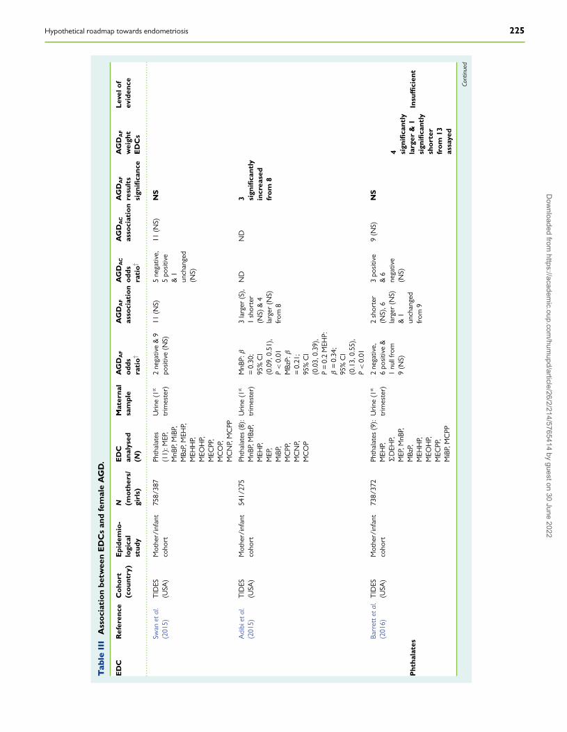
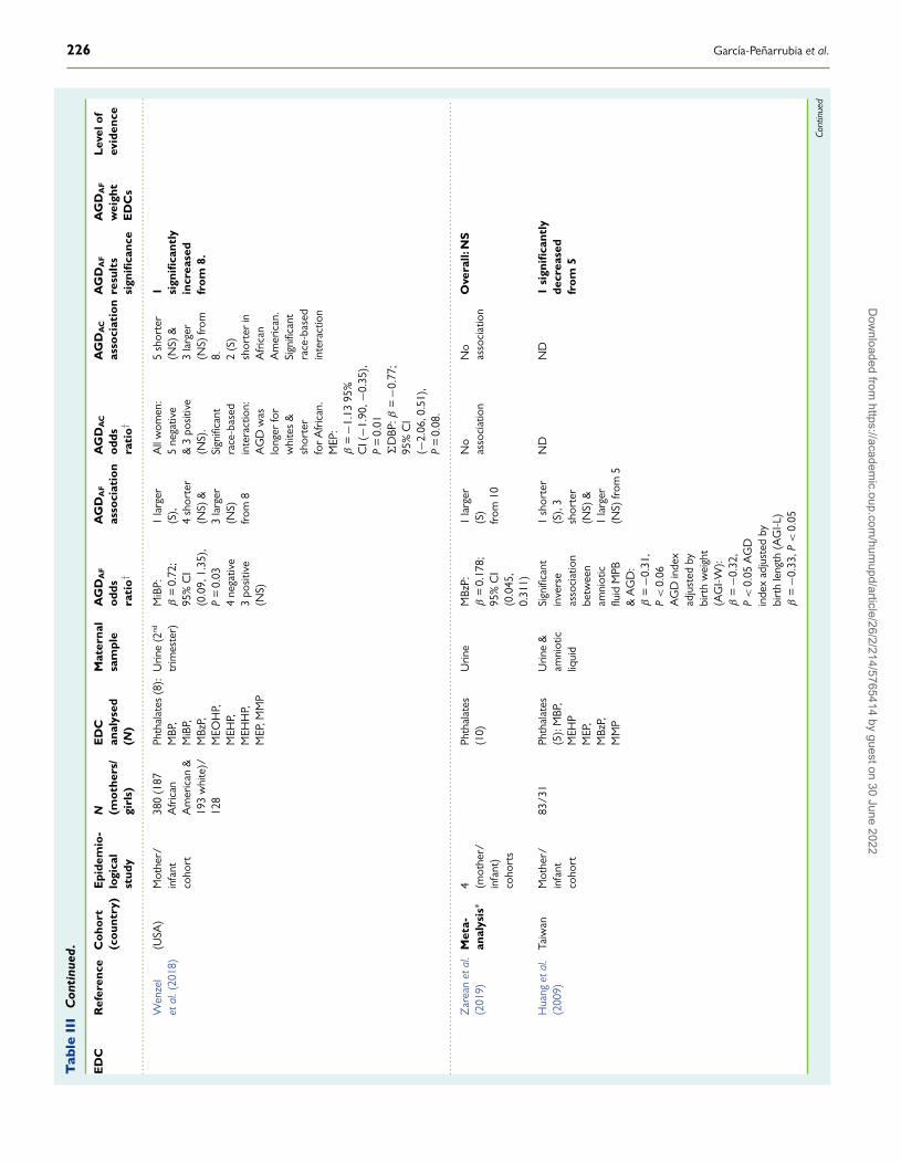
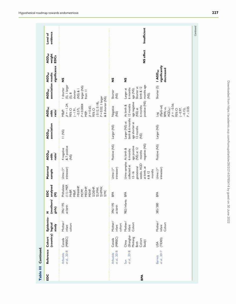
Therefore, recent cohort studies of pregnant mother–child pairshave analysed the AGD as a biomarker of the hormonal and EDCprenatal environment, although a definitive proof of cause–effectrelationships cannot be established from these type of cohorts and onlya few of them have examined prenatal EDC exposure and the AGDin female infants (Table III). Hence, as expected, these limited datashowed inconsistent and sometimes non-replicable results (Arbuckleet al., 2019). Thus, the meta-analysis of phthalates by Zarean et al.(2019) reported a non-significant association between phthalates andthe female AGD; Huang et al. (2009) found an inverse associationbetween monobutyl phthalate (MBP) in amniotic fluid and a shorteranogenital index adjusted by birth weight; and Arbuckle et al. (2018)reported a negative association of the anoclitoris distance (AGDAC)and monobenzyl phthalate (MBzP), but a positive association withmono-ethyl phthalate (MEP). Regarding the effect of prenatal exposureto BPA, only Barrett et al. (2017) described a shorter AGDAC, butthere were no differences in the AGDAF, while two other studies foundno significant associations (Arbuckle et al., 2018; Sun et al., 2018).Studies of polychlorinated biphenyls (PCBs) and organochlorine pes-ticides (OCPs) in girls showed contradictory results. Torres-Sanchezet al. (2008) and García-Villarino et al. (2018) found no association
Dow
nloaded from https://academ
ic.oup.com/hum
upd/article/26/2/214/5765414 by guest on 30 June 2022
224 García-Peñarrubia et al.
between the AGD and maternal serum levels of two polybrominateddiphenyl ethers (PBDEs) and several organochlorine compounds, butthe sample size was very small (only 34 and 14 girls, respectively).Loreto-Gómez et al. (2018) found an inverse association with thefemale AGDAF/H index and PCB 170, but it was positive with o,p′-DDT and the mixture of o,p′-DDT and p,p′-DEE from six PCBs andseveral OCPs (DDT) analysed. Bornman et al. (2016) found no asso-ciation with the level of p,p′-DDT/−DDE or o,p′-DDT, in mothers’serum, and the AGDAF at delivery and after one year, while o,p′-DDEwas negatively associated with the AGDAF in 1-year-old girls. On theother hand, Vafeiadi et al. (2013) found no association between dioxinand dioxin-like compounds and decreased AGD. In turn, two studiesdescribed a null effect of triclosan (Lassen et al., 2016; Arbuckle et al.,2018). Finally, Dalsager et al. (2018) found no significant changes in theAGD, at 3 months of age, with urine levels of organophosphate andpyrethroid insecticides and the herbicide 2,4-dichlorophenoxyaceticacid at 28 weeks of pregnancy. Hence, to date, there are limited dataand, thus, insufficient evidence to indicate that prenatal EDC exposureshortens the AGD in girls. Therefore, more studies are needed toinclude a greater number of people and to integrate their results withthe experimental outcomes in animals for a given mixture of EDCs toassess the association between prenatal EDC exposure and a shorterAGD in females (Bornehag et al., 2019).
The small number of studies and lack of accurate exposure measure-ments are the principal limitations (Zarean et al., 2019). Furthermore,inconsistencies in the specific literature can be caused by multipleuncontrolled variables and methodological gaps associated with thesecompounds and human cohorts, including the age at which the AGD ismeasured and training of the staff who measure it. Additionally, moststudies collected maternal EDC samples either from urine or blood ata unique and variable time point. In this regard, a recent prospectivecohort study suggested that AGD in humans, as in animals, is fixed inearly gestation (probably during the hypothetical masculinisation pro-gramming window, between 8 and 14 weeks) (Jain et al., 2018). Hence,the EDC concentrations determined beyond those dates will be uselessfor the AGD outcomes. Additionally, some of the most frequentlystudied compounds (i.e. phthalates and BPA) are rapidly metabolized;therefore, the measurement of a single time point does not reflect theactual dose and duration of prenatal exposure. In turn, it is known thatthe shape of dose-response curves for endocrine compounds are oftennon-monotonic (Xu et al., 2017), implying that it is possible that low-dose exposure to a specific EDC, such as BPA (Rubin et al., 2019) orDEHP (Do et al., 2012), produces phenotypic changes, while higherdoses do not produce those effects. Furthermore, it has been shownthat some neonatal outcomes are associated differently with prenatalexposure to individual EDC and EDC mixtures (Kelley et al., 2019). Inthis regard, to our knowledge, no human study has focused on prenatalexposure to defined mixtures of EDC and AGD. On the other hand,differences between individuals and inter-race in human studies are acommon issue that produces great heterogeneity of the recorded data(Wenzel et al., 2018), preventing significant conclusions from studiescarried out using a small sample size.
Nevertheless, despite the inconsistent results, there is growingexperimental and epidemiological evidence and concern about theeffects of environmental or synthetic EDCs on alterations of normalimmune cell development (Kelley et al., 2019; Nowak et al., 2019), FGTreproductive function (Crain et al., 2008; Patel et al., 2015; Leonardi
.
.
.
.
.
.
.
.
.
.
.
.
.
.
.
.
.
.
.
.
.
.
.
.
.
.
.
.
.
.
.
.
.
.
.
.
.
.
.
.
.
.
.
.
.
.
.
.
.
.
.
.
.
.
.
.
.
.
.
.
.
.
.
.
.
.
.
.
.
.
.
.
.
.
.
.
.
.
.
.
.
.
.
.
.
.
.
.
.
.
.
.
.
.
.
.
.
.
.
.
.
.
.
.
.
.
.
.
.
.
.
.
.
.
.
.
.
.
.
.
.
.
.
.
et al., 2017; Rashtian et al., 2019) and several gynaecologic diseases(Gore et al., 2015; Paulose et al., 2015; Hunt et al., 2016; Smarr et al.,2016; Piazza and Urbanetz, 2019; Wen et al., 2019). Concretely, arecent longitudinal study with pregnant mothers and their childrenuntil 9–13 years has revealed that prenatal exposure to phthalates,parabens and phenols during the in utero windows of susceptibility isassociated with earlier puberty in girls (Harley et al., 2019), althoughinconsistent results have also been described as reviewed by Lee et al.(2019).
According to these observations, it is tempting to speculate that ahigh level of exposure to EDCs or other still unknown compoundsduring prenatal life could not only modify the AGD and induce earliermenarche (considered a risk factor for endometriosis) (Nnoahamet al., 2012; Parasar et al., 2017; Parazzini et al., 2017; Shafrir et al.,2018) but also alter other sexual features, such as the normal devel-opment of uterine endometrial tissue or the length and morphologyof the uterus and fallopian tubes, which could be risk factors for thedevelopment and further progression of endometriosis. In this regard,studies in DES-daughter cohorts have revealed several morphologicand functional alterations of the FGT (Swan, 2000; Reed and Fenton,2013) and Missmer et al. (2004) found a higher risk of endometriosisamong women prenatally exposed to DES.
Therefore, based on those reported observations, we posit a directrelationship between higher prenatal exposure to oestrogens, anti-androgenic EDCs or other undetermined factors, a shorter AGD anda higher risk of developing endometriosis after menarche.
Human Female Genital TractMicrobiomeAnother risk factor increasingly implicated in the aetiology and patho-genesis of endometriosis is the composition of gut microbiota and itscorresponding metabolic activity (Kobayashi et al., 2014; Laschke andMenger, 2016; Khan et al., 2018). As mentioned above, there is anevident and close relationship between a healthy gut microbiota andcorrect development and function of the immune system (Smolinskaand O’Mahony, 2016; Levy et al., 2017; Pabst, 2017). In fact, theacquisition and maintenance of a suitable microbiota in early life seemto be a determining factor in maintaining a healthy state throughout life(Houghteling and Walker, 2015; Stinson et al., 2017). In this sense, thedegradation of certain glycans and mucin by commensal bacteria canact as a means of communication between the microbiota and host.Among them, short-chain fatty acids (SCFA) such as acetate, butyrateand propionate, produced from carbohydrate metabolism, can diffusethrough the mucus layer, reach the surface of epithelial cells and exertimportant biological functions (Corrêa-Oliveira et al., 2016; Morrisonand Preston, 2016).
Alteration of the composition and functionality of microbial popu-lations is known as dysbiosis, which is being increasingly linked to thesusceptibility and aggravation of several diseases (Thomas et al., 2017),including obesity (Riva et al., 2017), metabolic syndrome (Festi et al.,2014), chronic inflammatory diseases (gluten intolerance, inflammatorybowel disease) (Buttó and Haller, 2016), immune disorders (Levy et al.,2017; Felix et al., 2018) and even autism and depression (Rieder et al.,2017), although it remains unknown whether dysbiosis is itself a causeor a consequence of these pathologies.
Dow
nloaded from https://academ
ic.oup.com/hum
upd/article/26/2/214/5765414 by guest on 30 June 2022
Hypothetical roadmap towards endometriosis 225
Tab
leII
IA
ssoc
iati
onbe
twee
nE
DC
san
dfe
mal
eA
GD
.
ED
CR
efer
ence
Coh
ort
(cou
ntry
)E
pide
mio
-lo
gica
lst
udy
N (mot
hers
/gi
rls)
ED
Can
alys
ed(N
)
Mat
erna
lsa
mpl
eA
GD
AF
odds
rati
o†
AG
DA
F
asso
ciat
ion
AG
DA
C
odds
rati
o†
AG
DA
C
asso
ciat
ion
AG
DA
F
resu
lts
sign
ifica
nce
AG
DA
F
wei
ght
ED
Cs
Lev
elof
evid
ence
....
....
....
....
....
....
....
....
....
....
....
....
....
....
....
....
....
....
....
....
....
....
....
....
....
....
....
....
....
....
....
....
....
....
....
....
....
....
....
....
....
....
....
....
....
....
....
....
....
....
....
....
....
....
....
....
....
....
....
....
..Sw
anet
al.
(201
5)T
IDES
(USA
)M
othe
r/in
fant
coho
rt75
8/38
7Ph
thal
ates
(11)
:MEP
,M
nBP,
MiB
P,M
BzP,
MEH
P,M
EHH
P,M
EOH
P,M
ECPP
,M
CO
P,M
CN
P,M
CPP
Urin
e(1
st
trim
este
r)2
nega
tive
&9
posi
tive
(NS)
11(N
S)5
nega
tive,
5po
sitiv
e&
1un
chan
ged
(NS)
11(N
S)N
S
4 sign
ifica
ntly
larg
er&
1si
gnifi
cant
lysh
orte
rfr
om13
assa
yed
Insu
ffici
ent
Adi
biet
al.
(201
5)T
IDES
(USA
)M
othe
r/in
fant
coho
rt54
1/27
5Ph
thal
ates
(8):
MnB
P,M
BzP,
MEH
P,M
EP,
MiB
P,M
CPP
,M
CN
P,M
CO
P
Urin
e(1
st
trim
este
r)M
nBP:
β
=0.
30;
95%
CI
(0.0
9,0.
51),
P<
0.01
MBz
P:β
=0.
21;
95%
CI
(0.0
3,0.
39),
P=
0.2
MEH
P:β
=0.
34;
95%
CI
(0.1
3,0.
55),
P<
0.01
3la
rger
(S),
1sh
orte
r(N
S)&
4la
rger
(NS)
from
8
ND
ND
3 sign
ifica
ntly
incr
ease
dfr
om8
Pht
hala
tesBa
rret
teta
l.(2
016)
TID
ES(U
SA)
Mot
her/
infa
ntco
hort
738/
372
Phth
alat
es(9
):M
EHP,
�D
EHP,
MEP
,MnB
P,M
BzP,
MEH
HP,
MEO
HP,
MEC
PP,
MiB
P,M
CPP
Urin
e(1
st
trim
este
r)2
nega
tive,
6po
sitiv
e&
1nu
llfr
om9
(NS)
2sh
orte
r(N
S),6
larg
er(N
S)&
1un
chan
ged
from
9
3po
sitiv
e&
6ne
gativ
e(N
S)
9(N
S)N
S
Cont
inue
d
Dow
nloaded from https://academ
ic.oup.com/hum
upd/article/26/2/214/5765414 by guest on 30 June 2022
226 García-Peñarrubia et al.
Tab
leII
IC
onti
nued
.
ED
CR
efer
ence
Coh
ort
(cou
ntry
)E
pide
mio
-lo
gica
lst
udy
N (mot
hers
/gi
rls)
ED
Can
alys
ed(N
)
Mat
erna
lsa
mpl
eA
GD
AF
odds
rati
o†
AG
DA
F
asso
ciat
ion
AG
DA
C
odds
rati
o†
AG
DA
C
asso
ciat
ion
AG
DA
F
resu
lts
sign
ifica
nce
AG
DA
F
wei
ght
ED
Cs
Lev
elof
evid
ence
....
....
....
....
....
....
....
....
....
....
....
....
....
....
....
....
....
....
....
....
....
....
....
....
....
....
....
....
....
....
....
....
....
....
....
....
....
....
....
....
....
....
....
....
....
....
....
....
....
....
....
....
....
....
....
....
....
....
....
....
..W
enze
let
al.(
2018
)(U
SA)
Mot
her/
infa
ntco
hort
380
(187
Afr
ican
Am
eric
an&
193
whi
te)/
128
Phth
alat
es(8
):M
BP,
MiB
P,M
BzP,
MEO
HP,
MEH
P,M
EHH
P,M
EP,M
MP
Urin
e(2
nd
trim
este
r)M
iBP:
β=
0.72
;95
%C
I(0
.09,
1.35
),P
=0.
034
nega
tive
3po
sitiv
e(N
S)
1la
rger
(S),
4sh
orte
r(N
S)&
3la
rger
(NS)
from
8
All
wom
en:
5ne
gativ
e&
3po
sitiv
e(N
S).
Sign
ifica
ntra
ce-b
ased
inte
ract
ion:
AG
Dw
aslo
nger
for
whi
tes
&sh
orte
rfo
rA
fric
an.
MEP
:β
=−1
.13
95%
CI(
−1.9
0,−0
.35)
,P
=0.
01�
DBP
:β=
−0.7
7;95
%C
I(−
2.06
,0.5
1),
P=
0.08
.
5sh
orte
r(N
S)&
3la
rger
(NS)
from
8. 2(S
)sh
orte
rin
Afr
ican
Am
eric
an.
Sign
ifica
ntra
ce-b
ased
inte
ract
ion
1 sign
ifica
ntly
incr
ease
dfr
om8.
....
....
....
....
....
....
....
....
....
....
....
....
....
....
....
....
....
....
....
....
....
....
....
....
....
....
....
....
....
....
....
....
....
....
....
....
....
....
....
....
....
....
....
....
....
....
....
....
....
....
....
....
....
....
....
....
....
....
....
....
.Z
area
net
al.
(201
9)M
eta-
anal
ysis
∗4 (m
othe
r/in
fant
)co
hort
s
Phth
alat
es(1
0)U
rine
MBz
P:β
=0.
178;
95%
CI
(0.0
45,
0.31
1)
1la
rger
(S)
from
10
No
asso
ciat
ion
No
asso
ciat
ion
Ove
rall:
NS
Hua
nget
al.
(200
9)Ta
iwan
Mot
her/
infa
ntco
hort
83/3
1Ph
thal
ates
(5):
MBP
,M
EHP
MEP
,M
BzP,
MM
P
Urin
e&
amni
otic
liqui
d
Sign
ifica
ntin
vers
eas
soci
atio
nbe
twee
nam
niot
icflu
idM
PB&
AG
D:
β=
−0.3
1,P
<0.
06A
GD
inde
xad
just
edby
birt
hw
eigh
t(A
GI-W
):β
=−0
.32,
P<
0.05
AG
Din
dex
adju
sted
bybi
rth
leng
th(A
GI-L
)β
=−0
.33,
P<
0.051
shor
ter
(S),
3sh
orte
r(N
S)&
1la
rger
(NS)
from
5
ND
ND
1si
gnifi
cant
lyde
crea
sed
from
5
Cont
inue
d
Dow
nloaded from https://academ
ic.oup.com/hum
upd/article/26/2/214/5765414 by guest on 30 June 2022
Hypothetical roadmap towards endometriosis 227
Tab
leII
IC
onti
nued
.
ED
CR
efer
ence
Coh
ort
(cou
ntry
)E
pide
mio
-lo
gica
lst
udy
N (mot
hers
/gi
rls)
ED
Can
alys
ed(N
)
Mat
erna
lsa
mpl
eA
GD
AF
odds
rati
o†
AG
DA
F
asso
ciat
ion
AG
DA
C
odds
rati
o†
AG
DA
C
asso
ciat
ion
AG
DA
F
resu
lts
sign
ifica
nce
AG
DA
F
wei
ght
ED
Cs
Lev
elof
evid
ence
....
....
....
....
....
....
....
....
....
....
....
....
....
....
....
....
....
....
....
....
....
....
....
....
....
....
....
....
....
....
....
....
....
....
....
....
....
....
....
....
....
....
....
....
....
....
....
....
....
....
....
....
....
....
....
....
....
....
....
....
..A
rbuc
kle
etal
.,20
18C
anad
a(M
IREC
)M
othe
r/in
fant
coho
rt
396/
195
atbi
rth
Phth
alat
es(1
1):M
EP,
MnB
P,M
BzP,
MN
HH
P,M
CPP
,M
EOH
P,M
EHP,
�D
EHP,
�LM
W,
�H
MW
,�
MC
Urin
e(1
st
trim
este
r)4
nega
tive
&7
posi
tive
(NS)
11(N
S)M
BzP:
β=
−1.2
4;95
%C
I(−
1.91
,−0
.57)
,P
=0.
0004
MEP
:β
=0.
65;
95%
CI
(0.
12,0
.18)
,P
=0.
02;1
larg
er&
8sh
orte
r(N
S)1sh
orte
r(S
),1
larg
er(S
),8
shor
ter
(NS)
&1
larg
er(N
S)fr
om11
NS
....
....
....
....
....
....
....
....
....
....
....
....
....
....
....
....
....
....
....
....
....
....
....
....
....
....
....
....
....
....
....
....
....
....
....
....
....
....
....
....
....
....
....
....
....
....
....
....
....
....
....
....
....
....
....
....
....
....
....
...
Arb
uckl
eet
al.,
2018
Can
ada
(MIR
EC)
Mot
her/
infa
ntco
hort
396/
195
atbi
rth
BPA
Urin
e(1
st
trim
este
r)Po
sitiv
e(N
S)La
rger
(NS)
Neg
ativ
e(N
S)Sh
orte
r(N
S)N
S
NS
effec
tIn
suffi
cien
tB
PA
Sun
etal
.,20
18C
hina
(Sha
ngha
i-M
inha
ngBi
rth
Coh
ort
Stud
y)
Mot
her/
Infa
ntC
ohor
t
982/
infa
nts
BPA
Urin
esa
mpl
esco
llect
edat
12–1
6ge
stat
iona
lw
eeks
.AG
Dat
birt
h,at
6&
12m
onth
s
Atb
irth
&6
mon
ths
age
posi
tive
(NS)
at12
mon
ths
nega
tive
(NS)
Larg
er(N
S)at
birt
h&
6m
onth
sag
esh
orte
rat
12m
onth
s(N
S)
Atb
irth
&12
mon
ths
age
nega
tive
(NS)
at6
mon
ths
posi
tive
(NS)
Larg
erat
6m
onth
sag
e(N
S).
Shor
ter
atbi
rth
&12
mon
ths
age
(NS)
.
NS
Barr
ett
etal
.,20
17U
SA(T
IDES
)M
othe
r/In
fant
Coh
ort
385/
380
BPA
Urin
e(1
st
trim
este
r)Po
sitiv
e(N
S)La
rger
(NS)
Log
(fSp
G-a
djBP
A)
vs.
AG
DA
C:
β=
−0.5
6;95
%C
I(−
0.97
,−0
.15)
,P
<0.
05
Shor
ter
(S)
1A
GD
AC
sign
ifica
ntly
decr
ease
d
Cont
inue
d
Dow
nloaded from https://academ
ic.oup.com/hum
upd/article/26/2/214/5765414 by guest on 30 June 2022
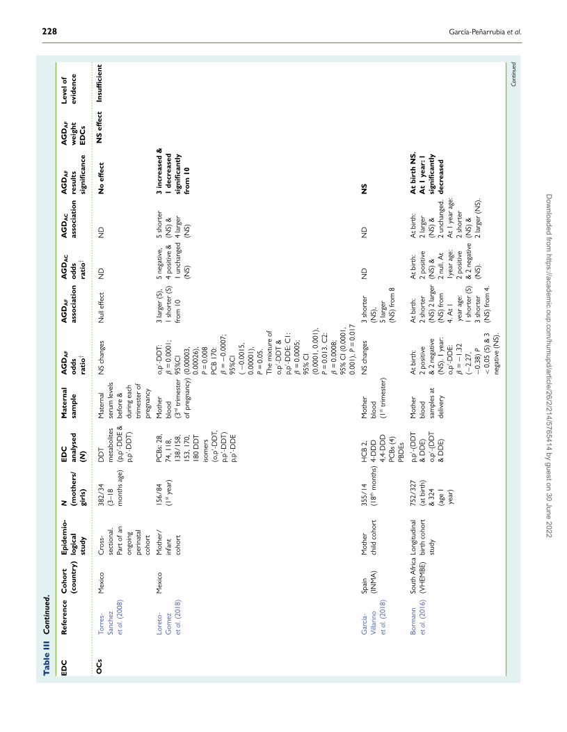
228 García-Peñarrubia et al.
Tab
leII
IC
onti
nued
.
ED
CR
efer
ence
Coh
ort
(cou
ntry
)E
pide
mio
-lo
gica
lst
udy
N (mot
hers
/gi
rls)
ED
Can
alys
ed(N
)
Mat
erna
lsa
mpl
eA
GD
AF
odds
rati
o†
AG
DA
F
asso
ciat
ion
AG
DA
C
odds
rati
o†
AG
DA
C
asso
ciat
ion
AG
DA
F
resu
lts
sign
ifica
nce
AG
DA
F
wei
ght
ED
Cs
Lev
elof
evid
ence
....
....
....
....
....
....
....
....
....
....
....
....
....
....
....
....
....
....
....
....
....
....
....
....
....
....
....
....
....
....
....
....
....
....
....
....
....
....
....
....
....
....
....
....
....
....
....
....
....
....
....
....
....
....
....
....
....
....
....
....
..O
Cs
Torr
es-
Sanc
hez
etal
.(20
08)
Mex
ico
Cro
ss-
sect
iona
l.Pa
rtof
anon
goin
gpe
rinat
alco
hort
382/
34(3
–18
mon
ths
age)
DD
Tm
etab
olite
s(p
,p′ -D
DE
&p,
p′ -DD
T)
Mat
erna
lse
rum
leve
lsbe
fore
&du
ring
each
trim
este
rof
preg
nanc
y
NS
chan
ges
Nul
leffe
ctN
DN
DN
oeff
ect
NS
effec
tIn
suffi
cien
t
Lore
to-
Gom
ezet
al.(
2018
)
Mex
ico
Mot
her/
infa
ntco
hort
156/
84(1
stye
ar)
PCBs
:28,
74,1
18,
138/
158,
153,
170,
180
DD
Tis
omer
s(o
,p’-D
DT,
p,p’
-DD
T)
p,p’
-DD
E
Mot
her
bloo
d(3
rdtr
imes
ter
ofpr
egna
ncy)
o,p′ -D
DT:
β=
0.00
01;
95%
CI
(0.0
0003
,0.
0002
6),
P=
0.00
8PC
B17
0:β
=−0
.000
7;95
%C
I(−0
.001
5,0.
0000
1),
P=
0.05
.T
hem
ixtu
reof
o,p′ -D
DT
&p,
p′ -DD
E:C
1:β
=0.
0005
;95
%C
I(0
.000
1,0.
001)
,P
=0.
013.
C2:
β=
0.00
08;
95%
CI(
0.00
01,
0.00
1),P
=0.
017
3la
rger
(S),
1sh
orte
r(S
)fr
om10
5ne
gativ
e,4
posi
tive
&1
unch
ange
d(N
S)
5sh
orte
r(N
S)&
4la
rger
(NS)
3in
crea
sed
&1
decr
ease
dsi
gnifi
cant
lyfr
om10
Gar
cía-
Vill
arin
oet
al.(
2018
)
Spai
n(I
NM
A)
Mot
her
child
coho
rt35
5/14
(18th
mon
ths)
HC
B2,
4-D
DD
4,4-
DD
DPC
Bs(4
)PB
DEs
Mot
her
bloo
d(1
sttr
imes
ter)
NS
chan
ges
3sh
orte
r(N
S),
5la
rger
(NS)
from
8
ND
ND
NS
Borm
ann
etal
.(20
16)
Sout
hA
fric
a(V
HEM
BE)
Long
itudi
nal
birt
hco
hort
stud
y
752/
327
(atb
irth
)&
324
(age
1ye
ar)
p,p′ -(
DD
T&
DD
E)o,
p′ -(D
DT
&D
DE)
Mot
her
bloo
dsa
mpl
esat
deliv
ery
Atb
irth
:2
posi
tive
&2
nega
tive
(NS)
.1ye
ar:
o,p′ -D
DE:
β=
−1.3
2(−
2.27
,−0
.38)
P<
0,05
(S)
&3
nega
tive
(NS)
.
Atb
irth
:2
shor
ter
(NS)
2la
rger
(NS)
from
4.A
t1ye
arag
e:1
shor
ter
(S)
3sh
orte
r(N
S)fr
om4.
Atb
irth
:2
posi
tive
(NS)
&2
null.
At
1yea
rag
e:2
posi
tive
&2
nega
tive
(NS)
.
Atb
irth
:2
larg
er(N
S)&
2un
chan
ged.
At1
year
age:
2sh
orte
r(N
S)&
2la
rger
(NS)
.
At
birt
hN
S.
At
1ye
ar:1
sign
ifica
ntly
decr
ease
d
Cont
inue
d
Dow
nloaded from https://academ
ic.oup.com/hum
upd/article/26/2/214/5765414 by guest on 30 June 2022
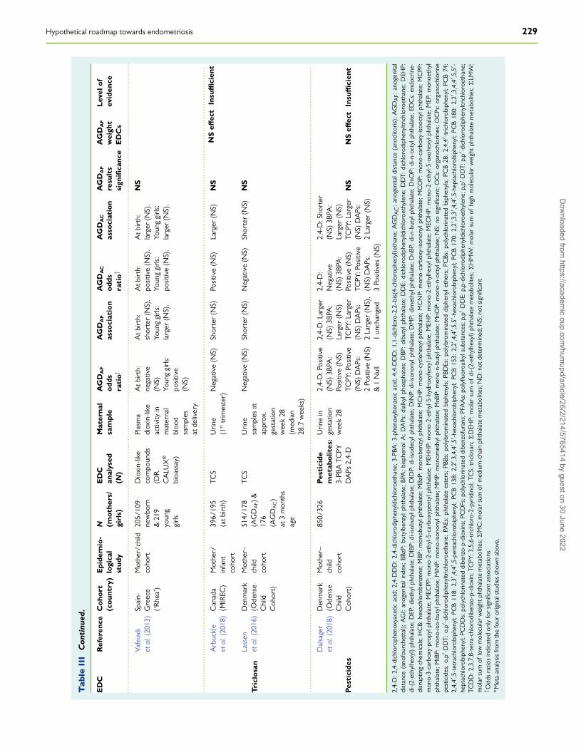
Hypothetical roadmap towards endometriosis 229
Tab
leII
IC
onti
nued
.
ED
CR
efer
ence
Coh
ort
(cou
ntry
)E
pide
mio
-lo
gica
lst
udy
N (mot
hers
/gi
rls)
ED
Can
alys
ed(N
)
Mat
erna
lsa
mpl
eA
GD
AF
odds
rati
o†
AG
DA
F
asso
ciat
ion
AG
DA
C
odds
rati
o†
AG
DA
C
asso
ciat
ion
AG
DA
F
resu
lts
sign
ifica
nce
AG
DA
F
wei
ght
ED
Cs
Lev
elof
evid
ence
....
....
....
....
....
....
....
....
....
....
....
....
....
....
....
....
....
....
....
....
....
....
....
....
....
....
....
....
....
....
....
....
....
....
....
....
....
....
....
....
....
....
....
....
....
....
....
....
....
....
....
....
....
....
....
....
....
....
....
....
..Va
feia
diet
al.(
2013
)Sp
ain-
Gre
ece
(‘R
hea’
)
Mot
her/
child
coho
rt20
5/10
9ne
wbo
rn&
219
youn
ggi
rls
Dio
xin-
like
com
poun
ds(D
RC
ALU
X®
bioa
ssay
)
Plas
ma
diox
in-li
keac
tivity
inm
ater
nal
bloo
dsa
mpl
esat
deliv
ery
Atb
irth
:ne
gativ
e(N
S)Yo
ung
girls
:po
sitiv
e(N
S)
Atb
irth
:sh
orte
r(N
S).
Youn
ggi
rls:
larg
er(N
S).
Atb
irth
:po
sitiv
e(N
S).
Youn
ggi
rls:
posi
tive
(NS)
.
Atb
irth
:la
rger
(NS)
.Yo
ung
girls
:la
rger
(NS)
.
NS
....
....
....
....
....
....
....
....
....
....
....
....
....
....
....
....
....
....
....
....
....
....
....
....
....
....
....
....
....
....
....
....
....
....
....
....
....
....
....
....
....
....
....
....
....
....
....
....
....
....
....
....
....
....
....
....
....
....
....
....
Tric
losa
n
Arb
uckl
eet
al.(
2018
)C
anad
a(M
IREC
)M
othe
r/in
fant
coho
rt
396/
195
(atb
irth
)TC
SU
rine
(1st
trim
este
r)N
egat
ive
(NS)
Shor
ter
(NS)
Posi
tive
(NS)
Larg
er(N
S)N
SN
Seff
ect
Insu
ffici
ent
Lass
enet
al.(
2016
)D
enm
ark
(Ode
nse
Chi
ldC
ohor
t)
Mot
her–
child
coho
rt
514/
178
(AG
DA
F)&
176
(AG
DA
C)
at3
mon
ths
age
TCS
Urin
esa
mpl
esat
appr
ox.
gest
atio
nw
eek
28(m
edia
n28
.7w
eeks
)
Neg
ativ
e(N
S)Sh
orte
r(N
S)N
egat
ive
(NS)
Shor
ter
(NS)
NS
....
....
....
....
....
....
....
....
....
....
....
....
....
....
....
....
....
....
....
....
....
....
....
....
....
....
....
....
....
....
....
....
....
....
....
....
....
....
....
....
....
....
....
....
....
....
....
....
....
....
....
....
....
....
....
....
....
....
....
....
Pest
icid
es
Dal
sage
ret
al.(
2018
)D
enm
ark
(Ode
nse
Chi
ldC
ohor
t)
Mot
her–
child
coho
rt
850/
326
Pest
icid
em
etab
olit
es:
3-PB
ATC
PYD
APs
2,4-
D
Urin
ein
gest
atio
nw
eek
28
2,4-
D:P
ositi
ve(N
S)3B
PA:
Posi
tive
(NS)
TCPY
:Pos
itive
(NS)
DA
Ps:
2Po
sitiv
e(N
S)&
1N
ull
2,4-
D:L
arge
r(N
S)3B
PA:
Larg
er(N
S)TC
PY:L
arge
r(N
S)D
APs
:2
Larg
er(N
S),
1un
chan
ged
2,4-
D:
Neg
ativ
e(N
S)3B
PA:
Posi
tive
(NS)
TCPY
:Pos
itive
(NS)
DA
Ps:
3Po
sitiv
es(N
S)
2,4-
D:S
hort
er(N
S)3B
PA:
Larg
er(N
S)TC
PY:L
arge
r(N
S)D
APs
:2
Larg
er(N
S)
NS
NS
effec
tIn
suffi
cien
t
2,4-
D:
2,4-
dich
loro
phen
oxya
cetic
acid
;2,
4-D
DD
:2,
4-di
chlo
rodi
phen
yldi
chlo
roet
hane
;3-
PBA
:3-
phen
oxyb
enzo
icac
id;
4,4-
DD
D:
1,1-
dich
loro
-2,2
–bis
(4-c
hlor
ophe
nyl)e
than
e;A
GD
AC
:an
ogen
itald
ista
nce
(ano
clito
ris);
AG
DA
F:an
ogen
ital
dist
ance
(ano
four
chet
tal);
AG
I:an
ogen
itali
ndex
;BB
zP:
buty
lben
zylp
htha
late
;BP
A:
bisp
heno
lA;
DA
Ps:
dial
kylp
hosp
hate
s;D
BP:
dibu
tylp
htha
late
;D
DE:
dich
loro
diph
enyl
dich
loro
ethy
lene
;D
DT:
dich
loro
diph
enyl
tric
hlor
oeth
ane;
DEH
P:di
-(2-
ethy
lhex
yl)
phth
alat
e;D
EP:d
ieth
ylph
thal
ate;
DIB
P:di
-isob
utyl
phth
alat
e;D
IDP:
di-is
odec
ylph
thal
ate;
DIN
P:di
-ison
onyl
phth
alat
e;D
MP:
dim
ethy
lpht
hala
te;D
nBP:
di-n
-but
ylph
thal
ate;
DnO
P:di
-n-o
ctyl
phth
alat
e;ED
Cs:
endo
crin
e-di
srup
ting
chem
ical
s;H
CB:
hexa
chlo
robe
nzen
e;M
BP:m
onob
utyl
phth
alat
e;M
BzP:
mon
oben
zylp
htha
late
;MC
HP:
mon
o-cy
cloh
exyl
phth
alat
e;M
CN
P:m
ono-
carb
oxy-
ison
onyl
phth
alat
e;M
CO
P:m
ono-
carb
oxy-
isoo
ctyl
phth
alat
e;M
CPP
:m
ono-
3-ca
rbox
y-pr
opyl
phth
alat
e;M
ECPP
:mon
o-2-
ethy
l-5-c
arbo
xype
ntyl
phth
alat
e;M
EHH
P:m
ono-
2-et
hyl-5
-hyd
roxy
hexy
lpht
hala
te;M
EHP:
mon
o-2-
ethy
lhex
ylph
thal
ate;
MEO
HP:
mon
o-2-
ethy
l-5-o
xohe
xylp
htha
late
;MEP
:mon
oeth
ylph
thal
ate;
MiB
P:m
ono-
iso-
buty
lpht
hala
te;M
iNP:
mon
o-is
onon
ylph
thal
ate;
MM
P:m
onom
ethy
lpht
hala
te;M
nBP:
mon
o-n-
buty
lpht
hala
te;M
nOP:
mon
o-n-
octy
lpht
hala
te;N
S:no
sign
ifica
nt;O
Cs:
orga
noch
lorin
es;O
CPs
:org
anoc
hlor
ine
pest
icid
es;
o,p′
-DD
T:o,
p′-d
ichl
orod
iphe
nyltr
ichl
oroe
than
e;PA
Es:
phth
alat
ees
ters
;PB
Bs:
poly
brom
inat
edbi
phen
yls;
PBD
Es:
poly
brom
inat
eddi
phen
ylet
hers
;PC
Bs:
poly
chlo
rinat
edbi
phen
yls;
PCB
28:
2,4,
4′-t
richl
orob
iphe
nyl;
PCB
74:
2,4,
4′,5
-tet
rach
loro
biph
enyl
;PC
B11
8:2,
3′,4
,4′ ,5
-pen
tach
loro
biph
enyl
;PC
B13
8:2,
2′,3
,4,4
′ ,5′ -h
exac
hlor
obip
heny
l;PC
B15
3:2,
2′,4
,4′ ,5
,5′ -h
exac
hlor
obip
heny
l;PC
B17
0:2,
2′,3
,3′ ,4
,4′ ,5
-hep
tach
loro
biph
enyl
;PC
B18
0:2,
2′,3
,4,4
′ ,5,5
′ -he
ptac
hlor
obip
heny
l;PC
DD
s:po
lych
lorin
ated
dibe
nzo-
p-di
oxin
s;PC
DFs
:pol
ychl
orin
ated
dibe
nzof
uran
s;PF
AA
s:po
lyflu
oroa
lkyl
subs
tanc
es;p
,p′ -D
DE:
p,p-
dich
loro
diph
enyl
dich
loro
ethy
lene
;p,p
’-DD
T:p,
p′-d
ichl
orod
iphe
nyltr
ichl
oroe
than
e;TC
DD
:2,3
,7,8
-tet
ra-c
hlor
odib
enzo
-p-d
ioxi
n;TC
PY:3
,5,6
-tric
hlor
o-2-
pyrid
inol
;TC
S:tr
iclo
san;
�D
EHP:
mol
arsu
mof
di-(
2-et
hylh
exyl
)ph
thal
ate
met
abol
ites;
�H
MW
:mol
arsu
mof
high
mol
ecul
arw
eigh
tph
thal
ate
met
abol
ites;
�LM
W:
mol
arsu
mof
low
mol
ecul
arw
eigh
tpht
hala
tem
etab
olite
s;�
MC
:mol
arsu
mof
med
ium
-cha
inph
thal
ate
met
abol
ites;
ND
:not
dete
rmin
ed;N
S:no
tsig
nific
ant
† Odd
sra
tios
indi
cate
don
lyfo
rsi
gnifi
cant
asso
ciat
ions
.∗ M
eta-
anal
ysis
from
the
four
orig
inal
stud
ies
show
nab
ove.
Dow
nloaded from https://academ
ic.oup.com/hum
upd/article/26/2/214/5765414 by guest on 30 June 2022
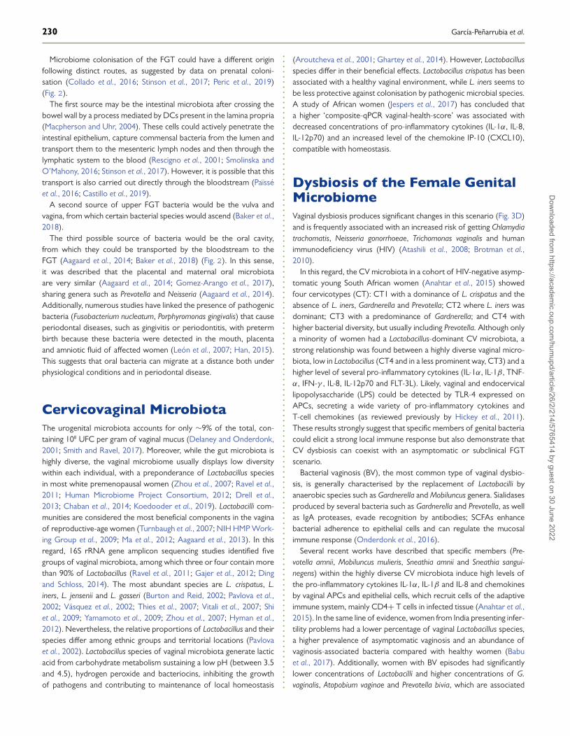
230 García-Peñarrubia et al.
Microbiome colonisation of the FGT could have a different originfollowing distinct routes, as suggested by data on prenatal coloni-sation (Collado et al., 2016; Stinson et al., 2017; Peric et al., 2019)(Fig. 2).
The first source may be the intestinal microbiota after crossing thebowel wall by a process mediated by DCs present in the lamina propria(Macpherson and Uhr, 2004). These cells could actively penetrate theintestinal epithelium, capture commensal bacteria from the lumen andtransport them to the mesenteric lymph nodes and then through thelymphatic system to the blood (Rescigno et al., 2001; Smolinska andO’Mahony, 2016; Stinson et al., 2017). However, it is possible that thistransport is also carried out directly through the bloodstream (Païsséet al., 2016; Castillo et al., 2019).
A second source of upper FGT bacteria would be the vulva andvagina, from which certain bacterial species would ascend (Baker et al.,2018).
The third possible source of bacteria would be the oral cavity,from which they could be transported by the bloodstream to theFGT (Aagaard et al., 2014; Baker et al., 2018) (Fig. 2). In this sense,it was described that the placental and maternal oral microbiotaare very similar (Aagaard et al., 2014; Gomez-Arango et al., 2017),sharing genera such as Prevotella and Neisseria (Aagaard et al., 2014).Additionally, numerous studies have linked the presence of pathogenicbacteria (Fusobacterium nucleatum, Porphyromonas gingivalis) that causeperiodontal diseases, such as gingivitis or periodontitis, with pretermbirth because these bacteria were detected in the mouth, placentaand amniotic fluid of affected women (León et al., 2007; Han, 2015).This suggests that oral bacteria can migrate at a distance both underphysiological conditions and in periodontal disease.
Cervicovaginal MicrobiotaThe urogenital microbiota accounts for only ∼9% of the total, con-taining 108 UFC per gram of vaginal mucus (Delaney and Onderdonk,2001; Smith and Ravel, 2017). Moreover, while the gut microbiota ishighly diverse, the vaginal microbiome usually displays low diversitywithin each individual, with a preponderance of Lactobacillus speciesin most white premenopausal women (Zhou et al., 2007; Ravel et al.,2011; Human Microbiome Project Consortium, 2012; Drell et al.,2013; Chaban et al., 2014; Koedooder et al., 2019). Lactobacilli com-munities are considered the most beneficial components in the vaginaof reproductive-age women (Turnbaugh et al., 2007; NIH HMP Work-ing Group et al., 2009; Ma et al., 2012; Aagaard et al., 2013). In thisregard, 16S rRNA gene amplicon sequencing studies identified fivegroups of vaginal microbiota, among which three or four contain morethan 90% of Lactobacillus (Ravel et al., 2011; Gajer et al., 2012; Dingand Schloss, 2014). The most abundant species are L. crispatus, L.iners, L. jensenii and L. gasseri (Burton and Reid, 2002; Pavlova et al.,2002; Vásquez et al., 2002; Thies et al., 2007; Vitali et al., 2007; Shiet al., 2009; Yamamoto et al., 2009; Zhou et al., 2007; Hyman et al.,2012). Nevertheless, the relative proportions of Lactobacillus and theirspecies differ among ethnic groups and territorial locations (Pavlovaet al., 2002). Lactobacillus species of vaginal microbiota generate lacticacid from carbohydrate metabolism sustaining a low pH (between 3.5and 4.5), hydrogen peroxide and bacteriocins, inhibiting the growthof pathogens and contributing to maintenance of local homeostasis
.
.
.
.
.
.
.
.
.
.
.
.
.
.
.
.
.
.
.
.
.
.
.
.
.
.
.
.
.
.
.
.
.
.
.
.
.
.
.
.
.
.
.
.
.
.
.
.
.
.
.
.
.
.
.
.
.
.
.
.
.
.
.
.
.
.
.
.
.
.
.
.
.
.
.
.
.
.
.
.
.
.
.
.
.
.
.
.
.
.
.
.
.
.
.
.
.
.
.
.
.
.
.
.
.
.
.
.
.
.
.
.
.
.
.
.
.
.
.
.
.
.
.
.
(Aroutcheva et al., 2001; Ghartey et al., 2014). However, Lactobacillusspecies differ in their beneficial effects. Lactobacillus crispatus has beenassociated with a healthy vaginal environment, while L. iners seems tobe less protective against colonisation by pathogenic microbial species.A study of African women (Jespers et al., 2017) has concluded thata higher ‘composite-qPCR vaginal-health-score’ was associated withdecreased concentrations of pro-inflammatory cytokines (IL-1α, IL-8,IL-12p70) and an increased level of the chemokine IP-10 (CXCL10),compatible with homeostasis.
Dysbiosis of the Female GenitalMicrobiomeVaginal dysbiosis produces significant changes in this scenario (Fig. 3D)and is frequently associated with an increased risk of getting Chlamydiatrachomatis, Neisseria gonorrhoeae, Trichomonas vaginalis and humanimmunodeficiency virus (HIV) (Atashili et al., 2008; Brotman et al.,2010).
In this regard, the CV microbiota in a cohort of HIV-negative asymp-tomatic young South African women (Anahtar et al., 2015) showedfour cervicotypes (CT): CT1 with a dominance of L. crispatus and theabsence of L. iners, Gardnerella and Prevotella; CT2 where L. iners wasdominant; CT3 with a predominance of Gardnerella; and CT4 withhigher bacterial diversity, but usually including Prevotella. Although onlya minority of women had a Lactobacillus-dominant CV microbiota, astrong relationship was found between a highly diverse vaginal micro-biota, low in Lactobacillus (CT4 and in a less prominent way, CT3) and ahigher level of several pro-inflammatory cytokines (IL-1α, IL-1β, TNF-α, IFN-γ , IL-8, IL-12p70 and FLT-3L). Likely, vaginal and endocervicallipopolysaccharide (LPS) could be detected by TLR-4 expressed onAPCs, secreting a wide variety of pro-inflammatory cytokines andT-cell chemokines (as reviewed previously by Hickey et al., 2011).These results strongly suggest that specific members of genital bacteriacould elicit a strong local immune response but also demonstrate thatCV dysbiosis can coexist with an asymptomatic or subclinical FGTscenario.
Bacterial vaginosis (BV), the most common type of vaginal dysbio-sis, is generally characterised by the replacement of Lactobacilli byanaerobic species such as Gardnerella and Mobiluncus genera. Sialidasesproduced by several bacteria such as Gardnerella and Prevotella, as wellas IgA proteases, evade recognition by antibodies; SCFAs enhancebacterial adherence to epithelial cells and can regulate the mucosalimmune response (Onderdonk et al., 2016).
Several recent works have described that specific members (Pre-votella amnii, Mobiluncus mulieris, Sneathia amnii and Sneathia sangui-negens) within the highly diverse CV microbiota induce high levels ofthe pro-inflammatory cytokines IL-1α, IL-1β and IL-8 and chemokinesby vaginal APCs and epithelial cells, which recruit cells of the adaptiveimmune system, mainly CD4+ T cells in infected tissue (Anahtar et al.,2015). In the same line of evidence, women from India presenting infer-tility problems had a lower percentage of vaginal Lactobacillus species,a higher prevalence of asymptomatic vaginosis and an abundance ofvaginosis-associated bacteria compared with healthy women (Babuet al., 2017). Additionally, women with BV episodes had significantlylower concentrations of Lactobacilli and higher concentrations of G.vaginalis, Atopobium vaginae and Prevotella bivia, which are associated
Dow
nloaded from https://academ
ic.oup.com/hum
upd/article/26/2/214/5765414 by guest on 30 June 2022
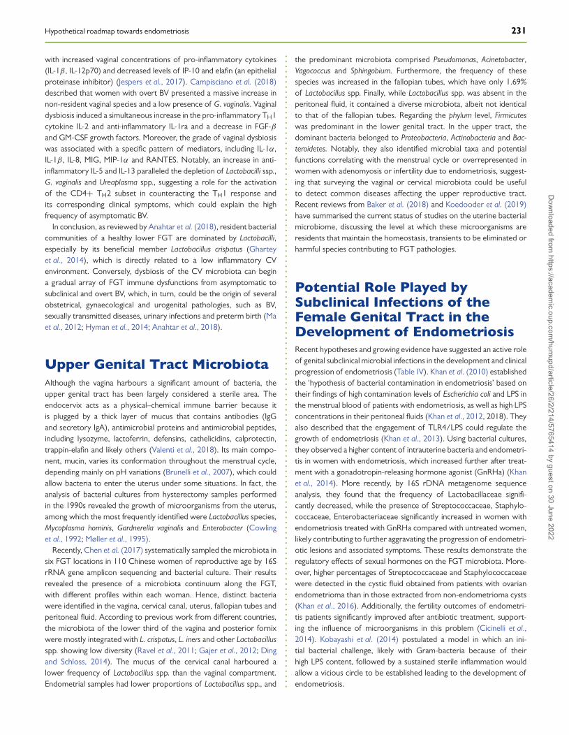
Hypothetical roadmap towards endometriosis 231
with increased vaginal concentrations of pro-inflammatory cytokines(IL-1β, IL-12p70) and decreased levels of IP-10 and elafin (an epithelialproteinase inhibitor) (Jespers et al., 2017). Campisciano et al. (2018)described that women with overt BV presented a massive increase innon-resident vaginal species and a low presence of G. vaginalis. Vaginaldysbiosis induced a simultaneous increase in the pro-inflammatory TH1cytokine IL-2 and anti-inflammatory IL-1ra and a decrease in FGF-βand GM-CSF growth factors. Moreover, the grade of vaginal dysbiosiswas associated with a specific pattern of mediators, including IL-1α,IL-1β, IL-8, MIG, MIP-1α and RANTES. Notably, an increase in anti-inflammatory IL-5 and IL-13 paralleled the depletion of Lactobacilli ssp.,G. vaginalis and Ureaplasma spp., suggesting a role for the activationof the CD4+ TH2 subset in counteracting the TH1 response andits corresponding clinical symptoms, which could explain the highfrequency of asymptomatic BV.
In conclusion, as reviewed by Anahtar et al. (2018), resident bacterialcommunities of a healthy lower FGT are dominated by Lactobacilli,especially by its beneficial member Lactobacillus crispatus (Gharteyet al., 2014), which is directly related to a low inflammatory CVenvironment. Conversely, dysbiosis of the CV microbiota can begina gradual array of FGT immune dysfunctions from asymptomatic tosubclinical and overt BV, which, in turn, could be the origin of severalobstetrical, gynaecological and urogenital pathologies, such as BV,sexually transmitted diseases, urinary infections and preterm birth (Maet al., 2012; Hyman et al., 2014; Anahtar et al., 2018).
Upper Genital Tract MicrobiotaAlthough the vagina harbours a significant amount of bacteria, theupper genital tract has been largely considered a sterile area. Theendocervix acts as a physical–chemical immune barrier because itis plugged by a thick layer of mucus that contains antibodies (IgGand secretory IgA), antimicrobial proteins and antimicrobial peptides,including lysozyme, lactoferrin, defensins, cathelicidins, calprotectin,trappin-elafin and likely others (Valenti et al., 2018). Its main compo-nent, mucin, varies its conformation throughout the menstrual cycle,depending mainly on pH variations (Brunelli et al., 2007), which couldallow bacteria to enter the uterus under some situations. In fact, theanalysis of bacterial cultures from hysterectomy samples performedin the 1990s revealed the growth of microorganisms from the uterus,among which the most frequently identified were Lactobacillus species,Mycoplasma hominis, Gardnerella vaginalis and Enterobacter (Cowlinget al., 1992; Møller et al., 1995).
Recently, Chen et al. (2017) systematically sampled the microbiota insix FGT locations in 110 Chinese women of reproductive age by 16SrRNA gene amplicon sequencing and bacterial culture. Their resultsrevealed the presence of a microbiota continuum along the FGT,with different profiles within each woman. Hence, distinct bacteriawere identified in the vagina, cervical canal, uterus, fallopian tubes andperitoneal fluid. According to previous work from different countries,the microbiota of the lower third of the vagina and posterior fornixwere mostly integrated with L. crispatus, L. iners and other Lactobacillusspp. showing low diversity (Ravel et al., 2011; Gajer et al., 2012; Dingand Schloss, 2014). The mucus of the cervical canal harboured alower frequency of Lactobacillus spp. than the vaginal compartment.Endometrial samples had lower proportions of Lactobacillus spp., and
.
.
.
.
.
.
.
.
.
.
.
.
.
.
.
.
.
.
.
.
.
.
.
.
.
.
.
.
.
.
.
.
.
.
.
.
.
.
.
.
.
.
.
.
.
.
.
.
.
.
.
.
.
.
.
.
.
.
.
.
.
.
.
.
.
.
.
.
.
.
.
.
.
.
.
.
.
.
.
.
.
.
.
.
.
.
.
.
.
.
.
.
.
.
.
.
.
.
.
.
.
.
.
.
.
.
.
.
.
.
.
.
.
.
.
.
.
.
.
.
.
.
.
.
the predominant microbiota comprised Pseudomonas, Acinetobacter,Vagococcus and Sphingobium. Furthermore, the frequency of thesespecies was increased in the fallopian tubes, which have only 1.69%of Lactobacillus spp. Finally, while Lactobacillus spp. was absent in theperitoneal fluid, it contained a diverse microbiota, albeit not identicalto that of the fallopian tubes. Regarding the phylum level, Firmicuteswas predominant in the lower genital tract. In the upper tract, thedominant bacteria belonged to Proteobacteria, Actinobacteria and Bac-teroidetes. Notably, they also identified microbial taxa and potentialfunctions correlating with the menstrual cycle or overrepresented inwomen with adenomyosis or infertility due to endometriosis, suggest-ing that surveying the vaginal or cervical microbiota could be usefulto detect common diseases affecting the upper reproductive tract.Recent reviews from Baker et al. (2018) and Koedooder et al. (2019)have summarised the current status of studies on the uterine bacterialmicrobiome, discussing the level at which these microorganisms areresidents that maintain the homeostasis, transients to be eliminated orharmful species contributing to FGT pathologies.
Potential Role Played bySubclinical Infections of theFemale Genital Tract in theDevelopment of EndometriosisRecent hypotheses and growing evidence have suggested an active roleof genital subclinical microbial infections in the development and clinicalprogression of endometriosis (Table IV). Khan et al. (2010) establishedthe ‘hypothesis of bacterial contamination in endometriosis’ based ontheir findings of high contamination levels of Escherichia coli and LPS inthe menstrual blood of patients with endometriosis, as well as high LPSconcentrations in their peritoneal fluids (Khan et al., 2012, 2018). Theyalso described that the engagement of TLR4/LPS could regulate thegrowth of endometriosis (Khan et al., 2013). Using bacterial cultures,they observed a higher content of intrauterine bacteria and endometri-tis in women with endometriosis, which increased further after treat-ment with a gonadotropin-releasing hormone agonist (GnRHa) (Khanet al., 2014). More recently, by 16S rDNA metagenome sequenceanalysis, they found that the frequency of Lactobacillaceae signifi-cantly decreased, while the presence of Streptococcaceae, Staphylo-coccaceae, Enterobacteriaceae significantly increased in women withendometriosis treated with GnRHa compared with untreated women,likely contributing to further aggravating the progression of endometri-otic lesions and associated symptoms. These results demonstrate theregulatory effects of sexual hormones on the FGT microbiota. More-over, higher percentages of Streptococcaceae and Staphylococcaceaewere detected in the cystic fluid obtained from patients with ovarianendometrioma than in those extracted from non-endometrioma cysts(Khan et al., 2016). Additionally, the fertility outcomes of endometri-tis patients significantly improved after antibiotic treatment, support-ing the influence of microorganisms in this problem (Cicinelli et al.,2014). Kobayashi et al. (2014) postulated a model in which an ini-tial bacterial challenge, likely with Gram-bacteria because of theirhigh LPS content, followed by a sustained sterile inflammation wouldallow a vicious circle to be established leading to the development ofendometriosis.
Dow
nloaded from https://academ
ic.oup.com/hum
upd/article/26/2/214/5765414 by guest on 30 June 2022
232 García-Peñarrubia et al.
Tab
leIV
Ass
ocia
tion
betw
een
mic
robi
ome
and
endo
met
rios
is.
Ass
ocia
tion
stud
ied
Ref
eren
ceS
tudy
(cou
ntry
)S
ampl
esi
zeE
ndom
etri
osis
diag
nost
icS
ampl
eM
etho
dsR
esul
tsA
ssoc
iati
onL
evel
ofev
iden
ce..
....
....
....
....
....
....
....
....
....
....
....
....
....
....
....
....
....
....
....
....
....
....
....
....
....
....
....
....
....
....
....
....
....
....
....
....
....
....
....
....
....
....
....
....
....
....
....
....
....
....
....
....
....
....
....
....
....
....
....
....
End
omet
rios
is/
Bac
teri
a
Ata
etal
.(2
019)
Pros
pect
ive
obse
rva-
tiona
l(T
urke
y)
14w
omen
w/
endo
met
riosi
s14 as
ympt
omat
icw
omen
(con
trol
)
Lapa
rosc
opy
(his
tolo
gy)
Stoo
l,va
gina
l& en
doce
rvic
alsw
abs
16S
rRN
Age
nese
quen
cing
(bac
teria
)
Cer
vix,
vagi
na&
guts
imila
rba
cter
ia(n
odi
ffere
nces
).D
iffer
ence
sat
the
genu
sle
veli
nen
dom
etrio
sis
patie
nts:
Abs
ence
ofAt
opob
ium
inva
gina
&ce
rvix
.Inc
reas
eof
Gar
dner
ella
,Str
epto
cocc
us,E
sche
richi
a,Sh
igel
la,&
Ure
apla
sma
inth
ece
rvix
.Sh
igel
la/E
sche
richi
ado
min
ants
tool
mic
robi
ome.
Posi
tive
asso
ciat
ion
but
NS
Insu
ffici
ent
....
....
....
....
....
....
....
....
....
....
....
....
....
....
....
....
....
....
....
....
....
....
....
....
....
....
....
....
....
....
....
....
....
....
....
....
....
....
....
....
....
....
....
....
....
....
....
....
....
....
....
....
....
....
....
Cam
pos
etal
.(2
018)
Cro
ss-
sect
iona
l(B
razi
l)
73w
omen
w/
endo
met
riosi
s31 as
ympt
omat
icw
omen
(con
trol
)
Lapa
rosc
opy
(his
tolo
gy)
Endo
cerv
ical
swab
sPe
riton
eal
fluid
biop
sies
from
lesi
ons
PCR
(bac
teria
)Lu
min
ex(c
ytok
ines
)
No
stat
istic
ally
sign
ifica
ntdi
ffere
nces
wer
ede
tect
edin
the
swab
&pe
riton
eal
sam
ples
inM
ycop
lasm
aho
min
is,M
.ge
nita
lium
,Ure
apla
sma
urea
lytic
um,&
U.
parv
um.U
reap
lasm
apa
rvum
was
asso
ciat
edw
/dy
spar
euni
a.M
ycop
lasm
age
nita
lium
was
asso
ciat
edw
/in
crea
sed
IFN
-γ,I
L-1β
.
NS
....
....
....
....
....
....
....
....
....
....
....
....
....
....
....
....
....
....
....
....
....
....
....
....
....
....
....
....
....
....
....
....
....
....
....
....
....
....
....
....
....
....
....
....
....
....
....
....
....
....
....
....
....
....
....
Kha
net
al.
(201
4)C
ase–
cont
rol
(Jap
an)
65w
omen
w/
endo
met
riosi
s55
cont
rol
wom
en
Lapa
rosc
opy
(his
tolo
gy)
Vagi
nal
smea
rsEn
dom
etria
lsa
mpl
es
Bact
eria
lcu
lture
Vagi
nalp
H≥4
.5:7
9.3%
endo
met
riosi
sve
rsus
58.4
%C
ontr
ol(P
<0.
03).
The
num
ber
of(C
FU/m
l)of
Gar
dner
ella
,α-S
trep
toco
ccus
,Ent
eroc
occi
&Es
cher
ichi
aco
liw
assi
gnifi
cant
lyhi
gher
inen
dom
etria
lsam
ples
from
wom
enw
/en
dom
etrio
sis
than
cont
rolw
omen
(P<
0.05
for
each
bact
eria
).
Hig
her
CF
U/m
LS
igni
fican
tdi
ffer
ence
....
....
....
....
....
....
....
....
....
....
....
....
....
....
....
....
....
....
....
....
....
....
....
....
....
....
....
....
....
....
....
....
....
....
....
....
....
....
....
....
....
....
....
....
....
....
....
....
....
....
....
....
....
....
....
Aki
yam
aet
al.
(201
9)C
ase–
cont
rol
(Jap
an)
30w
omen
w/
endo
met
riosi
s(s
tage
3/4)
39w
/oen
dom
etrio
sis
(w/
fibro
ids
orbe
nign
ovar
ian
tum
our)
Lapa
rosc
opy
(his
tolo
gy)
Cer
vica
lm
ucus
Bact
eria
lD
NA
extr
actio
n&
PCR
ampl
ifica
tion
of16
SrR
NA
gene
.A
mpl
icon
sequ
enci
ngus
ing
NG
S.
The
amou
ntof
Ente
roba
cter
iace
ae&
Stre
ptoc
occu
sw
assi
gnifi
cant
lyhi
gher
inth
ece
rvic
alm
ucus
ofen
dom
etrio
sis
grou
pth
anth
atin
the
cont
rolg
roup
(P<
0.05
).T
hepo
pula
tions
ofCo
ryne
bact
eriu
m,E
nter
obac
teria
ceae
,Fl
avob
acte
rium
,Pse
udom
onas
,&St
rept
ococ
cus
wer
ein
crea
sed
inth
een
dom
etrio
sis
grou
p.
Hig
her
sign
ifica
ntdi
ffer
ence
s
Cont
inue
d
Dow
nloaded from https://academ
ic.oup.com/hum
upd/article/26/2/214/5765414 by guest on 30 June 2022
Hypothetical roadmap towards endometriosis 233
Tab
leIV
Con
tinu
ed
Ass
ocia
tion
stud
ied
Ref
eren
ceS
tudy
(cou
ntry
)S
ampl
esi
zeE
ndom
e-tr
iosi
sdi
agno
stic
Sam
ple
Met
hods
Res
ults
Ass
ocia
tion
Lev
elof
evid
ence
....
....
....
....
....
....
....
....
....
....
....
....
....
....
....
....
....
....
....
....
....
....
....
....
....
....
....
....
....
....
....
....
....
....
....
....
....
....
....
....
....
....
....
....
....
....
....
....
....
....
....
....
....
....
....
....
....
....
....
....
..Ta
ieta
l.(2
018)
Ret
rosp
ectiv
eco
hort
(Tai
wan
)
2829
2w
omen
w/
pelv
icin
flam
mat
ory
dise
ase
(PID
)11
316
8w
/oPI
D(c
ontr
ol)
Ultr
ason
o-gr
aphy
orla
paro
scop
y
Com
orbi
litie
sas
soci
ated
toen
dom
etrio
sis
Dat
abas
eH
azar
dra
tiofo
ren
dom
etrio
sis/
PID
asso
ciat
ion:
β=
3.02
;95%
CI(
2.85
,3.2
),P
<0.
0001
Sig
nific
ant
asso
ciat
ion
Insu
ffici
ent
....
....
....
....
....
....
....
....
....
....
....
....
....
....
....
....
....
....
....
....
....
....
....
....
....
....
....
....
....
....
....
....
....
....
....
....
....
....
....
....
....
....
....
....
....
....
....
....
....
....
....
....
....
....
....
..E
ndom
e-tr
iosi
s/F
GT
infe
ctio
usdi
seas
es
Lin
etal
.(2
016)
Ret
rosp
ectiv
eba
sed
coho
rt(T
aiw
an)
7951
2w
omen
w/
low
erFG
Tin
flam
mat
ory
dise
ases
&79
512
w/o
FGT
infla
mm
ator
ydi
seas
es.
Ultr
ason
ogra
phy
orla
paro
scop
yD
atab
ase
Dat
abas
eH
azar
dra
tio:β
=2.
01;9
5%C
I(1.
91,2
.12)
,P
<0.
001
Inci
denc
eof
endo
met
riosi
sw
ashi
gher
amon
gin
flam
mat
ory
dise
ase
patie
nts
than
cont
rols.
Sig
nific
ant
asso
ciat
ion
....
....
....
....
....
....
....
....
....
....
....
....
....
....
....
....
....
....
....
....
....
....
....
....
....
....
....
....
....
....
....
....
....
....
....
....
....
....
....
....
....
....
....
....
....
....
....
....
....
....
....
....
....
....
....
..Ta
keba
yash
iet
al.(
2014
)C
ase–
cont
rol
(Jap
an)
34pa
tient
sw
/en
dom
etrio
-si
s.37
w/o
endo
met
rio-
sis.
28w
/ch
roni
cen
dom
etrit
is.43
w/o
chro
nic
endo
met
ritis.
Imm
uno-
hist
oche
mis
try
Ute
rus
from
hyst
erec
tom
yIm
mun
o-hi
stoc
hem
istr
yLo
gist
icre
gres
sion
anal
ysis
reve
aled
that
endo
met
riosi
sw
asas
soci
ated
w/
chro
nic
endo
met
ritis
Odd
sra
tio:3
.037
;95%
CI(
1.12
9,8.
174)
,P=
0.02
8
Sig
nific
ant
asso
ciat
ion
....
....
....
....
....
....
....
....
....
....
....
....
....
....
....
....
....
....
....
....
....
....
....
....
....
....
....
....
....
....
....
....
....
....
....
....
....
....
....
....
....
....
....
....
....
....
....
....
....
....
....
....
....
....
....
....
....
....
....
....
.E
ndom
e-tr
iosi
s/pe
riod
on-
tald
isea
se
Tho
mas
etal
.(20
18)
Cas
e–co
ntro
l(I
ndia
)25
wom
enw
/en
dom
etrio
-si
s.25
w/o
endo
met
riosi
s(c
ontr
ol).
Lapa
roco
py(h
isto
logy
)Pe
riodo
ntal
scre
enin
gC
linic
alex
amin
atio
nG
ingi
vali
ndex
was
seen
tobe
sign
ifica
ntly
high
erin
patie
nts
w/
endo
met
riosi
s:(1
.55
±0.
53)
inca
ses
&(1
.12
±0.
25)
inco
ntro
ls,P
=0.
001
Prop
ortio
nof
wom
enw
/m
oder
ate-
to-s
ever
epe
riodo
ntiti
sw
asse
ento
behi
gher
amon
gw
omen
w/
endo
met
riosi
s.Se
vere
perio
dont
itis
&en
dom
etrio
sis:
OR
:2.9
;95%
CI(
0.76
,11.
33),
P=
0.09
Posi
tive
asso
ciat
ion
but
NS
Insu
ffici
ent
Kav
ouss
iet
al.(
2009
)C
ross
-se
ctio
nal
(NH
AN
ES(U
SA))
4136
wom
enw
/pe
riodo
ntal
dise
ase
&en
dom
etrio
sis
Self-
repo
rted
endo
met
riosi
sD
atab
ase
Clin
ical
oral
heal
thex
amin
atio
n
Adj
uste
dO
R=
1.57
;95%
CI(
1.06
,2.3
)fo
rgi
ngiv
itis
plus
perio
dont
itis.
Wom
enw
/se
lf-re
port
eden
dom
etrio
sis
had
sign
ifica
ntly
(57%
)hi
gher
odds
ofha
ving
both
ging
iviti
s&
perio
dont
itis.
Sig
nific
ant
asso
ciat
ion
PID
:pel
vic
infla
mm
ator
ydi
seas
e;N
S:no
tsig
nific
ant.
Dow
nloaded from https://academ
ic.oup.com/hum
upd/article/26/2/214/5765414 by guest on 30 June 2022
234 García-Peñarrubia et al.
A recent study of a Taiwan cohort including a total of 79 512 patientswith lower FGT inflammatory diseases revealed that the presence ofendometriosis was statistically higher in patients than in control women(Lin et al., 2016). The association between chronic endometritis andendometriosis has also been reported (Takebayashi et al., 2014).The work of Chen et al. (2017) reported the presence (from thevagina up to peritoneal fluid) of Streptococci (45–60%), Staphylococci(35–87%), Ureaplasma (2–55%) and Chlamydia (0–3.2%) in womenwith endometriosis, agreeing with previous results obtained fromendometrial cultures of women with chronic endometritis (Cicinelliet al., 2008) and endometrial cancer (Walther-António et al., 2016).Wang et al. (2018) reported the presence of Proteobacteria andFirmicutes, followed by Actinobacteria, Bacteroides, Fusobacteriumand Tenericutes in the peritoneal fluid of women with and withoutendometriosis, without significant differences. More recently, froma small sample (14 endometriosis and 14 healthy women), Ataet al. (2019) observed some differences at the genus level, namely,complete absence of Atopobium in the vaginal and cervical microbiotaand an increased presence of Gardnerella, Streptococcus, Escherichia,Shigella and Ureaplasma in the cervical microbiota in women withendometriosis stage 3/4. Notably, most women at this stage hadShigella and Escherichia dominant stool microbiota. From a cross-sectional study, Campos et al. (2018) observed higher microbialdiversity in endocervical swabs and peritoneal fluid in endometriosispatients compared with the healthy group. Furthermore, dyspareuniawas associated with the presence of Ureaplasma parvum, whileMycoplasma genitalium was associated with higher secretion of IFN-γ and IL-1β. Additionally, genes associated with inflammation weredown-regulated in peritoneal endometriotic cells, especially in thepresence of M. genitalium. More recently, Chadchan et al. (2019)described that endometriotic lesions in mice treated with broad-spectrum antibiotics were significantly smaller with fewer proliferatingcells than untreated controls.
Based on this accumulated evidence, the question arises regardinghow alterations of the FGT microbiota could lead to inflammatoryor malignant diseases that threaten women’s health. The CV sentinelimmune cells would recognise different pathogen-associated molec-ular patterns (PAMPs), such as LPS, peptidoglycan, flagellin, glycansand damage-associated molecular patterns (DAMPs), including heatshock protein 70, calcium-modulated S100A13 and many others, bypattern recognition receptors (PRRs), including TLRs, NLRs, lectin-likereceptors and others, inducing the activation and secretion of pro-inflammatory cytokines and inflammatory mediators responsible for asubclinical grades of FGT inflammation (Sheldon et al., 2017). Thesealert signals, together with iron from both menstrual blood and ectopicendometrial tissue, are released into the peritoneal cavity, wherethey activate innate resident sentinel cells (i.e. macrophages, DCs andrecruited neutrophils, among others) that secrete pro-inflammatorycytokines and several angiogenic growth factors, which promote thegrowth, implantation and neoangiogenesis of peritoneal endometrioticlesions (Laux-Biehlmann et al., 2015). It is also likely that dysbiosis ofthe FGT microbiome and the subsequent inflammatory response mayaffect uterine contractility, enhancing retrograde menstruation and cor-responding endometrial cell adhesion in the peritoneal compartment(Pinto et al., 2015).
On the other hand, Laschke and Menger (2016) suggested a putativerole for intestinal microbiota in the pathogenesis of endometriosis
.
.
.
.
.
.
.
.
.
.
.
.
.
.
.
.
.
.
.
.
.
.
.
.
.
.
.
.
.
.
.
.
.
.
.
.
.
.
.
.
.
.
.
.
.
.
.
.
.
.
.
.
.
.
.
.
.
.
.
.
.
.
.
.
.
.
.
.
.
.
.
.
.
.
.
.
.
.
.
.
.
.
.
.
.
.
.
.
.
.
.
.
.
.
.
.
.
.
.
.
.
.
.
.
.
.
.
.
.
.
.
.
.
.
.
.
.
.
.
.
.
.
.
.
based on experimental evidence linking gut dysbiosis with inflammationand immune dysregulation, leading to numerous diseases. They postu-lated that the extent and quality of the initial immune response againstepisodes of retrograde menstruation could be determined by factorsassociated with the dysbiosis of gut microbiota, which will allow thedevelopment of endometriosis. This hypothesis was supported by thefinding of Bailey and Coe (2002) on the association of endometriosiswith an altered profile of the intestinal microbiota in rhesus monkeys.Moreover, there is a 50% increase in the risk of inflammatory boweldisease in women with endometriosis (Jess et al., 2012). In this regard, arecent work in a murine model of peritoneal endometriosis has shownan increased gut Firmicutes/Bacteroidetes ratio, indicative of intestinaldysbiosis, 42 days after the intraperitoneal injection of endometrialtissue, together with an elevated level of Bifidobacterium (Yuan et al.,2018). These results confirm the existence of a tight communicationbetween the host and intestinal microbiota (Alfano et al., 2018; Yuanet al., 2018). Nevertheless, a cause–effect relationship between micro-biota and endometriosis cannot be established because the describedcurrent evidence is still insufficient; thus, more studies are necessary toassess this theory.
On the other hand, there is emerging experimental evidencesuggesting that EDC exposure can modify the vaginal (Geller et al.,2018) and intestinal microbiome, leading to dysbiosis and impairmentof local immune system homeostasis (Reddivari et al., 2017; Rosenfeld,2017; Malaisé et al., 2018), preceding the other host alterationsdiscussed above, such as sexual/reproductive, gastrointestinal,metabolic and hormonal disorders. Therefore, a direct multifactorialrelationship can be established between genetic factors, EDCs, themicrobiome and hormones, as the origin and subsequent clinicalevolution of endometriosis.
Oestrogens and MicrobiotaLittle is known about the effect of sexual hormones on the FGTmicrobiome. Bezirtzoglou et al. (2008) revealed that Lactobacilli con-centrations in ovariectomised rats depended on the oestradiol levels.Furthermore, it was shown that the vaginal microbiota undergoestemporal shifts in the composition and abundance of crucial speciesunder the influence of age, sexual intercourse, hormonal variations,pharmacological treatments and hygiene habits (Shi et al., 2009; Ravelet al., 2011; Gajer et al., 2012; Ma et al., 2012; Chaban et al., 2014).Hyman et al. (2012), using a metagenomic approach, studied the rela-tionship between the vaginal microbiome and the levels of circulatingoestradiol and progesterone in IVF-ET (in vitro fertilisation-embryotransfer), concluding that, although the vaginal microbiome at theembryo transfer day affects the pregnancy outcome, significant changesin the vaginal microbiota were not associated with the hormonalprofiles. Nevertheless, it is well documented that the menstrual cycleis associated with changes in the vaginal microbiota. Thus, high lev-els of oestradiol favour a Lactobacilli-predominant community mainlyintegrated with L. crispatus, L. gasseri and/or L. jensenii while theirrelative frequency is lower under low oestrogen levels as occurs inthe first phase of the menstrual cycle or in menopausal women (Gajeret al., 2012). Moreover, the greater variability of vaginal microbiota isassociated with menstruation. Recent work exploring the timing andfluctuations of the vaginal microbiota throughout puberty has shown
Dow
nloaded from https://academ
ic.oup.com/hum
upd/article/26/2/214/5765414 by guest on 30 June 2022
Hypothetical roadmap towards endometriosis 235
that the vaginal microbiota of perimenarcheal girls mirrors that ofreproductive-age women, with a predominance of L. crispatus, L. iners,L. gasseri and L. jensenii, and, in some individuals, Streptococcus spp. werealso detected (Hickey et al., 2015). Further studies should be carriedout to increase knowledge about the link among oestrogens, vaginalglycogen levels and pH, the FGT immune system and microbiota inhealth and disease conditions.
On the other hand, there is increasing evidence supporting thatthe relationships between gut microbiota and oestrogens are directlyinvolved in the regulation of reproductive, neurological and metabolichomeostasis, as well as in the development of several types of cancer(e.g. breast and endometrium) (Baker et al., 2017). In this regard, it haslong been shown that numerous species of the gut microbiome haveenzymatic activity that can metabolise oestrogens (Kwa et al., 2016).Plottel and Blaser (2011) defined the estrobolome as ‘the aggregateof enteric bacterial genes whose products are capable of metabolisingoestrogens’. Oestrogens are endocrine hormones synthesised in theovaries, adrenal glands, adipose tissue and other locations, from wherethey are widely distributed by the bloodstream to target tissues and aremetabolised and conjugated in the liver. Conjugated hepatic oestrogenscan return to the bloodstream or be secreted in the intestine bythe bile. Intestinal oestrogens and phytoestrogens are deconjugatedfrom bile acids by the intestinal microbiota through the secretion ofβ-glucuronidases, glucosidases and hydroxysteroid dehydrogenases.Deconjugated intestinal oestrogens can be reabsorbed through theintestinal mucosa into the enterohepatic circulation and transported todistant sites, including FGT mucosal surfaces, among others. Finally,these oestrogens are eliminated by faecal and urinary excretion.Oestrogens and phytoestrogens bind to their specific oestrogenreceptor alpha (ERα) and oestrogen receptor beta (ERβ) expressedin several tissues, triggering intracellular signalling pathways thatresult in specific physiological functions (reviewed by Wira et al.,2015). Because oestrogens induce the proliferation of epithelialcells paving the female reproductive system, sustained increasedlevels of these hormones have been implicated in the pathogenesisof several proliferative diseases such as endometriosis (Machado-Linde et al., 2012), endometrial cancer (Rodriguez et al., 2019)and breast cancer (Kulkoyluoglu-Cotul et al., 2019; Rizner, 2009).Dysbiosis of the gut microbiome can disrupt the estrobolomecomposition, resulting in higher or lower levels of peripheral blood-circulating oestrogens. Hence, exogenous estrobolome-modifyingfactors, including antibiotics or other drugs, EDCs, diet and otherfactors, may play an indirect role in the pathophysiology of FGTproliferative and inflammatory diseases causing infertility, infections,cancer and, likely, endometriosis. In this regard, endometriosis isassociated with a hyper-proliferative endometrial cell state and neu-roimmune communication, mainly mediated by macrophages, causedby elevated oestrogen levels (Rizner, 2009; Laschke and Menger, 2016;Liang et al., 2018).
Therefore, the consequences of high EDC exposure as a possible riskfactor to develop endometriosis and promote its progression from theasymptomatic state to the different clinical stratifications of the diseasemay not only be a shorter AGD, which would favour frequent vulvo-vaginal faecal microbiota contamination during childhood and producea lower grade of FGT dysbiosis, but the disease could also be mediatedby alteration of the estrobolome, resulting in an anomalous level ofoestrogens.
.
.
.
.
.
.
.
.
.
.
.
.
.
.
.
.
.
.
.
.
.
.
.
.
.
.
.
.
.
.
.
.
.
.
.
.
.
.
.
.
.
.
.
.
.
.
.
.
.
.
.
.
.
.
.
.
.
.
.
.
.
.
.
.
.
.
.
.
.
.
.
.
.
.
.
.
.
.
.
.
.
.
.
.
.
.
.
.
.
.
.
.
.
.
.
.
.
.
.
.
.
.
.
.
.
.
.
.
.
.
.
.
.
.
.
.
.
.
.
.
.
.
.
.
Proposed Model ofEndometriosis DevelopmentBased on epidemiological and experimental evidence, we hypothe-sise that there may be a direct relationship between higher prenatalexposure to oestrogens, EDCs and/or pharmaceutical compounds, ashorter AGD or other FGT alterations and a higher risk of developingendometriosis in adulthood, as shown in Fig. 3.
Therefore, a single or mixed combination exposure to EDCs (result-ing in an oestrogen-like effect) throughout prenatal life could inducea shorter AGD and likely affect the physiological development ofother sexual characteristics, such as the normal development of uterineendometrial tissue or the length, morphology, curvature and contrac-tility of the uterus and fallopian tubes, which could favour neonataland/or future episodes of retrograde menstruation after menarcheand, therefore, increase the risk of developing endometriosis. Addi-tionally, throughout childhood, a shorter AGD would favour frequentfaecal microbiota contamination from the anus to the external andlower FGT (the vulva and vagina), producing an early FGT dysbiosis,which could disrupt local antimicrobial defences, mainly by increasingthe vaginal pH and breaking the beneficial versus pathogenic micro-bial competitive balance. Interruptions of this balance would subvertthe homeostasis state and generate an initial subclinical inflammatoryresponse, which could evolve to a long-term sustained immune acti-vation, closing the vicious circle responsible for the development ofendometriosis after menarcheal age. The translocation and transportof intestinal and/or oral microbiota by the haematogenous route toFGT could also play a role in the development of endometriosis. Infact, periodontitis has been associated with endometriosis, as shownin Table IV (Kavoussi et al., 2009; Thomas et al., 2018).
Compatibility of ThisHypothesis with PreviousTheoriesAs discussed above, there is strong scientific evidence that endometrio-sis is a chronic inflammatory oestrogen-dependent disease mediated byan altered and sustained immune response that induces cell prolifera-tion, along with increased cell adhesion and neo-angiogenic activity anda decreased apoptotic rate. On this basis, the mechanism proposedhere, which implies a multifactorial relationship established betweengenetic factors, EDCs, the microbiome, the immune system and sexhormones as the origin and clinical evolution of endometriosis, iscompatible with the remaining previously proposed theories. Sup-porting this, there is increasing evidence that EDCs dysregulate thedevelopment and function of human immune cells (Dunbar et al., 2012;Nowak et al., 2019). Additionally, some of these compounds inducealterations in human endometrial cells in vitro, such as an increasedactivation of the ERK/p38 MAPK and NFκB pathways, oxidative stressand expression of ERα (Cho et al., 2015). EDCs can also (i) affect thedevelopmental programming of Hox gene expression (necessary forthe development of the Müllerian system) (Block et al., 2000; Zanattaet al., 2010); (ii) alter the interactions of epithelial and stromal cellsin oestrogen-dependent prenatal cells (as shown in the foetal mam-mary gland) (Paulose et al., 2015); and (iii) produce irreversible foetalFGT abnormalities that could be a risk factor for the development
Dow
nloaded from https://academ
ic.oup.com/hum
upd/article/26/2/214/5765414 by guest on 30 June 2022
236 García-Peñarrubia et al.
of endometriosis in adults (Reed and Fenton, 2013). Additionally,there is increasing evidence that microbial dysbiosis can induce cellularoncogenesis by modulating host proliferative cell pathways or by inter-fering with the host’s hormonal or immune systems (Walther-Antónioet al., 2016; Levy et al., 2017; Vivarelli et al., 2019). Altogether, theseconsiderations not only support the new hypothesis but also make itcompatible with previous theories.
Future Directions inEndometriosis ResearchLongitudinal studies in large mother–infant cohorts should be con-ducted by carring out repeated measurements of serum/urine EDClevels during sensitive pregnancy windows for reproductive develop-ment and function. After childbirth, the CV microbiota and the AGDshould be analysed in newborn girls, as well as during the premenar-cheal stage until adulthood, carrying out a long-term follow-up on theappearance of endometriosis related to AGD and microbiota. Ongoingcohort studies in pregnant mother–girl pairs should be followed up untiladulthood, looking for the appearance of endometriosis.
Detailed studies of the morphological characteristics of the upperFGT in women with and without shorter or longer AGD should alsobe conducted.
The potential association of FGT, intestinal and oral microbiotawith the AGD and presence of endometriosis should be studied bycomparisons with unaffected women, taking care to avoid includingasymptomatic women with endometriosis in the reference group.
A stratification plan should be included in epidemiological studiesand other investigations, considering the characteristics noted in eachof the four major clinical presentations of endometriosis: superficialperitoneal endometriosis, ovarian endometriomas, deep infiltratingendometriosis (DIE) and extragenital endometriosis (e.g. lung, eye andbrain).
‘Keystone’ microbial species associated with endometriosis shouldbe identified.
The FGT, intestinal and oral microbiota of women with endometrio-sis should be studied after treatment to assess the possible associa-tion between the presence of specific microbiota characteristics andappearance of recurrences.
AcknowledgementsWe thank the donors’ support to the Precipita crowdfunding (PR200;Endometriosis), especially to Macarena Guillamón, Mercedes Jimenez,Ramón Diaz Petrel and Victoria Millor. We also thank the generousfinancial support from the association ‘Endomequé’, founded by LuciaArnaiz García to provide support to scientific endometriosis research.We thank Violeta Carmona Martínez for reading and approving thefinal version of the manuscript.
Authors’ rolesP.G.P. conceived the original hypothesis, wrote the manuscript, com-piled and reviewed the bibliography, drew the figures and approvedthe final version of the article. A.J.R.A. compiled, reviewed and edited
.
.
.
.
.
.
.
.
.
.
.
.
.
.
.
.
.
.
.
.
.
.
.
.
.
.
.
.
.
.
.
.
.
.
.
.
.
.
.
.
.
.
.
.
.
.
.
.
.
.
.
.
.
.
.
.
.
.
.
.
.
.
.
.
.
.
.
.
.
.
.
.
.
.
.
.
.
.
.
.
.
.
.
.
.
.
.
.
.
.
.
.
.
.
.
.
.
.
.
.
.
.
.
.
.
.
.
.
.
.
.
.
.
.
.
.
.
.
.
.
.
.
.
.
the bibliography, discussed the original hypothesis and revised andapproved the manuscript. M.M.E. discussed the original hypothesisand read and approved the manuscript. P.M. discussed the originalhypothesis and revised, read and approved the final version of themanuscript. F.M.L. discussed the original hypothesis and revised, readand approved the final version of the manuscript.
FundingThe authors declare that they do not have any public or commercialfinancial support.
Conflict of interestThe authors declare that they have no competing interests.
ReferencesAagaard K, Ma J, Antony KM, Ganu R, Petrosino J, Versalovic J. The pla-
centa harbors a unique microbiome. Sci Transl Med 2014;6:237ra65.Aagaard K, Petrosino J, Keitel W, Watson M, Katancik J, Garcia N, Patel
S, Cutting M, Madden T, Hamilton H et al. The Human MicrobiomeProject strategy for comprehensive sampling of the human micro-biome and why it matters. FASEB J 2013;27:1012–1022.
Abbas S, Ihle P, Köster I, Schubert I. Prevalence and incidence ofdiagnosed endometriosis and risk of endometriosis in patients withendometriosis-related symptoms: findings from a statutory healthinsurance-based cohort in Germany. Eur J Obstet Gynecol Reprod Biol2012;160:79–83.
Adeel M, Song X, Wang Y, Francis D, Yang Y. Environmental impact ofestrogens on human, animal and plant life: a critical review. EnvironInt 2017;99:107–119.
Adibi JJ, Lee MK, Naimi AI, Barrett E, Nguyen RH, SathyanarayanaS, Zhao Y, Thiet M-P, Redmon JB, Swan SH. Human chorionicgonadotropin partially mediates phthalate association with maleand female anogenital distance. J Clin Endocrinol Metab 2015;100:E1216–E1224.
Aerts L, Grangier L, Streuli I, Dällenbach P, Marci R, Wenger J-M, Pluchino N. Psychosocial impact of endometriosis: from co-morbidity to intervention. Best Pract Res Clin Obstet Gynaecol 2018;50:2–10.
Akiyama K, Nishioka K, Khan KN, Tanaka Y, Mori T, Nakaya T,Kitawaki J. Molecular detection of microbial colonization in cervicalmucus of women with and without endometriosis. Am J ReprodImmunol 2019;82:e13147.
Alfano M, Ferrarese R, Locatelli I, Ventimiglia E, Ippolito S, GallinaP, Cesana D, Canducci F, Pagliardini L, Viganò P et al. Testicularmicrobiome in azoospermic men-first evidence of the impact of analtered microenvironment. Hum Reprod 2018;33:1212–1217.
Aluvihare VR, Kallikourdis M, Betz AG. Regulatory T cells mediatematernal tolerance to the fetus. Nat Immunol 2004;5:266–271.
Anahtar MN, Byrne EH, Doherty KE, Bowman BA, Yamamoto HS,Soumillon M, Padavattan N, Ismail N, Moodley A, Sabatini ME et al.Cervicovaginal bacteria are a major modulator of host inflammatoryresponses in the female genital tract. Immunity 2015;42:965–976.
Dow
nloaded from https://academ
ic.oup.com/hum
upd/article/26/2/214/5765414 by guest on 30 June 2022
Hypothetical roadmap towards endometriosis 237
Anahtar MN, Gootenberg DB, Mitchell CM, Kwon DS. Cervicovaginalmicrobiota and reproductive health: the virtue of simplicity. Cell HostMicrobe 2018;23:159–168.
Arbuckle TE, Agarwal A, MacPherson SH, Fraser WD, SathyanarayanaS, Ramsay T, Dodds L, Muckle G, Fisher M, Foster W et al.Prenatal exposure to phthalates and phenols and infant endocrine-sensitive outcomes: the MIREC study. Environ Int 2018;120:572–583.
Arbuckle TE, MacPherson S, Barrett E, Muckle G, Séguin JR, Fos-ter WG, Sathyanarayana S, Dodds L, Fisher M, Agarwal A etal. Do stressful life events during pregnancy modify associationsbetween phthalates and anogenital distance in newborns? Environ Res2019;177:108593.
Aroutcheva A, Gariti D, Simon M, Shott S, Faro J, Simoes JA, GurguisA, Faro S. Defense factors of vaginal lactobacilli. Am J Obstet Gynecol2001;185:375–379.
Asghari S, Valizadeh A, Aghebati-Maleki L, Nouri M, Yousefi M.Endometriosis: perspective, lights, and shadows of etiology. BiomedPharmacother 2018;106:163–174.
As-Sanie S, Black R, Giudice LC, Gray Valbrun T, Gupta J, Jones B,Laufer MR, Milspaw AT, Missmer SA, Norman A et al. Assessingresearch gaps and unmet needs in endometriosis. Am J Obstet Gynecol2019;221:86–94.
Ata B, Yildiz S, Turkgeldi E, Brocal VP, Dinleyici EC, Moya A, Urman B.The endobiota study: comparison of vaginal, cervical and gut micro-biota between women with stage 3/4 endometriosis and healthycontrols. Sci Rep 2019;9:2204.
Atashili J, Poole C, Ndumbe PM, Adimora AA, Smith JS. Bacterialvaginosis and HIV acquisition: a meta-analysis of published studies.AIDS 2008;22:1493–1501.
Aznaurova YB, Zhumataev MB, Roberts TK, Aliper AM, Zha-voronkov AA. Molecular aspects of development and regulation ofendometriosis. Reprod Biol Endocrinol 2014;12:50.
Babu G, Singaravelu BG, Srikumar R, Reddy SV, Kokan A. Comparativestudy on the vaginal flora and incidence of asymptomatic vaginosisamong healthy women and in women with infertility problems ofreproductive age. J Clin Diagn Res 2017;11:DC18–DC22.
Bacci M, Capobianco A, Monno A, Cottone L, Di Puppo F, Camisa B,Mariani M, Brignole C, Ponzoni M, Ferrari S et al. Macrophages arealternatively activated in patients with endometriosis and requiredfor growth and vascularization of lesions in a mouse model ofdisease. Am J Pathol 2009;175:547–556.
Backonja U, Hediger ML, Chen Z, Lauver DR, Sun L, PetersonCM, Buck Louis GM. Beyond body mass index: using anthro-pometric measures and body composition indicators to assessodds of an endometriosis diagnosis. J Womens Health (Larchmt)2017;26:941–950.
Bailey MT, Coe CL. Endometriosis is associated with an altered profileof intestinal microflora in female rhesus monkeys. Hum Reprod2002;17:1704–1708.
Baker JM, Al-Nakkash L, Herbst-Kralovetz MM. Estrogen-gutmicrobiome axis: physiological and clinical implications. Maturitas2017;103:45–53.
Baker JM, Chase DM, Herbst-Kralovetz MM. Uterine microbiota: resi-dents, tourists, or invaders? Front Immunol 2018;9:208.
Barker DJ. The fetal and infant origins of adult disease. BMJ 1990;301:1111–1111.
.
.
.
.
.
.
.
.
.
.
.
.
.
.
.
.
.
.
.
.
.
.
.
.
.
.
.
.
.
.
.
.
.
.
.
.
.
.
.
.
.
.
.
.
.
.
.
.
.
.
.
.
.
.
.
.
.
.
.
.
.
.
.
.
.
.
.
.
.
.
.
.
.
.
.
.
.
.
.
.
.
.
.
.
.
.
.
.
.
.
.
.
.
.
.
.
.
.
.
.
.
.
.
.
.
.
.
.
.
.
.
.
.
.
.
.
.
.
.
.
.
.
.
.
Barrett ES, Hoeger KM, Sathyanarayana S, Abbott DH, Redmon JB,Nguyen RHN, Swan SH. Anogenital distance in newborn daughtersof women with polycystic ovary syndrome indicates fetal testos-terone exposure. J Dev Orig Health Dis 2018;9:307–314.
Barrett ES, Parlett LE, Redmon JB, Swan SH. Evidence for sexu-ally dimorphic associations between maternal characteristics andanogenital distance, a marker of reproductive development. Am JEpidemiol 2014;179:57–66.
Barrett ES, Parlett LE, Sathyanarayana S, Redmon JB, Nguyen RHN,Swan SH. Prenatal stress as a modifier of associations betweenphthalate exposure and reproductive development: results froma multicentre pregnancy cohort study. Paediatr Perinat Epidemiol2016;30:105–114.
Barrett ES, Sathyanarayana S, Mbowe O, Thurston SW, Redmon JB,Nguyen RHN, Swan SH. First-trimester urinary bisphenol a con-centration in relation to anogenital distance, an androgen-sensitivemeasure of reproductive development, in infant girls. Environ HealthPerspect 2017;125:077008.
de Barros IBL, Malvezzi H, Gueuvoghlanian-Silva BY, Piccinato CA,Rizzo LV, Podgaec S. What do we know about regulatory Tcells and endometriosis? A systematic review. J Reprod Immunol2017;120:48–55.
Bartemes K, Chen C-C, Iijima K, Drake L, Kita H. IL-33-responsivegroup 2 innate lymphoid cells are regulated by female sex hormonesin the uterus. J Immunol 2018;200:229–236.
Berger K, Eskenazi B, Balmes J, Kogut K, Holland N, Calafat AM,Harley KG, Kalayci Ö . Prenatal high molecular weight phthalates andbisphenol A, and childhood respiratory and allergic outcomes. PediatrAllergy Immunol 2019;30:36–46.
Bezirtzoglou E, Voidarou C, Papadaki A, Tsiotsias A, KotsovolouO, Konstandi M. Hormone therapy alters the composition of thevaginal microflora in ovariectomized rats. Microb Ecol 2008;55:751–759.
Black CA, Rohan LC, Cost M, Watkins SC, Draviam R, Alber S,Edwards RP. Vaginal mucosa serves as an inductive site for tolerance.J Immunol 2000;165:5077–5083.
Block K, Kardana A, Igarashi P, Taylor HS. In utero diethylstilbestrol(DES) exposure alters Hox gene expression in the developing mül-lerian system. FASEB J 2000;14:1101–1108.
Boberg J, Axelstad M, Svingen T, Mandrup K, Christiansen S, VinggaardAM, Hass U. Multiple endocrine disrupting effects in rats perinatallyexposed to butylparaben. Toxicol Sci 2016;152:244–256.
Borghese B, Sibiude J, Santulli P, Lafay Pillet M-C, Marcellin L, BrosensI, Chapron C, Xie X. Low birth weight is strongly associated with therisk of deep infiltrating endometriosis: results of a 743 case-controlstudy. PLoS One 2015;10:e0117387.
Borghese B, Zondervan KT, Abrao MS, Chapron C, Vaiman D. Recentinsights on the genetics and epigenetics of endometriosis. Clin Genet2017;91:254–264.
Bornehag C, Kitraki E, Stamatakis A, Panagiotidou E, Rudén C,Shu H, Lindh C, Ruegg J, Gennings CA. Novel approach tochemical mixture risk assessment-linking data from population-based epidemiology and experimental animal tests. Risk Anal 2019;risa.13323.
Bornman MS, Chevrier J, Rauch S, Crause M, Obida M, SathyanarayanaS, Barr DB, Eskenazi B. Dichlorodiphenyltrichloroethane exposureand anogenital distance in the Venda Health Examination of Mothers,
Dow
nloaded from https://academ
ic.oup.com/hum
upd/article/26/2/214/5765414 by guest on 30 June 2022
238 García-Peñarrubia et al.
Babies and their Environment (VHEMBE) birth cohort study, SouthAfrica. Andrology 2016;4:608–615.
Braun JM. Early-life exposure to EDCs: role in childhoodobesity and neurodevelopment. Nat Rev Endocrinol 2017;13:161–173.
Bredhult C, Bäcklin B-M, Olovsson M. Effects of some endocrinedisruptors on the proliferation and viability of human endometrialendothelial cells in vitro. Reprod Toxicol 2007;23:550–559.
Brehm E, Rattan S, Gao L, Flaws JA. Prenatal exposure todi(2-ethylhexyl) phthalate causes long-term transgenerationaleffects on female reproduction in mice. Endocrinology 2018;159:795–809.
Brotman RM, Klebanoff MA, Nansel TR, Yu KF, Andrews WW, ZhangJ, Schwebke JR. Bacterial vaginosis assessed by gram stain and dimin-ished colonization resistance to incident gonococcal, chlamydial, andtrichomonal genital infection. J Infect Dis 2010;202:1907–1915.
Brunelli R, Papi M, Arcovito G, Bompiani A, Castagnola M, ParasassiT, Sampaolese B, Vincenzoni F, De Spirito M. Globular structure ofhuman ovulatory cervical mucus. FASEB J 2007;21:3872–3876.
Buck Louis GM, Hediger ML, Pena JB. Intrauterine exposures and riskof endometriosis. Hum Reprod 2007;22:3232–3236.
Buck Louis GM, Hediger ML, Peterson CM, Croughan M, SundaramR, Stanford J, Chen Z, Fujimoto VY, Varner MW, Trumble Aet al. Incidence of endometriosis by study population and diagnosticmethod: the ENDO study. Fertil Steril 2011;96:360–365.
Buck Louis GM, Peterson CM, Chen Z, Croughan M, Sundaram R,Stanford J, Varner MW, Kennedy A, Giudice L, Fujimoto VY et al.Bisphenol A and phthalates and endometriosis: the endometrio-sis: natural history, diagnosis and outcomes study. Fertil Steril2013;100:162–169.e2.
Buonocore S, Ahern PP, Uhlig HH, Ivanov II, Littman DR, MaloyKJ, Powrie F. Innate lymphoid cells drive interleukin-23-dependentinnate intestinal pathology. Nature 2010;464:1371–1375.
Burton JP, Reid G. Evaluation of the bacterial vaginal flora of 20 post-menopausal women by direct (Nugent score) and molecular (poly-merase chain reaction and denaturing gradient gel electrophoresis)techniques. J Infect Dis 2002;186:1770–1780.
Buttó LF, Haller D. Dysbiosis in intestinal inflammation: cause orconsequence. Int J Med Microbiol 2016;306:302–309.
Callegari C, Everett S, Ross M, Brasel JA. Anogenital ratio: measure offetal virilization in premature and full-term newborn infants. J Pediatr1987;111:240–243.
Campisciano G, Zanotta N, Licastro D, De Seta F, ComarM. In vivo microbiome and associated immune markers: newinsights into the pathogenesis of vaginal dysbiosis. Sci Rep 2018;8:2307.
Campos GB, Marques LM, Rezende IS, Barbosa MS, Abrão MS,Timenetsky J. Mycoplasma genitalium can modulate the local immuneresponse in patients with endometriosis. Fertil Steril 2018;109:549–560.e4.
Cano-Sancho G, Ploteau S, Matta K, Adoamnei E, Louis GB, MendiolaJ, Darai E, Squifflet J, Le Bizec B, Antignac J-P. Human epidemiologicalevidence about the associations between exposure to organochlo-rine chemicals and endometriosis: systematic review and meta-analysis. Environ Int 2019;123:209–223.
Capobianco A, Rovere-Querini P. Endometriosis, a disease of themacrophage. Front Immunol 2013;4:9.
.
.
.
.
.
.
.
.
.
.
.
.
.
.
.
.
.
.
.
.
.
.
.
.
.
.
.
.
.
.
.
.
.
.
.
.
.
.
.
.
.
.
.
.
.
.
.
.
.
.
.
.
.
.
.
.
.
.
.
.
.
.
.
.
.
.
.
.
.
.
.
.
.
.
.
.
.
.
.
.
.
.
.
.
.
.
.
.
.
.
.
.
.
.
.
.
.
.
.
.
.
.
.
.
.
.
.
.
.
.
.
.
.
.
.
.
.
.
.
.
.
.
.
.
Castillo DJ, Rifkin RF, Cowan DA, Potgieter M. The healthy humanblood microbiome: fact or fiction? Front Cell Infect Microbiol2019;9:148.
Chaban B, Links MG, Jayaprakash T, Wagner EC, Bourque DK, LohnZ, Albert AY, van Schalkwyk J, Reid G, Hemmingsen SM et al. Char-acterization of the vaginal microbiota of healthy Canadian womenthrough the menstrual cycle. Microbiome 2014;2:23.
Chadchan SB, Cheng M, Parnell LA, Yin Y, Schriefer A, MysorekarIU, Kommagani R. Antibiotic therapy with metronidazole reducesendometriosis disease progression in mice: a potential role for gutmicrobiota. Hum Reprod 2019;34:1106–1116.
Chang K-K, Liu L-B, Jin L-P, Zhang B, Mei J, Li H, Wei C-Y, Zhou W-J,Zhu X-Y, Shao J et al. IL-27 triggers IL-10 production in Th17 cellsvia a c-Maf/RORγ t/Blimp-1 signal to promote the progression ofendometriosis. Cell Death Dis 2017;8:e2666.
Chen C, Song X, Wei W, Zhong H, Dai J, Lan Z, Li F, Yu X, FengQ, Wang Z et al. The microbiota continuum along the femalereproductive tract and its relation to uterine-related diseases. NatCommun 2017;8:875.
Chiappini F, Sánchez M, Miret N, Cocca C, Zotta E, Ceballos L,Pontillo C, Bilotas M, Randi A. Exposure to environmental con-centrations of hexachlorobenzene induces alterations associatedwith endometriosis progression in a rat model. Food Chem Toxicol2019;123:151–161.
Cho YJ, Park SB, Han M. Di-(2-ethylhexyl)-phthalate induces oxidativestress in human endometrial stromal cells in vitro. Mol Cell Endocrinol2015;407:9–17.
Choi H-J, Park M-J, Kim B-S, Choi H-J, Joo B, Lee KS, Choi J-H, ChungT-W, Ha K-T. Transforming growth factor β1 enhances adhesion ofendometrial cells to mesothelium by regulating integrin expression.BMB Rep 2017;50:429–434.
Christiansen S, Axelstad M, Boberg J, Vinggaard AM, Pedersen GA,Hass U. Low-dose effects of bisphenol A on early sexual develop-ment in male and female rats. Reproduction 2014;147:477–487.
Christodoulakos G, Augoulea A, Lambrinoudaki I, Sioulas V,Creatsas G. Pathogenesis of endometriosis: the role of defective“immunosurveillance”. Eur J Contracept Reprod Health Care 2007;12:194–202.
Chuang P-C, Wu M-H, Shoji Y, Tsai S-J. Downregulation of CD36results in reduced phagocytic ability of peritoneal macrophages ofwomen with endometriosis. J Pathol 2009;219:232–241.
Cicinelli E, De Ziegler D, Nicoletti R, Colafiglio G, Saliani N, RestaL, Rizzi D, De Vito D. Chronic endometritis: correlation amonghysteroscopic, histologic, and bacteriologic findings in a prospec-tive trial with 2190 consecutive office hysteroscopies. Fertil Steril2008;89:677–684.
Cicinelli E, Matteo M, Tinelli R, Pinto V, Marinaccio M, Indraccolo U, DeZiegler D, Resta L. Chronic endometritis due to common bacteriais prevalent in women with recurrent miscarriage as confirmed byimproved pregnancy outcome after antibiotic treatment. Reprod Sci2014;21:640–647.
Collado MC, Rautava S, Aakko J, Isolauri E, Salminen S. Human gutcolonisation may be initiated in utero by distinct microbial commu-nities in the placenta and amniotic fluid. Sci Rep 2016;6:23129.
Corrêa-Oliveira R, Fachi JL, Vieira A, Sato FT, Vinolo MAR. Regulationof immune cell function by short-chain fatty acids. Clin Transl Immunol2016;5:e73.
Dow
nloaded from https://academ
ic.oup.com/hum
upd/article/26/2/214/5765414 by guest on 30 June 2022
Hypothetical roadmap towards endometriosis 239
Cousins FL, Kirkwood PM, Saunders PTK, Gibson DA. Evidence for adynamic role for mononuclear phagocytes during endometrial repairand remodelling. Sci Rep 2016;6:36748.
Cowling P, McCoy DR, Marshall RJ, Padfield CJ, Reeves DS. Bacterialcolonization of the non-pregnant uterus: a study of pre-menopausalabdominal hysterectomy specimens. Eur J Clin Microbiol Infect Dis1992;11:204–205.
Crain DA, Janssen SJ, Edwards TM, Heindel J, Ho S, Hunt P, Iguchi T,Juul A, McLachlan JA, Schwartz J et al. Female reproductive disorders:the roles of endocrine-disrupting compounds and developmentaltiming. Fertil Steril 2008;90:911–940.
Crowley-Nowick PA, Bell M, Edwards RP, McCallister D, GoreH, Kanbour-Shakir A, Mestecky J, Partridge EE. Normal uter-ine cervix: characterization of isolated lymphocyte phenotypesand immunoglobulin secretion. Am J Reprod Immunol 1995;34:241–247.
Dalsager L, Christensen LE, Kongsholm MG, Kyhl HB, Nielsen F,Schoeters G, Jensen TK, Andersen HR. Associations of maternalexposure to organophosphate and pyrethroid insecticides and theherbicide 2,4-D with birth outcomes and anogenital distance at3 months in the Odense child cohort. Reprod Toxicol 2018;76:53–62.
Dean A, Sharpe RM. Clinical review: Anogenital distance or digit lengthratio as measures of fetal androgen exposure: relationship to malereproductive development and its disorders. J Clin Endocrinol Metab2013;98:2230–2238.
Dean A, Smith LB, Macpherson S, Sharpe RM. The effect of dihy-drotestosterone exposure during or prior to the masculinizationprogramming window on reproductive development in male andfemale rats. Int J Androl 2012;35:330–339.
Delaney ML, Onderdonk AB. Nugent score related to vaginal culturein pregnant women. Obstet Gynecol 2001;98:79–84.
Denny E. Mann CH. A clinical overview of endometriosis: a misunder-stood disease. Br J Nurs 2007;16:1112–1116.
Ding T, Schloss PD. Dynamics and associations of microbial communitytypes across the human body. Nature 2014;509:357–360.
Do RP, Stahlhut RW, Ponzi D, Vom Saal FS, Taylor JA. Non-monotonic dose effects of in utero exposure to di(2-ethylhexyl)phthalate (DEHP) on testicular and serum testosterone and anogen-ital distance in male mouse fetuses. Reprod Toxicol 2012;34:614–621.
Doisne J-M, Balmas E, Boulenouar S, Gaynor LM, Kieckbusch J, Gard-ner L, Hawkes DA, Barbara CF, Sharkey AM, Brady HJM et al.Composition, development, and function of uterine innate lymphoidcells. J Immunol 2015;195:3937–3945.
Drell T, Lillsaar T, Tummeleht L, Simm J, Aaspõllu A, VäinE, Saarma I, Salumets A, Donders GGG, Metsis M, FredricksDN. Characterization of the vaginal micro- and mycobiome inasymptomatic reproductive-age Estonian women. PLoS One 2013;8:e54379.
Duan J, Liu X, Wang H, Guo S-W. The M2a macrophage subset maybe critically involved in the fibrogenesis of endometriosis in mice.Reprod Biomed Online 2018;37:254–268.
Duluc D, Gannevat J, Anguiano E, Zurawski S, Carley M, Boreham M,Stecher J, Dullaers M, Banchereau J, Oh S. Functional diversity ofhuman vaginal APC subsets in directing T-cell responses. MucosalImmunol 2013;6:626–638.
.
.
.
.
.
.
.
.
.
.
.
.
.
.
.
.
.
.
.
.
.
.
.
.
.
.
.
.
.
.
.
.
.
.
.
.
.
.
.
.
.
.
.
.
.
.
.
.
.
.
.
.
.
.
.
.
.
.
.
.
.
.
.
.
.
.
.
.
.
.
.
.
.
.
.
.
.
.
.
.
.
.
.
.
.
.
.
.
.
.
.
.
.
.
.
.
.
.
.
.
.
.
.
.
.
.
.
.
.
.
.
.
.
.
.
.
.
.
.
.
.
.
.
.
Dunbar B, Patel M, Fahey J, Wira C. Endocrine control of mucosalimmunity in the female reproductive tract: impact of environmentaldisruptors. Mol Cell Endocrinol 2012;354:85–93.
Eberl G, Colonna M, Di Santo JP, McKenzie ANJ. Innate lymphoid cells:a new paradigm in immunology. Science 2015a;348:aaa6566.
Eberl G, Di Santo JP, Vivier E. The brave new world of innate lymphoidcells. Nat Immunol 2015b;16:1–5.
Elkabets M, Ribeiro VSG, Dinarello CA, Ostrand-Rosenberg S, DiSanto JP, Apte RN, Vosshenrich CAJ. IL-1β regulates a novel myeloid-derived suppressor cell subset that impairs NK cell development andfunction. Eur J Immunol 2010;40:3347–3357.
Felix KM, Tahsin S, H-JJ W. Host-microbiota interplay in mediatingimmune disorders. Ann N Y Acad Sci 2018;1417:57–70.
Festi D, Schiumerini R, Eusebi LH, Marasco G, Taddia M, ColecchiaA. Gut microbiota and metabolic syndrome. World J Gastroenterol2014;20:16079.
Filipovic I, Chiossone L, Vacca P, Hamilton RS, Ingegnere T, Doisne J-M, Hawkes DA, Mingari MC, Sharkey AM, Moretta L et al. Moleculardefinition of group 1 innate lymphoid cells in the mouse uterus. NatCommun 2018;9:4492.
Fung JN, Montgomery GW. Genetics of endometriosis: state of the arton genetic risk factors for endometriosis. Best Pract Res Clin ObstetGynaecol 2018;50:61–71.
Gajer P, Brotman RM, Bai G, Sakamoto J, Schütte UME, Zhong X,Koenig SSK, Fu L, Ma ZS, Zhou X et al. Temporal dynamics of thehuman vaginal microbiota. Sci Transl Med 2012;4:132ra52.
Gao M, Allebeck P, Mishra GD, Koupil I. Developmental origins ofendometriosis: a Swedish cohort study. J Epidemiol Community Health2019a;73:353–359.
Gao M, Scott K, Koupil I. Associations of perinatal characteristicswith endometriosis: a nationwide birth cohort study. Int J Epidemiol2019b.
García-Villarino M, Riaño-Galán I, Rodriguez-Dehli AC, Vizcaíno E,Grimalt JO, Tardón A, Fernández-Somoano A. Prenatal exposure topersistent organic pollutants and anogenital distance in children at 18months. Horm Res Paediatr 2018;90:116–122.
Geller RJ, Brotman RM, O’Brien KM, Fine DM, Zota AR. Phthalateexposure and odds of bacterial vaginosis among U.S. reproductive-aged women, NHANES 2001–2004. Reprod Toxicol 2018;82:1–9.
Ghartey JP, Smith BC, Chen Z, Buckley N, Lo Y, Ratner AJ, HeroldBC, Burk RD, Desvaux M. Lactobacillus crispatus dominant vagi-nal microbiome is associated with inhibitory activity of femalegenital tract secretions against Escherichia coli.PLoS One 2014;9:e96659.
Gogacz M, Winkler I, Bojarska-Junak A, Tabarkiewicz J, Semczuk A,Rechberger T, Adamiak A. T regulatory lymphocytes in patients withendometriosis. Mol Med Rep 2014;10:1072–1076.
Gomez-Arango LF, Barrett HL, McIntyre HD, Callaway LK, MorrisonM, Nitert MD. Contributions of the maternal oral and gut micro-biome to placental microbial colonization in overweight and obesepregnant women. Sci Rep 2017;7:2860.
Gordon S, Plüddemann A, Martinez Estrada F. Macrophage hetero-geneity in tissues: phenotypic diversity and functions. Immunol Rev2014;262:36–55.
Gore AC, Chappell VA, Fenton SE, Flaws JA, Nadal A, Prins GS,Toppari J, Zoeller RT. EDC-2: the Endocrine Society’s second
Dow
nloaded from https://academ
ic.oup.com/hum
upd/article/26/2/214/5765414 by guest on 30 June 2022
240 García-Peñarrubia et al.
scientific statement on endocrine-disrupting chemicals. Endocr Rev2015;36:E1–E150.
Gou Y, Li X, Li P, Zhang H, Xu T, Wang H, Wang B, Ma X, JiangX, Zhang Z. Estrogen receptor β upregulates CCL2 via NF-κBsignaling in endometriotic stromal cells and recruits macrophages topromote the pathogenesis of endometriosis. Hum Reprod 2019;34:646–658.
Gray LE, Ostby JS, Kelce WR. Developmental effects of an envi-ronmental antiandrogen: the fungicide vinclozolin alters sex dif-ferentiation of the male rat. Toxicol Appl Pharmacol 1994;129:46–52.
Guo S-W, Simsa P, Kyama CM, Mihalyi A, Fulop V, Othman E-ER,D’Hooghe TM. Reassessing the evidence for the link between dioxinand endometriosis: from molecular biology to clinical epidemiology.Mol Hum Reprod 2009;15:609–624.
Han YW. Fusobacterium nucleatum: a commensal-turned pathogen.Curr Opin Microbiol 2015;23:141–147.
Hanada T, Tsuji S, Nakayama M, Wakinoue S, Kasahara K, Kimura F,Mori T, Ogasawara K, Murakami T. Suppressive regulatory T cellsand latent transforming growth factor-β-expressing macrophagesare altered in the peritoneal fluid of patients with endometriosis.Reprod Biol Endocrinol 2018;16:9.
Harley KG, Berger KP, Kogut K, Parra K, Lustig RH, Greenspan LC,Calafat AM, Ye X, Eskenazi B. Association of phthalates, parabensand phenols found in personal care products with pubertal timing ingirls and boys. Hum Reprod 2019;34:109–117.
Hassa H, Tanir HM, Tekin B, Kirilmaz SD, Sahin Mutlu F. Cytokineand immune cell levels in peritoneal fluid and peripheral bloodof women with early- and late-staged endometriosis. Arch GynecolObstet 2009;279:891–895.
Hediger ML, Hartnett HJ, Buck Louis GM. Association of endometrio-sis with body size and figure. Fertil Steril 2005;84:1366–1374.
Heilier J-F, Donnez J, Nackers F, Rousseau R, Verougstraete V,Rosenkranz K, Donnez O, Grandjean F, Lison D, Tonglet R. Environ-mental and host-associated risk factors in endometriosis and deependometriotic nodules: a matched case–control study. Environ Res2007;103:121–129.
Hemmert R, Schliep KC, Willis S, Peterson CM, Louis GB, Allen-BradyK, Simonsen SE, Stanford JB, Byun J, Smith KR. Modifiable life stylefactors and risk for incident endometriosis. Paediatr Perinat Epidemiol2019;33:19–25.
Hickey DK, Patel MV, Fahey JV, Wira CR. Innate and adaptive immunityat mucosal surfaces of the female reproductive tract: stratificationand integration of immune protection against the transmission ofsexually transmitted infections. J Reprod Immunol 2011;88:185–194.
Hickey RJ, Zhou X, Settles ML, Erb J, Malone K, Hansmann MA, ShewML, Van Der Pol B, Fortenberry JD, Forney LJ. Vaginal microbiota ofadolescent girls prior to the onset of menarche resemble those ofreproductive-age women. MBio 2015;6:e00097–e00015.
Hoshino N, Tani E, Wako Y, Takahashi K. A two-generation reproduc-tive toxicity study of benzophenone in rats. J Toxicol Sci 2005;30SpecNo.:5–20.
Hotchkiss AK, Lambright CS, Ostby JS, Parks-Saldutti L, VandenberghJG, Gray LE. Prenatal testosterone exposure permanently mas-culinizes anogenital distance, nipple development, and reproduc-tive tract morphology in female Sprague-Dawley rats. Toxicol Sci2007;96:335–345.
.
.
.
.
.
.
.
.
.
.
.
.
.
.
.
.
.
.
.
.
.
.
.
.
.
.
.
.
.
.
.
.
.
.
.
.
.
.
.
.
.
.
.
.
.
.
.
.
.
.
.
.
.
.
.
.
.
.
.
.
.
.
.
.
.
.
.
.
.
.
.
.
.
.
.
.
.
.
.
.
.
.
.
.
.
.
.
.
.
.
.
.
.
.
.
.
.
.
.
.
.
.
.
.
.
.
.
.
.
.
.
.
.
.
.
.
.
.
.
.
.
.
.
.
Houghteling PD, Walker WA. From birth to “immunohealth,” allergiesand enterocolitis. J Clin Gastroenterol 2015;49:S7–S12.
Howard SG. Exposure to environmental chemicals and type 1 diabetes:an update. J Epidemiol Community Health 2019;73:483–488.
Huang F, Cao J, Liu Q, Zou Y, Li H, Yin. MAPK/ERK signal path-way involved expression of COX-2 and VEGF by IL-1β inducedin human endometriosis stromal cells in vitro. Int J Clin Exp Pathol2013;6:2129–2136.
Huang P-C, Kuo P-L, Chou Y-Y, Lin S-J, Lee C-C. Association betweenprenatal exposure to phthalates and the health of newborns. EnvironInt 2009;35:14–20.
Huang Q, Chen Y, Chen Q, Zhang H, Lin Y, Zhu M, Dong S. Dioxin-like rather than non-dioxin-like PCBs promote the developmentof endometriosis through stimulation of endocrine-inflammationinteractions. Arch Toxicol 2017;91:1915–1924.
Human Microbiome Project Consortium. Structure, functionand diversity of the healthy human microbiome. Nature2012;486:207–214.
Hunt PA, Sathyanarayana S, Fowler PA, Trasande L. Female repro-ductive disorders, diseases, and costs of exposure to endocrinedisrupting chemicals in the European Union. J Clin Endocrinol Metab2016;101:1562–1570.
Hyman RW, Fukushima M, Jiang H, Fung E, Rand L, Johnson B, VoKC, Caughey AB, Hilton JF, Davis RW et al. Diversity of the vaginalmicrobiome correlates with preterm birth. Reprod Sci 2014;21:32–40.
Hyman RW, Herndon CN, Jiang H, Palm C, Fukushima M, Bern-stein D, Vo KC, Zelenko Z, Davis RW, Giudice LC. The dynam-ics of the vaginal microbiome during infertility therapy with invitro fertilization-embryo transfer. J Assist Reprod Genet 2012;29:105–115.
Jain VG, Goyal V, Chowdhary V, Swarup N, Singh RJ, Singal A,Shekhawat P. Anogenital distance is determined during early gesta-tion in humans. Hum Reprod 2018;33:1619–1627.
Jespers V, Kyongo J, Joseph S, Hardy L, Cools P, Crucitti T, MwauraM, Ndayisaba G, Delany-Moretlwe S, Buyze J et al. A longitudinalanalysis of the vaginal microbiota and vaginal immune mediators inwomen from sub-Saharan Africa. Sci Rep 2017;7:11974.
Jess T, Frisch M, Jørgensen KT, Pedersen BV, Nielsen NM. Increasedrisk of inflammatory bowel disease in women with endometriosis: anationwide Danish cohort study. Gut 2012;61:1279–1283.
Jones G, Jenkinson C, Kennedy S. The impact of endometriosis uponquality of life: a qualitative analysis. J Psychosom Obstet Gynaecol2004;25:123–133.
Kang Y-J, Jeung IC, Park A, Park Y-J, Jung H, Kim T-D, Lee HG, ChoiI, Yoon SR. An increased level of IL-6 suppresses NK cell activity inperitoneal fluid of patients with endometriosis via regulation of SHP-2 expression. Hum Reprod 2014;29:2176–2189.
Katz TA, Yang Q, Treviño LS, Walker CL, Al-Hendy A.Endocrine-disrupting chemicals and uterine fibroids. Fertil Steril2016;106:967–977.
Kavoussi SK, West BT, Taylor GW, Lebovic DI. Periodontal diseaseand endometriosis: analysis of the National Health and NutritionExamination Survey. Fertil Steril 2009;91:335–342.
Kelley AS, Banker M, Goodrich JM, Dolinoy DC, Burant C, DominoSE, Smith YR, Song PXK, Padmanabhan V. Early pregnancy expo-sure to endocrine disrupting chemical mixtures are associated with
Dow
nloaded from https://academ
ic.oup.com/hum
upd/article/26/2/214/5765414 by guest on 30 June 2022
Hypothetical roadmap towards endometriosis 241
inflammatory changes in maternal and neonatal circulation. Sci Rep2019;9:5422.
Khan KN, Fujishita A, Hiraki K, Kitajima M, Nakashima M, Fushiki S,Kitawaki J. Bacterial contamination hypothesis: a new concept inendometriosis. Reprod Med Biol 2018;17:125–133.
Khan KN, Fujishita A, Kitajima M, Hiraki K, Nakashima M,Masuzaki H. Intra-uterine microbial colonization and occurrence ofendometritis in women with endometriosis†. Hum Reprod 2014;29:2446–2456.
Khan KN, Fujishita A, Masumoto H, Muto H, Kitajima M, Masuzaki H,Kitawaki J. Molecular detection of intrauterine microbial colonizationin women with endometriosis. Eur J Obstet Gynecol Reprod Biol2016;199:69–75.
Khan KN, Kitajima M, Hiraki K, Yamaguchi N, Katamine S, MatsuyamaT, Nakashima M, Fujishita A, Ishimaru T, Masuzaki H. Escherichia colicontamination of menstrual blood and effect of bacterial endotoxinon endometriosis. Fertil Steril 2010;94:2860–3.e1-3.
Khan KN, Kitajima M, Inoue T, Tateishi S, Fujishita A, Nakashima M,Masuzaki H. Additive effects of inflammation and stress reaction ontoll-like receptor 4-mediated growth of endometriotic stromal cells.Hum Reprod 2013;28:2794–2803.
Khan KN, Kitajima M, Yamaguchi N, Fujishita A, Nakashima M, IshimaruT, Masuzaki H. Role of prostaglandin E2 in bacterial growth inwomen with endometriosis. Hum Reprod 2012;27:3417–3424.
Kim HS, Kim MA, Lee M, Suh DH, Kim K, No JH, Chung HH, KimYB, Song YS. Effect of endometriosis on the prognosis of ovarianclear cell carcinoma: a two-center cohort study and meta-analysis.Ann Surg Oncol 2015a;22:2738–2745.
Kim HS, Kim TH, Chung HH, Song YS. Risk and prognosis of ovariancancer in women with endometriosis: a meta-analysis. Br J Cancer2014a;110:1878–1890.
Kim SH, Cho S, Ihm HJ, Oh YS, Heo S-H, Chun S, Im H, Chae HD,Kim C-H, Kang BM. Possible role of phthalate in the pathogenesisof endometriosis: in vitro, animal, and human data. J Clin EndocrinolMetab 2015b;100:E1502–E1511.
Kim TH, Lee HH, Kim JM, Yang YJ, Kim SY, Hong YP. The routinevalue of anogenital distance as an anthropometric measurement innewborns. Clin Exp Obstet Gynecol 2014b;41:283–287.
Klemmt PAB, Starzinski-Powitz A. Molecular and cellular patho-genesis of endometriosis. Curr Women s Heal Rev 2018;14:106–116.
Kobayashi H, Higashiura Y, Shigetomi H, Kajihara H. Pathogenesis ofendometriosis: the role of initial infection and subsequent sterileinflammation (review). Mol Med Rep 2014;9:9–15.
Koedooder R, Mackens S, Budding A, Fares D, Blockeel C, Laven J,Schoenmakers S. Identification and evaluation of the microbiomein the female and male reproductive tracts. Hum Reprod Update2019;25:298–325.
Koike E, Yasuda Y, Shiota M, Shimaoka M, Tsuritani M, Konishi H,Yamasaki H, Okumoto K, Hoshiai H. Exposure to ethinyl estradiolprenatally and/or after sexual maturity induces endometriotic andprecancerous lesions in uteri and ovaries of mice. Congenit Anom(Kyoto) 2013;53:9–17.
Kuessel L, Wenzl R, Proestling K, Balendran S, Pateisky P, Yotova YG,Streubel B, Husslein H. Soluble VCAM-1/soluble ICAM-1 ratio isa promising biomarker for diagnosing endometriosis. Hum Reprod2017;32:770–779.
.
.
.
.
.
.
.
.
.
.
.
.
.
.
.
.
.
.
.
.
.
.
.
.
.
.
.
.
.
.
.
.
.
.
.
.
.
.
.
.
.
.
.
.
.
.
.
.
.
.
.
.
.
.
.
.
.
.
.
.
.
.
.
.
.
.
.
.
.
.
.
.
.
.
.
.
.
.
.
.
.
.
.
.
.
.
.
.
.
.
.
.
.
.
.
.
.
.
.
.
.
.
.
.
.
.
.
.
.
.
.
.
.
.
.
.
.
.
.
.
.
.
.
.
Kulkoyluoglu-Cotul E, Arca A, Madak-Erdogan Z. Crosstalk betweenestrogen signaling and breast cancer metabolism. Trends EndocrinolMetab 2019;30:25–38.
Kwa M, Plottel CS, Blaser MJ, Adams S. The intestinal microbiome andestrogen receptor–positive female breast cancer. JNCI J Natl CancerInst 2016;108.
Laschke MW, Menger MD. The gut microbiota: a puppet mas-ter in the pathogenesis of endometriosis? Am J Obstet Gynecol2016;215:68.e1–68.e4.
Lassen TH, Frederiksen H, Kyhl HB, Swan SH, Main KM, AnderssonA-M, Lind DV, Husby S, Wohlfahrt-Veje C, Skakkebæk NE et al.Prenatal Triclosan exposure and anthropometric measures includ-ing anogenital distance in Danish infants. Environ Health Perspect2016;124:1261–1268.
Laux-Biehlmann A, d’Hooghe T, Zollner TM. Menstruation pulls thetrigger for inflammation and pain in endometriosis. Trends PharmacolSci 2015;36:270–276.
Lee JE, Jung HW, Lee YJ, Lee YA. Early-life exposure to endocrine-disrupting chemicals and pubertal development in girls. Ann PediatrEndocrinol Metab 2019;24:78–91.
Lee M-Y, Kim SH, Oh YS, Heo S-H, Kim K-H, Chae HD, KimC-H, Kang BM. Role of interleukin-32 in the pathogenesis ofendometriosis: in vitro, human and transgenic mouse data. HumReprod 2018;33:807–816.
León R, Silva N, Ovalle A, Chaparro A, Ahumada A, Gajardo M,Martinez M, Gamonal J. Detection of Porphyromonas gingivalis inthe amniotic fluid in pregnant women with a diagnosis of threatenedpremature labor. J Periodontol 2007;78:1249–1255.
Leonardi A, Cofini M, Rigante D, Lucchetti L, Cipolla C, Penta L,Esposito S. The effect of bisphenol A on puberty: a critical review ofthe medical literature. Int J Environ Res Public Health 2017;14:1044.
Levy M, Kolodziejczyk AA, Thaiss CA, Elinav E. Dysbiosis and theimmune system. Nat Rev Immunol 2017;17:219–232.
Li M-Q, Li H-P, Meng Y-H, Wang X-Q, Zhu X-Y, Mei J, Li D-J.Chemokine CCL2 enhances survival and invasiveness of endometrialstromal cells in an autocrine manner by activating Akt and MAP-K/Erk1/2 signal pathway. Fertil Steril 2012;97:919–929.e1.
Li S, Fu X, Wu T, Yang L, Hu C, Wu R. Role of interleukin-6 and itsreceptor in endometriosis. Med Sci Monit 2017;23:3801–3807.
Li Y, Liu Y-D, Chen S-L, Chen X, Ye D-S, Zhou X-Y, Zhe J, Zhang J.Down-regulation of long non-coding RNA MALAT1 inhibits gran-ulosa cell proliferation in endometriosis by up-regulating P21 viaactivation of the ERK/MAPK pathway. Mol Hum Reprod 2019;25:17–29.
Liang Y, Xie H, Wu J, Liu D, Yao S. Villainous role of estro-gen in macrophage-nerve interaction in endometriosis. Reprod BiolEndocrinol 2018;16:122.
Lin W-C, Chang CY-Y, Hsu Y-A, Chiang J-H, Wan L. Increased risk ofendometriosis in patients with lower genital tract infection. Medicine(Baltimore) 2016;95:e2773.
Lite C, Ahmed SSSJ, Santosh W, Seetharaman B. Prenatal expo-sure to bisphenol-A altered miRNA-224 and protein expressionof aromatase in ovarian granulosa cells concomitant with elevatedserum estradiol levels in F1 adult offspring. J Biochem Mol Toxicol2019;e22317.
Liu C, Xu X, Huo X. Anogenital distance and its application in environ-mental health research. Environ Sci Pollut Res Int 2014;21:5457–5464.
Dow
nloaded from https://academ
ic.oup.com/hum
upd/article/26/2/214/5765414 by guest on 30 June 2022
242 García-Peñarrubia et al.
Loreto-Gómez C, Farías P, Moreno-Macías H, Guzmán C, Riojas-Rodríguez H. Prenatal exposure to persistent organic compoundsand their association with anogenital distance in infants. ReprodBiomed Online 2018;37:732–740.
Ma B, Forney LJ, Ravel J. Vaginal microbiome: rethinking health anddisease. Annu Rev Microbiol 2012;66:371–389.
Machado-Linde F, Pelegrin P, Sanchez-Ferrer ML, Leon J, Cascales P,Parrilla JJ. 2-Methoxyestradiol in the pathophysiology of endometrio-sis: focus on angiogenesis and therapeutic potential. Reprod Sci2012;19:1018–1029.
Machado-Linde F, Sánchez-Ferrer ML, Cascales P, Torroba A, OrozcoR, Silva Sánchez Y, Nieto A, Fiol G. Prevalence of endometriosis inepithelial ovarian cancer. Analysis of the associated clinical featuresand study on molecular mechanisms involved in the possible causal-ity. Eur J Gynaecol Oncol 2015;36:21–24.
Macpherson AJ, Uhr T. Induction of protective IgA by intesti-nal dendritic cells carrying commensal bacteria. Science 2004;303:1662–1665.
Malaisé Y, Ménard S, Cartier C, Lencina C, Sommer C, GaultierE, Houdeau E, Guzylack-Piriou L. Consequences of bisphenol aperinatal exposure on immune responses and gut barrier functionin mice. Arch Toxicol 2018;92:347–358.
Mao K, Baptista AP, Tamoutounour S, Zhuang L, Bouladoux N, MartinsAJ, Huang Y, Gerner MY, Belkaid Y, Germain RN. Innate andadaptive lymphocytes sequentially shape the gut microbiota and lipidmetabolism. Nature 2018;554:255–259.
Matalliotakis IM, Cakmak H, Fragouli YG, Goumenou AG, MahutteNG, Arici A. Epidemiological characteristics in women with andwithout endometriosis in the Yale series. Arch Gynecol Obstet 2008;277:389–393.
Matalliotakis M, Zervou MI, Matalliotaki C, Rahmioglu N, KoumantakisG, Kalogiannidis I, Prapas I, Zondervan K, Spandidos DA, Matallio-takis I et al. The role of gene polymorphisms in endometriosis. MolMed Rep 2017;16:5881–5886.
Matsuura I, Saitoh T, Ashina M, Wako Y, Iwata H, Toyota N, IshizukaY, Namiki M, Hoshino N, Tsuchitani M. Evaluation of a two-generation reproduction toxicity study adding endpoints to detectendocrine disrupting activity using vinclozolin. J Toxicol Sci 2005;30Spec No.:163–188.
Matsuzaki S, Darcha C. Co-operation between the AKT and ERKsignaling pathways may support growth of deep endometriosisin a fibrotic microenvironment in vitro. Hum Reprod 2015;30:1606–1616.
McKinnon BD, Kocbek V, Nirgianakis K, Bersinger NA, MuellerMD. Kinase signalling pathways in endometriosis: potential tar-gets for non-hormonal therapeutics. Hum Reprod Update 2016;22:382–403.
McLaren J. Vascular endothelial growth factor and endometriotic angio-genesis. Hum Reprod Update 2000;6:45–55.
Mendiola J, Roca M, Mínguez-Alarcón L, Mira-Escolano M-P, López-Espín JJ, Barrett ES, Swan SH, Torres-Cantero AM. Anogenital dis-tance is related to ovarian follicular number in young Spanish women:a cross-sectional study. Environ Health 2012;11:90.
Mendiola J, Sánchez-Ferrer ML, Jiménez-Velázquez R, Cánovas-López L, Hernández-Peñalver AI, Corbalán-Biyang S, Carmona-Barnosi A, Prieto-Sánchez MT, Nieto A, Torres-Cantero AM.Endometriomas and deep infiltrating endometriosis in adulthood
.
.
.
.
.
.
.
.
.
.
.
.
.
.
.
.
.
.
.
.
.
.
.
.
.
.
.
.
.
.
.
.
.
.
.
.
.
.
.
.
.
.
.
.
.
.
.
.
.
.
.
.
.
.
.
.
.
.
.
.
.
.
.
.
.
.
.
.
.
.
.
.
.
.
.
.
.
.
.
.
.
.
.
.
.
.
.
.
.
.
.
.
.
.
.
.
.
.
.
.
.
.
.
.
.
.
.
.
.
.
.
.
.
.
.
.
.
.
.
.
.
.
.
.
are strongly associated with anogenital distance, a biomarkerfor prenatal hormonal environment. Hum Reprod 2016;31:2377–2383.
Miller D, Motomura K, Garcia-Flores V, Romero R, Gomez-Lopez N.Innate lymphoid cells in the maternal and fetal compartments. FrontImmunol 2018;9:2396.
Miller JE, Ahn SH, Monsanto SP, Khalaj K, Koti M, Tayade C. Implica-tions of immune dysfunction on endometriosis associated infertility.Oncotarget 2017;8:7138–7147.
Mira-Escolano M, Mendiola J, Mínguez-Alarcón L, Melgarejo M,Cutillas-Tolín A, Roca M, López-Espín J, Noguera-Velasco J, Torres-Cantero A. Longer anogenital distance is associated with highertestosterone levels in women: a cross-sectional study. BJOG An IntJ Obstet Gynaecol 2014;121:1359–1364.
Missmer SA, Hankinson SE, Spiegelman D, Barbieri RL, Michels KB,Hunter DJ. In utero exposures and the incidence of endometriosis.Fertil Steril 2004;82:1501–1508.
Møller BR, Kristiansen FV, Thorsen P, Frost L, Mogensen SC. Steril-ity of the uterine cavity. Acta Obstet Gynecol Scand 1995;74:216–219.
Montgomery GW, Nyholt DR, Zhao ZZ, Treloar SA, PainterJN, Missmer SA, Kennedy SH, Zondervan KT. The search forgenes contributing to endometriosis risk. Hum Reprod Update2008;14:447–457.
Morrison DJ, Preston T. Formation of short chain fatty acids by thegut microbiota and their impact on human metabolism. Gut Microbes2016;7:189–200.
Mselle TF, Meadows SK, Eriksson M, Smith JM, Shen L, Wira CR,Sentman CL. Unique characteristics of NK cells throughout thehuman female reproductive tract. Clin Immunol 2007;124:69–76.
Mu F, Harris HR, Rich-Edwards JW, Hankinson SE, Rimm EB,Spiegelman D, Missmer SA. A prospective study of inflamma-tory markers and risk of endometriosis. Am J Epidemiol 2018;187:515–522.
NIH HMP Working Group, Peterson J, Garges S, Giovanni M, McInnes P, Wang L, Schloss JA, Bonazzi V, JE ME, Wetterstrand KAet al. The NIH human microbiome project. Genome Res 2009;19:2317–2323.
Nnoaham KE, Webster P, Kumbang J, Kennedy SH, Zondervan KT.Is early age at menarche a risk factor for endometriosis? A sys-tematic review and meta-analysis of case-control studies. Fertil Steril2012;98:702–712.e6.
Nothnick W, Alali Z. Recent advances in the understanding ofendometriosis: the role of inflammatory mediators in disease patho-genesis and treatment. F1000Research 2016;5:186.
Nowak K, Jabłonska E, Ratajczak-Wrona W. Immunomodulatoryeffects of synthetic endocrine disrupting chemicals on the devel-opment and functions of human immune cells. Environ Int2019;125:350–364.
Onderdonk AB, Delaney ML, Fichorova RN. The human microbiomeduring bacterial vaginosis. Clin Microbiol Rev 2016;29:223–238.
Pabst O. Correlation, consequence, and functionality in microbiome-immune interplay. Immunol Rev 2017;279:4–7.
Païssé S, Valle C, Servant F, Courtney M, Burcelin R, Amar J, LelouvierB. Comprehensive description of blood microbiome from healthydonors assessed by 16S targeted metagenomic sequencing. Transfu-sion 2016;56:1138–1147.
Dow
nloaded from https://academ
ic.oup.com/hum
upd/article/26/2/214/5765414 by guest on 30 June 2022
Hypothetical roadmap towards endometriosis 243
Pandiyan P, Bhaskaran N, Zou M, Schneider E, Jayaraman S, Huehn J.Microbiome dependent regulation of Tregs and Th17 cells in mucosa.Front Immunol 2019;10:426.
Parasar P, Ozcan P, Terry KL. Endometriosis: epidemiology, diagnosisand clinical management. Curr Obstet Gynecol Rep 2017;6:34–41.
Parazzini F, Esposito G, Tozzi L, Noli S, Bianchi S. Epidemiology ofendometriosis and its comorbidities. Eur J Obstet Gynecol Reprod Biol2017;209:3–7.
Patel BG, Lenk EE, Lebovic DI, Shu Y, Yu J, Taylor RN. Pathogenesisof endometriosis: interaction between endocrine and inflammatorypathways. Best Pract Res Clin Obstet Gynaecol 2018;50:50–60.
Patel S, Zhou C, Rattan S, Flaws JA. Effects of endocrine-disruptingchemicals on the ovary. Biol Reprod 2015;93:20.
Patiño-García D, Cruz-Fernandes L, Buñay J, Palomino J, MorenoRD. Reproductive alterations in chronically exposed female miceto environmentally relevant doses of a mixture of phthalates andalkylphenols. Endocrinology 2018;159:1050–1061.
Paulose T, Speroni L, Sonnenschein C, Soto AM. Estrogens in thewrong place at the wrong time: fetal BPA exposure and mammarycancer. Reprod Toxicol 2015;54:58–65.
Pavlova SI, Kilic AO, Kilic SS, So J-S, Nader-Macias ME, Simoes JA, TaoL. Genetic diversity of vaginal lactobacilli from women in differentcountries based on 16S rRNA gene sequences. J Appl Microbiol2002;92:451–459.
Pearce CL, Templeman C, Rossing MA, Lee A, Near AM, Webb PM,Nagle CM, Doherty JA, Cushing-Haugen KL, Wicklund KG et al.Association between endometriosis and risk of histological subtypesof ovarian cancer: a pooled analysis of case-control studies. LancetOncol 2012;13:385–394.
Pepe G, Locati M, Della Torre S, Mornata F, Cignarella A, Maggi A,Vegeto E. The estrogen-macrophage interplay in the homeostasisof the female reproductive tract. Hum Reprod Update 2018;24:652–672.
Peric A, Weiss J, Vulliemoz N, Baud D, Stojanov M. Bacterial col-onization of the female upper genital tract. Int J Mol Sci 2019;20:3405.
Peterson CM, Johnstone EB, Hammoud AO, Stanford JB, Varner MW,Kennedy A, Chen Z, Sun L, Fujimoto VY, Hediger ML et al. Risk fac-tors associated with endometriosis: importance of study populationfor characterizing disease in the ENDO Study. Am J Obstet Gynecol2013;208:451.e1–451.e11.
Piazza MJ, Urbanetz AA. Environmental toxins and the impact of otherendocrine disrupting chemicals in women’s reproductive health. JBRAAssist Reprod 2019.
Pinto V, Matteo M, Tinelli R, Mitola PC, De Ziegler D, Cicinelli E.Altered uterine contractility in women with chronic endometritis.Fertil Steril 2015;103:1049–1052.
Plottel CS, Blaser MJ. Microbiome and malignancy. Cell Host Microbe2011;10:324–335.
Priskorn L, Petersen JH, Jørgensen N, Kyhl HB, Andersen MS, Main KM,Andersson A-M, Skakkebaek NE, Jensen TK. Anogenital distance as aphenotypic signature through infancy. Pediatr Res 2018;83:573–579.
Prusinski L, Al-Hendy A, Yang Q. Developmental exposure toendocrine disrupting chemicals alters the epigenome: identificationof reprogrammed targets. Gynecol Obstet Res open J 2016;3:1–6.
Quirós-Alcalá L, Hansel NN, McCormack MC, Matsui EC. Parabenexposures and asthma-related outcomes among children from
.
.
.
.
.
.
.
.
.
.
.
.
.
.
.
.
.
.
.
.
.
.
.
.
.
.
.
.
.
.
.
.
.
.
.
.
.
.
.
.
.
.
.
.
.
.
.
.
.
.
.
.
.
.
.
.
.
.
.
.
.
.
.
.
.
.
.
.
.
.
.
.
.
.
.
.
.
.
.
.
.
.
.
.
.
.
.
.
.
.
.
.
.
.
.
.
.
.
.
.
.
.
.
.
.
.
.
.
.
.
.
.
.
.
.
.
.
.
.
.
.
.
.
.
the US general population. J Allergy Clin Immunol 2019;143:948–956.e4.
Rahman MF, Yanful EK, Jasim SY. Endocrine disrupting compounds(EDCs) and pharmaceuticals and personal care products (PPCPs)in the aquatic environment: implications for the drinking waterindustry and global environmental health. J Water Health 2009;7:224–243.
Rakhila H, Al-Akoum M, Bergeron M-E, Leboeuf M, Lemyre M, AkoumA, Pouliot M. Promotion of angiogenesis and proliferation cytokinespatterns in peritoneal fluid from women with endometriosis. J ReprodImmunol 2016;116:1–6.
Rashtian J, Chavkin DE, Merhi Z. Water and soil pollution as deter-minant of water and food quality/contamination and its impact onfemale fertility. Reprod Biol Endocrinol 2019;17:5.
Rattan S, Brehm E, Gao L, Flaws JA. Di(2-Ethylhexyl) phthalateexposure during prenatal development causes adverse transgen-erational effects on female fertility in mice. Toxicol Sci 2018;163:420–429.
Ravel J, Gajer P, Abdo Z, Schneider GM, Koenig SSK, McCulle SL,Karlebach S, Gorle R, Russell J, Tacket CO et al. Vaginal micro-biome of reproductive-age women. Proc Natl Acad Sci 2011;108:4680–4687.
Reddivari L, Veeramachaneni DNR, Walters WA, Lozupone C, PalmerJ, Hewage MKK, Bhatnagar R, Amir A, Kennett MJ, Knight R etal. Perinatal bisphenol A exposure induces chronic inflammation inrabbit offspring via modulation of gut bacteria and their metabolites.Sharpton T (ed) mSystems 2017;2.
Reed CE, Fenton SE. Exposure to diethylstilbestrol during sensitive lifestages: a legacy of heritable health effects. Birth Defects Res Part CEmbryo Today Rev 2013;99:134–146.
Repouskou A, Panagiotidou E, Panagopoulou L, Bisting PL, Tuck AR,Sjödin MOD, Lindberg J, Bozas E, Rüegg J, Gennings C et al. Gesta-tional exposure to an epidemiologically defined mixture of phthalatesleads to gonadal dysfunction in mouse offspring of both sexes. Sci Rep2019;9:6424.
Rescigno M, Urbano M, Valzasina B, Francolini M, Rotta G, BonasioR, Granucci F, Kraehenbuhl JP, Ricciardi-Castagnoli P. Dendriticcells express tight junction proteins and penetrate gut epithelialmonolayers to sample bacteria. Nat Immunol 2001;2:361–367.
Riccio L d GC, Santulli P, Marcellin L, Abrão MS, Batteux F, ChapronC. Immunology of endometriosis. Best Pract Res Clin Obstet Gynaecol2018;50:39–49.
Rieder R, Wisniewski PJ, Alderman BL, Campbell SC. Microbes andmental health: a review. Brain Behav Immun 2017;66:9–17.
Riva A, Borgo F, Lassandro C, Verduci E, Morace G, Borghi E, BerryD. Pediatric obesity is associated with an altered gut microbiotaand discordant shifts in Firmicutes populations. Environ Microbiol2017;19:95–105.
Rizner TL. Estrogen metabolism and action in endometriosis. Mol CellEndocrinol 2009;307:8–18.
Rodriguez AC, Blanchard Z, Maurer KA, Gertz J. Estrogen signalingin endometrial cancer: a key oncogenic pathway with several openquestions. Horm Cancer 2019;10:51–63.
Romagnani S, Maggi E, Liotta F, Cosmi L, Annunziato F. Properties andorigin of human Th17 cells. Mol Immunol 2009;47:3–7.
Rosenfeld CS. Gut dysbiosis in animals due to environmental chemicalexposures. Front Cell Infect Microbiol 2017;7:396.
Dow
nloaded from https://academ
ic.oup.com/hum
upd/article/26/2/214/5765414 by guest on 30 June 2022
244 García-Peñarrubia et al.
Rubin BS, Schaeberle CM, Soto AM. The case for BPA as an obe-sogen: contributors to the controversy. Front Endocrinol (Lausanne)2019;10:30.
Saha R, Kuja-Halkola R, Tornvall P, Marions L. Reproductiveand lifestyle factors associated with endometriosis in a largecross-sectional population sample. J Womens Health (Larchmt)2017;26:152–158.
Salazar-Martinez E, Romano-Riquer P, Yanez-Marquez E, LongneckerMP, Hernandez-Avila M. Anogenital distance in human male andfemale newborns: a descriptive, cross-sectional study. Environ Health2004;3:8.
Sampson JA. Metastatic or embolic endometriosis, due to the men-strual dissemination of endometrial tissue into the venous circulation.Am J Pathol 1927;3:93–110.43.
Sánchez-Ferrer ML, Jiménez-Velázquez R, Mendiola J, Prieto-SánchezMT, Cánovas-López L, Carmona-Barnosi A, Corbalán-Biyang S,Hernández-Peñalver AI, Adoamnei E, Nieto A et al. Accuracyof anogenital distance and anti-Müllerian hormone in the diag-nosis of endometriosis without surgery. Int J Gynaecol Obstet2019;144:90–96.
Sánchez-Ferrer ML, Mendiola J, Jiménez-Velázquez R, Cánovas-LópezL, Corbalán-Biyang S, Hernández-Peñalver AI, Carmona-BarnosiA, Maldonado-Cárceles AB, Prieto-Sánchez MT, Machado-Linde Fet al. Investigation of anogenital distance as a diagnostic tool inendometriosis. Reprod Biomed Online 2017;34:375–382.
Schliep KC, Mumford SL, Peterson CM, Chen Z, Johnstone EB,Sharp HT, Stanford JB, Hammoud AO, Sun L, Buck Louis GM.Pain typology and incident endometriosis. Hum Reprod 2015;30:2427–2438.
Schwartz CL, Christiansen S, Vinggaard AM, Axelstad M, Hass U,Svingen T. Anogenital distance as a toxicological or clinical markerfor fetal androgen action and risk for reproductive disorders. ArchToxicol 2019;93:253–272.
Scsukova S, Rollerova E, Bujnakova Mlynarcikova A. Impact ofendocrine disrupting chemicals on onset and development of femalereproductive disorders and hormone-related cancer. Reprod Biol2016;16:243–254.
Serafini N, Vosshenrich CAJ, Di Santo JP. Transcriptional regulation ofinnate lymphoid cell fate. Nat Rev Immunol 2015;15:415–428.
Shafrir AL, Farland LV, Shah DK, Harris HR, Kvaskoff M, ZondervanK, Missmer SA. Risk for and consequences of endometriosis: acritical epidemiologic review. Best Pract Res Clin Obstet Gynaecol2018;51:1–15.
Shao J, Zhang B, Yu J-J, Wei C-Y, Zhou W-J, Chang K-K, Yang H-L,Jin L-P, Zhu X-Y, Li M-Q. Macrophages promote the growth andinvasion of endometrial stromal cells by downregulating IL-24 inendometriosis. Reproduction 2016;152:673–682.
Sheldon IM, Owens S-E, Turner ML. Innate immunity and the sensingof infection, damage and danger in the female genital tract. J ReprodImmunol 2017;119:67–73.
Shi Y, Chen L, Tong J, Xu C. Preliminary characterization of vaginalmicrobiota in healthy Chinese women using cultivation-independentmethods. J Obstet Gynaecol Res 2009;35:525–532.
Sifakis S, Androutsopoulos VP, Tsatsakis AM, Spandidos DA. Humanexposure to endocrine disrupting chemicals: effects on themale and female reproductive systems. Environ Toxicol Pharmacol2017;51:56–70.
.
.
.
.
.
.
.
.
.
.
.
.
.
.
.
.
.
.
.
.
.
.
.
.
.
.
.
.
.
.
.
.
.
.
.
.
.
.
.
.
.
.
.
.
.
.
.
.
.
.
.
.
.
.
.
.
.
.
.
.
.
.
.
.
.
.
.
.
.
.
.
.
.
.
.
.
.
.
.
.
.
.
.
.
.
.
.
.
.
.
.
.
.
.
.
.
.
.
.
.
.
.
.
.
.
.
.
.
.
.
.
.
.
.
.
.
.
.
.
.
.
.
.
.
Signorile PG, Baldi F, Bussani R, Viceconte R, Bulzomi P, D’ArmientoM, D’Avino A, Baldi A. Embryologic origin of endometriosis:analysis of 101 human female fetuses. J Cell Physiol 2012;227:1653–1656.
Signorile PG, Spugnini EP, Mita L, Mellone P, D’Avino A, Bianco M,Diano N, Caputo L, Rea F, Viceconte R et al. Pre-natal exposure ofmice to bisphenol A elicits an endometriosis-like phenotype in femaleoffspring. Gen Comp Endocrinol 2010;168:318–325.
Sikora J, Smycz-Kubanska M, Mielczarek-Palacz A, Kondera-Anasz Z.Abnormal peritoneal regulation of chemokine activation-the roleof IL-8 in pathogenesis of endometriosis. Am J Reprod Immunol2017;77:e12622.
Simoens S, Dunselman G, Dirksen C, Hummelshoj L, Bokor A, Bran-des I, Brodszky V, Canis M, Colombo GL, DeLeire T et al. Theburden of endometriosis: costs and quality of life of women withendometriosis and treated in referral centres. Hum Reprod 2012;27:1292–1299.
Simoens S, Hummelshoj L, D’Hooghe T. Endometriosis: costestimates and methodological perspective. Hum Reprod Update2007;13:395–404.
Smarr MM, Kannan K, Buck Louis GM. Endocrine disrupting chemicalsand endometriosis. Fertil Steril 2016;106:959–966.
Smith SB, Ravel J. The vaginal microbiota, host defence and reproduc-tive physiology. J Physiol 2017;595:451–463.
Smolinska S, O’Mahony L. Microbiome–host immune system interac-tions. Semin Liver Dis 2016;36:317–326.
Somigliana E, Vigano P, Abbiati A, Paffoni A, Benaglia L, Vercellini P,Fedele L. Perinatal environment and endometriosis. Gynecol ObstetInvest 2011;72:135–140.
Soto AM, Brisken C, Schaeberle C, Sonnenschein C. Does cancerstart in the womb? Altered mammary gland development and pre-disposition to breast cancer due to in utero exposure to endocrinedisruptors. J Mammary Gland Biol Neoplasia 2013;18:199–208.
Sourial S, Tempest N, Hapangama DK. Theories on the pathogenesisof endometriosis. Int J Reprod Med 2014;2014:179515.
Stinson LF, Payne MS, Keelan JA. Planting the seed: origins, compo-sition, and postnatal health significance of the fetal gastrointestinalmicrobiota. Crit Rev Microbiol 2017;43:352–369.
Street ME, Angelini S, Bernasconi S, Burgio E, Cassio A, Catellani C,Cirillo F, Deodati A, Fabbrizi E, Fanos V et al. Current knowledgeon endocrine disrupting chemicals (EDCs) from animal biology tohumans, from pregnancy to adulthood: highlights from a NationalItalian Meeting. Int J Mol Sci 2018;19:1647.
Sun H, Li D, Yuan M, Li Q, Zhen Q, Li N, Wang G. Macrophagesalternatively activated by endometriosis-exosomes contribute tothe development of lesions in mice. Mol Hum Reprod 2019;25:5–16.
Sun X, Li D, Liang H, Miao M, Song X, Wang Z, Zhou Z, Yuan W.Maternal exposure to bisphenol A and anogenital distance through-out infancy: a longitudinal study from Shanghai, China. Environ Int2018;121:269–275.
Swan SH. Intrauterine exposure to diethylstilbestrol: long-term effectsin humans. APMIS 2000;108:793–804.
Swan SH, Sathyanarayana S, Barrett ES, Janssen S, Liu F, NguyenRHN, Redmon JB, TIDES Study Team, Liu F, Scher E et al. Firsttrimester phthalate exposure and anogenital distance in newborns.Hum Reprod 2015;30:963–972.
Dow
nloaded from https://academ
ic.oup.com/hum
upd/article/26/2/214/5765414 by guest on 30 June 2022
Hypothetical roadmap towards endometriosis 245
Symons LK, Miller JE, Kay VR, Marks RM, Liblik K, Koti M, TayadeC. The immunopathophysiology of endometriosis. Trends Mol Med2018;24:748–762.
Tai F-W, Chang CY-Y, Chiang J-H, Lin W-C, Wan L. Association ofpelvic inflammatory disease with risk of endometriosis: a nation-wide cohort study involving 141,460 individuals. J Clin Med 2018;7:379.
Takagi H, Shibutani M, Masutomi N, Uneyama C, Takahashi N, Mit-sumori K, Hirose M. Lack of maternal dietary exposure effects ofbisphenol A and nonylphenol during the critical period for brainsexual differentiation on the reproductive/endocrine systems in laterlife. Arch Toxicol 2004;78:97–105.
Takebayashi A, Kimura F, Kishi Y, Ishida M, Takahashi A, YamanakaA, Takahashi K, Suginami H, Murakami T, Critchley HOD. Theassociation between endometriosis and chronic endometritis. PLoSOne 2014;9:e88354.
Takebayashi A, Kimura F, Kishi Y, Ishida M, Takahashi A, YamanakaA, Wu D, Zheng L, Takahashi K, Suginami H et al. Subpopula-tions of macrophages within eutopic endometrium of endometriosispatients. Am J Reprod Immunol 2015;73:221–231.
Thankamony A, Ong KK, Dunger DB, Acerini CL, Hughes IA. Anogen-ital distance from birth to 2 years: a population study. Environ HealthPerspect 2009;117:1786–1790.
Thankamony A, Pasterski V, Ong KK, Acerini CL, Hughes IA. Anogen-ital distance as a marker of androgen exposure in humans. Andrology2016;4:616–625.
Thies FL, König W, König B. Rapid characterization of the normaland disturbed vaginal microbiota by application of 16S rRNA geneterminal RFLP fingerprinting. J Med Microbiol 2007;56:755–761.
Thomas S, Izard J, Walsh E, Batich K, Chongsathidkiet P, Clarke G,Sela DA, Muller AJ, Mullin JM, Albert K et al. The host microbiomeregulates and maintains human health: a primer and perspective fornon-microbiologists. Cancer Res 2017;77:1783–1812.
Thomas V, Uppoor AS, Pralhad S, Naik DG, Kushtagi P. Towards acommon etiopathogenesis: periodontal disease and endometriosis. JHum Reprod Sci 2018;11:269–273.
Torres-Sanchez L, Zepeda M, Cebrián ME, Belkind-Gerson J,Garcia-Hernandez RM, Belkind-Valdovinos U, López-CarrilloL. Dichlorodiphenyldichloroethylene exposure during the firsttrimester of pregnancy alters the anal position in male infants. AnnN Y Acad Sci 2008;1140:155–162.
Trifonova RT, Lieberman J, van Baarle D. Distribution of immune cellsin the human cervix and implications for HIV transmission. Am JReprod Immunol 2014;71:252–264.
Turnbaugh PJ, Ley RE, Hamady M, Fraser-Liggett CM, Knight R, GordonJI. The human microbiome project. Nature 2007;449:804–810.
Ulukus M, Ulukus EC, Tavmergen Goker EN, Tavmergen E, ZhengW, Arici A. Expression of interleukin-8 and monocyte chemotac-tic protein 1 in women with endometriosis. Fertil Steril 2009;91:687–693.
Upson K, Sathyanarayana S, Scholes D, Holt VL. Early-life factors andendometriosis risk. Fertil Steril 2015;104:964–971.e5.
Urata Y, Osuga Y, Akiyama I, Nagai M, Izumi G, Takamura M,Hasegawa A, Harada M, Hirata T, Hirota Y et al. Interleukin-4 andprostaglandin E2 synergistically up-regulate 3β-hydroxysteroid dehy-drogenase type 2 in endometrioma stromal cells. J Clin EndocrinolMetab 2013;98:1583–1590.
.
.
.
.
.
.
.
.
.
.
.
.
.
.
.
.
.
.
.
.
.
.
.
.
.
.
.
.
.
.
.
.
.
.
.
.
.
.
.
.
.
.
.
.
.
.
.
.
.
.
.
.
.
.
.
.
.
.
.
.
.
.
.
.
.
.
.
.
.
.
.
.
.
.
.
.
.
.
.
.
.
.
.
.
.
.
.
.
.
.
.
.
.
.
.
.
.
.
.
.
.
.
.
.
.
.
.
.
.
.
.
.
.
.
.
.
.
.
.
.
.
.
.
.
Vacca P, Montaldo E, Croxatto D, Loiacono F, Canegallo F, VenturiniPL, Moretta L, Mingari MC. Identification of diverse innate lymphoidcells in human decidua. Mucosal Immunol 2015;8:254–264.
Vafeiadi M, Agramunt S, Papadopoulou E, Besselink H, MathianakiK, Karakosta P, Spanaki A, Koutis A, Chatzi L, Vrijheid M et al. Inutero exposure to dioxins and dioxin-like compounds and anogenitaldistance in newborns and infants. Environ Health Perspect 2013;121:125–130.
Valenti P, Rosa L, Capobianco D, Lepanto MS, Schiavi E, Cutone A,Paesano R, Mastromarino P. Role of lactobacilli and lactoferrin inthe mucosal cervicovaginal defense. Front Immunol 2018;9:376.
Vannuccini S, Lazzeri L, Orlandini C, Tosti C, Clifton VL, PetragliaF. Potential influence of in utero and early neonatal exposureson the later development of endometriosis. Fertil Steril 2016;105:997–1002.
Vásquez A, Jakobsson T, Ahrné S, Forsum U, Molin G. Vaginallactobacillus flora of healthy Swedish women. J Clin Microbiol2002;40:2746–2749.
Veillat V, Carli C, Metz CN, Al-Abed Y, Naccache PH, AkoumA. Macrophage migration inhibitory factor elicits an angiogenicphenotype in human ectopic endometrial cells and triggersthe production of major angiogenic factors via CD44, CD74,and MAPK signaling pathways. J Clin Endocrinol Metab 2010;95:E403–E412.
Vercellini P, Viganò P, Somigliana E, Fedele L. Endometriosis: pathogen-esis and treatment. Nat Rev Endocrinol 2014;10:261–275.
Vitali B, Pugliese C, Biagi E, Candela M, Turroni S, Bellen G, DondersGGG, Brigidi P. Dynamics of vaginal bacterial communities in womendeveloping bacterial vaginosis, candidiasis, or no infection, analyzedby PCR-denaturing gradient gel electrophoresis and real-time PCR.Appl Environ Microbiol 2007;73:5731–5741.
Vivarelli S, Salemi R, Candido S, Falzone L, Santagati M, Stefani S, TorinoF, Banna GL, Tonini G, Libra M. Gut microbiota and cancer: frompathogenesis to therapy. Cancers (Basel) 2019;11:38.
Walther-António MRS, Chen J, Multinu F, Hokenstad A, Distad TJ,Cheek EH, Keeney GL, Creedon DJ, Nelson H, Mariani A et al.Potential contribution of the uterine microbiome in the developmentof endometrial cancer. Genome Med 2016;8:122.
Wang X-M, Ma Z-Y, Song N. Inflammatory cytokines IL-6, IL-10, IL-13, TNF-α and peritoneal fluid flora were associated with infer-tility in patients with endometriosis. Eur Rev Med Pharmacol Sci2018;22:2513–2518.
Wang X-Q, Zhou W-J, Luo X-Z, Tao Y, Li D-J. Synergistic effect ofregulatory T cells and proinflammatory cytokines in angiogenesis inthe endometriotic milieu. Hum Reprod 2017;32:1304–1317.
Wei J-J, William J, Bulun S. Endometriosis and ovarian cancer: a reviewof clinical, pathologic, and molecular aspects. Int J Gynecol Pathol2011;30:553–568.
Wei M, Chen X, Zhao Y, Cao B, Zhao W. Effects of prenatal environ-mental exposures on the development of endometriosis in femaleoffspring. Reprod Sci 2016;23:1129–1138.
Wen X, Xiong Y, Qu X, Jin L, Zhou C, Zhang M, Zhang Y. Therisk of endometriosis after exposure to endocrine-disrupting chem-icals: a meta-analysis of 30 epidemiology studies. Gynecol Endocrinol2019;35:645–650.
Wenzel AG, Bloom MS, Butts CD, Wineland RJ, Brock JW, Cruze L,Unal ER, Kucklick JR, Somerville SE, Newman RB. Influence of race
Dow
nloaded from https://academ
ic.oup.com/hum
upd/article/26/2/214/5765414 by guest on 30 June 2022
246 García-Peñarrubia et al.
on prenatal phthalate exposure and anogenital measurements amongboys and girls. Environ Int 2018;110:61–70.
Wira CR, Rodriguez-Garcia M, Patel MV. The role of sex hormonesin immune protection of the female reproductive tract. Nat RevImmunol 2015;15:217–230.
Wolf CJ, Hotchkiss A, Ostby JS, LeBlanc GA, Gray LE. Effects ofprenatal testosterone propionate on the sexual development ofmale and female rats: a dose-response study. Toxicol Sci 2002;65:71–86.
Wolff EF, Sun L, Hediger ML, Sundaram R, Peterson CM, Chen Z, BuckLouis GM. In utero exposures and endometriosis: the Endometrio-sis, Natural History, Disease, Outcome (ENDO) study. Fertil Steril2013;99:790–795.
Wu J, Xie H, Yao S, Liang Y. Macrophage and nerve interaction inendometriosis. J Neuroinflammation 2017a;14:53.
Wu MY, Yang JH, Chao KH, Hwang JL, Yang YS, Ho HN. Increasein the expression of killer cell inhibitory receptors on peritonealnatural killer cells in women with endometriosis. Fertil Steril 2000;74:1187–1191.
Wu Y, Zhong G, Chen S, Zheng C, Liao D, Xie M. Polycystic ovarysyndrome is associated with anogenital distance, a marker of prenatalandrogen exposure. Hum Reprod 2017b;32:1–7.
Xu Z, Liu J, Wu X, Huang B, Pan X. Nonmonotonic responsesto low doses of xenoestrogens: a review. Environ Res 2017;155:199–207.
Yamamoto T, Zhou X, Williams CJ, Hochwalt A, Forney LJ. Bacterialpopulations in the vaginas of healthy adolescent women. J PediatrAdolesc Gynecol 2009;22:11–18.
Yu J-J, Sun H-T, Zhang Z-F, Shi R-X, Liu L-B, Shang W-Q, Wei C-Y,Chang K-K, Shao J, Wang M-Y et al. IL15 promotes growth andinvasion of endometrial stromal cells and inhibits killing activity ofNK cells in endometriosis. Reproduction 2016;152:151–160.
.
.
.
.
.
.
.
.
.
.
.
.
.
.
.
.
.
.
.
.
.
.
.
.
.
.
.
.
.
.
.
.
.
.
.
.
.
.
.
.
.
.
.
.
.
.
.
.
.
.
.
.
.
.
.
.
.
.
.
.
.
.
.
.
.
.
.
.
.
.
Yuan M, Li D, Zhang Z, Sun H, An M, Wang G. Endometriosisinduces gut microbiota alterations in mice. Hum Reprod 2018;33:607–616.
Zanatta A, Rocha AM, Carvalho FM, Pereira RMA, Taylor HS, MottaELA, Baracat EC, Serafini PC. The role of the Hoxa10/HOXA10gene in the etiology of endometriosis and its related infertility: areview. J Assist Reprod Genet 2010;27:701–710.
Zarean M, Keikha M, Feizi A, Kazemitabaee M, Kelishadi R. Therole of exposure to phthalates in variations of anogenital distance:a systematic review and meta-analysis. Environ Pollut 2019;247:172–179.
Zhang T, De Carolis C, Man GCW, Wang CC. The link betweenimmunity, autoimmunity and endometriosis: a literature update.Autoimmun Rev 2018a;17:945–955.
Zhang T, Zhou J, Man GCW, Leung KT, Liang B, Xiao B, Ma X, Huang S,Huang H, Hegde VL et al. MDSCs drive the process of endometriosisby enhancing angiogenesis and are a new potential therapeutic target.Eur J Immunol 2018b;48:1059–1073.
Zhou C, Gao L, Flaws JA. Prenatal exposure to an environmentallyrelevant phthalate mixture disrupts reproduction in F1 female mice.Toxicol Appl Pharmacol 2017a;318:49–57.
Zhou C, Gao L, Flaws JA. Exposure to an environmentally relevantphthalate mixture causes transgenerational effects on female repro-duction in mice. Endocrinology 2017b;158:1739–1754.
Zhou JZ, Way SS, Chen K. Immunology of the uterine and vaginalmucosae. Trends Immunol 2018;39:302–314.
Zhou X, Brown CJ, Abdo Z, Davis CC, Hansmann MA, Joyce P, FosterJA, Forney LJ. Differences in the composition of vaginal microbialcommunities found in healthy Caucasian and black women. ISME J2007;1:121–133.
Zondervan KT, Becker CM, Koga K, Missmer SA, Taylor RN, ViganòP. Endometriosis. Nat Rev Dis Prim 2018;4:9.
Dow
nloaded from https://academ
ic.oup.com/hum
upd/article/26/2/214/5765414 by guest on 30 June 2022