preclinical efficacy spectrum and pharmacokinetics of ixabepilone
TRANSCRIPT
ORIGINAL ARTICLE
Preclinical efficacy spectrum and pharmacokinetics of ixabepilone
Francis Y. F. Lee Æ Richard Smykla Æ Kathy Johnston Æ Krista Menard ÆKelly McGlinchey Æ Russell W. Peterson Æ Amy Wiebesiek Æ Gregory Vite ÆCraig R. Fairchild Æ Robert Kramer
Received: 9 January 2008 / Accepted: 29 February 2008 / Published online: 19 March 2008
� The Author(s) 2008
Abstract
Purpose Ixabepilone, a semisynthetic analog of natural
epothilone B, was developed for use in cancer treatment.
This study extends previous findings regarding the efficacy
of ixabepilone and its low susceptibility to tumor resistance
mechanisms and describes the pharmacokinetics of this
new antineoplastic agent.
Methods The cytotoxicity of ixabepilone was assessed in
vitro in breast, lung, and colon tumor cell lines and in vivo
in human xenografts in mice. Antitumor activities of ixab-
epilone and taxanes were compared in multidrug-resistant
models in vivo. Differential drug uptake of ixabepilone and
paclitaxel was assessed in a P-glycoprotein (P-gp)-resistant
colon cancer model in vitro. The pharmacokinetic profile of
ixabepilone was established in mice and humans.
Results Ixabepilone demonstrated potent cytotoxicity in a
broad range of human cancer cell lines in vitro and in a
wide range of xenografts in vivo. Ixabepilone was *3-fold
more potent than docetaxel in the paclitaxel-resistant
Pat-21 xenograft model (resistant due to overexpression
of bIII-tubulin and a lack of bII-tubulin). Ixabepilone
activity against P-gp-overexpressing breast and colon
cancer was confirmed in in vivo models. Cellular uptake of
ixabepilone, but not paclitaxel, was established in a P-gp-
overexpressing model. The pharmacokinetics of ixabepi-
lone was characterized by rapid tissue distribution and
extensive tissue binding.
Conclusions Cytotoxicity studies against a range of
tumor types in vitro and in vivo demonstrate that ixab-
epilone has potent and broad-spectrum antineoplastic
activity. This is accompanied by favorable pharmacoki-
netics. Ixabepilone has reduced susceptibility to resistance
due to P-gp overexpression, tubulin mutations, and altera-
tions in b-tubulin isotype expression.
Keywords Ixabepilone � Antineoplastic �Pharmacokinetics � Drug resistance � P-glycoprotein �Taxane � Tubulin � Breast cancer � Epothilone
Introduction
Since chemotherapy was first developed for the treatment
of cancer over four decades ago, a wide range of effective
agents have been identified. Despite these advances, the
therapeutic benefits of chemotherapy have been limited by
the ability of tumors to develop drug resistance [1]. If
tumor cells are repeatedly exposed to an antineoplastic
agent, cross-resistance to related agents of the same drug
class generally is seen. However, the tumor is likely to
remain sensitive to drugs from different classes due to their
different mechanisms of action [2]. Even so, in many cases,
tumors display multidrug resistance (MDR), where cross-
resistance occurs to multiple drugs that are neither struc-
turally nor functionally related, and to which the tumor has
never been exposed.
Several different mechanisms exist whereby tumors
become drug-resistant. One key mechanism is overex-
pression of the P-glycoprotein (P-gp) efflux pump (encoded
by MDR1), which can result in subtherapeutic concentra-
tions of cytotoxic agents such as anthracyclines and
taxanes being retained in tumor cells [3, 4]. For drugs that
F. Y. F. Lee (&) � R. Smykla � K. Johnston � K. Menard �K. McGlinchey � R. W. Peterson � A. Wiebesiek � G. Vite �C. R. Fairchild � R. Kramer
Oncology Drug Discovery, Bristol–Myers Squibb
Pharmaceutical Research Institute, P.O. Box 4000, K22-03,
Princeton, NJ 08540, USA
e-mail: [email protected]
123
Cancer Chemother Pharmacol (2009) 63:201–212
DOI 10.1007/s00280-008-0727-5
target microtubules, such as taxanes, other key mechanisms
of resistance include overexpression of the bIII-tubulin
isotype and tubulin mutations [5, 6]. Multidrug resistance
poses a significant challenge to the treatment of cancer and
drives the continued search for new compounds without
such limitations.
The epothilones are a novel class of microtubule-stabi-
lizing agents produced by the myxobacterium Sorangium
cellulosum. Four main natural epothilones are produced by
S. cellulosum: A and B and, to a lesser extent, C and D [7].
These 16-membered macrolide antibiotics are highly
effective at inducing cell arrest at the G2/M phase, resulting
in apoptosis [8, 9]. However, although the naturally
occurring epothilones demonstrate impressive activity in
vitro, it has been difficult to demonstrate antitumor activity
in vivo [10]. This is, at least in part, due to the unfavorable
pharmacokinetic characteristics and relatively narrow
therapeutic window of the naturally occurring epothilones.
Ixabepilone is a semisynthetic analog of epothilone B,
chemically modified to retain the highly favorable in vitro
characteristics of natural epothilone B while improving the
pharmacokinetic profile [11]. Specifically, the lactone
oxygen is replaced with a lactam. In previous studies, ix-
abepilone has demonstrated the ability to overcome tumor
resistance due to a range of mechanisms in vivo, showing
antitumor activity in the following established models:
Pat-7 ovarian carcinoma, HCT116/VM46 human colon
carcinoma (both resistant due to P-gp overexpression),
A2780Tax ovarian carcinoma (resistant due to a tubulin
mutation), clinically derived, paclitaxel-resistant Pat-21
breast carcinoma (resistant due to overexpression of bIII-
tubulin), and the inherently paclitaxel-refractory murine
fibrosarcoma M5076 (unknown mechanism of resistance,
non-MDR) [5].
These promising preclinical findings have since translated
to the clinic. Single-agent ixabepilone has shown encour-
aging antitumor activity in a broad range of tumor types
during phase I [12–15] and phase II [16–28] clinical trials.
The results reported herein extend these previous reports
regarding the preclinical efficacy of ixabepilone, explore
the susceptibility of ixabepilone to tumor resistance
mechanisms, and describe the pharmacokinetics of this
new antineoplastic agent.
Materials and methods
Materials
Chemicals
Ixabepilone and paclitaxel were synthesized in the
Oncology Chemistry Department at Bristol–Myers Squibb
Pharmaceutical Research Institute. Unless specified,
chemicals and solutions used for the maintenance of cell
culture were obtained from GIBCO-BRL (Grand Island,
NY). Sterile tissue culture ware was obtained from Corning
(New York, NY). All other reagents were obtained from
Sigma Chemical Company (St Louis, MO) at the highest
grade available.
Drug administration
For administration of ixabepilone to rodents, two different
excipients were used: ethanol/water (1:9, v/v) and Crem-
ophor�/ethanol/water (1:1:8, v/v). Ixabepilone was first
dissolved in ethanol or a mixture of Cremophor�/ethanol
(50:50). Final dilution to the required dosage strength was
made less than 1 h before drug administration. For paren-
teral administration, dilution was made with water so that
the dosing solutions contained the specified excipient
composition described above. Docetaxel was administered
IV Q43d in ethanol/Tween-80/normal saline (1:1:8, v/v)
and paclitaxel at Q2D 95 in Cremophor�/ethanol/normal
saline (1:1:8, v/v).
Tumor cell lines and xenografts
All in vitro cell lines were maintained in RPMI 1640 cul-
ture medium and 10% fetal bovine serum (FBS), apart from
HCT116 human carcinoma and HCT116/VM46 (an MDR
variant) [29], which were maintained in McCoy’s 5A
medium (Gibco BRL) and 10% FBS. The majority of cell
lines used in tissue culture studies were obtained from
American Type Culture Collection (Manassas, VA).
Subcutaneous (SC) tumors used in nude mice were
obtained from the American Type Culture Collection
(Manassas, VA), with the exception of the following tumor
lines: Pat-7 is derived from an ovarian tumor biopsy from a
patient who developed resistance to paclitaxel (Fox Chase
Cancer Center, Philadelphia, PA); Pat-14 was provided by
the Cancer Institute of New Jersey (New Brunswick, NJ);
Pat-21 is derived from a breast biopsy from a patient who
failed paclitaxel and was provided by the Cancer Institute
of New Jersey (New Brunswick, NJ); and Pat-24, Pat-25,
and Pat-26 were derived from pancreatic carcinomas and
were provided by the Fox Chase Cancer Center (Phila-
delphia, PA). Pat-27 is derived from a patient biopsy from
a taxane-resistant tumor and provided by Drs. Hait,
Hamilton, and Hoffman (Fox Chase Cancer Center, Phil-
adelphia, PA); KPL-4 was provided by Dr. Kurebayashi
(Kowasaki Medical School, Japan); L2987 (lung carci-
noma) and CD228 (ovarian carcinoma) were derived by
Bristol–Myers Squibb/Oncogene Company (Princeton,
NJ); GEO (colon carcinoma) was provided by Dr. M.
Brattain (Baylor College of Medicine, Houston, TX);
202 Cancer Chemother Pharmacol (2009) 63:201–212
123
A2780Tax (ovarian carcinoma) was provided by Dr. T.
Fojo (National Cancer Institute, Bethesda, MD); MW387
(ovarian carcinoma) was provided by The James P. Wilmot
Cancer Center (University of Rochester Medical Center,
Rochester, NY); PA354 (ovarian carcinoma) was provided
by University of Rochester (Rochester, NY); CWR-22 and
LuCap35 (prostate carcinomas) were provided by Dr. R.
Vessella (University of Washington, Seattle, WA); and
MDA-PCa-2b (prostate carcinoma generated in vitro
from bone metastasis-derived, androgen-dependent MDA-
PCa-2b human PC cells) was provided by the University of
Texas MD Anderson Cancer Center (Houston, TX).
Animals
Rodents were obtained from Harlan Sprague Dawley
Company (Indianapolis, IN), and maintained in an
ammonia-free environment in a defined and pathogen-free
colony. The animal care program of Bristol–Myers Squibb
Pharmaceutical Research Institute is fully accredited by the
American Association for Accreditation of Laboratory
Animal Care.
Methods
Cytotoxicity assay
In vitro cytotoxicity was assessed in tumor cells by a tet-
razolium-based colorimetric assay, which takes advantage
of the metabolic conversion of MTS (3-[4,5-dimethylthia-
zol-2-yl]-5-[3-carboxymethoxyphenyl]-2-[4-sulphenyl]-
2H-tetrazolium, inner salt) to a reduced form that absorbs
light at 492 nm [30]. Cells were seeded 24 h prior to drug
addition. Following a 72-h incubation at 37�C with serially
diluted compound, MTS was added to the cells, in com-
bination with the electron-coupling agent phenazine
methosulfate. The incubation was continued for 3 h, after
which the absorbency of the medium at 492 nm was
measured with a spectrophotometer to obtain the number of
surviving cells relative to control populations. The in vitro
cytotoxicity of ixabepilone was evaluated in three tissue-
specific tumor cell line panels: breast (35 lines), colon (20
lines), and lung (23 lines); the results are expressed as IC50
values.
Clonogenic cell survival assay
The potency with which ixabepilone kills clonogenic tumor
cells (cells that are able to divide indefinitely to form a
colony) in proliferating and nonproliferating states in vitro
was evaluated by a colony-formation assay. Following
16 h of drug exposure, cells were trypsinized, plated at
various densities, and allowed to form colonies. After
10 days of colony formation, cell colonies were stained
with crystal violet solution for 5 min, allowed to dry, and
then counted. The concentration needed to kill 90% of
clonogenic cancer cells (IC90) was determined.
Cellular uptake of paclitaxel and ixabepilone
in HCT116 and HCT116/VM46 cell lines
Cells were seeded at a density of 3 9 105 cells in a T75
flask with RPMI 1640 medium containing 10% FBS and
25 mM HEPES buffer. Cells were grown in a 37�C CO2
incubator with 5% CO2 for 2 days. On Day 2, supernatants
were removed from the flask and 10 mL of complete
medium containing 20 nM paclitaxel or 20 nM ixabepilone
was added to the flasks. At 10, 30 min; 1, 2, and 17 h after
drug treatment, drugs were removed from the flask and the
cells were washed with cold PBS. Cells were removed
from the flask by trypsinization at room temperature. The
cell pellets were quickly frozen on dry ice until determi-
nation of cellular uptake by high pressure liquid
chromatography/mass spectrometry (HPLC/MS) analysis.
Determination of cellular drug uptake by HPLC/MS
Cell pellet samples (20 lL) were mechanically disrupted
and then deproteinized with two volumes of acetonitrile
containing 2 lg/mL BMS-188797 as internal standard (IS).
After centrifugation to remove precipitated protein, a
10 lL portion of clear supernatant was analyzed by HPLC/
MS/MS (Hewlett Packard model 1100 HPLC/Autosampler
combination). The column used was a Phenomenex Luna
Phenyl-Hexyl, 2 mm 9 50 mm, 3 lm particles, main-
tained at 40�C and a flow rate of 0.5 mL/min. The mobile
phase consisted of 5 mM ammonium formate in 90%
water/10% acetonitrile (at pH 3.75) (A) and acetonitrile
(B). The initial mobile phase composition was 75% A, 25%
B, ramped to 35% B in 1 min, followed by a second ramp
to 40% B over 3 min. A final increase to 60% B was
performed over 1 min and held until all components were
eluted. The HPLC was interfaced to a Finnigan LCQ
Advantage ion-trap mass spectrometer operated in the
positive ion electrospray, full MS/MS mode. For ixabepi-
lone, fragmentation of m/z 507 yielded daughter ions for
quantitation at m/z 420. For paclitaxel, fragmentation of
m/z 876 (sodium adduct of taxol) yielded daughter ions
for quantitation at m/z 591.1. For the IS, m/z 870 was
fragmented to yield daughters at m/z 525. Helium was
the collision gas. The retention times for ixabepilone,
paclitaxel, and the IS were 2.49, 5.46, and 5.83 min,
respectively. The standard curve ranged from 4 nM to
6.3 lM and was fitted with a quadratic regression weighted
by reciprocal concentration (1/x). Limit of quantitation
(LOQ) for the purposes of this assay was 10 nM. Quality
Cancer Chemother Pharmacol (2009) 63:201–212 203
123

control samples at two levels in the range of the standard
curve were used to accept individual analytical sets.
In vivo antitumor testing
The human tumors were maintained in BALB/c nu/nu
nude or Beige-SCID mice. In C3H mice, 16/C and 16C/
ADR tumors were maintained. Tumors were propagated
as SC transplants in the appropriate mouse strain using
tumor fragments obtained from donor mice. Tumor pas-
sage occurred biweekly for murine tumors and
approximately every 2–8 weeks for the various human
tumor lines.
Each of the required number of animals needed to detect
a meaningful response was given an SC implant of a tumor
fragment (*50 mg) with a 13-gauge trocar. For treatment
of early-stage tumors, the animals were pooled before
distribution to the various treatment and control groups.
For treatment of animals with advanced-stage disease,
tumors were allowed to grow to the predetermined size
window, and animals were evenly distributed to various
treatment and control groups. Tumors outside the prede-
termined size range were excluded from analysis.
Treatment of each animal was based on individual body
weight. Treated animals were checked daily for treatment-
related toxicity/mortality. Each group of animals was
weighed before the initiation of treatment (weight 1) and
then again following the last treatment dose (weight 2).
The difference in body weight (weight 2 - weight 1)
provided a measure of treatment-related toxicity.
Tumor response was determined by measurement of
tumors with a caliper twice a week, until the tumors
reached a predetermined ‘‘target’’ size of 500 or 1,000 mg.
Tumor weight, in mg, was estimated from the formula:
Tumor weight ¼ ðlength� width2Þ=2:
Antitumor activity was evaluated at the maximum
tolerated dose (MTD), defined as the dose level
immediately below which excessive toxicity (i.e., more
than one death) occurred. The MTD was frequently
equivalent to overdose. When death occurred, the day of
death was recorded. Treated mice that died prior to having
their tumors reach target size were considered to have died
from drug toxicity. Treatment groups with more than one
death caused by drug toxicity were considered to have had
excessively toxic treatments, and their data were not
included in the evaluation of a compound’s antitumor
activity.
Tumor response endpoint was expressed in terms of
tumor growth delay (T - C value), defined as the differ-
ence in time in days required for the treated tumors (T) to
reach a predetermined target size compared with those of
the control group (C).
To estimate tumor cell kill (TCK) [log cell kill (LCK)],
the tumor volume doubling time (TVDT) was first calcu-
lated with the formula:
TVDT ¼ ½median time in days for control
tumors to reach target size�
� ½median time in days for control
tumors to reach half the target size�:
The LCK was then determined as follows:
LCK ¼ T � C=ð3:32� TVDTÞ
where indicated, tumor response was also characterized as
partial regression (PR), complete regression (CR), or ‘‘long-
term absence of measurable disease’’; PR was defined as a
decrease in tumor volume by [ 50% from pretreatment
volume, and CR was defined as the disappearance of any
visible or palpable tumor mass for 2 consecutive tumor
measurements. ‘‘Long-term absence of measurable dis-
ease’’ was defined as the disappearance of any visible or
palpable tumor mass for a period greater than ten times the
TVDT. Statistical evaluations of the data were performed
using Gehan’s generalized Wilcoxon test [31].
Head-to-head comparison of ixabepilone and paclitaxel
against MDR breast cancer models
Head-to-head comparative studies were conducted to
determine the relative antitumor efficacies of ixabepilone
and paclitaxel against two P-gp-positive MDR breast
cancer models: Note that 16C/ADR and MCF7/ADR, 16C/
ADR, and MCF7/ADR breast cancer xenografts were
derived as previously described [32]. Briefly, tumor 16C
xenograft-bearing animals were dosed with adriamycin
(ADR) (12 mg/kg) until tumors developed repression, but
not long-term absence, of measurable disease. Treatments
were continued for repeated cycles until no further
repression was seen and 16C tumors were ADR resistant.
The MCF-7/ADR model was provided by the National
Cancer Institute (NCI). Head-to-head potency comparisons
of ixabepilone and docetaxel were also performed using the
same methodology as above, but using the paclitaxel-
resistant, Pat-21 breast xenograft model.
Pharmacokinetic analysis
The pharmacokinetic analysis was conducted on female
BALB/c nu/nu nude mice, aged 6–8 weeks. For drug
administration, ixabepilone was first dissolved in a mix-
ture of Cremophor�/ethanol (50:50), and then diluted 1:4
with 5% dextrose to the required dosage strength less than
1 h before drug administration. Mice were injected
204 Cancer Chemother Pharmacol (2009) 63:201–212
123
intravenously (IV) through the tail vein at a volume of
0.01 mL/gm.
Each timepoint contained three mice, and blood samples
were collected by cardiac puncture. Plasma was obtained
by centrifugation, and the supernatant and standards were
deproteinized by the addition of two volumes of acetoni-
trile containing the IS, epothilone A. The supernatant was
analyzed by HPLC/MS/MS, using a Phenomenex Prodigy
C18-ODS3 column, 2 mm 9 100 mm, 3 lm particles,
maintained at 40�C and a flow rate of 0.3 mL/min. The
mobile phase consisted of 5 mM ammonium acetate in
90% water/10% acetonitrile (A) and 5 mM ammonium
acetate in 10% water/90% acetonitrile (B). The initial
mobile phase composition was 70% A/30% B over 5 min,
and was held at that composition for an additional 6 min.
The mobile phase was then returned to initial conditions
and the column re-equilibrated. The HPLC was interfaced
to a Finnigan LCQ ion-trap mass spectrometer operated in
the positive electrospray, full MS/MS mode. For ixabepi-
lone, fragmentation of m/z 507 yielded daughter ions for
quantitation at m/z values of 320, 402, and 420. For the IS,
m/z 594 was fragmented to yield daughters for quantitation
at m/z values of 406, 506, and 576. Helium was the col-
lision gas. The retention times for ixabepilone and the IS
were 8.3 and 10.4, respectively. The standard curve ranged
from 10 nM to 40 lM and was fitted with a quadratic
regression weighted by reciprocal concentration (1/x). The
level of quantification and quality control were as previ-
ously described for cellular uptake experiments.
The area under the plasma ixabepilone concentration-
time curve (AUC) was calculated using the trapezoidal
method and extrapolated to infinity. The terminal slope for
the plasma concentration-time curve was derived by linear
regression after log transformation of the plasma concen-
trations, and this slope was used in the extrapolation of the
AUC to infinity and estimation of the terminal half-life.
The total body clearance was derived from the dose/AUC0–
infinity, and the volume of distribution at steady state (Vdss)
was calculated from the area under the moment curve
extrapolated to infinity.
Results
Ixabepilone demonstrates broad antitumor activity
in vitro and in vivo
In vitro cytotoxic activity of ixabepilone
In vitro assays in a panel of almost 80 breast, colon, and
lung tumor cell lines demonstrated potent cytotoxic activity
for ixabepilone. Figure 1a summarizes the results from 35
human breast cancer cell lines in which ixabepilone
demonstrated potent cytotoxicity, with the majority of the
IC50 values between 1.4 and 45.7 nM. Only four of 35 cell
lines exhibited significant resistance, as evidenced by IC50
values in the range [ 100 nM. A series of 20 human colon
tumor cell lines were also shown to be highly sensitive to
ixabepilone, with most IC50 values ranging from 4.7 to
49.7 nM; only two cell lines had IC50 values that approa-
ched or exceeded 100 nM (Fig. 1b). Finally, in a panel of
23 human lung carcinoma cell lines (Fig. 1c), all were
shown to be sensitive to ixabepilone, with IC50 values in
the range of 2.3–19.2 nM.
In vivo antitumor testing
In vivo evaluation of ixabepilone mirrored the in vitro
data described above, with robust antitumor activity
demonstrated against a wide array of human cancer xe-
nografts in nude mice. Ixabepilone demonstrated
significant antitumor activity in 33 of 35 human cancer
xenografts evaluated [consisting of eight breast, four non-
small cell lung cancer (NSCLC), four pancreatic, eight
ovarian, four prostate, four colon, one small cell lung
cancer (SCLC), one gastric, and one squamous cell car-
cinoma xenograft] (Table 1). In the majority of tumors,
prolonged tumor growth delay C 1 LCK was achieved.
This was accompanied by significant tumor regression
rates, both partial and complete. In addition, in about 50%
of tumor types, ‘‘long-term absence of measurable dis-
ease’’ was observed (Table 1). No treated mice died
bearing tumors less than target size, indicating acceptable
toxicity of the treatment administered.
Ixabepilone demonstrates greater activity in
chemotherapy-resistant models compared with taxanes
Ixabepilone is more active than paclitaxel in P-gp-resistant
breast cancer models
Having demonstrated the robust antitumor activity of
ixabepilone against the range of in vivo human xenografts
described above, it was important to establish whether
activity was maintained in models displaying MDR. Con-
sequently, head-to-head comparative studies were
conducted in mice to determine the relative antitumor
efficacy of ixabepilone and paclitaxel against two P-gp-
positive MDR breast cancer models, 16C/ADR and MCF7/
ADR (Table 2). Ixabepilone was significantly more active
than paclitaxel in the 16C/ADR model (P = 0.0048). In
the MCF7/ADR model, ixabepilone produced 0.5 LCK,
compared with 0 LCK for paclitaxel; the difference
between the two treatments was statistically significant
(P = 0.04). These data are in agreement with previous
Cancer Chemother Pharmacol (2009) 63:201–212 205
123

findings that ixabepilone possessed significant antitumor
activity against other paclitaxel-resistant carcinomas,
where resistance is attributed either to P-gp overexpression
(Pat-7) or a tubulin mutation (A2780Tax) [5].
Ixabepilone is more active than paclitaxel in P-gp-resistant
colon cancer models
The parent HCT116 human colon carcinoma cell line and
its P-gp overexpressing variant HCT116/VM46 were tested
for sensitivity to paclitaxel and ixabepilone using a colony
formation assay (Fig. 2a). Based on IC90 values, the P-gp
(MDR1)-overexpressing HCT116/VM46 was 25-fold more
resistant to paclitaxel compared with its parent line
HCT116. In contrast, HCT116/VM46 was only 2.2-fold
more resistant to ixabepilone compared with the parental
HCT116 cells.
In order to determine whether this difference in relative
cytotoxicity was due, at least in part, to differences in P-gp-
mediated cellular uptake, intracellular drug concentrations
were evaluated in both cell lines. Paclitaxel and ixabepi-
lone were incubated with the parent HCT116 cells and the
MDR variant HCT116/VM46 cells at the therapeutic con-
centration of 20 nM. Intracellular drug concentrations were
assayed at interval from 0 to 17 h after drug incubation.
The results of this evaluation demonstrated that, whereas
both ixabepilone and paclitaxel accumulated significantly
in HCT116 cells, ixabepilone accumulated far more
effectively in HCT116/VM46 cells compared with paclit-
axel (Fig. 2b, c, only data up to 2 h were shown, results at
17 h were similar to 2 h). Thus, the ratios of drug con-
centrations in HCT116 versus HCT116/VM46 cells at the
end of the 2-h incubation period were 48 and 4, respec-
tively, for paclitaxel and ixabepilone (Fig. 2d), reflecting
the decreased susceptibility of the latter compound to the
efflux mechanism mediated by P-gp. These results may
contribute to the increased therapeutic effect of ixabepilone
compared with paclitaxel in this MDR cell line. Interest-
ingly, while ixabepilone and paclitaxel had comparable
Fig. 1 a Cytotoxicity spectrum of ixabepilone against a panel of
human breast cancer cell lines. Individual IC50 results are the
mean ± standard deviation (SD) from C3 separate experiments.
Breast cell panel evaluation was performed in two separate stages:
stage 1 = cell lines 1–23 (mean panel IC50 value 0.0098 lM); and
stage 2 = cell lines 24–35 (mean panel IC50 value 0.0067 lM). The
mean bar graph depicts the difference between the log of the
individual cell line IC50 values relative to the mean log of all of the
IC50 values. Left-projecting bars show resistant cell lines. b Cytotox-
icity spectrum of ixabepilone against a panel of human colon cancer
cell lines. Individual IC50 results are the mean ± SD from C3
separate experiments. The mean colon panel IC50 value was
0.0201 lM. The mean bar graph depicts the difference between the
log of the individual cell line IC50 values relative to the mean log of
all of the IC50 values. Left-projecting bars show resistant cell lines.
c Cytotoxicity spectrum of ixabepilone against a panel of human lung
cancer cell lines. Individual IC50 results are the mean ± SD from C3
separate experiments. The mean lung panel IC50 value was
0.0065 lM. The mean bar graph depicts the difference between the
log of the individual cell line IC50 values relative to the mean log of
all of the IC50 values. Left-projecting bars show resistant cell lines
b
206 Cancer Chemother Pharmacol (2009) 63:201–212
123

Table 1 Antitumor efficacy of
ixabepilone as monotherapy
against human tumor
subcutaneous xenografts in
nude mice
CR complete response, IVintravenous, LCK log10 cell kill,
MTD maximum tolerated dose,
ND not determined, PR partial
response, Q7/4d every 7/4 daysa All treatments were IV and at
the MTD
Tumor
name
Dose
(mg/m2)aSchedule Growth
delay
(days)
LCK PR (%) CR (%) Long-term
absence of
measurable
disease (%)
Breast
BT-474 6 Q7d 93 [128 [3.4 75 13 0
KPL-4 6 Q4d 95 [66.5 [4.5 100 100 100
MCF7 10 Q4d 93 54 2.7 57 0 0
MCF7/ADR 9 Q4d 93 23.3 0.6 0 0 0
MDA-MB-231 13 Q4d 93 40 3.0 50 38 13
MDA-MB-435 13 Q4d 93 57.5 2.9 100 13 0
Pat-14 9 Q4d 93 [76.5 [1.6 100 100 0
Pat-21 11.6 Q4d 93 71 2.3 100 29 5
Lung
A549 11 Q4d 93 47.5 1.3 17 4 0
Calu-6 13 Q4d 93 30.7 3.1 25 0 0
L2987 10 Q4d 93 66.2 4 100 100 75
LX-1 10 Q4d 93 [67 [7.5 100 38 0
Pancreas
Pat-24 10 Q4d 93 [45.8 [1.8 71 71 29
Pat-25 15 Q4d 93 106 1.8 38 25 13
Pat-26 10 Q4d 93 23.5 1.2 20 0 0
Pat-27 10 Q4d 93 [76 [2.5 100 71 14
Ovarian
A2780 s 16 Q4d 93 [47.5 [5.3 100 50 50
A2780Tax 6.3 Q2d 95 23 2.5 0 0 0
CD228 10 Q4d 93 [165.8 [6.7 100 100 50
MW387 10 Q4d 93 37.5 0.8 75 13 13
PA354 10 Q4d 93 41.3 1.3 86 29 14
Pat-7 10 Q4d 93 25 2.4 88 0 0
Pat-18 10 Q4d 93 [90.3 [2.6 100 75 25
Pat-22 10 Q4d 93 [156 [3.6 100 71 57
Prostate
CWR-22 8 Q4d 93 38.8 5.3 13 0 0
LuCap35 8 Q4d 93 [28 [1 ND ND ND
MDA-PCa-2b 12 Q4d 93 58 2.2 ND ND ND
PC3 12 Q4d 93 69.5 4 100 50 13
Colon
GEO 10 Q4d 93 15 1.1 0 0 0
HCT-116 10 Q4d 93 [47.5 [6.3 100 63 63
HCT-116/VM46 16 Q4d 93 26.8 2 0 0 0
HT29 13 Q4d 93 58.3 2.3 100 86 0
SCLC
NCI-H69 13 Q4d 93 [84.8 [7.3 100 88 75
Gastric
N87 10 Q4d 93 [101.5 [3.3 100 63 63
Squamous
A431 13 Q4d 93 20.5 1.5 38 13 13
Cancer Chemother Pharmacol (2009) 63:201–212 207
123
IC50 values against the non-MDR line HCT116 (Fig. 2a),
the concentration of paclitaxel in these cells was approxi-
mately threefold higher than that of ixabepilone. This
suggests that ixabepilone is more potent than paclitaxel,
consistent with previous in vitro tubulin polymerization
results [5].
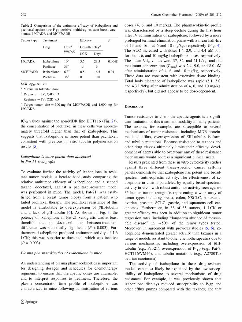
Ixabepilone is more potent than docetaxel
in Pat-21 xenografts
To evaluate further the activity of ixabepilone in resis-
tant tumor models, a head-to-head study comparing the
relative antitumor efficacy of ixabepilone and a second
taxane, docetaxel, against a paclitaxel-resistant model
was performed in mice. The model, Pat-21, was estab-
lished from a breast tumor biopsy from a patient who
failed paclitaxel therapy. The paclitaxel resistance of this
model is attributable to overexpression of bIII-tubulin
and a lack of bII-tubulin [6]. As shown in Fig. 3, the
potency of ixabepilone in Pat-21 xenografts was at least
threefold that of docetaxel; this between-treatment
difference was statistically significant (P \ 0.003). Fur-
thermore, ixabepilone produced antitumor activity of 1.6
LCK; this was superior to docetaxel, which was inactive
(P = 0.003).
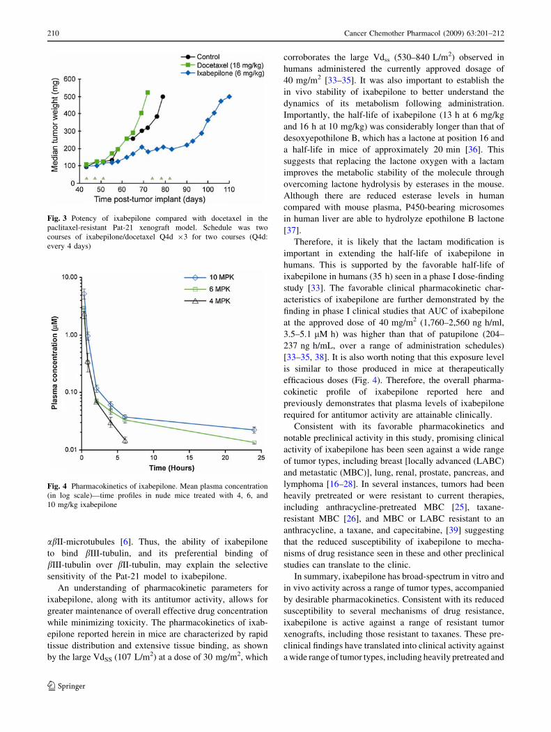
Plasma pharmacokinetics of ixabepilone in mice
An understanding of plasma pharmacokinetics is important
for designing dosages and schedules for chemotherapy
regimens, to ensure that therapeutic doses are attainable,
and to interpret responses to treatment. Therefore, the
plasma concentration-time profile of ixabepilone was
characterized in mice following administration of various
doses (4, 6, and 10 mg/kg). The pharmacokinetic profile
was characterized by a steep decline during the first hour
after IV administration of ixabepilone, followed by a more
prolonged terminal elimination phase with a mean half-life
of 13 and 16 h at 6 and 10 mg/kg, respectively (Fig. 4).
The AUC increased with dose: 1.4, 2.9, and 4.4 lM 9 h
for the 4, 6, and 10 mg/kg ixabepilone doses, respectively.
The mean Vdss values were 37, 32, and 21 L/kg, and the
maximum concentration (Cmax) was 2.4, 9.0, and 8.0 lM
after administration of 4, 6, and 10 mg/kg, respectively.
These data are consistent with extensive tissue binding.
Total body clearance of ixabepilone was rapid (5.1, 5.0,
and 4.3 L/h/kg after administration of 4, 6, and 10 mg/kg,
respectively), but did not appear to be dose-dependent.
Discussion
Tumor resistance to chemotherapeutic agents is a signifi-
cant limitation of this treatment modality in many patients.
The taxanes, for example, are susceptible to several
mechanisms of tumor resistance, including MDR protein-
mediated efflux, overexpression of bIII-tubulin isoform,
and tubulin mutations. Because resistance to taxanes and
other drug classes ultimately limits their efficacy, devel-
opment of agents able to overcome any of these resistance
mechanisms would address a significant clinical need.
Results presented from these in vitro cytotoxicity studies
against three different tissue-specific, cancer cell-line
panels demonstrate that ixabepilone has potent and broad-
spectrum antineoplastic activity. The effectiveness of ix-
abepilone in vitro is paralleled by equally broad-spectrum
activity in vivo, with robust antitumor activity seen against
35 human tumor xenografts representing a wide array of
tumor types including breast, colon, NSCLC, pancreatic,
ovarian, prostate, SCLC, gastric, and squamous cell car-
cinomas. Furthermore, in 33 of 35 tumors, 1 LCK or
greater efficacy was seen in addition to significant tumor
regression rates, including ‘‘long-term absence of measur-
able disease’’ in *50% of the tumor types tested.
Moreover, in agreement with previous studies [5, 6], ix-
abepilone demonstrated greater activity than taxanes in a
range of models resistant to other chemotherapeutics due to
various mechanisms, including overexpression of bIII-
tubulin (e.g., Pat-21), overexpression of P-gp (e.g., Pat-7,
HCT116/VM46), and tubulin mutations (e.g., A2780Tax
ovarian carcinoma).
The activity of ixabepilone in these drug-resistant
models can most likely be explained by the low suscep-
tibility of ixabepilone to several mechanisms of drug
resistance. For example, it was previously shown that
ixabepilone displays reduced susceptibility to P-gp and
other efflux pumps compared with the taxanes, and that
Table 2 Comparison of the antitumor efficacy of ixabepilone and
paclitaxel against two P-gp-positive multidrug resistant breast carci-
nomas: 16C/ADR and MCF7/ADR
Tumor type Treatment Efficacy P
Drug Dosea
(mg/kg)
Growth delayd
LCK Days
16C/ADR Ixabepilone 10b 3.5 23.5 0.0048
Paclitaxel 36c 1.4 9
MCF7/ADR Ixabepilone 6.3c 0.5 16.5 0.04
Paclitaxel 36c 0 0.8
LCK log10 cell killa Maximum tolerated doseb Regimen = IV, Q4D 93c Regimen = IV, Q2D 95d Target tumor size = 500 mg for MCF7/ADR and 1,000 mg for
16C/ADR
208 Cancer Chemother Pharmacol (2009) 63:201–212
123

ixabepilone does not induce expression of MDR proteins
[5]. The cellular uptake of ixabepilone, but not paclitaxel,
in P-gp-overexpressing cells shown in the present study
supports this observation, and is consistent with the
activity of ixabepilone in MDR models such as HCT116/
VM46. Ixabepilone is also able to overcome resistance
conferred by overexpression of the bIII-tubulin isoform,
as exemplified by its activity in the paclitaxel-resistant
Pat-21 model. The precise mechanism of Pat-21 resis-
tance was unknown until quite recently. However, recent
studies suggest that a combination of loss of bII-tubulin
and overexpression of bIII-tubulin may be responsible for
the paclitaxel resistance of Pat-21 [6]. In the studies
reported here, the head-to-head comparison of ixabepilone
versus docetaxel in the Pat-21 model showed that
ixabepilone was significantly more potent than docetaxel.
The activity of ixabepilone against this taxane-resistant
model may be explained by the fact that the tubulin-
binding mode of ixabepilone affects the microtubule
dynamics of multiple tubulin isoforms, including bIII-
tubulin. Moreover, ixabepilone preferentially suppresses
dynamic instability of abIII-microtubules compared with
Fig. 2 Clonogenic cytotoxicity
and differential cellular uptake
of ixabepilone and paclitaxel in
paclitaxel-resistant HCT116/
VM46 P-gp-overexpressing
colon carcinoma cell line.
a Clonogenic cytotoxicity based
on IC90 values in a clonogenic
cell survival assay. b Paclitaxel
and c ixabepilone uptake in
HCT116 and HCT116/VM46
(P-gp overexpressing) human
colon carcinoma cell lines.
d Uptake ratios in HCT116
versus HCT116/VM46 for
paclitaxel and ixabepilone
Cancer Chemother Pharmacol (2009) 63:201–212 209
123
abII-microtubules [6]. Thus, the ability of ixabepilone
to bind bIII-tubulin, and its preferential binding of
bIII-tubulin over bII-tubulin, may explain the selective
sensitivity of the Pat-21 model to ixabepilone.
An understanding of pharmacokinetic parameters for
ixabepilone, along with its antitumor activity, allows for
greater maintenance of overall effective drug concentration
while minimizing toxicity. The pharmacokinetics of ixab-
epilone reported herein in mice are characterized by rapid
tissue distribution and extensive tissue binding, as shown
by the large VdSS (107 L/m2) at a dose of 30 mg/m2, which
corroborates the large Vdss (530–840 L/m2) observed in
humans administered the currently approved dosage of
40 mg/m2 [33–35]. It was also important to establish the
in vivo stability of ixabepilone to better understand the
dynamics of its metabolism following administration.
Importantly, the half-life of ixabepilone (13 h at 6 mg/kg
and 16 h at 10 mg/kg) was considerably longer than that of
desoxyepothilone B, which has a lactone at position 16 and
a half-life in mice of approximately 20 min [36]. This
suggests that replacing the lactone oxygen with a lactam
improves the metabolic stability of the molecule through
overcoming lactone hydrolysis by esterases in the mouse.
Although there are reduced esterase levels in human
compared with mouse plasma, P450-bearing microsomes
in human liver are able to hydrolyze epothilone B lactone
[37].
Therefore, it is likely that the lactam modification is
important in extending the half-life of ixabepilone in
humans. This is supported by the favorable half-life of
ixabepilone in humans (35 h) seen in a phase I dose-finding
study [33]. The favorable clinical pharmacokinetic char-
acteristics of ixabepilone are further demonstrated by the
finding in phase I clinical studies that AUC of ixabepilone
at the approved dose of 40 mg/m2 (1,760–2,560 ng h/ml,
3.5–5.1 lM h) was higher than that of patupilone (204–
237 ng h/mL, over a range of administration schedules)
[33–35, 38]. It is also worth noting that this exposure level
is similar to those produced in mice at therapeutically
efficacious doses (Fig. 4). Therefore, the overall pharma-
cokinetic profile of ixabepilone reported here and
previously demonstrates that plasma levels of ixabepilone
required for antitumor activity are attainable clinically.
Consistent with its favorable pharmacokinetics and
notable preclinical activity in this study, promising clinical
activity of ixabepilone has been seen against a wide range
of tumor types, including breast [locally advanced (LABC)
and metastatic (MBC)], lung, renal, prostate, pancreas, and
lymphoma [16–28]. In several instances, tumors had been
heavily pretreated or were resistant to current therapies,
including anthracycline-pretreated MBC [25], taxane-
resistant MBC [26], and MBC or LABC resistant to an
anthracycline, a taxane, and capecitabine, [39] suggesting
that the reduced susceptibility of ixabepilone to mecha-
nisms of drug resistance seen in these and other preclinical
studies can translate to the clinic.
In summary, ixabepilone has broad-spectrum in vitro and
in vivo activity across a range of tumor types, accompanied
by desirable pharmacokinetics. Consistent with its reduced
susceptibility to several mechanisms of drug resistance,
ixabepilone is active against a range of resistant tumor
xenografts, including those resistant to taxanes. These pre-
clinical findings have translated into clinical activity against
a wide range of tumor types, including heavily pretreated and
Fig. 3 Potency of ixabepilone compared with docetaxel in the
paclitaxel-resistant Pat-21 xenograft model. Schedule was two
courses of ixabepilone/docetaxel Q4d 93 for two courses (Q4d:
every 4 days)
Fig. 4 Pharmacokinetics of ixabepilone. Mean plasma concentration
(in log scale)—time profiles in nude mice treated with 4, 6, and
10 mg/kg ixabepilone
210 Cancer Chemother Pharmacol (2009) 63:201–212
123

drug-resistant tumors. One phase II neoadjuvant study has
correlated response to ixabepilone in breast cancer patients
with the expression of specific cellular genes such as ER [40].
In addition, a phase III study in patients with anthracycline-
and taxane-resistant MBC demonstrated superior efficacy
for ixabepilone in combination with capecitabine versus
capecitabine alone, with 40% prolongation of PFS and
2.5-fold higher rate of response [41].
Acknowledgments We thank Kelly Covello for careful reading of
the manuscript and members of the Bristol–Myers Squibb Veterinary
Science Department for the care of the study animals.
Open Access This article is distributed under the terms of the
Creative Commons Attribution Noncommercial License which per-
mits any noncommercial use, distribution, and reproduction in any
medium, provided the original author(s) and source are credited.
References
1. Longley D, Johnston P (2005) Molecular mechanisms of drug
resistance. J Pathol 205:275–292
2. Moscow JMC, Cowan KH (2003) Drug resistance and its clinical
circumvention. In: Kufe D, Pollock R, Weischselbaum R, Bast R,
Gansler T, Holland J, Frei E (eds) Cancer medicine. BC Decker,
Hamilton
3. Endicott JA, Ling V (1989) The biochemistry of P-glycoprotein-
mediated multidrug resistance. Annu Rev Biochem 58:137–171
4. Gottesman MM, Pastan I (1993) Biochemistry of multidrug
resistance mediated by the multidrug transporter. Annu Rev
Biochem 62:385–427
5. Lee FYF, Borzilleri R, Fairchild CR, Kim S-H, Long BH,
Raventoz-Suarez C, Vite GD, Rose WC, Kramer RA (2001)
BMS-247550: a novel epothilone analog with a mode of action
similar to paclitaxel but possessing superior antitumor activity.
Clin Cancer Res 7:1429–1437
6. Jordan M, Miller H, Ni L, Castenada S, Inigo I, Kan D, Lewin A,
Ryseck R, Kramer R, Wilson L, Lee FY (2006) The Pat-21 breast
cancer model derived from a patient with primary Taxol� resis-
tance recapitulates the phenotype of its origin, has altered beta-
tubulin expression and is sensitive to ixabepilone. In: Proc Am
assoc cancer res 97th annual meeting, LB-280
7. Altaha R, Fojo T, Reed E, Abraham J (2003) Epothilones: a novel
class of non-taxane microtubule-stabilizing agents. Curr Pharm
Des 8:1707–1712
8. Bollag DM, McQueney PA, Zhu J, Hensens O, Koupal L, Liesch
J, Goetz M, Lazarides E, Woods CM (1995) Epothilones, a new
class of microtubule-stabilizing agents with a Taxol-like mech-
anism of action. Cancer Res 55:2325–2333
9. Bode CJ, Gupta ML, Reiff EA, Suprenant KA, Georg GI, Himes
RH (2002) Epothilone and paclitaxel: unexpected differences in
promoting the assembly and stabilization of yeast microtubules.
Biochemistry 41:3870–3874
10. Chou T-C, Zhang X-G, Bolag A, Su D-S, Meng D, Salvin K,
Bertino J, Danishefsky SJ (1998) Desoxyepothilone B: an effi-
cacious microtubule-targeted antitumor agent with a promising in
vivo profile relative to epothilone B. Proc Natl Acad Sci USA
95:9642–9647
11. Lee F, Borzilerri R, fairchild C, Kamath A, Smykla R, Kramer R,
Vite G (2008) Preclinical discovery of ixabepilone, a highly active
antineoplastic agent. Cancer Chemother Pharmacol (in press)
12. Awada A, Burris D, de Valeriola D (2001) Phase I clinical and
pharmacology study of the epothilone B analog BMS-247550
given weekly in patients with advanced solid tumors. Clin Cancer
Res 7:3801S
13. Loruzzo P, Wozniak A, Flaherty L, Shields A, Wright J, Lebwhol
D (2001) Phase I clinical trial of BMS-247550 (aka Epothilone B
Analog; NSC710428) in adult patients with advanced solid
tumors. Proc Am Soc Clin Oncol 20 (abstr. #2125)
14. Spriggs D, Soignet S, Bienvenu B, Letrent S, Lebwohl D, Jones
S, Burris HI (2001) Phase I first-in-man study of the epothilone B
analog BMS-247550 in patients with advanced cancer. Proc Am
Soc Clin Oncol 20 (abstr. 428)
15. Hao D, Hammond LA, deBono JS, Tolcher AW, Berg KE, Bass
A, Mays TA, Smith LS, Drengler R, Rowinsky EK (2002)
Continuous weekly administration of the epothilone-B derivative,
BMS247,550 (NSC710428): a phase I and pharmacokinetic (PK)
study. Proc Am Soc Clin Oncol 21 (abstr. #411)
16. Ajani JA, Shah MA, Bokemeyer C, Lenz H-J, Burris HA, Cutsem
EV, Usakewicz J, Peeters O, Voi M, Lebwohl D, Safran H (2002)
Phase II study of the novel epothilone BMS-247550 in patients
(pts) with metastatic gastric adenocarcinoma previously treated
with a taxane. Proc Am Soc Clin Oncol 21 (abstr. 619)
17. Baselga J, Gianni L, Llombart A, Manikhas G, Kubista E, Steger
G (2005) Predicting response to ixabepilone: genomics study in
patients receiving single agent ixabepilone as neoadjuvant treat-
ment for breast cancer (BC). Breast Cancer Res Treat 94:S31
(abstr. 305)
18. Fojo A, Menefee M, Poruchynsky M, Edgerly M, Mickley L, Li
N, Tapia E, Merino M, Balis F, Bates S (2005) A translational
study of ixabepilone (BMS-247550) in renal cell cancer (RCC):
assessment of its activity and demonstration of target engagement
in tumor cells. J Clin Oncol 23:388S (abstr 4541)
19. Galsky MD, Small EJ, Oh WK, Chen I, Smith DC, Colevas AD,
Martone L, Curley T, DeLaCruz A, Scher HI, Kelly WK (2005)
Multi-Institutional randomized phase II trial of the epothilone B
analog ixabepilone (BMS-247550) with or without estramustine
phosphate in patients with progressive castrate metastatic prostate
cancer. J Clin Oncol 23:1439–1446
20. Hussain M, Tangen CM, Lara PN Jr, Vaishampayan UN, Petrylak
DP, Colevas AD, Sakr WA, Crawford ED (2005) Ixabepilone
(epothilone B analogue BMS-247550) is active in chemotherapy-
naive patients with hormone-refractory prostate cancer: a South-
west Oncology Group Trial S0111. J Clin Oncol 23:8724–8729
21. O’Connor O, Straus D, Moskowitz C, Hamlin P, Portlock C,
Gerecitano J, Neylon E, Colevas D, Zelenetz A (2005) Targeting
the microtubule apparatus in indolent and mantle cell lymphoma
with the novel epothilone analog BMS 247550 induces major and
durable remissions in very drug resistant disease. J Clin Oncol
23:16S (abstr. 6569)
22. Smith SM, Pro B, Besien Kv, Conner K, Karrison T, Wong S,
Stiff P, Vokes E (2005) A phase II study of epothilone B analog
BMS-247550 (NSC 710428) in patients with relapsed aggressive
non-Hodgkin’s lymphomas. J Clin Oncol 23:16S (abstr. 6625)
23. Conte P, Thomas E, Martin M, Klimovsky J, Tabernero J (2006)
Phase II study of ixabepilone in patients (pts) with taxane-resis-
tant metastatic breast cancer (MBC): final report. J Clin Oncol
24:18S (abstr. 10505)
24. Whitehead RP, McCoy S, Rivkin SE, Gross HM, Conrad ME,
Doolittle GC, Wolff RA, Goodwin JW, Dakhil SR, Abbruzzese
JL (2006) A Phase II trial of epothilone B analogue BMS-247550
(NSC #710428) ixabepilone, in patients with advanced pancreas
cancer: a Southwest Oncology Group study. Invest New Drugs
24:512–520
25. Roche H, Yelle L, Cognetti F, Mauriac L, Bunnell C, Sparano J,
Kerbrat P, Delord J-P, Vahdat L, Peck R, Lebwohl D, Ezzeddine
R, Cure H (2007) Phase II clinical trial of ixabepilone
Cancer Chemother Pharmacol (2009) 63:201–212 211
123

(BMS-247550), an epothilone B analog, as first-line therapy in
patients with metastatic breast cancer previously treated with
anthracycline chemotherapy. J Clin Oncol 25:3415–3420
26. Thomas E, Tabernero J, Fornier M, Conte P, Fumoleau P, Lluch
A, Vahdat LT, Bunnell CA, Burris HA, Viens P, Baselga J,
Rivera E, Guarneri V, Poulart V, Klimovsky J, Lebwohl D,
Martin M (2007) Phase II clinical trial of ixabepilone (BMS-
247550), an epothilone B analog, in patients with taxane-resistant
metastatic breast cancer. J Clin Oncol 25:3399–3406
27. Vansteenkiste J, Lara PN Jr, Le Chevalier T, Breton J-L, Bonomi
P, Sandler AB, Socinski MA, Delbaldo C, McHenry B, Lebwohl
D, Peck R, Edelman M (2007) Phase II clinical trial of the
epothilone B analog, ixabepilone, in patients with non small-cell
lung cancer whose tumors have failed first-line platinum-based
chemotherapy. J Clin Oncol 25:3448–3455
28. Zhuang SH, Hung YE, Hung L, Robey RW, Sackett DL, Linehan
WM, Bates SE, Fojo T, Poruchynsky MS (2007) Evidence for
microtubule target engagement in tumors of patients receiving
ixabepilone. Clin Cancer Res 13:7480–7486
29. Long BH, Wang L, Lorico A, Wang RRC, Brattain MG, Casazza
AM (1991) Mechanisms of resistance to etoposide and teniposide
in acquired resistant human colon and lung carcinoma cell lines.
Cancer Res 51:5275–5284
30. Riss TL, Moravec RA (1992) Comparison of MTT, XTT, and a
novel tetrazolium compound MTS for in vitro proliferation and
chemosensitivity assays. Mol Biol Cell 3(Suppl):184a
31. Gehan EA (1965) A generalized Wilcoxon test for comparing
arbitrarily singly-censored samples. Biometrika 52:203–223
32. Lee F, Sciandra J, Siemann D (1989) A study of the mechanism
of resistance to Adriamycin in vivo. Glutathione metabolism,
P-glycoprotein expression and drug transport. Biochem Pharma-
col 38:3697–3705
33. Mani S, McDaid H, Hamilton A, Hochster H, Cohen MB,
Khabelle D, Griffin T, Lebwohl DE, Liebes L, Muggia F,
Horwitz SB (2004) Phase I clinical and pharmacokinetic study of
BMS-247550, a novel derivative of epothilone B, in solid tumors.
Clin Cancer Res 10:1289–1298
34. Gadgeel SM, Wozniak A, Boinpally RR, Wiegand R, Heilbrun
LK, Jain V, Parchment R, Colevas D, Cohen MB, LoRusso PM
(2005) Phase I clinical trial of BMS-247550, a derivative of
epothilone B, using accelerated titration 2B design. Clin Cancer
Res 11:6233–6239
35. Aghajanian C, Burris HA III, Jones S, Spriggs DR, Cohen MB,
Peck R, Sabbatini P, Hensley ML, Greco FA, Dupont J,
O’Connor OA (2007) Phase I study of the novel epothilone
analog ixabepilone (BMS-247550) in patients with advanced
solid tumors and lymphomas. J Clin Oncol 25:1082–1088
36. Chou T-C, O’Connor OA, Tong WP, Guan Y, Zhang Z-G,
Stachel SJ, Lee C, Danishefsky SJ (2001) The synthesis,
discovery, and development of a highly promising class of
microtubule stabilization agents: curative effects of desoxyepo-
thilones B and F against human tumor xenografts in nude mice.
PNAS 98:8113–8118
37. Lee FYF, Vite GD, Hofle G, Kim SH, Clark J, Fager K, Kennedy
K, Smykla R, Wen M, Leavitt K, Johnston KA, Peterson RW,
Kamath A, Franchini M, Schulze G, Fairchild C, Raghavan K,
Long BH, Kramer R (2002) The discovery of BMS-310705: a
water-soluble and chemically stable semi-synthetic epothilone
possessing potent parenteral and oral antitumor activity against
models of taxane-sensitive and -resistant human tumors in vivo.
Proc Am Assoic Cancer Res 43:792–793
38. Rubin EH, Rothermel J, Tesfaye F, Chen T, Hubert M, Ho Y-Y,
Hsu C-H, Oza AM (2005) Phase I dose-finding study of weekly
single-agent patupilone in patients with advanced solid tumors.
J Clin Oncol 23:9120–9129
39. Perez EA, Lerzo G, Pivot X, Thomas E, Vahdat L, Bosserman L,
Viens P, Cai C, Mullaney B, Peck R, Hortobagyi GN (2007)
Efficacy and safety of ixabepilone (BMS-247550) in a phase II
study of patients with advanced breast cancer resistant to an
anthracycline, a taxane, and capecitabine. J Clin Oncol 25:3407–
3414
40. Lee H, Xu L, Wu S, Paul B, Baselga J, Llombart A, Steger GG,
Galbraith S, Clark E (2006) Predictive biomarker discovery and
validation for the targeted chemotherapeutic ixabepilone. J Clin
Oncol 24:18S (abstr. 3011)
41. Thomas ES, Gomez HL, Li RK, Chung H-C, Fein LE, Chan VF,
Jassem J, Pivot XB, Klimovsky JV, de Mendoza FH, Xu B,
Campone M, Lerzo GL, Peck RA, Mukhopadhyay P, Vahdat LT,
Roche HH (2007) Ixabepilone plus capecitabine for metastatic
breast cancer progressing after anthracycline and taxane treat-
ment. J Clin Oncol 25:5210–5217
212 Cancer Chemother Pharmacol (2009) 63:201–212
123