pparγ ligands regulate nadph oxidase, enos, and barrier function in the lung following chronic...
TRANSCRIPT

PPARγ Ligands Regulate NADPH Oxidase, eNOS, and BarrierFunction in the Lung Following Chronic Alcohol Ingestion
Matthew C. Wagner1, Samantha M. Yeligar1,2, Lou Ann Brown2, and C. Michael Hart11Department of Medicine, Atlanta Veterans' Affairs and Emory University Medical Centers,Decatur2Department of Pediatrics, Division of Neonatal-Perinatal Medicine, Emory University, Atlanta,Georgia
AbstractBACKGROUND—Chronic alcohol ingestion increases the incidence and severity of the AcuteRespiratory Distress Syndrome (ARDS), where reactive species contribute to alveolar-capillarybarrier dysfunction and non-cardiogenic pulmonary edema. Previous studies demonstrated thatchronic alcohol ingestion increased lung NADPH oxidase and endothelial nitric oxide synthase(eNOS) expression and that ligands for the peroxisome proliferator-activated receptor gamma(PPARγ) reduced NADPH oxidase expression. Therefore, we hypothesized that the PPARγ ligand,rosiglitazone, would attenuate alcohol-induced NADPH oxidase expression and pulmonary barrierdysfunction.
METHODS—C57Bl/6 mice were treated ± alcohol in drinking water (20% w/v) for 12 weeks.During the final week of alcohol treatment, mice were gavaged with rosiglitazone (10 mg/kg/day)or vehicle. Selected animals were treated twice with lipopolysaccharide (LPS, 2 mg/kg IP) prior tosacrifice. Pulmonary barrier dysfunction was estimated from protein content of bronchoalveolarlavage (BAL) fluid.
RESULTS—LPS treatment increased BAL protein in alcohol-fed but not control mice, androsiglitazone attenuated LPS and alcohol-induced pulmonary barrier dysfunction. Alcohol- andLPS-induced increases in lung eNOS, Nox1, and Nox4 expression were attenuated byrosiglitazone. In vitro, alcohol (0.10% w/v) increased H2O2 production, barrier dysfunction,eNOS, Nox1, and Nox4 expression in human umbilical vein endothelial cell (HUVEC)monolayers, effects also attenuated by rosiglitazone (10 μM). Alcohol-induced HUVEC barrierdysfunction was attenuated by inhibition of NOS or addition of the eNOS cofactor,tetrahydrobiopterin.
CONCLUSIONS—These results indicate that PPARγ activation reduced expression of eNOS,Nox1, Nox4, the production of reactive species, and barrier dysfunction caused by chronic alcoholingestion and suggest that PPARγ represents a novel therapeutic target for strategies designed toreduce the risk of lung injury in patients with a history of chronic alcohol ingestion.
Keywordsrosiglitazone; Acute Respiratory Distress Syndrome; lung injury
Address correspondence to: C. Michael Hart, M.D. Associate Chief of Staff for Research Atlanta VAMC Professor of MedicineEmory University 1670 Clairmont Rd. (151-P) Decatur, GA 30033 Phone: 404 - 321 - 6111 ext 6170 Fax: 404 - 728 - [email protected] The authors have no financial conflicts to disclose.
NIH Public AccessAuthor ManuscriptAlcohol Clin Exp Res. Author manuscript; available in PMC 2013 February 1.
Published in final edited form as:Alcohol Clin Exp Res. 2012 February ; 36(2): 197–206. doi:10.1111/j.1530-0277.2011.01599.x.
NIH
-PA Author Manuscript
NIH
-PA Author Manuscript
NIH
-PA Author Manuscript

IntroductionThe Acute Respiratory Distress Syndrome (ARDS) is a severe form of acute lung injurycharacterized by noncardiogenic pulmonary edema following activation of proinflammatorycascades in response to diverse clinical conditions, such as infection, trauma, or aspiration(Ware and Matthay, 2000). A critical component of the pathophysiology of lung injuryinvolves impaired alveolar-capillary barrier function (Ware and Matthay, 2000). Thiscondition, which affects as many as 75,000–100,000 individuals each year in the UnitedStates alone, is associated with significant morbidity and mortality due to a lack of specifictherapies that directly modulate ARDS pathogenesis (Ware and Matthay, 2000).
Factors that predispose patients to ARDS include chronic alcohol abuse, a comorbid variableknown to increase the incidence of ARDS 2–4-fold in critically ill patients (Moss et al.,2003). While chronic alcohol ingestion likely increases ARDS incidence through multiplemechanisms, previous studies in a rat model of chronic alcohol ingestion (using the LieberDeCarli diet) (Guidot et al., 2000, Moss et al., 2000, Polikandriotis et al., 2006) haveindicated that oxidative stress plays a key role in susceptibility to lung injury. Inexperimental animals, chronic alcohol ingestion enhanced sepsis-induced barrierdysfunction in the lung, a derangement attenuated by treatment with glutathione precursors(Holguin et al., 1998). Chronic alcohol ingestion also altered the production of both reactiveoxygen and nitrogen species in the lung through increased expression of NADPH oxidaseand endothelial nitric oxide synthase (eNOS), respectively (Polikandriotis et al., 2007,Polikandriotis et al., 2006). These findings have led to the working hypothesis that chronicethanol ingestion “primes” the lung by increasing the expression of enzymatic sources ofreactive species. These alcohol-induced derangements while not sufficient to cause lunginjury by themselves, contribute to susceptibility to injury caused by subsequentinflammatory stimuli. Based on this “two hit” model and evidence that oxidative stresspromotes endothelial barrier dysfunction in vitro and in vivo (Lum and Roebuck, 2001), wehave sought to identify new interventions that might reduce the priming effect of alcohol onthe lung and / or the response of the alcohol-treated lung to inflammatory stimuli.
Treatment with peroxisome proliferator-activated receptor gamma (PPARγ ligandsrepresents a novel strategy to regulate NADPH oxidase expression. PPARγ is a ligand-activated transcription factor that is widely expressed and participates in the regulation ofdiverse biological processes, including adipocyte differentiation (Barak et al., 1999), lipidmetabolism (Schoonjans et al., 1996), smooth muscle cell proliferation (Meredith et al.,2009), and insulin sensitivity (Hevener et al., 2003, Rangwala et al., 2003). Ligands for thePPARγ receptor include naturally occurring fatty acids and their metabolites as well assynthetic thiazolidinedione (TZD) medications such as pioglitazone and rosiglitazone, whichhave been used to increase insulin sensitivity in patients with type 2 diabetes (Yki-Jarvinen,2004). Rosiglitazone reduced vascular NADPH oxidase expression and activity in culturedendothelial cells (Hwang et al., 2005), in the vasculature of diabetic mice (Hwang et al.,2007), and in the lungs of hypoxia-exposed mice (Nisbet et al., 2010). Based on thesestudies, we hypothesized that PPARγ ligands would reduce NADPH oxidase expression andoxidative stress in the lung following chronic alcohol ingestion, thereby reducing alcohol-induced susceptibility to barrier dysfunction. We explored this hypothesis by examining theability of the PPARγ ligand, rosiglitazone, to attenuate alcohol-induced oxidative stress andbarrier dysfunction in endothelial cells in vitro and in the mouse lung in vivo.
Our results demonstrate that administration of alcohol in the drinking water of mice for 12weeks increased LPS-induced barrier dysfunction and that treatment with rosiglitazone bygavage over the final week of alcohol administration attenuated barrier dysfunction as wellas alcohol-induced increases in the expression of eNOS, Nox1 and Nox4 in the lung. LPS-
Wagner et al. Page 2
Alcohol Clin Exp Res. Author manuscript; available in PMC 2013 February 1.
NIH
-PA Author Manuscript
NIH
-PA Author Manuscript
NIH
-PA Author Manuscript

induced increases in Nox1, Nox2, Nox4 and eNOS mRNA were also attenuated byrosiglitazone. Similarly, alcohol caused endothelial monolayer barrier dysfunction, H2O2production, and increased eNOS, Nox1, and Nox4 expression in vitro which were attenuatedby rosiglitazone treatment. Collectively, these results indicate that the PPARγ receptor mayrepresent a new therapeutic target in strategies designed to reduce oxidative stress andbarrier dysfunction in the lung caused by chronic alcohol ingestion.
Materials and MethodsMouse Model of Chronic Alcohol Ingestion
Male, C57Bl/6 mice aged 8 weeks were given 20% ethanol (EtOH) in their drinking wateralong with ad lib access to standard mouse chow. Mice were acclimated to EtOH byincreasing the EtOH concentration in 5% increments from 0% to the target 20% (w/v) overthe course of two weeks then maintaining the 20% concentration for an additional tenweeks. This regimen replicates blood alcohol levels following chronic EtOH ingestion inhuman subjects (Jerrells et al., 2007, Song et al., 2002). In preliminary studies, blood alcoholconcentrations were measured with a rapid, high-performance plasma alcohol analyzer(Analox Instruments Ltd., London, UK) according to the manufacturer's protocol. Theseanalyses confirmed that this ethanol regimen produced clinically relevant elevations in bloodalcohol concentration (0.12% ± 0.03, n=24). During the final week of EtOH treatment, allmice were gavaged daily for 7 days with either rosiglitazone (10 mg/kg/day in 100 μlmethylcellulose vehicle) or vehicle alone as previously reported (Hwang et al., 2007, Nisbetet al., 2010). Selected mice were treated with Escherichia coli lipopolysaccharide (Sigma-Aldrich, St. Louis MO, 2 mg/kg IP at 6 and 3 hours prior to sacrifice) as an inflammatorystimulus to promote pulmonary dysfunction. The timing of these studies was based on recentreports showing significant LPS-mediated increases in lung leak in C57Bl/6 mice 6-hoursafter LPS administration (Rojas et al., 2005). After sacrifice, blood was collected via cardiacpuncture, and bronchoalveolar lavage (BAL) performed via tracheotomy. Proteinconcentration in the BAL fluid was measured using the bicinchoninic acid (BCA) assay(Thermo Scientific, Rockford IL), and values were corrected for dilution based on the ratioof blood to BAL urea nitrogen, assayed with a commercially available kit (Pointe Scientific,Inc., Canton MI) as previously reported (Rennard et al., 1986). Lung tissue was collected forsubsequent analyses as described below following perfusion of the pulmonary artery withsterile phosphate buffered saline (PBS, Cellgro, Manassas, VA). All animal studies wereapproved by the Atlanta Veteran's Affairs Medical Center Animal Care and Use Committee.
Human Umbilical Vein Endothelial Cell (HUVEC) CulturesHUVEC (Clonetics) monolayers were grown at 37°C in endothelial growth medium(Clonetics) containing 10% fetal bovine serum, 10 ng/mL human epidermal growth factor,1.0 μg/mL hydrocortisone, 12 μg/mL bovine brain extract, 50 μg/mL gentamicin, and 50 ng/mL amphotericin B in a 5% CO2 atmosphere. Confluent cells were maintained in mediumwith 2% fetal bovine serum. In some experiments, cells (passage 4–8) were plated on 0.2%gelatin-coated 100 mm or 35 mm plastic tissue culture dishes and treated ± ethanol (0.10%w/v) and ± rosiglitazone (10 μM) for 72 hours as we have previously reported (Hwang et al.,2005, Polikandriotis et al., 2005a, Polikandriotis et al., 2005b). All cell cultures treated withethanol were maintained in a sealed incubator (Billups-Rothenburg, Del Mar, CA) with areservoir containing 20 ml standard medium with 0.10% ethanol in order to minimizeethanol evaporation. HUVEC media were replaced every 24 hours. In selected studies, toassess HUVEC monolayer barrier function, cells were plated on 0.2% gelatin-coated 12 mmTranswell Permeable Supports (Costar, Wilkes Barre, PA) with 0.4 μm polycarbonatemembranes and treated with or without EtOH for 72 hours. Selected HUVEC monolayers intranswell supports were treated with rosiglitazone (10 μM), tetrahydrobiopterin (20 μM), or
Wagner et al. Page 3
Alcohol Clin Exp Res. Author manuscript; available in PMC 2013 February 1.
NIH
-PA Author Manuscript
NIH
-PA Author Manuscript
NIH
-PA Author Manuscript

L-NG-Nitroarginine methyl ester hydrochloride (LNAME, 100 –M). After 72 hours, alltranswells were emptied, 750 μl Evan's blue dye-associated albumin (0.04 g/ml in standardmedium) was added to the upper, luminal chamber, and 1.2 ml standard medium was addedto the lower, abluminal chamber. Transmonolayer protein passage was determined at 0.5,1.5, and 3 hours by withdrawing 100 μl samples from the stirred abluminal chamber andmeasuring the spectrophotometric absorbance (595 nm) of Evan's blue dye as we havereported (Knepler et al., 2001). Following sample collection, corresponding quantities offresh media were added to the abluminal chamber to minimize the development ofhydrostatic pressure gradients across the monolayer.
Western Analysis of Lung or HUVEC HomogenatesLung tissue was homogenized in lysis buffer [20 mM Tris pH 7.4, 2.5 mM EDTA, 100 mMNaCl, 10 mM NaF, 1 mM Na3VO4, 1% Triton X-100, 0.1% SDS, 1% Na deoxycholate, 1tablet/10 ml EDTA-free complete protease inhibitor cocktail (Roche, Indianapolis IN), 1mM β-glycerolphosphate, 2.5 mM Na pyrophosphate] and frozen in liquid nitrogen. Thelysate was spun at 28,000 × g for 15 minutes, the supernatants transferred to new tubes, andprotein concentrations were determined using a BCA assay. Equal amounts of sampleprotein (15–20 μg/lane) were loaded into wells of a 4–12% bis-tris polyacrylamide gelelectrophoresis (PAGE) mini gel. HUVEC were collected in lysis buffer, and lysates werecentrifuged at 16,000 × g for 20 minutes. Supernatants were transferred to new tubes, andprotein concentrations were detected using a BCA assay. Sample proteins (40 μg/lane) wereloaded into the wells of 4–12% bis-tris PAGE mini gels. Proteins were separated byelectrophoresis and blotted to polyvinylidene fluoride (PVDF) or nitrocellulose membranes.Membranes were incubated with polyclonal antibodies to eNOS (1:1000 dilution; BDTransduction Laboratories), Nox1 (1:500 dilution; Santa Cruz Biotechnology, Santa Cruz,CA), Nox2 (1:500 dilution; Santa Cruz Biotechnology), Nox4 (1:2500 dilution; Dr. DavidLambeth, Emory University), CDK4 (1:1000 dilution; Santa Cruz Biotechnology), orGAPDH (1:500 dilution; Santa Cruz Biotechnology) in non-fat dry milk (5%) and tris-buffered saline for 8–24 hours at 4°C. Proteins were visualized using a peroxidase-coupledanti-rabbit, anti-goat or anti-mouse IgG in the presence of LumiGlo reagent while exposingin a Biorad Chemidoc XRS/HQ (Hercules, CA). Densitometric analysis was accomplishedusing Biorad Quantity One (Version 4.5.0) software. The densitometric intensity of eachsample was normalized to the density of CDK4 or GAPDH in that sample.
Real Time PCR Analysis of Lung or HUVEC HomogenatesReal-time PCR was performed to quantify mRNA levels of eNOS, Nox1, Nox2 (gp91phox),and Nox4. RNA was extracted from whole lung homogenates or cultured HUVECmonolayers with Tri Reagent (Molecular Research Center Inc., Cincinnati, OH) according tothe manufacturer's protocol. mRNA was reverse transcribed to cDNA with the I-ScriptcDNA synthesis kit (Bio Rad), employing Sybr green for detection. Primers for Nox1(sequence: F 5'-GTTTTAC CGCTCCCAGCAGAA-3', R 5'-GGATGCCATTCCAGGAGAGAG-3'), Nox2 (sequence: F 5'-CCACATACAGGCCCCCTTCAG-3', R 5'-GTTGGGGCTGAATGTCTTCCTCTTT-3'), Nox4 (sequence: F 5'-CTGGTCTGACGGGTGTCTGCATGGTG-3', R 5'-CTCCGCA CAATAAAGGCACAAAGGTCCAG-3'), and eNOS (sequence:F 5'- CCCACACAGAAG GTCTCACA-3', R 5'- ACACGGCTGGAGGAACT G-3') wereemployed on cDNA derived from lung homogenates or HUVEC monolayers and quantifiedto starting copy counts through standard curves. Results were normalized to S9 expression(sequence: F 5'-ATCCGCCAACGTCACAT TA-3', R 5'-TCTTCACTCGGCCTGGAC-3')in each sample and confirmed by generation of a standard curve with stock mRNA for eachtype.
Wagner et al. Page 4
Alcohol Clin Exp Res. Author manuscript; available in PMC 2013 February 1.
NIH
-PA Author Manuscript
NIH
-PA Author Manuscript
NIH
-PA Author Manuscript

Amplex Red Assay of HUVEC H2O2 ProductionDetection of HUVEC H2O2 production was performed with the Amplex Red assay(Invitrogen, Eugene, OR). Amplex red (10-acetyl-3,7,-dihydroxyphenoxazine) reacts withH2O2 in the presence of horseradish peroxidase to generate a highly fluorescent product,resorufin. Confluent HUVEC cultures in 35 mm Petri dishes were washed twice with sterilePBS and incubated for 30 minutes with 1000 μl of Amplex Red (20 μM) and horseradishperoxidase (0.1 U/ml) at 37°C in the dark in a 5% CO2 incubator. Fluorescence was thenmeasured in triplicate in a fluorescence plate reader (excitation λ 540, emission λ 590).Sample H2O2 concentrations were calculated based on standard curves generated withknown H2O2 concentrations. Cell cultures were then rinsed twice with PBS, scraped into150 μl of lysis buffer and sonicated. The resulting solution was centrifuged at 16,000 × g,and the supernatant protein concentrations were determined using a BCA assay. H2O2concentrations were normalized to these protein concentrations and expressed relative toaverage control values.
Statistical AnalysisAll data are expressed as mean ± SEM. Statistical analyses were accomplished byperforming ANOVA (with repeated measures where appropriate) followed by a Tukeyposthoc test to detect differences between individual experimental groups. p<0.05 wasconsidered significant. ANOVA and posthoc tests were conducted using GraphPad Prism(ver. 5, La Jolla, CA).
ResultsChronic EtOH Ingestion Increased Pulmonary Barrier Dysfunction Caused by LPS:Attenuation by Rosiglitazone
As illustrated in Figure 1, compared to BAL protein levels of control mice, chronic EtOHingestion alone caused a small but not statistically significant increase in pulmonary barrierdysfunction. Similarly, treatment with LPS had a small but not statistically significant effecton BAL protein in control mice. However, treatment with LPS significantly increased BALprotein in EtOH-treated mice. Treatment with rosiglitazone had no significant impact onlung barrier function in control animals regardless of LPS treatment. However, rosiglitazonetreatment significantly attenuated BAL protein leak in mice that were treated with LPSfollowing chronic EtOH ingestion.
Rosiglitazone Attenuated EtOH-Induced Barrier Dysfunction in Endothelial Monolayers inVitro
Having shown that chronic EtOH ingestion enhances LPS-induced barrier dysfunction in themouse lung and that this barrier dysfunction was attenuated by administration of the PPARγligand, rosiglitazone, we examined the ability of rosiglitazone to directly modulate EtOHeffects on endothelial barrier function. As shown in Figure 2, EtOH significantly increasedthe transmonolayer passage of albumin through cultured HUVEC monolayers consistentwith impaired endothelial barrier function. Treatment with rosiglitazone had no effect onbarrier function of control monolayers but significantly attenuated the observed barrierdysfunction in endothelial monolayers treated with EtOH. Furthermore, treatment witheither the non-specific NOS inhibitor, LNAME, or the NOS cofactor, tetrahydrobiopterin,attenuated EtOH-induced barrier dysfunction in HUVEC monolayers.
Rosiglitazone Attenuated EtOH-Induced H2O2 Production in VitroBecause oxidative stress represents a critical mediator of alcohol effects on the lung(Holguin et al., 1998, Moss et al., 2000), we examined the impact of rosiglitazone treatment
Wagner et al. Page 5
Alcohol Clin Exp Res. Author manuscript; available in PMC 2013 February 1.
NIH
-PA Author Manuscript
NIH
-PA Author Manuscript
NIH
-PA Author Manuscript

on EtOH-induced H2O2 production in HUVECs. H2O2 production was measured with theAmplex Red assay in HUVEC monolayers treated ± EtOH (0.10% w/v) and ± rosiglitazone(10 μM). As shown in Figure 3, EtOH significantly increased H2O2 production, and thisincrease was attenuated by treatment with rosiglitazone.
Rosiglitazone Attenuated EtOH-induced eNOS Expression in the Lung and in HUVECmonolayers
To further examine the mechanisms by which rosiglitazone treatment attenuates barrierdysfunction and oxidative stress, western blots of lung tissue from mice ± EtOH for 12weeks, treated with or without rosiglitazone, were examined for changes in the expression ofeNOS. As illustrated in Figure 4A, EtOH significantly increased eNOS protein expression inlung tissue, and treatment with rosiglitazone attenuated eNOS expression in mice followingchronic EtOH ingestion. Similarly, EtOH stimulation increased eNOS mRNA levels inHUVEC monolayers, and these increases were attenuated by rosiglitazone treatment (Figure4B).
Rosiglitazone Attenuated EtOH-induced NADPH Oxidase Expression in the Lung and inHUVEC Monolayers
Because we have also reported that chronic EtOH ingestion increased the expression andactivity of NADPH oxidases in the rat lung (Polikandriotis et al., 2006), we examined theexpression of Nox2 and its homologs, Nox1 and Nox4, in the mouse lung following chronicEtOH ingestion. As shown in Figure 5 (A,B), chronic EtOH ingestion significantly increasedthe expression of Nox1 and Nox4 protein in the mouse lung. Rosiglitazone attenuated Nox1and Nox4 expression in EtOH-treated lung but had no significant effect on the expression ofNADPH oxidase protein in lungs from control animals. Similar results were found incultured HUVEC, where EtOH-induced Nox1 and Nox4 protein levels were attenuated bytreatment with rosiglitazone (Figure 5, C, D). Nox2 expression was below the detectionthreshold for all treatments in both lung tissue and cell culture (data not shown). As shownin Figure 6, RT-PCR analysis of lung or HUVEC mRNA levels demonstrated that EtOHincreased levels of Nox1 and Nox4 mRNA and that rosiglitazone treatment attenuated theseincreases both in vivo (A,E) and in vitro (B, F). Rosiglitazone treatment alone had nosignificant effect on mRNA expression levels under control conditions. In contrast to theprotein results, Nox2 mRNA levels were detectable in lung and HUVEC homogenates(C,D), and although EtOH treatment failed to significantly increase Nox2 mRNA levels,rosiglitazone reduced levels in mouse lung and HUVECs following EtOH treatment.
Rosiglitazone Attenuated LPS-Induced Nox1, Nox2, Nox4, and eNOS mRNA Levels in theLung
In addition to modulating the responses of the lung to EtOH, we also examined ifrosiglitazone directly modulates responses to LPS. As illustrated in Figure 7, treatment withLPS increased the mRNA levels of Nox1, Nox2, Nox4, and eNOS in the mouse lung.Treatment with rosiglitazone had no effect on the mRNA levels of these targets in controlanimals but significantly attenuated LPS-induced increases in the levels of Nox1, Nox2,Nox4, and eNOS mRNA in the mouse lung.
DiscussionPrevious studies demonstrated that chronic alcohol ingestion in rats increased oxidativestress in the lung (Guidot et al., 2000, Polikandriotis et al., 2006) and enhancedsusceptibility to endotoxin-mediated lung injury (Holguin et al., 1998). Furthermore,glutathione precursors attenuated EtOH-induced oxidative stress and reduced the effects ofEtOH on the lung in vivo (Guidot et al., 2000) and on lung cells in vitro (Brown et al.,
Wagner et al. Page 6
Alcohol Clin Exp Res. Author manuscript; available in PMC 2013 February 1.
NIH
-PA Author Manuscript
NIH
-PA Author Manuscript
NIH
-PA Author Manuscript

2007). While the mechanisms of alcohol-induced oxidative stress in the lung remainincompletely defined, chronic EtOH ingestion increased the expression of NADPH oxidase(Polikandriotis et al., 2006) and eNOS (Polikandriotis et al., 2007) in the rat lung. Theseenzymes produce reactive oxygen and nitrogen species that can contribute to oxidative stressand barrier dysfunction. Because PPARγ ligands attenuated the expression of NADPHoxidases in vitro (Hwang et al., 2005) and in vivo (Hwang et al., 2007, Nisbet et al., 2010),the primary objectives of the current study were to determine if treatment with the PPARγligand, rosiglitazone, could attenuate EtOH-induced alterations in the expression of eNOSand NADPH oxidases in the lung.
We employed a mouse model where EtOH was added to the animals' water for 12 weeks.This regimen of EtOH ingestion produced clinically relevant blood alcohol concentrations inmice that were comparable to alcohol concentrations in rats fed the liquid Lieber-DeCarlidiet for six weeks (unpublished observations). The current findings indicate that EtOHcaused barrier dysfunction in vitro in endothelial monolayers, but had no effect on lungbarrier function by itself, consistent with clinical observations that chronic alcohol ingestionalone does not cause lung injury. However, the EtOH regimen employed in the current studyenhanced lung barrier dysfunction in response to LPS, as reflected by increased proteinconcentrations in BAL fluid, consistent with previous reports examining rat (Guidot et al.,2000, Holguin et al., 1998) or human (Martin et al., 2005) subjects. These findings areconsistent with clinical observations where patients with a history of chronic alcohol abusedemonstrate enhanced susceptibility to lung injury (Moss et al., 2003, Moss et al., 1996).We speculate that while chronic alcohol ingestion alone may “prime” the lung and causesubclinical alterations in lung cell or pulmonary function, it is only following aninflammatory stimulus or “second hit” that susceptibility to acute lung injury becomesmanifest.
Previous reports have emphasized that oxidative stress in the lung plays an important role inalcohol-induced susceptibility to lung injury (Guidot and Hart, 2005). While alcohol as wellas its metabolites may contribute to derangements in cell and tissue function, our findingsindicate that EtOH can directly perturb endothelial barrier function and increase theexpression of eNOS and NADPH oxidases in human endothelial cells in vitro as well astheir production of reactive oxygen species. Comparable EtOH-induced increases in Nox1,Nox4, and eNOS in the mouse lung in vivo provide additional evidence that alcohol-inducedchanges in eNOS and NADPH oxidase expression in the lung may contribute to oxidativestress and susceptibility to lung injury as previously reported in rat models of chronicalcohol ingestion.
NADPH oxidases are membrane-bound enzymes that produce superoxide and hydrogenperoxide (Bedard and Krause, 2007). eNOS produces nitric oxide which, in the presence ofsuperoxide, reacts to produce the oxidant, peroxynitrite (ONOO−) which we have previouslyreported to cause endothelial barrier dysfunction (Knepler et al., 2001). Hydrogen peroxidecan increase the expression and activity of eNOS in endothelial cells (Cai et al., 2003, Cai etal., 2002, Drummond et al., 2000, Thomas et al., 2002), and over expression of eNOS(Bendall et al., 2005, Ozaki et al., 2002) can lead to uncoupling of the enzyme and shiftproduction from NO to superoxide due to insufficient availability of the eNOS cofactor,tetrahydrobiopterin (Bendall et al., 2005). Our in vitro studies demonstrated that EtOH-induced endothelial barrier dysfunction was prevented by the NOS inhibitor, LNAME,suggesting that increases in NOS activity play an important role in EtOH-inducedendothelial barrier dysfunction. Further, attenuation of EtOH-induced endothelial barrierdysfunction by treatment with tetrahydrobiopterin suggests that eNOS may be uncoupled inthis system and generating superoxide or peroxynitrite rather than nitric oxide. Takentogether, these findings illustrate that EtOH-induced upregulation of NADPH oxidases and
Wagner et al. Page 7
Alcohol Clin Exp Res. Author manuscript; available in PMC 2013 February 1.
NIH
-PA Author Manuscript
NIH
-PA Author Manuscript
NIH
-PA Author Manuscript

eNOS can generate reactive species through multiple pathways that contribute to oxidativestress in the lung.
The current report extends our previous findings by demonstrating that the PPARγ ligand,rosiglitazone, attenuated alcohol-induced barrier dysfunction in vitro and LPS-induced lungbarrier dysfunction in vivo, as well as H2O2 production in vitro and expression of eNOS,Nox1 and Nox4 in vitro and in vivo. Although alcohol failed to significantly increase Nox2expression in the mouse lung, in contrast to our previous observations in the rat lung(Polikandriotis et al., 2006), rosiglitazone treatment significantly reduced Nox2 mRNAlevels in the lungs of EtOH-fed mice and in HUVEC monolayers cultured in the presence ofalcohol. Collectively, these results provide correlative evidence for the hypothesis thatalcohol-induced increases in eNOS and NADPH oxidases play an important role inpulmonary oxidative stress and susceptibility to lung injury.
Previous studies have shown that chronic EtOH ingestion activated the reninangiotensinsystem (Linkola et al., 1979, Puddey et al., 1987, Wright et al., 1986). Angiotensin II (AngII), a major effector in this pathway, is increased in patients with ARDS (Wenz et al., 1997)and potently stimulates NADPH oxidase expression and superoxide production in vivo(Griendling et al., 1994, Zafari et al., 1998). We reported that inhibiting the production ofAng II with the angiotensin converting enzyme (ACE) inhibitor, lisinopril, prevented EtOH-induced increases in NADPH oxidase (Polikandriotis et al., 2006) and eNOS expression(Polikandriotis et al., 2007) as well as oxidative stress in the rat lung. However, thesepotential therapeutic effects of ACE inhibition required that the ACE inhibitor be present forthe duration of alcohol ingestion (unpublished observations). Thus we sought to identifyinterventions that could be introduced following the onset of chronic alcohol ingestion, in atherapeutically relevant paradigm, but retain the ability to attenuate the deleterious effects ofalcohol on the lung. The current study examined rosiglitazone because PPARγ ligandsattenuated the expression of NADPH oxidases (Hwang et al., 2005, Hwang et al., 2007,Nisbet et al., 2010) and increased NO production (Bagi et al., 2004, Calnek et al., 2003,Hwang et al., 2005, Polikandriotis et al., 2005a) in vitro and in vivo. Syntheticthiazolidinedione PPARγ ligands, including rosiglitazone and pioglitazone, enhance insulinsensitivity and have been employed in the treatment of type 2 diabetes(Yki-Jarvinen, 2004).Although the long-term use of rosiglitazone in diabetic patients has been associated withadverse outcomes (Home et al., 2007, Nissen and Wolski, 2007, Rosen, 2007), it is not clearif similar adverse effects would be observed in short-term applications of this drug in non-diabetic subjects. Further, pioglitazone was not associated with adverse cardiovascularoutcomes in diabetic subjects (Dormandy et al., 2005, Erdmann et al., 2007, Lincoff et al.,2007, Wilcox et al., 2007). Taken together, these considerations along with the currentfindings suggest that targeting PPARγ with existing pharmacological tools represents anintriguing strategy for reducing adverse effects of alcohol on the lung.
In addition to their effects on alcohol-induced alterations in gene expression in the lung, ourresults indicate that PPARγ ligands also attenuated LPS-induced increases in lung NADPHoxidase and eNOS expression. These findings are consistent with previous reports thatPPARγ ligands attenuate inflammatory gene expression in vascular cells and macrophages(Asada et al., 2004, Pascual et al., 2005, Zingarelli et al., 2003). Within the context of the“two hit” model, these findings suggest that PPARγ ligands can attenuate not only alcohol-induced priming and alterations in gene expression in the lung (the “first hit”) but alsoresponses to the subsequent inflammatory stimulus (the “second hit”). These considerationsfurther support the potential relevance of pursuing PPARγ as a therapeutic target instrategies to reduce the deleterious effects of alcohol on the lung.
Wagner et al. Page 8
Alcohol Clin Exp Res. Author manuscript; available in PMC 2013 February 1.
NIH
-PA Author Manuscript
NIH
-PA Author Manuscript
NIH
-PA Author Manuscript

To our knowledge, while this is the first study to examine PPARγ ligands as a strategy toreduce the effects of chronic alcohol ingestion on the lung, the study has several importantlimitations. First and foremost, these studies only establish that alterations in eNOS andNADPH oxidase expression caused by alcohol are associated with pulmonary barrierdysfunction. These studies were not designed to demonstrate a causative role for theseenzymes in lung barrier dysfunction. However, by demonstrating that chronic alcoholingestion in mice induces derangements in the lung that are similar to those previouslyreported in a rat model, the current findings indicate that these alterations are not an artifactof a single experimental alcohol ingestion paradigm or a manifestation of chronic alcoholingestion limited to a single species. As a result, this model should prove useful for futurestudies employing knockout and transgenic models examining mechanistic pathways. Inaddition, although the dosage of rosiglitazone employed in the current study on a mg/kgbasis is comparable to that employed in many previous animal studies, it exceeds dosing inhuman patients. While the higher metabolic rate of rodents may reduce this discrepancy, thelack of pharmacokinetic studies of thiazolidinediones in rodents prevents precise dosemodeling in mice. In conclusion, despite these limitations, the current study providesevidence that short term treatment with PPARγ ligands can attenuate alcohol-inducedalterations in the lung in a mouse model and thus might represent a novel and effectivestrategy for reducing the risk of ARDS in alcoholic patients at risk for lung injury.
AcknowledgmentsFunding for these studies was provided through an NIAAA T32 training grant (5 T32AA013528-09) and TheEmory Alcohol and Lung Biology Center (5P50 AA013 757-07) and from the Research Service of the Atlanta VAMedical Center.
Funding was provided through an NIAAA T32 training grant (5 T32AA013528-09) and The Emory Alcohol andLung Biology Center (5P50 AA013 757-07) and from the Research Service of the Atlanta VA Medical Center.
ReferencesAsada K, Sasaki S, Suda T, Chida K, Nakamura H. Antiinflammatory roles of peroxisome proliferator-
activated receptor gamma in human alveolar macrophages. Am J Respir Crit Care Med. 2004;169:195–200. [PubMed: 14563653]
Bagi Z, Koller A, Kaley G. PPARgamma activation, by reducing oxidative stress, increases NObioavailability in coronary arterioles of mice with Type 2 diabetes. Am J Physiol Heart CircPhysiol. 2004; 286:H742–8. [PubMed: 14551045]
Barak Y, Nelson MC, Ong ES, Jones YZ, Ruiz-Lozano P, Chien KR, Koder A, Evans RM. PPARgamma is required for placental, cardiac, and adipose tissue development. Mol Cell. 1999; 4:585–95. [PubMed: 10549290]
Bechara RI, Pelaez A, Palacio A, Joshi PC, Hart CM, Brown LA, Raynor R, Guidot DM. AngiotensinII mediates glutathione depletion, transforming growth factor-beta1 expression, and epithelialbarrier dysfunction in the alcoholic rat lung. Am J Physiol Lung Cell Mol Physiol. 2005; 289:L363–70. [PubMed: 15908476]
Bedard K, Krause KH. The NOX family of ROS-generating NADPH oxidases: physiology andpathophysiology. Physiol Rev. 2007; 87:245–313. [PubMed: 17237347]
Bendall JK, Alp NJ, Warrick N, Cai S, Adlam D, Rockett K, Yokoyama M, Kawashima S, ChannonKM. Stoichiometric relationships between endothelial tetrahydrobiopterin, endothelial NO synthase(eNOS) activity, and eNOS coupling in vivo: insights from transgenic mice with endothelial-targeted GTP cyclohydrolase 1 and eNOS overexpression. Circ Res. 2005; 97:864–71. [PubMed:16179591]
Brown LA, Ping XD, Harris FL, Gauthier TW. Glutathione availability modulates alveolarmacrophage function in the chronic ethanol-fed rat. Am J Physiol Lung Cell Mol Physiol. 2007;292:L824–32. [PubMed: 17122355]
Wagner et al. Page 9
Alcohol Clin Exp Res. Author manuscript; available in PMC 2013 February 1.
NIH
-PA Author Manuscript
NIH
-PA Author Manuscript
NIH
-PA Author Manuscript

Cai H, Li Z, Davis ME, Kanner W, Harrison DG, Dudley SC Jr. Akt-dependent phosphorylation ofserine 1179 and mitogen-activated protein kinase kinase/extracellular signal-regulated kinase 1/2cooperatively mediate activation of the endothelial nitric-oxide synthase by hydrogen peroxide. MolPharmacol. 2003; 63:325–31. [PubMed: 12527803]
Cai H, Li Z, Dikalov S, Holland SM, Hwang J, Jo H, Dudley SC Jr. Harrison DG. NAD(P)H oxidase-derived hydrogen peroxide mediates endothelial nitric oxide production in response to angiotensinII. J Biol Chem. 2002; 277:48311–7. [PubMed: 12377764]
Calnek DS, Mazzella L, Roser S, Roman J, Hart CM. Peroxisome proliferator-activated receptorgamma ligands increase release of nitric oxide from endothelial cells. Arterioscler Thromb VascBiol. 2003; 23:52–7. [PubMed: 12524224]
Cook RT, Schlueter AJ, Coleman RA, Tygrett L, Ballas ZK, Jerrells TR, Nashelsky MB, Ray NB,Haugen TH, Waldschmidt TJ. Thymocytes, pre-B cells, and organ changes in a mouse model ofchronic ethanol ingestion--absence of subset-specific glucocorticoid-induced immune cell loss.Alcohol Clin Exp Res. 2007; 31:1746–58. [PubMed: 17681030]
Dormandy JA, Charbonnel B, Eckland DJ, Erdmann E, Massi-Benedetti M, Moules IK, Skene AM,Tan MH, Lefebvre PJ, Murray GD, Standl E, Wilcox RG, Wilhelmsen L, Betteridge J, BirkelandK, Golay A, Heine RJ, Koranyi L, Laakso M, Mokan M, Norkus A, Pirags V, Podar T, Scheen A,Scherbaum W, Schernthaner G, Schmitz O, Skrha J, Smith U, Taton J. Secondary prevention ofmacrovascular events in patients with type 2 diabetes in the PROactive Study (PROspectivepioglitAzone Clinical Trial In macroVascular Events): a randomised controlled trial. Lancet. 2005;366:1279–89. [PubMed: 16214598]
Drummond GR, Cai H, Davis ME, Ramasamy S, Harrison DG. Transcriptional and posttranscriptionalregulation of endothelial nitric oxide synthase expression by hydrogen peroxide. Circ Res. 2000;86:347–54. [PubMed: 10679488]
Erdmann E, Dormandy JA, Charbonnel B, Massi-Benedetti M, Moules IK, Skene AM. The effect ofpioglitazone on recurrent myocardial infarction in 2,445 patients with type 2 diabetes and previousmyocardial infarction: results from the PROactive (PROactive 05) Study. J Am Coll Cardiol.2007; 49:1772–80. [PubMed: 17466227]
Griendling KK, Minieri CA, Ollerenshaw JD, Alexander RW. Angiotensin II stimulates NADH andNADPH oxidase activity in cultured vascular smooth muscle cells. Circ Res. 1994; 74:1141–8.[PubMed: 8187280]
Guidot DM, Hart CM. Alcohol abuse and acute lung injury: epidemiology and pathophysiology of arecently recognized association. J Investig Med. 2005; 53:235–45.
Guidot DM, Modelska K, Lois M, Jain L, Moss IM, Pittet JF, Brown LA. Ethanol ingestion viaglutathione depletion impairs alveolar epithelial barrier function in rats. Am J Physiol Lung CellMol Physiol. 2000; 279:L127–35. [PubMed: 10893211]
Hevener AL, He W, Barak Y, Le J, Bandyopadhyay G, Olson P, Wilkes J, Evans RM, Olefsky J.Muscle-specific Pparg deletion causes insulin resistance. Nat Med. 2003; 9:1491–7. [PubMed:14625542]
Holguin F, Moss I, Brown LA, Guidot DM. Chronic ethanol ingestion impairs alveolar type II cellglutathione homeostasis and function and predisposes to endotoxin-mediated acute edematouslung injury in rats. J Clin Invest. 1998; 101:761–8. [PubMed: 9466970]
Home PD, Pocock SJ, Beck-Nielsen H, Gomis R, Hanefeld M, Jones NP, Komajda M, Mcmurray JJ.Rosiglitazone evaluated for cardiovascular outcomes--an interim analysis. N Engl J Med. 2007;357:28–38. [PubMed: 17551159]
Hwang J, Kleinhenz DJ, Lassegue B, Griendling KK, Dikalov S, Hart CM. Peroxisome proliferator-activated receptor-gamma ligands regulate endothelial membrane superoxide production. Am JPhysiol Cell Physiol. 2005; 288:C899–905. [PubMed: 15590897]
Hwang J, Kleinhenz DJ, Rupnow HL, Campbell AG, Thule PM, Sutliff RL, Hart CM. ThePPARgamma ligand, rosiglitazone, reduces vascular oxidative stress and NADPH oxidaseexpression in diabetic mice. Vascul Pharmacol. 2007; 46:456–62. [PubMed: 17337254]
Jerrells TR, Pavlik JA, Devasure J, Vidlak D, Costello A, Strachota JM, Wyatt TA. Association ofchronic alcohol consumption and increased susceptibility to and pathogenic effects of pulmonaryinfection with respiratory syncytial virus in mice. Alcohol. 2007; 41:357–69. [PubMed: 17889312]
Wagner et al. Page 10
Alcohol Clin Exp Res. Author manuscript; available in PMC 2013 February 1.
NIH
-PA Author Manuscript
NIH
-PA Author Manuscript
NIH
-PA Author Manuscript

Knepler JL Jr. Taher LN, Gupta MP, Patterson C, Pavalko F, Ober MD, Hart CM. Peroxynitrite causesendothelial cell monolayer barrier dysfunction. Am J Physiol Cell Physiol. 2001; 281:C1064–75.[PubMed: 11502585]
Lincoff AM, Wolski K, Nicholls SJ, Nissen SE. Pioglitazone and risk of cardiovascular events inpatients with type 2 diabetes mellitus: a meta-analysis of randomized trials. Jama. 2007;298:1180–8. [PubMed: 17848652]
Linkola J, Fyhrquist F, Ylikahri R. Renin, aldosterone and cortisol during ethanol intoxication andhangover. Acta Physiol Scand. 1979; 106:75–82. [PubMed: 463581]
Lum H, Roebuck KA. Oxidant stress and endothelial cell dysfunction. Am J Physiol Cell Physiol.2001; 280:C719–41. [PubMed: 11245588]
Martin GS, Eaton S, Mealer M, Moss M. Extravascular lung water in patients with severe sepsis: aprospective cohort study. Crit Care. 2005; 9:R74–82. [PubMed: 15774053]
Meredith D, Panchatcharam M, Miriyala S, Tsai YS, Morris AJ, Maeda N, Stouffer GA, Smyth SS.Dominant-negative loss of PPARgamma function enhances smooth muscle cell proliferation,migration, and vascular remodeling. Arterioscler Thromb Vasc Biol. 2009; 29:465–71. [PubMed:19179641]
Moss M, Bucher B, Moore FA, Moore EE, Parsons PE. The role of chronic alcohol abuse in thedevelopment of acute respiratory distress syndrome in adults. JAMA. 1996; 275:50–4. [PubMed:8531287]
Moss M, Guidot DM, Wong-Lambertina M, Ten Hoor T, Perez RL, Brown LA. The effects of chronicalcohol abuse on pulmonary glutathione homeostasis. Am J Respir Crit Care Med. 2000; 161:414–9. [PubMed: 10673179]
Moss M, Parsons PE, Steinberg KP, Hudson LD, Guidot DM, Burnham EL, Eaton S, Cotsonis GA.Chronic alcohol abuse is associated with an increased incidence of acute respiratory distresssyndrome and severity of multiple organ dysfunction in patients with septic shock. Crit Care Med.2003; 31:869–77. [PubMed: 12626999]
Nisbet RE, Bland JM, Kleinhenz DJ, Mitchell PO, Walp ER, Sutliff RL, Hart CM. Rosiglitazoneattenuates chronic hypoxia-induced pulmonary hypertension in a mouse model. Am J Respir CellMol Biol. 2010; 42:482–90. [PubMed: 19520921]
Nissen SE, Wolski K. Effect of rosiglitazone on the risk of myocardial infarction and death fromcardiovascular causes. N Engl J Med. 2007; 356:2457–71. [PubMed: 17517853]
Ozaki M, Kawashima S, Yamashita T, Hirase T, Namiki M, Inoue N, Hirata K, Yasui H, Sakurai H,Yoshida Y, Masada M, Yokoyama M. Overexpression of endothelial nitric oxide synthaseaccelerates atherosclerotic lesion formation in apoE-deficient mice. J Clin Invest. 2002; 110:331–40. [PubMed: 12163452]
Pascual G, Fong AL, Ogawa S, Gamliel A, Li AC, Perissi V, Rose DW, Willson TM, Rosenfeld MG,Glass CK. A SUMOylation-dependent pathway mediates transrepression of inflammatoryresponse genes by PPAR-gamma. Nature. 2005; 437:759–63. [PubMed: 16127449]
Polikandriotis JA, Mazzella LJ, Rupnow HL, Hart CM. Peroxisome proliferator-activated receptorgamma ligands stimulate endothelial nitric oxide production through distinct peroxisomeproliferator-activated receptor gamma-dependent mechanisms. Arterioscler Thromb Vasc Biol.2005a; 25:1810–6. [PubMed: 16020752]
Polikandriotis JA, Rupnow HL, Brown LA, Hart CM. Chronic ethanol ingestion increases nitric oxideproduction in the lung. Alcohol. 2007; 41:309–16. [PubMed: 17889307]
Polikandriotis JA, Rupnow HL, Elms SC, Clempus RE, Campbell DJ, Sutliff RL, Brown LA, GuidotDM, Hart CM. Chronic ethanol ingestion increases superoxide production and NADPH oxidaseexpression in the lung. Am J Respir Cell Mol Biol. 2006; 34:314–9. [PubMed: 16284359]
Polikandriotis JA, Rupnow HL, Hart CM. Chronic ethanol exposure stimulates endothelial cell nitricoxide production through PI-3 kinase-and hsp90-dependent mechanisms. Alcohol Clin Exp Res.2005b; 29:1932–8. [PubMed: 16340449]
Puddey IB, Beilin LJ, Vandongen R. Regular alcohol use raises blood pressure in treated hypertensivesubjects. A randomised controlled trial. Lancet. 1987; 1:647–51. [PubMed: 2882082]
Wagner et al. Page 11
Alcohol Clin Exp Res. Author manuscript; available in PMC 2013 February 1.
NIH
-PA Author Manuscript
NIH
-PA Author Manuscript
NIH
-PA Author Manuscript

Rangwala SM, Rhoades B, Shapiro JS, Rich AS, Kim JK, Shulman GI, Kaestner KH, Lazar MA.Genetic modulation of PPARgamma phosphorylation regulates insulin sensitivity. Dev Cell. 2003;5:657–63. [PubMed: 14536066]
Rennard SI, Basset G, Lecossier D, O'donnell KM, Pinkston P, Martin PG, Crystal RG. Estimation ofvolume of epithelial lining fluid recovered by lavage using urea as marker of dilution. J ApplPhysiol. 1986; 60:532–8. [PubMed: 3512509]
Rojas M, Woods CR, Mora AL, Xu J, Brigham KL. Endotoxin-induced lung injury in mice: structural,functional, and biochemical responses. Am J Physiol Lung Cell Mol Physiol. 2005; 288:L333–41.[PubMed: 15475380]
Rosen CJ. The rosiglitazone story--lessons from an FDA Advisory Committee meeting. N Engl J Med.2007; 357:844–6. [PubMed: 17687124]
Schoonjans K, Peinado-Onsurbe J, Lefebvre AM, Heyman RA, Briggs M, Deeb S, Staels B, Auwerx J.PPARalpha and PPARgamma activators direct a distinct tissue-specific transcriptional responsevia a PPRE in the lipoprotein lipase gene. Embo J. 1996; 15:5336–48. [PubMed: 8895578]
Song K, Coleman RA, Zhu X, Alber C, Ballas ZK, Waldschmidt TJ, Cook RT. Chronic ethanolconsumption by mice results in activated splenic T cells. J Leukoc Biol. 2002; 72:1109–16.[PubMed: 12488491]
Thevananther S, Brecher AS. Interaction of acetaldehyde with plasma proteins of the renin-angiotensinsystem. Alcohol. 1994; 11:493–9. [PubMed: 7865150]
Thomas SR, Chen K, Keaney JF Jr. Hydrogen peroxide activates endothelial nitric-oxide synthasethrough coordinated phosphorylation and dephosphorylation via a phosphoinositide 3-kinase-dependent signaling pathway. J Biol Chem. 2002; 277:6017–24. [PubMed: 11744698]
Ware LB, Matthay MA. The acute respiratory distress syndrome. N Engl J Med. 2000; 342:1334–49.[PubMed: 10793167]
Wenz M, Steinau R, Gerlach H, Lange M, Kaczmarczyk G. Inhaled nitric oxide does not changetranspulmonary angiotensin II formation in patients with acute respiratory distress syndrome.Chest. 1997; 112:478–83. [PubMed: 9266887]
Wilcox R, Bousser MG, Betteridge DJ, Schernthaner G, Pirags V, Kupfer S, Dormandy J. Effects ofpioglitazone in patients with type 2 diabetes with or without previous stroke: results fromPROactive (PROspective pioglitAzone Clinical Trial In macroVascular Events 04). Stroke. 2007;38:865–73. [PubMed: 17290029]
Wright JW, Morseth SL, Abhold RH, Harding JW. Elevations in plasma angiotensin II with prolongedethanol treatment in rats. Pharmacol Biochem Behav. 1986; 24:813–8. [PubMed: 3012594]
Yki-Jarvinen H. Thiazolidinediones. N Engl J Med. 2004; 351:1106–18. [PubMed: 15356308]Zafari AM, Ushio-Fukai M, Akers M, Yin Q, Shah A, Harrison DG, Taylor WR, Griendling KK. Role
of NADH/NADPH oxidase-derived H2O2 in angiotensin II-induced vascular hypertrophy.Hypertension. 1998; 32:488–95. [PubMed: 9740615]
Zingarelli B, Sheehan M, Hake PW, O'connor M, Denenberg A, Cook JA. Peroxisome proliferatoractivator receptor-gamma ligands, 15-deoxy-Delta(12,14)-prostaglandin J2 and ciglitazone, reducesystemic inflammation in polymicrobial sepsis by modulation of signal transduction pathways. JImmunol. 2003; 171:6827–37. [PubMed: 14662889]
Wagner et al. Page 12
Alcohol Clin Exp Res. Author manuscript; available in PMC 2013 February 1.
NIH
-PA Author Manuscript
NIH
-PA Author Manuscript
NIH
-PA Author Manuscript

Figure 1. Rosiglitazone Attenuated LPS-Mediated Increases in BAL Protein in EtOH-fed MiceAnimals were provided control (Con) or ethanol-containing (EtOH, 20% w/v) water for 12weeks. During the final week of this 12-week regimen, mice were treated with eitherrosiglitazone (Rosi, 10 mg/kg) or vehicle by gavage daily for 7 days. Selected animals ineach group were then treated with two IP injections of E.coli lipopolysaccharide (LPS, 2mg/kg) at 6 and 3 hours prior to sacrifice. After sacrifice, BAL was performed viatracheostomy in each animal and subjected to protein assays. Each bar represents the mean ±SEM BAL protein concentration, corrected for dilution, from 6–9 animals in each group. * p≤ 0.05 compared to Con + LPS; +p ≤ 0.001 compared to EtOH + LPS.
Wagner et al. Page 13
Alcohol Clin Exp Res. Author manuscript; available in PMC 2013 February 1.
NIH
-PA Author Manuscript
NIH
-PA Author Manuscript
NIH
-PA Author Manuscript

Figure 2. Rosiglitazone Attenuated EtOH-Induced Endothelial Barrier Dysfunction in VitroHUVEC monolayers were grown to confluence in transwell dishes then incubated in controlmedia (Con) or media with 0.10% w/v ethanol (EtOH) for 72 hours. Selected monolayerswere treated with rosiglitazone (Rosi, 10 μM), LNAME (100 μM), or tetrahydrobiopterin(20 μM) as indicated. The transmonolayer clearance of albumin was then measured for 3hours as described. Each point represents the mean absorbance value for the albumin/Evan'sblue dye complex ± SEM from 19–20 monolayers (A), 9 monolayers (B), or 6 monolayers(C). **p<0.001 compared to Control; *p<0.05 compared to Control; +p<0.05 compared toEtOH.
Wagner et al. Page 14
Alcohol Clin Exp Res. Author manuscript; available in PMC 2013 February 1.
NIH
-PA Author Manuscript
NIH
-PA Author Manuscript
NIH
-PA Author Manuscript
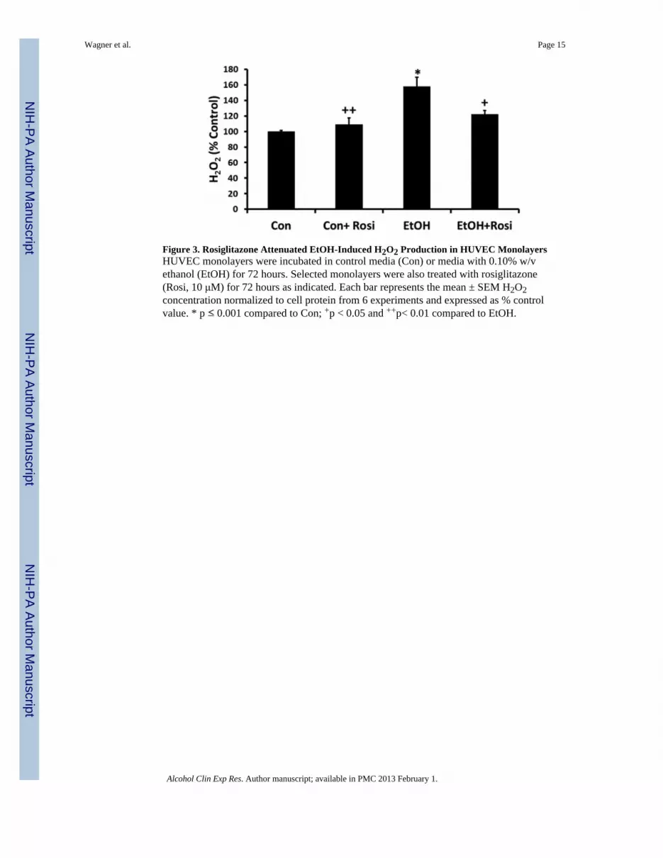
Figure 3. Rosiglitazone Attenuated EtOH-Induced H2O2 Production in HUVEC MonolayersHUVEC monolayers were incubated in control media (Con) or media with 0.10% w/vethanol (EtOH) for 72 hours. Selected monolayers were also treated with rosiglitazone(Rosi, 10 μM) for 72 hours as indicated. Each bar represents the mean ± SEM H2O2concentration normalized to cell protein from 6 experiments and expressed as % controlvalue. * p ≤ 0.001 compared to Con; +p < 0.05 and ++p< 0.01 compared to EtOH.
Wagner et al. Page 15
Alcohol Clin Exp Res. Author manuscript; available in PMC 2013 February 1.
NIH
-PA Author Manuscript
NIH
-PA Author Manuscript
NIH
-PA Author Manuscript

Figure 4. Rosiglitazone Attenuated EtOH-induced Increases in Lung eNOS Protein Levels inMice and mRNA eNOS Levels in HUVEC CultureA) Selected mice were treated with ethanol (EtOH, 20% w/v) in drinking water for 12weeks, with or without rosiglitazone (Rosi) by gavage (10 mg/kg) during the 12th week oftreatment. Each bar represents the mean densitometric intensity of eNOS protein in the lungrelative to CDK4 levels in the same sample ± SEM expressed as % Control. Blots arerepresentative from 6–9 mice. * p ≤ 0.001 compared to Con; +p ≤ 0.01 compared to EtOH.B) HUVEC monolayers were grown to confluency on 100 mm plastic tissue culture dishesand treated with ethanol (0.10% w/v) for 72 hours ± rosiglitazone (10 μM). Monolayerswere homogenized, and mRNA was extracted and analyzed via RT-PCR. Results arenormalized to 9s levels and expressed as % control for n=3. * p ≤ 0.05 compared to Con; +p≤ 0.05 compared to EtOH.
Wagner et al. Page 16
Alcohol Clin Exp Res. Author manuscript; available in PMC 2013 February 1.
NIH
-PA Author Manuscript
NIH
-PA Author Manuscript
NIH
-PA Author Manuscript

Figure 5. Rosiglitazone Attenuated EtOH-induced Increases in Nox1 and 4 Protein Levels inMouse Lung and HUVEC CulturesSelected mice were treated with ethanol (EtOH, 20% w/v) in drinking water for 12 weeks,with or without rosiglitazone (Rosi) by gavage (10 mg/kg) during the 12th week oftreatment. HUVEC monolayers were incubated in control media (Con) or media with 0.10%w/v ethanol (EtOH) for 72 hours. Where indicated, selected monolayers were treated withrosiglitazone (Rosi, 10 μM). Lung tissue (A,B) or harvested HUVEC cultures (C,D) werehomogenized, and protein was extracted and analyzed via western blotting. Each barrepresents the mean densitometric intensity of Nox1 or Nox4 relative to GAPDH levels inthe same sample ± SEM expressed as % Control. Blots are representative from n=3–4. * p ≤0.05 compared to Con; +p ≤ 0.05 compared to EtOH.
Wagner et al. Page 17
Alcohol Clin Exp Res. Author manuscript; available in PMC 2013 February 1.
NIH
-PA Author Manuscript
NIH
-PA Author Manuscript
NIH
-PA Author Manuscript

Figure 6. Rosiglitazone Attenuated EtOH-induced Increases in Nox1 and 4 mRNA Levels inMouse Lung and HUVEC CulturesSelected mice were treated with ethanol (EtOH, 20% w/v) in drinking water for 12 weeks,with or without rosiglitazone (Rosi) by gavage (10 mg/kg) during the 12th week oftreatment. HUVEC monolayers were incubated in control media (Con) or media with 0.10%w/v ethanol (EtOH) for 72 hours. Where indicated, selected monolayers were treated withrosiglitazone (Rosi, 10μM). Lung tissue or HUVEC monolayers were homogenized, andmRNA was extracted and analyzed via RT-PCR. Results are normalized to 9s levels andexpressed as % control for A) Nox1 lung tissue (n=8–9), B) Nox1 HUVEC (n=3), C) Nox2lung tissue (n=8–11), D) Nox2 HUVEC (n=3), E) Nox4 lung tissue (n=8–11), and F) Nox4HUVEC (n=3). * p ≤ 0.05 compared to Con;+ p ≤ 0.05 compared to EtOH.
Wagner et al. Page 18
Alcohol Clin Exp Res. Author manuscript; available in PMC 2013 February 1.
NIH
-PA Author Manuscript
NIH
-PA Author Manuscript
NIH
-PA Author Manuscript

Figure 7. Rosiglitazone Attenuated LPS-Mediated Increases in Nox1, Nox2, Nox4 and eNOSmRNA levels in Mouse LungC57Bl/6 mice were treated with either rosiglitazone (RSG, 10 mg/kg) or vehicle by gavagedaily for 7 days. Selected animals were then treated with two IP injections of E.colilipopolysaccharide (LPS, 2 mg/kg) at 6 and 3 hours prior to sacrifice. After sacrifice, lungtissue was homogenized, and mRNA was extracted and analyzed via RT-PCR. Results arenormalized to 9s levels and expressed as fold-change relative to lungs from animals lackingeither LPS or RSG treatment. Each bar represents the mean ± SEM fold-change in Nox1,Nox2, Nox4, or eNOS mRNA levels from 4 animals in each group. * p ≤ 0.05 compared toall other groups.
Wagner et al. Page 19
Alcohol Clin Exp Res. Author manuscript; available in PMC 2013 February 1.
NIH
-PA Author Manuscript
NIH
-PA Author Manuscript
NIH
-PA Author Manuscript