non-dna binding, dominant-negative, human pparγ mutations cause lipodystrophic insulin resistance
TRANSCRIPT

Non-DNA binding, dominant-negative, human PPARγ mutationscause lipodystrophic insulin resistance
Maura Agostini1,12, Erik Schoenmakers1,12, Catherine Mitchell1, Istvan Szatmari3, DavidSavage2, Aaron Smith1, Odelia Rajanayagam1, Robert Semple2, Jian'an Luan4, LouiseBath5, Anthony Zalin6, Mourad Labib6, Sudhesh Kumar7, Helen Simpson1, Dirk Blom8,David Marais8, John Schwabe9, Inês Barroso10, Richard Trembath11, Nicholas Wareham4,Laszlo Nagy3, Mark Gurnell1, Stephen O'Rahilly1,2, and Krishna Chatterjee1∗1Department of Medicine, University of Cambridge, United Kingdom.
2Department of Clinical Biochemistry, University of Cambridge, United Kingdom.
3Department of Biochemistry and Molecular Biology, University of Debrecen, Hungary.
4Medical Research Council Epidemiology Unit, Cambridge, United Kingdom.
5Royal Hospital for Sick Children, Edinburgh, United Kingdom.
6Wordsley Hospital, Stourbridge, United Kingdom.
7Department of Medicine, University of Warwick, Coventry, United Kingdom.
8Department of Internal Medicine, University of Cape Town, South Africa.
9Medical Research Council, Laboratory of Molecular Biology, Cambridge, United Kingdom.
10Metabolic Disease Group, Wellcome Trust Sanger Institute, Cambridgeshire, United Kingdom.
11Department of Medical and Molecular Genetics, King's College, London, United Kingdom.
SummaryPPARγ is essential for adipogenesis and metabolic homeostasis. We describe mutations in the DNAand ligand binding domains of human PPARγ in lipodystrophic, severe insulin resistance. Thesereceptor mutants lack DNA binding and transcriptional activity but can translocate to the nucleus,interact with PPARγ coactivators and inhibit coexpressed wild-type receptor. Expression ofPPARγ target genes is markedly attenuated in mutation-containing versus receptor haploinsufficentprimary cells, indicating that such dominant-negative inhibition operates in vivo. Our observationssuggest that these mutants restrict wild-type PPARγ action via a non-DNA binding, transcriptionalinterference mechanism, which may involve sequestration of functionally limiting coactivators.
IntroductionThe nuclear receptor (NR) peroxisome proliferator-activated receptor γ (PPARγ) is a ligand-inducible transcription factor that is essential for adipocyte differentiation (Tontonoz et al.,1994b; Barak et al., 1999; Rosen et al., 1999). Alternative splicing and differential promoter
© 2006 Elsevier Inc.∗Corresponding author [email protected] authors contributed equally to this work.This document was posted here by permission of the publisher. At the time of deposit, it included all changes made during peer review,copyediting, and publishing. The U.S. National Library of Medicine is responsible for all links within the document and for incorporatingany publisher-supplied amendments or retractions issued subsequently. The published journal article, guaranteed to be such by Elsevier,is available for free, on ScienceDirect.
Sponsored document fromCell Metabolism
Published as: Cell Metab. 2006 October ; 4(4): 303–311.
Sponsored Docum
ent Sponsored D
ocument
Sponsored Docum
ent
usage generates two protein isoforms: PPARγ2, expressed from a single γ2 promoter, containsan additional 28 amino-terminal amino acids and is nearly adipose-specific; PPARγ1, whoseexpression can be regulated by multiple (γ1, γ3, γ4) promoters, is more ubiquitously distributed.In addition to adipogenesis, PPARγ also plays an important role in adipocyte lipid metabolism,regulating target genes (lipoprotein lipase, fatty-acid transport protein, aquaporin) that mediatetriglyceride hydrolysis and fatty acid and glycerol uptake, together with genes (acylCoAsynthetase, PEPCK, glycerol kinase) involved in fatty acid re-esterification and lipid storage(Lehrke and Lazar, 2005; Savage, 2005). The thiazolidinedione (TZD) class of antidiabeticagents are synthetic, high-affinity PPARγ ligands (Lehmann et al., 1995) and putativeendogenous activators include fatty acids, eicosanoids, and prostaglandin derivatives(Desvergne and Wahli, 1999) as well as undefined ligands produced during adipocytedifferentiation (Tzameli et al., 2004).
The most common population genetic variant of PPARγ is a polymorphism replacing alaninefor proline at codon 12 (Pro12Ala) in PPARγ2, with a meta-analysis of association studiesshowing that the Pro allele confers a modest but significant increase in diabetes risk (Altshuleret al., 2000). The discovery that PPARγ is a target for TZDs, which act by enhancing tissueinsulin sensitivity, prompted screening of a cohort of subjects with severe insulin resistance,with identification of two missense PPARγ mutations (P467L, V290M) in unrelated cases(Barroso et al., 1999). Functional studies showed that these mutant receptors retain DNAbinding but exhibit significant impairment of transcriptional activation and coactivatorrecruitment in response to different ligands (Barroso et al., 1999; Agostini et al., 2004), due tothe mutations destabilizing the carboxyterminal α helix of PPARγ (Kallenberger et al., 2003),which mediates these functions. Consonant with heterozygosity in affected subjects anddominant inheritance in one kindred, the P467L and V290M mutant receptors inhibited thetranscriptional activity of wild-type (WT) PPARγ in a dominant-negative manner (Barrosoet al., 1999). Subsequently, two further heterozygous mutations in the ligand binding domain(LBD) of PPARγ (R425C; F388L) have been described, with recognition that in addition toinsulin resistance the phenotype also includes a stereotyped pattern of partial lipodystrophy(PLD) (Hegele et al., 2002; Agarwal and Garg, 2002; Savage et al., 2003).
Following this, we described several individuals who were heterozygous for a frameshift/premature stop codon mutation, ([A553ΔAAAiT]fs.185[stop186]-hereafter abbreviated toFSX) in the DNA binding domain (DBD) of PPARγ, with this truncation mutant lacking DNAbinding, transcriptional, and dominant-negative activity. Significantly, heterozygosity for theFSX mutation alone was not associated with insulin resistance, but individuals who weredoubly heterozygous, with an additional defect in an unrelated gene encoding the muscle-specific regulatory subunit of protein phosphatase 1 (PPP1R3A), exhibited severe insulinresistance (Savage et al., 2002). Heterozygosity for a single nucleotide substitution in thepromoter of human PPARγ4 leading to its altered expression in vitro has been associated withPLD and insulin resistance in one family, but the authors did not exclude the possibility ofinteraction with a defect at a second genetic locus to produce this phenotype (Al-Shali et al.,2004).
Here, we describe the identification of five heterozygous human PPARγ mutations (C114R,C131Y, C162W, R357X, [A935ΔC]fs.312[stop315]-hereafter abbreviated to FS315X) notassociated with a PPP1R3A gene defect, in unrelated cases of lipodystrophic insulin resistanceand show that these mutants inhibit WT receptor action via a non-DNA binding, dominant-negative mechanism.
Agostini et al. Page 2
Published as: Cell Metab. 2006 October ; 4(4): 303–311.
Sponsored Docum
ent Sponsored D
ocument
Sponsored Docum
ent

Results and DiscussionHeterozygous PPARγ mutations are associated with lipodystrophic insulin resistance
The case histories (see the Supplemental Data available with this article online) andcharacterization (Table 1) of index subjects (S1–S5) harboring PPARγ mutations indicate manyof the features associated with previously described cases (Barroso et al., 1999; Hegele et al.,2002; Agarwal and Garg, 2002; Savage et al., 2003). All subjects showed marked fastinghyperinsulinaemia (Table 1) with acanthosis nigricans in a subset (S3, S4, S5), denoting severeinsulin resistance; total body fat was reduced in all individuals, and imaging indicated astereotyped pattern of partial lipodystrophy affecting gluteal (Figure S1) and peripheral limbdepots; hepatic steatosis and marked dyslipidaemia (raised triglycerides, low high-densitylipoprotein cholesterol [HDL-C]) with secondary complications (cutaneous eruptivexanthomata S3, S4; pancreatitis S5) were features of all cases; several individuals (S2, S3, S5)exhibited early-onset hypertension.
We sequenced the γ4 promoter, coding exons and splice junctions of PPARG and identifiedheterozygous, missense mutations in the DBD (S1–S3), or premature stop mutations in theLBD (S4, S5) of the receptor in index cases. PPARG has also been sequenced by us in 215additional subjects, comprising 93 patients from our severe insulin resistance cohort (Barrosoet al., 1999), 48 CEPH individuals of European descent and 27 Europid, hyperinsulinaemicparticipants in the Ely study (Williams et al., 1995), and 47 controls from four different ethnicgroups, or sequenced by others in 24 African and 23 CEPH European individuals (Seattle SNPsproject, http://pga.gs.washington.edu), and other than the Pro12Ala polymorphism neitherthese or other mutations have been identified. We also sequenced PPP1R3A in each probandand identified no mutations or polymorphisms, excluding a second genetic defect at this locusas described previously (Savage et al., 2002).
Heterozygosity for PPARγ mutations in a parent and grandparent of S3 and a parent of S5segregated with phenotype, constituting a dominant inheritance pattern in two families; onesibling of S2 with dyslipidaemia and insulin resistance was heterozygous for the PPARγmutation whereas another genetically unaffected sibling was biochemically normal; theascertainable family members of S1 were unaffected and normal and no relatives of S4 couldbe contacted (Figure 1B).
PPARγ mutants fail to bind DNA and are transcriptionally inactiveThree missense mutations involve highly conserved cysteine residues within (C114R, C131Y,C162W) the DBD and two further nonsense (R357X) or frameshift/premature stop (FS315X)mutations truncate the receptor within the central part of its LBD (Figure 1A), predicting loss-of-function of the mutant proteins. We therefore characterised and compared the properties ofthese PPARγ mutants with the FSX mutant described previously (Savage et al., 2002).
The receptor mutants exhibited negligible transcriptional activity, lacking constitutive basalactivity noted previously with WT PPARγ (Agostini et al., 2004; Zamir et al., 1997) as wellas any response to rosiglitazone, a TZD receptor agonist (Figure 1C). Such complete loss offunction was similar to the FSX mutant and might be anticipated with analogous truncationmutants (FS315X, R357X) not possessing the transactivation (AF2) domain at the receptorcarboxyterminus (Figure 1A) (Zamir et al., 1997; Wu et al., 2003), but the lack of functionwith DBD mutants (C114R, C131Y, C162W), prompted further investigation of their DNAbinding properties.
PPARγ heterodimerizes with the retinoid X receptor (RXR) and this complex has been shownto bind a DNA response element (PPARE), consisting of a direct repeat (DR1) of the consensussequence (AGGTCA) separated by a single nucleotide (Ijpenberg et al., 1997) and a recent
Agostini et al. Page 3
Published as: Cell Metab. 2006 October ; 4(4): 303–311.
Sponsored Docum
ent Sponsored D
ocument
Sponsored Docum
ent
study has suggested that the stringency of PPARγ binding to some response elements isrelatively relaxed, not needing complete integrity of its DBD (Temple et al., 2005). A rangeof previously documented or predicted PPAREs from known target genes were therefore testedin electrophoretic mobility shift assays and both DBD and LBD truncation receptor mutantsshowed negligible heterodimeric binding (Figure 1D). To examine interaction of mutantreceptors with RXR, we coexpressed VP16-full length PPARγ fusions with Gal4DBD-RXRin a mammalian two-hybrid assay. In keeping with preservation of the dimerization interface(Gampe et al., 2000) within their intact LBD (Figure 1A), the DBD mutants interacted readilywhereas the FS315X, R357X, and FSX mutants lacking this interface failed to be recruited toGal4-RXR (Figure S2). It was therefore conceivable that the DBD mutants could be recruitedindirectly to a PPARE by binding RXR (Gampe et al., 2000), or conversely, that the LBDtruncation mutants might bind a PPARE monomerically as has been documented with thethyroid hormone receptor (TR) (Lazar et al., 1991). However, unlike WT receptor, VP16-fulllength, mutant PPARγ fusions were unable to activate a PPARE-containing reporter gene(Figure S3), indicating that like FSX, these PPARγ mutants do not bind DNA directly orindirectly.
PPARγ mutants translocate to the nucleus and interact with cofactorsThe intracellular localization of WT PPARγ is predominantly nuclear (Akiyama et al., 2002)and, analogous to steroid/thyroid hormone receptors, may be dependent on a putative nuclearlocalisation signal (NLS) located between its DBD and LBD (Figure 1A) (Guiochon-Manteland Milgrom, 1993; Zhu et al., 1998). Studies of GFP- PPARγ fusions showed that, in keepingwith preservation of the putative NLS, both DBD and LBD truncation mutants localized to thenucleus comparably to WT, whereas the FSX truncation mutant, which lacks this sequence,remained cytoplasmic similar to GFP alone (Figure 1E).
We next examined whether the PPARγ mutants might also retain the ability to interact withtranscriptional coactivators. Steroid receptor coactivator-1 (SRC1/NCoA1) (Onate et al.,1995) and PPARγ binding protein/thyroid receptor-associated protein 220 (PBP/TRAP220)interact directly with the AF2 domain of PPARγ, with the latter cofactor being required forreceptor-mediated adipogenesis (Zhu et al., 1996, 1997; Ge et al., 2002). Consistent withpreservation of their AF2 domains, protein-protein interaction assays showed ligand-dependent binding of SRC1 or TRAP220 to the DBD mutants, but no specific interaction withFSX or LBD truncation mutants, which lack this region (Figure 1F). Conversely, wehypothesized that the PPARγ LBD truncation mutants would retain the ability to recruitcoactivators, which can interact with receptor independently of its AF2 domain. PPARγcoactivator-1 (PGC1), which augments receptor action in fat cells (Puigserver and Spiegelman,2003), can bind PPARγ via its DBD and hinge region (αα 128-229) (Puigserver et al., 1999);PDIP, isolated in a two hybrid assay using the DBD/hinge region of PPARγ (Tomaru et al.,2006), is a coactivator that also enhances PPARα activity (Surapureddi et al., 2002). BothPGC1 and PDIP bound WT or FS315X and R357X LBD truncation mutants in protein-proteininteraction assays, whereas the FSX mutant showed negligible interaction (Figure 1G).
PPARγ signaling is reduced in mutation-containing primary cells ex vivo or mutant-expressing cells in vitro
The observation that these PPARγ mutants translocate to the nucleus and interact withcoactivators raised the possibility that they might interfere with WT receptor signaling. Themurine adipocyte P2 (aP2) gene is a classical target of PPARγ action (Tontonoz et al., 1994a;Guan et al., 2005) and the human homolog (FABP4) is similarly responsive (Pelton et al.,1999). When coexpressed with WT PPARγ at equivalent levels in 3T3-L1 adipocytes, the DBDand LBD mutants blocked WT receptor mediated activation of the human aP2/FABP4 genepromoter comparably to an artificial, dominant-negative PPARγ mutant (AF2) described
Agostini et al. Page 4
Published as: Cell Metab. 2006 October ; 4(4): 303–311.
Sponsored Docum
ent Sponsored D
ocument
Sponsored Docum
ent
previously (Gurnell et al., 2000), whereas FSX lacked dominant-negative inhibitory activity(Figure 2A).
We wished to determine whether such divergent dominant-negative inhibition by thesePPARγ mutants versus FSX might operate in vivo. PPARγ is highly expressed in immaturedendritic cells (IDCs) derived from primary human blood monocytes and mediates markedreceptor responsiveness, with strong ligand-dependent induction of aP2 expression in thesecells (Szatmari et al., 2004). Induction of aP2/FABP4 expression in IDCs containing DBD orLBD PPARγ mutations was severely attenuated compared to responses in control cells fromeither normal individuals (WT) or from subjects (IR) with comparable insulin resistancewithout a PPARγ gene defect. Significantly, aP2 induction in FSX mutation-containing cellswas comparable to responses from control subjects (Figure 2B). We examined other PPARγtarget genes, identified from extensive microarray profiling of normal IDCs (I.S. and L.N.,unpublished data) and found that responses to PPARγ agonist in DBD and LBD truncationmutation-containing cells were markedly attenuated whereas FSX mutation-containing cellsexhibited responses that were either similar or only slightly reduced compared to WT cells(Figure 2C). PPARγ mRNA levels in control and mutation-containing primary cells weresimilar (data not shown), suggesting that differential responsiveness was not due to alteredreceptor expression. Furthermore, PPARγ mRNA from both WT and R357X alleles wasexpressed in mutation-containing IDCs (Figure 2D), indicating that the R357X transcript isnot subject to nonsense-mediated decay (Culbertson, 1999) and both WT and R357X mutantPPARγ proteins were also expressed in these cells (Figure 2E).
Finally, we determined whether dominant-negative inhibition by a non-DNA bindingPPARγ mutant could interfere with a receptor-mediated biological process. Compared tocontrol, WT PPARγ or GFP adenovirus-transduced human preadipocyte cells, both cellulardifferentiation (Figure 3A) and aP2 gene expression (Figure 3B) in cells transduced withC114R mutant PPARγ adenovirus were significantly reduced.
Transcriptional interference via a non-DNA binding mechanismWe have shown previously that dominant-negative inhibition by PPARγ mutants (P467L,V290M), is mediated by repression of target genes by DNA-bound mutant receptors, analogousto mechanisms of other mutant nuclear receptors (e.g., the v-erbA oncogene, TRβ mutants inResistance to Thyroid Hormone, PZLF-RARα fusion proteins in promyelocytic leukaemia)(Love et al., 2000). In contrast, the missense DBD and LBD truncation mutants identified hereare unable to bind DNA, yet can inhibit WT PPARγ action, suggesting a different mechanismof transcriptional interference. Competition for shared cofactors by NRs was postulated toexplain mutual antagonism of progesterone and estrogen receptor signaling (Meyer et al.,1989) and the subsequent observation that SRC1, a shared coactivator, could relieve such“squelching”, validated this hypothesis (Onate et al., 1995). Ligand-activated NRs have beenshown to inhibit either their own function (Barettino et al., 1994) or that of heterologousreceptors (Zhang et al., 1996) by limiting the availability of coactivators that are recruited totheir transactivation domains. Our observations indicate that non-DNA binding, dominant-negative PPARγ mutants can recruit coactivators, suggesting an analogous cofactorsequestration mechanism for thereby restricting WT receptor function. Evidence suggests thatsimilar mechanisms operate to inhibit PPAR signaling in other contexts: analogous to ournatural DBD mutants, others have generated artificial, dominant-negative, PPARγ DBDmutants, which block either adipogenesis (Park et al., 2003) or neural stem cell differentiation(Wada et al., 2006); γORF4 is a newly identified human PPARγ splice variant with a truncatedLBD (αα273), which has dominant-negative activity and is selectively overexpressed incolorectal neoplasia (Sabatino et al., 2005); a dominant-negative PPARα splice variant with atruncated LBD (αα 174), is expressed in human tissues including liver (Gervois et al., 1999).
Agostini et al. Page 5
Published as: Cell Metab. 2006 October ; 4(4): 303–311.
Sponsored Docum
ent Sponsored D
ocument
Sponsored Docum
ent
Interestingly, heterozygous, non-DNA binding mutations in some nuclear receptors do notmediate a phenotype: mutations in the DBD of VDR only cause vitamin D resistance in thehomozygous state (Malloy et al., 1999); a “knock-in” mutation in the DBD of murine TRβdoes not produce thyroid hormone resistance (Shibusawa et al., 2003). Possibly due to itspivotal role in regulating transcription of genes mediating both adipocyte formation andfunction (Lehrke and Lazar, 2005), we suggest that PPARγ signaling may be particularlysensitive to interference via the postulated “squelching” mechanism, with deleteriousmetabolic consequences. A corollary of this may be that even modest enhancement of normalreceptor activity in key tissues could be beneficial, supporting attempts to develop partial ortissue-specific PPARγ agonists (Reginato et al., 1998; Rocchi et al., 2001; Berger et al.,2003).
Experimental proceduresSequencing of PPARγ and PPP1R3A genes
The PPP1R3A (exons 1-4) and PPARγ (exons 1-6, B and promoter region of PPARγ4) geneswere amplified using specific primers (available upon request) and sequenced as decribedpreviously (Savage et al., 2002).
Construction of PPARγ mutants and other vectorsFull length WT and mutant PPARγ1 cDNAs were cloned in pGEX4T (Amersham PharmaciaBiotech), pCMX-VP16 (kind gift from R. Evans), pSG424 (Sadowski and Ptashne, 1989) andpEGFP-C1 (Clontech), to yield GST-PPARγ1, VP16-PPARγ1, Gal4DBD-PPARγ and GFP-PPARγ1 fusions respectively.
Electrophoretic mobility shift assaysElectrophoretic mobility shift assays (EMSA) were performed as described (Collingwoodet al., 1994) with different natural PPAREs: aP2, derived by alignment of human and murinepromoter sequences (Graves et al., 1992); Adiponectin (Iwaki et al., 2003): ACoABP (Helledieet al., 2002); mCPT1, (Mascaro et al., 1998); LXRα, (Laffitte et al., 2001); CAP1, (Baumannet al., 2000); LPL, (Schoonjans et al., 1996); ACoAOx, (Varanasi et al., 1996); ACoAOx(Zamir et al., 1997).
Transfection assays293EBNA cells, cultured in DMEM/10%FCS were transfected with Lipofectamine2000- orcalcium phosphate-mediated in 96- or 24-well plates respectively and assayed for luciferaseand β-galactosidase activity as described (Collingwood et al., 1994) following 36 hr with orwithout ligand. 3T3-L1 adipocyte cells were cultured and transfected with Lipofectamine2000in 24-well plates as described above.
Cellular localisation of EGFP-tagged mutants293EBNA cells, grown on glass well slides were transfected using Lipofectamine 2000 with1μg of EGFP-PPARγ1 fusions, fixed with 4% paraformaldehyde, mounted using vectashieldand fluorescence was visualized by digital microscopy.
Peripheral blood monocyte purification and IDC cultureWith ethical approval, monocytes were harvested from peripheral blood by Ficoll gradientcentrifugation and immunomagnetic cell separation using anti-CD14-conjugated microbeads(VarioMACS; Miltenyi Biotec), resuspended in 6-well plates at a density of 1.5 × 10⁶ cells/ml and cultured in RPMI 1640 plus 10% FBS containing 800U/ml GM-CSF (Leucomax) and
Agostini et al. Page 6
Published as: Cell Metab. 2006 October ; 4(4): 303–311.
Sponsored Docum
ent Sponsored D
ocument
Sponsored Docum
ent

500U/ml IL-4 (Peprotech) to generate IDCs as described (Sallusto and Lanzavecchia, 1994)with or without exposure to ligand for 24 hr.
Quantitative real-time PCR analysis of gene expression100ng of total RNA from IDCs, isolated using TRIZOL (Invitrogen), was reverse transcribedand analyzed by Taqman quantitative real-time PCR (qPCR) as described (Szatmari et al.,2004). The sequences of primers and probes are available upon request.
Taqman qPCR low density arrays (TLDA) were used to quantify the expression of multipletarget genes in IDCs, according to the manufacturer's instructions.
To obtain cDNA, RNA was reverse transcribed using a High Capacity cDNA Archive kit(Applied Biosystems). The following commercially available Taqman assays (AppliedBiosystems) were used: ADRP/ADFP (Hs00605340_m1), APOC1 (Hs00155790_m1),CLDN1 (Hs00221623_m1), aP2/FABP4 (Hs00609791_m1), CLECSF5 (Hs00183780_m1),CD1E (Hs00229421_m1), MYO1B (Hs00362654_m1), IL1R2 (Hs00174759_m1), OAS1(Hs00242943_m1), p30 (Hs00396457_m1), cyclophilinA/PPIA (Hs99999904_m1). Thecomparative Ct method was used to quantify transcripts and normalize to cyclophilinAexpression levels, which did not vary with ligand treatment. Thereafter, data were furthernormalized to expression levels in ligand-treated WT IDC samples using GeneSpring 7.2software (Agilent).
RFLP analysis of PPARγ transcriptsPPARγ cDNAs were amplified from WT or R357X mutation-containing IDCs by RT-PCRusing forward (CTCCTTGATGAATAAAGATGGGG) and reverse(ATGTCTTCAATGGGCTTCACAT) primers, the PCR products were digested with Cac8Ienzyme (New England Biolabs) and analyzed by agarose gel electrophoresis.
Immunoprecipitation and Western blot analysisIDCs, harvested from 200ml of peripheral blood, were lysed in RIPA buffer containing aprotease inhibitor cocktail (Roche) and cell supernatants immunoprecipitated using a mousemonoclonal anti-PPARγ antibody (K8713, Perseus Proteomics) and analyzed by SDS-PAGE.Western blotting was carried out using a rabbit polyclonal anti-PPARγ antibody (H-100, SantaCruz Biotechnology).
Adenovirus construction and expressionRecombinant type 5 adenoviruses (Ad5) expressing GFP alone or with either WT or C114Rmutant PPARγ1 were generated using the AdEasy Vector System (Quantum Biotechnologies,Montreal), amplified and purified as described (Gurnell et al., 2000). 6-well plates of Chub-S7 human preadipocyte cells were cultured and infected with 2x10⁷ pfu/well of recombinantvirus 24 hr prior to differentiation in the presence of 100nM rosiglitazone as described(Darimont et al., 2003). Comparable infection efficiency was verified by fluorescencemicroscopy with subsequent qPCR analysis on days 0, 3, 5 and 7. Fully differentiated cellswere fixed and stained with Oil Red-O as described (Adams et al., 1997).
Supplemental dataRefer to Web version on PubMed Central for supplementary material.
Agostini et al. Page 7
Published as: Cell Metab. 2006 October ; 4(4): 303–311.
Sponsored Docum
ent Sponsored D
ocument
Sponsored Docum
ent
Acknowledgments
We would like to thank the Imaging Department (Royal Orthopaedic Hospital, Stanmore) for supplying control MRIdata and Teturou Satoh (Gunma University) for providing PDIP1α ahead of publication. This work was supported bythe Wellcome Trust (V.K.K.C., S.O.R., L.N., I.B., D.B.S.) and the Medical Research Council (N.J.W., J.W.R.S.).
ReferencesAdams et al, 1997. Adams M. Reginato M.J. Shao D. Lazar M.A. Chatterjee V.K.K. Transcriptional
activation by peroxisome proliferator-activated receptor γ is inhibited by phosphorylation at aconsensus mitogen-activated protein kinase site. J. Biol. Chem. 1997;272:5128–5132. [PubMed:9030579]
Agarwal and Garg, 2002. Agarwal A.K. Garg A. A novel heterozygous mutation in peroxisomeproliferator-activated receptor-gamma gene in a patient with familial partial lipodystrophy. J. Clin.Endocrinol. Metab. 2002;87:408–411. [PubMed: 11788685]
Agostini et al, 2004. Agostini M. Gurnell M. Savage D.B. Wood E.M. Smith A.G. Rajanayagam O.Garnes K.T. Levinson S.H. Xu H.E. Schwabe J.W. Tyrosine agonists reverse the molecular defectsassociated with dominant-negative mutations in human peroxisome proliferator-activated receptorgamma. Endocrinology 2004;145:1527–1538. [PubMed: 14657011]
Akiyama et al, 2002. Akiyama T.E. Baumann C.T. Sakai S. Hager G.L. Gonzalez F.J. Selectiveintranuclear redistribution of PPAR isoforms by RXR alpha. Mol. Endocrinol. 2002;16:707–721.[PubMed: 11923467]
Al-Shali et al, 2004. Al-Shali K. Cao H. Knoers N. Hermus A.R. Tack C.J. Hegele R.A. A single-basemutation in the peroxisome proliferator-activated receptor gamma4 promoter associated withaltered in vitro expression and partial lipodystrophy. J. Clin. Endocrinol. Metab. 2004;89:5655–5660. [PubMed: 15531525]
Altshuler et al, 2000. Altshuler D. Hirschhorn J.N. Klannemark M. Lindgren C.M. Vohl M.C. NemeshJ. Lane C.R. Schaffner S.F. Bolk S. Brewer C. The common PPARgamma Pro12Ala polymorphismis associated with decreased risk of type 2 diabetes. Nat. Genet. 2000;26:76–80. [PubMed:10973253]
Barak et al, 1999. Barak Y. Nelson M.C. Ong E.S. Jones Y.Z. Ruiz-Lozano P. Chien K.R. Koder A.Evans R.M. PPARγ is required for placental, cardiac and adipose tissue development. Mol. Cell1999;4:585–595. [PubMed: 10549290]
Barettino et al, 1994. Barettino D. Vivanco Ruiz M.D.M. Stunnenberg H.G. Characterization of theligand-dependent transactivation domain of thyroid hormone receptor. EMBO J. 1994;13:3039–3049. [PubMed: 8039499]
Barroso et al, 1999. Barroso I. Gurnell M. Crowley V.E.F. Agostini M. Schwabe J.W. Soos M.A. MaslenG.L.I. Williams T.D.M. Lewis H. Schafer A.J. Dominant negative mutations in human PPARγ areassociated with severe insulin resistance, diabetes mellitus and hypertension. Nature 1999;402:880–883. [PubMed: 10622252]
Baumann et al, 2000. Baumann C.A. Chokshi N. Saltiel A.R. Ribon V. Cloning and characterization ofa functional peroxisome proliferator activator receptor-gamma-responsive element in the promoterof the CAP gene. J. Biol. Chem. 2000;275:9131–9135. [PubMed: 10734046]
Berger et al, 2003. Berger J.P. Petro A.E. Macnaul K.L. Kelly L.J. Zhang B.B. Richards K. Elbrecht A.Johnson B.A. Zhou G. Doebber T.W. Distinct properties and advantages of a novel peroxisomeproliferator-activated protein [gamma] selective modulator. Mol. Endocrinol. 2003;17:662–676.[PubMed: 12554792]
Black et al, 1983. Black D. James W.P.T. Besser G.M. Brock C.G.D. Craddock D. Garrow J.S. HockadayT.D.R. Lewis B. Pilkington T.R.E. Silverstone J.T. Obesity: Areport of the College of Physicians.J. Roy. Coll. Phys. London 1983;17:5–65.
Collingwood et al, 1994. Collingwood T.N. Adams M. Tone Y. Chatterjee V.K.K. Spectrum oftranscriptional dimerization and dominant negative properties of twenty different mutant thyroidhormone β receptors in thyroid hormone resistance syndrome. Mol. Endocrinol. 1994;8:1262–1277.[PubMed: 7838159]
Culbertson, 1999. Culbertson M.R. RNA surveillance. Unforeseen consequences for gene expression,inherited genetic disorders and cancer. Trends Genet. 1999;15:74–80. [PubMed: 10098411]
Agostini et al. Page 8
Published as: Cell Metab. 2006 October ; 4(4): 303–311.
Sponsored Docum
ent Sponsored D
ocument
Sponsored Docum
ent

Darimont et al, 2003. Darimont C. Zbinden I. Avanti O. Leone-Vautravers P. Giusti V. Burckhardt P.Pfeifer A.M. Mace K. Reconstitution of telomerase activity combined with HPV-E7 expressionallow human preadipocytes to preserve their differentiation capacity after immortalization. CellDeath Differ. 2003;10:1025–1031. [PubMed: 12934077]
Desvergne and Wahli, 1999. Desvergne B. Wahli W. Peroxisome proliferator-activated receptors: nuclearcontrol of metabolism. Endocr. Rev. 1999;20:649–688. [PubMed: 10529898]
Gampe et al, 2000. Gampe J.R.T. Montana V.G. Lambert M.H. Miller A.B. Bledsoe R.K. Milburn M.V.Kliewer S.A. Willson T.M. Xu H.E. Asymmetry in the PPARγ/RXRα crystal structure reveals themolecular basis of heterodimerization among nuclear receptors. Mol. Cell 2000;5:545–555.[PubMed: 10882139]
Ge et al, 2002. Ge K. Guermah M. Yuan C.X. Ito M. Wallberg A.E. Spiegelman B.M. Roeder R.G.Transcription coactivator TRAP220 is required for PPAR gamma 2-stimulated adipogenesis.Nature 2002;417:563–567. [PubMed: 12037571]
Gervois et al, 1999. Gervois P. Torra I.P. Chinetti G. Grotzinger T. Dubois G. Fruchart J.C. Fruchart-Najib J. Leitersdorf E. Staels B. A truncated human peroxisome proliferator-activated receptor alphasplice variant with dominant negative activity. Mol. Endocrinol. 1999;13:1535–1549. [PubMed:10478844]
Graves et al, 1992. Graves R.A. Tontonoz P. Platt K.A. Ross S.R. Spiegelman B.M. Identification of afat cell enhancer: analysis of requirements for adipose tissue-specific gene expression. J. Cell.Biochem. 1992;49:219–224. [PubMed: 1644859]
Guan et al, 2005. Guan H.P. Ishizuka T. Chui P.C. Lehrke M. Lazar M.A. Corepressors selectively controlthe transcriptional activity of PPARgamma in adipocytes. Genes Dev. 2005;19:453–461. [PubMed:15681609]
Guiochon-Mantel and Milgrom, 1993. Guiochon-Mantel A. Milgrom E. Cytoplasmic-nuclear traffickingof steroid hormone receptors. Tr Endocrinol Metab 1993;10:322–328.
Gurnell et al, 2000. Gurnell M. Wentworth J.M. Agostini M. Adams M. Collingwood T.N. ProvenzanoC. Browne P.O. Rajanayagam O. Burris T.P. Schwabe J.W. A dominant negative PeroxisomeProliferator-activated Receptor γ (PPARγ) mutant is a constitutive repressor and inhibits PPARγ-mediated adipogenesis. J. Biol. Chem. 2000;275:5754–5759. [PubMed: 10681562]
Hegele et al, 2002. Hegele R.A. Cao H. Frankowski C. Mathews S.T. Leff T. PPARG F388L, atransactivation-deficient mutant, in familial partial lipodystrophy. Diabetes 2002;51:3586–3590.[PubMed: 12453919]
Helledie et al, 2002. Helledie T. Grontved L. Jensen S.S. Kiilerich P. Rietveld L. Albrektsen T. BoysenM.S. Nohr J. Larsen L.K. Fleckner J. The gene encoding the Acyl-CoA-binding protein is activatedby peroxisome proliferator-activated receptor gamma through an intronic response elementfunctionally conserved between humans and rodents. J. Biol. Chem. 2002;277:26821–26830.[PubMed: 12015306]
Ijpenberg et al, 1997. Ijpenberg A. Jeannin E. Wahli W. Desvergne B. Polarity and specific sequencerequirements of peroxisome proliferator-activated receptor (PPAR)/retinoid X receptor heterodimerbinding to DNA. A functional analysis of the malic enzyme gene PPAR response element. J. Biol.Chem. 1997;272:20108–20117. [PubMed: 9242684]
Iwaki et al, 2003. Iwaki M. Matsuda M. Maeda N. Funahashi T. Matsuzawa Y. Makishima M. ShimomuraI. Induction of adiponectin, a fat-derived antidiabetic and antiatherogenic factor, by nuclearreceptors. Diabetes 2003;52:1655–1663. [PubMed: 12829629]
Kallenberger et al, 2003. Kallenberger B.C. Love J.D. Chatterjee V.K. Schwabe J.W. A dynamicmechanism of nuclear receptor activation and its perturbation in a human disease. Nat. Struct. Biol.2003;10:136–140. [PubMed: 12536206]
Laffitte et al, 2001. Laffitte B.A. Joseph S.B. Walczak R. Pei L. Wilpitz D.C. Collins J.L. Tontonoz P.Autoregulation of the human liver X receptor alpha promoter. Mol. Cell. Biol. 2001;21:7558–7568.[PubMed: 11604492]
Lazar et al, 1991. Lazar M.A. Berrodin T.J. Harding H.P. Differential DNA binding by monomeric,homodimeric, and potentially heteromeric forms of the thyroid hormone receptor. Mol. Cell. Biol.1991;11:5005–5015. [PubMed: 1922030]
Agostini et al. Page 9
Published as: Cell Metab. 2006 October ; 4(4): 303–311.
Sponsored Docum
ent Sponsored D
ocument
Sponsored Docum
ent

Lehmann et al, 1995. Lehmann J.M. Moore L.B. Smith-Oliver T.A. Wilkison W.O. Willson T.M. KliewerS.A. An antidiabetic thiazolidinedione is a high affinity ligand for peroxisome proliferator-activatedreceptor γ (PPARγ). J. Biol. Chem. 1995;270:12953–12956. [PubMed: 7768881]
Lehrke and Lazar, 2005. Lehrke M. Lazar M.A. The many faces of PPARgamma. Cell 2005;123:993–999. [PubMed: 16360030]
Love et al, 2000. Love J.D. Gooch J.T. Nagy L. Chatterjee V.K.K. Schwabe J.W.R. Transcriptionalrepression by nuclear receptors: mechanisms and role in disease. Biochem. Soc. Trans.2000;28:390–396. [PubMed: 10961926]
Malloy et al, 1999. Malloy P.J. Pike J.W. Feldman D. The vitamin D receptor and the syndrome ofhereditary 1,25-dihydroxyvitamin D-resistant rickets. Endocr. Rev. 1999;20:156–188. [PubMed:10204116]
Mascaro et al, 1998. Mascaro C. Acosta E. Ortiz J.A. Marrero P.F. Hegardt F.G. Haro D. Control ofhuman muscle-type carnitine palmitoyltransferase I gene transcription by peroxisome proliferator-activated receptor. J. Biol. Chem. 1998;273:8560–8563. [PubMed: 9535828]
Meyer et al, 1989. Meyer M.E. Gronemeyer H. Turcotte B. Bocquel M.T. Tasset D. Chambon P. Steroidhormone receptors compete for factors that mediate their enhancer function. Cell 1989;57:433–442.[PubMed: 2720778]
Onate et al, 1995. Onate S.A. Tsai S.Y. Tsai M.-J. O'Malley B.W. Sequence and characterization of acoactivator for the steroid hormone receptor superfamily. Science 1995;270:1354–1357. [PubMed:7481822]
Park et al, 2003. Park Y. Freedman B.D. Lee E.J. Park S. Jameson J.L. A dominant negative PPARgammamutant shows altered cofactor recruitment and inhibits adipogenesis in 3T3-L1 cells. Diabetologia2003;46:365–377. [PubMed: 12687335]
Pelton et al, 1999. Pelton P.D. Zhou L. Demarest K.T. Burris T.P. PPARγ activation induces theexpression of the adipocyte fatty acid binding protein gene in human monocytes. Biochem. Biophys.Res. Commun. 1999;261:456–458. [PubMed: 10425206]
Puigserver et al, 1999. Puigserver P. Adelmant G. Wu Z. Fan M. Xu J. O'Malley B. Spiegelman B.M.Activation of PPARgamma coactivator-1 through transcription factor docking. Science1999;286:1368–1371. [PubMed: 10558993]
Puigserver and Spiegelman, 2003. Puigserver P. Spiegelman B.M. Peroxisome proliferator-activatedreceptor-gamma coactivator 1 alpha (PGC-1 alpha): transcriptional coactivator and metabolicregulator. Endocr. Rev. 2003;24:78–90. [PubMed: 12588810]
Reginato et al, 1998. Reginato M.J. Bailey S.T. Krakow S.L. Minami C. Ishii S. Tanaka H. Lazar M.A.A potent antidiabetic thiazolidinedione with unique peroxisome proliferator-activated receptor γ-activating properties. J. Biol. Chem. 1998;273:32679–32684. [PubMed: 9830009]
Rocchi et al, 2001. Rocchi S. Picard F. Vamecq J. Gelman L. Potier N. Zeyer D. Dubuquoy L. Bac P.Champy M.F. Plunket K.D. A unique PPARgamma ligand with potent insulin-sensitizing yet weakadipogenic activity. Mol. Cell 2001;8:737–747. [PubMed: 11684010]
Rosen et al, 1999. Rosen E.D. Sarraf P. Troy A.E. Bradwin G. Moore K. Milstone D.S. Spiegelman B.M.Mortensen R.M. PPAR gamma is required for the differentiation of adipose tissue in vivo and invitro. Mol. Cell 1999;4:611–617. [PubMed: 10549292]
Sabatino et al, 2005. Sabatino L. Casamassimi A. Peluso G. Barone M.V. Capaccio D. Migliore C. BonelliP. Pedicini A. Febbraro A. Ciccodicola A. A novel peroxisome proliferator-activated receptorgamma isoform with dominant negative activity generated by alternative splicing. J. Biol. Chem.2005;280:26517–26525. [PubMed: 15857827]
Sadowski and Ptashne, 1989. Sadowski I. Ptashne M. A vector for expressing GAL4(1-147) fusions inmammalian cells. Nucleic Acids Res. 1989;17:7539. [PubMed: 2798115]
Sallusto and Lanzavecchia, 1994. Sallusto F. Lanzavecchia A. Efficient presentation of soluble antigenby cultured human dendritic cells is maintained by granulocyte/macrophage colony-stimulatingfactor plus interleukin 4 and downregulated by tumor necrosis factor alpha. J. Exp. Med.1994;179:1109–1118. [PubMed: 8145033]
Savage, 2005. Savage D.B. PPARgamma as a metabolic regulator: insights from genomics andpharmacology. Expert Rev. Mol. Med. 2005;2005:1–16. [PubMed: 15673477]
Agostini et al. Page 10
Published as: Cell Metab. 2006 October ; 4(4): 303–311.
Sponsored Docum
ent Sponsored D
ocument
Sponsored Docum
ent

Savage et al, 2002. Savage D.B. Agostini M. Barroso I. Gurnell M. Luan J. Meirhaeghe A. Harding A.H.Ihrke G. Rajanayagam O. Soos M.A. Digenic inheritance of severe insulin resistance in a humanpedigree. Nat. Genet. 2002;31:379–384. [PubMed: 12118251]
Savage et al, 2003. Savage D.B. Tan G.D. Acerini C.L. Jebb S.A. Agostini M. Gurnell M. Williams R.L.Umpleby A.M. Thomas E.L. Bell J.D. Human metabolic syndrome resulting from dominant-negative mutations in the nuclear receptor peroxisome proliferator-activated receptor-gamma.Diabetes 2003;52:910–917. [PubMed: 12663460]
Schoonjans et al, 1996. Schoonjans K. Staels B. Auwerx J. Role of the peroxisome proliferator-activatedreceptor (PPAR) in mediating the effects of fibrates and fatty acids on gene expression. J. LipidRes. 1996;37:907–925. [PubMed: 8725145]
Shibusawa et al, 2003. Shibusawa N. Hashimoto K. Nikrodhanond A.A. Liberman M.C. Applebury M.L.Liao X.H. Robbins J.T. Refetoff S. Cohen R.N. Wondisford F.E. Thyroid hormone action in theabsence of thyroid hormone receptor DNA-binding in vivo. J. Clin. Invest. 2003;112:588–597.[PubMed: 12925699]
Surapureddi et al, 2002. Surapureddi S. Yu S. Bu H. Hashimoto T. Yeldandi A.V. Kashireddy P.Cherkaoui-Malki M. Qi C. Zhu Y.J. Rao M.S. Identification of a transcriptionally active peroxisomeproliferator-activated receptor alpha -interacting cofactor complex in rat liver and characterizationof PRIC285 as a coactivator. Proc. Natl. Acad. Sci. USA 2002;99:11836–11841. [PubMed:12189208]
Szatmari et al, 2004. Szatmari I. Gogolak P. Im J.S. Dezso B. Rajnavolgyi E. Nagy L. Activation ofPPARgamma specifies a dendritic cell subtype capable of enhanced induction of iNKT cellexpansion. Immunity 2004;21:95–106. [PubMed: 15345223]
Temple et al, 2005. Temple K.A. Cohen R.N. Wondisford S.R. Yu C. Deplewski D. Wondisford F.E. Anintact DNA-binding domain is not required for peroxisome proliferator-activated receptor gamma(PPARgamma) binding and activation on some PPAR response elements. J. Biol. Chem.2005;280:3529–3540. [PubMed: 15572375]
Tomaru et al, 2006. Tomaru T. Satoh T. Yoshino S. Ishizuka T. Hashimoto K. Monden T. Yamada M.Mori M. Isolation and characterization of a transcriptional cofactor and its novel isoform that bindthe deoxyribonucleic acid-binding domain of peroxisome proliferator-activated receptor-gamma.Endocrinology 2006;147:377–388. [PubMed: 16239304]
Tontonoz et al, 1994a. Tontonoz P. Hu E. Graves R.A. Budavari A.I. Spiegelman B.M. mPPARγ2: tissue-specific regulator of an adipocyte enhancer. Genes Dev. 1994;8:1224–1234. [PubMed: 7926726]
Tontonoz et al, 1994b. Tontonoz P. Hu E. Spiegelman B.M. Stimulation of adipogenesis in fibroblastsby PPARγ2, a lipid-activated transcription factor. Cell 1994;79:1147–1156. [PubMed: 8001151]
Tzameli et al, 2004. Tzameli I. Fang H. Ollero M. Shi H. Hamm J.K. Kievit P. Hollenberg A.N. FlierJ.S. Regulated production of a peroxisome proliferator-activated receptor-gamma ligand during anearly phase of adipocyte differentiation in 3T3-L1 adipocytes. J. Biol. Chem. 2004;279:36093–36102. [PubMed: 15190061]
Varanasi et al, 1996. Varanasi U. Chu R. Huang Q. Castellon R. Yeldandi A.V. Reddy J.K. Identificationof a peroxisome proliferator-responsive element upstream of the human peroxisomal fatty acylcoenzyme A oxidase gene. J. Biol. Chem. 1996;271:2147–2155. [PubMed: 8567672]
Wada et al, 2006. Wada K. Nakajima A. Katayama K. Kudo C. Shibuya A. Kubota N. Terauchi Y.Tachibana M. Miyoshi H. Kamisaki Y. Peroxisome proliferator-activated receptor gamma -mediated regulation of neural stem cell proliferation and differentiation. J. Biol. Chem.2006;281:12673–12681. [PubMed: 16524877]
Williams et al, 1995. Williams D.R. Wareham N.J. Brown D.C. Byrne C.D. Clark P.M. Cox B.D. CoxL.J. Day N.E. Hales C.N. Palmer C.R. Undiagnosed gluocse intolerance in the community: The Isleof Ely Diabetes Project. Diabet. Med. 1995;12:30–35. [PubMed: 7712700]
Wu et al, 2003. Wu Y. Chin W.W. Wang Y. Burris T.P. Ligand and coactivator identity determines therequirement of the charge clamp for coactivation of the peroxisome proliferator-activated receptorgamma. J. Biol. Chem. 2003;278:8637–8644. [PubMed: 12502716]
Zamir et al, 1997. Zamir I. Zhang J. Lazar M.A. Stoichiometric and steric principles governing repressionby nuclear hormone receptors. Genes Dev. 1997;11:835–846. [PubMed: 9106656]
Agostini et al. Page 11
Published as: Cell Metab. 2006 October ; 4(4): 303–311.
Sponsored Docum
ent Sponsored D
ocument
Sponsored Docum
ent
Zhang et al, 1996. Zhang X. Jeyakumar M. Bagchi M.K. Ligand-dependent cross-talk between steroidand thyroid hormone receptors. Evidence for common transcriptional coactivator(s). J. Biol. Chem.1996;271:14825–14833. [PubMed: 8662980]
Zhu et al, 1998. Zhu X.G. Hanover J.A. Hager G.L. Cheng S.Y. Hormone-induced translocation of thyroidhormone receptors in living cells visualized using a receptor green fluorescent protein chimera. J.Biol. Chem. 1998;273:27058–27063. [PubMed: 9765220]
Zhu et al, 1996. Zhu Y. Qi C. Calandra C. Rao M.S. Reddy J.K. Cloning and identification of mousesteroid receptor coactivator-1 (mSRC-1), as a coactivator of peroxisome proliferator-activatedreceptor gamma. Gene Expr. 1996;6:185–195. [PubMed: 9041124]
Zhu et al, 1997. Zhu Y. Qi C. Jain S. Rao M.S. Reddy J.K. Isolation and characterization of PBP, a proteinthat interacts with peroxisome proliferator-activated receptor. J. Biol. Chem. 1997;272:25500–25506. [PubMed: 9325263]
Agostini et al. Page 12
Published as: Cell Metab. 2006 October ; 4(4): 303–311.
Sponsored Docum
ent Sponsored D
ocument
Sponsored Docum
ent
Agostini et al. Page 13
Published as: Cell Metab. 2006 October ; 4(4): 303–311.
Sponsored Docum
ent Sponsored D
ocument
Sponsored Docum
ent
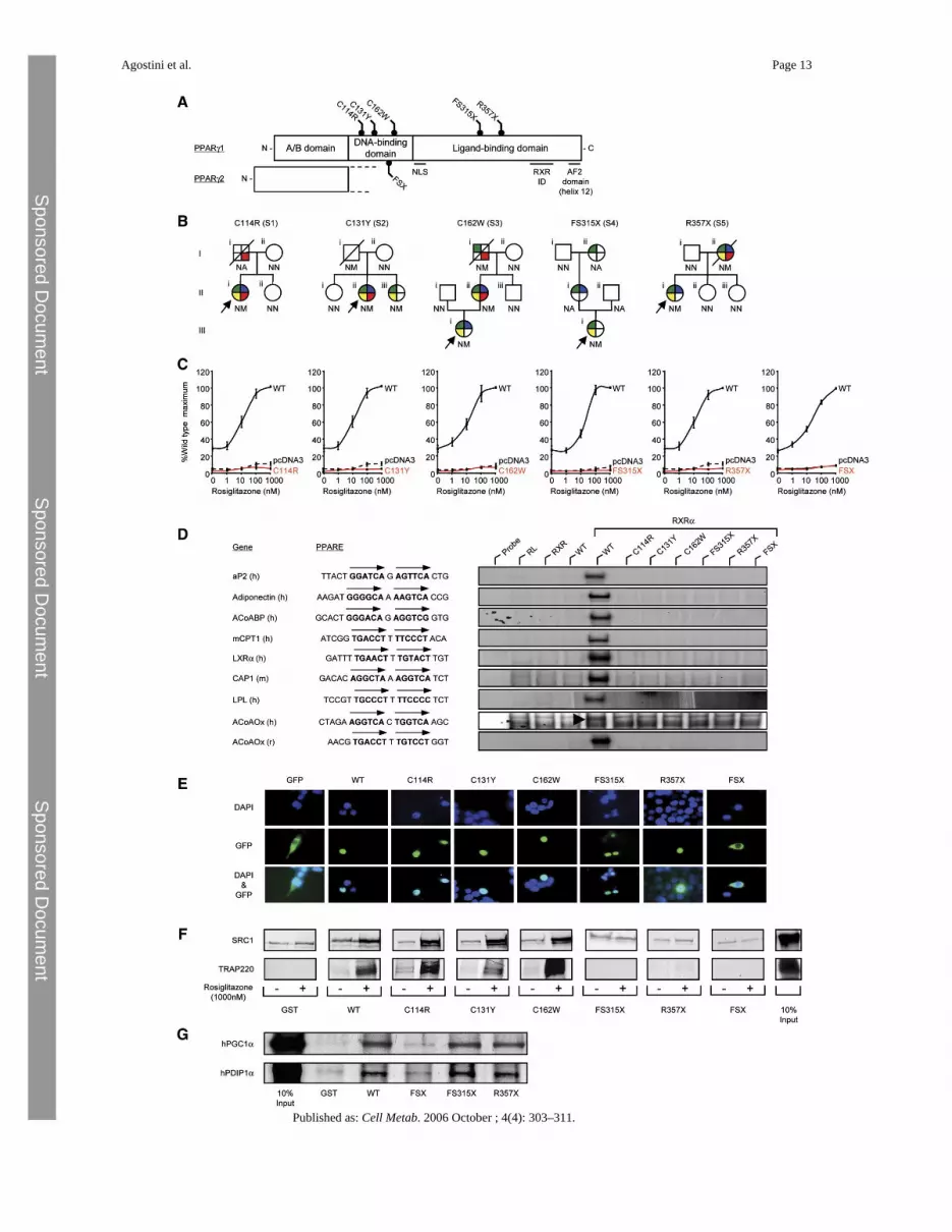
Figure 1.Identification and characterization of loss-of-function mutations in human PPARγA) Schematic representation of the three major domains of PPARγ, showing the locations ofthe five mutations (C114R, C131Y, C162W, FS315X, and R357X – PPARγ1 nomenclature)and the previously reported FSX mutation. NLS, nuclear localisation signal; RXR ID, retinoidX receptor interaction domain; AF2, activation function 2 domain.B) Family pedigrees showing genotypes (N, wild-type allele; M, mutant allele; NA, notavailable) and phenotypes (colored segments denote the presence of specific traits: green, type2 diabetes mellitus/impaired glucose tolerance/hyperinsulinaemia; yellow,hypertriglyceridaemia; blue, hypertension; red, ischemic heart disease). Squares and circlesrepresent male and female family members; slashed symbols denote deceased family membersand arrows denote probands.C) PPARγ mutants are unable to mediate ligand-dependent transactivation. 293EBNA cellswere transfected with 100 ng of wild-type (WT), mutant, or empty (pcDNA3) expressionvectors, together with 500 ng of (PPARE)3TKLUC reporter construct and 100 ng of Bos-β-gal internal control plasmid, and increasing concentrations of rosiglitazone. Results areexpressed as a percentage of the maximum activation with WT PPARγ1 and represent the mean± SEM of at least three independent experiments in triplicate.D) PPARγ mutants are unable to bind to DNA. EMSA with in vitro translated wild-type (WT)or mutant PPARγ1 (C114R, C131Y, C162W, FS315X, R357X, or FSX) and RXR proteinscoincubated with oligonucleotide duplexes corresponding to various natural PPAREs. aP2,adipocyte protein 2; ACoABP, acyl coenzyme A binding protein; mCPT1, muscle carnitinepalmitoyl transferase 1; LXRα, liver X receptor α; CAP, cbl-associated protein; LPL,lipoprotein lipase, ACoAOx, acyl coenzyme A oxidase; h, human; m, mouse; r, rat; RL,reticulocyte lysate.E) The C114R, C131Y, C162W, FS315X, and R357X mutants translocate to the nucleuswhereas the FSX mutant remains cytoplasmic. 293EBNA cells were transfected as described.Top panels show DAPI-staining (blue) of nuclei, middle panels the cellular localisation ofGFP-tagged receptors, and lower panels merged images.F) The DBD PPARγ mutants recruit SRC1 and TRAP220 coactivators, whereas the FS315X,R357X, and FSX truncation mutants do not interact. GST alone or WT and mutant GST-PPARγ fusion proteins were tested with 35S-labeled in vitro translated SRC1 (upper panel) orTRAP220 (lower panel) in the absence or presence of rosiglitazone. Coomassie-stained gelsconfirmed comparable protein loading (data not shown). G) The LBD truncation mutants(FS315X, R357X) recruit PGC1α and PDIP1α coactivators, whereas the FSX mutant fails tointeract. GST alone or WT and mutant GST-PPARγ fusion proteins were tested with 35S-labeled in vitro translated human PGC1α and human PDIP1α in the absence of ligand.Coomassie-stained gels confirmed comparable protein loading (data not shown).
Agostini et al. Page 14
Published as: Cell Metab. 2006 October ; 4(4): 303–311.
Sponsored Docum
ent Sponsored D
ocument
Sponsored Docum
ent

Figure 2.PPARγ mutants exhibit dominant-negative activityA) The C114R, C131Y, C162W, FS315X, and R357X PPARγ mutants inhibit transactivationby wild-type (WT) receptor, comparably to AF2, an artificial PPARγ mutant describedpreviously, whereas the FSX mutant lacks dominant-negative activity (upper panel). 3T3-L1cells were cotransfected with 33 ng of WT receptor plus an equal amount of either empty(pcDNA3) or WT or mutant expression vector, together with 265 ng of human aP2LUC reporterplasmid and 65 ng of the internal control plasmid Bos-β-gal. The dotted and dashed lines denotetranscriptional activity of WT receptor in the absence and presence of ligand respectively.Results are expessed as fold induction relative to empty vector (pcDNA3 + pcDNA3) andrepresent the mean ± SEM of at least three independent experiments in triplicate. Expressionof wild-type and mutant receptor proteins was confirmed by Western blotting (lower panel)and the positions of WT, C114R, C131Y, C162W, and AF2 PPARγ (open arrow) and FSX,FS315X and R357X truncation mutants (solid arrows) are indicated.B and C) Ligand-dependent regulation of PPARγ target genes in IDCs from subjects withPPARγ mutations. (B) Induction of the aP2 gene by rosiglitazone, measured by qPCR, ismarkedly impaired in IDCs derived from subjects with the C114R, C131Y, FS315X, andR357X mutations, compared to responses in cells from normal (WT), severely insulin resistant
Agostini et al. Page 15
Published as: Cell Metab. 2006 October ; 4(4): 303–311.
Sponsored Docum
ent Sponsored D
ocument
Sponsored Docum
ent
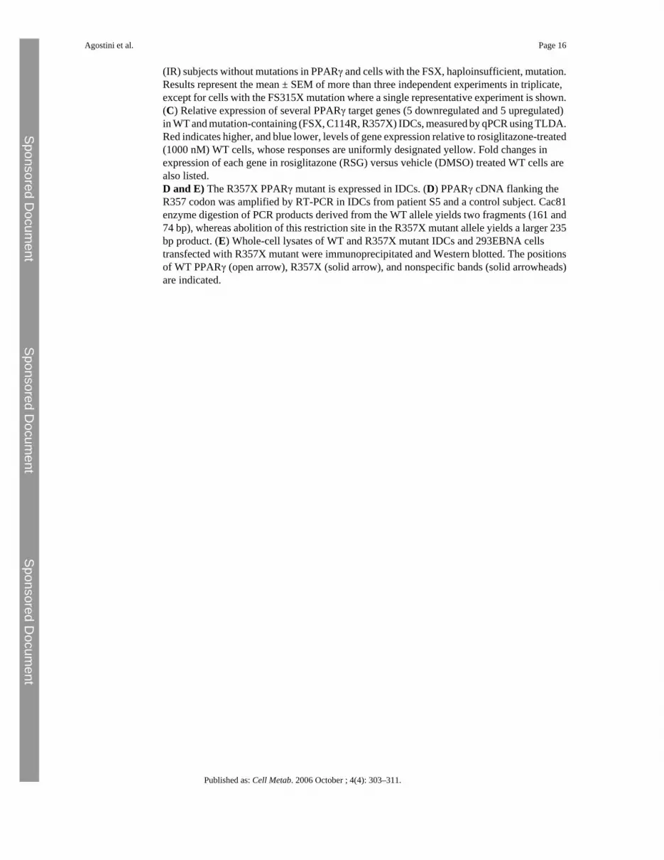
(IR) subjects without mutations in PPARγ and cells with the FSX, haploinsufficient, mutation.Results represent the mean ± SEM of more than three independent experiments in triplicate,except for cells with the FS315X mutation where a single representative experiment is shown.(C) Relative expression of several PPARγ target genes (5 downregulated and 5 upregulated)in WT and mutation-containing (FSX, C114R, R357X) IDCs, measured by qPCR using TLDA.Red indicates higher, and blue lower, levels of gene expression relative to rosiglitazone-treated(1000 nM) WT cells, whose responses are uniformly designated yellow. Fold changes inexpression of each gene in rosiglitazone (RSG) versus vehicle (DMSO) treated WT cells arealso listed.D and E) The R357X PPARγ mutant is expressed in IDCs. (D) PPARγ cDNA flanking theR357 codon was amplified by RT-PCR in IDCs from patient S5 and a control subject. Cac81enzyme digestion of PCR products derived from the WT allele yields two fragments (161 and74 bp), whereas abolition of this restriction site in the R357X mutant allele yields a larger 235bp product. (E) Whole-cell lysates of WT and R357X mutant IDCs and 293EBNA cellstransfected with R357X mutant were immunoprecipitated and Western blotted. The positionsof WT PPARγ (open arrow), R357X (solid arrow), and nonspecific bands (solid arrowheads)are indicated.
Agostini et al. Page 16
Published as: Cell Metab. 2006 October ; 4(4): 303–311.
Sponsored Docum
ent Sponsored D
ocument
Sponsored Docum
ent
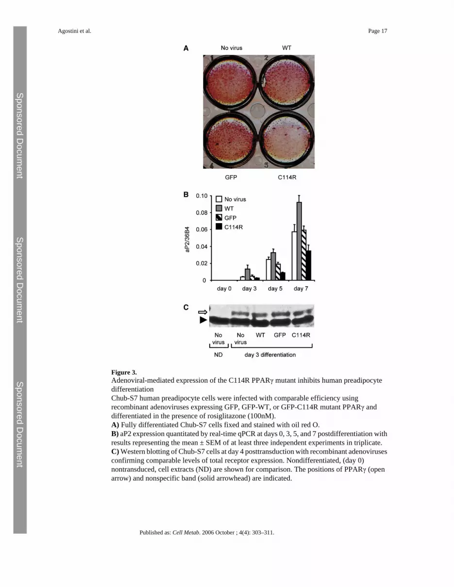
Figure 3.Adenoviral-mediated expression of the C114R PPARγ mutant inhibits human preadipocytedifferentiationChub-S7 human preadipocyte cells were infected with comparable efficiency usingrecombinant adenoviruses expressing GFP, GFP-WT, or GFP-C114R mutant PPARγ anddifferentiated in the presence of rosiglitazone (100nM).A) Fully differentiated Chub-S7 cells fixed and stained with oil red O.B) aP2 expression quantitated by real-time qPCR at days 0, 3, 5, and 7 postdifferentiation withresults representing the mean ± SEM of at least three independent experiments in triplicate.C) Western blotting of Chub-S7 cells at day 4 posttransduction with recombinant adenovirusesconfirming comparable levels of total receptor expression. Nondifferentiated, (day 0)nontransduced, cell extracts (ND) are shown for comparison. The positions of PPARγ (openarrow) and nonspecific band (solid arrowhead) are indicated.
Agostini et al. Page 17
Published as: Cell Metab. 2006 October ; 4(4): 303–311.
Sponsored Docum
ent Sponsored D
ocument
Sponsored Docum
ent
Sponsored Docum
ent Sponsored D
ocument
Sponsored Docum
ent
Agostini et al. Page 18
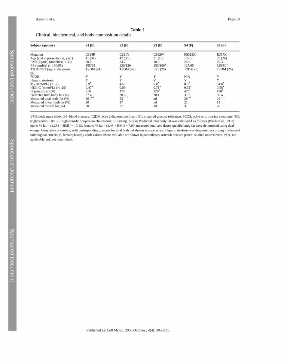
Table 1Clinical, biochemical, and body composition details
Subject (gender) S1 (F) S2 (F) S3 (F) S4 (F) S5 (F)
Mutation C114R C131Y C162W FS315X R357XAge (and at presentation, year) 41 (34) 42 (35) 31 (19) 13 (8) 35 (26)BMI (kg/m2) (nonobese < 30) 30.0 24.2 30.5 25.9 29.3BP (mmHg) (< 130/85) 155/95 220/120 150/100∗ 125/65 125/80∗T2DM/IGT (age at diagnosis,yr)
T2DM (41) T2DM (42) IGT (29) T2DM (8) T2DM (26)
PCOS Y Y Y N/A YHepatic steatosis Y Y Y Y YTG (mmol/L) (<1.7) 8.9∗ 4.5 5.0∗ 8.3∗ 34.8∗HDL-C (mmol/L) (>1.29) 0.47∗ 0.89 0.71∗ 0.72∗ 0.56∗FI (pmol/L) (<60) 310 174 220∗ 475∗ 170∗Predicted total body fat (%) 37.4 28.8 38.1 31.3 36.4Measured total body fat (%) 26 −0.8 23 −1.2 nd 26 nd 21 −1.1
Measured lower limb fat (%) 20 17 nd 21 11Measured truncal fat (%) 30 27 nd 31 28
BMI, body mass index; BP, blood pressure; T2DM, type 2 diabetes mellitus; IGT, impaired glucose tolerance; PCOS, polycystic ovarian syndrome; TG,triglycerides; HDL-C, high-density lipoprotein cholesterol; FI, fasting insulin; Predicted total body fat was calculated as follows (Black et al., 1983):males % fat = (1.281 × BMI) − 10.13: females % fat = (1.48 × BMI) − 7.00; measured total and depot-specific body fat were determined using dual-energy X-ray absorptiometry, with corresponding z scores for total body fat shown as superscript; Hepatic steatosis was diagnosed according to standardradiological criteria; F, female; healthy adult values where available are shown in parentheses; asterisk denotes patient studied on treatment; N/A, notapplicable; nd, not determined.
Published as: Cell Metab. 2006 October ; 4(4): 303–311.