potent inhibitors of a shikimate pathway enzyme from mycobacterium tuberculosis: combining...
TRANSCRIPT

Potent Inhibitors of a Shikimate Pathway Enzyme fromMycobacterium tuberculosisCOMBINING MECHANISM- AND MODELING-BASED DESIGN*□S
Received for publication, December 13, 2010, and in revised form, February 7, 2011 Published, JBC Papers in Press, March 15, 2011, DOI 10.1074/jbc.M110.211649
Sebastian Reichau‡1, Wanting Jiao‡2, Scott R. Walker‡, Richard D. Hutton‡, Edward N. Baker§, and Emily J. Parker‡3
From the ‡Biomolecular Interaction Centre and Department of Chemistry, University of Canterbury, Christchurch 8140 and the§Maurice Wilkins Centre for Molecular Biodiscovery and School of Biological Sciences, University of Auckland,Auckland 1010, New Zealand
Tuberculosis remains a serious global health threat, withthe emergence of multidrug-resistant strains highlighting theurgent need for novel antituberculosis drugs. The enzyme 3-de-oxy-D-arabino-heptulosonate 7-phosphate synthase (DAH7PS)catalyzes the first step of the shikimate pathway for the biosyn-thesis of aromatic compounds. This pathway has been shown tobe essential in Mycobacterium tuberculosis, the pathogenresponsible for tuberculosis. DAH7PS catalyzes a condensationreaction between P-enolpyruvate and erythrose 4-phosphate togive 3-deoxy-D-arabino-heptulosonate 7-phosphate. Theenzyme reaction mechanism is proposed to include a tetrahe-dral intermediate, which is formed by attack of an active sitewater on the central carbon of P-enolpyruvate during the courseof the reaction. Molecular modeling of this intermediate intothe active site reported in this study shows a configurationalpreference consistent with water attack from the re face ofP-enolpyruvate. Based on this model, we designed and synthe-sized an inhibitor of DAH7PS that mimics this reaction inter-mediate. Both enantiomers of this intermediate mimic werepotent inhibitors of M. tuberculosis DAH7PS, with inhibitoryconstants in the nanomolar range. The crystal structure of theDAH7PS-inhibitor complexwas solved to 2.35 A. Both the posi-tion of the inhibitor and the conformational changes of activesite residues observed in this structure correspond closely to thepredictions from the intermediatemodeling. This structure alsoidentifies a water molecule that is located in the appropriateposition to attack the re face of P-enolpyruvate during thecourse of the reaction, allowing the catalyticmechanism for thisenzyme to be clearly defined.
Tuberculosis remains a serious global health threat with overa million deaths per year. The recent emergence of multidrug-
resistant strains of Mycobacterium tuberculosis, the pathogenthat causes the lung disease, highlights the need for rapid devel-opment of new antibacterial drugs to combat tuberculosis(1–4).3-Deoxy-D-arabino-heptulosonate 7-phosphate synthase
(DAH7PS)4 is an important enzyme in M. tuberculosis andother pathogens. It catalyzes the first committed step in theshikimate pathway, which is responsible for the biosynthesis ofaromatic amino acids and other essential aromatic metabolitesin microorganisms, plants, and apicomplexan parasites (5–7).This pathway is absent in humans, and inhibitors of amino acidbiosynthesis have been shown to be effective antimicrobial andherbicidal agents (8, 9). Gene disruption studies have demon-strated that M. tuberculosis is not viable if the shikimate path-way is not operational (10). These findings make DAH7PS anattractive target for drug development.DAH7PS catalyzes the aldol-like condensation of P-enolpy-
ruvate and D-erythrose 4-phosphate (E4P) to yield 3-deoxy-D-arabino-heptulusonate 7-phosphate (Fig. 1a). The reactionmechanism has been subject to extensive study, and many ofthe key details of themechanism have been elucidated (11–16).The reaction occurs stereospecifically with respect to both sub-strates, with the si face of P-enolpyruvate attacking the re face ofE4P. A divalent metal ion present in the active site is essentialfor activity. The reaction takes place with cleavage of the C–Obond of P-enolpyruvate rather than the O–P bond, requiringwater to attack C2 of P-enolpyruvate at some stage during thereaction.A mechanism consistent with the data published to date
starts with nucleophilic attack of P-enolpyruvate at the E4Paldehyde moiety, resulting in the formation of oxocarbeniumspecies 1 (Fig. 1b). This oxocarbenium ion 1 can be attacked byan active site water to form phosphohemiketal 2. It is notewor-thy thatwater can potentially attack fromeither face of 1, givingrise to two possible diastereoisomers of tetrahedral intermedi-ate 2, differing in their absolute configuration at C2. Althoughthis stereogenic center is transient and the stereochemicalinformation is lost by elimination of phosphate in the final stepto generate the product DAH7P (3), the geometry of theenzyme active site is likely to favor stereoselective attack of
* This research was funded in part by the Maurice Wilkins Centre for Molecu-lar Biodiscovery.
The atomic coordinates and structure factors (code 3PFP) have been deposited inthe Protein Data Bank, Research Collaboratory for Structural Bioinformatics,Rutgers University, New Brunswick, NJ (http://www.rcsb.org/).
□S The on-line version of this article (available at http://www.jbc.org) containssupplemental text and Figs. S1–S4.
1 Supported by a University of Canterbury Doctoral Scholarship and a NewZealand International Doctoral Research Scholarship (NZIDRS).
2 Supported by a New Zealand Tertiary Education Commission Top AchieverDoctoral Scholarship.
3 To whom correspondence should be addressed: Dept. of Chemistry, Univer-sity of Canterbury, Private Bag 4800, Christchurch. Tel.: 64-3-364-2871; Fax:64-3-364-2110; E-mail: [email protected].
4 The abbreviations used are: DAH7PS, 3-deoxy-D-arabino-heptulosonate7-phosphate synthase; MtuDAH7PS, M. tuberculosis DAH7PS; E4P, D-erythrose 4-phosphate; G3P, glycerol-3-phosphate; KDO8PS, 3-deoxy-D-manno-octulosonate-8-phosphate synthase; iPr, isopropyl.
THE JOURNAL OF BIOLOGICAL CHEMISTRY VOL. 286, NO. 18, pp. 16197–16207, May 6, 2011© 2011 by The American Society for Biochemistry and Molecular Biology, Inc. Printed in the U.S.A.
MAY 6, 2011 • VOLUME 286 • NUMBER 18 JOURNAL OF BIOLOGICAL CHEMISTRY 16197
by guest on April 6, 2016
http://ww
w.jbc.org/
Dow
nloaded from
by guest on April 6, 2016
http://ww
w.jbc.org/
Dow
nloaded from
by guest on April 6, 2016
http://ww
w.jbc.org/
Dow
nloaded from
by guest on April 6, 2016
http://ww
w.jbc.org/
Dow
nloaded from
by guest on April 6, 2016
http://ww
w.jbc.org/
Dow
nloaded from
by guest on April 6, 2016
http://ww
w.jbc.org/
Dow
nloaded from
by guest on April 6, 2016
http://ww
w.jbc.org/
Dow
nloaded from
by guest on April 6, 2016
http://ww
w.jbc.org/
Dow
nloaded from
by guest on April 6, 2016
http://ww
w.jbc.org/
Dow
nloaded from
by guest on April 6, 2016
http://ww
w.jbc.org/
Dow
nloaded from
by guest on April 6, 2016
http://ww
w.jbc.org/
Dow
nloaded from
by guest on April 6, 2016
http://ww
w.jbc.org/
Dow
nloaded from
by guest on April 6, 2016
http://ww
w.jbc.org/
Dow
nloaded from
by guest on April 6, 2016
http://ww
w.jbc.org/
Dow
nloaded from
by guest on April 6, 2016
http://ww
w.jbc.org/
Dow
nloaded from
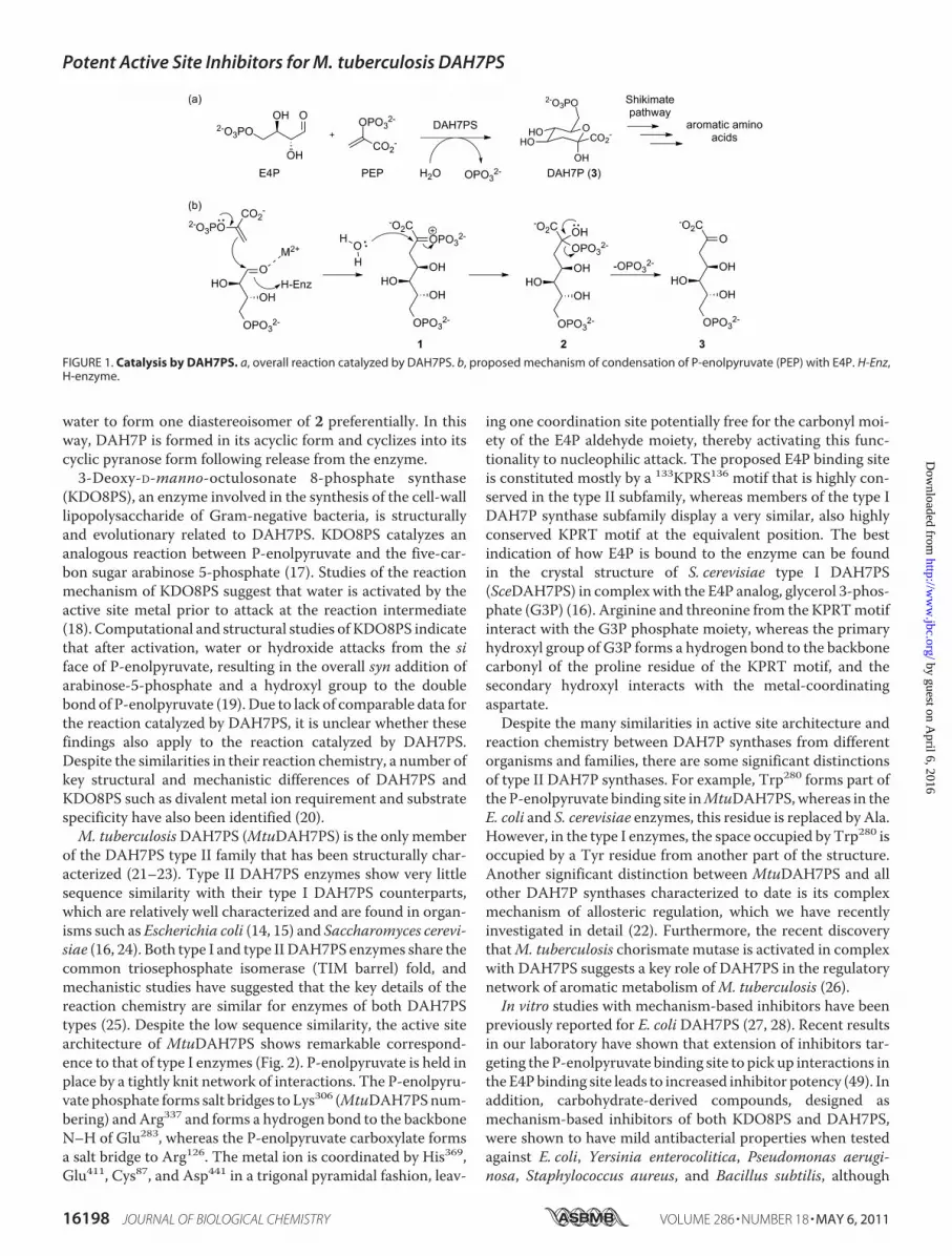
water to form one diastereoisomer of 2 preferentially. In thisway, DAH7P is formed in its acyclic form and cyclizes into itscyclic pyranose form following release from the enzyme.3-Deoxy-D-manno-octulosonate 8-phosphate synthase
(KDO8PS), an enzyme involved in the synthesis of the cell-walllipopolysaccharide of Gram-negative bacteria, is structurallyand evolutionary related to DAH7PS. KDO8PS catalyzes ananalogous reaction between P-enolpyruvate and the five-car-bon sugar arabinose 5-phosphate (17). Studies of the reactionmechanism of KDO8PS suggest that water is activated by theactive site metal prior to attack at the reaction intermediate(18). Computational and structural studies of KDO8PS indicatethat after activation, water or hydroxide attacks from the siface of P-enolpyruvate, resulting in the overall syn addition ofarabinose-5-phosphate and a hydroxyl group to the doublebond of P-enolpyruvate (19). Due to lack of comparable data forthe reaction catalyzed by DAH7PS, it is unclear whether thesefindings also apply to the reaction catalyzed by DAH7PS.Despite the similarities in their reaction chemistry, a number ofkey structural and mechanistic differences of DAH7PS andKDO8PS such as divalent metal ion requirement and substratespecificity have also been identified (20).M. tuberculosisDAH7PS (MtuDAH7PS) is the only member
of the DAH7PS type II family that has been structurally char-acterized (21–23). Type II DAH7PS enzymes show very littlesequence similarity with their type I DAH7PS counterparts,which are relatively well characterized and are found in organ-isms such as Escherichia coli (14, 15) and Saccharomyces cerevi-siae (16, 24). Both type I and type II DAH7PS enzymes share thecommon triosephosphate isomerase (TIM barrel) fold, andmechanistic studies have suggested that the key details of thereaction chemistry are similar for enzymes of both DAH7PStypes (25). Despite the low sequence similarity, the active sitearchitecture of MtuDAH7PS shows remarkable correspond-ence to that of type I enzymes (Fig. 2). P-enolpyruvate is held inplace by a tightly knit network of interactions. The P-enolpyru-vate phosphate forms salt bridges to Lys306 (MtuDAH7PSnum-bering) and Arg337 and forms a hydrogen bond to the backboneN–H of Glu283, whereas the P-enolpyruvate carboxylate formsa salt bridge to Arg126. The metal ion is coordinated by His369,Glu411, Cys87, and Asp441 in a trigonal pyramidal fashion, leav-
ing one coordination site potentially free for the carbonyl moi-ety of the E4P aldehyde moiety, thereby activating this func-tionality to nucleophilic attack. The proposed E4P binding siteis constituted mostly by a 133KPRS136 motif that is highly con-served in the type II subfamily, whereas members of the type IDAH7P synthase subfamily display a very similar, also highlyconserved KPRT motif at the equivalent position. The bestindication of how E4P is bound to the enzyme can be foundin the crystal structure of S. cerevisiae type I DAH7PS(SceDAH7PS) in complexwith the E4P analog, glycerol 3-phos-phate (G3P) (16). Arginine and threonine from the KPRTmotifinteract with the G3P phosphate moiety, whereas the primaryhydroxyl group of G3P forms a hydrogen bond to the backbonecarbonyl of the proline residue of the KPRT motif, and thesecondary hydroxyl interacts with the metal-coordinatingaspartate.Despite the many similarities in active site architecture and
reaction chemistry between DAH7P synthases from differentorganisms and families, there are some significant distinctionsof type II DAH7P synthases. For example, Trp280 forms part ofthe P-enolpyruvate binding site inMtuDAH7PS,whereas in theE. coli and S. cerevisiae enzymes, this residue is replaced by Ala.However, in the type I enzymes, the space occupied by Trp280 isoccupied by a Tyr residue from another part of the structure.Another significant distinction between MtuDAH7PS and allother DAH7P synthases characterized to date is its complexmechanism of allosteric regulation, which we have recentlyinvestigated in detail (22). Furthermore, the recent discoverythatM. tuberculosis chorismate mutase is activated in complexwith DAH7PS suggests a key role of DAH7PS in the regulatorynetwork of aromatic metabolism ofM. tuberculosis (26).In vitro studies with mechanism-based inhibitors have been
previously reported for E. coliDAH7PS (27, 28). Recent resultsin our laboratory have shown that extension of inhibitors tar-geting the P-enolpyruvate binding site to pick up interactions inthe E4Pbinding site leads to increased inhibitor potency (49). Inaddition, carbohydrate-derived compounds, designed asmechanism-based inhibitors of both KDO8PS and DAH7PS,were shown to have mild antibacterial properties when testedagainst E. coli, Yersinia enterocolitica, Pseudomonas aerugi-nosa, Staphylococcus aureus, and Bacillus subtilis, although
FIGURE 1. Catalysis by DAH7PS. a, overall reaction catalyzed by DAH7PS. b, proposed mechanism of condensation of P-enolpyruvate (PEP) with E4P. H-Enz,H-enzyme.
Potent Active Site Inhibitors for M. tuberculosis DAH7PS
16198 JOURNAL OF BIOLOGICAL CHEMISTRY VOLUME 286 • NUMBER 18 • MAY 6, 2011
by guest on April 6, 2016
http://ww
w.jbc.org/
Dow
nloaded from

further in vitro studies were suggested to confirm that thesecompounds indeed target KDO8PS and/or DAH7PS (29).However, to date, no inhibitors for the DAH7PS from thepathogenM. tuberculosis have been reported. Herein we reportthe design, synthesis, and testing of an intermediate mimic asthe first potent inhibitor of MtuDAH7PS in both its racemicand its enantiomerically pure forms. The crystal structure ofthe target enzyme in complex with the inhibitor shows that thesimplified intermediate analog binds in a fashion in accordancewith its design and thus provides valuable insight into both theDAH7PS reaction mechanism and a lead structure for novelpotent antimycobacterial drugs targetingMtuDAH7PS.
EXPERIMENTAL PROCEDURES
Analytical Methods—Optical rotations [�]D20 were measuredat room temperature on a PerkinElmer Life SciencesModel 341polarimeter; analyte concentrations are given in g/100 ml. Spe-cific rotations are reported. NMR spectroscopy was carried outon aVarianUNITY300MHzorVarian INOVA500MHzNMRspectrometer. 1H and 13CNMR spectra are referenced to exter-nal tetramethylsilane; 31PNMR spectra are referenced to exter-nal 85% phosphoric acid.Mass spectrometry was performed onBrukermaXis 3G orMicromass LCTby electrospray ionizationin positive or negative ionization modes.UV-visible spectrophotometry was carried out on a Varian
Cary� One UV-visible spectrophotometer, in stoppered quartz
cells. The temperature was continuously controlled at 303 K(30 °C) by the use of a jacketedmulticell holder, connected to anexternal Varian Peltier temperature controller filled with eitherwater or ethylene glycol.Expression, Purification, and Crystallization of MtuDAH7PS—
M. tuberculosis DAH7PS was overexpressed and purified asdescribed previously (21–23). A reservoir solution containing400 �M inhibitor was prepared by dissolving an appropriateamount of solid rac-4 in crystallization buffer (0.1 M Tris-HClbuffer at pH 8, 1.5 M ammonium sulfate, and 12% v/v glycerol).500 �l of enzyme stock solution was concentrated to �50 �lusing centrifugation with a 10-kDa cutoff membrane, and theresulting solution was diluted to �500 �l using the inhibitorsolution. This process was repeated twice to buffer-exchangethe solution as completely as possible. 1 �l of the buffer-ex-changed enzyme solution (concentration 3–5 mg/ml) wasmixedwith 1�l of crystallization buffer. Crystalswere grownbysitting drop vapor diffusion over 300 �l of reservoir solutionand appeared within 24 h at 25 °C. Crystals were flash-frozen inliquid nitrogen in a cryoprotectant solution consisting of crys-tallization buffer and 20% v/v glycerol.Structure Determination—X-ray diffraction data were col-
lected at the MX2 beamline at the Australian Synchrotron (seeTable 1). The dataset was integrated using iMosflm (30). Thespace group and cell parameters were identical to those of pre-
FIGURE 2. Active site architecture of DAH7PS. a, stereo view of the active site of M. tuberculosis DAH7PS (PDB code 3NV8) (22) showing P-enolpyruvate (PEP,yellow carbons) in its binding site. The manganese iron is represented as a purple sphere. Major interactions between P-enolpyruvate and the enzyme and themetal coordination sphere are shown as dashed lines. b, stereo view of the active site of S. cerevisiae DAH7PS in complex with P-enolpyruvate, G3P (yellowcarbons), and a Co2� ion (magenta sphere)(PDB code 1OF8) (16). A water molecule (WAT) on the re side of P-enolpyruvate (left in this orientation), which isproposed to play the role of the substrate water, is shown as a red sphere.
Potent Active Site Inhibitors for M. tuberculosis DAH7PS
MAY 6, 2011 • VOLUME 286 • NUMBER 18 JOURNAL OF BIOLOGICAL CHEMISTRY 16199
by guest on April 6, 2016
http://ww
w.jbc.org/
Dow
nloaded from

viously reported structures of MtuDAH7PS allowing phasescalculated from the original structure (Protein Data Bank(PDB) code 2B7O) (21) to be used to solve the structure. Thestructure was refined using the CCP4 software package (30) bymethods analogous to the ones described for the native struc-ture (21). Both enantiomers of inhibitor 4 were built using theDundee PRODRG2 server (31). The two enantiomers of 4wereinitially positionedmanually into the electron density observedin the active site using COOT and then refined at half-occu-pancy. The model was optimized using repetitive cycles ofmodel building with COOT and refinement with REFMAC5.Water molecules were added automatically in COOT and ver-ified using �2Fo � Fc� and �Fo � Fc� maps and potential to hydro-gen-bond to at least one protein atom or other water molecule.A bias-removed �Fo � Fc� omit map (see Fig. 5a) was generatedby deleting the ligand and active site waters from themodel andsubjecting the model to simulated annealing and refinement inPHENIX. The �2Fo � Fc� map obtained by this procedure (seeFig. 5a) shows clear continuous electron density for ligand 4.The structure was validated using SFCHECK, with 97.5% ofresidues in the most favored region of the Ramachandran plot(Table 1). The final model included two protein chains eachcontaining all 462 natural residues and two residues from Histag cleavage, 554 water molecules, two manganese ions, onesulfate ion, one chloride ion, and (R)- and (S)-4.Inhibition Assays—Enzyme activity was monitored by fol-
lowing the loss of absorbance at 232 nm (� � 2800 liters mol�1
cm�1) due to the consumption of P-enolpyruvate (32). Assayswere carried out in 50 mM 1,3-bis(tris(hydroxymethyl)methyl-amino)propane buffer at pH 7.5 containing 1 mM tris(2-car-boxyethyl)phosphine at 30 °C. The buffer solution was pre-pared using ultrapure water, which was stirred over Chelexresin (Bio-Rad) and filtered before use. Manganese(II) sulfatestock solution (10 mM) was made up in ultrapure water pre-treated with Chelex resin. P-enolpyruvate and E4P substratesolutions were made up to�10mM by dissolving the P-enolpy-ruvate monopotassium salt and E4P monosodium salt(Aldrich) in buffer solution. Accurate substrate concentrationswere determined by enzymatic reaction; the absorbancechanges of solutions containing one substrate in excess weredetermined before and after conversion of the limiting sub-strate by addedDAH7PShad occurred, and the extinction coef-ficient of P-enolpyruvate was used to calculate the concentra-tion of the limiting substrate. The absorbance change wascorrected by the absorbance change causedwhenDAH7PSwasadded to a control solution that did not contain E4P. For inhi-bition studies, assay solutions containing manganese (II), vary-ing concentrations of P-enolpyruvate and inhibitors, and bufferwere equilibrated at 30 °C. 2 �l (1.3 mg/ml) of M. tuberculosisDAH7PSwas added, and themixturewas equilibrated for 2minbefore the reaction was initiated by the addition of E4P (11 �l).Final assay conditions were 10 �M Mn2�, 5.2 nM DAH7PS, 100�M E4P, 57.6–115.2 �M P-enolpyruvate, 0–940 nM inhibitorwith an appropriate amount of buffer tomake up a final volumeof 1 ml.Lanzetta Phosphate Assay—Lanzetta reagent (33) was pre-
pared fresh as required from the following components: 3 parts0.045% w/v malachite green in water, 1 part 4.2% w/v ammo-
niummolybdate in 4 M HCl, 0.1 parts 1.5% v/v Triton X-100 inwater. The components were mixed in the dark and stirred for1 h before the solution was filtered through a 0.45-�m syringefilter. For the qualitative detection of phosphate-containingfractions after anion exchange chromatography, a 20-�l sampleof each fraction wasmixed with 250 �l of Lanzetta reagent, andthe color change was judged by optical inspection. For thequantitative determination of inhibitor concentration, 300 �lof the inhibitor solutions was incubated with 10 �l of calf alka-line phosphatase solution (5 units/ml in 4 mM MgCl2) for atleast 2 h. To 100 �l of the digested sample was added 700 �l ofLanzetta reagent, and the absorbance at 630 nm was deter-mined after 20min.A calibration curve for the determination ofphosphate concentrationwas obtained from analogous analysisof solutions of appropriate concentrations (6–150 �M) ofKH2PO4, which had been dried in high vacuum for at least 3 hbefore use. As a control, a glucose-6-phosphate solution ofknown concentration was also digested with calf alkaline phos-phatase and analyzed.Modeling Procedure for Linear Intermediate and the
Designed Inhibitors—Modeling studies were carried out usingsoftware packages from Schrodinger Suite 2006 software pack-age (34–37); more detailed computational methods can beobtained from the supplemental material. The previouslyreported crystal structure of MtuDAH7PS (PDB code 2B7O)(21) was used as the receptor. As a starting point for dockingstudies, an ensemble of low-energy conformations of theligands (R)- and (S)-4 and both epimers of the tetrahedral inter-mediate 2 were identified by conformational searches. Themodeling of the tetrahedral intermediate epimers intothe active site of MtuDAH7PS was carried out with theSchrodinger Suite 2006 Induced Fit Docking protocol (36). Theinduced fit docking procedure lead to an intermediate adaptedprotein receptor in which several residues in the active siteadopted conformations different from the ones observed in thenative crystal structure (PDB code 2B7O). This intermediateadapted structure of MtuDAH7PS was then used as a rigidreceptor for docking studies of compounds (R)- and (S)-4,which were conducted using Glide (37).Synthesis—All reactions were carried out under an inert
atmosphere of nitrogen in flame-dried glassware unless other-wise stated. Organic solvents were dried before use by standardmethods (38). Dess-Martin periodinane (39, 40) was preparedaccording to literature procedures. m-Chloroperbenzoic acidwas recrystallized from dichloromethane before use, and allother reagents were purchased from Sigma-Aldrich and usedwithout further purification.Preparative procedures and characterization of previously
unreported compounds can be found in the supplementalmaterial. A representative procedure for the final step of thesynthesis and characterization of inhibitor4 is outlined in detailbelow.HeptanoicAcid 2,7-Bisphosphate 4—Protected bisphosphate
11 (177 mg, 0.24 mmol) was dissolved in ethyl acetate (15 ml),and palladium on charcoal (10% palladium/carbon, 57 mg,0.053 mmol of palladium) was added. The reaction flask waspurged with hydrogen gas with three freeze-pump-thaw cycles.The reaction mixture was stirred for 16 h, after which TLC
Potent Active Site Inhibitors for M. tuberculosis DAH7PS
16200 JOURNAL OF BIOLOGICAL CHEMISTRY VOLUME 286 • NUMBER 18 • MAY 6, 2011
by guest on April 6, 2016
http://ww
w.jbc.org/
Dow
nloaded from

indicated complete consumption of starting material 11. Thesuspension was filtered through a Celite pad, the Celite waswashed with ethyl acetate and methanol, and the solvent wasevaporated under reduced pressure. The resulting residuewas dissolved in 1 M aqueous potassium hydroxide solution (7ml) and stirred vigorously for 2.5 h. The reaction mixture waspassed down a column consisting of freshly regeneratedDOWEX50WX4-200 (H�) resin (�5� 1 cm), and the columnwas eluted with 50ml of water. The eluate was neutralized with1 M potassium hydroxide solution and lyophilized. The crudeproduct was purified by anion exchange chromatography onSOURCE-Q resin eluting with a gradient of water/aqueousammonium bicarbonate solution. Fractions containing thedesired product were identified using the qualitative Lanzettaassay (see below), pooled, and lyophilized to give 42.5 mg of awhite powder consisting of the product and residual ammo-nium bicarbonate. Lanzetta phosphate assay (see above) estab-lished that the purity of the white powder yielded was 80% byweight; the corrected yield for this step is thus 34%.
1HNMR (500MHz, D2O) � 4.34 (td, J� 5.6, 9.0Hz, 1H), 3.73(q, J� 6.4Hz, 2H), 1.63–1.76 (m, 2H), 1.49–1.59 (m, 2H), 1.22–1.42 (m, 4H). 13C NMR (126 MHz, D2O) � 24.0 (br), 25.2 (br),30.1 (br), 33.7 (br), 65.4 (br), 76.1 (br), 180.5 (br). 31P NMR (121MHz, D2O) � ppm 3.31 (t, J � 6.0 Hz, 1P), 1.85 (d, J � 8.8 Hz,1P). HRMS[ESI,neg] calculated for C7H14O10P2 321.0140,found 321.0131 [M-H]�. [�]D20 (R)-4, -0.9 (c 1.8, H2O), (S)-4,�0.8 (c 1.9, H2O).
RESULTS
Modeling of the Reaction Intermediate into MtuDAH7PSActive Site—To inform inhibitor design, the predicted tetrahe-dral reaction intermediate (2, Fig. 3) was modeled into theactive site ofMtuDAH7PS. Because it is unknown from which
side the water attacks the P-enolpyruvate substrate during thereaction, both possible epimers of the linear intermediate weremodeled.Themodeling study produced a total of 30 possible poses for
the two possible C2 diastereoisomers of the tetrahedral inter-mediate in the active site, out of which 17 retained the expectedP-enolpyruvate binding interactions. All 17 poses with correctP-enolpyruvate orientation are from the S-isomer of the inter-mediate, consistent with water attacking the C3 carbon ofP-enolpyruvate from its re face, which is the side opposite theactive site Mn2� ion.
The best pose from themodeling study (Fig. 3a) was selectedbased on two criteria. The first is that the P-enolpyruvate part ofthe tetrahedral intermediate retains the same interactions asobserved in the original crystal structure (PDBcode 2B7O). Thesecond is that the positioning of the E4P phosphate groupshould be as close as possible to the phosphate group of G3P inthe crystal structure of SceDAH7PS (PDB code 1OF8), which isconsidered to provide the best prediction for the E4Pphosphateposition. In the best pose (Fig. 3a), the P-enolpyruvate part ofthe molecule interacts with Arg126, Lys306, Arg337, Arg284, andGlu283. Arg284 bridges the phosphate groups of P-enolpyruvateand E4P through salt bridges and hydrogen bonds. The E4Pphosphate group interacts with Arg284, Arg135, and Ser136,which suggests that part of the KPRS motif (Arg135 and Ser136)plays a key role in positioning of the E4P phosphate, as expectedfrom the G3P-bound SceDAH7PS structure. The E4P aldehydegroup, now converted to a hydroxyl group in the linear inter-mediate, is at a distance of 2.2 Å from the metal ion. This indi-cates that the aldehyde of E4P may be held in place by coordi-nation to the metal ion before reaction occurs. The hydroxylgroup that corresponds to the incoming water molecule forms
FIGURE 3. Molecular modeling studies and inhibitor design. a, the modeled best pose of the tetrahedral open chain intermediate 2 (S-isomer) from inducedfit docking to the active site of MtuDAH7PS superimposed on the crystal structure of SceDAH7PS (PDB code 1OF8). The active site carbons from the modeledstructure are in green, and the active site residues of SceDAH7PS are shown with yellow carbons. The modeled linear intermediate molecule is shown withmagenta carbons, and the C2-hydroxyl group is pointing behind the plane. The residues are labeled according to the numbering in MtuDAH7PS. The metal ionis displayed as a purple sphere. Hydrogen bonds are represented by dashed lines. b, left, linear intermediate 2. Center, molecular model of linear intermediate 2showing the preferred 2S-configuration. Right, simplified intermediate analog 4, synthetic target for this study.
Potent Active Site Inhibitors for M. tuberculosis DAH7PS
MAY 6, 2011 • VOLUME 286 • NUMBER 18 JOURNAL OF BIOLOGICAL CHEMISTRY 16201
by guest on April 6, 2016
http://ww
w.jbc.org/
Dow
nloaded from

hydrogen bonds with Trp280, Glu248, and Lys133. Two of theseresidues, Glu248 and Lys133, are conserved across the entireDAH7PS family, whereas Trp280 is only conserved in the type IIenzymes.Inhibitor Design—Combining results from the intermediate
modeling and previous work on mechanism-based inhibitors(27, 28), we designed bisphosphate 4 (Fig. 3b) as a simplifiedanalog of the tetrahedral intermediate. The 2-phosphorylcar-boxylic acid moiety targets the P-enolpyruvate binding site andresembles phospholactate, which has been reported as an effi-cient inhibitor of E. coli DAH7PS (27). Bisphosphate 4 repre-sents a simplified analog of tetrahedral intermediate 2; thephosphoryl and carboxyl moieties of compound 4 are pro-posed to act as charged anchors arranged in a similar geom-etry to those of intermediate 2 and held together by a non-functionalized hydrocarbon linker. Given its low degree offunctionalization, bisphosphate 4 may be used as a leadstructure for specific introduction of functionality toenhance enzyme-ligand affinity. In light of the results formolecular modeling of the tetrahedral intermediate 2, whichsuggest a preference for the S-configuration at C2, wedevised two syntheses leading to target compound 4 to assesspossible enantio-discrimination: one racemic synthesis,which had already been proven useful in our laboratory, aswell as a chiral pool synthesis, which allowed control of theabsolute configuration at C2.Synthesis of Racemic 4—The synthetic strategy toward rac-4
relies on the establishment of the key�-hydroxyestermoiety byaDarzens-type chain elongation of an aldehyde, amethodologythat has been used for the synthesis of related compounds (Fig.4) (29, 41). The final deprotection was achieved by hydrog-enolysis of the benzyl groups followed by hydrolysis of the iso-propyl ester. The final product, rac-4, was purified by anionexchange chromatography eluting with an ammonium bicar-bonate gradient.Crystal Structure—The racemic inhibitor 4 was co-crystal-
lized with DAH7PS from M. tuberculosis. Although generallyboth purification of the enzyme and crystal growth are difficultin the absence of P-enolpyruvate in the buffer solution, x-raydiffraction quality crystals were obtained by buffer exchange ofthe purified enzyme solution with a solution containing inhib-
itor 4 (Table 1). The crystal structure was solved by molecularreplacement using the previously determined MtuDAH7PSstructure (PDB code 2B7O) (21) as search model and wasrefined using x-ray diffraction to 2.35 Å (Table 1) The asym-metric unit contains two enzyme molecules as in otherMtuDAH7PS crystal structures; tertiary and quaternary struc-tures are essentially unaffected by binding of inhibitor 4. Super-imposition on the wild-type protein (PDB code 3NV8) (22)showed that 5029 atoms could be matched with a root meansquare positional difference of 0.22 Å.
FIGURE 4. Synthesis of racemic inhibitor 4. (i) TBDMSCl, imidazole, dichloromethane, 61%. (ii) Dess-Martin periodinane, dichloromethane, 70% (crude). (iii)isopropyl dichloroacetate, KOiPr, iPrOH, Et2O. iv, NaCNBH3, iPrOH, 60% (two steps). (v) tetrabutylammonium fluoride, tetrahydrofuran, 56%. (vi) 1)(BnO)2PN(iPr)2, 1H-tetrazole, dichloromethane; 2) meta-chloroperbenzoic acid, 50%. (vii) 1) H2, palladium/carbon; 2) KOH, H2O; 3) DOWEX-H�; 4) SOURCE-Qanion exchange chromatography, 42%.
TABLE 1Data collection and refinement statistics for M. tuberculosis DAH7PS incomplex with 4
Data collectionCrystal system TrigonalSpace group P3221Unit cell parametersa (Å) 203.58b (Å) 203.58c (Å) 66.49
Resolution range (Å) 55.641–2.35 Å (2.48–2.35)Measurements 726,170Unique reflections 65,888Redundancy 11.0 (11.2)Completeness (%) 100 (100)I/�(I) 5.0 (1.9)Rmerge (%) 13.0 (39.2)Wilson B-value (Å2) 26.5
RefinementResolution (Å) 51.16–2.35 (2.41–2.35)Rcryst (%) 16.4Rfree (%) 19.6Amino acids (chain length464 residues)
464 � 464 res.; 7136 atom sites
Water molecules 554Other 2 Mn2�a, 1 SO4
2�, 1 Cl�, (R)-4a, (S)-4aMean B (Å2)Protein 25.13Water 32.06Other 12.12
r.m.s.d.b from target valuesBond lengths (Å) 0.010Bond angles (°) 1.214Dihedral angles (°) 5.634
RamachandranMost favored (%) 98.3Allowed (%) 1.2
Disallowed (%) 0.5PDB code 3PFP
a Refined at reduced occupancy.b r.m.s.d., root mean square deviation.
Potent Active Site Inhibitors for M. tuberculosis DAH7PS
16202 JOURNAL OF BIOLOGICAL CHEMISTRY VOLUME 286 • NUMBER 18 • MAY 6, 2011
by guest on April 6, 2016
http://ww
w.jbc.org/
Dow
nloaded from
Clear continuous electron density for inhibitor 4 was foundin both active sites. The �Fo� Fc� omitmap generated by remov-ing the inhibitor and active site waters from the model andsubjecting the resulting model to simulated annealing andrefinement shows clear continuous density for inhibitor 4and for awatermolecule in the active site (Fig. 5a). Based on theelectron density maps obtained, it could not be concludedwhether there is preferential binding of one of the enantiomersfrom the racemic mixture. To represent this ambiguity, ourfinal model includes both (R)-4 and (S)-4 at half-occupancy.Inhibitor 4 binds as predicted with its 2-phosphoryl and car-
boxylate moieties in the P-enolpyruvate binding site and in anextended conformation so that the phosphate moiety on C7 islocated near the proposed E4P phosphate binding site (Fig. 5b).Active site residues interacting with the P-enolpyruvate-mim-icking portion of ligand 4 show little movement, consistentwith this portion of the inhibitor being a goodmimic of P-enol-pyruvate. As expected, the conformations of the metal bindingresidues were unaffected by binding of compound 4. The2-phosphate moiety of inhibitor 4 forms salt bridges withArg284, Arg337, and Lys306 and a hydrogen bond to the back-bone N–H of Glu283. The carboxyl group forms salt bridgeswith Lys133 and Arg126 and coordinates to the manganese ionpresent in the active site. The 7-phosphate moiety extends intothe tentative E4P phosphate binding site, forming salt bridgeswith Lys380 and Arg135 and hydrogen bonds with the Ser136hydroxyl group and backbone N–H. Conformational changesrelative to the P-enolpyruvate-liganded structure wereobserved for the KPRS motif associated with inhibitor binding(Fig. 5c). Arg135 and Ser136 interact with the 7-phosphate moi-ety of inhibitor 4, moving closer into the active site pocket. Theside chain of Lys133 shows the biggest conformational change ofall active site residues, establishing a salt bridge to the carbox-ylate group of ligand 4. This big movement may also be partlydue to increased electrostatic repulsion with the guanidiniumgroup of Arg135, which has moved closer into the active site.Interestingly, the side chainmovement of Lys133 correlates wellwith the movement that the induced fit molecular modelingresults predict for this residue to accommodate the proposedreaction intermediate (Fig. 5d).The superposition of structures of the DAH7PS-4-complex
and the induced fit-modeling results (Fig. 5d) show clearly thatthe experimental binding mode of bisphosphate 4 closelymatches the in silico modeling of the proposed reaction inter-mediate into the active site. The carboxylate and both phos-phate groups of inhibitor 4 occupy positions that were pre-dicted for the corresponding moieties of the intermediatemodel, with minor deviations found in the carbon chain. Acrystallographically located water molecule occupies a positionvery close to the predicted position of the intermediate 2-hydroxyl group. This water is held in place by hydrogen bondsto Trp280 and Glu248. The distance of this water to C2 of theligand is 3.4 Åwhen (R)-4 is modeled into the active site and 3.2Å for the model containing (S)-4. Preliminary testing of rac-4indicated that its Ki againstMtuDAH7PS was in the submicro-molar range. Encouraged by these results, and to assesswhetherone enantiomer of 4 is preferentially bound by the enzyme, wesynthesized compounds (R)-4 and (S)-4 for inhibition assays.
Synthesis of (R)-4 and (S)-4—Because the previously de-scribed synthesis of rac-4 offers little potential with respect tostereoselective introduction of the �-hydroxyester moiety,another synthetic route was developed in which the chirality isderived from a cheap and readily available chiral pool startingmaterial (Fig. 6). The chiral epoxyester 14 of appropriate con-figuration was prepared from D- or L-serine, respectively, fol-lowing a previously reported method (42). Ethyl glycidate 14was ring-opened regioselectively to yield �-hydroxyester 15.Hydrogenolysis of ester 15 liberated the 7-hydroxyl group togenerate dihydroxyester 16, which could then be converted to(R)-4 or (S)-4 using the same procedures as described above forthe racemic synthesis.Inhibition Studies—(R)-4 and (S)-4were tested for inhibition
ofMtuDAH7PS. Although inhibitor 4was designed as a bisub-strate inhibitor, it is known that DAH7PS operates by a sequen-tial ordered reaction mechanism in which P-enolpyruvate hasto bind to the enzyme first (25). Thismeans that although com-petition with P-enolpyruvate is most important to assess theinhibitory potency, the fixed concentration of the second sub-strate can influence the inhibition. The kinetic assays were car-ried outwith P-enolpyruvate concentrations ranging from57 to115 �M at a fixed E4P concentration of 100 �M, which corre-sponds to four times the value determined for the Michaelisconstant for E4P (Km(E4P)). The concentrations of (R)-4 and(S)-4 were varied from 0 to 940 nM. Even at these high levels ofE4P, (R)-4 and (S)-4 gave inhibitory constants of 360 � 50 and620 � 110 nM respectively (see supplemental Figs. S1 and S2).(R)-4 and (S)-4 represent the first potent inhibitors reported forMtuDAH7PS and the first inhibitors with substantially greateraffinity than the substrate P-enolpyruvate (Km(PEP) � 29 �M
forMtuDAH7PS) for any DAH7P synthase.Modeling of (R)-4 and (S)-4 into MtuDAH7PS Active Site—
Both (R)-4 and (S)-4weremodeled into theMtuDAH7PS activesite, and the results showed that there is little preference forbinding between the two isomers of inhibitor 4. Both isomersadopt similar bindingmodes, which are also very similar to thatof the linear intermediate in the active site, with the R-isomerhaving a slightly better docking score than the S-isomer. Theinteractions found in the best poses from modeling of the twoisomers are the same as those observed in the crystal structureof the MtuDAH7PS-4-complex (supplemental Fig. S3). Thesemodeling results in conjunction with the inhibition dataobtained for (R)- and (S)-4 and the tetrahedral intermediatemodeling results suggest that a fully functionalized C2-carbonof the tetrahedral intermediate bearing carboxylate, phosphate,and hydroxyl substituents is necessary to observe stereoselec-tive recognition in the enzyme active site.
DISCUSSION
The study presented here demonstrates the successful com-bination of synthetic, structural, and computationalmethods toinform the design of novel lead structures of inhibitors forMtuDAH7PS. Although both the substrate and the product ofthe enzymatic transformation, P-enolpyruvate and DAH7P,possess planar geometry atC2,mechanistic considerations sug-gest that the enzyme active site is also set up to accommodatepreferentially a tetrahedral reaction intermediate. The high
Potent Active Site Inhibitors for M. tuberculosis DAH7PS
MAY 6, 2011 • VOLUME 286 • NUMBER 18 JOURNAL OF BIOLOGICAL CHEMISTRY 16203
by guest on April 6, 2016
http://ww
w.jbc.org/
Dow
nloaded from

FIGURE 5. Stereo view of the active site of the enzyme in complex with 4 (only the (R)-enantiomer is shown for clarity). a, �Fo � Fc� simulatedannealing omit map contoured at 3� showing the electron density for inhibitor 4 (blue carbons) in the active site. WAT, water. b, interactions of 4 withthe active site residues (green carbons) and metal ion (purple sphere). c, overlay of P-enolpyruvate (yellow carbons)-DAH7PS structure 3NV8 (cyan carbons)with DAH7PS-4-complex (green carbons). d, overlay of the preferred pose of the linear intermediate (magenta carbons) and 4 (blue carbons; the oxygenson the C2-phosphate were omitted for clarity). The enzyme structure experimentally obtained from the DAH7PS-4-complex (green carbons) superim-poses well onto the enzyme structure obtained by induced fit modeling of the linear intermediate (yellow carbons, peptide; magenta carbons,intermediate).
Potent Active Site Inhibitors for M. tuberculosis DAH7PS
16204 JOURNAL OF BIOLOGICAL CHEMISTRY VOLUME 286 • NUMBER 18 • MAY 6, 2011
by guest on April 6, 2016
http://ww
w.jbc.org/
Dow
nloaded from

inhibitory potencies of (R)- and (S)-4 confirm that intermediateanalogueswith tetrahedral C2 are a promising scaffold for high-affinity inhibitors of DAH7PS.The fact that the experimentally determined structure and
the induced-fit model are in good agreement firstly demon-strates that inhibitor 4 is a good intermediate mimic, and sec-ondly, verifies that our modeling approach is effective for pre-dictions of enzyme-ligand interactions for MtuDAH7PS. Thestructural information from the enzyme-inhibitor complex canbe used to inform future inhibitor design; based on the bindingmode of inhibitor 4 in the active site, modifications to the leadstructure allow the introduction of specific new interactionswith the enzyme. For example, additional hydroxyl groups atC4 and C6 of inhibitor 4 with the appropriate absolute con-figuration are expected to form hydrogen bonds to Asp441,interactions that were observed in the modeling of the inter-mediate (Fig. 3). Introduction of a C4-hydroxyl or other met-al-coordinating group such as an amine or thiol group couldalso lead to additional enzyme-inhibitor interactionsthrough metal coordination.Inhibitor 4 is a highly charged molecule, featuring two phos-
phate groups and a carboxylate moiety. This characteristic ofthis molecule is expected to significantly limit membrane per-meability. Phosphates are abundant in naturally occurringmol-ecules, making them highly interestingmoieties in the develop-ment of drugs. Strategies to generate phosphate prodrugscontaining suitable masking groups that facilitate membranepermeability and allow the release of the active drug inside thecell have been developed, and examples of these prodrugs are inclinical trials (43–45).Although both enantiomers of inhibitor 4 displayed a high
affinity to the enzyme active site, the small difference in affinityof the two enantiomers provides valuable insight into theenzyme mechanism and its inhibition. The observation that(S)-4 proved to be a slightly less potent inhibitor may be due tothe fact that the C2-hydrogen of (S)-4 points in the direction ofa water molecule also present in the active site, with interatomdistances that suggest a steric clash between these moieties(supplemental Fig. S4). For the related enzyme KDO8PS, datafromcomputational and crystallographic studies have led to the
proposition that the substrate water attacks P-enolpyruvatefrom the si side during the course of the reaction (18, 19, 46). It,however, so far remains unclear whether this mechanistic pro-posal can be transferred to MtuDAH7PS or even DAH7P syn-thases in general. In the crystal structure reported in this study,a water molecule was located in close proximity to C2 of theinhibitor on what would be the re side of P-enolpyruvate. The re-semblance of this arrangement of inhibitor and water to thelocation of the 2-hydroxyl group of the modeled tetrahedralreaction intermediate, combined with the configurational pref-erence observed for the docking of the intermediate, suggestthat inMtuDAH7PS, water attack proceeds from the re side ofP-enolpyruvate. This is further supported by studies of type IDAH7P synthases from S. cerevisiae (16), E. coli (14), andTher-motogamaritima (47), which located awatermolecule that wassuggested to attack P-enolpyruvate during the course of theenzymatic reaction in a very similar environment. The glutamicacid residue has been proposed as a general base catalyst tofacilitate nucleophilic attack of water. The highly conservednature of the arrangement of water in the P-enolpyruvate bind-ing site suggests a common catalytic mechanism for all DAH7Psynthases that involves water attacking the re face of P-enolpy-ruvate during the course of the reaction.The combined results from intermediate modeling and the
structural and kinetic study of intermediate analog 4 allow sig-nificant refinements of the proposed catalytic mechanism ofMtuDAH7PS (Fig. 7). P-enolpyruvate and E4P binding hasalready been discussed in detail. We propose that the substratewater is bound on the re face of P-enolpyruvate through hydro-gen-bonding interactions with Trp280 and Glu248. Nucleophilicattack of P-enolpyruvate at the E4P aldehyde moiety is facili-tated by metal coordination, and the resulting oxyanion is pro-tonated by Lys133. Both the intermediate modeling results andthe crystal structure of the intermediate analog show significantmovement of the Lys133 side chain upon ligand binding, posi-tioning it in a suitable position to fulfill this role as a generalacid. The substrate water is activated for attack at C2 of P-enol-pyruvate by deprotonation with Glu248.A similar mechanism has been previously suggested in stud-
ies of S. cerevisiae andT. maritima type IDAH7PS (16, 47). The
FIGURE 6. Synthesis of enantiopure inhibitor 4. Yields are given for the synthesis of (R)-4; analogous procedures using L-serine as the starting material wereused for the synthesis of (S)-4 in comparable yields. Please refer to the supplemental material for more detail. (i) HNO2, KBr, H2O then KOH/EtOH, 37% (twosteps). (ii) EtBr, Bn(Et)3NCl, CH2Cl2, 66%. (iii) BnO(CH2)4Br, magnesium, tetrahydrofuran, room temperature, then CuBr�SMe2, �78 °C, then 14, 32%. (iv) H2,palladium/carbon, EtOAc, 66%. (v) (BnO)2P-N(iPr)2, 1H-tetrazole, dichloromethane, then meta-chloroperbenzoic acid 64%. (vi) H2, palladium/carbon, EtOAc,then KOH/H2O, then DOWEX-H�, anion exchange chromatography, 27%.
Potent Active Site Inhibitors for M. tuberculosis DAH7PS
MAY 6, 2011 • VOLUME 286 • NUMBER 18 JOURNAL OF BIOLOGICAL CHEMISTRY 16205
by guest on April 6, 2016
http://ww
w.jbc.org/
Dow
nloaded from

fact that a glutamate residue is found at the equivalent positionfor M. tuberculosis type II DAH7PS and the crystallographiclocalization of a suitable substrate water molecule in theMtuDAH7PS-4-complex in this study as well as our modelingresults further support this mechanistic proposition. In thenext step, the now neutral Lys133 can abstract the proton fromGlu248, either by direct deprotonation or relayed via an activesite water molecule. This step regenerates both the Lys133
ammonium and the Glu248 carboxylate group necessary for thenext catalytic cycle. Lys306, which is positioned within hydro-gen-bonding distance of the P-enolpyruvate phosphate, couldaid the elimination of phosphate from tetrahedral intermediate2 by protonating the phosphate, thus making it a better leavinggroup. Elimination of hydrogen phosphate yieldsDAH7PS pro-tonated on the 2-keto moiety; this proton can then be removedby Lys306. After dissociation of the product DAH7P, all activesite residues have returned to the protonation state that isrequired for the next catalytic turnover. This mechanism is thesimplest that is in accordance with the data published to date,and it accounts for all protonation and deprotonation stepsrequired on active site residues and intermediates. The onlymajor side chain movement of an active site residue is requiredfrom the proton-shuttling Lys133, a residue that is not welldefined or possesses high B-factor values in some crystal struc-tures of DAH7P synthases, indicating mobility of its side chain(11, 21). Further support for the important role of both Lys133
and Lys306 comes from results in our laboratory that show thatboth residues are essential for catalytic activity.5
CONCLUSION
By combining molecular modeling-guided and mechanism-based design, we have synthesized the first potent inhibitor ofMtuDAH7PS. The crystal structure of the enzyme-inhibitorcomplex shows that the structural features of the inhibitordesigned to mimic features of the natural substrates and pro-posed reaction intermediate indeed lead to the predicted spe-cific enzyme inhibitor interactions. Inhibitor 4 represents apromising lead structure for antimycobacterial drugs and offersample opportunity for further modification to establish struc-ture-activity relationships. The crystallographic location of anactive site water molecule has clarified the enzyme catalyticmechanism.
Acknowledgments—This researchwas undertaken on theMX2beam-line at the Australian Synchrotron, Victoria, Australia (48). Weacknowledge travel funding provided by the International Synchro-tron Access Program (ISAP) managed by the Australian Synchrotronand the New Zealand Synchrotron Group.
REFERENCES1. Sharma, S. K., and Mohan, A. (2006) Chest 130, 261–2722. Meya, D. B., and McAdam, K. P. (2007) J. Intern. Med. 261, 309–3293. World Health Organization (2009). Global Tuberculosis Control: Epide-
miology, Strategy, and Financing, WHO Report 2009, pp. 6–33, WHOPress, Geneva
4. Barry, C. E., 3rd, and Blanchard, J. S. (2010) Curr. Opin. Chem. Biol. 14,456–466
5. Campbell, S. A., Richards, T. A., Mui, E. J., Samuel, B. U., Coggins, J. R.,McLeod, R., and Roberts, C. W. (2004) Int. J. Parasitol. 34, 5–13
6. Roberts, F., Roberts, C. W., Johnson, J. J., Kyle, D. E., Krell, T., Coggins,J. R., Coombs, G. H., Milhous, W. K., Tzipori, S., Ferguson, D. J.,Chakrabarti, D., and McLeod, R. (1998) Nature 393, 801–8055 L. R. Schofield and E. J. Parker, unpublished results.
FIGURE 7. Refined catalytic mechanism for MtuDAH7PS in accordance with the results obtained from this study.
Potent Active Site Inhibitors for M. tuberculosis DAH7PS
16206 JOURNAL OF BIOLOGICAL CHEMISTRY VOLUME 286 • NUMBER 18 • MAY 6, 2011
by guest on April 6, 2016
http://ww
w.jbc.org/
Dow
nloaded from
7. Bentley, R. (1990) Crit. Rev. Biochem. Mol. Biol. 25, 307–3848. Sikorski, J. A., and Gruys, K. J. (1997) Acc. Chem. Res. 30, 2–89. Schonbrunn, E., Eschenburg, S., Shuttleworth, W. A., Schloss, J. V., Am-
rhein, N., Evans, J. N., and Kabsch, W. (2001) Proc. Natl. Acad. Sci. U.S.A.98, 1376–1380
10. Parish, T., and Stoker, N. G. (2002)Microbiology 148, 3069–307711. Schofield, L. R., Anderson, B. F., Patchett, M. L., Norris, G. E., Jameson,
G. B., and Parker, E. J. (2005) Biochemistry 44, 11950–1196212. Williamson, R.M., Pietersma, A. L., Jameson, G. B., and Parker, E. J. (2005)
Bioorg. Med. Chem. Lett. 15, 2339–234213. DeLeo, A. B., Dayan, J., and Sprinson, D. B. (1973) J. Biol. Chem. 248,
2344–235314. Shumilin, I. A., Bauerle, R., and Kretsinger, R. H. (2003) Biochemistry 42,
3766–377615. Wagner, T., Shumilin, I. A., Bauerle, R., andKretsinger, R. H. (2000) J.Mol.
Biol. 301, 389–39916. Konig, V., Pfeil, A., Braus, G. H., and Schneider, T. R. (2004) J. Mol. Biol.
337, 675–69017. Ray, P. H. (1980) J. Bacteriol. 141, 635–64418. Kona, F., Xu, X., Martin, P., Kuzmic, P., and Gatti, D. L. (2007) Biochem-
istry 46, 4532–454419. Kona, F., Tao, P., Martin, P., Xu, X., and Gatti, D. L. (2009) Biochemistry
48, 3610–363020. Ahn, M., Pietersma, A. L., Schofield, L. R., and Parker, E. J. (2005) Org.
Biomol. Chem. 3, 4046–404921. Webby, C. J., Baker, H. M., Lott, J. S., Baker, E. N., and Parker, E. J. (2005)
J. Mol. Biol. 354, 927–93922. Webby, C. J., Jiao,W., Hutton, R. D., Blackmore, N. J., Baker, H.M., Baker,
E. N., Jameson, G. B., and Parker, E. J. (2010) J. Biol. Chem. 285,30567–30576
23. Webby, C. J., Lott, J. S., Baker, H. M., Baker, E. N., and Parker, E. J. (2005)Acta Crystallogr. Sect. F Struct. Biol. Cryst. Commun. 61, 403–406
24. Hartmann, M., Schneider, T. R., Pfeil, A., Heinrich, G., Lipscomb, W. N.,and Braus, G. H. (2003) Proc. Natl. Acad. Sci. U.S.A. 100, 862–867
25. Webby, C. J., Patchett, M. L., and Parker, E. J. (2005) Biochem. J. 390,223–230
26. Sasso, S., Okvist, M., Roderer, K., Gamper, M., Codoni, G., Krengel, U.,and Kast, P. (2009) EMBO J. 28, 2128–2142
27. Walker, S. R., Cumming, H., and Parker, E. J. (2009)Org. Biomol. Chem. 7,
3031–303528. Walker, S. R., and Parker, E. J. (2006) Bioorg. Med. Chem. Lett. 16,
2951–295429. Grison, C., Petek, S., Finance, C., and Coutrot, P. (2005) Carbohydr. Res.
340, 529–53730. Collaborative Computational Project Number 4 (1994) Acta Crystallogr.
D Biol. Crystallogr. 50, 760–76331. Schuttelkopf, A. W., and van Aalten, D. M. F. (2004) Acta Crystallogr. D
Biol. Crystallogr. 60, 1355–136332. Schoner, R., and Herrmann, K. M. (1976) J. Biol. Chem. 251, 5440–544733. Lanzetta, P. A., Alvarez, L. J., Reinach, P. S., and Candia, O. A. (1979)Anal.
Biochem. 100, 95–9734. Schrodinger, LLC (2005) MacroModel, Version 9.1, Schrodinger, LLC,
New York35. Schrodinger, LLC (2005) Maestro, Version 7.5, Schrodinger, LLC, New
York36. Schrodinger, LLC (2005) Schrodinger Suite 2006 Induced Fit Protocol,
Prime version 1.5, Schrodinger, LLC, New York37. Schrodinger, LLC (2005)Glide, Version 4.0, Schrodinger, LLC, New York38. Armarego, W. L., and Chai, C. (2003). Purification of Laboratory Chemi-
cals, 5th Ed., Butterworth-Heinemann, Oxford39. Dess, D. B., and Martin, J. C. (1991) J. Am. Chem. Soc. 113, 7277–728740. Ireland, R. E., and Liu, L. (1993) J. Org. Chem. 58, 2899–289941. Grison, C., Coutrot, F., Comoy, C., Lemilbeau, C., and Coutrot, P. (2000)
Tetrahedron Lett. 41, 6571–657442. Petit, Y., and Larcheveque, M. (1998) Org. Synth. 75, 37–4443. Friis, G. J., and Bundgaard, H. (1996) Eur. J. Pharm. Sci. 4, 49–5944. Schultz, C. (2003) Bioorg. Med. Chem. 11, 885–89845. Hecker, S. J., and Erion, M. D. (2008) J. Med. Chem. 51, 2328–234546. Wang, J., Duewel, H. S., Woodard, R.W., and Gatti, D. L. (2001) Biochem-
istry 40, 15676–1568347. Shumilin, I. A., Bauerle, R., Wu, J., Woodard, R. W., and Kretsinger, R. H.
(2004) J. Mol. Biol. 341, 455–46648. McPhillips, T. M., McPhillips, S. E., Chiu, H. J., Cohen, A. E., Deacon,
A. M., Ellis, P. J., Garman, E., Gonzalez, A., Sauter, N. K., Phizackerley,R. P., Soltis, S. M., and Kuhn, P. (2002) J. Synchrotron Rad. 9, 401–406
49. Walker, S. R., Jiao, W., and Parker, E. J. (2011) Bioorg. Med. Chem. Lett.,10:10.1016/j.bmcl.2011.03.071
Potent Active Site Inhibitors for M. tuberculosis DAH7PS
MAY 6, 2011 • VOLUME 286 • NUMBER 18 JOURNAL OF BIOLOGICAL CHEMISTRY 16207
by guest on April 6, 2016
http://ww
w.jbc.org/
Dow
nloaded from

S1
SUPPLEMENTAL DATA
POTENT INHIBITORS OF A SHIKIMATE PATHWAY ENZYME
FROM MYCOBACTERIUM TUBERCULOSIS: COMBINING
MECHANISM- AND MODELING-BASED DESIGN
Sebastian Reichau1, Wanting Jiao1, Scott R. Walker1, Richard D. Hutton1, Edward N. Baker2
and Emily J. Parker1*
Running title: Potent active site inhibitors for M. tuberculosis DAH7PS
1Biomolecular Interaction Centre and Department of Chemistry, University of Canterbury,
Christchurch 8040, New Zealand
2Centre for Molecular Biodiscovery and School of Biological Sciences, University of Auckland,
Auckland, New Zealand

S2
CONTENTS
SYNTHESIS AND CHARACTERIZATION OF COMPOUNDS ..................................................................... 3
INHIBITION STUDIES ............................................................................................................................ 8
INHIBITOR MODELING RESULTS ........................................................................................................ 10
REFERENCES ..................................................................................................................................... 13
S3
SYNTHESIS AND CHARACTERIZATION OF COMPOUNDS
Compound 6 (1) and compounds 12-14 (2) were synthesized according to literature procedures and
had analytical data consistent with those reported.
Preparation of 4-benzyloxy-1-butanol
Silver(I) oxide was prepared freshly as follows: NaOH (7.99 g, 0.20 mol) was dissolved in water
(200 mL) and heated to 80-90°C. This solution was added to a solution of AgNO3 (34 g, 0.20 mol) in
water (200 mL) at 80-90°C. The hot suspension was filtered to collect the dark brown precipitate that
formed. The precipitate was washed with hot water (200 mL) and ethanol (200 mL) before it was
dried for 4 h under high vacuum while heated with an oil bath at 100°C. The silver oxide obtained
from this procedure (18.74 g, 0.08 mol) was added to a solution of 1,4-butanediol (4.47 g, 0.05 mol)
and benzyl bromide (10.2 g, 7.1 mL, 0.06 mol) in dichloromethane. The mixture was stirred at room
temperature for 10 h, then filtered through a plug of celite. The celite was washed with
dichloromethane and the solvent evaporated under reduced pressure. The crude product was purified
by flash column chromatography on silica gel eluted with a gradient of ethylacetate/PET ether (20 %
v/v to 50 % v/v) solvent to yield 5.35 g (59 %) of the desired product. 1H NMR (500 MHz,
CHLOROFORM-d) δ 7.28 - 7.39 (m, 5H), 4.52 (s, 2H), 3.60 (t, J = 6.2 Hz, 2H), 3.51 (t, J = 6.1 Hz,
2H), 3.13 (br. s., 1H), 1.61 - 1.74 (m, 4H). 13C NMR (126 MHz, CHLOROFORM-d) δ 138.0, 128.2,
127.5, 127.4, 72.8, 70.1, 62.2, 29.6, 26.3. HRMS[ESI,pos] Calcd for C11H16O2Na 203.1043 found
203.1047 [M+Na]+.
Preparation of 4-benzyloxy-1-bromobutane
Triphenylphosphine (5.58 g, 0.02 mol) and 4-benzyloxy1-butanol (2.513 g, 0.014 mol) were dissolved
in dichloromethane (25 mL) and cooled to 0°C. N-bromosuccinimide (3.730 g, 0.021 mol) was added
in batches with stirring. The reaction mixture was stirred at 0°C for 1.5 h, was the allowed to room
temperature and stirred for an additional 3 h. The solvent was evaporated under reduced pressure and
the resulting brown viscous oil was triturated with PET ether (3x50 mL). The combined PET ether
extracts were evaporated under reduced pressure to yield a colorless oil. The crude product was
purified by flash column chromatography on silica gel (10% ethyl acetate/PET ether) to yield 2.6 g
(78%) of the desired bromide. 1H NMR (500 MHz, CHLOROFORM-d) δ 7.28 - 7.40 (m, 5H), 4.53

S4
(s, 2H), 3.53 (t, J = 6.2 Hz, 2H), 3.46 (t, J = 6.8 Hz, 2H), 1.97 - 2.04 (m, 2H), 1.75 - 1.82 (m, 2H). 13C
NMR (126 MHz, CHLOROFORM-d) δ 138.3, 128.3, 127.5, 127.5, 72.8, 69.2, 33.7, 29.6, 28.3.
HRMS[ESI,pos] Calcd for C11H15OBrNa 265.0198 found 265.0204 [M+Na]+.
Preparation of propan-2-yl 7-[(tert-butyldimethylsilyl)oxy]-2-hydroxyheptanoate (9)
Alcohol 6 (2.65 g, 12.1 mmol) in dichloromethane solution (5 mL) was added to a suspension of
Dess-Martin Periodinane (5.65 g, 13.3 mmol) in dichloromethane (20 mL). The solution was stirred
for 1 h, after which complete consumption of starting material 6 was indicated by TLC analysis. The
reaction mixture was poured into ether (100 mL) and aqueous 1 M sodium hydroxide solution was
added (200 mL) until all the white precipitate had dissolved. The organic phase was separated, the
aqueous phase was extracted with ether (2x120 mL) and the combined organic extracts washed with
1 M aqueous NaOH (50 mL) and brine (100 mL). The organic phase was dried over sodium sulphate,
filtered and the solvent evaporated under reduced pressure to give 1.945 g of a pale yellow oil. This
crude aldehyde 7 was used in the next step without further purification.
18 mL of a freshly prepared potassium isopropoxide solution (potassium (810 mg, 20.7 mmol)
dissolved in 20 mL isopropyl alcohol) was added to a solution of isopropyl-2,2-dichloroacetate (3.1 g,
18 mmol) in a mixture of isopropanol (5 mL) and ether (10 mL) at 0°C. The crude aldehyde 7 (1.95 g,
6.30 mmol) was added and the mixture stirred at 0°C for 3 h, during which the solution turned dark
orange. Ether (50 mL) and water (25 mL) were added and the solution was stirred until the precipitate
dissolved. The organic phase was separated, the aqueous phase was extracted with ether (100 mL) and
the combined organic extracts dried over sodium sulphate. After filtration, the solvent was evaporated
under reduced pressure to yield 3.4 g of crude chloroepoxide 8 which was used immediately in the
next step without further purification.
Chloroepoxide 8 was dissolved in isopropanol (20 mL) and sodium cyanoborohydride (587 mg,
9.3 mmol) was added as a solid. The reaction mixture was heated to reflux for 19 h and then quenched
by the addition of saturated aqueous ammonium chloride solution (10 mL). Volatile components were
removed by evaporation under reduced pressure, the residue was taken up in dichloromethane and
water added until all the precipitate dissolved. The organic phase was separated and the aqueous
phase was extracted with dichloromethane (200 and 150 mL). The combined organic extracts were
dried with brine and sodium sulphate and the solvent evaporated under reduced pressure. Flash

S5
column chromatography on silica gel (eluent 20% ethyl acetate/PET ether) afforded 1.6 g (42% yield
from alcohol 6) of the desired α-hydroxyester 9 as a colorless oil. 1H NMR (300 MHz,
CHLOROFORM-d) δ 5.10 (spt, J = 6.3 Hz, 1H), 4.13 (ddd, J = 4.4, 5.8, 7.1 Hz, 1H), 3.60 (t, J = 6.5
Hz, 2H), 2.78 (d, J = 5.9 Hz, 1H), 1.70 - 1.85 (m, 1H), 1.33 - 1.62 (m, 7H), 1.29 (d, J = 2.6 Hz, 3H),
1.27 (d, J = 2.9 Hz, 3H), 0.89 (s, 9H), 0.05 (s, 6H). 13C NMR (75 MHz, CHLOROFORM-d) δ 174.9,
70.4, 69.4, 63.1, 34.4, 32.7, 26.0, 25.6, 24.5, 21.8, 18.3, -5.3. HRMS[ESI,pos] Calcd for
C16H34NaO4Si 341.2119, found 341.2122 [M+Na]+.
Synthesis of propan-2-yl 2,7-dihydroxyheptanoate (10)
α-Hydroxyester 9 (342 mg, 1.07 mmol) was dissolved in THF (10 mL) and a 1 M solution of
tetrabutylammoniumfluoride in THF (3.6 mL, 3.6 mmol) added. The reaction mixture was stirred at
room temperature for 3 h after which volatile compounds were removed under reduced pressure. The
crude product was purified by flash column chromatography on silica gel (eluent 50% ethyl
acetate/PET ether) to afford 123 mg (56%) of the desired dihydroxyester 10 as a colorless oil. 1H
NMR (300 MHz, CHLOROFORM-d) δ 5.03 (spt, J = 6.3 Hz, 1H), 4.07 (dd, J = 4.1, 7.3 Hz, 1H),
3.56 (t, J = 6.5 Hz, 2H), 2.96 (br. s., 2H), 1.31 - 1.78 (m, 8H), 1.23 (d, J = 2.6 Hz, 3H), 1.20 (d, J =
2.6 Hz, 3H). 13C NMR (75 MHz, CHLOROFORM-d) δ 174.7, 70.2, 69.1, 62.3, 34.1, 32.3, 25.3, 24.3,
21.6, 21.6. HRMS [ESI, pos] Calcd for C10H21O4 205.1440, found 205.1433 [M+H]+.
Preparation of propan-2-yl 2,7-di{[bis(benzyloxy)phosphoryl]oxy}heptanoate (11)
1H-Tetrazole (300 mg, 4.28 mmol) was added to a solution of dihydroxyester 10 (123 mg,
0.60 mmol) and dibenzyl N,N-diisopropylphosphoramidite (810 µL, 787 mg, 2.30 mmol) in
dichloromethane (7 mL). After stirring the reaction mixture for 3 h at room temperature, it was cooled
to 0°C and m-chloroperbenzoic acid (633 mg, 3.70 mmol) was added. After 2 h, the reaction mixture
was allowed to warm to room temperature and after an additional hour the reaction was quenched by

S6
addition of 10% w/v aqueous sodium thiosulfate solution (15 mL). The organic phase was separated,
washed with 10% w/v aqueous sodium thiosulfate solution (2x15 mL), saturated aqueous sodium
bicarbonate solution (2x15 mL) and dried with brine (15 mL) and sodium sulfate. Filtration and
removal of the solvent under reduced pressure afforded the crude product as an oil. Flash column
chromatography on silica gel (eluent 1% methanol/dichloromethane) yielded 218 mg (50%) of the
desired product 11 as a pale yellow oil. 1H NMR (500 MHz, CHLOROFORM-d) δ 7.28 - 7.40 (m,
20H), 4.98 - 5.19 (m, 8H), 4.74 (td, J = 6.1, 7.9 Hz, 1H), 3.94 (apparent q, J = 6.6 Hz, 2H), 1.73 - 1.80
(m, 2H), 1.50 - 1.58 (m, 2H), 1.24 - 1.39 (m, 4H), 1.23 (d, J = 6.2 Hz, 5H). 13C NMR (126 MHz,
CHLOROFORM-d) δ 169.4 (d, JPC=3.3 Hz), 135.9, 135.8, 135.7, 135.6, 128.5, 128.5, 128.5, 128.4,
128.4, 127.9, 127.9, 127.8, 75.5 (d, JPC=6.1 Hz), 69.4, 69.3, 69.3, 69.2, 69.1, 67.6 (d, JPC= 6.1 Hz),
32.7 (d, JPC=6.2 Hz), 29.9 (d, JPC=7.2 Hz), 24.9, 24.0, 21.6. 31P NMR (121 MHz, CHLOROFORM-d)
δ ppm -2.8 (app spt, J=7.9 Hz, 1 P), -3.6 (app sxt, J=7.8 Hz, 1 P). HRMS[ESI,pos] Calcd for
C38H46O10P2Na 747.2464, found 747.2448 [M+Na]+.
Synthesis of ethyl (2R)-7-(benzyloxy)-2-hydroxyheptanoate (R)-15
Cuprous bromide dimethylsulfide complex was prepared following the procedure the described by
Nakamura et al.(3) Cuprous bromide dimethylsulfide complex was recrystallized fresh before each
use from a solution in dimethylsulfide by addition of PET ether. The resulting white solid was dried in
a stream of nitrogen for 3-5 h.
Magnesium turnings (185 mg, 7.60 mmol) were ground under a stream of nitrogen and then
suspended in THF (4 mL). After addition of 1,2-dibromoethane (15.0 µL, 32.7 mg, 0.17 mmol) the
solution was heated to reflux for 10 minutes. The solvent was discarded, the activated magnesium
turnings were washed with fresh THF (4 mL) which was also discarded. The turnings were suspended
in fresh THF (4 mL) and 4-benzyloxy-1-bromobutane (738 mg, 3.00 mmol) in THF solution (2 mL)
was added. The reaction mixture was heated to reflux for 1 h, during which a green solution formed.
The excess magnesium was left to settle and the Grignard solution transferred dropwise via cannula to
a flask containing a suspension of cuprous bromide dimethyl sulfide complex (626 mg, 3.00 mmol) in
THF cooled to -78°C. The solution was stirred at -78°C for 20 minutes before (R)-ethylglycidate ((R)-
14, 174 mg, 1.50 mmol) was added in THF solution (2 mL). The mixture was stirred at -78°C for 2 h,
quenched at that temperature by the addition of saturated aqueous ammonium chloride solution
(15 mL) and allowed to warm to room temperature. The mixture was transferred to a separating
S7
funnel and water was added until all precipitate had dissolved. The organic phase was separated and
the aqueous phase extracted three times with ether (2x50 mL and 100 mL). The combined organic
phases were washed with brine (2x40 mL), dried over sodium sulphate, filtered and evaporated. The
crude product was purified by flash column chromatography on silica gel (20% ethyl acetate/PET
ether) to yield 133 mg (32%) of α-hydroxyester (R)-15 as a colorless oil. The preparation of (S)-15 on
a 6.6 mmol scale (referring to (S)-ethylglycidate) following a similar procedure yielded 483 mg (26%)
of the desired product. Spectral data are identical to those reported for (R)-15 with the exception of
optical rotation (see below). 1H NMR (500 MHz, CHLOROFORM-d) δ 7.28 - 7.39 (m, 5H), 4.48 -
4.53 (m, 2H), 4.24 (q, J = 7.3 Hz, 2H), 4.17 (dd, J = 4.2, 7.5 Hz, 1H), 3.48 (t, J = 6.4 Hz, 2H), 1.76 -
1.84 (m, 1H), 1.59 - 1.70 (m, 3H), 1.37 - 1.52 (m, 4H), 1.30 (t, J = 7.2 Hz, 3H). 13C NMR (126 MHz,
CHLOROFORM-d) δ 175.3, 138.6, 128.3, 127.6, 127.5, 72.9, 70.3, 70.2, 61.6, 34.3, 29.6, 25.9, 24.6,
14.2. HRMS[ESI,pos] Calcd for C16H24O4Na 303.1566, found 303.1567 [M+Na]+. [α]D20 (R)-15 -1.4
(c 2.3, CHCl3), (S)-15 +0.9 (c 2.1, CHCl3).
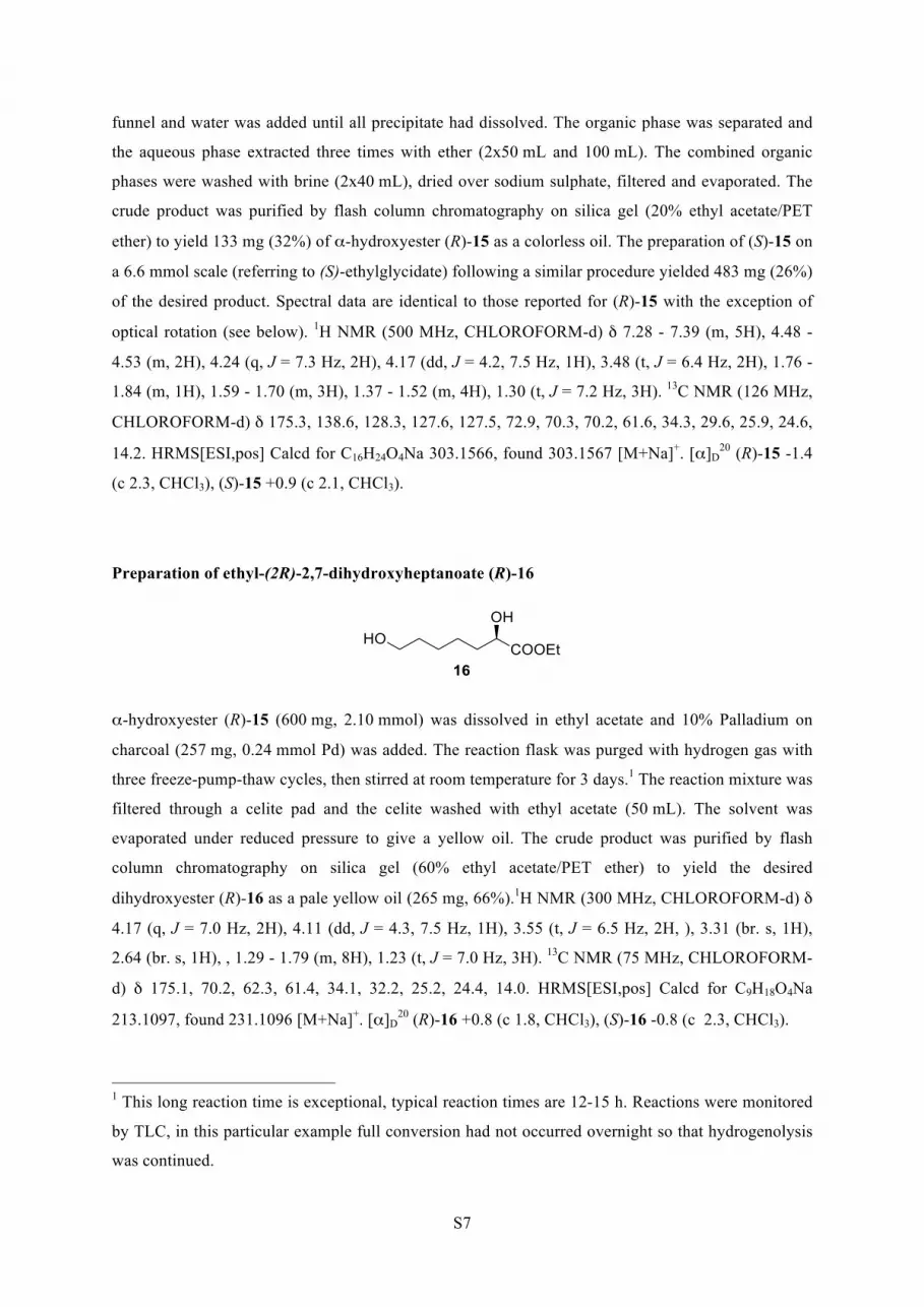
Preparation of ethyl-(2R)-2,7-dihydroxyheptanoate (R)-16
α-hydroxyester (R)-15 (600 mg, 2.10 mmol) was dissolved in ethyl acetate and 10% Palladium on
charcoal (257 mg, 0.24 mmol Pd) was added. The reaction flask was purged with hydrogen gas with
three freeze-pump-thaw cycles, then stirred at room temperature for 3 days.1 The reaction mixture was
filtered through a celite pad and the celite washed with ethyl acetate (50 mL). The solvent was
evaporated under reduced pressure to give a yellow oil. The crude product was purified by flash
column chromatography on silica gel (60% ethyl acetate/PET ether) to yield the desired
dihydroxyester (R)-16 as a pale yellow oil (265 mg, 66%).1H NMR (300 MHz, CHLOROFORM-d) δ
4.17 (q, J = 7.0 Hz, 2H), 4.11 (dd, J = 4.3, 7.5 Hz, 1H), 3.55 (t, J = 6.5 Hz, 2H, ), 3.31 (br. s, 1H),
2.64 (br. s, 1H), , 1.29 - 1.79 (m, 8H), 1.23 (t, J = 7.0 Hz, 3H). 13C NMR (75 MHz, CHLOROFORM-
d) δ 175.1, 70.2, 62.3, 61.4, 34.1, 32.2, 25.2, 24.4, 14.0. HRMS[ESI,pos] Calcd for C9H18O4Na
213.1097, found 231.1096 [M+Na]+. [α]D20 (R)-16 +0.8 (c 1.8, CHCl3), (S)-16 -0.8 (c 2.3, CHCl3).
1 This long reaction time is exceptional, typical reaction times are 12-15 h. Reactions were monitored
by TLC, in this particular example full conversion had not occurred overnight so that hydrogenolysis
was continued.

S8
Preparation of ethyl-(2R)-2,7-di{[bis(benzyloxy)phosphoryl]oxy}heptanoate (R)/(S)-17
(R)/(S)-17 were synthesized from (R)/(S)-17 in an analogous procedure to the synthesis of 11. (R)-
and (S)-17 could be obtained in 64% and 74% yield respectively. Spectral data for the two
enantiomers were identical except for optical rotation, the data for (R)-17 is listed below. 1H NMR
(500 MHz, CHLOROFORM-d) δ 7.29 - 7.39 (m, 20H), 4.99 - 5.18 (m, 8H), 4.77 (td, J= 5.9, 8.0 Hz,
1H), 4.18 (m, 2H), 3.94 (q, J= 6.6 Hz, 2H), 1.77 (q, J = 6.7 Hz, 2H), 1.54 (m, 2H), 1.27 - 1.37 (m,
4H), 1.25 (t, J= 7.2 Hz, 3H). 13C NMR (126 MHz, CHLOROFORM-d) δ 169.9 (d, JPC=3.3 Hz),
135.9, 135.82, 135.80, 135.73, 135.70, 135.6, 128.51, 128.49, 128.47, 128.46, 128.42, 127.90, 127.86,
127.8, 75.4 (d, JPC=5.4 Hz), 69.4 (m), 69.1 (m), 67.6 (d, JPC=6.0 Hz), 61.5, 32.7 (d, JPC=6.4 Hz), 29.9
(d, JPC=6.9 Hz), 24.9, 24.1, 14.1. 31P NMR (121 MHz, CHLOROFORM-d) δ -0.4 (sept, J=7.9 Hz,
1P, P-2), -1.24 (sext, J=7.8 Hz, 1P, P-7). [α]D20 (R)-17 +2.9° (c 2.2, CHCl3), (S)-17 -2.9° (c 2.2,
CHCl3). HRMS[ESI,pos] Calcd for C37H45O10P2 711.2488, found 711.2478 [M+H]+.
INHIBITION STUDIES
Assay conditions are described in the main manuscript. Initial rates were obtained by a linear least
square fit of the initial rate data. Each rate measurement is the average of at least duplicate
measurement with less than 10% standard deviation. Rate measurements were repeated until at least
two rates were found to agree within 10% standard deviation. The negative rates obtained from
consumption of PEP were converted into progress rates using Beer’s Law and the extinction
coefficient of PEP ε=2800 L mol-1 cm-1(30°C, 232 nm) (4). Error bars in the graphs (Figure S1 and
Figure S2) show the standard deviation of rates used to obtain average rates. The kinetic data obtained
was globally fitted to the equation
! =!!"#× !
!!× 1 + !!!
+ !
using the computer program GraFit®.(5) Vmax represents the maximum progress rate, [S] represents
the concentration of the substrate PEP, Km represents the Michaelis-constant for PEP, [I] represents
the concentration of inhibitor present and Ki represents the inhibition constant with respect to PEP.
Grafit® data and fits were replotted using ORIGIN®(6) to allow inclusion of error bars.

S9
Figure S1: Results of Inhibition assays with R-4, represented as Michaelis-Menten-Plot. Inhibitor concentrations: squares – no inhibitor; circles [R-4]= 470 nM; triangles – [R-4]=940 nM. Error bars are standard deviations of averaged rates, [PEP] has an error of ±10%. Solid lines represent the global fit obtained using all assay measurements (21 data points in total) in Grafit® to a model of competitive inhibition, giving Vmax=15 ± 2 µM min-1, Km=120 ± 30 µM , Ki= 360 ± 50 nM.
Figure S2: Results of Inhibition assays with S-4, represented as Michaelis-Menten-Plot. Inhibitor concentrations: squares – no inhibitor; circles [S-4]=390 nM; triangles – [S-4]=780 nM. Error bars are standard deviations of averaged rates. [PEP] has an error of ±10%. Solid lines represent the global fit obtained using all assay measurements (21 data points in total) in Grafit® to a model of competitive inhibition, in Grafit®, giving Vmax=13 ± 2 µM min-1, Km=100 ± 30 µM , Ki= 620 ± 110 nM.

S10
DETAILED INHIBITOR MODELING PROCEDURE AND RESULTS
The structures of both possible diastereoisomers of the linear intermediate and the designed inhibitor
4 were built using the protein builder in Maestro (7) of the Schrödinger Suite 2006 and then
minimized with MacroModel (8). The Polak-Ribiere Conjugate Gradient (PRCG) minimization
method was used with up to 5000 iterations and a gradient convergence threshold of δ=0.05
kJ/(mol*Å). A conformational search was then carried out for each isomer in order to generate an
ensemble of low-energy conformers to find a suitable starting conformation for the later docking
calculation. Conformational searches were conducted with the MCMM Serial Torsional Sampling
method and were run with a GB/SA water model, using the OPLS2005 force field, with 3000 steps for
the conformational search and an energy window of 12.0 kJ/mol for collecting appropriate
conformers. All the conformers from the conformational search were clustered by consideration of
torsional RMS deviation of the backbones, and the representatives were selected based on the
clustering statistics, which were then used in the docking calculations as the starting conformation for
each epimer of the linear intermediate.
The modeling of the linear intermediate epimers into the active site of MtuDAH7PS was carried out
with the Schrödinger Suite 2006 Induced Fit Docking protocol (9). The active site in chain A of the
unliganded crystal structure of MtuDAH7PS was used as the receptor. The center of the receptor grid
was defined as the centroid of residues 87, 126, 133, 134, 135, 136, 137, 248, 250, 280, 282, 283, 284,
306, 337, 366, 369, 380, 382, 409, 411, 440, and 441 as defined in the wild type MtuDAH7PS crystal
structure (PDB-code 2B7O) (10). For the initial docking, the van der Waals radii of the linear
intermediate atoms were scaled to 0.8. The 20 best solutions from the initial docking were kept. All
residues of the protein within a 5 Å distance of the respective linear molecule solution were refined.
The ligands were re-docked, with a van der Waals radius scale of 0.8, to the top 20 newly generated
protein structures if the energy was within 30 kcal/mol of the best protein structure.
The (R)- and (S)-isomers of inhibitor 4 were modeled into MtuDAH7PS by docking flexible inhibitors
into the rigid receptor with the “intermediate adapted” conformation of the active site. This grid is
generated based on the best pose from induced fit modeling of the linear intermediate molecule, from
which the active site conformation, especially the residues in close proximity to the modeled linear
intermediate molecule (within 5 Å), have adopted different conformations from the original crystal
structure (2B7O), to optimize interactions with the modeled linear intermediate. Therefore, this active
site conformation can be considered as “intermediate-adapted” and can be used in modeling of
inhibitors that are designed to mimic the binding mode of the linear intermediate molecule. The
inhibitor docking was conducted in Glide (11) with OPLS2005 force field and extra precision (XP)
mode. 90000 poses per ligand were kept for initial docking, and scoring window for keeping initial
poses was 5000. The best 1000 poses per ligand were kept for energy minimisation with a distance

S11
dependent dielectric constant of 2 and a maximum of 5000 conjugate gradient steps. Ten poses per
ligand were saved for evaluation.
Figure S3: Best pose of inhibitors (magenta carbons) (R)-4 (top) and (S)-4 (bottom) when modeled into the intermediate adapted conformation of MtuDAH7PS (carbon: green, nitrogen: blue, oxygen: red, sulfur: yellow, manganese: gray sphere). All hydrogen atoms except for the C2 hydrogen of 4 highlighting the inhibitor configuration were omitted for clarity, the C2 hydrogen of (S)-4 points away from the viewer into the plane. Enzyme conformation and inhibitor binding closely resemble the experimentally determined structure.

S12
Figure S4: Side-on view (Figure S3 rotated approximately 90° around vertical axis) of the best poses of inhibitors (R)-4 and (S)-4 (magenta carbons) when modeled into the intermediate adapted conformation of MtuDAH7PS. This view illustrates the hydrogen pointing towards ((S)-4) and away from ((R)-4) Glu248 and Trp280 in the foreground on the left. This potential steric clash of (S)-4 with a water molecule (red sphere) coordinated to these residues can be used to rationalize the slightly lower inhibitory potency of (S)-4. The distance between H2 of (S)-4 and the water molecule (black dashed line) is only 1.7 Å.

S13
REFERENCES
1. McDougal, P. G., Rico, J. G., Oh, Y. I. & Condon, B. D. (1986) J. Org. Chem. 51, 3388-3390.
2. Petit, Y. & Larcheveque, M. (1998) Org. Synth. 75, 37-44. 3. Matsuo, Y., Muramatsu, A., Tahara, K., Koide, M. & Nakamura, E.
(2006) Org. Synth. 83, 80-87. 4. Schoner, R. & Herrmann, K. M. (1976) Journal of Biological Chemistry
251, 5440-5447. 5. GraFit, Version 5.0.13, Erithacus Software Limited, 2006. 6. Origin Pro 8.1 SR3, v8.1.34.90, OriginLab Corporation, 2010. 7. Maestro, Version 7.5, Schrodinger, LLC, 2005. 8. MacroModel, Version 9.1, Schrodinger, LLC, 2005. 9. Schrodinger Suite 2006 Induced Fit Protocol; Glide version 4.0; Prime
version 1.5, Schrodinger, LLC, 2005. 10. Webby, C. J., Baker, H. M., Lott, J. S., Baker, E. N. & Parker, E. J.
(2005) J. Mol. Biol. 354, 927-939. 11. Glide, Version 4.0, Schrodinger, LLC, 2005.

and Emily J. ParkerSebastian Reichau, Wanting Jiao, Scott R. Walker, Richard D. Hutton, Edward N. Baker
: COMBINING MECHANISM- AND MODELING-BASED DESIGNtuberculosisMycobacteriumPotent Inhibitors of a Shikimate Pathway Enzyme from
doi: 10.1074/jbc.M110.211649 originally published online March 15, 20112011, 286:16197-16207.J. Biol. Chem.
10.1074/jbc.M110.211649Access the most updated version of this article at doi:
Alerts:
When a correction for this article is posted•
When this article is cited•
to choose from all of JBC's e-mail alertsClick here
Supplemental material:
http://www.jbc.org/content/suppl/2011/03/15/M110.211649.DC1.html
http://www.jbc.org/content/286/18/16197.full.html#ref-list-1
This article cites 43 references, 6 of which can be accessed free at
by guest on April 6, 2016
http://ww
w.jbc.org/
Dow
nloaded from