pdgf induces reorganization of vimentin filaments
TRANSCRIPT

1973Journal of Cell Science 111, 1973-1980 (1998)Printed in Great Britain © The Company of Biologists Limited 1998JCS4567
PDGF induces reorganization of vimentin filaments
Sigrídur Valgeirsdóttir 1,*, Lena Claesson-Welsh 2, Erik Bongcam-Rudloff 3, Ulf Hellman 1, Bengt Westermark 4
and Carl-Henrik Heldin 1
1Ludwig Institute for Cancer Research, Biomedical Center, Uppsala, Sweden2Department of Medical Biochemistry and Microbiology, Biomedical Center, Uppsala, Sweden3Department of Cell Biology, The Wenner-Gren Institute, Stockholm, Sweden4Department of Pathology, Uppsala, Sweden*Author for correspondence
Accepted 11 May; published on WWW 30 June 1998
In this study we demonstrate that stimulation with platelet-derived growth factor (PDGF) leads to a markedreorganization of the vimentin filaments in porcine aorticendothelial (PAE) cells ectopically expressing the PDGF β-receptor. Within 20 minutes after stimulation, the well-spread fine fibrillar vimentin was reorganized as thefilaments aggregated into a dense coil around the nucleus.The solubility of vimentin upon Nonidet-P40-extraction ofcells decreased considerably after PDGF stimulation,indicating that PDGF caused a redistribution of vimentinto a less soluble compartment. In addition, an increasedtyrosine phosphorylation of vimentin was observed. Theredistribution of vimentin was not a direct consequence ofits tyrosine phosphorylation, since treatment of cells withan inhibitor for the cytoplasmic tyrosine kinase Src,attenuated phosphorylation but not redistribution ofvimentin. These changes in the distribution of vimentinoccurred in conjunction with reorganization of actinfilaments. In PAE cells expressing a Y740/751F mutantreceptor that is unable to bind and activatephosphatidylinositol 3′-kinase (PI3-kinase), the
distribution of vimentin was virtually unaffected by PDGFstimulation. Thus, PI3-kinase is important for vimentinreorganization, in addition to its previously demonstratedrole in actin reorganization. The small GTPase Rac haspreviously been shown to be involved downstream of PI3-kinase in the reorganization of actin filaments. In PAE cellsoverexpressing dominant negative Rac1 (N17Rac1), nochange in the fine fibrillar vimentin network was seen afterPDGF-BB stimulation, whereas in PAE cellsoverexpressing constitutively active Rac1 (V12Rac1), therewas a dramatic change in vimentin filament organizationindependent of PDGF stimulation. These data indicate thatPDGF causes a reorganization of microfilaments as well asintermediate filaments in its target cells and suggest animportant role for Rac downstream of PI3-kinase in thePDGF stimulated reorganization of both actin andvimentin filaments.
Key words: Platelet-derived growth factor (PDGF), Vimentin,Phosphatidylinositol 3′-kinase (PI3-kinase), Rac1, Src
SUMMARY
dc
fnt
,
e
In
;ed
INTRODUCTION
Platelet-derived growth factor (PDGF), a mitogen fomesenchymal cells, is a homo- or heterodimeric protecomposed of disulfide-bonded A- and B-polypeptide cha(PDGF-AA, PDGF-BB and PDGF-AB). The PDGF isoformelicit their effects on target cells by binding to two relatetyrosine kinase receptors denoted α- and β-receptors (reviewedby Claesson-Welsh, 1994). The binding of PDGF inducreceptor dimerization leading to activation of the intrinstyrosine kinase activity of the receptors. The kinase activityexerted both on the receptor itself and on intracellusubstrates.
PDGF has been assigned in vivo functions in tissue repand in embryonal development as well as in pathologiprocesses, such as formation of atherosclerotic plaq(reviewed by Heldin and Westermark, 1996). In vitro, PDGstimulates cell division, reorganization of actin microfilamen
rin
inssd
esic islar
aircaluesFts
and chemotaxis of fibroblasts, smooth muscle cells anphagocytic cells. An essential feature of the chemotactiresponse is the formation of lamellipodia with membraneruffling extending in the direction of movement. In adherentcells, membrane ruffling which is caused by reorganization othe actin filament system, appears in response to differegrowth factors (Ridley et al., 1992). Another aspect of actinreorganization is the formation of circular membrane ruffleswhich is a transient response transduced by the PDGF β-receptor (Mellström et al., 1988). Previous studies havindicated a role for phosphatidylinositol 3′-kinase (PI3-kinase)in PDGF-induced actin reorganization and cell motilityresponses (Kundra et al., 1994; Wennström et al., 1994a,b). addition, the small GTP-binding protein Rac, is a majoreffector of PI3-kinase in this pathway (Hawkins et al., 1995Nobes et al., 1995). Thus, microinjection of dominant negativRac1 (N17Rac1) has been shown to abolish PDGF-inducemembrane ruffling, while injection of constitutively active

1974
ctscell 2h2),
mstti-mdnti-nee
l,edrere
rteP-
of in,,
-ngr
nt.5
n 26
re
re
r
by
eyeoror
y
S. Valgeirsdóttir and others
Rac1 (V12Rac1) is sufficient to induce plasma membraruffling and subsequent formation of actin stress fibers in Sw3T3 cells (Ridley et al., 1992).
Intermediate filaments constitute one of the three macomponents of the cytoskeleton of eukaryotic cells, alowith microfilaments and microtubules. Intermediatfilaments comprise a diverse and heterogeneous familyproteins that can be divided into six different groups on tbasis of tissue-specific expression and molecular analyVimentin belongs to class III of intermediate filamenttogether with desmin, glial fibrillary acidic protein andperipherin (van de Klundert et al., 1993; Franke, 1993Vimentin is present in the majority of mesenchymal cetypes and, in addition, it is expressed in most exponentiagrowing cells in culture, independent of which cell typespecific intermediate filament proteins are expressed in viExpression of the vimentin gene is growth regulated several cell types in culture with an altered expression durmitosis and after stimulation of quiescent cells with seruor growth factors (Colucci-Guyon et al., 1994; van dKlundert et al., 1993).
Recent data have documented that intermediate filamentshighly dynamic structures in living cells. The protein subunare continually undergoing a reversible cycling of disassemand reassembly, in response to a variety of stimuli and durthe cell cycle (Steinert et al., 1993). Certain protein kinasincluding protein kinase A, protein kinase C, cdc2 kinase aCa2+-calmodulin-dependent protein kinase II (CaMKII), havbeen shown to phosphorylate the intermediate filamentsserine and threonine residues, and changes in phosphorylaof the intermediate filaments have been suggested as a potemechanism for modulating their assembly and distributi(reviewed by Inagaki et al., 1996). The dynamic, as well mechanical properties, of intermediate filaments appear toexerted via interactions with components at the plasmembrane, at the nuclear envelope and with other cytoskelelements such as microtubules and microfilaments. Studiesthe mechanisms underlying these interactions have revealnumber of intermediate filament-associated proteins that essential for maintaining the integrity of intermediate filamenetwork (reviewed by Chou et al., 1997).
The aim of this study was to examine the effect of PDGstimulation on vimentin organization. We report a remarkabchange in the organization of vimentin after 2-20 minutstimulation with PDGF, with a concomitant decrease Nonidet-P40-soluble vimentin detectable in the cytoplasPI3-kinase and Rac appear to be important for regulationvimentin distribution, in addition to the role in regulating thorganization of actin.
MATERIALS AND METHODS
Cells and tissue culturePorcine aortic endothelial (PAE) cells expressing the wild-type PDGβ-receptor, Y740/751F, Y579F, Y581F mutant PDGF β-receptors orwild-type fibroblast growth factor receptor-1 (FGFR-1), have bedescribed earlier (Mori et al., 1993; Landgren et al., 1995; Wennstret al., 1994b). The PAE cells stably transfected with the PDGFβ-receptor and the Lac repressor (a kind gift from L. Stephens, DepDevelopment and Signalling, the Babraham Inst., Cambridge, U
neiss
jornge ofhesis.s,
).lllly-
vo.iningme
areitsblyinges,nde ontionntial
onas bemaetal on
ed aarent
Fle
esinm. ofe
F
enöm t ofK),
were used as recipient for transfection of mutant Rac1 construcontaining the Lac operon (Hooshmand-Rad et al., 1997). These lines include a dominant negative mutant of Rac1 inducible withmM IPTG (N17Rac1), and constitutively active Rac1 inducible wit50 µM IPTG (V12Rac1). PAE cells were cultured in Ham’s F-1medium (Gibco), supplemented with 10% fetal calf serum (Gibco100 units/ml of penicillin and 100 µg/ml streptomycin.
Antisera and reagentsThe monoclonal anti-phosphotyrosine antibody PY20, was froTransduction Laboratories. The monoclonal antibody againvimentin and fluorescein-isothiocyanate (FITC)-conjugated goat anmouse-Ig antibodies were from DAKOPATTS. The rabbit antiseruagainst PDGF β-receptor (PDGFR-3), has been previously describe(Claesson-Welsh et al., 1989). Peroxidase-conjugated sheep amouse-Ig antibodies were from Amersham. Tetramethylrhodamiisomer R (TRITC)-conjugated phalloidin was from Sigma. Thspecific PI3-kinase inhibitor, 2-(4-morpholinyl)-8-phenyl-4H-1-benzopyran-4-one (LY294002) was purchased from BiomoPlymouth Meeting, PA. The Src specific inhibitor, PP1, was purchasfrom Calbiochem-Novabiochem Corp., La Jolla, CA. The cells weincubated with 1 µM PP1 or DMSO alone for 10 minutes befostimulation with PDGF-BB.
Immunoblotting Serum-starved cells were stimulated with 100 ng/ml PDGF-BB fodifferent time periods at 37°C. After washing with ice-cold phosphabuffered saline (PBS), cells were lysed in a buffer containing 1% N40, 10% glycerol, 20 mM Hepes, pH 7.5, 150 mM NaCl, 1 mMphenylmethylsulfonyl fluoride (PMSF), 1% Trasylol (Bayer), 100 µMNa3VO4. Immunoprecipitation and immunoblotting were performedas previously described (Valgeirsdóttir et al., 1995). For detection tyrosine phosphorylated vimentin, serum-starved cells were lyseda RIPA-buffer containing 1.0% Triton X-100, 1.0% deoxycholic acid0.1% sodium dodecyl sulfate (SDS), 20 mM Tris-HCl, pH 7.4150 mM NaCl, 2.5 mM EDTA, 10% glycerol, 10 mM Na3P2O7, 1 mM PMSF, 1% Trasylol, 100 µM Na3VO4, followed byimmunoprecipitation with PY20 and separation by SDSpolyacrylamide gel electrophoresis (SDS-PAGE) under nonreduciconditions and immunoblotting with antibodies against vimentin. Fopreparation of total cell lysates, cells were stimulated for differetime periods, washed with PBS and thereafter lysed by addition of 0ml boiling 1% SDS, 10 mM Tris-HCl, pH 7.4. The lysates were theboiled for an additional 5 minutes, passed several times through agauge needle and centrifuged for 5 minutes.
ImmunostainingPAE cells expressing the PDGF β-receptors were cultured on glasscoverslips in 6-well plates and serum-starved overnight befostimulation with 100 ng/ml PDGF-BB at 37°C for different timeperiods. After washing three times with PBS at 37°C, the cells wefixed in 1% glutaraldehyde in PBS for 1 minutes at roomtemperature, and permeabilized in 0.5% Triton X-100, 0.5%glutaraldehyde for 4 minutes followed by treatment with NaBH4 for7 minutes. Vimentin was visualized using anti-vimentin antibody fo1 hour, followed by incubation for 1 hour with FITC-conjugatedanti-mouse antibodies. Filamentous actin was detected incubating the cells with 0.1 µg/ml TRITC-conjugated phalloidin(Sigma) for 30 minutes. After washing three times with PBS, thcells were mounted in Fluoromount-G (Southern BiotechnologAssociates Inc., Birmingham, AL), and viewed by fluorescencmicroscopy. In the case where the PI3-kinase specific inhibitLY294002 was used, the cells were incubated with 1.4 µM inhibitat 37°C for 10 minutes prior to stimulation with PDGF. Thisinhibitor concentration leads to 50% inhibition of PI3-kinase activitaccording to previous studies (Vlahos et al., 1994).

1975PDGF-induced vimentin reorganization
dyddrofrd
RESULTS
Reorganization of the vimentin network upon PDGFstimulationTo investigate the effect of PDGF-stimulation on the vimennetwork, PAE cells expressing PDGF β-receptors were seededon coverslips, cultured to subconfluency, and stimulated w
rea.ertse
ginm
heeneenhees,to
of ahlls,le2se
Fig. 1.PDGF induces reorganization of vimentin filaments uponPDGF stimulation. PAE cells expressing the PDGF β-receptor werestimulated with 100 ng/ml PDGF-BB for 0 (A), 10 (B), 20 (C) or 30(D) minutes at 37°C. The cells were then fixed, permeabilized, andstained for distribution of vimentin (green) and actin filaments (red
tin
ith
PDGF for different time periods. The cells were then fixed animmunostained with antibodies against vimentin followed bstaining for filamentous actin, using rhodamine-conjugatephalloidin. In unstimulated cells, vimentin formed an extendenetwork, from the perinuclear region to the cell periphery. Aftestimulation with PDGF, a marked change in the organization the vimentin network was observed (Fig. 1); the fine fibrillaand well-spread vimentin filaments present in unstimulatecells, aggregated into dense structures at the perinuclear aThe maximal effect on vimentin redistribution was seen aft20 minutes of stimulation, thereafter the vimentin filamenstarted to redistribute into the peripheral cytoplasm. Thrhodamine-phalloidin staining revealed actin-containinmembrane ruffles after 2-5 minutes of PDGF-stimulation, agreement with previous reports (Nistér et al., 1988; Wennströet al., 1994b). Circular ruffles were also noticed in some of tcells upon stimulation for 5-20 minutes. In cells with extensivmembrane and circular ruffling, an extensive vimentireorganization was observed, suggesting a cross-talk betwthe actin and vimentin filament networks. We also estimated torganization of vimentin filaments in PAE cells expressing thfibroblast growth factor receptor-1 (FGFR-1). In these cellthere was virtually no reorganization of vimentin in response stimulation with FGF (data not shown).
In unstimulated PAE cells, we detected a small amount vimentin present in a soluble form which was extractable withmild NP-40-containing lysis buffer. This is in agreement witprevious studies showing that in the cytoplasm of cultured cea small amount of the vimentin exists in soluble form, extractabin near-physiological buffers (Soellner et al., 1985). After minutes of stimulation with PDGF there was a marked decrea
).
Fig. 2.Vimentin is redistributed to an NP-40-insoluble pool uponPDGF stimulation. PAE cells expressing the PDGF β-receptor werestimulated with 100 ng/ml PDGF-BB at 37°C for 0, 2, 5, 10, 20 or 30minutes. Cells were lysed with a mild detergent (1% NP-40),separated by SDS-PAGE and immunoblotted with vimentinantibodies (A) or PY20 to evaluate the phosphorylation of the PDGFβ-receptor (C). Parallel cultures of cells were lysed in 1% SDS and10 mM Tris-HCl, pH 7.4, followed by analysis by SDS-PAGE andimmunoblotting with vimentin antibodies (B). The positions ofvimentin (arrow) and PDGF β-receptor (arrowhead) are indicated tothe right.
1976
AE
eithne
hhethn
,e
ed
nt
81loe
r
toreF
S. Valgeirsdóttir and others
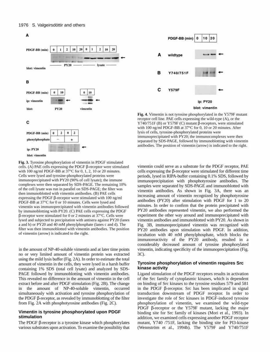
Fig. 3.Tyrosine phosphorylation of vimentin in PDGF stimulatedcells. (A) PAE cells expressing the PDGF β-receptor were stimulatedwith 100 ng/ml PDGF-BB at 37°C for 0, 1, 2, 10 or 20 minutes.Cells were lysed and tyrosine-phosphorylated proteins wereimmunoprecipitated with PY20 (90% of cell lysate); the immunecomplexes were then separated by SDS-PAGE. The remaining 10of the cell lysate was run in parallel on SDS-PAGE; the filter wasthen immunoblotted with vimentin antibodies. (B) PAE cellsexpressing the PDGF β-receptor were stimulated with 100 ng/mlPDGF-BB at 37°C for 0 or 10 minutes. Cells were lysed andvimentin was immunoprecipitated with vimentin antibodies followeby immunoblotting with PY20. (C) PAE cells expressing the PDGFβ-receptor were stimulated for 0 or 2 minutes at 37°C. Cells werelysed and subjected to precipitation with antisera against PY20 (laa and b) or PY20 and 40 mM phenylphosphate (lanes c and d). Thfilter was then immunoblotted with vimentin antibodies. The positioof vimentin (arrow) is indicated to the right.
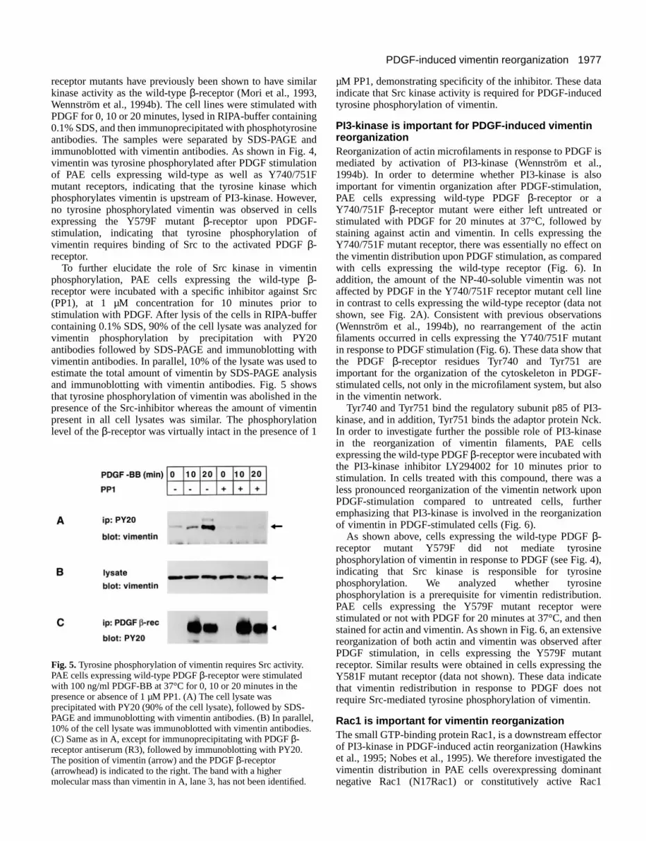
Fig. 4.Vimentin is not tyrosine phosphorylated in the Y579F mutantreceptor cell line. PAE cells expressing the wild-type (A), or theY740/751F (B) or Y579F (C) mutant β-receptors, were stimulatedwith 100 ng/ml PDGF-BB at 37°C for 0, 10 or 20 minutes. Afterlysis of cells, tyrosine-phosphorylated proteins wereimmunoprecipitated with PY20; the immunocomplexes were thenseparated by SDS-PAGE, followed by immunoblotting with vimentinantibodies. The position of vimentin (arrow) is indicated to the right.
in the amount of NP-40-soluble vimentin and at later time poino or very limited amount of vimentin protein was extractusing the mild lysis buffer (Fig. 2A). In order to estimate the toamount of vimentin in the cells, they were lysed in a harsh bucontaining 1% SDS (total cell lysate) and analyzed by SDPAGE followed by immunoblotting with vimentin antibodiesThis revealed no difference in the amount of vimentin in the cextract before and after PDGF stimulation (Fig. 2B). The chanin the amount of NP-40-soluble vimentin, occurresimultaneously with activation and tyrosine phosphorylation the PDGF β-receptor, as revealed by immunoblotting of the filtfrom Fig. 2A with phosphotyrosine antibodies (Fig. 2C).
Vimentin is tyrosine phosphorylated upon PDGFstimulationThe PDGF β-receptor is a tyrosine kinase which phosphorylatvarious substrates upon activation. To examine the possibility
ntsedtalfferS-.ellgedof
er
esthat
vimentin could serve as a substrate for the PDGF receptor, Pcells expressing the β-receptor were stimulated for different timeperiods, lysed in RIPA-buffer containing 0.1% SDS, followed byimmunoprecipitation with phosphotyrosine antibodies. Thsamples were separated by SDS-PAGE and immunoblotted wvimentin antibodies. As shown in Fig. 3A, there was aincreasing amount of vimentin recognized by phosphotyrosinantibodies (PY20) after stimulation with PDGF for 1 to 20minutes. In order to confirm that the protein precipitated witPY20 antibodies represented vimentin, we also performed texperiment the other way around and immunoprecipitated wivimentin antibodies and immunoblotted with PY20. As shown iFig. 3B, immunoprecipitated vimentin was recognized withPY20 antibodies upon stimulation with PDGF. In additionincubation with 40 mM phenylphosphate, which blocks thimmunoreactivity of the PY20 antibody, resulted in aconsiderably decreased amount of tyrosine phosphorylatvimentin, indicating specificity of the immunoprecipitation (Fig.3C).
Tyrosine phosphorylation of vimentin requires Srckinase activityLigand stimulation of the PDGF receptors results in activatioof the Src family of cytoplasmic kinases, which is dependenon binding of Src kinases to the tyrosine residues 579 and 5in the PDGF β-receptor. Src has been implicated in signatransduction downstream of PDGF receptor. In order tinvestigate the role of Src kinases in PDGF-induced tyrosinphosphorylation of vimentin, we examined the wild-typePDGF β-receptor or the Y579F mutant, lacking the majobinding site for Src family of kinases (Mori et al., 1993). Inaddition, we examined cells expressing another PDGF recepmutant, Y740 /751F, lacking the binding site for PI3-kinas(Wennström et al., 1994b). The Y579F and Y740/751
%
d
nesen
1977PDGF-induced vimentin reorganization
tad
isl.,so,
ryheond
noteot
nstinnt
hate-
so
-k.
ses
receptor mutants have previously been shown to have simkinase activity as the wild-type β-receptor (Mori et al., 1993,Wennström et al., 1994b). The cell lines were stimulated wPDGF for 0, 10 or 20 minutes, lysed in RIPA-buffer containin0.1% SDS, and then immunoprecipitated with phosphotyrosantibodies. The samples were separated by SDS-PAGE immunoblotted with vimentin antibodies. As shown in Fig. vimentin was tyrosine phosphorylated after PDGF stimulatiof PAE cells expressing wild-type as well as Y740/751mutant receptors, indicating that the tyrosine kinase whphosphorylates vimentin is upstream of PI3-kinase. Howevno tyrosine phosphorylated vimentin was observed in ceexpressing the Y579F mutant β-receptor upon PDGF-stimulation, indicating that tyrosine phosphorylation ovimentin requires binding of Src to the activated PDGF β-receptor.
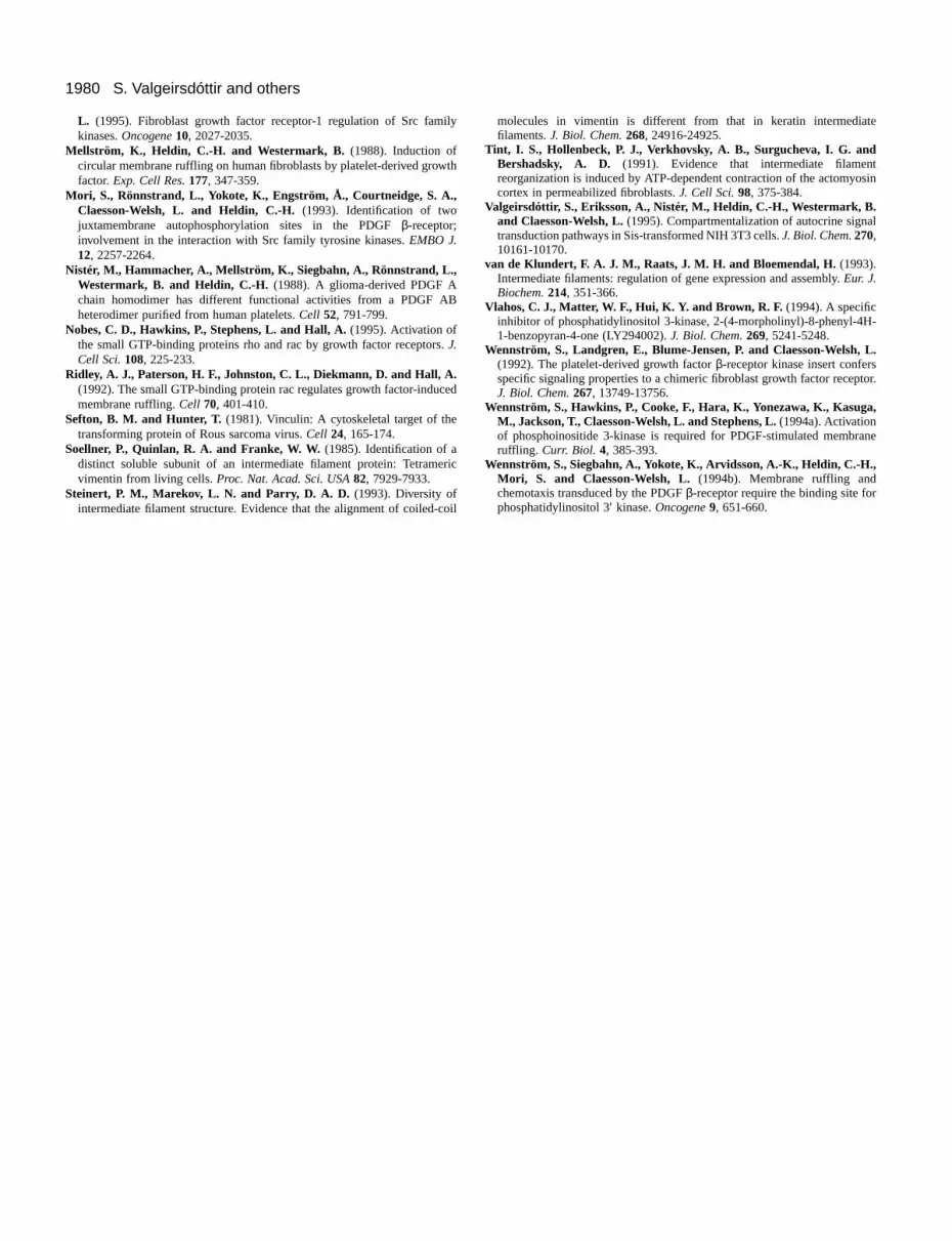
To further elucidate the role of Src kinase in vimentphosphorylation, PAE cells expressing the wild-type β-receptor were incubated with a specific inhibitor against S(PP1), at 1 µM concentration for 10 minutes prior stimulation with PDGF. After lysis of the cells in RIPA-buffecontaining 0.1% SDS, 90% of the cell lysate was analyzedvimentin phosphorylation by precipitation with PY20antibodies followed by SDS-PAGE and immunoblotting witvimentin antibodies. In parallel, 10% of the lysate was usedestimate the total amount of vimentin by SDS-PAGE analyand immunoblotting with vimentin antibodies. Fig. 5 showthat tyrosine phosphorylation of vimentin was abolished in tpresence of the Src-inhibitor whereas the amount of vimenpresent in all cell lysates was similar. The phosphorylatilevel of the β-receptor was virtually intact in the presence of
aonr
on
e4),ee.ren
veternttheateot
tors
thet1
Fig. 5.Tyrosine phosphorylation of vimentin requires Src activity.PAE cells expressing wild-type PDGF β-receptor were stimulatedwith 100 ng/ml PDGF-BB at 37°C for 0, 10 or 20 minutes in thepresence or absence of 1 µM PP1. (A) The cell lysate wasprecipitated with PY20 (90% of the cell lysate), followed by SDS-PAGE and immunoblotting with vimentin antibodies. (B) In parallel10% of the cell lysate was immunoblotted with vimentin antibodies(C) Same as in A, except for immunoprecipitating with PDGF β-receptor antiserum (R3), followed by immunoblotting with PY20.The position of vimentin (arrow) and the PDGF β-receptor(arrowhead) is indicated to the right. The band with a highermolecular mass than vimentin in A, lane 3, has not been identified
ilar
ithgineand4,onF
icher,lls
f
in
rctor for
h tosisshetinon 1
µM PP1, demonstrating specificity of the inhibitor. These daindicate that Src kinase activity is required for PDGF-inducetyrosine phosphorylation of vimentin.
PI3-kinase is important for PDGF-induced vimentinreorganizationReorganization of actin microfilaments in response to PDGFmediated by activation of PI3-kinase (Wennström et a1994b). In order to determine whether PI3-kinase is alimportant for vimentin organization after PDGF-stimulationPAE cells expressing wild-type PDGF β-receptor or aY740/751F β-receptor mutant were either left untreated ostimulated with PDGF for 20 minutes at 37°C, followed bstaining against actin and vimentin. In cells expressing tY740/751F mutant receptor, there was essentially no effect the vimentin distribution upon PDGF stimulation, as comparewith cells expressing the wild-type receptor (Fig. 6). Iaddition, the amount of the NP-40-soluble vimentin was naffected by PDGF in the Y740/751F receptor mutant cell linin contrast to cells expressing the wild-type receptor (data nshown, see Fig. 2A). Consistent with previous observatio(Wennström et al., 1994b), no rearrangement of the acfilaments occurred in cells expressing the Y740/751F mutain response to PDGF stimulation (Fig. 6). These data show tthe PDGF β-receptor residues Tyr740 and Tyr751 arimportant for the organization of the cytoskeleton in PDGFstimulated cells, not only in the microfilament system, but alin the vimentin network.
Tyr740 and Tyr751 bind the regulatory subunit p85 of PI3kinase, and in addition, Tyr751 binds the adaptor protein NcIn order to investigate further the possible role of PI3-kinain the reorganization of vimentin filaments, PAE cellexpressing the wild-type PDGF β-receptor were incubated withthe PI3-kinase inhibitor LY294002 for 10 minutes prior tostimulation. In cells treated with this compound, there wasless pronounced reorganization of the vimentin network upPDGF-stimulation compared to untreated cells, furtheemphasizing that PI3-kinase is involved in the reorganizatiof vimentin in PDGF-stimulated cells (Fig. 6).
As shown above, cells expressing the wild-type PDGF β-receptor mutant Y579F did not mediate tyrosinphosphorylation of vimentin in response to PDGF (see Fig. indicating that Src kinase is responsible for tyrosinphosphorylation. We analyzed whether tyrosinphosphorylation is a prerequisite for vimentin redistributionPAE cells expressing the Y579F mutant receptor westimulated or not with PDGF for 20 minutes at 37°C, and thestained for actin and vimentin. As shown in Fig. 6, an extensireorganization of both actin and vimentin was observed afPDGF stimulation, in cells expressing the Y579F mutareceptor. Similar results were obtained in cells expressing Y581F mutant receptor (data not shown). These data indicthat vimentin redistribution in response to PDGF does nrequire Src-mediated tyrosine phosphorylation of vimentin.
Rac1 is important for vimentin reorganizationThe small GTP-binding protein Rac1, is a downstream effecof PI3-kinase in PDGF-induced actin reorganization (Hawkinet al., 1995; Nobes et al., 1995). We therefore investigated vimentin distribution in PAE cells overexpressing dominannegative Rac1 (N17Rac1) or constitutively active Rac
,.
.

1978
isF,s,
edr
of
al,gin
tins
S. Valgeirsdóttir and others
Fig. 6.PI3-kinase is important for vimentin reorganization. PAE cellsexpressing the wild-type PDGF β-receptor (A and B) the Y740/751Fmutant PDGF β-receptor (C and D) or Y579F mutant β-receptor (Gand H), were stimulated with 100 ng/ml PDGF-BB for 0 or 20minutes at 37°C. The cells were then fixed and stained fordistribution of vimentin (green) and actin (red) filaments. (E and F)PAE cells expressing the PDGF β-receptor were incubated with thePI3-kinase inhibitor LY294002 (1.4 µM), for 10 minutes prior tostimulation and then analyzed as described above.
Fig. 7.Correlation between reorganization of actin and vimentinfilaments in PAE cells overexpressing Rac1 mutants. Serum-starvedcells were incubated for 20 minutes in the absence (A,C,E,G) or inthe presence (B,D,F,H) of 100 ng/ml PDGF-BB. The IPTG-inducibleclones (N17Rac1 and V12Rac1) were either induced (C,D,G,H) ornot induced (A,B,E,F) with IPTG for 16 hours prior to stimulationwith PDGF. The cells were fixed and stained for the distribution ofvimentin filaments (green) and actin filaments (red).
+LY294002
(V12Rac1), in an isopropyl β-D-galactopyranoside (IPTG)-regulated manner. The cells were either left untreated stimulated with PDGF for 20 minutes at 37°C, with or withouprior induction with IPTG, followed by staining for vimentinand actin filaments. In IPTG-treated cells overexpressN17Rac1, PDGF stimulation failed to induce membraruffling, in agreement with previous studies (Hooshmand-Ret al., 1997; Ridley et al., 1992). Furthermore, there was change in the fine fibrillar vimentin network in these cells response to PDGF (Fig. 7A). On the other hand, in ceoverexpressing V12Rac1 upon IPTG induction, a dramachange in vimentin filament organization was observeincluding a collapse of the filaments into dense structuindependent of PDGF stimulation (Fig. 7B). As previousreported (Hooshmand-Rad et al., 1997), these cells ashowed extensive membrane ruffling independent of PDGstimulation. Thus, these data show that Rac1 is an imporeffector of the reorganization of both vimentin filaments anactin filaments.
ort
ingneadnoinllsticd,
reslylsoF
tantd
DISCUSSION
In this study we show that activation of the PDGF β-receptorleads to reorganization of the vimentin filament network. Theffect was seen after 5-10 minutes of stimulation with PDGwith a maximum effect after 20 minutes of stimulation, avisualized by immunostaining of vimentin. Furthermoreimmunoblotting of vimentin from PDGF-stimulated cellsrevealed that the amount of NP-40-soluble vimentin decreasin the lysate after 2 minutes of stimulation. However, to ouknowledge, this is the first report demonstrating an effect short-term PDGF-stimulation on vimentin filamentdistribution. It has previously been reported that corneepithelial cells treated with 10 ng/ml PDGF-AB for 24 hoursshow a disorganization and disruption of actin-containinmicrofilament bundles as well as of microtubules and vimentfilaments (Knorr et al., 1992).
Prior studies have shown a dramatic reorganization of acfilaments in cells stimulated with PDGF, with loss of stres

1979PDGF-induced vimentin reorganization
ted
he,F
t,refl
e
sto
dyal
In
tal
t
fibers and formation of edge ruffles with a maximum effect afabout 2-5 minutes of stimulation and circular ruffles appearsomewhat later. The results of the present investigation shocorrelation between PDGF-induced actin reorganization vimentin reorganization. Thus, in cells expressing the PDreceptor mutant Y740/751F which is unable to bind and activPI3-kinase, or in wild-type receptor expressing cells treawith the PI3-kinase inhibitor LY294002, neither actin nvimentin filaments were rearranged upon PDGF stimulatiFurthermore, reorganization of actin or vimentin filaments wnot detected in cells expressing dominant negative Rawhereas expression of constitutively active Rac1 led tocollapse of both the actin and the vimentin filament systeindependent of PDGF. These observations suggest thcommon signal transduction pathway is involved in treorganization of both microfilaments and intermediafilaments. Our finding that activation of the FGFR-1, did nresult in reorganization of vimentin further supports this notiosince FGFR-1 does not activate PI3-kinase (Wennström et1992). It remains to be determined whether vimentin filamreorganization is dependent on prior reorganization of the afilament system. Previous studies have suggested thatmaintenance of the intermediate filament network requintact microtubule and microfilament systems. Thus, a drinduced aggregation of the vimentin filaments relies on interaction with the microfilament system of the ce(Hollenbeck et al., 1989; Tint et al., 1991). Direct interactibetween vimentin filaments and microfilament/microfilameassociated proteins have also been reported and it appearthe C-terminal domain of vimentin is involved in the binding actin filaments (Cary et al., 1994). In addition, peptide-inducdisruption of the vimentin network in fibroblasts waaccompanied by rounding-up of cells and disassembly of bmicrotubules and microfilaments, indicating a role for tintermediate filaments in maintaining cell shape, cytoskeleintegrity and mechanical property of the cytoplasm in vi(Goldman et al., 1996). In contrast, fibroblasts from vimentknockout mice did not show any significant modifications their microtubular and actin filament systems (Colucci-Guyet al., 1994). However, it remains to be determined whetfibroblasts from vimentin-deficient mice retain the ability form actin-containing lamellipodia or circular ruffles iresponse to growth factor stimulation.
There is accumulating evidence that the organizationintermediate filaments is regulated by phosphorylatiPhosphorylation of the intermediate filaments by specprotein kinases promotes disassembly of the filamefollowed by subsequent dephosphorylation by phosphataallowing spontaneous reassembly (Steinert et al., 19Vimentin has been shown to be phosphorylated by cerprotein serine/threonine kinases which phosphorylate the hdomain of vimentin. In this study we report that PDGF inductyrosine phosphorylation of vimentin. Vimentin was tyrosinphosphorylated in PAE cells expressing the Y740/751F mureceptor, to a similar extent as in cells expressing the wild-treceptor. On the other hand, tyrosine phosphorylation wabolished in PAE cells treated with a Src specific inhibi(PP1) and in cells expressing a PDGF β-receptor mutated onthe Src kinase binding site, Tyr 579, indicating that the PDGinduced tyrosine phosphorylation of vimentin is mediated members of the Src family. This is in line with previous da
teringw a
andGFatetedoron.asc1, a
ms,at aheteotn, al.,entctin theiresug-thellsonnt-s thattoedsoth
hetal
voin-ofonherton
ofon.ificnts,ses
93).taineadese
tantype
astor
F-byta
showing that trace amounts of phosphotyrosine were detecin vimentin in chicken cells transformed with v-src, whereasvimentin isolated from normal chicken cells did not containany detectable phosphotyrosine (Sefton and Hunter, 1981). Tvimentin filaments as well as actin microfilaments werehowever, extensively reorganized in response to PDGstimulation in PAE cells expressing the Y579F mutanindicating that tyrosine phosphorylation is not required foPDGF-induced reorganization of vimentin filaments. Thfunctional role of PDGF-induced tyrosine phosphorylation ovimentin remains to be determined. About 0.5% of the totaamount of vimentin was recognized by phosphotyrosinantibodies after PDGF-stimulation. An interesting possibilityis that tyrosine phosphorylated vimentin binds and activateSH2-domain containing molecules and thus contributes signal transduction events.
We thank Len Stephens for kindly providing the N17Rac1 anV12Rac1 mutant PAE cell lines, Pontus Aspenström for criticallreading the manuscript and Ingegärd Schiller for secretariassistance.
REFERENCES
Cary, R. B., Klymkowsky, M. W., Evans, R. M., Domingo, A., Dent, J. A.and Backhus, L. E.(1994). Vimentin’s tail interacts with actin-containingstructures in vivo. J. Cell Sci.107, 1609-1622.
Chou, Y.-H., Skalli, O. and Goldman, R. D.(1997). Intermediate filamentsand cytoplasmic networking: new connections and more functions. Curr.Opin. Cell Biol.9, 49-53.
Claesson-Welsh, L., Hammacher, A., Westermark, B., Heldin, C.-H. andNistér, M. (1989). Identification and structural analysis of the A typereceptor for platelet-derived growth factor. Similarities with the B typereceptor. J. Biol. Chem.264, 1742-1747.
Claesson-Welsh, L.(1994). Platelet-derived growth factor receptor signals. J.Biol. Chem.269, 32023-32026.
Colucci-Guyon, E., Portier, M.-M., Dunia, I., Paulin, D., Pournin, S. andBabinet, C. (1994). Mice lacking vimentin develop and reproduce withoutan obvious phenotype. Cell 79, 679-694.
Franke, W. W. (1993). The intermediate filaments and associated proteins. Guidebook to the Cytoskeletal and Motor Proteins(ed. T. Kreis and R. Vale),pp. 137-143. Oxford University Press Inc., New York.
Goldman, R. D., Khuon, S., Chou, Y. H., Opal, P. and Steinert, P. M.(1996). The function of intermediate filaments in cell shape and cytoskeleintegrity. J. Cell Biol.134, 971-983.
Hawkins, P. T., Eguinoa, A., Qiu, R.-G., Stokoe, D., Cooke, F. T., Walters,R., Wennström, S., Claesson-Welsh, L., Evans, T., Symons, M. andStephens, L. (1995). PDGF stimulates an increase in GTP-Rac viaactivation of phosphoinositide 3-kinase. Curr. Biol. 5, 393-403.
Heldin, C.-H. and Westermark, B. (1996). Role of platelet-derived growthfactor in vivo. In The Molecular and Cellular Biology of Wound Repair(ed.R. A. F. Clark), pp. 249-273. Plenum Press, New York.
Hollenbeck, P. J., Bershadsky, A. D., Pletjushkina, O. Y., Tint, I. S. andVasiliev, J. M. (1989). Intermediate filament collapse is an ATP-dependenand actin-dependent process. J. Cell Sci.92, 621-631.
Hooshmand-Rad, R., Claesson-Welsh, L., Wennström, S., Yokote, K.,Siegbahn, A. and Heldin, C.-H. (1997). Involvement ofphosphatidylinositide 3′-kinase and Rac in platelet-derived growth factor-induced actin reorganization and chemotaxis. Exp. Cell Res.234, 434-441.
Inagaki, M., Matsuoka, Y., Tsujimura, K. ando, S., Tokui, T., Takahashi,T. and Inagaki, N. (1996). Dynamic property of intermediate filaments:regulation by phosphorylation. BioEssays18, 481-487.
Knorr, M., Hoppe, J., Steuhl, K. P. and Dartsch, P. C.(1992). Effect ofPDGF-AB heterodimer on a corneal epithelial cell line. Eur. J. Cell Biol.57, 202-209.
Kundra, V., Escobedo, J. A., Kazlauskas, A., Kim, H. K., Rhee, S. G.,Williams, L. T. and Zetter, B. R. (1994). Regulation of chemotaxis by theplatelet-derived growth factor receptor-β. Nature367, 474-476.
Landgren, E., Blume-Jensen, P., Courtneidge, S. A. and Claesson-Welsh,
1980
e
sin
l
or.
ne
S. Valgeirsdóttir and others
L. (1995). Fibroblast growth factor receptor-1 regulation of Src famikinases. Oncogene10, 2027-2035.
Mellström, K., Heldin, C.-H. and Westermark, B. (1988). Induction ofcircular membrane ruffling on human fibroblasts by platelet-derived growfactor. Exp. Cell Res.177, 347-359.
Mori, S., Rönnstrand, L., Yokote, K., Engström, Å., Courtneidge, S. A.,Claesson-Welsh, L. and Heldin, C.-H.(1993). Identification of twojuxtamembrane autophosphorylation sites in the PDGF β-receptor;involvement in the interaction with Src family tyrosine kinases. EMBO J.12, 2257-2264.
Nistér, M., Hammacher, A., Mellström, K., Siegbahn, A., Rönnstrand, L.,Westermark, B. and Heldin, C.-H. (1988). A glioma-derived PDGF Achain homodimer has different functional activities from a PDGF Aheterodimer purified from human platelets. Cell 52, 791-799.
Nobes, C. D., Hawkins, P., Stephens, L. and Hall, A.(1995). Activation ofthe small GTP-binding proteins rho and rac by growth factor receptorsJ.Cell Sci.108, 225-233.
Ridley, A. J., Paterson, H. F., Johnston, C. L., Diekmann, D. and Hall, A.(1992). The small GTP-binding protein rac regulates growth factor-inducmembrane ruffling. Cell 70, 401-410.
Sefton, B. M. and Hunter, T. (1981). Vinculin: A cytoskeletal target of thetransforming protein of Rous sarcoma virus. Cell 24, 165-174.
Soellner, P., Quinlan, R. A. and Franke, W. W.(1985). Identification of adistinct soluble subunit of an intermediate filament protein: Tetramevimentin from living cells. Proc. Nat. Acad. Sci. USA82, 7929-7933.
Steinert, P. M., Marekov, L. N. and Parry, D. A. D.(1993). Diversity ofintermediate filament structure. Evidence that the alignment of coiled-c
ly
th
B
.
ed
ric
oil
molecules in vimentin is different from that in keratin intermediatfilaments. J. Biol. Chem.268, 24916-24925.
Tint, I. S., Hollenbeck, P. J., Verkhovsky, A. B., Surgucheva, I. G. andBershadsky, A. D. (1991). Evidence that intermediate filamentreorganization is induced by ATP-dependent contraction of the actomyocortex in permeabilized fibroblasts. J. Cell Sci.98, 375-384.
Valgeirsdóttir, S., Eriksson, A., Nistér, M., Heldin, C.-H., Westermark, B.and Claesson-Welsh, L.(1995). Compartmentalization of autocrine signatransduction pathways in Sis-transformed NIH 3T3 cells. J. Biol. Chem.270,10161-10170.
van de Klundert, F. A. J. M., Raats, J. M. H. and Bloemendal, H.(1993).Intermediate filaments: regulation of gene expression and assembly. Eur. J.Biochem.214, 351-366.
Vlahos, C. J., Matter, W. F., Hui, K. Y. and Brown, R. F.(1994). A specificinhibitor of phosphatidylinositol 3-kinase, 2-(4-morpholinyl)-8-phenyl-4H-1-benzopyran-4-one (LY294002). J. Biol. Chem.269, 5241-5248.
Wennström, S., Landgren, E., Blume-Jensen, P. and Claesson-Welsh, L.(1992). The platelet-derived growth factor β-receptor kinase insert confersspecific signaling properties to a chimeric fibroblast growth factor receptJ. Biol. Chem.267, 13749-13756.
Wennström, S., Hawkins, P., Cooke, F., Hara, K., Yonezawa, K., Kasuga,M., Jackson, T., Claesson-Welsh, L. and Stephens, L.(1994a). Activationof phosphoinositide 3-kinase is required for PDGF-stimulated membraruffling. Curr. Biol. 4, 385-393.
Wennström, S., Siegbahn, A., Yokote, K., Arvidsson, A.-K., Heldin, C.-H.,Mori, S. and Claesson-Welsh, L. (1994b). Membrane ruffling andchemotaxis transduced by the PDGF β-receptor require the binding site forphosphatidylinositol 3′ kinase. Oncogene9, 651-660.