pandemic (h1n1) 2009 influenza virus induces weaker host immune responses in vitro: a possible...
TRANSCRIPT
RESEARCH Open Access
Pandemic (H1N1) 2009 influenza virus inducesweaker host immune responses in vitro:a possible mechanism of high transmissibilitySanjay Mukherjee, Veena C Vipat, Akhilesh C Mishra, Shailesh D Pawar and Alok K Chakrabarti*
Abstract
Background: The world has recently overcome the first influenza pandemic of the 21st century caused by a novelH1N1 virus (pH1N1) which is a triple reassortant comprising genes derived from avian, human, and swine influenzaviruses and antigenically quite different from seasonal H1N1 strains. Although the case fatality rates have decreasedin many developed countries, the situation is still alarming in many developing countries including India whereconsiderable numbers of new cases are appearing everyday. There is still a high morbidity and mortality ofsusceptible adult as well as young population without having underlying health issues due to the influenzainfection.
Results: To achieve a better understanding of the risk posed by the pH1N1 and to understand its pathogenicity,we studied the host gene expression response to Indian isolate of pH1N1 infection and compared it with seasonalH1N1 infection. The response was studied at four different time points (4, 8, 16 and 24 h) post infection (hpi) inA549 cells using microarray platform. We found that pH1N1 induces immune response earlier than seasonal H1N1viruses, but at the later stages of infection there is a suppression of host immune responses. The infection withpH1N1 resulted in considerable decrease in the expression of cytokine and other immune genes namely IL8,STAT1, B2 M and IL4 compared to seasonal H1N1.
Conclusion: We propose that the inability to induce strong innate immune response could be a reason for thehigh transmissibility, pathogenicity and mortality caused by pH1N1 virus.
BackgroundThe pandemic (H1N1) 2009 influenza A virus (pH1N1)has already killed more than 19,000 people worldwidesince it appeared in April 2009 [1]. Although on 10th ofAugust 2010, the Director General of the World HealthOrganization (WHO) has announced that the world isno longer in phase 6 of influenza pandemic alert and weare now moving into the post-pandemic period, thevirus transmission is still highly active in many parts ofSouth Asia, West Africa, and Central America [2]. InAsia, the most active areas of pandemic influenza virustransmission currently are in parts of India, Bangladesh,Bhutan, Myanmar Nepal, and Thailand. The virus(pH1N1) is still a serious threat to children as well as
susceptible young and old population in developingcountries like India.Till date, there are reports of 2720 deaths from pan-
demic H1N1 influenza virus infection in India which isapproximately 14% of the total world mortality http://mohfw-h1n1.nic.in/august.html; http://netindian.in/news/2010/11/15/0008699/6-h1n1-deaths-india-during-past-week-govt. The pandemic H1N1 virus is antigeni-cally distinct from seasonal influenza viruses and themajority of human population lacks immunity againstthis virus [3-5]. The pathogenesis and transmission ofthe pH1N1 in humans is not completely known andmany studies are underway. Animal studies have shownthat this virus has a higher replicative power than theseasonal influenza virus [6,7]. Studies on human macro-phages have shown that pH1N1 is a weak inducer ofcytokine responses as compared to seasonal H1N1viruses [8]. In addition, pH1N1 replicates efficiently in
* Correspondence: [email protected] Containment Complex, National Institute of Virology, Sus Road,Pashan, Pune -411021, India
Mukherjee et al. Virology Journal 2011, 8:140http://www.virologyj.com/content/8/1/140
© 2011 Mukherjee et al; licensee BioMed Central Ltd. This is an Open Access article distributed under the terms of the CreativeCommons Attribution License (http://creativecommons.org/licenses/by/2.0), which permits unrestricted use, distribution, andreproduction in any medium, provided the original work is properly cited.
non-human primates, causes more severe pathologicallesions in the lungs than currently circulating seasonalhuman H1N1 virus [9,10]. Earlier findings indicate thatpandemic H1N1 are more pathogenic in mammalianmodels than seasonal H1N1 influenza viruses[6-8,11-13]. The pH1N1 isolates tested in mice and fer-rets, were found to be replicating more efficiently thancurrently circulating human H1N1 viruses [6-8].The respiratory tract is the primary site of infection
for all the mammalian influenza viruses [13,14]. In thisstudy we have used human lung epithelial cells (A549)to study the host gene expression responses to infectionwith Indian isolate of pH1N1, isolated in August 2009and compared it with seasonal H1N1 infection in orderto assess the pathogenicity and transmissibility of pan-demic (2009) H1N1 influenza virus.
Materials and methodsViruses and cell linePandemic Influenza virus A/Jalna/NIV9436/2009(H1N1) and seasonal human influenza virus A/NIV/0914864/2009(H1N1) isolated in the influenza divisionof the National Institute of Virology, Pune, India wereused for the study. Human lung epithelial (A549) cellline was used as host and maintained in Dulbecco’smodified Eagle’s tissue culture medium (Invitrogen LifeTechnologies, Carlsbad, CA, USA) containing 10% fetalcalf serum,100 units/ml penicillin, 100 ug/ml streptomy-cin in tissue culture flasks (Corning, USA) at 37°C in aCO2 incubator.
Virus infectionA549 cells were infected with pH1N1 and seasonalinfluenza viruses at a multiplicity of infection (MOI) of3 as described earlier [15]. After 1 hour of adsorptionperiod, the inoculum was removed and the cells werewashed twice with phosphate buffer saline (PBS) andsupplemented with growth media. Four sets of tissueculture flasks containing monolayer of A549 cells wereinfected corresponding to four different time points postinfection for both the viruses. Mock infected cells ateach time point served as controls. Infection of pH1N1was performed in BSL-2 laboratory following WorldHealth Organization norm for handling of pandemicH1N1 viruses.
Microarray HybridizationInfected cells were harvested at different time pointspost infection, total RNA was extracted from theinfected cells at 4, 8, 16 and 24 hpi using Trizol reagent(Invitrogen Life Technologies, Carlsbad, CA, USA) andpurified using the RNeasy kit (Qiagen, Germany).Amplification of RNA and indirect labeling of Cy-dyewas done using Amino Allyl MessageAmp II aRNA
amplification kit (Ambion, Austin, TX, USA) followingmanufacturer’s instruction. One hundred nanograms oftotal RNA from control and infected cells were used forthe experiments. The RNA was reverse transcribed andamplified according to manufacturer’s protocol. Thepurified amino allyl aRNA was labeled with Cy3 andCy5 for control and experimental samples respectively.Purified samples were lyophilized, resuspended in hybri-dization buffer (Pronto Universal Hybridization kit,Corning) and hybridized on human Discover chip(Arrayit corporation, Sunnyvale, CA, USA). Hybridiza-tion was carried out in a Hybstation (GenomicSolutions, Ann Arbor, MI) and the conditions used were55°C for 6 h, 50°C for 6 h, and 42°C for 6 h. Scanningwas performed at 5-micron resolutions with the Scanarray express (PerkinElmer, Waltham, MI). Grid align-ment was done by gene annotation files and raw datawere extracted into MS EXCEL [15].
Data AnalysisMicroarray data analysis was carried out with GENO-WIZ Microarray data and pathway analysis tool (Oci-mum Biosolutions, Hyderabad, India). Replicated valuesfor genes were merged and median values of theexpression ratios were considered for the dataset. Unde-tected spots were removed by filtering. Dye bias wasnullified by applying Loess normalization. Log transfor-mation (log2) was done to stabilize the variation indataset and median centering was performed to bringdown data distribution of dataset close to zero. In orderto detect highly expressed genes, fold change analysiswas done. Genes with 1.5 folds up/down-regulationwere considered as differentially expressed at a p-value<0.05, Student’s t-test. Functional classification of thegenes was performed using gene ontology and pathwayanalysis [15].
Quantitative RT-PCR of host genes using SYBR Green IMicroarray gene expression data was validated by quan-titative RT-PCR as described earlier [15]. The PCR reac-tion was performed in triplicates using ABI 7300real-time PCR system (Applied Biosystems, Foster City,CA, USA) with Quanti Tect SYBR green RT-PCR kit(Qiagen, Germany). Reaction efficiency was calculatedby using serial 10-fold dilutions of the housekeepinggene- b-actin and the sample genes. Melting curve ana-lysis was performed to verify product specificity. Allquantitations (threshold cycle [CT] values) were normal-ized to that of b-actin to generate ΔCT, and the differ-ence among the ΔCT value of the sample and that ofthe reference (uninfected sample) was calculated as-ΔΔCT. The relative level of gene expression wasexpressed as 2-ΔΔCT. Primer sequences for the genes ofinterest were obtained from primer bank and also
Mukherjee et al. Virology Journal 2011, 8:140http://www.virologyj.com/content/8/1/140
Page 2 of 10
designed using Primer Express (Applied Biosystems).The primer sequences used in this study are as follows:Beta-Actin_F 5’- CATGAAGTGTGACGTGGA-
CATCC-3’; Beta-Actin_R 5’-GCTGATCCACATCTGCTGGAAGG-3’; TNFRSF1A_F 5’-TTGCATCCTAGCCCAGCAG-3’; TNFRSF1A_R 5’-CTGACCCTGGAAAGAAAAGTC-3’; IL8_F 5’-TGCCAAGGAGTGCTAAAG-3’; IL8_R 5’-CTCCACAACCCTCTGCAC-3’;B2 M (Beta-2-microglobin)_F 5’-ATGTCTCGCTCCGTGGCCTTA-3’; B2 M (Beta-2-microglobin)_R 5’-ATCTTGGGCTGTGACAAAGTC-3’; STAT1_F 5’- CCATCCTTTGGTACAACATGC-3’; STAT1_R 5’-TGCACATGGTGGAGTCAGG-3’; IFNb_F 5’-CAGCAATTTTCAGTGTCAGAAGC-3’; IFNb_R 5’-TCATCCTGTCCTTGAGGCAGT-3’.
Western Blot analysisTotal cellular protein from control and infected A549 cellsat different time points post infection were isolated usingRIPA lysis buffer. Equal amount of proteins (10 μg) fromcell extracts were separated by 12.5% SDS-polyacrylamidegel electrophoresis (12.5% SDS-PAGE) and transferred toHybond-C (Amersham Biosciences) membrane with anelectrotransfer apparatus (Cleaver Scientific Ltd) at 10Volts (100 mA) for 1 h 30 min. Primary and secondaryantibody interaction was performed in phosphate-bufferedsaline (pH 7.5). Primary antibodies used were rabbit anti-STAT1, mouse anti-CASP3 and mouse anti-b-actin anti-body (Santa Cruz Biotechnology, Inc. Santa Cruz, CA.USA). The secondary antibodies were mouse anti-rabbitand goat anti-mouse secondary antibodies (Santa CruzBiotechnology, Inc. Santa Cruz, CA. USA) labeled withHorseradish peroxidase (HRP). The protein bands weredeveloped with 3, 3’-Diaminobenzidine tetrahydrochloride(DABT) and Hydrogen peroxide (H2O2) staining.
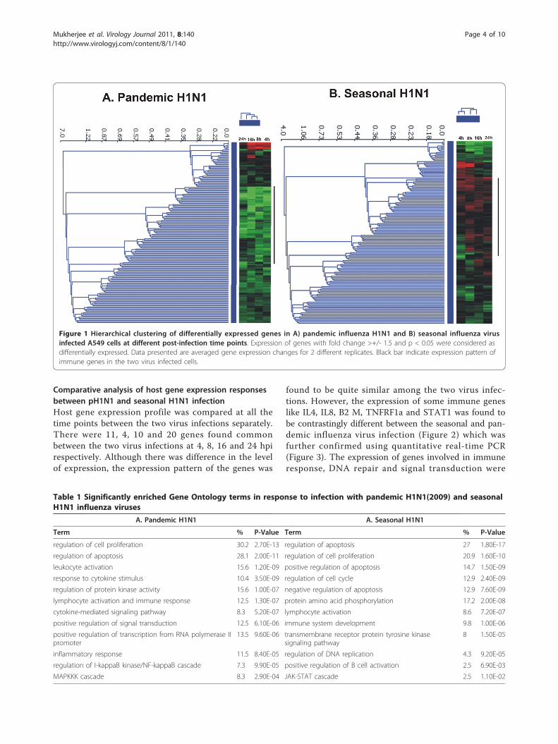
ResultsHost gene expression profile in response to pandemic(H1N1) 2009 virus infectionThe gene expression profile to pH1N1 infection wasstudied at 4 different time points post infection in orderto understand the host responses at different stages ofvirus infection. Figure 1A shows overall gene expressionprofile in response to pH1N1 infection. The genesshowing increased expression compared to controls atall the time points were mainly involved in T-cell activa-tion and proliferation and enzyme linked protein signal-ing whereas, genes showing decrease in expression weremostly involved in regulation of apoptosis and NF-�Bmediated signaling (Table 1A).At the early stages of virus infection i.e. at 4 hpi, we
found up-regulation of immune genes like TNF-a3,EGR-1, IL6R, v-FOS, v-JUN. However, IL13RA, IL3RA,IL4, STAT1, STAT4 were down-regulated at this time
point of infection. At 8 hpi there was further increase inthe expression of EGR-1, IL6R, TNF-a3, v-FOS andv-JUN genes and up-regulation of other immune respon-sive genes like IL-8. Surprisingly, we observed morenumber of immune genes getting down-regulated at thisstage as compared to 4 hpi. The down-regulation of thisset of immune genes was more prominent at later stages(16 and 24 hpi) of infection with pandemic H1N1. Genesinvolved in intracellular signaling and DNA repair likeTopoisomerase II, MAP2K6 were also found to be down-regulated at 8 hpi. Higher expression of IL8, TNF-a3,TNFR-6, CXCR4, EGR-1, v-JUN was found at 16 hpi.Gene coding for IL13RA showed continued down-regula-tion at this stage of infection. Interestingly, at 24 h postinfection there was no further increase in immuneresponsive genes but down-regulation of STAT4, TNFR,IL4R, and TNF6 genes were found.
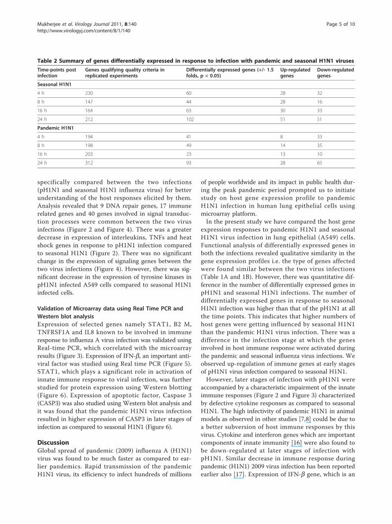
Host gene expression profile in response to seasonalH1N1 virus infectionIn case of seasonal H1N1 infection there was up-regula-tion of very few immune responsive genes at the earlystage (4 hpi) of infection. In fact, there was down-regu-lation of genes involved in innate immune response likeIL2, IL15 and STAT1 at early stages (4 and 8 hpi) ofinfection. Figure 1B shows hierarchical clustering ofoverall cellular gene expression profile in response toseasonal H1N1 virus infection in A549 cells. Geneontology analysis of differentially expressed genes at allthe time points showed not much differences in thetype and functions of host genes affected by the pH1N1and seasonal influenza virus infections (Table 1A andTable 1B). The total number of differentially expressedgenes at different post infection time points during thetwo virus infections is given in Table 2.At 4 hour post infection with seasonal H1N1, genes
encoding for the ribosomal proteins and DNA modify-ing enzymes were up-regulated and continued to beup-regulated at all the time points post infection studiedin this experiment. Increased level of the expression ofimmune genes was observed from 16 hpi. Cytokines likeCCL5, small inducible cytokine A2, IL8 and otherimmune responsive genes like STAT1, IRF1 and B2 Mwere found to be up-regulated at this time point postinfection with seasonal influenza virus. At 24 hpi therewas further increase in the expression of immune andribosomal genes. There was up-regulation of additionaltranscription factors and signaling molecules at 24 hpi.However, some of the cytokines, signaling genes andDNA repair genes were selectively down regulated at allthe post infection time points. These genes mainlyincluded IGF2R, Topoisomerase I and IL13RA1. Genesinvolved in cell cycle like Cyclin G1, G2 were down-regulated at all the time points post infection.
Mukherjee et al. Virology Journal 2011, 8:140http://www.virologyj.com/content/8/1/140
Page 3 of 10
Comparative analysis of host gene expression responsesbetween pH1N1 and seasonal H1N1 infectionHost gene expression profile was compared at all thetime points between the two virus infections separately.There were 11, 4, 10 and 20 genes found commonbetween the two virus infections at 4, 8, 16 and 24 hpirespectively. Although there was difference in the levelof expression, the expression pattern of the genes was
found to be quite similar among the two virus infec-tions. However, the expression of some immune geneslike IL4, IL8, B2 M, TNFRF1a and STAT1 was found tobe contrastingly different between the seasonal and pan-demic influenza virus infection (Figure 2) which wasfurther confirmed using quantitative real-time PCR(Figure 3). The expression of genes involved in immuneresponse, DNA repair and signal transduction were
Figure 1 Hierarchical clustering of differentially expressed genes in A) pandemic influenza H1N1 and B) seasonal influenza virusinfected A549 cells at different post-infection time points. Expression of genes with fold change >+/- 1.5 and p < 0.05 were considered asdifferentially expressed. Data presented are averaged gene expression changes for 2 different replicates. Black bar indicate expression pattern ofimmune genes in the two virus infected cells.
Table 1 Significantly enriched Gene Ontology terms in response to infection with pandemic H1N1(2009) and seasonalH1N1 influenza viruses
A. Pandemic H1N1 A. Seasonal H1N1
Term % P-Value Term % P-Value
regulation of cell proliferation 30.2 2.70E-13 regulation of apoptosis 27 1.80E-17
regulation of apoptosis 28.1 2.00E-11 regulation of cell proliferation 20.9 1.60E-10
leukocyte activation 15.6 1.20E-09 positive regulation of apoptosis 14.7 1.50E-09
response to cytokine stimulus 10.4 3.50E-09 regulation of cell cycle 12.9 2.40E-09
regulation of protein kinase activity 15.6 1.00E-07 negative regulation of apoptosis 12.9 7.60E-09
lymphocyte activation and immune response 12.5 1.30E-07 protein amino acid phosphorylation 17.2 2.00E-08
cytokine-mediated signaling pathway 8.3 5.20E-07 lymphocyte activation 8.6 7.20E-07
positive regulation of signal transduction 12.5 6.10E-06 immune system development 9.8 1.00E-06
positive regulation of transcription from RNA polymerase IIpromoter
13.5 9.60E-06 transmembrane receptor protein tyrosine kinasesignaling pathway
8 1.50E-05
inflammatory response 11.5 8.40E-05 regulation of DNA replication 4.3 9.20E-05
regulation of I-kappaB kinase/NF-kappaB cascade 7.3 9.90E-05 positive regulation of B cell activation 2.5 6.90E-03
MAPKKK cascade 8.3 2.90E-04 JAK-STAT cascade 2.5 1.10E-02
Mukherjee et al. Virology Journal 2011, 8:140http://www.virologyj.com/content/8/1/140
Page 4 of 10
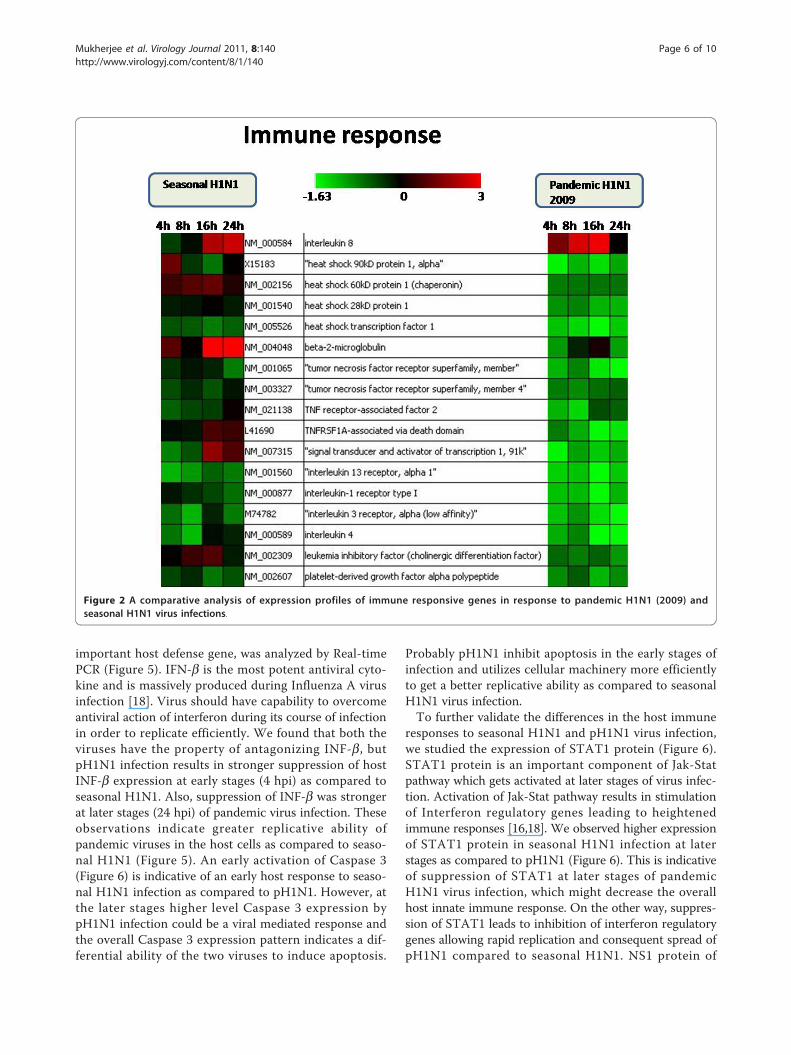
specifically compared between the two infections(pH1N1 and seasonal H1N1 influenza virus) for betterunderstanding of the host responses elicited by them.Analysis revealed that 9 DNA repair genes, 17 immunerelated genes and 40 genes involved in signal transduc-tion processes were common between the two virusinfections (Figure 2 and Figure 4). There was a greaterdecrease in expression of interleukins, TNFs and heatshock genes in response to pH1N1 infection comparedto seasonal H1N1 (Figure 2). There was no significantchange in the expression of signaling genes between thetwo virus infections (Figure 4). However, there was sig-nificant decrease in the expression of tyrosine kinases inpH1N1 infected A549 cells compared to seasonal H1N1infected cells.
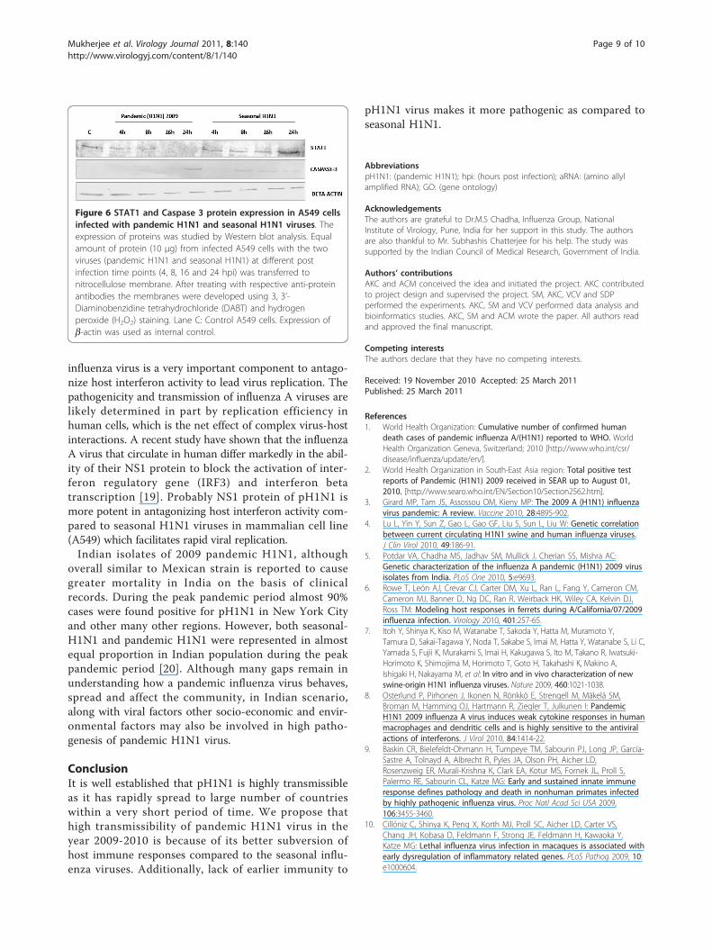
Validation of Microarray data using Real Time PCR andWestern blot analysisExpression of selected genes namely STAT1, B2 M,TNFRSF1A and IL8 known to be involved in immuneresponse to influenza A virus infection was validated usingReal-time PCR, which correlated with the microarrayresults (Figure 3). Expression of IFN-b, an important anti-viral factor was studied using Real time PCR (Figure 5).STAT1, which plays a significant role in activation ofinnate immune response to viral infection, was furtherstudied for protein expression using Western blotting(Figure 6). Expression of apoptotic factor, Caspase 3(CASP3) was also studied using Western blot analysis andit was found that the pandemic H1N1 virus infectionresulted in higher expression of CASP3 in later stages ofinfection as compared to seasonal H1N1 (Figure 6).
DiscussionGlobal spread of pandemic (2009) influenza A (H1N1)virus was found to be much faster as compared to ear-lier pandemics. Rapid transmission of the pandemicH1N1 virus, its efficiency to infect hundreds of millions
of people worldwide and its impact in public health dur-ing the peak pandemic period prompted us to initiatestudy on host gene expression profile to pandemicH1N1 infection in human lung epithelial cells usingmicroarray platform.In the present study we have compared the host gene
expression responses to pandemic H1N1 and seasonalH1N1 virus infection in lung epithelial (A549) cells.Functional analysis of differentially expressed genes inboth the infections revealed qualitative similarity in thegene expression profiles i.e. the type of genes affectedwere found similar between the two virus infections(Table 1A and 1B). However, there was quantitative dif-ference in the number of differentially expressed genes inpH1N1 and seasonal H1N1 infections. The number ofdifferentially expressed genes in response to seasonalH1N1 infection was higher than that of the pH1N1 at allthe time points. This indicates that higher numbers ofhost genes were getting influenced by seasonal H1N1than the pandemic H1N1 virus infection. There was adifference in the infection stage at which the genesinvolved in host immune response were activated duringthe pandemic and seasonal influenza virus infections. Weobserved up-regulation of immune genes at early stagesof pH1N1 virus infection compared to seasonal H1N1.However, later stages of infection with pH1N1 were
accompanied by a characteristic impairment of the innateimmune responses (Figure 2 and Figure 3) characterizedby defective cytokine responses as compared to seasonalH1N1. The high infectivity of pandemic H1N1 in animalmodels as observed in other studies [7,8] could be due toa better subversion of host immune responses by thisvirus. Cytokine and interferon genes which are importantcomponents of innate immunity [16] were also found tobe down-regulated at later stages of infection withpH1N1. Similar decrease in immune response duringpandemic (H1N1) 2009 virus infection has been reportedearlier also [17]. Expression of IFN-b gene, which is an
Table 2 Summary of genes differentially expressed in response to infection with pandemic and seasonal H1N1 viruses
Time-points postinfection
Genes qualifying quality criteria inreplicated experiments
Differentially expressed genes (+/- 1.5folds, p < 0.05)
Up-regulatedgenes
Down-regulatedgenes
Seasonal H1N1
4 h 230 60 28 32
8 h 147 44 28 16
16 h 164 63 30 33
24 h 212 102 51 51
Pandemic H1N1
4 h 194 41 8 33
8 h 198 49 14 35
16 h 203 23 13 10
24 h 312 93 28 65
Mukherjee et al. Virology Journal 2011, 8:140http://www.virologyj.com/content/8/1/140
Page 5 of 10
important host defense gene, was analyzed by Real-timePCR (Figure 5). IFN-b is the most potent antiviral cyto-kine and is massively produced during Influenza A virusinfection [18]. Virus should have capability to overcomeantiviral action of interferon during its course of infectionin order to replicate efficiently. We found that both theviruses have the property of antagonizing INF-b, butpH1N1 infection results in stronger suppression of hostINF-b expression at early stages (4 hpi) as compared toseasonal H1N1. Also, suppression of INF-b was strongerat later stages (24 hpi) of pandemic virus infection. Theseobservations indicate greater replicative ability ofpandemic viruses in the host cells as compared to seaso-nal H1N1 (Figure 5). An early activation of Caspase 3(Figure 6) is indicative of an early host response to seaso-nal H1N1 infection as compared to pH1N1. However, atthe later stages higher level Caspase 3 expression bypH1N1 infection could be a viral mediated response andthe overall Caspase 3 expression pattern indicates a dif-ferential ability of the two viruses to induce apoptosis.
Probably pH1N1 inhibit apoptosis in the early stages ofinfection and utilizes cellular machinery more efficientlyto get a better replicative ability as compared to seasonalH1N1 virus infection.To further validate the differences in the host immune
responses to seasonal H1N1 and pH1N1 virus infection,we studied the expression of STAT1 protein (Figure 6).STAT1 protein is an important component of Jak-Statpathway which gets activated at later stages of virus infec-tion. Activation of Jak-Stat pathway results in stimulationof Interferon regulatory genes leading to heightenedimmune responses [16,18]. We observed higher expressionof STAT1 protein in seasonal H1N1 infection at laterstages as compared to pH1N1 (Figure 6). This is indicativeof suppression of STAT1 at later stages of pandemicH1N1 virus infection, which might decrease the overallhost innate immune response. On the other way, suppres-sion of STAT1 leads to inhibition of interferon regulatorygenes allowing rapid replication and consequent spread ofpH1N1 compared to seasonal H1N1. NS1 protein of
Figure 2 A comparative analysis of expression profiles of immune responsive genes in response to pandemic H1N1 (2009) andseasonal H1N1 virus infections.
Mukherjee et al. Virology Journal 2011, 8:140http://www.virologyj.com/content/8/1/140
Page 6 of 10
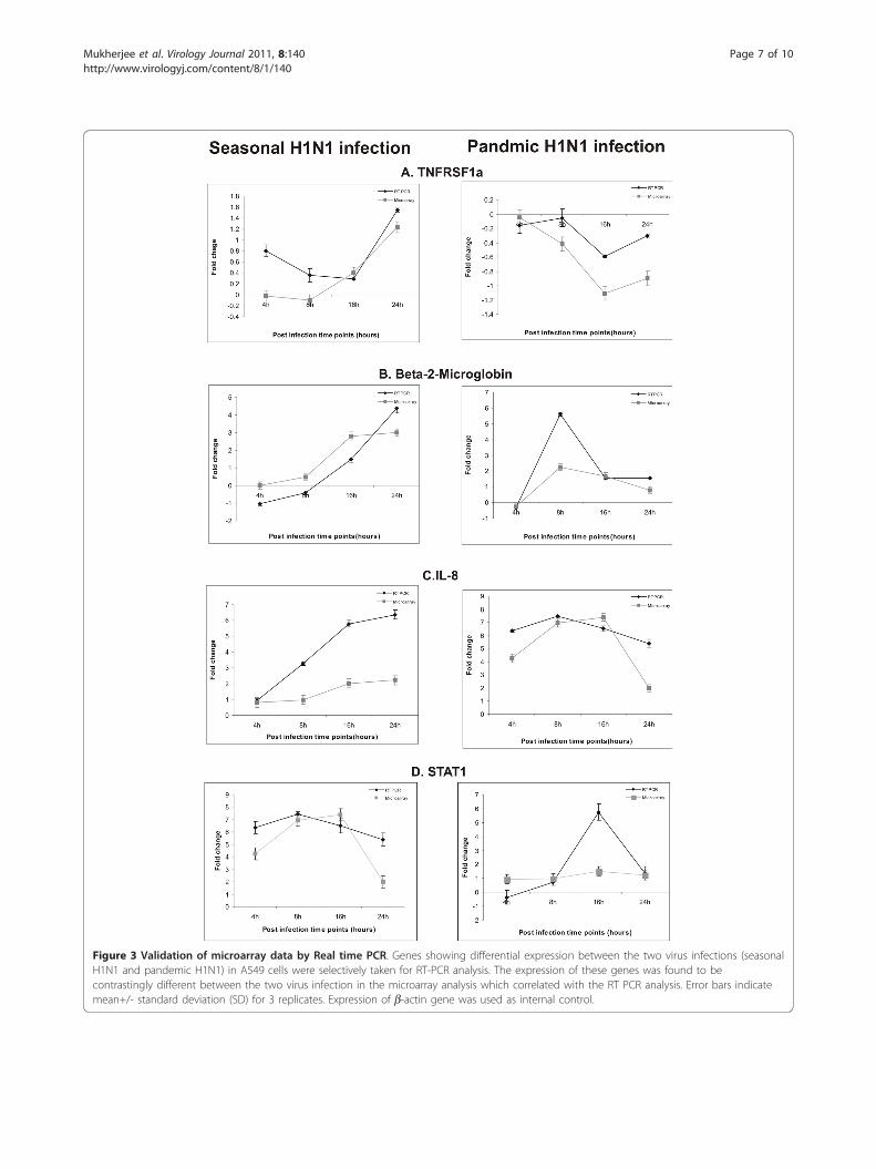
Figure 3 Validation of microarray data by Real time PCR. Genes showing differential expression between the two virus infections (seasonalH1N1 and pandemic H1N1) in A549 cells were selectively taken for RT-PCR analysis. The expression of these genes was found to becontrastingly different between the two virus infection in the microarray analysis which correlated with the RT PCR analysis. Error bars indicatemean+/- standard deviation (SD) for 3 replicates. Expression of b-actin gene was used as internal control.
Mukherjee et al. Virology Journal 2011, 8:140http://www.virologyj.com/content/8/1/140
Page 7 of 10
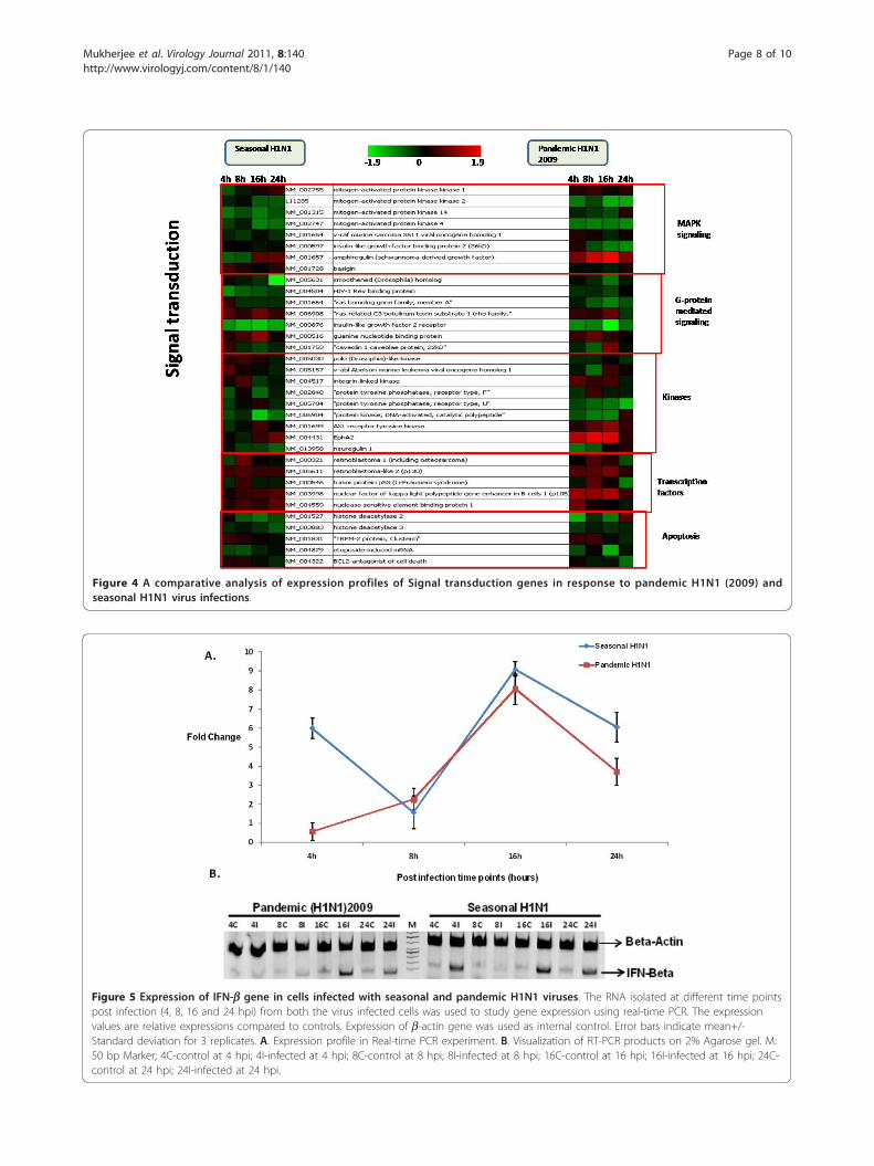
Figure 4 A comparative analysis of expression profiles of Signal transduction genes in response to pandemic H1N1 (2009) andseasonal H1N1 virus infections.
Figure 5 Expression of IFN-b gene in cells infected with seasonal and pandemic H1N1 viruses. The RNA isolated at different time pointspost infection (4, 8, 16 and 24 hpi) from both the virus infected cells was used to study gene expression using real-time PCR. The expressionvalues are relative expressions compared to controls. Expression of b-actin gene was used as internal control. Error bars indicate mean+/-Standard deviation for 3 replicates. A. Expression profile in Real-time PCR experiment. B. Visualization of RT-PCR products on 2% Agarose gel. M:50 bp Marker; 4C-control at 4 hpi; 4I-infected at 4 hpi; 8C-control at 8 hpi; 8I-infected at 8 hpi; 16C-control at 16 hpi; 16I-infected at 16 hpi; 24C-control at 24 hpi; 24I-infected at 24 hpi.
Mukherjee et al. Virology Journal 2011, 8:140http://www.virologyj.com/content/8/1/140
Page 8 of 10
influenza virus is a very important component to antago-nize host interferon activity to lead virus replication. Thepathogenicity and transmission of influenza A viruses arelikely determined in part by replication efficiency inhuman cells, which is the net effect of complex virus-hostinteractions. A recent study have shown that the influenzaA virus that circulate in human differ markedly in the abil-ity of their NS1 protein to block the activation of inter-feron regulatory gene (IRF3) and interferon betatranscription [19]. Probably NS1 protein of pH1N1 ismore potent in antagonizing host interferon activity com-pared to seasonal H1N1 viruses in mammalian cell line(A549) which facilitates rapid viral replication.Indian isolates of 2009 pandemic H1N1, although
overall similar to Mexican strain is reported to causegreater mortality in India on the basis of clinicalrecords. During the peak pandemic period almost 90%cases were found positive for pH1N1 in New York Cityand other many other regions. However, both seasonal-H1N1 and pandemic H1N1 were represented in almostequal proportion in Indian population during the peakpandemic period [20]. Although many gaps remain inunderstanding how a pandemic influenza virus behaves,spread and affect the community, in Indian scenario,along with viral factors other socio-economic and envir-onmental factors may also be involved in high patho-genesis of pandemic H1N1 virus.
ConclusionIt is well established that pH1N1 is highly transmissibleas it has rapidly spread to large number of countrieswithin a very short period of time. We propose thathigh transmissibility of pandemic H1N1 virus in theyear 2009-2010 is because of its better subversion ofhost immune responses compared to the seasonal influ-enza viruses. Additionally, lack of earlier immunity to
pH1N1 virus makes it more pathogenic as compared toseasonal H1N1.
AbbreviationspH1N1: (pandemic H1N1); hpi: (hours post infection); aRNA: (amino allylamplified RNA); GO: (gene ontology)
AcknowledgementsThe authors are grateful to Dr.M.S Chadha, Influenza Group, NationalInstitute of Virology, Pune, India for her support in this study. The authorsare also thankful to Mr. Subhashis Chatterjee for his help. The study wassupported by the Indian Council of Medical Research, Government of India.
Authors’ contributionsAKC and ACM conceived the idea and initiated the project. AKC contributedto project design and supervised the project. SM, AKC, VCV and SDPperformed the experiments. AKC, SM and VCV performed data analysis andbioinformatics studies. AKC, SM and ACM wrote the paper. All authors readand approved the final manuscript.
Competing interestsThe authors declare that they have no competing interests.
Received: 19 November 2010 Accepted: 25 March 2011Published: 25 March 2011
References1. World Health Organization: Cumulative number of confirmed human
death cases of pandemic influenza A/(H1N1) reported to WHO. WorldHealth Organization Geneva, Switzerland; 2010 [http://www.who.int/csr/disease/influenza/update/en/].
2. World Health Organization in South-East Asia region: Total positive testreports of Pandemic (H1N1) 2009 received in SEAR up to August 01,2010. [http://www.searo.who.int/EN/Section10/Section2562.htm].
3. Girard MP, Tam JS, Assossou OM, Kieny MP: The 2009 A (H1N1) influenzavirus pandemic: A review. Vaccine 2010, 28:4895-902.
4. Lu L, Yin Y, Sun Z, Gao L, Gao GF, Liu S, Sun L, Liu W: Genetic correlationbetween current circulating H1N1 swine and human influenza viruses.J Clin Virol 2010, 49:186-91.
5. Potdar VA, Chadha MS, Jadhav SM, Mullick J, Cherian SS, Mishra AC:Genetic characterization of the influenza A pandemic (H1N1) 2009 virusisolates from India. PLoS One 2010, 5:e9693.
6. Rowe T, León AJ, Crevar CJ, Carter DM, Xu L, Ran L, Fang Y, Cameron CM,Cameron MJ, Banner D, Ng DC, Ran R, Weirback HK, Wiley CA, Kelvin DJ,Ross TM: Modeling host responses in ferrets during A/California/07/2009influenza infection. Virology 2010, 401:257-65.
7. Itoh Y, Shinya K, Kiso M, Watanabe T, Sakoda Y, Hatta M, Muramoto Y,Tamura D, Sakai-Tagawa Y, Noda T, Sakabe S, Imai M, Hatta Y, Watanabe S, Li C,Yamada S, Fujii K, Murakami S, Imai H, Kakugawa S, Ito M, Takano R, Iwatsuki-Horimoto K, Shimojima M, Horimoto T, Goto H, Takahashi K, Makino A,Ishigaki H, Nakayama M, et al: In vitro and in vivo characterization of newswine-origin H1N1 influenza viruses. Nature 2009, 460:1021-1038.
8. Osterlund P, Pirhonen J, Ikonen N, Rönkkö E, Strengell M, Mäkelä SM,Broman M, Hamming OJ, Hartmann R, Ziegler T, Julkunen I: PandemicH1N1 2009 influenza A virus induces weak cytokine responses in humanmacrophages and dendritic cells and is highly sensitive to the antiviralactions of interferons. J Virol 2010, 84:1414-22.
9. Baskin CR, Bielefeldt-Ohmann H, Tumpeye TM, Sabourin PJ, Long JP, García-Sastre A, Tolnayd A, Albrecht R, Pyles JA, Olson PH, Aicher LD,Rosenzweig ER, Murali-Krishna K, Clark EA, Kotur MS, Fornek JL, Proll S,Palermo RE, Sabourin CL, Katze MG: Early and sustained innate immuneresponse defines pathology and death in nonhuman primates infectedby highly pathogenic influenza virus. Proc Natl Acad Sci USA 2009,106:3455-3460.
10. Cillóniz C, Shinya K, Peng X, Korth MJ, Proll SC, Aicher LD, Carter VS,Chang JH, Kobasa D, Feldmann F, Strong JE, Feldmann H, Kawaoka Y,Katze MG: Lethal influenza virus infection in macaques is associated withearly dysregulation of inflammatory related genes. PLoS Pathog 2009, 10:e1000604.
Figure 6 STAT1 and Caspase 3 protein expression in A549 cellsinfected with pandemic H1N1 and seasonal H1N1 viruses. Theexpression of proteins was studied by Western blot analysis. Equalamount of protein (10 μg) from infected A549 cells with the twoviruses (pandemic H1N1 and seasonal H1N1) at different postinfection time points (4, 8, 16 and 24 hpi) was transferred tonitrocellulose membrane. After treating with respective anti-proteinantibodies the membranes were developed using 3, 3’-Diaminobenzidine tetrahydrochloride (DABT) and hydrogenperoxide (H2O2) staining. Lane C: Control A549 cells. Expression ofb-actin was used as internal control.
Mukherjee et al. Virology Journal 2011, 8:140http://www.virologyj.com/content/8/1/140
Page 9 of 10
11. Bermejo-Martin JF, Martin-Loeches I, Rello J, Antón A, Almansa R, Xu L,Lopez-Campos G, Pumarola T, Ran L, Ramirez P, Banner D, Cheuk Ng D,Socias L, Loza A, Andaluz D, Maravi E, Gómez-Sánchez MJ, Gordón M,Gallegos MC, Fernandez V, Aldunate S, León C, Merino P, Blanco J, Martin-Sanchez F, Rico L, Varillas D, Iglesias V, Marcos MA, Gandía F, Bobillo F,Nogueira B, Rojo S, Resino S, Castro C, Ortiz de Lejarazu R, Kelvin D: Hostadaptive immunity deficiency in severe pandemic influenza. Crit Care2010, 14:R167.
12. Giamarellos-Bourboulis EJ, Raftogiannis M, Antonopoulou A, Baziaka F,Koutoukas P, Savva A, Kanni T, Georgitsi M, Pistiki A, Tsaganos T,Pelekanos N, Athanassia S, Galani L, Giannitsioti E, Kavatha D, Kontopidou F,Mouktaroudi M, Poulakou G, Sakka V, Panagopoulos P, Papadopoulos A,Kanellakopoulou K, Giamarellou H: Effect of the novel influenza A (H1N1)virus in the human immune system. PLoS One 2009, 4:e8393.
13. Lee SMY, Gardy JL, Cheung CY, Cheung TKW, Hui KPY, Ip NY, Guan Y,Hancock REW, Peiris JSM: Systems-Level Comparison of Host-ResponsesElicited by Avian H5N1 and Seasonal H1N1 Influenza Viruses in PrimaryHuman Macrophages. PLoS One 2009, 4:e8072.
14. Mansfield KG: Viral Tropism and the Pathogenesis of Influenza in theMammalian Host. American Journal of Pathology 2007, 171:1089-1092.
15. Chakrabarti AK, Vipat VC, Mukherjee S, Singh R, Pawar SD, Mishra AC: Hostgene expression profiling in influenza A virus-infected lung epithelial(A549) cells: A comparative analysis between highly pathogenic andmodified viruses. Virol J 2010, 7:219.
16. Koyama S, Ishii KJ, Coban C, Akira S: Innate immune response to viralinfection. Cytokine 2008, 43:336-341.
17. Arankalle VA, Lole KS, Arya RP, Tripathy AS, Ramdasi AY, Chadha MS,Sangle SA, Kadam DB: Role of host immune response and viral load inthe differential outcome of pandemic H1N1 (2009) influenza virusinfection in Indian patients. PLoS One 2010, 5:e13099.
18. Le Goffic RL, Bouguyon E, Chevalier C, Vidic J, Da Costa B, Leymarie O,Bourdieu C, Decamps L, Dhorne-Pollet S, Delmas B: Influenza A VirusProtein PB1-F2 exacerbates IFN-b Expression of Human RespiratoryEpithelial Cells. J Immunol 2010, 185:4812-4823.
19. Kuo RL, Zhao C, Malur M, Krug RM: Influenza A virus strains that circulatein humans differ in the ability of their NS1 proteins to block theactivation of IRF3 and interferon-β transcription. J Virol 2010, 408:146-158.
20. Mishra AC, Chadha MS, Choudhary ML, Potdar VA: Pandemic Influenza(H1N1) 2009 Is Associated with Severe Disease in India. PLoS One 2010,5:e10540.
doi:10.1186/1743-422X-8-140Cite this article as: Mukherjee et al.: Pandemic (H1N1) 2009 influenzavirus induces weaker host immune responses in vitro: a possiblemechanism of high transmissibility. Virology Journal 2011 8:140.
Submit your next manuscript to BioMed Centraland take full advantage of:
• Convenient online submission
• Thorough peer review
• No space constraints or color figure charges
• Immediate publication on acceptance
• Inclusion in PubMed, CAS, Scopus and Google Scholar
• Research which is freely available for redistribution
Submit your manuscript at www.biomedcentral.com/submit
Mukherjee et al. Virology Journal 2011, 8:140http://www.virologyj.com/content/8/1/140
Page 10 of 10