s influenza a(h1n1)pdm09 infection in the context o - archive
TRANSCRIPT
HAL Id: hal-02050564https://hal.archives-ouvertes.fr/hal-02050564
Submitted on 11 May 2021
HAL is a multi-disciplinary open accessarchive for the deposit and dissemination of sci-entific research documents, whether they are pub-lished or not. The documents may come fromteaching and research institutions in France orabroad, or from public or private research centers.
L’archive ouverte pluridisciplinaire HAL, estdestinée au dépôt et à la diffusion de documentsscientifiques de niveau recherche, publiés ou non,émanant des établissements d’enseignement et derecherche français ou étrangers, des laboratoirespublics ou privés.
Clinical management and viral genomic diversityanalysis of a child's influenza A(H1N1)pdm09
infection in the context of a severe combinedimmunodeficiency
Maxime Pichon, Caroline Picard, Bruno Simon, Alexandre Gaymard, CécileRenard, Bruno Massenavette, Christophe Malcus, Guillaume Monneret,
Florence Morfin-Sherpa, Martine Valette, et al.
To cite this version:Maxime Pichon, Caroline Picard, Bruno Simon, Alexandre Gaymard, Cécile Renard, et al.. Clinicalmanagement and viral genomic diversity analysis of a child's influenza A(H1N1)pdm09 infectionin the context of a severe combined immunodeficiency. Antiviral Research, Elsevier Masson, 2018, 160,pp. 1-9. �10.1016/j.antiviral.2018.10.009�. �hal-02050564�
Contents lists available at ScienceDirect
Antiviral Research
journal homepage: www.elsevier.com/locate/antiviral
Clinical management and viral genomic diversity analysis of a child'sinfluenza A(H1N1)pdm09 infection in the context of a severe combinedimmunodeficiency
Maxime Pichona,b,3, Caroline Picarda,b,1,2,3, Bruno Simona, Alexandre Gaymarda,b,Cécile Renardc, Bruno Massenavetted, Christophe Malcuse, Guillaume Monnerete,Florence Morfin-Sherpaa,b, Martine Valettea,b, Etienne Javouheyd, Gilles Millatf, Bruno Linaa,b,Laurence Josseta,b, Vanessa Escureta,b,∗
aHospices Civils de Lyon, Centre National de Référence des virus des infections respiratoires, Laboratoire de Virologie, Institut des Agents Infectieux, Groupement HospitalierNord, F-69317, Lyon Cedex 04, FrancebUniv Lyon, CIRI, Inserm U1111, CNRS UMR5308, ENS, UCBL1, équipe Virpath, Faculté de Médecine Lyon Est, 7-11 Rue Guillaume Paradin, F-69372, Lyon Cedex 08,FrancecHospices Civils de Lyon, Institut d’Hématologie et d’Oncologie Pédiatrique, Unité Protégée, 1 Place Joseph Renaut, F-69008, Lyon, FrancedHospices Civils de Lyon, Service de Réanimation Pédiatrique, Hôpital Femme Mère Enfant, Groupement Hospitalier Est, 59 Boulevard Pinel, F-69677, Bron, FranceeHospices Civils de Lyon, Laboratoire d’Immunologie, Groupement Hospitalier Edouard Herriot, 5 Place d’Arsonval, F-69437, Lyon Cedex 03, FrancefHospices Civils de Lyon, Plateforme de séquençage diagnostique, Centre de Biologie et de Pathologie Est, Groupement Hospitalier Est, F-69677, Bron, France
A R T I C L E I N F O
Keywords:Influenza A(H1N1)pdm09 virusOseltamivir resistanceIntravenous zanamivirNext-generation sequencingNA-H275Y substitutionNA-E119A substitution
A B S T R A C T
Introduction: A child with severe combined immunodeficiency (SCID) had an influenza A(H1N1)pdm09 infec-tion with viral excretion longer than 6 months, during 2013–2014 influenza season, despite cord blood trans-plantation and antiviral treatments.Methods: Conventional real-time RT-PCR methods were used to estimate viral load and to detect the presence ofthe common N1 neuraminidase (NA) H275Y substitution responsible for oseltamivir resistance. Next-generationsequencing (NGS) of influenza viruses was performed retrospectively to characterize viral quasispecies in spe-cimens.Results: The patient was first treated with oral oseltamivir, leading to detection of low-levels of NA-H275Ysubstitution. Concomitant cord blood cell transplantation, intravenous administration of zanamivir and im-munoglobulins led to an increase in white blood cells and influenza viral load decrease. A viral rebound occurredas soon as the antiviral treatment was discontinued. Eventually, influenza viral load was negated with immunereconstitution. NGS found influenza quasispecies harboring NA-E119A substitution (10.3%). Moreover, NGSshowed that viral genomic diversity evolved under antiviral treatment and immune status.Conclusions: Conventional virological techniques were sufficient for influenza infection follow-up but NGSperformances allowed characterization of viral variants evolution in this specific case of prolonged influenzavirus infection. New and efficient treatments against influenza in immunocompromised patients are needed.
1. Introduction
The duration of influenza viral shedding is usually about 6 days inimmunocompetent patients but increase in function of the immunityimpairment (Baz et al., 2006; Boivin et al., 2002; Memoli et al., 2014).
In immunocompromised patients, influenza virus infections are moresevere, with an elevated risk of antiviral resistance after selection intreated patients (Memoli et al., 2014).
Neuraminidase (NA) inhibitors (NAI) are the only anti-influenzadrugs recommended by WHO. In France, only oral oseltamivir and
https://doi.org/10.1016/j.antiviral.2018.10.009Received 11 May 2018; Received in revised form 3 October 2018; Accepted 8 October 2018
∗ Corresponding author. Laboratoire de Virologie – Institut des Agents Infectieux, Groupement Hospitalier Nord, F-69317, Lyon Cedex 04, France.
1 CIRI, Centre National de référence des fièvres hémorragiques virales, Unité de biologie des infections virales émergentes, Institut Pasteur, F-69007, Lyon, France.2 correspond to present address of author no longer at the institution where the work was performed.3 These authors contributed equally to the work.
E-mail address: [email protected] (V. Escuret).
Antiviral Research 160 (2018) 1–9
Available online 11 October 20180166-3542/ © 2018 Elsevier B.V. All rights reserved.
T
intravenous (IV) zanamivir (upon special authorization) are available.In immunocompromised patients, IV zanamivir may be considered anoption for the first line of treatment (Marty et al., 2017), but there iscurrently no consensus recommendation. However, oseltamivir andzanamivir combined is not recommended as it could lead to a compe-titive antagonism (Duval et al., 2010). Furthermore, a phase II clinicaltrial conducted in patients at risk of complications that investigated thecombination oseltamivir, amantadine and ribavirin versus oseltamivirmonotherapy did not find any clinical benefit of the combinationtreatment; the median duration of symptoms was 4.5 days in thecombination arm and 4.0 days in the monotherapy arm (Beigel et al.,2017).
The 2007/2008 winter season was marked by the emergence ofseasonal A (H1N1) influenza viruses bearing a NA-H275Y substitutionresponsible for oseltamivir resistance in untreated patients. However,since the 2009 pandemic, oseltamivir treatment in im-munocompromised patients is the leading cause of oseltamivir re-sistance (Hurt et al., 2012; Okomo-Adhiambo et al., 2015; Takashitaet al., 2013).
Currently, influenza virus genomic diversity study is focused onhemagglutinin antigenic evolution and matching with current vaccines.Next-generation sequencing (NGS) allows precise influenza genomicdiversity assessment of individual specimens and a longitudinal eva-luation over the course of infection and the immune status of a patient.However, NGS has been rarely used to characterize prolonged influenzaA(H1N1)pdm09 or A(H3N2) infection in immunocompromised patients(Eshaghi et al., 2014; Ghedin et al., 2011, 2012; Trebbien et al., 2017),and only two papers report viral quasispecies investigation in severecombined immunodeficiency (SCID) patients, infected with respiratorysyncytial virus (RSV) (Grad et al., 2014) or influenza A(H3N2) virus(Rogers et al., 2015).
In this paper, we describe severe influenza A(H1N1)pdm09 virusinfection in a SCID child with more than 6 months of viral excretion.Viral genomic diversity under immune and antiviral treatment selectionwas studied using conventional techniques and NGS.
2. Materials and methods
2.1. Ethics
This study was approved by the ethics committee of Hospices Civilsde Lyon on July 18, 2017. To protect anonymity of the patient theprecise dates of specimen collection was not given. Respiratory sampleswere collected for regular disease management during hospital stay andno additional samples were taken.
2.2. Virological diagnosis of influenza virus infection
A total of 12 nasopharyngeal aspirates (NPA) and 21 nasophar-yngeal swabs (NS) were collected from March to August 2014.Subsequently, virus culture medium was added to obtain at least 1.5 mLfinal volume. All clinical samples were screened for the presence ofinfluenza virus using the respiratory MWS Influenza A/B R-GENE® realtime reverse transcriptase quantitative polymerase chain reaction (rtRT-qPCR) assay (Argène bioMérieux, Marcy-l’étoile, France) that candetect both influenza A and B viruses. RNA was extracted from 200 μLof sample using the automated NucliSENS® easyMAG® system(bioMérieux, Marcy-l’étoile, France). Elution was performed in 50 μL.Influenza A subtyping was performed using in-house RT-qPCR (CNR desvirus des infections respiratoires, Institut Pasteur, Paris).
2.3. Cells and viral culture
Madin-Darby canine kidney (MDCK) cells (obtained from ATCC;CCL34) were maintained in serum-free medium (EMEM; Lonza,Verviers, Belgium) supplemented with 1% L-Glutamine (200 nM;
Lonza), 2% of penicillin-streptomycin (10,000U penicillin/mL;10,000U streptomycin/mL; Lonza). Cells were maintained at 37 °C and5% CO2. Respiratory samples were cultured on MDCK cells to isolatevirus; two passages were performed prior to NA inhibition assays. Viralculture was performed at 34 °C and 5% CO2.
2.4. NA activity and inhibition assays
Oseltamivir carboxylate was provided by Hoffmann-La Roche(Roche Diagnostics GmbH, Mannheim, Germany), and zanamivir byGlaxoSmithKline (GSK, Brentford, UK). The fluorometric inhibitionassays were performed using a MFX fluorometer (Dynex technologies,Chantilly, VA, USA) as described previously, except MES buffer was atpH 6.4 (Ferraris et al., 2005). The NA inhibition assay was performedusing a standardized amount of NA activity (10 nmol/h/mL). The NAactivity was calculated as the quantity of 2′-(4-methylumbelliferyl)-α-D-N-acetylneuraminic acid (MUNANA) substrate (Sigma-Aldrich, StLouis, MO, USA) degraded to 4-methylumbelliferone in 1 h per mL ofviral suspensions (nmol/h/mL). The inhibitory concentration (IC50) isthe drug concentration (nM) able to inhibit 50% of the NA activity andwas calculated using SigmaPlot software v 8.0 (Systat software, SanJose, CA, USA).
2.5. RT-qPCR for quantification of the NA-H275Y substitution in clinicalsamples
RT-qPCR for NA-H275Y substitution quantification in N1 was per-formed on 28 specimens as previously described (Escuret et al., 2012).Briefly, probes were specific for 275Y (FAM-probe) or H275 (VIC-probe). The assay was performed on the ABI7500 thermocycler (Ap-plied Biosystem, Foster city, CA, USA) that was programmed as follows:50 °C for 15min (RT), 95 °C for 2min (polymerase activation), then 45cycles at 95 °C for 15 s (denaturation), and 60 °C for 40 s (annealing;elongation; reading of fluorescence). A Ct value higher than 45 wasconsidered as negative for the rest of the study.
2.6. Influenza A(H1N1)pdm09 next-generation sequencing
Six NPA samples (collected on post-infection day 14, 99, 111, 115,121, and 136) and two NS specimens (collected on post-infection day40 and 72) were included in this study. Extraction was performed as forthe diagnosis step. Nucleic acids were stored at −80 °C. Sequencescorresponding to each influenza A genomic segment were obtained by acommercial multiplex RT-PCR using the PathAmp™ Flu A reagents kit(Life Technologies, Carlsbad, CA, USA), controlled on a 1% agarose gel.After purification using Agencourt AMPure XP beads (Beckman Coulter,Beverly, MA, USA). Quality and quantity of DNA was evaluated using aNanoDrop1000 spectrophotometer and Qubit 2.0 HS DNA kit (ThermoFisher Scientific, Waltham, MA, USA).
Sequencing of these amplicons was performed using two differentNGS methodologies. On the one hand, samples were sequenced usingPGM technologies, as previously described (Pichon et al., 2017). Li-brary was prepared manually using 100ng of DNA; using Ion Xpress™plus Library Preparation kit (Life Technologies). After purificationusing Agencourt AMPure XP beads, quality and quantity of each librarywas verified using the 2100 Bioanalyzer (DNA High Sensitivity Chip,Agilent Technologies, Santa Clara, CA, USA). They were then dilutedand pooled in equimolar amounts (26pM) before emulsification andsequencing. Emulsified libraries were finally loaded onto a Ion 318v2chip (Life Technologies) then sequenced on an Ion-Torrent sequencerfollowing the manufacturer's instructions (Ion PGM™ 200 Sequencingv2 protocol, Life Technologies).
On the other hand, amplicons were sequenced using an Illuminasequencing platform (Illumina, San Diego, CA, USA) to reinforce ob-served results on PGM platform. Briefly, after mechanical shearing onM220 Focused-ultrasonicator™, (Covaris Inc., Woburn, MA, USA), end-
M. Pichon et al. Antiviral Research 160 (2018) 1–9
2
repair, adenylation, and adapter ligation were performed usingNextFlex™ Rapid DNA Sequencing Bundle (Bio Scientific, Austin, TX,USA). Size selection was performed to obtain 150-250 to 700 bp DNAc(corresponding to a 150-bp-insert). Amplification was performed ac-cording to the manufacturer's recommendations, quality was thenchecked using Agilent 2100 Bioanalyzer (Agilent Technologies) andfollowed by quantification of each library on Lab Chip® GX Touch DNAHigh-Sensitivity (PerkinElmer, Waltham, MA, USA). All libraries werepooled and sequenced on Illumina NextSeq 500® (Illumina) using aMidOutput cartridge (Illumina). The reads obtained on the sequencingplatform were submitted to NCBI's Sequence Read Archive and can befound under project number SUB3709326.
2.7. Bioinformatic analysis
The sequence reads, obtained from Ion PGM and Illumina NextSeq,were analyzed separately but followed the same bioinformatic proce-dure except for technology-dependent settings. The reads were trimmedusing cutadapt (v0.4.4) (Martin, 2011), for adapters and low basequality removal (Phred score< 20). Reads smaller than 50 bases and,for Illumina NextSeq only, the paired reads with which they were as-sociated, were excluded. Since the studied strain belonged to clade 6, itsrepresentative, A/South Africa/3626/2013 (GISAID Isolate 175880),was used as reference for further steps (Supplementary Figure) (Bogneret al., 2006). Mapping was conducted with the BWA-MEM algorithm(v0.7.15) (Li, 2014) using default settings except for gap opening andextension penalties set to 10 and 2, respectively, to increase indelstringency. Sorted mapped reads were manually inspected throughFastqc (v0.11.5) to verify read quality. Single nucleotide polymorphism(SNP) calling was divided into two steps. First, variant calling files (vcf)were generated with naive variant caller (Biomina Galaxy platform)(Blankenberg et al., 2014)), only excluding bases presenting a base- or amapping- Phred score < 20. Second, a homemade vcf analysis pythonscript was used that applied filters to validate SNPs retrieved previouslyand generate end-user workable files. Briefly, to this purpose, a quan-titative strand bias (SB) score was calculated to estimate an uncertaintyfor variants frequencies reported (Guo et al., 2012a, 2012b). For eachposition, a depth-adapted threshold was calculated following a t-testdistribution and corresponded to the lowest variant frequency detectedwith 99% power of detection for a given depth. These SNPs required tohave a SB score lesser than 1.0, corresponding to an uncertainty at leastsmaller than reported frequency, and an estimated minimal frequencygreater than both an arbitrary (5%) and a depth-adapted threshold. SBscore could only be calculated for paired-end files; therefore, no cor-rection could be applied to Ion PGM SNP frequencies. Results reportedin this study were based on validated SNPs found in both Ion PGM andIllumina Nextseq sequencing and their estimated minimal frequency.Segment sequences with a low coverage (mean depth < 2,000X) wereexcluded from analysis.
3. Results
3.1. Patient clinical history
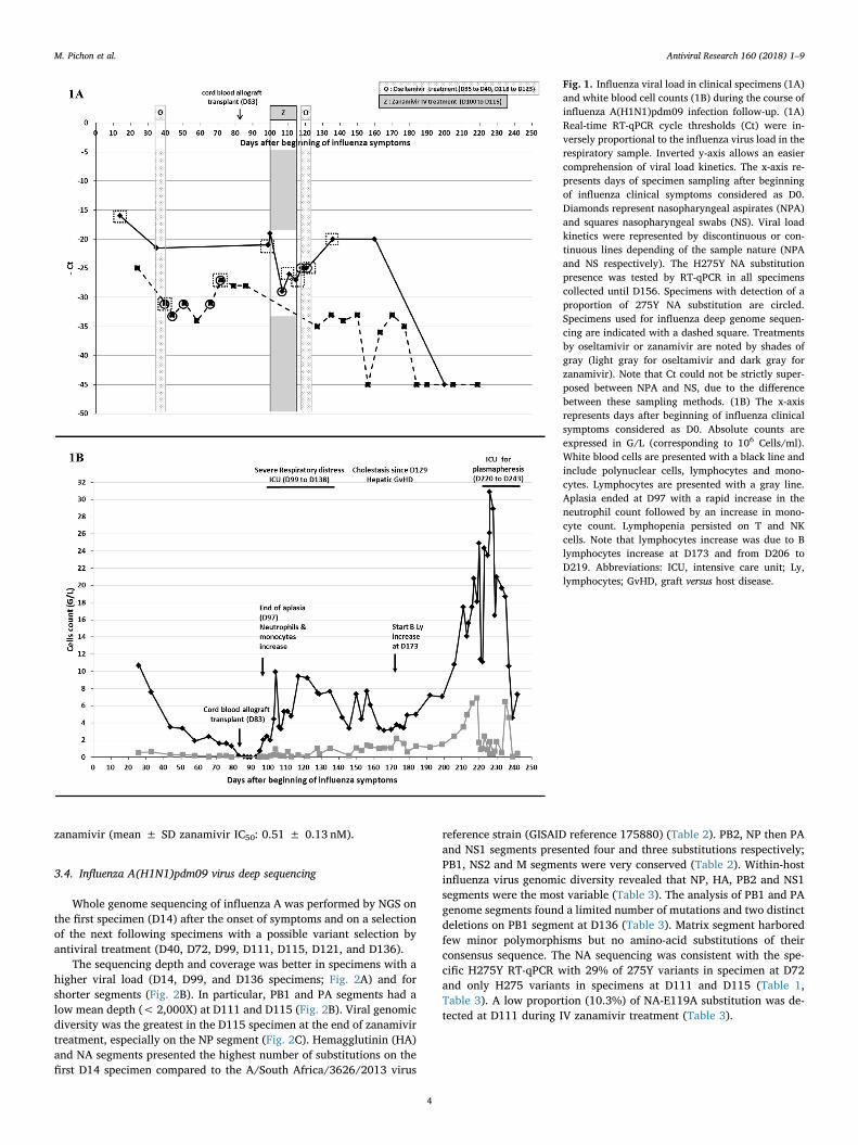
A five-month-old child was admitted for oxygen-requiring inter-stitial pneumonia associated with bronchiolitis symptoms. These re-spiratory symptoms had started 14 days before hand (considered as Day0, D0) and were due to an influenza virus infection diagnosed on NPA atD14 (Fig. 1A). Within his first months of life, he had presented twoepisodes of acute pyelonephritis and an episode of wheezing compli-cated by oral candidiasis due to steroid treatment. These iterative in-fections led to immunological analyses to be performed that foundagammaglobulinemia and a defect of T and NK cells suggesting an X-SCID.
In order to correct the SCID, and despite the ongoing influenza in-fection, the patient underwent allogeneic cord blood transplantation on
D83, previously conditioned by fludarabine, melphalan and anti-lym-phocyte serum association. Immunosuppressive therapy was based oncyclosporin and steroids. Aplasia ended on D97 with an increase inneutrophils and monocytes but lymphopenia persisted until lympho-cytes increased on D173 and from D206 to D219 (Fig. 1B). The patient'scondition deteriorated in the weeks following cord blood transplanta-tion; severe respiratory distress (due to influenza infection concomitantwith innate immunity reconstitution but with persistent lymphopenia)leading to intensive care unit (ICU) admission from D99 to D138. Agraft-versus-host-disease (GvHD) was suspected due to hepatic im-pairment characterized by moderate hepatic cytolysis (2-fold increaseof liver transaminases on D123) and important cholestasis (35-fold in-crease of gamma glutamyl transferases on D129). Thus, im-munosuppression with cyclosporin and steroids was maintained butthese drugs were responsible for hypertension. Due to persistent cho-lestasis, the patient was admitted again to the ICU on D220 to undergoplasmapheresis, and cyclosporin was replaced by tacrolimus on D222.The patient was also infected by a bocavirus and norovirus II whoseviral loads also varied according to the immunity and were still de-tected on D236 and D240. Finally, the patient died on D243.
3.2. Influenza viral infection and antiviral treatment
Influenza virus detection remained positive in respiratory samplesfor 6 months (last positive specimen on D177) despite NAI treatment(Fig. 1A). Oral oseltamivir (15mg twice daily) was administered fromD35 to D40. A temporary authorization for the compassionate use of IVzanamivir was obtained on D100 since an oseltamivir-resistant sub-population was detected. This treatment, associated with IV im-munoglobulins, was extended during 15 days. This treatment, con-comitant with partial immune reconstitution, allowed an influenza viralload decrease around 2 log10 copies/mL on NPA (Fig. 1A, Table 1).However, insufficient data on a potential toxicity during extended useled to discontinue IV zanamivir and initiate another oseltamivir therapyfrom D118 to D123, as the oseltamivir-resistant subpopulation hadlowered, but this treatment was inefficient. The viral load was corre-lated to immune reconstitution (Fig. 1). The white blood cells decreasebetween D117 and D146, associated to the antiviral stop led to a viralload rebound (very close Ct for NPA specimens at D136 and D100).Finally, the viral clearance was obtained when immune reconstitutionwas sufficient.
3.3. Evaluation of susceptibility of influenza virus to NAI using conventionaltechniques
During the clinical management of the patient, 11 NPA and 17 NS,collected between D14 to D177 after the onset of symptoms, were foundto be positive for influenza A viruses (Table 1) and secondarily sub-typed as an A(H1N1)pdm09 virus. The viral loads for NPA sampleswere higher as this sampling method allows recovering more cells thanNS (Fig. 1A). A proportion of 11%–29% of influenza viruses bearing aNA-H275Y substitution were detected, after oseltamivir therapy, on NSperformed on D40, D44, D51, D66 and D72 but not D58 when viral loadwas very low (Ct= 34). This subpopulation appeared just after osel-tamivir treatment (from D35 to D40), persisted at least up to D121(Table 1) during which there was low immune pressure (Fig. 1B). Aftercord blood allograft transplant, on D83, the respiratory distress wor-sened as viral load increased at D86 at which time no 275Y variantscould be detected. On D107, the highest proportion of 275Y variants(24%) were detected and the viral load was the lowest since transplant(Ct= 29; Table 1).
The 275Y substitution was detected on several specimens but onlyat a low proportion (≤29%) (Table 1, Table 3) and only wild-typeviruses were selected after cell culture. Fourteen influenza virus isolateswere obtained after culture and all presented normal inhibition byoseltamivir (mean ± SD oseltamivir IC50: 0.38 ± 0.18 nM) and
M. Pichon et al. Antiviral Research 160 (2018) 1–9
3
zanamivir (mean ± SD zanamivir IC50: 0.51 ± 0.13 nM).
3.4. Influenza A(H1N1)pdm09 virus deep sequencing
Whole genome sequencing of influenza A was performed by NGS onthe first specimen (D14) after the onset of symptoms and on a selectionof the next following specimens with a possible variant selection byantiviral treatment (D40, D72, D99, D111, D115, D121, and D136).
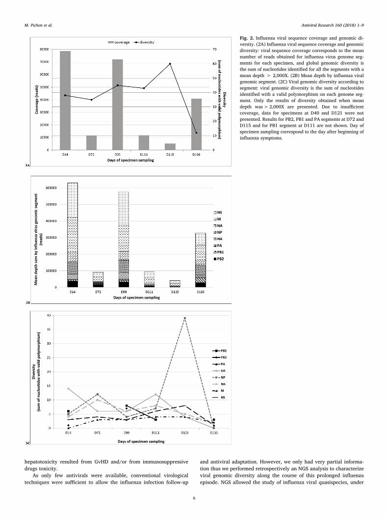
The sequencing depth and coverage was better in specimens with ahigher viral load (D14, D99, and D136 specimens; Fig. 2A) and forshorter segments (Fig. 2B). In particular, PB1 and PA segments had alow mean depth (< 2,000X) at D111 and D115 (Fig. 2B). Viral genomicdiversity was the greatest in the D115 specimen at the end of zanamivirtreatment, especially on the NP segment (Fig. 2C). Hemagglutinin (HA)and NA segments presented the highest number of substitutions on thefirst D14 specimen compared to the A/South Africa/3626/2013 virus
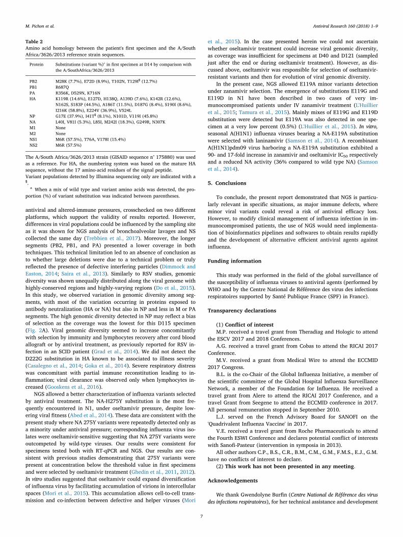
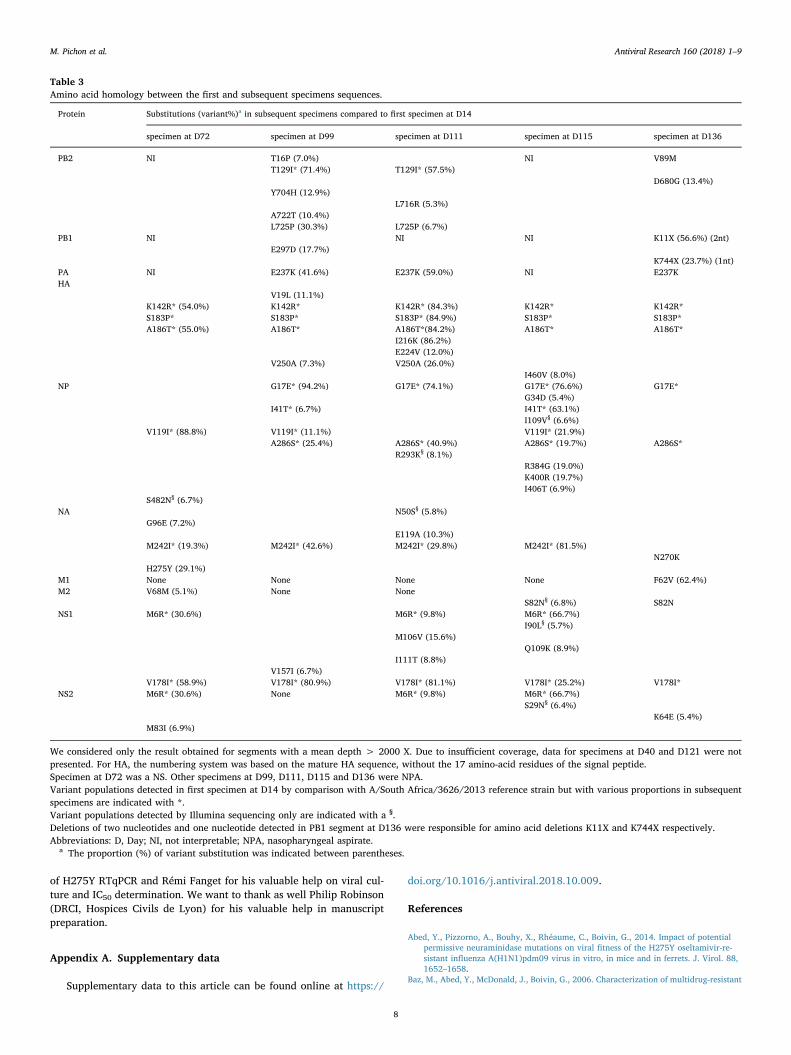
reference strain (GISAID reference 175880) (Table 2). PB2, NP then PAand NS1 segments presented four and three substitutions respectively;PB1, NS2 and M segments were very conserved (Table 2). Within-hostinfluenza virus genomic diversity revealed that NP, HA, PB2 and NS1segments were the most variable (Table 3). The analysis of PB1 and PAgenome segments found a limited number of mutations and two distinctdeletions on PB1 segment at D136 (Table 3). Matrix segment harboredfew minor polymorphisms but no amino-acid substitutions of theirconsensus sequence. The NA sequencing was consistent with the spe-cific H275Y RT-qPCR with 29% of 275Y variants in specimen at D72and only H275 variants in specimens at D111 and D115 (Table 1,Table 3). A low proportion (10.3%) of NA-E119A substitution was de-tected at D111 during IV zanamivir treatment (Table 3).
Fig. 1. Influenza viral load in clinical specimens (1A)and white blood cell counts (1B) during the course ofinfluenza A(H1N1)pdm09 infection follow-up. (1A)Real-time RT-qPCR cycle thresholds (Ct) were in-versely proportional to the influenza virus load in therespiratory sample. Inverted y-axis allows an easiercomprehension of viral load kinetics. The x-axis re-presents days of specimen sampling after beginningof influenza clinical symptoms considered as D0.Diamonds represent nasopharyngeal aspirates (NPA)and squares nasopharyngeal swabs (NS). Viral loadkinetics were represented by discontinuous or con-tinuous lines depending of the sample nature (NPAand NS respectively). The H275Y NA substitutionpresence was tested by RT-qPCR in all specimenscollected until D156. Specimens with detection of aproportion of 275Y NA substitution are circled.Specimens used for influenza deep genome sequen-cing are indicated with a dashed square. Treatmentsby oseltamivir or zanamivir are noted by shades ofgray (light gray for oseltamivir and dark gray forzanamivir). Note that Ct could not be strictly super-posed between NPA and NS, due to the differencebetween these sampling methods. (1B) The x-axisrepresents days after beginning of influenza clinicalsymptoms considered as D0. Absolute counts areexpressed in G/L (corresponding to 106 Cells/ml).White blood cells are presented with a black line andinclude polynuclear cells, lymphocytes and mono-cytes. Lymphocytes are presented with a gray line.Aplasia ended at D97 with a rapid increase in theneutrophil count followed by an increase in mono-cyte count. Lymphopenia persisted on T and NKcells. Note that lymphocytes increase was due to Blymphocytes increase at D173 and from D206 toD219. Abbreviations: ICU, intensive care unit; Ly,lymphocytes; GvHD, graft versus host disease.
M. Pichon et al. Antiviral Research 160 (2018) 1–9
4
4. Discussion
The present study combines specimen whole influenza genome se-quencing, clinical data, and antiviral treatment of a SCID patient in-fected by influenza A(H1N1)pdm09 virus.
The late SCID diagnosis led to perform the cord blood transplanta-tion with an ongoing infection, which is known to have a poor outcome(Rivers and Gaspar, 2015). Influenza infections in im-munocompromised patients require effective antiviral agents but onlyoseltamivir and zanamivir are available.
IV zanamivir as late add-on therapy has been described to have alimited effectiveness; however most patients were im-munocompromised (Fraaij et al., 2011). In the case presented here, weobserved a partial viral response with a 2 log10 copies/mL decrease inseven days. This observation is consistent with a median decrease inviral load of 1.81 log10 copies/mL after 2 days of IV zanamivir treat-ment of children (phase II assay) (Bradley et al., 2017). In previouslypublished cases, IV zanamivir was also associated with viral load de-crease although with WB cells increase and persistent lymphopenia
(Dulek et al., 2010). It was reported to be effective in stem cell trans-planted teenagers; however, although IV zanamivir decreased influenzaviral load, genome clearing was only observed more than 4 monthslater, following immune reconstitution (Ghosh et al., 2012). IV zana-mivir was discontinued after the usual 15 days of treatment (Dohna-Schwake et al., 2010; Dulek et al., 2010; Gaur et al., 2010). In anotherclinical case, IV zanamivir was administered for a total of 34 dayswithout notification of particular adverse events (Ghosh et al., 2012). Inthis situation, owing to important cholestasis on D129, the hepato-toxicity of IV zanamivir was questioned. Hepatotoxicity of IV zanamivirwas hypothesized in a case of pediatric liver transplant (Dohna-Schwake et al., 2010) without any serious adverse events detected inother patients (Dulek et al., 2010; Fraaij et al., 2011). IV zanamivir inadults led to liver enzyme elevations during a Phase II evaluation(Marty et al., 2014). However no increased hepatotoxicity was noted inthe IV zanamivir groups in the phase III trial in adults (Marty et al.,2017) and in the phase II trial in children (Bradley et al., 2017). In thischild's history, the 14-day interval between IV zanamivir treatmentdiscontinuation and appearance of liver injury signs suggests that
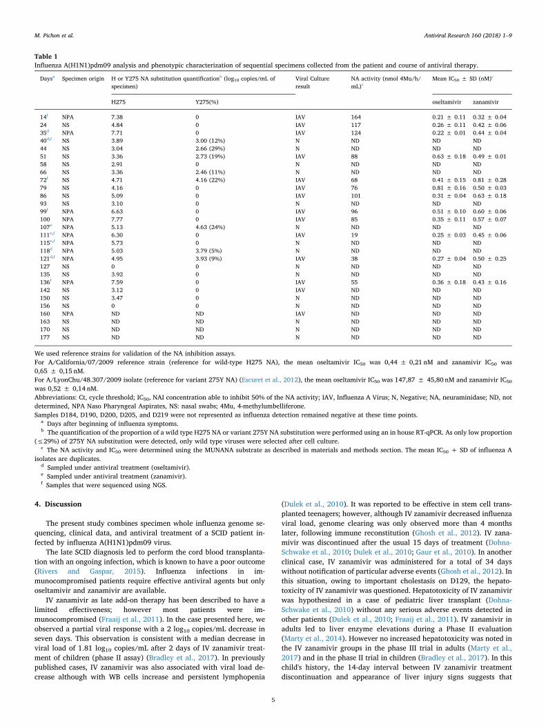
Table 1Influenza A(H1N1)pdm09 analysis and phenotypic characterization of sequential specimens collected from the patient and course of antiviral therapy.
Daysa Specimen origin H or Y275 NA substitution quantificationb (log10 copies/mL ofspecimen)
Viral Cultureresult
NA activity (nmol 4Mu/h/mL)c
Mean IC50 ± SD (nM)c
H275 Y275(%) oseltamivir zanamivir
14f NPA 7.38 0 IAV 164 0.21 ± 0.11 0.32 ± 0.0424 NS 4.84 0 IAV 117 0.26 ± 0.11 0.42 ± 0.0635d NPA 7.71 0 IAV 124 0.22 ± 0.01 0.44 ± 0.0440d,f NS 3.89 3.00 (12%) N ND ND ND44 NS 3.04 2.66 (29%) N ND ND ND51 NS 3.36 2.73 (19%) IAV 88 0.63 ± 0.18 0.49 ± 0.0158 NS 2.91 0 N ND ND ND66 NS 3.36 2.46 (11%) N ND ND ND72f NS 4.71 4.16 (22%) IAV 68 0.41 ± 0.15 0.81 ± 0.2879 NS 4.16 0 IAV 76 0.81 ± 0.16 0.50 ± 0.0386 NS 5.09 0 IAV 101 0.31 ± 0.04 0.63 ± 0.1893 NS 3.10 0 N ND ND ND99f NPA 6.63 0 IAV 96 0.51 ± 0.10 0.60 ± 0.06100 NPA 7.77 0 IAV 85 0.35 ± 0.11 0.57 ± 0.07107e NPA 5.13 4.63 (24%) N ND ND ND111e,f NPA 6.30 0 IAV 19 0.25 ± 0.03 0.45 ± 0.06115e,f NPA 5.73 0 N ND ND ND118d NPA 5.03 3.79 (5%) N ND ND ND121d,f NPA 4.95 3.93 (9%) IAV 38 0.27 ± 0.04 0.50 ± 0.25127 NS 0 0 N ND ND ND135 NS 3.92 0 N ND ND ND136f NPA 7.59 0 IAV 55 0.36 ± 0.18 0.43 ± 0.16142 NS 3.12 0 IAV ND ND ND150 NS 3.47 0 N ND ND ND156 NS 0 0 N ND ND ND160 NPA ND ND IAV ND ND ND163 NS ND ND N ND ND ND170 NS ND ND N ND ND ND177 NS ND ND N ND ND ND
We used reference strains for validation of the NA inhibition assays.For A/California/07/2009 reference strain (reference for wild-type H275 NA), the mean oseltamivir IC50 was 0,44 ± 0,21 nM and zanamivir IC50 was0,65 ± 0,15 nM.For A/LyonChu/48.307/2009 isolate (reference for variant 275Y NA) (Escuret et al., 2012), the mean oseltamivir IC50 was 147,87 ± 45,80 nM and zanamivir IC50
was 0,52 ± 0,14 nM.Abbreviations: Ct, cycle threshold; IC50, NAI concentration able to inhibit 50% of the NA activity; IAV, Influenza A Virus; N, Negative; NA, neuraminidase; ND, notdetermined, NPA Naso Pharyngeal Aspirates, NS: nasal swabs; 4Mu, 4-methylumbelliferone.Samples D184, D190, D200, D205, and D219 were not represented as influenza detection remained negative at these time points.
a Days after beginning of influenza symptoms.b The quantification of the proportion of a wild type H275 NA or variant 275Y NA substitution were performed using an in house RT-qPCR. As only low proportion
(≤29%) of 275Y NA substitution were detected, only wild type viruses were selected after cell culture.c The NA activity and IC50 were determined using the MUNANA substrate as described in materials and methods section. The mean IC50 + SD of influenza A
isolates are duplicates.d Sampled under antiviral treatment (oseltamivir).e Sampled under antiviral treatment (zanamivir).f Samples that were sequenced using NGS.
M. Pichon et al. Antiviral Research 160 (2018) 1–9
5
hepatotoxicity resulted from GvHD and/or from immunosuppressivedrugs toxicity.
As only few antivirals were available, conventional virologicaltechniques were sufficient to allow the influenza infection follow-up
and antiviral adaptation. However, we only had very partial informa-tion thus we performed retrospectively an NGS analysis to characterizeviral genomic diversity along the course of this prolonged influenzaepisode. NGS allowed the study of influenza viral quasispecies, under
Fig. 2. Influenza viral sequence coverage and genomic di-versity. (2A) Influenza viral sequence coverage and genomicdiversity: viral sequence coverage corresponds to the meannumber of reads obtained for influenza virus genome seg-ments for each specimen, and global genomic diversity isthe sum of nucleotides identified for all the segments with amean depth > 2,000X. (2B) Mean depth by influenza viralgenomic segment. (2C) Viral genomic diversity according tosegment: viral genomic diversity is the sum of nucleotidesidentified with a valid polymorphism on each genome seg-ment. Only the results of diversity obtained when meandepth was>2,000X are presented. Due to insufficientcoverage, data for specimens at D40 and D121 were notpresented. Results for PB2, PB1 and PA segments at D72 andD115 and for PB1 segment at D111 are not shown. Day ofspecimen sampling correspond to the day after beginning ofinfluenza symptoms.
M. Pichon et al. Antiviral Research 160 (2018) 1–9
6
antiviral and altered-immune pressures, crosschecked on two differentplatforms, which support the validity of results reported. However,differences in viral populations could be influenced by the sampling siteas it was shown for NGS analysis of bronchoalveolar lavages and NScollected the same day (Trebbien et al., 2017). Moreover, the longersegments (PB2, PB1, and PA) presented a lower coverage in bothtechniques. This technical limitation led to an absence of conclusion asto whether large deletions were due to a technical problem or trulyreflected the presence of defective interfering particles (Dimmock andEaston, 2014; Saira et al., 2013). Similarly to RSV studies, genomicdiversity was shown unequally distributed along the viral genome withhighly-conserved regions and highly-varying regions (Do et al., 2015).In this study, we observed variation in genomic diversity among seg-ments, with most of the variation occurring in proteins exposed toantibody neutralization (HA or NA) but also in NP and less in M or PAsegments. The high genomic diversity detected in NP may reflect a biasof selection as the coverage was the lowest for this D115 specimen(Fig. 2A). Viral genomic diversity seemed to increase concomitantlywith selection by immunity and lymphocytes recovery after cord bloodallograft or by antiviral treatment, as previously reported for RSV in-fection in an SCID patient (Grad et al., 2014). We did not detect theD222G substitution in HA known to be associated to illness severity(Casalegno et al., 2014; Goka et al., 2014). Severe respiratory distresswas concomitant with partial immune reconstitution leading to in-flammation; viral clearance was observed only when lymphocytes in-creased (Gooskens et al., 2016).
NGS allowed a better characterization of influenza variants selectedby antiviral treatment. The NA-H275Y substitution is the most fre-quently encountered in N1, under oseltamivir pressure, despite low-ering viral fitness (Abed et al., 2014). These data are consistent with thepresent study where NA 275Y variants were repeatedly detected only asa minority under antiviral pressure; corresponding influenza virus iso-lates were oseltamivir-sensitive suggesting that NA 275Y variants wereoutcompeted by wild-type viruses. Our results were consistent forspecimens tested both with RT-qPCR and NGS. Our results are con-sistent with previous studies demonstrating that 275Y variants werepresent at concentration below the threshold value in first specimensand were selected by oseltamivir treatment (Ghedin et al., 2011, 2012).In vitro studies suggested that oseltamivir could expand diversificationof influenza virus by facilitating accumulation of virions in intercellularspaces (Mori et al., 2015). This accumulation allows cell-to-cell trans-mission and co-infection between defective and helper viruses (Mori
et al., 2015). In the case presented herein we could not ascertainwhether oseltamivir treatment could increase viral genomic diversity,as coverage was insufficient for specimens at D40 and D121 (sampledjust after the end or during oseltamivir treatment). However, as dis-cussed above, oseltamivir was responsible for selection of oseltamivir-resistant variants and then for evolution of viral genomic diversity.
In the present case, NGS allowed E119A minor variants detectionunder zanamivir selection. The emergence of substitutions E119G andE119D in N1 have been described in two cases of very im-munocompromised patients under IV zanamivir treatment (L'Huillieret al., 2015; Tamura et al., 2015). Mainly mixes of E119G and E119Dsubstitution were detected but E119A was also detected in one spe-cimen at a very low percent (0.5%) (L'Huillier et al., 2015). In vitro,seasonal A(H1N1) influenza viruses bearing a NA-E119A substitutionwere selected with laninamivir (Samson et al., 2014). A recombinantA(H1N1)pdm09 virus harboring a NA-E119A substitution exhibited a90- and 17-fold increase in zanamivir and oseltamivir IC50 respectivelyand a reduced NA activity (36% compared to wild type NA) (Samsonet al., 2014).
5. Conclusions
To conclude, the present report demonstrated that NGS is particu-larly relevant in specific situations, as major immune defects, whereminor viral variants could reveal a risk of antiviral efficacy loss.However, to modify clinical management of influenza infection in im-munocompromised patients, the use of NGS would need implementa-tion of bioinformatics pipelines and softwares to obtain results rapidlyand the development of alternative efficient antiviral agents againstinfluenza.
Funding information
This study was performed in the field of the global surveillance ofthe susceptibility of influenza viruses to antiviral agents (performed byWHO and by the Centre National de Référence des virus des infectionsrespiratoires supported by Santé Publique France (SPF) in France).
Transparency declarations
(1) Conflict of interestM.P. received a travel grant from Theradiag and Hologic to attend
the ESCV 2017 and 2018 Conferences.A.G. received a travel grant from Cobas to attend the RICAI 2017
Conference.M.V. received a grant from Medical Wire to attend the ECCMID
2017 Congress.B.L. is the co-Chair of the Global Influenza Initiative, a member of
the scientific committee of the Global Hospital Influenza SurveillanceNetwork, a member of the Foundation for Influenza. He received atravel grant from Alere to attend the RICAI 2017 Conference, and atravel Grant from Seegene to attend the ECCMID conference in 2017.All personal remuneration stopped in September 2010.
L.J. served on the French Advisory Board for SANOFI on the'Quadrivalent Influenza Vaccine' in 2017.
V.E. received a travel grant from Roche Pharmaceuticals to attendthe Fourth ESWI Conference and declares potential conflict of interestswith Sanofi-Pasteur (intervention in symposia in 2013).
All other authors C.P., B.S., C.R., B.M., C.M., G.M., F.M.S., E.J., G.M.have no conflicts of interest to declare.
(2) This work has not been presented in any meeting.
Acknowledgements
We thank Gwendolyne Burfin (Centre National de Référence des virusdes infections respiratoires), for her technical assistance and development
Table 2Amino acid homology between the patient's first specimen and the A/SouthAfrica/3626/2013 reference strain sequences.
Protein Substitutions (variant %)a in first specimen at D14 by comparison withthe A/SouthAfrica/3626/2013
PB2 M28K (7.7%), E72D (8.9%), T102N, T129I§ (12.7%)PB1 R687QPA R356K, D529N, K716NHA K119R (14.6%), E127D, H138Q, A139D (7.6%), K142R (12.6%),
N162S, S183P (44.5%), A186T (11.5%), D187G (8.4%), S190I (8.6%),I216K (58.8%), E224V (36.9%), V524L
NP G17E (37.9%), I41T§ (8.1%), N101D, V119I (45.8%)NA L40I, V81I (5.3%), L85I, M242I (18.3%), G249R, N307KM1 NoneM2 NoneNS1 M6R (57.5%), T76A, V178I (15.4%)NS2 M6R (57.5%)
The A/South Africa/3626/2013 strain (GISAID sequence n° 175880) was usedas a reference. For HA, the numbering system was based on the mature HAsequence, without the 17 amino-acid residues of the signal peptide.Variant populations detected by Illumina sequencing only are indicated with a§.
a When a mix of wild type and variant amino acids was detected, the pro-portion (%) of variant substitution was indicated between parentheses.
M. Pichon et al. Antiviral Research 160 (2018) 1–9
7
of H275Y RTqPCR and Rémi Fanget for his valuable help on viral cul-ture and IC50 determination. We want to thank as well Philip Robinson(DRCI, Hospices Civils de Lyon) for his valuable help in manuscriptpreparation.
Appendix A. Supplementary data
Supplementary data to this article can be found online at https://
doi.org/10.1016/j.antiviral.2018.10.009.
References
Abed, Y., Pizzorno, A., Bouhy, X., Rhéaume, C., Boivin, G., 2014. Impact of potentialpermissive neuraminidase mutations on viral fitness of the H275Y oseltamivir-re-sistant influenza A(H1N1)pdm09 virus in vitro, in mice and in ferrets. J. Virol. 88,1652–1658.
Baz, M., Abed, Y., McDonald, J., Boivin, G., 2006. Characterization of multidrug-resistant
Table 3Amino acid homology between the first and subsequent specimens sequences.
Protein Substitutions (variant%)a in subsequent specimens compared to first specimen at D14
specimen at D72 specimen at D99 specimen at D111 specimen at D115 specimen at D136
PB2 NI T16P (7.0%) NI V89MT129I* (71.4%) T129I* (57.5%)
D680G (13.4%)Y704H (12.9%)
L716R (5.3%)A722T (10.4%)L725P (30.3%) L725P (6.7%)
PB1 NI NI NI K11X (56.6%) (2nt)E297D (17.7%)
K744X (23.7%) (1nt)PA NI E237K (41.6%) E237K (59.0%) NI E237KHA
V19L (11.1%)K142R* (54.0%) K142R* K142R* (84.3%) K142R* K142R*S183P* S183P* S183P* (84.9%) S183P* S183P*A186T* (55.0%) A186T* A186T*(84.2%) A186T* A186T*
I216K (86.2%)E224V (12.0%)
V250A (7.3%) V250A (26.0%)I460V (8.0%)
NP G17E* (94.2%) G17E* (74.1%) G17E* (76.6%) G17E*G34D (5.4%)
I41T* (6.7%) I41T* (63.1%)I109V§ (6.6%)
V119I* (88.8%) V119I* (11.1%) V119I* (21.9%)A286S* (25.4%) A286S* (40.9%) A286S* (19.7%) A286S*
R293K§ (8.1%)R384G (19.0%)K400R (19.7%)I406T (6.9%)
S482N§ (6.7%)NA N50S§ (5.8%)
G96E (7.2%)E119A (10.3%)
M242I* (19.3%) M242I* (42.6%) M242I* (29.8%) M242I* (81.5%)N270K
H275Y (29.1%)M1 None None None None F62V (62.4%)M2 V68M (5.1%) None None
S82N§ (6.8%) S82NNS1 M6R* (30.6%) M6R* (9.8%) M6R* (66.7%)
I90L§ (5.7%)M106V (15.6%)
Q109K (8.9%)I111T (8.8%)
V157I (6.7%)V178I* (58.9%) V178I* (80.9%) V178I* (81.1%) V178I* (25.2%) V178I*
NS2 M6R* (30.6%) None M6R* (9.8%) M6R* (66.7%)S29N§ (6.4%)
K64E (5.4%)M83I (6.9%)
We considered only the result obtained for segments with a mean depth > 2000 X. Due to insufficient coverage, data for specimens at D40 and D121 were notpresented. For HA, the numbering system was based on the mature HA sequence, without the 17 amino-acid residues of the signal peptide.Specimen at D72 was a NS. Other specimens at D99, D111, D115 and D136 were NPA.Variant populations detected in first specimen at D14 by comparison with A/South Africa/3626/2013 reference strain but with various proportions in subsequentspecimens are indicated with *.Variant populations detected by Illumina sequencing only are indicated with a §.Deletions of two nucleotides and one nucleotide detected in PB1 segment at D136 were responsible for amino acid deletions K11X and K744X respectively.Abbreviations: D, Day; NI, not interpretable; NPA, nasopharyngeal aspirate.
a The proportion (%) of variant substitution was indicated between parentheses.
M. Pichon et al. Antiviral Research 160 (2018) 1–9
8
influenza A/H3N2 viruses shed during 1 year by an immunocompromised child. Clin.Infect. Dis. 43, 1555–1561.
Beigel, J.H., Bao, Y., Beeler, J., Manosuthi, W., Slandzicki, A., Dar, S.M., Panuto, J.,Beasley, R.L., Perez-Patrigeon, S., Suwanpimolkul, G., Losso, M.H., McClure, N.,Bozzolo, D.R., Myers, C., Holley, H.P., Hoopes, J., Lane, H.C., Hughes, M.D., Davey,R.T., Team, I.S., 2017. Oseltamivir, amantadine, and ribavirin combination antiviraltherapy versus oseltamivir monotherapy for the treatment of influenza: a multicentre,double-blind, randomised phase 2 trial. Lancet Infect. Dis. 17, 1255–1265.
Blankenberg, D., Von Kuster, G., Bouvier, E., Baker, D., Afgan, E., Stoler, N., Taylor, J.,Nekrutenko, A., Team, G., 2014. Dissemination of scientific software with GalaxyToolShed. Genome Biol. 15, 403.
Bogner, P., Capua, I., Cox, N.J., Lipman, D.J., 2006. A global initiative on sharing avianflu data. Nature 442, 981.
Boivin, G., Goyette, N., Bernatchez, H., 2002. Prolonged excretion of amantadine-re-sistant influenza a virus quasi species after cessation of antiviral therapy in an im-munocompromised patient. Clin. Infect. Dis. 34, E23–E25.
Bradley, J.S., Blumer, J.L., Romero, J.R., Michaels, M.G., Munoz, F.M., Kimberlin, D.W.,Pahud, B., DeBiasi, R.L., Yamamoto, G., Roberts, G., Hossain, M., Shortino, D., Yates,P.J., Adams, B., Peppercorn, A., 2017. Intravenous zanamivir in hospitalized patientswith influenza. Pediatrics 140.
Casalegno, J.-S., Ferraris, O., Escuret, V., Bouscambert, M., Bergeron, C., Lines, L.,Excoffier, T., Valette, M., Frobert, E., Pillet, S., Pozzetto, B., Lina, B., Ottmann, M.,2014. Functional balance between the hemagglutinin and neuraminidase of influenzaA(H1N1)pdm09 HA D222 variants. PloS One 9.
Dimmock, N.J., Easton, A.J., 2014. Defective interfering influenza virus RNAs: time toreevaluate their clinical potential as broad-spectrum antivirals? J. Virol. 88,5217–5227.
Do, L.A., Wilm, A., Van Doorn, H.R., Lam, H.M., Sim, S., Sukumaran, R., Tran, A.T.,Nguyen, B.H., Tran, T.T., Tran, Q.H., Vo, Q.B., Dac, N.A., Trinh, H.N., Nguyen, T.T.,Binh, B.T., Le, K., Nguyen, M.T., Thai, Q.T., Vo, T.V., Ngo, N.Q., Dang, T.K., Cao,N.H., Tran, T.V., Ho, L.V., Farrar, J., Jong, M., Chen, S., Nagarajan, N., Bryant, J.E.,Hibberd, M.L., 2015. Direct whole-genome deep-sequencing of human respiratorysyncytial virus A and B from Vietnamese children identifies distinct patterns of inter-and intra-host evolution. J. Gen. Virol. 96, 3470–3483.
Dohna-Schwake, C., Schweiger, B., Felderhoff-Müser, U., Fiedler, M., Kaiser, G.M., Paul,A., Gerner, P., Lainka, E., Hoyer, P.F., 2010. Severe H1N1 infection in a pediatricliver transplant recipient treated with intravenous zanamivir: efficiency and com-plications. Transplantation 90, 223–224.
Dulek, D.E., Williams, J.V., Creech, C.B., Schulert, A.K., Frangoul, H.A., Domm, J.,Denison, M.R., Chappell, J.D., 2010. Use of intravenous zanamivir after developmentof oseltamivir resistance in a critically Ill immunosuppressed child infected with 2009pandemic influenza A (H1N1) virus. Clin. Infect. Dis. 50, 1493–1496.
Duval, X., van der Werf, S., Blanchon, T., Mosnier, A., Bouscambert-Duchamp, M., Tibi,A., Enouf, V., Charlois-Ou, C., Vincent, C., Andreoletti, L., Tubach, F., Lina, B.,Mentré, F., Leport, C., Group, B.S., 2010. Efficacy of oseltamivir-zanamivir combi-nation compared to each monotherapy for seasonal influenza: a randomized placebo-controlled trial. PLoS Med. 7, e1000362.
Escuret, V., Cornu, C., Boutitie, F., Enouf, V., Mosnier, A., Bouscambert-Duchamp, M.,Gaillard, S., Duval, X., Blanchon, T., Leport, C., Gueyffier, F., Van Der Werf, S., Lina,B., 2012. Oseltamivir-zanamivir biotherapy compared to oseltamivir monotherapy inthe treatment of pandemic 2009 influenza A(H1N1) virus infections. Antivir. Res. 96,130–137.
Eshaghi, A., Shalhoub, S., Rosenfeld, P., Li, A.M., Higgins, R.R., Stogios, P.J., Savchenko,A., Bastien, N., Li, Y., Rotstein, C., Gubbaya, J.B., 2014. Multiple influenza a (H3N2)mutations conferring resistance to neuraminidase inhibitors in a bone marrowtransplant recipient. Antimicrob. Agents Chemother. 58, 7188–7197.
FastQC A Quality Control tool for High Throughput Sequence Data. Available at http://bioinformaticq.babraham.ac.uk/projects/fastqc/. Date accessed 07/02/2018.
Ferraris, O., Kessler, N., Lina, B., 2005. Sensitivity of influenza viruses to zanamivir andoseltamivir: a study performed on viruses circulating in France prior to the in-troduction of neuraminidase inhibitors in clinical practice. Antivir. Res. 68, 43–48.
Fraaij, P.L., van der Vries, E., Beersma, M.F., Riezebos-Brilman, A., Niesters, H.G., van derEijk, A.A., de Jong, M.D., Reis Miranda, D., Horrevorts, A.M., Ridwan, B.U.,Wolfhagen, M.J., Houmes, R.J., van Dissel, J.T., Fouchier, R.A., Kroes, A.C.,Koopmans, M.P., Osterhaus, A.D., Boucher, C.A., 2011. Evaluation of the antiviralresponse to zanamivir administered intravenously for treatment of critically ill pa-tients with pandemic influenza A (H1N1) infection. J. Infect. Dis. 204, 777–782.
Gaur, A.H., Bagga, B., Barman, S., Hayden, R., Lamptey, A., Hoffman, J.M., Bhojwani, D.,Flynn, P.M., Tuomanen, E., Webby, R., 2010. Intravenous zanamivir for oseltamivir-resistant 2009 H1N1 influenza. N. Engl. J. Med. 362, 88–89.
Ghedin, E., Holmes, E.C., DePasse, J.V., Pinilla, L.T., Fitch, A., Hamelin, M.E., Papenburg,J., Boivin, G., 2012. Presence of oseltamivir-resistant pandemic A/H1N1 minorvariants before drug therapy with subsequent selection and transmission. J. Infect.Dis. 206, 1504–1511.
Ghedin, E., Laplante, J., DePasse, J., Wentworth, D.E., Santos, R.P., Lepow, M.L., Porter,J., Stellrecht, K., Lin, X., Operario, D., Griesemer, S., Fitch, A., Halpin, R.A.,Stockwell, T.B., Spiro, D.J., Holmes, E.C., St George, K., 2011. Deep sequencing re-veals mixed infection with 2009 pandemic influenza A (H1N1) virus strains and theemergence of oseltamivir resistance. J. Infect. Dis. 203, 168–174.
Ghosh, S., Adams, O., Schuster, F.R., Borkhardt, A., Meisel, R., 2012. Efficient control ofpandemic 2009 H1N1 virus infection with intravenous zanamivir despite the lack ofimmune function. Transpl. Infect. Dis. 14, 657–659.
Goka, E.A., Vallely, P.J., Mutton, K.J., Klapper, P.E., 2014. Mutations associated withseverity of the pandemic influenza A(H1N1)pdm09 in humans: a systematic reviewand meta-analysis of epidemiological evidence. Arch. Virol. 159, 3167–3183.
Gooskens, J., Marijt, W.A., van Essen, E.H., Rimmelzwaan, G.F., Kroes, A.C., 2016. Hostimmunity dictates influenza A(H1N1)pdm09 infection outcome in hematology-on-cology patients. Bone Marrow Transplant. 51, 138–141.
Grad, Y.H., Newman, R., Zody, M., Yang, X., Murphy, R., Qu, J., Malboeuf, C.M., Levin,J.Z., Lipsitch, M., DeVincenzo, J., 2014. Within-host whole-genome deep sequencingand diversity analysis of human respiratory syncytial virus infection reveals dynamicsof genomic diversity in the absence and presence of immune pressure. J. Virol. 88,7286–7293.
Guo, Y., Cai, Q., Samuels, D.C., Ye, F., Long, J., Li, C.I., Winther, J.F., Tawn, E.J., Stovall,M., Lähteenmäki, P., Malila, N., Levy, S., Shaffer, C., Shyr, Y., Shu, X.O., Boice, J.D.,2012a. The use of next generation sequencing technology to study the effect of ra-diation therapy on mitochondrial DNA mutation. Mutat. Res. 744, 154–160.
Guo, Y., Li, J., Li, C.I., Long, J., Samuels, D.C., Shyr, Y., 2012b. The effect of strand bias inIllumina short-read sequencing data. BMC Genomics 13, 666.
Hurt, A.C., Hardie, K., Wilson, N.J., Deng, Y.M., Osbourn, M., Leang, S.K., Lee, R.T.,Iannello, P., Gehrig, N., Shaw, R., Wark, P., Caldwell, N., Givney, R.C., Xue, L.,Maurer-Stroh, S., Dwyer, D.E., Wang, B., Smith, D.W., Levy, A., Booy, R., Dixit, R.,Merritt, T., Kelso, A., Dalton, C., Durrheim, D., Barr, I.G., 2012. Characteristics of awidespread community cluster of H275Y oseltamivir-resistant A(H1N1)pdm09 in-fluenza in Australia. J. Infect. Dis. 206, 148–157.
L'Huillier, A.G., Abed, Y., Petty, T.J., Cordey, S., Thomas, Y., Bouhy, X., Schibler, M.,Simon, A., Chalandon, Y., van Delden, C., Zdobnov, E., Boquete-Suter, P., Boivin, G.,Kaiser, L., 2015. E119D neuraminidase mutation conferring Pan-resistance to neur-aminidase inhibitors in an A(H1N1)pdm09 isolate from a stem-cell transplant re-cipient. J. Infect. Dis. 212, 1726–1734.
Li, H., 2014. Toward better understanding of artifacts in variant calling from high-cov-erage samples. Bioinformatics 30, 2843–2851.
Martin, M., 2011. Cutadapt removes adapter sequences from high-throughput sequencingreads. EMBnet J. 17, 10–12.
Marty, F.M., Man, C.Y., van der Horst, C., Francois, B., Garot, D., Mánez, R., Thamlikitkul,V., Lorente, J.A., Alvarez-Lerma, F., Brealey, D., Zhao, H.H., Weller, S., Yates, P.J.,Peppercorn, A.F., 2014. Safety and pharmacokinetics of intravenous zanamivirtreatment in hospitalized adults with influenza: an open-label, multicenter, single-arm, phase II study. J. Infect. Dis. 209, 542–550.
Marty, F.M., Vidal-Puigserver, J., Clark, C., Gupta, S.K., Merino, E., Garot, D., Chapman,M.J., Jacobs, F., Rodriguez-Noriega, E., Husa, P., Shortino, D., Watson, H.A., Yates,P.J., Peppercorn, A.F., 2017. Intravenous zanamivir or oral oseltamivir for hospita-lised patients with influenza: an international, randomised, double-blind, double-dummy, phase 3 trial. Lancet Respir. Med. 5, 135–146.
Memoli, M.J., Athota, R., Reed, S., Czajkowski, L., Bristol, T., Proudfoot, K., Hagey, R.,Voell, J., Fiorentino, C., Ademposi, A., Shoham, S., Taubenberger, J.K., 2014. Thenatural history of influenza infection in the severely immunocompromised vs non-immunocompromised hosts. Clin. Infect. Dis. 58, 214–224.
Mori, K., Murano, K., Ohniwa, R.L., Kawaguchi, A., Nagata, K., 2015. Oseltamivir expandsquasispecies of influenza virus through cell-to-cell transmission. Sci. Rep. 5, 9163.
Okomo-Adhiambo, M., Fry, A.M., Su, S., Nguyen, H.T., Elal, A.A., Negron, E., Hand, J.,Garten, R.J., Barnes, J., Xiyan, X., Villanueva, J.M., Gubareva, L.V., Group, U.I.A.W.,2015. Oseltamivir-resistant influenza A(H1N1)pdm09 viruses, United States, 2013-14. Emerg. Infect. Dis. 21, 136–141.
Pichon, M., Gaymard, A., Josset, L., Valette, M., Millat, G., Lina, B., Escuret, V., 2017.Characterization of oseltamivir-resistant influenza virus populations in im-munosuppressed patients using digital-droplet PCR: comparison with qPCR and nextgeneration sequencing analysis. Antivir. Res. 145, 160–167.
Rivers, L., Gaspar, H.B., 2015. Severe combined immunodeficiency: recent developmentsand guidance on clinical management. Arch. Dis. Child. 100, 667–672.
Rogers, M.B., Song, T., Sebra, R., Greenbaum, B.D., Hamelin, M.E., Fitch, A., Twaddle, A.,Cui, L., Holmes, E.C., Boivin, G., Ghedin, E., 2015. Intrahost dynamics of antiviralresistance in influenza A virus reflect complex patterns of segment linkage, re-assortment, and natural selection. mBio 6.
Saira, K., Lin, X., DePasse, J.V., Halpin, R., Twaddle, A., Stockwell, T., Angus, B., Cozzi-Lepri, A., Delfino, M., Dugan, V., Dwyer, D.E., Freiberg, M., Horban, A., Losso, M.,Lynfield, R., Wentworth, D.N., Holmes, E.C., Davey, R., Wentworth, D.E., Ghedin, E.,Group, I.F.S., Group, I.F.S., 2013. Sequence analysis of in vivo defective interfering-like RNA of influenza A H1N1 pandemic virus. J. Virol. 87, 8064–8074.
Samson, M., Abed, Y., Desrochers, F.M., Hamilton, S., Luttick, A., Tucker, S.P., Pryor,M.J., Boivin, G., 2014. Characterization of drug-resistant influenza virus A(H1N1)and A(H3N2) variants selected in vitro with laninamivir. Antimicrob. AgentsChemother. 58, 5220–5228.
Takashita, E., Fujisaki, S., Kishida, N., Xu, H., Imai, M., Tashiro, M., Odagiri, T., Japan,I.V.S.G.o., 2013. Characterization of neuraminidase inhibitor-resistant influenzaA(H1N1)pdm09 viruses isolated in four seasons during pandemic and post-pandemicperiods in Japan. Influenza Other Respir. Viruses 7, 1390–1399.
Tamura, D., DeBiasi, R.L., Okomo-Adhiambo, M., Mishin, V.P., Campbell, A.P., Loechelt,B., Wiedermann, B.L., Fry, A.M., Gubareva, L.V., 2015. Emergence of multidrug-re-sistant influenza A(H1N1)pdm09 virus variants in an immunocompromised childtreated with oseltamivir and zanamivir. J. Infect. Dis. 212, 1209–1213.
Trebbien, R., Pedersen, S.S., Vorborg, K., Franck, K.T., Fischer, T.K., 2017. Developmentof oseltamivir and zanamivir resistance in influenza A(H1N1)pdm09 virus, Denmark,2014. Euro Surveill. 22.
M. Pichon et al. Antiviral Research 160 (2018) 1–9
9