osteopontin is required for unloading-induced osteoclast recruitment and modulation of rankl...
TRANSCRIPT
Osteopontin is Required for Unloading-Induced OsteoclastRecruitment and Modulation of RANKL Expression during ToothDrift-associated Bone Remodeling, but Not for Super-Eruption
CAMERON G. WALKERa,*, SMIT DANGARIAa,*, YOSHIHIRO ITOa, XIANGHONG LUANa,b,and THOMAS G. H. DIEKWISCHa,bCAMERON G. WALKER: [email protected]; SMIT DANGARIA: [email protected]; YOSHIHIRO ITO: [email protected];XIANGHONG LUAN: [email protected]; THOMAS G. H. DIEKWISCH: [email protected] Department of Oral Biology, University of Illinois at Chicago, Chicago, Illinois, USAb Department of Orthodontics, University of Illinois at Chicago, Chicago, Illinois, USA
AbstractUnloading of teeth results in extensive alveolar bone remodeling, causing teeth to move in bothvertical (“super-eruption”) and horizontal direction (“drift”). In order to decipher the molecularmechanisms of unloading-induced bone remodeling during tooth movement, we focused on the roleof osteopontin (OPN) in the un-opposed molar model, comparing wild-type (WT) and OPN-nullmice. Our data indicated that OPN was not required for the continuous eruption of un-opposed teethwhile OPN was necessary for the drift of teeth. OPN expression and osteoclast counts were greatlyincreased on alveolar bone surfaces facing the direction of the drift in WT mice, while osteoclastcounts were diminished in OPN−/− mice. RANKL expression in the distal periodontal ligament ofWT molars increased significantly by day 6 following unloading, while overall levels of RANKLexpression were decreased in both WT and OPN-null mice. In vitro treatment of MC3T3 cells, WTBMCs and OPN−/− BMCs with recombinant OPN resulted in significantly increased RANKLexpression in all three cell types. The PI3K and MEK/ERK pathway inhibitors Ly294002 and U0126reduced RANKL expression levels in vitro. Treatment of BMCs and MC3T3 with OPN also resultedin increased ERK phosphorylation and reduced OPG levels. Together, our studies suggest thatincreased OPN expression during unloading-induced drifting of teeth enhances localized RANKLexpression and osteoclast activity on drift-direction alveolar bone surfaces via extracellular matrixsignaling pathways.
KeywordsOsteopontin; RANKL; PI3K; ERK; Bone remodeling; Mechanical Stress
Corresponding author: Thomas G.H. Diekwisch, Allan G. Brodie Laboratory for Craniofacial Genetics, University of Illinois at ChicagoCollege of Dentistry, 801 South Paulina M/C 841, Chicago, IL. 60612, USA. Tel.: +1-312-413-9683; fax: +1-312-996-0873;[email protected].*Contributed equallyPublisher's Disclaimer: This is a PDF file of an unedited manuscript that has been accepted for publication. As a service to our customerswe are providing this early version of the manuscript. The manuscript will undergo copyediting, typesetting, and review of the resultingproof before it is published in its final citable form. Please note that during the production process errors may be discovered which couldaffect the content, and all legal disclaimers that apply to the journal pertain.
NIH Public AccessAuthor ManuscriptBone. Author manuscript; available in PMC 2011 December 1.
Published in final edited form as:Bone. 2010 December ; 47(6): 1020–1029. doi:10.1016/j.bone.2010.08.025.
NIH
-PA Author Manuscript
NIH
-PA Author Manuscript
NIH
-PA Author Manuscript
IntroductionLoading and unloading of bone results in continuous modeling and remodeling of the skeletonthroughout life [1–3]. On a molecular level, the mechanical stresses-induced remodelingcaused by loading and unloading of bone is mediated by the extracellular matrixphosphoprotein osteopontin (OPN) [4–8]. Osteopontin is an extracellular glycoproteincommonly found in bone that contains an Arg-Gly-Asp integrin binding domain and binds tomultiple organic or mineral ligands such as the integrin receptor αVβ3, CD44, andhydroxyapatite [9–12].
OPN−/− mice subjected to force induced tooth movement have decreased bone resorption andosteoclast counts, demonstrating the vital role of OPN in force initiated remodeling [5,13]. Theuse of transgenic animals with reporter constructs driven by varying lengths of OPN promoter,demonstrated OPN to have an upstream promoter region that is activated in response tomechanical stress [13]. OPN is also important for bone remodeling that occurs as a result ofunloading or disuse. Tail suspension studies with OPN−/− mice have established OPN to beessential for unloading induced enhancement of bone resorption and suppression of boneformation [7,8,14].
OPN expression induced by mechanical loading or unloading is essential for the migration ofosteoclasts to remodeling sites [5,14] and facilitates osteoclast chemotaxis, motility, andanchorage to bone through CD44, ανβ3 integrin, and G-protein coupled receptors [5,11,15–17]. OPN has also been shown to be a suppressor of osteoblastic bone formation during boneremodeling induced by unloading [7,14,18]. Moreover, bone resorption and osteoclast functionin OPN deficient animals is greatly reduced in response to orthodontic force [5,13,19].Furthermore, mechanical stress induces OPN expression via ATP through P2Y1 and throughRho Kinase pathway receptors in human periodontal ligament cells [20,21].
Osteoclast recruitment at bone resorption sites is facilitated by the TNF-related ligand RANKL,which binds to its cellular receptor RANK in order to direct osteoclast differentiation,activation, survival and apoptosis [22–25]. The relationship between osteoclasts andosteoblasts in bone metabolism is greatly affected by the balance between RANKL andosteoprotegerin (OPG), a non-signaling receptor decoy for RANKL [26–29], which modulatesthe level of bone resorption on bone surfaces.
Movement of teeth within bone, either through changes in biological loads or throughmechanical appliances, is one of the premier examples of mechanical stress-induced boneremodeling. Appliance-driven tooth movement models have contributed several key findingsrelated to the function of OPN during bone remodeling [5,13]. During tooth movement,mechanical stress stimulates osteocytes to express OPN to initiate bone remodeling [5,13,19],resulting in bone resorption and bone apposition on opposing sides of the tooth. Supportingthe pivotal function of OPN in unloading-induced enhancement of bone resorption andsuppression of bone formation during tooth movement, bone resorption and osteoclast functionin OPN deficient animals were greatly reduced in response to mechanical force [5,7,13,14,18].
In previous studies we have shown that unloading-induced biological tooth movement in miceis a result of osteoclastic bone resorption on the distal aspect of the alveolar socket combinedwith alveolar bone and cementum formation on the mesial and apical aspects of the alveolarsocket [30,31]. However, the detailed molecular mechanism underlying the contribution ofindividual molecules on unloading-induced bone turnover is not yet known. Based on the keyrole of OPN in the mediation of force response in bone, we have now hypothesized that OPNis essential for the remodeling of alveolar bone that takes place during the distal drift of un-opposed molars. In order to determine the role of OPN as it relates to key events in the complex
WALKER et al. Page 2
Bone. Author manuscript; available in PMC 2011 December 1.
NIH
-PA Author Manuscript
NIH
-PA Author Manuscript
NIH
-PA Author Manuscript
mineralized tissue remodeling processes, we have assayed key bone remodeling factors suchas RANKL and OPG and tested their behavior during unloading-induced tooth movement inwild-type and OPN−/− models.
Materials and MethodsTreatment of Mice
Osteopontin (OPN or Spp1) knockout mice (strain B6.129S6(Cg)- Spp1tm1Blh/JB40) and wild-type (WT) C57BL/6J mice were obtained from Jackson Labs (Bar Harbor, Maine, USA). TheseOPN−/− (Spp1−/−) mice were generated by injecting targeted ES cells into C57BL/6blastocysts and the resulting chimeric animals were crossed to outbred Black Swiss, maintainedon the mixed Black Swiss, 129S6 background and then backcrossed to C57BL/6 for 10generations (http://jaxmice.jax.org/strain/004936.html). In our study, C57BL/6J and OPN−/−mice were obtained from Jackson and cross-bred in our lab, and littermates were used forexperiments. Male OPN−/− and WT (40 days of age) were separated into various experimentalgroups. The presence or absence of OPN was confirmed by PCR amplification of genomicDNA extracted from tail snips using Taq polymerase (Clontech, Mountain View, CA) andprimer sequences provided by Jackson Labs. All animal experiments and procedures followedthe guidelines of the University of Illinois at Chicago Animal Care Committee.
Unloading of the right-side mandibular teeth was accomplished by extraction of the right-sidemaxillary molars. Functional occlusion of the molars on the left side was maintained as detailedpreviously [30,31]. Anesthesia for the procedure was accomplished using Ketamine (100 mg/kg) and Xylazine (5 mg/kg). Mice were also given Buprenorphine (0.05 mg/kg)intraperitoneally post procedure to manage any pain.
Skeletonization and Measurement of MovementIn order to measure unloading-induced tooth movement, groups of OPN−/− and WT mice (n= 3 each) were maintained in the unopposed state for 12 days. Wild-type and OPN−/− controlmice (n = 3 each) were maintained in normal occlusion for a period of 12 days after which theywere sacrificed. Control and treatment groups contained mice of the same age and weresacrificed together on the same day. Skeletonization of the mandibles was completedatraumatically by Dermestes maculatus. Beetles of the Dermestidae family are scavengers thatfeed on animal flesh and hairs. Anatomists and taxidermists take advantage of the dietarypreference of dermestid beetles to clean skeletons.
In preparation for morphological analysis, mandibles were photographed at uniformmagnification after which distances were scaled and measured using image software (AdobeSystems, San Jose, CA). Molar drifting was measured as the difference of distances from theanterior most point of the molars to the condyles on the right and left sides (Fig 1A.b).Measurements based on these landmarks were highly reproducible [30–32]. The magnitude ofdrift was graphed using the formula [(L – R) + 1], where L and R are the distances from anteriormost point of the first molar to the condyle on the left and right sides respectively. In a similarmanner, the magnitude of molar eruption was determined and graphed using the formula [(L−R) +1], where L and R are the distances from left and right molar cusp tips to the planeconnecting the superior borders of the left and right mental foramina (Fig 1A.f).
Tissue ProcessingWT and OPN −/− mice were maintained in the unopposed state for periods of 0, 1, 3, and 6days (n = 10 each). The experiments were timed so that all mice subjected to varying treatmentlengths were of the same age upon sacrifice. Collected mandibles were fixed in 4%paraformaldehyde for 24 h followed by decalcification for 2 wk with 5% EDTA and 2%
WALKER et al. Page 3
Bone. Author manuscript; available in PMC 2011 December 1.
NIH
-PA Author Manuscript
NIH
-PA Author Manuscript
NIH
-PA Author Manuscript
paraformaldehyde. Specimens were dehydrated, embedded in paraffin, and cut in 6μm sagitalsections along the long axis of the molar teeth or in cross section to be used for TRAP stainingor immunohistochemistry.
Tartrate resistant acid phosphatase staining and osteoclast countingOsteoclasts were visualized using a tartrate resistant acid phosphatase (TRAP) stainingprocedure. Paraffin sections were deparaffinized, rehydrated and incubated in acetate bufferedsolution containing naphthol AS-MX phosphate, Fast Garnet GBC salt, and tartrate solution(.67 mol/L) (Sigma, St Louis, MO, USA) for 60 min. Sections were counterstained withhematoxylin and Villanueva osteochrome bone stain. Only cross sections of mandibular secondmolar roots were used for cell counting. These sections were oriented parallel to the occlusaltable. For statistical evaluation, three sections each from five mice per group were subjectedto morphometry. Areas of 175 × 700μm were defined on the mesial and distal side ofmandibular second molar roots (Fig 3D.a). As a reference, “mesial” is the direction that pointstoward the middle of the front of the jaw while “distal” is the opposite direction. Root heightsof sections selected for counting were determined by the thickness of bone in between mesialand distal roots and an absence of cellular cementum. A representation of the root level of thesections is depicted by the lines in figure 3D.b. Multinucleated TRAP positive cells locatedadjacent to bone tissue in the predefined areas were counted as osteoclasts.
ImmunohistochemistryAnti-rabbit RANKL polyclonal antibody (ab62516) (Abcam) and anti-OPN mousemonoclonal antibody (SC-21742) (Santa Cruz Biotechnology) were used for immunostainingof paraffin sections as previously described [33]. Briefly, sections were deparaffinized,rehydrated and treated with 6% peroxide and methanol followed by a brief incubation in 10mM sodium citrate buffer with .05% Tween 20 at pH 6.0 for antigen retrieval. Sections werethen incubated with 1% bovine serum albumin (BSA) for 30 minutes at room temperature toblock nonspecific binding of the antibody. After blocking sections were incubated with anti-OPN at dilution of 1:200, or anti-RANKL at a concentration of 5μg/ml and washed three timesin PBS. Antigen localization in tissues was visualized using a Histomouse Broad SpectrumAEC kit (Zymed) containing secondary antibody, streptavidin enzyme conjugate, and an AECsubstrate chromogen mixture. Incubation times and washes were according to themanufacturer’s instructions and Hematoxylin was used as a counterstain. As a negative control,non-immune rabbit serum was used instead of the primary antibody. For dual TRAP and OPNimmunoassayed slides, sections were first stained with anti-OPN as described above, afterwhich they were incubated in a naphthol AS-MX phosphate, Fast Blue Salt (Sigma), andtartrate solution as described above. Dual stained sections were counterstained with acid fastgreen.
Cell culture and treatment with OPN, P13-Kinase and MEK/ERK InhibitorTo verify the effect of OPN on RANKL expression we cultured bone marrow stromal cellsfrom WT and OPN−/− mice and cells from the osteoblast precursor cell line MC3T3-E1,subclone 4 (ATCC), in the presence or absence of recombinant osteopontin (rOPN). Bonemarrow stromal cells (BMCs) were obtained by dissection of soft tissue free femurs of 40 day-old OPN −/− and WT mice (n = 15 each). Cells were seeded at a density of 106 cells/cm2 ontoculture flasks and incubated at 37 °C, 5% CO2. Nonadherent cells were removed by changingthe medium at day 3 and adding fresh complete expansion medium every 4 days for 12 daysuntil cells reached 70% confluency.
BMCs and MC-3T3 cells (3×10⁴/cm2) were cultured for 12 hours in the presence or absenceof 1μg/ml OPN (R&D Systems, Minneapolis, MN) and pathway inhibitors. The MEK/ERKinhibitor, UO126 (Sigma), and the PI3K inhibitor, LY294002 (Sigma), [38] were used at 10
WALKER et al. Page 4
Bone. Author manuscript; available in PMC 2011 December 1.
NIH
-PA Author Manuscript
NIH
-PA Author Manuscript
NIH
-PA Author Manuscript
μM to test for potential mechanisms for OPN induced RANKL expression. All experimentswere conducted in triplicate.
Analysis of gene expression by real-time quantitative PCRApical tissues of unloaded molars from OPN−/− and WT mice treated for periods of 0 (control),1, 3, and 6 days (n = 5 per time point), were carefully removed as described previously [30].RNA was extracted from tissues and cultured cells using TRIZOL LS Reagent (Invitrogen)according to the manufacturer’s instructions. The expression of RANKL, OPN, and OPG inthe periodontal tissues was assessed by real-time quantitative PCR. RNAs underwent RT-PCRusing Sprint RT Complete kit (Clontech). Real-time quantitative PCR was conducted usingTaqman Fast Universal PCR Master Mix (Applied Biosystems) with DLUX fluorogenicprimers. Commercially available primers RANKL (MLUX3313140), OPN (MLUX3312179),OPG (MLUX3309083) were designed and synthesized by Invitrogen (FAM labeled LUXDesigner primers, sequences maintained by Invitrogen, Carlsbad, CA, USA). Samples werenormalized using ribosome 18s RNA (JOE labeled LUX primer set, Invitrogen, Carlsbad, CA,USA). Reaction condition were as follows: 2 min at 50 °C (one cycle), 10 min at 95 °C (onecycle), and 15 s at 95 °C and 1 min at 60 °C (40 cycles). PCR products were continuouslymonitored with an ABI PRISM 7900 detection system (Applied Biosystems). Relativeexpression levels were calculated using the 2 −ΔΔ Ct method (34) and values were graphed asthe mean expression level ± standard deviation.
Western blot analysisAfter 12 hrs of culture, cells were lysed and proteins extracted as described [32]. Proteins werethen subjected to sodium dodecylsulfate-polyacrylamide gel electrophoresis (SDS-PAGE).Alternatively, mouse molar root tissues at day 0, day 3, day 6 in our unloading model andcontrol, OPN−/− and OPN−/− unloaded mouse molars were homogenized and lysed in SDS-PAGE sample buffer, and total proteins extracted. Identical amounts of protein extracts fromall regenerates were separated on a 10% SDS-PAGE gel and transferred to a PVDF membranein a semi-dry blotting apparatus containing transfer buffer (25mM tris, 40mM glycine, 10%methanol) for 45 minutes at 15V. The PVDF membrane was then blocked with 2.5% BSA for1 hour at room temperature and the blot was incubated with 1:1000 dilution of ERK1/2(Millipore, Billerica, MA), ERK1/2-Phospho (Millipore), 1 μg/mL RANKL, (Abcam) orGAPDH (Abcam) antibodies for 2 hours, washed with TBST 3 times and incubated with 1:2500dilution of HRP conjugated anti-rabbit or anti-mouse secondary antibody respectively (Zymed,South San Francisco, CA) for 1 hour, and further washed 3 times with TBST. HRP detectionwas performed using a chemiluminescent substrate (Supersignal West Pico ChemiluminescentSubstrate, Pierce). Histograms were calculated from n=3 samples and analyzed in triplicate.
Statistical AnalysisSignificance of the tooth movement data was calculated using Student’s T-test. Osteoclastcounts on mesial and distal surfaces at 0, 1, 3, and 6 days were analyzed for significance by 1-way analysis of variance (ANOVA) followed by a Tukey-Kramer pairwise test of significanceat α = .05. Statistical analysis of Real-time RT-PCR data was performed using 1-way analysisof variance (ANOVA) followed by a Tukey-Kramer pairwise test of significance at α = .05.Graphic visualization of RT-PCR values, tooth movement studies, TRAP number count, andWestern blots displays data as mean +/− standard deviation.
WALKER et al. Page 5
Bone. Author manuscript; available in PMC 2011 December 1.
NIH
-PA Author Manuscript
NIH
-PA Author Manuscript
NIH
-PA Author Manuscript
ResultsOPN is required for tooth drift as documented in OPN−/− mice
In order to determine the role of OPN in unloading-induced drifting of teeth, WT and OPN−/− mice were maintained in the unopposed state for 12 days and compared to mice with intactocclusion (Fig. 1). Unloading of teeth by unilateral maxillary molar extraction resulted insignificant distal drifting of mandibular molars in WT mice after 12 days compared to WTcontrols (P < 0.01). In contrast, OPN−/− mice teeth did not display significant distal driftingcompared to OPN−/− controls (P > 0.4) or WT controls (P > 0.3), demonstrating that OPN isessential for unloading-induced distal tooth drift.
OPN is not required for tooth super-eruption in OPN−/− miceTo determine if OPN has a function in tooth eruption, the level of vertical tooth displacementwas analyzed in WT and OPN−/− mice (Fig. 1). Unloading of WT mouse molars resulted insignificant molar eruption (P < 0.001) compared to controls with opposing teeth. Similar toWT mice, OPN−/− mice also displayed significant eruption of unloaded molar teeth after 12days (P < 0.001) compared to OPN−/− controls (Fig. 1C), indicating that OPN is not requiredfor tooth super-eruption.
During tooth drift in WT mice, increased localized OPN expression on distal alveolar surfaceswas associated with osteoclast recruitment and increased RANKL expression in the distalperiodontal ligament, while overall OPG levels were decreased or not affected
In order to determine the role of OPN in unloading-induced tooth drift, real-time quantitativePCR and immunhistochemistry were used to detect and localize changes in OPN expression.Analysis of gene expression by quantitative real-time PCR revealed that OPN expressionincreased significantly at 1, 3, and 6 days compared to controls (Fig. 2A). OPN was visualizedby immunostaining on alveolar bone surfaces in control mice and at days 1, 3, and 6 ofunloading (Fig. 2B). In the day 0 control, extracellular osteopontin was localized on alveolarbone surfaces mesial and distal to tooth roots, but not immediately apical to root apices. Byday 6 of unloading, OPN was localized almost exclusively on alveolar bone distal to rootsurfaces (“distal alveolar bone”). Dual TRAP and OPN immunostained sections revealedosteoclast mediated bone resorption in areas of OPN expression. Although osteopontin waslocalized in cementum, where it reportedly has a role in mineralization (9), no change incementum OPN expression was detected during unloading.
To further clarify the role of osteopontin in osteoclast response and bone resorption duringdistal drifting of unloaded teeth, TRAP stained osteoclasts were counted on molar teeth sectionsat days 0, 1, 3 and 6 (Fig. 3A–D). Osteoclast counts on bone surfaces distal to tooth rootsgradually increased through day 6, demonstrating a 5-fold increase compared to the control(Fig. 3B). In contrast, no significant increase in osteoclasts was detected on alveolar bonemesial to tooth roots (“mesial alveolar bone”)(Fig. 3C).
In force-induced orthodontic tooth movement, increased osteoclast counts are well correlatedwith increases in RANKL expression [28,29,39]. In order to test the effect of OPN onosteoclastogenesis and bone remodeling in the un-opposed molar model, RANKL expressionduring unloading-induced drifting of teeth was analyzed by quantitative real-time PCR (Fig.4). Due to the large 5-fold increases in osteoclasts, we predicted a large increase in RANKLexpression. Unexpectedly, there was a 6-fold decrease in RANKL expression in the WT groupat day 1, followed by similarly significant decreases at days 3, and 6 compared to the control.In order to address this discrepancy, we analyzed the regional distribution of RANKLexpression by immunohistochemistry. Stained sections of WT mice at day 0 (control) revealeduniform RANKL distribution throughout the periodontal ligament. However, at day six of
WALKER et al. Page 6
Bone. Author manuscript; available in PMC 2011 December 1.
NIH
-PA Author Manuscript
NIH
-PA Author Manuscript
NIH
-PA Author Manuscript
unloading, RANKL expression was markedly localized to areas distal of tooth roots comparedto the mesial areas, which displayed reduced signal compared to the day 0 control. Hence, asa consequence of unloading, overall RANKL expression decreased in the periodontal tissues,except in cells lining alveolar bone distal to tooth roots.
Osteoclast formation and activity can be modulated by the RANKL/OPG expression ratio[28,29,39]. For this reason, overall OPG expression in periodontal tissues was examined byquantitative real-time PCR. Changes in OPG expression during unloading were not significantexcept for a brief decrease at day 1 (P < 0.05). Together, these data indicate that tooth drift inWT mice was associated with increased RANKL and OPG expression and osteoclastrecruitment at distal alveolar surfaces, while OPG levels were decreased or not affected.
Following unloading of OPN−/− mouse molars, osteoclast counts increased less than in WTcounterparts, while OPG expression was increased and RANKL was little affected
To determine the effect of OPN expression during unloading-induced drifting of teeth,osteoclast counts, RANKL, and OPG expression were compared between OPN−/− and WTmice. Osteoclast numbers on bone surfaces distal to tooth roots in OPN−/− mice at day 6 weresignificantly less than in the WT group (P <0.001) at day 6, highlighting the importance ofOPN in bone remodeling during unloading-induced distal drifting. However, the greatlyreduced osteoclast response in OPN −/− mice at day 6 still constituted a significant increase(P <0.05) relative to the OPN−/− controls. Similar to the WT mice, no significant increases inosteoclast counts were recorded on the alveolar bone surfaces mesial to tooth roots in OPN−/− mice (Fig. 3). Also similar to WT mice, unloading of teeth in OPN−/− mice resulted in anoverall decrease in RANKL expression in the periodontal tissues. Real-time quantitative PCRanalysis revealed a significant decrease in RANKL expression in OPN−/− mice at days 1 and6 relative to OPN−/− controls. However, RANKL expression in OPN−/− mice wasdramatically reduced in the day 0 control (P < 0.003) and during unloading at days 3 (P < 0.02)and 6 (P < 0.001) compared to WT mice of corresponding treatment length (Fig. 4C).
RANKL expression detected by immunohistochemistry in OPN−/− mice at day 0, revealed ahomogenous distribution of RANKL similar to day 0 WT mice, although less intense. Unlikethe WT group, sections from OPN−/− mice after 6 days of unloading did not display discernabledifferences in RANKL signal localization or intensity distal to tooth roots (Fig. 4B). Moreover,while WT mice exhibited a decrease or little change in OPG expression during unloading-induced drift, OPG expression in OPN−/− mice increased from day 0 to 6, reaching significantlevels at day 6 (P <0.05). Thus, in OPN−/− mice compared to WT mice, RANKL and osteoclastaccumulation at distal alveolar bone surfaces was reduced or no longer detectable, while overallOPG expression was increased during tooth unloading.
In cell culture, lack of OPN in OPN null BMCs corresponded with a decrease in RANKLexpression, which was partially ameliorated by adding exogenous OPN
To test whether there was a potential relationship between OPN and RANKL expression, WTBMCs, OPN−/− BMCs, and pre-osteoblast MC3T3-E1 cells with or without recombinant OPN(rOPN) were cultured and subjected to real-time quantitative PCR to measure RANKL geneexpression (Fig. 5A). The addition of rOPN significantly increased RANKL expression in WTBMCs (P<0.001), OPN−/− BMCs (P<0.002) and MC3T3 cells (P<0.003) compared to rOPNabsent controls (Fig. 5A). WT BMCs displayed the greatest base level of RANKL expression(Fig. 5A). Furthermore, addition of rOPN to the OPN−/− BMCs increased RANKL expression3.6 fold in WT BMCs, 2.3 fold in OPN−/− BMCs, and 2.9 fold in MC3T3 cells (Fig. 5A).Thus, the decrease in RANKL expression observed in cultured OPN−/− BMCs compared tothe WT corresponded to a lack of OPN, an effect that was partially ameliorated by addition ofexogenous OPN. In another set of quantitative real-time RT-PCR experiments, the effect of
WALKER et al. Page 7
Bone. Author manuscript; available in PMC 2011 December 1.
NIH
-PA Author Manuscript
NIH
-PA Author Manuscript
NIH
-PA Author Manuscript
OPN on OPG expression was analyzed, both in MC3T3 cells and in BMCs. This datasetrevealed that OPG expression was reduced following addition of recombinant OPN. The OPNinduced reduction of OPG was significant in BMCs (P < 0.05), while the change was notsignificant in MC3T3 cells (Fig. 5B).
PI3K/Akt and MEK/ERK pathway inhibitors blocked OPN induced RANKL expression andenhanced ERK phosphorylation
Investigation of potential pathways involved in OPN induced RANKL expression wasconducted using various pathway inhibitors in the presence or absence of recombinant OPN.Addition of OPN alone resulted in a more than two-fold increase in RANKL expression (Fig.5C) and also enhanced ERK phosphorylation in BMCs and MC3T3 cells (Fig. 5E). The PI3K/Akt pathway inhibitor Ly294002 completely blocked the effect of OPN on RANKL expressionand the MEK/ERK pathway inhibitor U0126 resulted in a partial block (Fig. 5D and F).Together, these data suggest that OPN mediates tissue-specific RANKL expression throughthe PI3K and MEK/ERK pathways.
DiscussionIn the present study we have used the un-opposed (unloaded) molar model system to decipherthe role of OPN in horizontal and vertical components of physiological tooth movement.Previous studies had demonstrated that unloading mandibular mouse molars by removingopposing teeth resulted in rapid super-eruption and drift in the direction of overall tooth axisorientation [30,31]. In this model system, two types of controls are commonly employed, (i)teeth from the contralateral side and (ii) teeth from untreated mice. Vital dye stains from earlierstudies indicated a lack of mesio-distal drift on the contralateral side after 12 days [30],supporting the overall concept of our paper that detectable degrees of drift only occurred inun-opposed molars. Thus, untreated mice were used as standard controls because contra-lateralmolars showed slight remodeling due to changed loading. In the present study we have usedthe un-opposed molar system to detect the function of OPN and its effect on RANKLexpression, bone resorption, and rapid distal drift induced by unloading of the teeth.
Eruption and distal drift induced by unloading are a result of distal bone resorption and mesial/apical bone apposition [31]. In the present study we demonstrated that unloading of teeth inOPN−/− mice resulted in tooth eruption without distal drift. Previous studies using OPN−/−mice have indicated that osteopontin is essential for the enhancement of bone resorption andinhibition of bone formation that occurs as a result of unloading [14]. Thus, the lack of distaldrift demonstrated by OPN−/− mice suggests that OPN is essential for the enhancement ofdistal bone resorption during unloading. We therefore conclude that OPN is involved in thetransduction of mechanical force signals that mediate unloading-induced distal resorption ofalveolar bone, but not alveolar bone and cementum apposition.
Previous studies have reported that OPN is expressed on the surface of alveolar bone and inosteocytes in response to mechanical stress [5,19]. Here we have demonstrated that OPN waslocalized on bone surfaces mesial and distal to tooth roots in normal occluding controls, whileincreased OPN expression was found exclusively distal to tooth roots during unloading. Theexpression of OPN on mesial and distal bone surfaces during normal occlusion is corroboratedby previous findings from our laboratory indicating that OPN expression is localized onalveolar bone that provides primary support against occlusal force [33]. However, distalinclinations of mouse molar teeth cause greater functional stress to alveolar bone distal to rootsurfaces than to the mesial aspects [31,40]. We propose that the greater distal forces ofocclusion may account for the exclusively distal expression of OPN when unloaded, especiallysince previous studies have indicated that OPN is rapidly upregulated in osteocytes deprivedof mechanical loading [18]. We therefore suggest that the increased mechanical stress on
WALKER et al. Page 8
Bone. Author manuscript; available in PMC 2011 December 1.
NIH
-PA Author Manuscript
NIH
-PA Author Manuscript
NIH
-PA Author Manuscript
alveolar bone distal to tooth roots induces increased OPN expression on distal alveolar bonesurfaces following unloading (Fig. 6).
As mentioned previously, OPN modulates bone remodeling by inhibiting bone formation whileincreasing osteoclast resorption activity, attachment to bone, and chemotaxis [11,14,17,41].Here we demonstrated significant increases in unloading induced osteoclast counts in areas ofOPN expression in WT mice compared to OPN null mice. We interpret these findings toindicate that the absence of distal drift in OPN−/− mice in concert with decreased osteoclastnumbers implicates that OPN is necessary for osteoclast activation and function duringunloading-induced movement. Continued bone apposition and super-eruption in tandem withinhibited distal alveolar resorption and drift in OPN−/− mice also suggests that distal alveolarresorption is a necessary first step and a requirement for mesial alveolar bone apposition anddrift of teeth. In the context of the continuous distal “drift” in rodents [32], we argue that distalbone resorption causes “unloading” of the mesial alveolar walls, which in turn results in newbone formation at mesial alveolar walls (Fig. 6). The concept of distal alveolar bone resorptionpreceding bone apposition at mesial alveolar walls is further supported by the Takahashi/Frosttheorem postulating that bone resorption preceeds apposition [42].
RANKL has been shown to be an important regulator of osteoclast activation duringorthodontic force-induced tooth movement [28,29]. Remarkably little is known about therelationship between RANKL and OPN, even though it has been shown that OPN up-regulatedRANKL and down-regulated OPG in arthritis cell culture models [35]. Our study revealedspecific increases in RANKL expression in cells adjacent to the alveolar bone distal to toothroots of WT mice, areas that also displayed increased OPN expression and osteoclast countsin response to unloading. In contrast, differences in RANKL signal localization were notdetected mesial or distal in OPN−/− mice in response to unloading, indicating that OPN mayfacilitate localized increases in RANKL expression stimulating bone resorption in WT mice.Moreover, previous studies of gene expression in bone during unloading reported activationof NF-κB pathway factors in WT mice that were not seen in the absence of OPN [43]. Otherstudies have shown that OPN−/− mice have a reduced odontoclastic response and less rootresorption during orthodontic tooth movement [44]. Together, these data suggest that OPNexpression during unloading may stimulate bone lining cells or PDL derived effector cells toproduce RANKL and subsequently activate osteoclasts and facilitate bone resorption.
Osteoprotegerin (OPG) is a soluble RANKL decoy receptor that inhibits the binding ofRANKL to its receptor [26,27]. Consequently, increases in OPG expression relative to RANKLresult in inhibition of osteoclastogenesis during tooth movement [29]. High ratios of RANKLto OPG expression have also been closely correlated with increased root resorption [45,46].Our studies support the concept of a key role of OPN in controlling the classic RANKL/OPGantagonism. Absence of OPN in OPN−/− mice resulted in increased OPG during unloadingcompared to WT counterparts as well as an abolishment of the unloading induced increase inRANKL. In contrast, increased OPN reduced OPG in BMC culture, while exogenous OPNwas able to ameliorate a decrease of RANKL in OPN−/− cells, suggesting that bone lining orPDL cells increase RANKL expression levels in response to OPN. Together, our studiessuggest that unloading-induced OPN expression offsets the RANKL/OPG balance at distalalveolar wall bone resorption sites in favor of RANKL, resulting in osteoclast recruitment,bone resorption, and subsequent drift.
We identified the PI3K and MEK/ERK pathways as potential intermediary pathways to explainOPN-mediated RANKL induction during unloading-induced bone remodeling. The PI3K andMEK/ERK signaling pathways plays an important role in the regulation of various cellularprocesses such as proliferation, differentiation, and development [47,48]. Here our dataindicate that increased ERK expression and phosphorylation might contribute to the effects of
WALKER et al. Page 9
Bone. Author manuscript; available in PMC 2011 December 1.
NIH
-PA Author Manuscript
NIH
-PA Author Manuscript
NIH
-PA Author Manuscript
OPN on bone remodeling. We propose that members of the PI3K and MEK/ERK signalingpathways provide a molecular interface between rising extracellular levels of OPN as aresponse to mechanical stress and increased levels of RANKL expression as direct effectorsrelated to bone remodeling.
In summary, we have characterized OPN expression during unloading-induced movement ofteeth and demonstrated that OPN is essential for unloading-induced distal drifting, but noteruption of mouse teeth. While tooth eruption continued in unloaded OPN−/− teeth similar toWT controls, tooth drift was entirely abolished in OPN−/− mice. We suggest that OPN mediatesosteoclast activity, RANKL expression and bone resorption at un-loaded alveolar bone wallsusing a PI3K and ERK-dependent mechanism. We propose that the site-directed skeletal tissueresorption at distal alveolar walls facilitates bone apposition at mesial alveolar walls,explaining the distal drift of teeth as an osteopontin-dependent process (Fig. 6).
AcknowledgmentsStudies were generously supported by the National Institute of Dental and Craniofacial Research, National Instituteof Health Grants R01 DE15425 to TGHD and F-30 FDE 018298A (to CGW), and the Brodie Endowment to theDepartment of Orthodontics, UIC College of Dentistry, Chicago, IL.
References1. Wolff J. Über die innere Architectur der Knochen und ihre Bedeutung für die Frage vom
Knochenwachstum. Virchows Arch Pathol Anat Physiol Klin Med 1870;50:389–463.2. Raisz LG. Physiology and pathophysiology of bone remodeling. Clin Chem 1999;45:1353–8.
[PubMed: 10430818]3. Frost HM. Bone's mechanostat: a 2003 update. Anat Rec A Discov Mol Cell Evol Biol 2003;275:1081–
101. [PubMed: 14613308]4. Storey E, Feik SA. Remodelling of bone and bones: effects of altered mechanical stress on caudal
vertebrae. J Anat 1985;140 ( Pt 1):37–48. [PubMed: 4066469]5. Terai K, Takano-Yamamoto T, Ohba Y, Hiura K, Sugimoto M, Sato M, Kawahata H, Inaguma N,
Kitamura Y, Nomura S. Role of osteopontin in bone remodeling caused by mechanical stress. J BoneMiner Res 1999;14:839–49. [PubMed: 10352091]
6. Nomura S, Takano-Yamamoto T. Molecular events caused by mechanical stress in bone. Matrix Biol2000;19:91–6. [PubMed: 10842092]
7. Ishijima M, Tsuji K, Rittling SR, Yamashita T, Kurosawa H, Denhardt DT, Nifuji A, Noda M.Resistance to unloading-induced three-dimensional bone loss in osteopontin-deficient mice. J BoneMiner Res 2002;17:661–7. [PubMed: 11918223]
8. Ishijima M, Tsuji K, Rittling SR, Yamashita T, Kurosawa H, Denhardt DT, Nifuji A, Ezura Y, NodaM. Osteopontin is required for mechanical stress-dependent signals to bone marrow cells. J Endocrinol2007;193:235–43. [PubMed: 17470514]
9. Sodek J, Ganss B, McKee MD. Osteopontin. Crit Rev Oral Biol Med 2000;11:279–303. [PubMed:11021631]
10. Kazanecki CC, Uzwiak DJ, Denhardt DT. Control of osteopontin signaling and function by post-translational phosphorylation and protein folding. J Cell Biochem 2007;102:912–24. [PubMed:17910028]
11. Zhu B, Suzuki K, Goldberg HA, Rittling SR, Denhardt DT, McCulloch CA, Sodek J. Osteopontinmodulates CD44-dependent chemotaxis of peritoneal macrophages through G-protein-coupledreceptors: evidence of a role for an intracellular form of osteopontin. J Cell Physiol 2004;198:155–67. [PubMed: 14584055]
12. Zohar R, Suzuki N, Suzuki K, Arora P, Glogauer M, McCulloch CA, Sodek J. Intracellular osteopontinis an integral component of the CD44-ERM complex involved in cell migration. J Cell Physiol2000;184:118–30. [PubMed: 10825241]
WALKER et al. Page 10
Bone. Author manuscript; available in PMC 2011 December 1.
NIH
-PA Author Manuscript
NIH
-PA Author Manuscript
NIH
-PA Author Manuscript
13. Fujihara S, Yokozeki M, Oba Y, Higashibata Y, Nomura S, Moriyama K. Function and regulationof osteopontin in response to mechanical stress. J Bone Miner Res 2006;21:956–64. [PubMed:16753026]
14. Ishijima M, Rittling SR, Yamashita T, Tsuji K, Kurosawa H, Nifuji A, Denhardt DT, Noda M.Enhancement of osteoclastic bone resorption and suppression of osteoblastic bone formation inresponse to reduced mechanical stress do not occur in the absence of osteopontin. J Exp Med2001;193:399–404. [PubMed: 11157060]
15. Weber GF, Ashkar S, Glimcher MJ, Cantor H. Receptor-ligand interaction between CD44 andosteopontin (Eta-1). Science 1996;271:509–12. [PubMed: 8560266]
16. Chellaiah MA, Hruska KA. The integrin alpha(v)beta(3) and CD44 regulate the actions of osteopontinon osteoclast motility. Calcif Tissue Int 2003;72:197–205. [PubMed: 12469249]
17. Suzuki K, Zhu B, Rittling SR, Denhardt DT, Goldberg HA, McCulloch CA, Sodek J. Colocalizationof intracellular osteopontin with CD44 is associated with migration, cell fusion, and resorption inosteoclasts. J Bone Miner Res 2002;17:1486–97. [PubMed: 12162503]
18. Gross TS, King KA, Rabaia NA, Pathare P, Srinivasan S. Upregulation of osteopontin by osteocytesdeprived of mechanical loading or oxygen. J Bone Miner Res 2005;20:250–6. [PubMed: 15647819]
19. Kuroda S, Balam TA, Sakai Y, Tamamura N, Takano-Yamamoto T. Expression of osteopontin mRNAin odontoclasts revealed by in situ hybridization during experimental tooth movement in mice. J BoneMiner Metab 2005;23:110–13. [PubMed: 15750688]
20. Wongkhantee S, Yongchaitrakul T, Pavasant P. Mechanical stress induces osteopontin expression inhuman periodontal ligament cells through rho kinase. J Periodontol 2007;78:1113–9. [PubMed:17539726]
21. Wongkhantee S, Yongchaitrakul T, Pavasant P. Mechanical stress induces osteopontin via ATP/P2Y1in periodontal cells. J Dent Res 2008;87:564–8. [PubMed: 18502966]
22. Boyle WJ, Simonet WS, Lacey DL. Osteoclast differentiation and activation. Nature 2003;423:337–42. [PubMed: 12748652]
23. Teitelbaum SL. Bone resorption by osteoclasts. Science 2000;289:1504–8. [PubMed: 10968780]24. Boyce BF, Xing L. Functions of RANKL/RANK/OPG in bone modeling and remodeling. Arch
Biochem Biophys 2008;473:139–46. [PubMed: 18395508]25. Wu X, Pan G, McKenna MA, Zayzafoon M, Xiong WC, McDonald JM. RANKL regulates Fas
expression and Fas-mediated apoptosis in osteoclasts. J Bone Miner Res 2005;20:107–16. [PubMed:15619676]
26. Lacey DL, Timms E, Tan HL, Kelley MJ, Dunstan CR, Burgess T, Elliott R, Colombero A, ElliottG, Scully S, Hsu H, Sullivan J, Hawkins N, Davy E, Capparelli C, Eli A, Qian YX, Kaufman S,Sarosi I, Shalhoub V, Senaldi G, Guo J, Delaney J, Boyle WJ. Osteoprotegerin ligand is a cytokinethat regulates osteoclast differentiation and activation. Cell 1998;93:165–76. [PubMed: 9568710]
27. Burgess TL, Qian Y, Kaufman S, Ring BD, Van G, Capparelli C, Kelley M, Hsu H, Boyle WJ,Dunstan CR, Hu S, Lacey DL. The ligand for osteoprotegerin (OPGL) directly activates matureosteoclasts. J Cell Biol 1999;145:527–38. [PubMed: 10225954]
28. Kanzaki H, Chiba M, Shimizu Y, Mitani H. Dual regulation of osteoclast differentiation by periodontalligament cells through RANKL stimulation and OPG inhibition. J Dent Res 2001;80:887–91.[PubMed: 11379890]
29. Kanzaki H, Chiba M, Takahashi I, Haruyama N, Nishimura M, Mitani H. Local OPG gene transferto periodontal tissue inhibits orthodontic tooth movement. J Dent Res 2004;83:920–25. [PubMed:15557398]
30. Holliday S, Schneider B, Galang MT, Fukui T, Yamane A, Luan X, Diekwisch TG. Bones, teeth, andgenes: a genomic homage to Harry Sicher's "Axial Movement of Teeth". World J Orthod 2005;6:61–70. [PubMed: 15794043]
31. Luan X, Ito Y, Holliday S, Walker C, Daniel J, Galang TM, Fukui T, Yamane A, Begole E, EvansC, Diekwisch TG. Extracellular matrix-mediated tissue remodeling following axial movement ofteeth. J Histochem Cytochem 2007;55:127–40. [PubMed: 17015623]
32. Luan X, Diekwisch TG. Vienna-Chicago: the cultural transformation of the model system of the un-opposed molar. Bioessays 2007;29:819–30. [PubMed: 17621674]
WALKER et al. Page 11
Bone. Author manuscript; available in PMC 2011 December 1.
NIH
-PA Author Manuscript
NIH
-PA Author Manuscript
NIH
-PA Author Manuscript
33. Walker CG, Ito Y, Dangaria S, Luan X, Diekwisch TG. RANKL, osteopontin, and osteoclasthomeostasis in a hyperocclusion mouse model. Eur J Oral Sci 2008;116:312–8. [PubMed: 18705798]
34. Livak KJ, Schmittgen TD. Analysis of relative gene expression data using real-time quantitative PCRand the 2(-Delta Delta C(T)) Method. Methods 2001;25:402–8. [PubMed: 11846609]
35. Ishii T, Ohshima S, Ishida T, Mima T, Tabunoki Y, Kobayashi H, Maeda M, Uede T, Liaw L,Kinoshita N, Kawase I, Saeki Y. Osteopontin as a positive regulator in the osteoclastogenesis ofarthritis. Biochem Biophys Res Commun 2004;316:809–15. [PubMed: 15033472]
36. Bourguignon LY, Peyrollier K, Gilad E, Brightman A. Hyaluronan-CD44 interaction with neuralWiskott-Aldrich syndrome protein (N-WASP) promotes actin polymerization and ErbB2 activationleading to beta-catenin nuclear translocation, transcriptional up-regulation, and cell migration inovarian tumor cells. J Biol Chem 2007;282:1265–80. [PubMed: 17092940]
37. Cao JJ, Singleton PA, Majumdar S, Boudignon B, Burghardt A, Kurimoto P, Wronski TJ,Bourguignon LY, Halloran BP. Hyaluronan increases RANKL expression in bone marrow stromalcells through CD44. J Bone Miner Res 2005;20:30–40. [PubMed: 15619667]
38. Mori K, Blanchard F, Charrier C, Battaglia S, Ando K, Duplomb L, Shultz LD, Redini F, HeymannD. Conditioned media from mouse osteosarcoma cells promote MC3T3-E1 cell proliferation usingJAKs and PI3-K/Akt signal crosstalk. Cancer Sci 2008;99:2170–6. [PubMed: 18957057]
39. Oshiro T, Shiotani A, Shibasaki Y, Sasaki T. Osteoclast induction in periodontal tissue duringexperimental movement of incisors in osteoprotegerin-deficient mice. Anat Rec 2002;266:218–25.[PubMed: 11920384]
40. Schneider BJ, Meyer J. Experimental Studies on the Interrelations of Condylar Growth and AlveolarBone Formation. Angle Orthod 1965;35:187–99. [PubMed: 14331019]
41. Yao Z, Xing L, Qin C, Schwarz EM, Boyce BF. Osteoclast precursor interaction with bone matrixinduces osteoclast formation directly by an interleukin-1-mediated autocrine mechanism. J BiolChem 2008;283:9917–24. [PubMed: 18250170]
42. Takahashi H, Epker B, Frost HM. Resorption Precedes Formative Activity. Surg Forum 1964;15:437–8. [PubMed: 14189452]
43. Ishijima M, Ezura Y, Tsuji K, Rittling SR, Kurosawa H, Denhardt DT, Emi M, Nifuji A, Noda M.Osteopontin is associated with nuclear factor kappaB gene expression during tail-suspension-inducedbone loss. Exp Cell Res 2006;312:3075–83. [PubMed: 16889770]
44. Chung CJ, Soma K, Rittling SR, Denhardt DT, Hayata T, Nakashima K, Ezura Y, Noda M. OPNdeficiency suppresses appearance of odontoclastic cells and resorption of the tooth root induced byexperimental force application. J Cell Physiol 2008;214:614–20. [PubMed: 17894420]
45. George A, Evans C. Detection of root resorption using dentin and bone markers. Orthod CraniofacRes 2009;12:229–35. [PubMed: 19627525]
46. Yamaguchi M, Aihara N, Kojima T, Kasai K. RANKL increase in compressed periodontal ligamentcells from root resorption. J Dent Res 2006;85:751–6. [PubMed: 16861294]
47. Shaul YD, Seger R. The MEK/ERK cascade: from signaling specificity to diverse functions. BBA2007;1773:1213–26. [PubMed: 17112607]
48. Zehorai E, Yao Z, Plotnikov A, Seger R. The subcellular localization of MEK and ERK – A novelnuclear translocation signal (NTS) paves a way to the nucleus. Mol Cell Endocrinol 2010;314:213–20. [PubMed: 19406201]
WALKER et al. Page 12
Bone. Author manuscript; available in PMC 2011 December 1.
NIH
-PA Author Manuscript
NIH
-PA Author Manuscript
NIH
-PA Author Manuscript
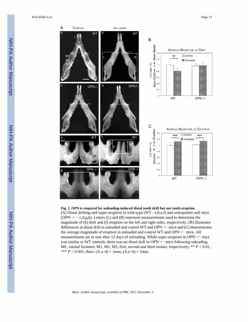
Fig. 1. OPN is required for unloading-induced distal tooth drift but not tooth eruption(A) Distal drifting and super-eruption in wild-type (WT - a,b,e,f) and osteopontin null mice(OPN−/− - c,d,g,h). Letters (L) and (R) represent measurements used to determine themagnitude of (b) drift and (f) eruption on the left and right sides, respectively. (B) illustratesdifferences in distal drift in unloaded and control WT and OPN−/− mice and (C) demonstratesthe average magnitude of eruption in unloaded and control WT and OPN−/− mice. Allmeasurements are in mm after 12 days of unloading. While super-eruption in OPN−/− micewas similar to WT controls, there was no distal drift in OPN−/− mice following unloading.MF, mental foramen; M1, M2, M3, first, second and third molars, respectively; ** P < 0.01,*** P < 0.001; Bars: (A.a–d) = 1mm; (A.e–h) = 1mm.
WALKER et al. Page 13
Bone. Author manuscript; available in PMC 2011 December 1.
NIH
-PA Author Manuscript
NIH
-PA Author Manuscript
NIH
-PA Author Manuscript
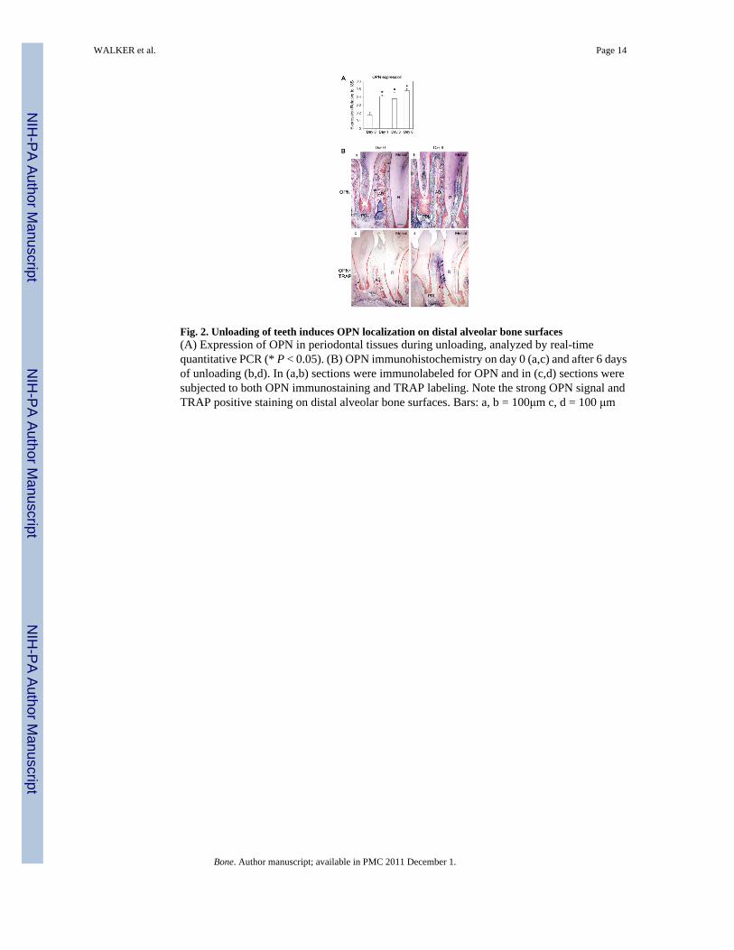
Fig. 2. Unloading of teeth induces OPN localization on distal alveolar bone surfaces(A) Expression of OPN in periodontal tissues during unloading, analyzed by real-timequantitative PCR (* P < 0.05). (B) OPN immunohistochemistry on day 0 (a,c) and after 6 daysof unloading (b,d). In (a,b) sections were immunolabeled for OPN and in (c,d) sections weresubjected to both OPN immunostaining and TRAP labeling. Note the strong OPN signal andTRAP positive staining on distal alveolar bone surfaces. Bars: a, b = 100μm c, d = 100 μm
WALKER et al. Page 14
Bone. Author manuscript; available in PMC 2011 December 1.
NIH
-PA Author Manuscript
NIH
-PA Author Manuscript
NIH
-PA Author Manuscript
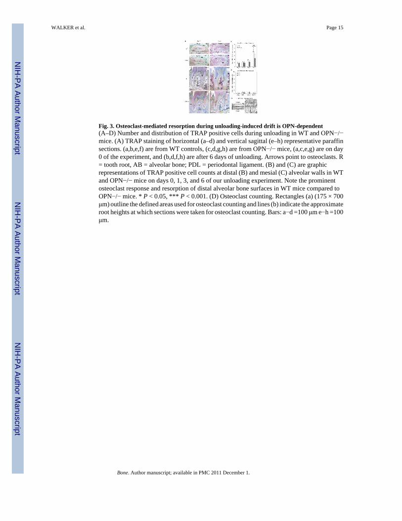
Fig. 3. Osteoclast-mediated resorption during unloading-induced drift is OPN-dependent(A–D) Number and distribution of TRAP positive cells during unloading in WT and OPN−/−mice. (A) TRAP staining of horizontal (a–d) and vertical sagittal (e–h) representative paraffinsections. (a,b,e,f) are from WT controls, (c,d,g,h) are from OPN−/− mice, (a,c,e,g) are on day0 of the experiment, and (b,d,f,h) are after 6 days of unloading. Arrows point to osteoclasts. R= tooth root, AB = alveolar bone; PDL = periodontal ligament. (B) and (C) are graphicrepresentations of TRAP positive cell counts at distal (B) and mesial (C) alveolar walls in WTand OPN−/− mice on days 0, 1, 3, and 6 of our unloading experiment. Note the prominentosteoclast response and resorption of distal alveolar bone surfaces in WT mice compared toOPN−/− mice. * P < 0.05, *** P < 0.001. (D) Osteoclast counting. Rectangles (a) (175 × 700μm) outline the defined areas used for osteoclast counting and lines (b) indicate the approximateroot heights at which sections were taken for osteoclast counting. Bars: a−d =100 μm e−h =100μm.
WALKER et al. Page 15
Bone. Author manuscript; available in PMC 2011 December 1.
NIH
-PA Author Manuscript
NIH
-PA Author Manuscript
NIH
-PA Author Manuscript
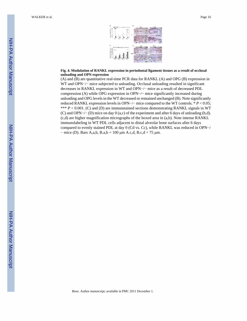
Fig. 4. Modulation of RANKL expression in periodontal ligament tissues as a result of occlusalunloading and OPN expression(A) and (B) are quantitative real-time PCR data for RANKL (A) and OPG (B) expression inWT and OPN−/− mice subjected to unloading. Occlusal unloading resulted in significantdecreases in RANKL expression in WT and OPN−/− mice as a result of decreased PDLcompression (A) while OPG expression in OPN−/− mice significantly increased duringunloading and OPG levels in the WT decreased or remained unchanged (B). Note significantlyreduced RANKL expression levels in OPN−/− mice compared to the WT controls. * P < 0.05;*** P < 0.001. (C) and (D) are immunstained sections demonstrating RANKL signals in WT(C) and OPN−/− (D) mice on day 0 (a,c) of the experiment and after 6 days of unloading (b,d).(c,d) are higher magnification micrographs of the boxed area in (a,b). Note intense RANKLimmunolabeling in WT PDL cells adjacent to distal alveolar bone surfaces after 6 dayscompared to evenly stained PDL at day 0 (Cd vs. Cc), while RANKL was reduced in OPN−/− mice (D). Bars A,a,b, B.a,b = 100 μm A.c,d, B.c,d = 75 μm.
WALKER et al. Page 16
Bone. Author manuscript; available in PMC 2011 December 1.
NIH
-PA Author Manuscript
NIH
-PA Author Manuscript
NIH
-PA Author Manuscript
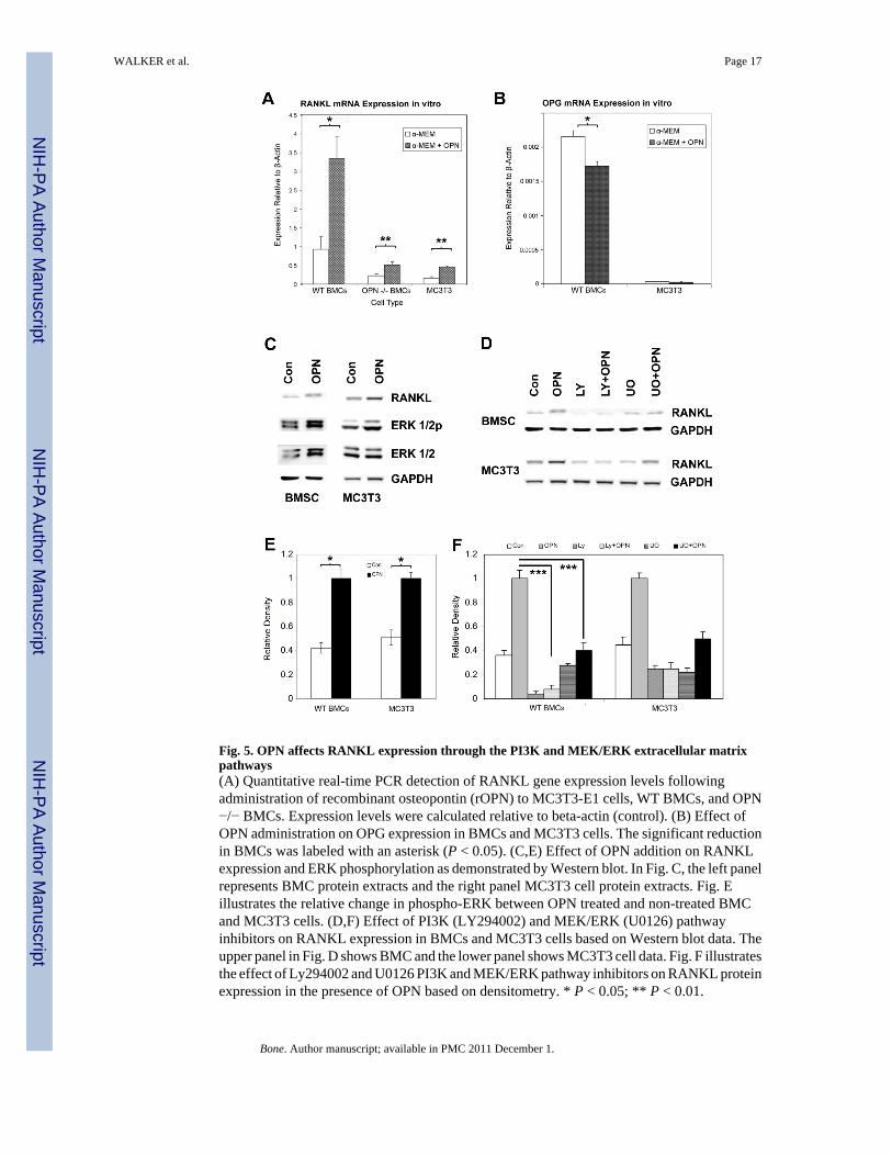
Fig. 5. OPN affects RANKL expression through the PI3K and MEK/ERK extracellular matrixpathways(A) Quantitative real-time PCR detection of RANKL gene expression levels followingadministration of recombinant osteopontin (rOPN) to MC3T3-E1 cells, WT BMCs, and OPN−/− BMCs. Expression levels were calculated relative to beta-actin (control). (B) Effect ofOPN administration on OPG expression in BMCs and MC3T3 cells. The significant reductionin BMCs was labeled with an asterisk (P < 0.05). (C,E) Effect of OPN addition on RANKLexpression and ERK phosphorylation as demonstrated by Western blot. In Fig. C, the left panelrepresents BMC protein extracts and the right panel MC3T3 cell protein extracts. Fig. Eillustrates the relative change in phospho-ERK between OPN treated and non-treated BMCand MC3T3 cells. (D,F) Effect of PI3K (LY294002) and MEK/ERK (U0126) pathwayinhibitors on RANKL expression in BMCs and MC3T3 cells based on Western blot data. Theupper panel in Fig. D shows BMC and the lower panel shows MC3T3 cell data. Fig. F illustratesthe effect of Ly294002 and U0126 PI3K and MEK/ERK pathway inhibitors on RANKL proteinexpression in the presence of OPN based on densitometry. * P < 0.05; ** P < 0.01.
WALKER et al. Page 17
Bone. Author manuscript; available in PMC 2011 December 1.
NIH
-PA Author Manuscript
NIH
-PA Author Manuscript
NIH
-PA Author Manuscript
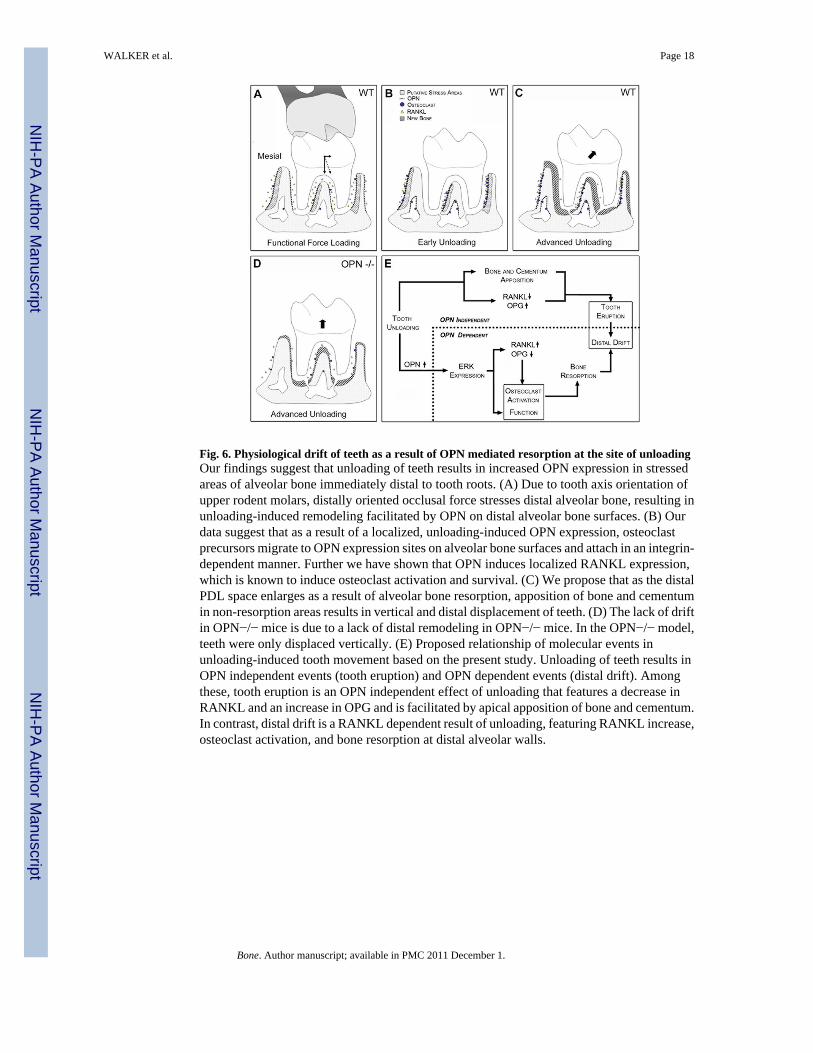
Fig. 6. Physiological drift of teeth as a result of OPN mediated resorption at the site of unloadingOur findings suggest that unloading of teeth results in increased OPN expression in stressedareas of alveolar bone immediately distal to tooth roots. (A) Due to tooth axis orientation ofupper rodent molars, distally oriented occlusal force stresses distal alveolar bone, resulting inunloading-induced remodeling facilitated by OPN on distal alveolar bone surfaces. (B) Ourdata suggest that as a result of a localized, unloading-induced OPN expression, osteoclastprecursors migrate to OPN expression sites on alveolar bone surfaces and attach in an integrin-dependent manner. Further we have shown that OPN induces localized RANKL expression,which is known to induce osteoclast activation and survival. (C) We propose that as the distalPDL space enlarges as a result of alveolar bone resorption, apposition of bone and cementumin non-resorption areas results in vertical and distal displacement of teeth. (D) The lack of driftin OPN−/− mice is due to a lack of distal remodeling in OPN−/− mice. In the OPN−/− model,teeth were only displaced vertically. (E) Proposed relationship of molecular events inunloading-induced tooth movement based on the present study. Unloading of teeth results inOPN independent events (tooth eruption) and OPN dependent events (distal drift). Amongthese, tooth eruption is an OPN independent effect of unloading that features a decrease inRANKL and an increase in OPG and is facilitated by apical apposition of bone and cementum.In contrast, distal drift is a RANKL dependent result of unloading, featuring RANKL increase,osteoclast activation, and bone resorption at distal alveolar walls.
WALKER et al. Page 18
Bone. Author manuscript; available in PMC 2011 December 1.
NIH
-PA Author Manuscript
NIH
-PA Author Manuscript
NIH
-PA Author Manuscript