characterization of osteopontin expression and function after status epilepticus
TRANSCRIPT
Characterization of osteopontin expression
and function after status epilepticus∗yKarin Borges, zMarla Gearing, xSusan Rittling, ¶Esben S. Sorensen,
#Robert Kotloski, ∗∗David T. Denhardt, and yRaymond Dingledine
∗Department of Pharmaceutical Sciences, Texas Tech Health Sciences Center, Amarillo, Texas, U.S.A.;
yDepartment of Pharmacology, Emory University, Atlanta, Georgia, U.S.A.; zDepartment of Pathology and
Laboratory Medicine, Emory University, Atlanta, Georgia, U.S.A.; xDepartment of Cell Biology and Neuroscience,
The Forsyth Institute, Boston, Massachusetts, U.S.A.; ¶Protein Chemistry Laboratory, Department of Molecular
Biology, University of Aarhus, Aarhus C, Denmark; #Department of Neurobiology, Duke University, Durham,
North Carolina, U.S.A.; and ∗∗Department of Cell Biology and Neuroscience, Rutgers University, Piscataway,
New Jersey, U.S.A.
SUMMARY
Purpose: Osteopontin is a cytokine found in many
tissues and plays a role in tissue injury and repair.
This study had two goals: to characterize osteo-
pontin expression after status epilepticus (SE),
and to test the hypotheses that osteopontin affects
the susceptibility to seizures or alters cell death
and inflammation after SE.
Methods: Pilocarpine was used to induce SE in
OPN)/) and OPN+/+ mice to compare seizure sus-
ceptibility, neuropathological markers including
real time PCR for inflammatory genes, and osteo-
pontin immunohistochemistry. The effect of
added osteopontin on excitotoxicity by N-methyl-
D-aspartate in neuronal cultures of ONP)/) mice
was determined.
Results: Neurons undergoing degeneration
showed osteopontin immunoreactivity 2–3 days
after SE. After 10 to 31 days degenerating axons in
the thalamus were osteopontin-positive. The sus-
ceptibility to seizures of OPN)/) and OPN+/+ mice
in the pilocarpine, fluorothyl, and maximal elec-
troshock models was similar. There were no signif-
icant differences in the extent of neuronal damage
after pilocarpine-induced SE, the expression of
several neuropathological markers or the RNA
levels of selected inflammatory genes. Recombi-
nant and natural bovine osteopontin did not affect
the extent of NMDA-induced cell death in OPN)/)
mouse neuronal cultures.
Conclusion: We demonstrated that osteopontin is
up-regulated in response to SE in distinct temporal
sequences in the hippocampus, specifically in dege-
nerating neurons and axons. However, osteopontin
did not appear to regulate neurodegeneration or
inflammation within the first 3 days after SE.
KEY WORDS: Seizure, Pilocarpine, Inflammation,
Axonal degeneration, Neuronal degeneration.
Osteopontin (gene symbol spp1) is a highly glycosylatedmultifunctional phosphoprotein that influences adhesion,migration, cell survival, and inflammation (reviewed inDenhardt et al., 2001a, 2001b; Scatena et al., 2007). It isexpressed in many tissues and cell types, such as bone,kidney, epithelial cells, arterial smooth muscles, inflam-matory cells, certain brain areas, and in tumors. Osteopon-
tin is usually secreted and is present in all body fluids butcan also be found underneath the plasma membrane inassociation with the intracellular domain of CD44 (Zoharet al., 2000). Osteopontin is upregulated after injury inmany peripheral tissues.
Osteopontin associates with the hyaluronin receptorCD44 and extracellular matrix molecules, includingfibronectin, type I collagen, osteocalcin, and mediatescell attachment via its arginine-glycine-aspartate (RGD)sequence by binding to integrins. It also plays a role incell migration and matrix reorganization after injury andwound healing (Liaw et al., 1998). Furthermore,osteopontin acts as a cytokine because it potently attractsmacrophages and regulates secretion of cytokines from
Accepted March 10, 2008; Early View publication June 3, 2008.Address correspondence to Karin Borges, Ph.D., Department of Phar-
maceutical Sciences, Texas Tech University Health Sciences, 1300 SCoulter, Amarillo, TX 79106, U.S.A. E-mail: [email protected]
Wiley Periodicals, Inc.ª 2008 International League Against Epilepsy
Epilepsia, 49(10):1675–1685, 2008doi: 10.1111/j.1528-1167.2008.01613.x
FULL-LENGTH ORIGINAL RESEARCH
1675
macrophages and T-cells. On the other hand, osteopontincan also limit inflammation by inhibition of matrixmetalloproteinase 2 and inducible nitric oxide synthase(iNOS; reviewed in Denhardt et al., 2001b; Scatena et al.,2007).
Osteopontin is normally not detected in the healthytel-encephalon and diencephalon, but has been found inother brain areas, such as the substantia nigra (Iczkiewiczet al., 2006; Schroeter et al., 2006), midbrain, brain stem,and the olfactory bulb (Shin et al., 1999) and in peripheralneurons (Ichikawa et al., 2001). Osteopontin has beenreported to be upregulated in various brain disorders orafter injury, e.g., after ischemia (Ellison et al., 1998; Wanget al., 1998; Lee et al., 1999; Choi et al., 2007), kainate-induced seizures (Kim et al., 2002), cryolesions (Shinet al., 2005), experimental autoimmune encephalomyelitis(EAE, Chabas et al., 2001), and spinal cord injury(Hashimoto et al., 2007). In human brain tissue osteopontinimmunoreactivity was found in multiple sclerosis plaques(Chabas et al., 2001; Diaz-Sanchez et al., 2006) and in peri-ventricular leukomalacia (Tanaka et al., 2000). Osteo-pontin appears to be protective against ischemic injury inthe brain (Meller et al., 2005) and kidney (Noiri et al.,1999; Denhardt et al., 2001a, 2001b), but in contrast it alsopromotes inflammation and exacerbates autoinmmunediseases, such as EAE (Chabas et al., 2001).
In a microarray survey, we found that osteopontin wasthe most prominently upregulated among approximately60 upregulated genes in the CA3 pyramidal cell layer 3days after pilocarpine-induced status epilepticus (SE, JimDoherty and Raymond Dingledine, unpublished). Givenosteopontin's many roles in inflammation and tissue injuryand its protective effects against ischemia, we hypothe-sized that osteopontin might alter neuropathologicalevents after SE, including cell death and inflammation, ormight affect the susceptibility to seizures. Here, we char-acterized osteopontin expression and its potential roleafter SE in a chronic seizure model.
METHODS
Mice and seizure modelsThe pilocarpine-injected CF1 mouse model used in
this study was described earlier (Borges et al., 2003,2004). Outbred CF1 mice (6–10 weeks old, 30–42 g)were obtained from Charles River (Wilmington, MA,U.S.A.). Mice deficient for osteopontin expression andtheir wild type counterparts were obtained fromDr. Susan Rittling (OPN)/) (SR) and OPN+/+(SR);Rittling et al., 1998) and Dr. Lucy Liaw (OPN)/) (LL)and OPN+/+(LL); Liaw et al., 1998). The OPN)/) andOPN+/+ (SR) mice are derived from AB2.1 stem cells andare therefore a substrain of 129Sv mice from Jacksonlabs. OPN)/) and OPN+/+ (LL) mice were bred into the129/SvJ strain from Taconic Farms Inc. (Germantown,
NY, U.S.A.; Liaw et al., 1998). All mice were housedunder a 12 h light dark cycle with food and water ad libi-dum. Mice were injected with methylscopolamine andterbutaline (2 mg/kg each i.p. in 0.9% NaCl) to minimizeperipheral side effects followed after 15–30 min by dif-ferent doses of pilocarpine for CF1 mice (254–290 mg/kg, i.p.), OPN)/) and OPN+/+ mice from Dr. Susan Rit-tling (255–310 mg/kg, i.p.) and Dr. Lucy Liaw (230–240 mg/kg, i.p.). Approximately 30% of injected miceexperienced behavioral SE lasting about 5 h as definedby continuous seizure activity consisting mainly ofwhole body continuous clonic seizures. SE onset wasdefined as the onset of whole clonic body seizures thatbecame continuous. SE severity was scored by the mostsevere seizure activity observed, with whole body clonicseizures scored as (1), severe whole body clonic seizuresscored as (2), and tonic–clonic seizures or jumpingscored as (3). We did not measure the end of SE, becausebehavioral assessment is highly subjective at this timepoint and electrographic SE can continue sporadically forat least 24 h (Kris Bough & Raymond Dingledine,unpublished). About 6 h after pilocarpine injection, allmice were injected with 0.5–0.8 ml 5% dextrose in lac-tate Ringer’s solution (i.p.). Mice were fed moistenedhigh-fat rodent chow and were monitored daily andinjected with 5% dextrose in lactate Ringer's solutionwhen needed. ‘‘Control’’ mice received terbutaline andmethylscopolamine but no pilocarpine. CF1 mice thathad experienced SE after repeated kainate injection werefrom a previous study (Borges et al., 2004). Briefly,kainate (5–20 mg/kg, i.p. in phosphate-buffered saltsolution) was injected every 30 min until SE wasreached. After 4–5 h of behavioral SE, seizures wereterminated by injection of 25 mg/kg pentobarbital (i.p.).
For electroshock seizures one or two drops of localanesthetic (lidocaine, 1%) were placed in each conjunc-tiva of the animal. Approximately 30 s later, the animalwas picked up gently and a cup electrode was placed overeach cornea and current was administered through theelectrodes (Wahlquist Instrument Co., Salt Lake City, UT,U.S.A.). To induce maximal seizures, a stimulus of 200mA at 60 Hz for 200 ms duration was administered.Seizure onset occurred virtually instantaneously with theonset of current flow. The durations of tonic hind limbflexion and tonic hind limb extension were recorded.
For the fluorothyl model, 18–26 g OPN)/) (SR) andOPN+/+(SR) mice were placed individually into a clearplastic chamber (15 cm · 20 cm · 28 cm) and 20 ll/minfluorothyl was constantly dripped onto a filter paperplaced on a platform near the top of the container. Thevolatile fluorothyl evaporates and the latencies requiredfor the first myoclonic jerk and the first tonic–clonicseizure were timed.
All experiments were approved by the InstitutionalAnimal Care and Use Committee (IACUC) of Emory
1676
K. Borges et al.
Epilepsia, 49(10):1675–1685, 2008doi: 10.1111/j.1528-1167.2008.01613.x
University and conducted in accordance with its guide-lines. Every effort was made to minimize animal suffering.
Immunohistochemistry and neuropathologyAs described earlier (Borges et al., 2003) mice were
sacrificed under deep isoflurane anesthesia, and theirbrains were removed and immersed in 4% paraformalde-hyde fixative for at least 10 h. Brains were sliced coronallyin 1–2 mm slices, dehydrated in a graded series of alcoholsand xylenes and infiltrated with paraffin using an auto-mated tissue processor (Shandon Hypercenter XP,Pittsburgh, PA, U.S.A.), then embedded in paraffinblocks. Eight lm sections were cut using a ShandonAS325 microtome.
To compare the extent of cell death in wild type andOPN)/) mice after pilocarpine-induced SE, sections werestained with hematoxylin. Healthy neurons could be easilydistinguished from nonneuronal cells and injured neuronsby their large size, medium-intensity staining and darknucleoli. After SE, many pyramidal cells were shrunkenand stained darkly or had disappeared at late time points.
The amount of hippocampal pyramidal cell damage inmice 2–3 days after pilocarpine-induced SE and controlmice was assessed between )1.8 and )2.5 mm bregma(corresponding to mid-level hippocampal sections accord-ing to the atlas for C57BL/6 mice, Hof 2000) by a blindedinvestigator. The same investigator (KB) conducted allsubjective ratings. The amount of damage in the hippo-campal pyramidal CA1 and CA3 regions (a–c sectors con-sidered together for CA3) were scored for each mouse ona 0–4 scale, by estimating in 25% increments the numberof healthy cells remaining relative to control mice. Score 0was given if the section was undistinguishable from con-trol mice, i.e., the number of healthy neurons appearednormal, even if a few pyknotic cells were found. Score 1:>75% healthy pyramidal cells remaining, but with clearevidence of cell death; 2: 50–74%; 3: 25–49%; 4: <25%healthy pyramidal neurons remaining. The median scorewas calculated for each mouse from at least three or fourhippocampal sections.
Eight-micron thick paraffin-embedded sections wereimmunohistochemically labeled as described (Gearinget al., 1993; Borges et al., 2003, 2004). Briefly, sectionswere deparaffinized in a series of xylenes and graded alco-hols and were blocked with normal serum and then incu-bated with primary antibody, followed by biotinylatedsecondary antibody and avidin-biotin-peroxidase complex(ABC Elite Kit, Vector Laboratories, Burlingame, CA,U.S.A.), the MOM kit (Vector Laboratories) or the DakoEnvision System (Dako Corporation, Carpinteria, CA,U.S.A.) as instructed by the manufacturer. The chromagenused for color development was 3,3¢-diaminobenzidine,and sections were counterstained with hematoxylin. Forosteopontin staining, we used the monoclonal mouse 2A1antibody (1:100) followed by the MOM kit (Vector Labo-
ratories) or the rabbit LF-123 antibody (1:800, Fisheret al., 1995) with the Dako Envision System. For the amy-loid precursor protein (APP) antibody (1:200, BoehringerMannheim, GmbH, Mannheim, Germany), the biotinylat-ed secondary antibody (1:8,000, Vector Laboratories) wasfollowed by avidin-conjugated alkaline phosphatase, withnitroblue tetrazolium and 5-bromo, 4-chloro, 3-indolylphosphate for color development with nuclear fast redcounterstain. Other antibodies used to characterize theOPN)/) and OPN+/+ mice after seizures were rabbit anti-cow glial fibrillary acidic protein (GFAP,1:500, DakoCorporation), rabbit anti-neuropeptide Y (NPY, 1:500,Bachem Americas, Inc., Torrance, CA, U.S.A.), goat anti-b2 microglobulin (M20, 1:200, Santa Cruz Biotechnol-ogy, Inc., Santa Cruz, CA, U.S.A.), and rat anti-CD44antibodies (Km114, 1:100, and Im7, 1:50, both BDPharmingen, San Jose, CA, U.S.A.); these were visualizedwith the ABC elite kit as described earlier (Borges et al.,2003, 2004). Positive and negative controls were includedin each staining experiment. For all antibodies, negativecontrols included sections incubated in the absence of pri-mary antibody. For the OPN antibodies, additional nega-tive controls consisted of sections from OPN)/) mice 3days after SE incubated in the presence of primary anti-body. Positive controls included sections after 1–3 daysafter SE or human brain sections known to stain with APP.All antibodies labeled specific structures and cell popula-tions as described here or earlier.
Real-time PCRTwenty-four hours after pilocarpine-induced SE,
OPN)/) (SR) and OPN+/+(SR) mice were decapitatedunder deep isoflurane anesthesia. Hippocampi were dis-sected out and frozen on dry ice. RNA was extracted usingTri reagent (Sigma-Aldrich, St. Louis, MO, U.S.A.) andtreated with RNAse-free DNase on RNeasy columns (bothQiagen, Valencia, CA, U.S.A.). After elution, RNA wasquantitated using Ribogreen and 250 ng of RNA was
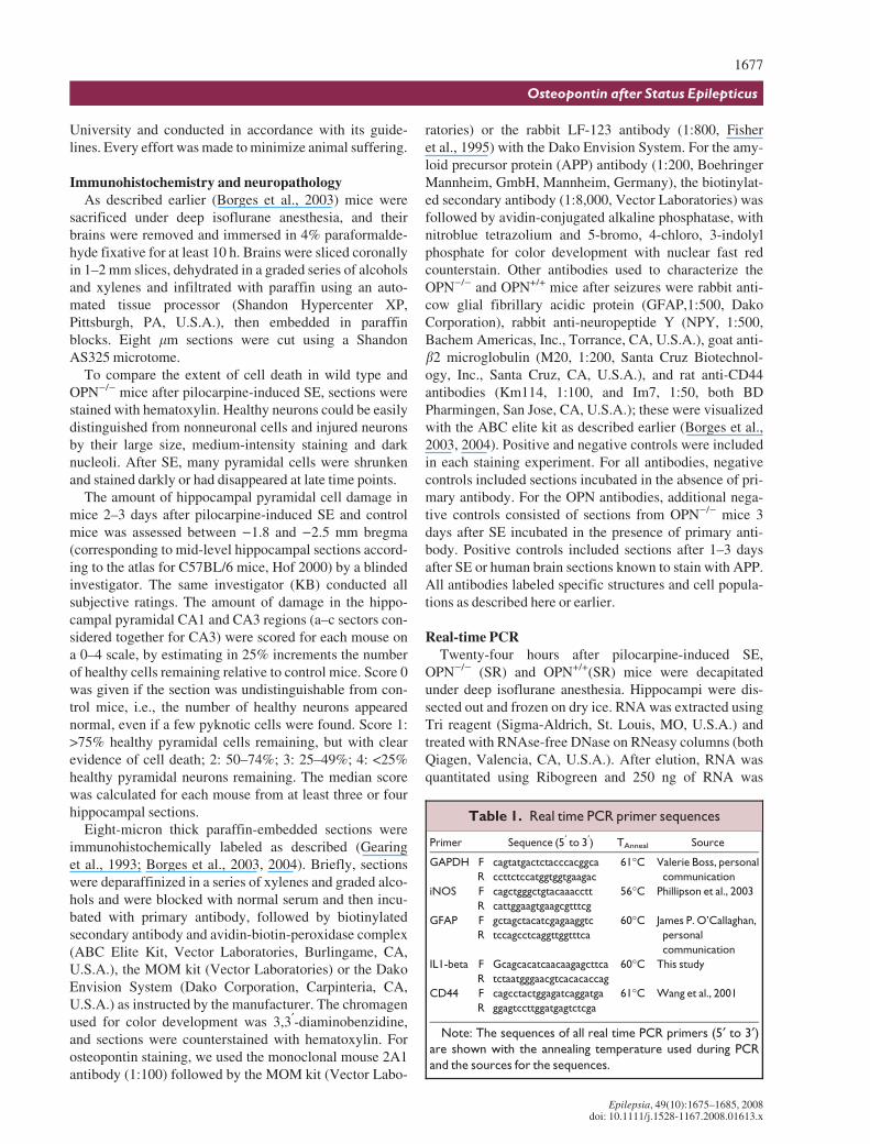
Table 1. Real time PCR primer sequences
Primer Sequence (5¢ to 3¢) TAnneal Source
GAPDH F cagtatgactctacccacggca 61�C Valerie Boss, personal
R ccttctccatggtggtgaagac communication
iNOS F cagctgggctgtacaaacctt 56�C Phillipson et al., 2003
R cattggaagtgaagcgtttcg
GFAP F gctagctacatcgagaaggtc 60�C James P. O’Callaghan,
R tccagcctcaggttggtttca personal
communication
IL1-beta F Gcagcacatcaacaagagcttca 60�C This study
R tctaatgggaacgtcacacaccag
CD44 F cagcctactggagatcaggatga 61�C Wang et al., 2001
R ggagtccttggatgagtctcga
Note: The sequences of all real time PCR primers (5¢ to 3¢)are shown with the annealing temperature used during PCR
and the sources for the sequences.
1677
Osteopontin after Status Epilepticus
Epilepsia, 49(10):1675–1685, 2008doi: 10.1111/j.1528-1167.2008.01613.x
reverse transcribed using oligo-dT primers andSuperscript II (Invitrogen, Carlsbad, CA, U.S.A.). For realtime PCR first strand cDNA from the equivalent of 3.6 ngRNA was reacted for 40 cycles with specific primer setsand SYBR Green PCR Master Mix (Applied Biosystems,Foster City, CA, U.S.A.) in an iCycler (Biorad, Hercules,CA, U.S.A.). All samples were run in duplicate. Real timeprimers were ordered from Integrated DNA Technologies,Inc. (Coralville, IA, U.S.A.; Table 1). All primer setsresulted in a single PCR product as judged from meltcurves and had similar PCR efficiencies (94–114%) com-pared to glyceraldehyde 3-phosphate dehydrogenase(GAPDH) primers (105%). Threshold cycles were deter-mined for GFAP, CD44, interleukin-1b (IL-1b), and iNOSand were normalized to the threshold cycles for GAPDHfor every animal to account for small differences in cDNAstarting concentrations (DCtiRNA ¼ CtiRNA ) CtGAPDH).The fold increase of each inflammatory RNA (iRNA) wascalculated for each animal 1 day after SE relative to theaverage amount of RNA found in six control animals (foldinduction ¼ 2 to the power of DCtiRNA(SE) ) averageDCtiRNA(CON)).
Cell culture assaysE14 mouse neuronal cell cultures were prepared from
timed pregnant OPN)/) (SR) mice. Briefly, the motherwas sacrificed by isoflurane anesthesia followed by cervi-cal dislocation. The embryos were removed and decapi-tated immediately. Forebrains were dissected out, themeninges removed, and neural cells were triturated to asingle cell suspension in cold Neurobasal medium. Cellswere seeded in Neurobasal medium with 1 mM glutaminesupplemented with B27 (Invitrogen) at a density of about75,000 cells/well into 24 well plates coated with 60 lg/mlpoly-D-lysine. After 11–14 days in vitro, medium waspartially removed for lactate dehydrogenase (LDH) mea-surements and 50–250 ng/ml mouse recombinant osteo-pontin (Sigma) or cow osteopontin isolated from milk(Sorensen & Petersen, 1993), or 250 ng/ml bovine serumalbumin (BSA, Sigma) was added. After 2 h preincubationwith osteopontin different concentrations of N-methyl-D-aspartate (NMDA) were added overnight for about 18 h.Neurobasal medium contains 400 lM glycine, so addi-tional glycine was unnecessary. To quantify neuronal celldeath the percentage of released LDH was essayed with anLDH cytotoxicity kit (Roche, Basel, Switzerland). Doseresponse curves were constructed with Origin software.The data from three different cultures were normalized bysetting the maximum percentage of LDH release withNMDA and BSA to 100%.
StatisticsFor parametric values we calculated mean and standard
error of the mean (s.e.m.), and for scores we report themedian. Unpaired two-tailed Student's t-tests were used to
compare seizure thresholds in the fluorothyl and electro-shock models, the induction of iRNAs in OPN)/) versusOPN+/+ mice, and the log EC50s of NMDA-induced celldeath with cow osteopontin versus BSA. The Mann–Whitney nonparametric test was used to compare SE sever-ity scores and neuronal damage scores. One-way ANOVAswere employed to compare the threshold cycles forGAPDH among the four groups of mice and the log EC50sof NMDA-induced cell death with two different concentra-tions of recombinant osteopontin versus BSA. GraphpadInstat was employed for all statistical comparisons.
RESULTS
Osteopontin expression in degenerating neuronsafter SE
Osteopontin upregulation was initially found 3 daysafter pilocarpine-induced SE in a microarray study (JimDoherty & Raymond Dingledine, unpublished). To assessthe time course of osteopontin protein upregulation in thischronic epilepsy model two antibodies, mouse 2A1 andrabbit LF-123, were used to stain brains 1–31 days afterpilocarpine-induced SE in CF1 mice (Figs. 1 and 2); bothantibodies resulted in similar staining patterns (e.g., com-pare Figs. 1B3 and 1D). Osteopontin immunoreactivitywas negative in untreated control mice (Fig. 1C), butappeared 1 day after pilocarpine-induced SE (Fig. 1A1,1A2, and 1A3) and was strongest 2–3 days after SE (Figs.1B1, 1B2, 1B3, and 1D). One day after SE only a fewsmall cells, potentially microglia (Fig. 1A3), and someneurons identified by their nuclear morphology (Fig.1A2), were labeled in the hippocampus and thalamus.Neuronal nuclei are large, stain light blue with the hema-toxylin counterstain and contain several darker nucleoli.After 2–3 days extracellular matrix (Figs. 1B1, 1B2, 1B3,and 1D) and areas showing neuronal damage, such as thehippocampus, thalamus, amygdala, and piriform cortexshowed strong and abundant osteopontin labeling, specifi-cally in small cells with dark nuclei, many resemblingdying neurons (Figs. 1B1, 1B2, 1B3, and 1D) and somepotentially representing microglia. Similarly, after SEinduced by repeated kainate injections, cells in the pyra-midal cell layer die (Borges et al., 2004) and small cellswith dark nuclei were osteopontin-positive in the damagedhippocampal pyramidal cell layers (Fig. 1E). In someexperiments, osteopontin-specific antibodies labeledastrocytes after 3 days of SE. However, this stainingappears to be nonspecific because similar astrocyte label-ing was found in OPN)/) mice after SE (not shown).
Osteopontin expression in degenerating axons after SETen to 31 days after pilocarpine-induced-SE punctate
osteopontin immunoreactivity was found in the thalamusin several nuclei, including the nucleus reuniens, ventrome-dial nucleus, lateral dorsal nucleus, the lateral geniculate
1678
K. Borges et al.
Epilepsia, 49(10):1675–1685, 2008doi: 10.1111/j.1528-1167.2008.01613.x
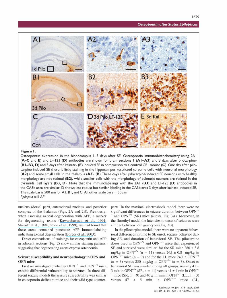
nucleus (dorsal part), anterodorsal nucleus, and posteriorcomplex of the thalamus (Figs. 2A and 2B). Previously,when assessing axonal degeneration with APP, a markerfor degenerating axons (Kawarabayashi et al., 1991;Sherriff et al., 1994; Stone et al., 1999), we had found thatthese areas contained punctuate APP immunolabelingindicating axonal degeneration (Borges et al., 2003).
Direct comparisons of stainings for osteopontin and APPin adjacent sections (Fig. 2) show similar staining patterns,suggesting that degenerating axons express osteopontin.
Seizure susceptibility and neuropathology in OPN andOPN mice
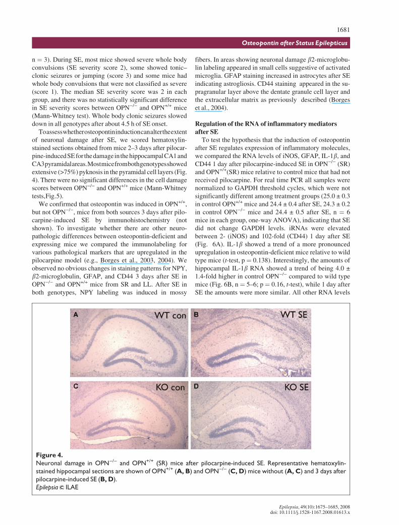
First we investigated whether OPN)/) and OPN+/+ miceexhibit differential vulnerability to seizures. In three dif-ferent seizure models the seizure susceptibility was similarin osteopontin-deficient mice and their wild type counter-
parts. In the maximal electroshock model there were nosignificant differences in seizure duration between OPN)/
) and OPN+/+ (SR) mice (t-tests, Fig. 3A). Moreover, inthe flurothyl model the latencies to onset of seizures weresimilar between both genotypes (Fig. 3B).
In the pilocarpine model, there were no apparent behav-ioral differences in time to SE onset, seizure behavior dur-ing SE, and duration of behavioral SE. The pilocarpinedoses used in OPN+/+ and OPN)/) mice that experiencedSE and survived were similar: for the SR mice 280 € 3.8mg/kg in OPN+/+ (n ¼ 11) versus 265 € 0.8 mg/kg inOPN)/) mice (n ¼ 9) and for the LL mice 240 in OPN+/+
(n ¼ 3) versus 230 mg/kg in OPN)/) (n ¼ 3). Onset tobehavioral SE was similar among all groups, namely 41 €3 min in OPN+/+ (SR, n¼ 11) versus 41 € 4 min in OPN)/
) mice (SR, n¼ 9) and 40 € 11 min in OPN+/+ (LL, n¼ 3)versus 47 € 5 min in OPN)/) mice (LL,
Figure 1.
Osteopontin expression in the hippocampus 1–3 days after SE. Osteopontin immunohistochemistry using 2A1
(A–C and E) and LF-123 (D) antibodies are shown for brain sections 1 (A1–A3) and 3 days after pilocarpine-
(B1–B3, D) and 3 days after kainate- (E) induced SE in comparison to a control CF1 mouse (C). One day after pilo-
carpine-induced SE there is little staining in the hippocampus restricted to some cells with neuronal morphology
(A2) and some small cells in the thalamus (A3). (B) Three days after pilocarpine-induced SE neurons with healthy
morphology are not stained (B2), while smaller cells with the morphology of pyknotic neurons are stained in the
pyramidal cell layers (B3, D). Note that the immunolabelings with the 2A1 (B3) and LF-123 (D) antibodies in
the CA3b area are similar. D shows less robust but similar labeling in the CA3b area 3 days after kainate-induced SE.
The scale bar is 500 lm for A1, B1, and C. All other scale bars¼ 50 lm
Epilepsia ILAE
1679
Osteopontin after Status Epilepticus
Epilepsia, 49(10):1675–1685, 2008doi: 10.1111/j.1528-1167.2008.01613.x
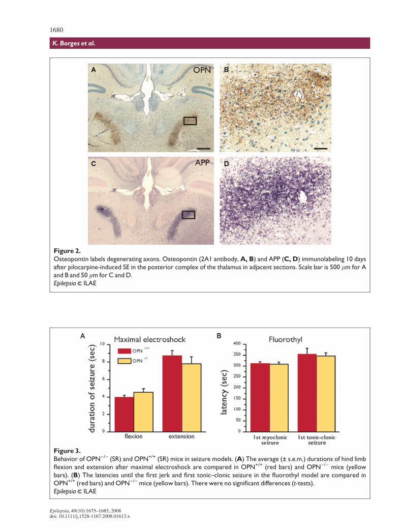
Figure 2.
Osteopontin labels degenerating axons. Osteopontin (2A1 antibody, A, B) and APP (C, D) immunolabeling 10 days
after pilocarpine-induced SE in the posterior complex of the thalamus in adjacent sections. Scale bar is 500 lm for A
and B and 50 lm for C and D.
Epilepsia ILAE
Figure 3.
Behavior of OPN)/) (SR) and OPN+/+ (SR) mice in seizure models. (A) The average (± s.e.m.) durations of hind limb
flexion and extension after maximal electroshock are compared in OPN+/+ (red bars) and OPN)/) mice (yellow
bars). (B) The latencies until the first jerk and first tonic–clonic seizure in the fluorothyl model are compared in
OPN+/+ (red bars) and OPN)/) mice (yellow bars). There were no significant differences (t-tests).
Epilepsia ILAE
1680
K. Borges et al.
Epilepsia, 49(10):1675–1685, 2008doi: 10.1111/j.1528-1167.2008.01613.x
n ¼ 3). During SE, most mice showed severe whole bodyconvulsions (SE severity score 2), some showed tonic–clonic seizures or jumping (score 3) and some mice hadwhole body convulsions that were not classified as severe(score 1). The median SE severity score was 2 in eachgroup, and there was no statistically significant differencein SE severity scores between OPN)/) and OPN+/+ mice(Mann-Whitney test). Whole body clonic seizures sloweddown in all genotypes after about 4.5 h of SE onset.
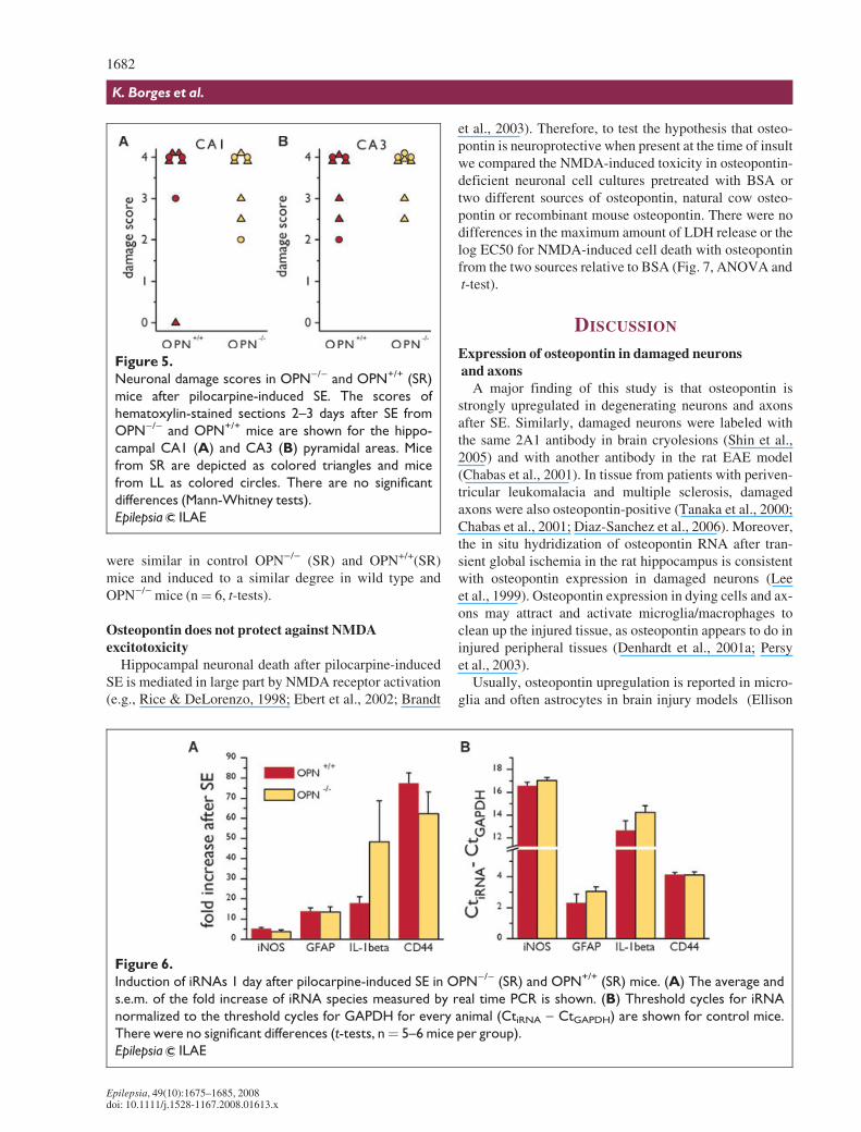
Toassesswhetherosteopontininductioncanaltertheextentof neuronal damage after SE, we scored hematoxylin-stained sections obtained from mice 2–3 days after pilocar-pine-inducedSEfor thedamageinthehippocampalCA1andCA3pyramidalareas.Mostmicefrombothgenotypesshowedextensive (>75%) pyknosis in the pyramidal cell layers (Fig.4). There were no significant differences in the cell damagescores between OPN)/) and OPN+/+ mice (Mann-Whitneytests,Fig.5).
We confirmed that osteopontin was induced in OPN+/+,but not OPN)/), mice from both sources 3 days after pilo-carpine-induced SE by immunohistochemistry (notshown). To investigate whether there are other neuro-pathologic differences between osteopontin-deficient andexpressing mice we compared the immunolabeling forvarious pathological markers that are upregulated in thepilocarpine model (e.g., Borges et al., 2003, 2004). Weobserved no obvious changes in staining patterns for NPY,b2-microglobulin, GFAP, and CD44 3 days after SE inOPN)/) and OPN+/+ mice from SR and LL. After SE inboth genotypes, NPY labeling was induced in mossy
fibers. In areas showing neuronal damage b2-microglobu-lin labeling appeared in small cells suggestive of activatedmicroglia. GFAP staining increased in astrocytes after SEindicating astrogliosis. CD44 staining appeared in the su-pragranular layer above the dentate granule cell layer andthe extracellular matrix as previously described (Borgeset al., 2004).
Regulation of the RNA of inflammatory mediatorsafter SE
To test the hypothesis that the induction of osteopontinafter SE regulates expression of inflammatory molecules,we compared the RNA levels of iNOS, GFAP, IL-1b, andCD44 1 day after pilocarpine-induced SE in OPN)/) (SR)and OPN+/+(SR) mice relative to control mice that had notreceived pilocarpine. For real time PCR all samples werenormalized to GAPDH threshold cycles, which were notsignificantly different among treatment groups (25.0 € 0.3in control OPN+/+ mice and 24.4 € 0.4 after SE, 24.3 € 0.2in control OPN)/) mice and 24.4 € 0.5 after SE, n ¼ 6mice in each group, one-way ANOVA), indicating that SEdid not change GAPDH levels. iRNAs were elevatedbetween 2- (iNOS) and 102-fold (CD44) 1 day after SE(Fig. 6A). IL-1b showed a trend of a more pronouncedupregulation in osteopontin-deficient mice relative to wildtype mice (t-test, p¼ 0.138). Interestingly, the amounts ofhippocampal IL-1b RNA showed a trend of being 4.0 €1.4-fold higher in control OPN)/) compared to wild typemice (Fig. 6B, n¼ 5–6; p¼ 0.16, t-test), while 1 day afterSE the amounts were more similar. All other RNA levels
Figure 4.
Neuronal damage in OPN)/) and OPN+/+ (SR) mice after pilocarpine-induced SE. Representative hematoxylin-
stained hippocampal sections are shown of OPN+/+ (A, B) and OPN)/) (C, D) mice without (A, C) and 3 days after
pilocarpine-induced SE (B, D).
Epilepsia ILAE
1681
Osteopontin after Status Epilepticus
Epilepsia, 49(10):1675–1685, 2008doi: 10.1111/j.1528-1167.2008.01613.x
were similar in control OPN)/) (SR) and OPN+/+(SR)mice and induced to a similar degree in wild type andOPN)/) mice (n¼ 6, t-tests).
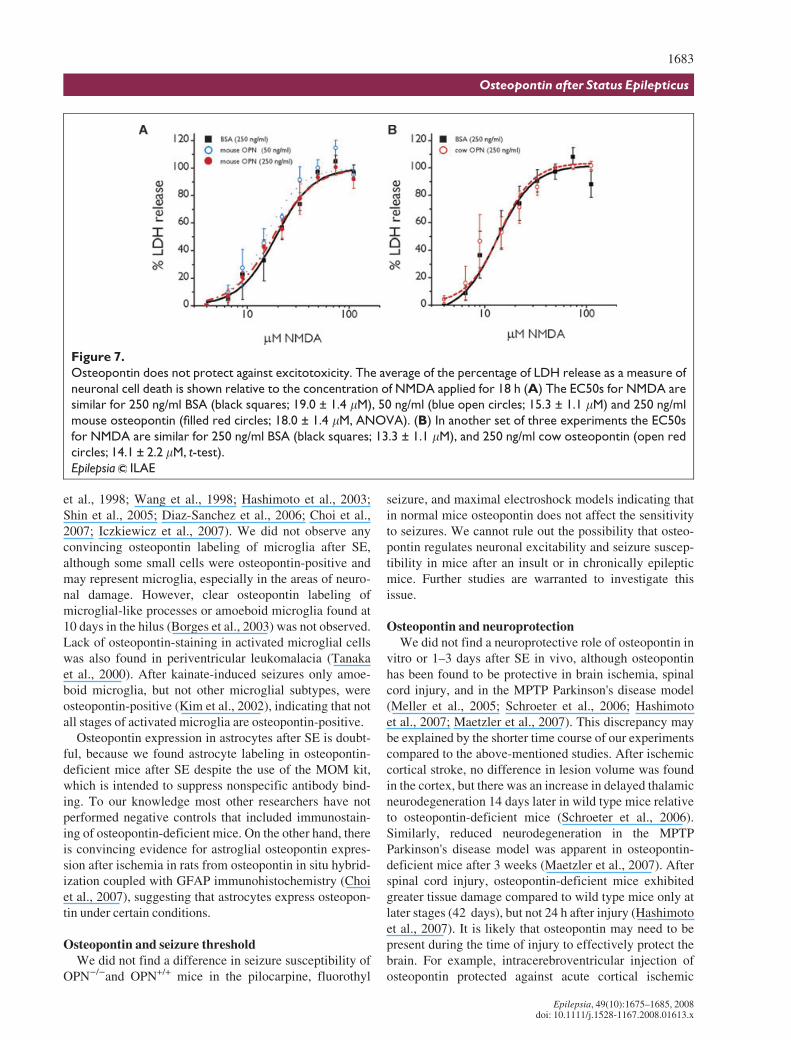
Osteopontin does not protect against NMDAexcitotoxicity
Hippocampal neuronal death after pilocarpine-inducedSE is mediated in large part by NMDA receptor activation(e.g., Rice & DeLorenzo, 1998; Ebert et al., 2002; Brandt
et al., 2003). Therefore, to test the hypothesis that osteo-pontin is neuroprotective when present at the time of insultwe compared the NMDA-induced toxicity in osteopontin-deficient neuronal cell cultures pretreated with BSA ortwo different sources of osteopontin, natural cow osteo-pontin or recombinant mouse osteopontin. There were nodifferences in the maximum amount of LDH release or thelog EC50 for NMDA-induced cell death with osteopontinfrom the two sources relative to BSA (Fig. 7, ANOVA andt-test).
DISCUSSION
Expression of osteopontin in damaged neuronsand axons
A major finding of this study is that osteopontin isstrongly upregulated in degenerating neurons and axonsafter SE. Similarly, damaged neurons were labeled withthe same 2A1 antibody in brain cryolesions (Shin et al.,2005) and with another antibody in the rat EAE model(Chabas et al., 2001). In tissue from patients with periven-tricular leukomalacia and multiple sclerosis, damagedaxons were also osteopontin-positive (Tanaka et al., 2000;Chabas et al., 2001; Diaz-Sanchez et al., 2006). Moreover,the in situ hydridization of osteopontin RNA after tran-sient global ischemia in the rat hippocampus is consistentwith osteopontin expression in damaged neurons (Leeet al., 1999). Osteopontin expression in dying cells and ax-ons may attract and activate microglia/macrophages toclean up the injured tissue, as osteopontin appears to do ininjured peripheral tissues (Denhardt et al., 2001a; Persyet al., 2003).
Usually, osteopontin upregulation is reported in micro-glia and often astrocytes in brain injury models (Ellison
Figure 6.
Induction of iRNAs 1 day after pilocarpine-induced SE in OPN)/) (SR) and OPN+/+ (SR) mice. (A) The average and
s.e.m. of the fold increase of iRNA species measured by real time PCR is shown. (B) Threshold cycles for iRNA
normalized to the threshold cycles for GAPDH for every animal (CtiRNA ) CtGAPDH) are shown for control mice.
There were no significant differences (t-tests, n¼ 5–6 mice per group).
Epilepsia ILAE
Figure 5.
Neuronal damage scores in OPN)/) and OPN+/+ (SR)
mice after pilocarpine-induced SE. The scores of
hematoxylin-stained sections 2–3 days after SE from
OPN)/) and OPN+/+ mice are shown for the hippo-
campal CA1 (A) and CA3 (B) pyramidal areas. Mice
from SR are depicted as colored triangles and mice
from LL as colored circles. There are no significant
differences (Mann-Whitney tests).
Epilepsia ILAE
1682
K. Borges et al.
Epilepsia, 49(10):1675–1685, 2008doi: 10.1111/j.1528-1167.2008.01613.x
et al., 1998; Wang et al., 1998; Hashimoto et al., 2003;Shin et al., 2005; Diaz-Sanchez et al., 2006; Choi et al.,2007; Iczkiewicz et al., 2007). We did not observe anyconvincing osteopontin labeling of microglia after SE,although some small cells were osteopontin-positive andmay represent microglia, especially in the areas of neuro-nal damage. However, clear osteopontin labeling ofmicroglial-like processes or amoeboid microglia found at10 days in the hilus (Borges et al., 2003) was not observed.Lack of osteopontin-staining in activated microglial cellswas also found in periventricular leukomalacia (Tanakaet al., 2000). After kainate-induced seizures only amoe-boid microglia, but not other microglial subtypes, wereosteopontin-positive (Kim et al., 2002), indicating that notall stages of activated microglia are osteopontin-positive.
Osteopontin expression in astrocytes after SE is doubt-ful, because we found astrocyte labeling in osteopontin-deficient mice after SE despite the use of the MOM kit,which is intended to suppress nonspecific antibody bind-ing. To our knowledge most other researchers have notperformed negative controls that included immunostain-ing of osteopontin-deficient mice. On the other hand, thereis convincing evidence for astroglial osteopontin expres-sion after ischemia in rats from osteopontin in situ hybrid-ization coupled with GFAP immunohistochemistry (Choiet al., 2007), suggesting that astrocytes express osteopon-tin under certain conditions.
Osteopontin and seizure thresholdWe did not find a difference in seizure susceptibility of
OPN)/)and OPN+/+ mice in the pilocarpine, fluorothyl
seizure, and maximal electroshock models indicating thatin normal mice osteopontin does not affect the sensitivityto seizures. We cannot rule out the possibility that osteo-pontin regulates neuronal excitability and seizure suscep-tibility in mice after an insult or in chronically epilepticmice. Further studies are warranted to investigate thisissue.
Osteopontin and neuroprotectionWe did not find a neuroprotective role of osteopontin in
vitro or 1–3 days after SE in vivo, although osteopontinhas been found to be protective in brain ischemia, spinalcord injury, and in the MPTP Parkinson's disease model(Meller et al., 2005; Schroeter et al., 2006; Hashimotoet al., 2007; Maetzler et al., 2007). This discrepancy maybe explained by the shorter time course of our experimentscompared to the above-mentioned studies. After ischemiccortical stroke, no difference in lesion volume was foundin the cortex, but there was an increase in delayed thalamicneurodegeneration 14 days later in wild type mice relativeto osteopontin-deficient mice (Schroeter et al., 2006).Similarly, reduced neurodegeneration in the MPTPParkinson's disease model was apparent in osteopontin-deficient mice after 3 weeks (Maetzler et al., 2007). Afterspinal cord injury, osteopontin-deficient mice exhibitedgreater tissue damage compared to wild type mice only atlater stages (42 days), but not 24 h after injury (Hashimotoet al., 2007). It is likely that osteopontin may need to bepresent during the time of injury to effectively protect thebrain. For example, intracerebroventricular injection ofosteopontin protected against acute cortical ischemic
Figure 7.
Osteopontin does not protect against excitotoxicity. The average of the percentage of LDH release as a measure of
neuronal cell death is shown relative to the concentration of NMDA applied for 18 h (A) The EC50s for NMDA are
similar for 250 ng/ml BSA (black squares; 19.0 ± 1.4 lM), 50 ng/ml (blue open circles; 15.3 ± 1.1 lM) and 250 ng/ml
mouse osteopontin (filled red circles; 18.0 ± 1.4 lM, ANOVA). (B) In another set of three experiments the EC50s
for NMDA are similar for 250 ng/ml BSA (black squares; 13.3 ± 1.1 lM), and 250 ng/ml cow osteopontin (open red
circles; 14.1 ± 2.2 lM, t-test).
Epilepsia ILAE
1683
Osteopontin after Status Epilepticus
Epilepsia, 49(10):1675–1685, 2008doi: 10.1111/j.1528-1167.2008.01613.x
injury (Meller et al., 2005). Based on this knowledge andthe fact that osteopontin expression peaked around 3 days,but not 1 day after SE, it would be worthwhile to investi-gate long-term changes after SE in osteopontin-deficientmice, including neuronal sprouting, rewiring, and sponta-neous seizure behavior.
We preincubated rat cortical cultures from OPN)/)
mice with recombinant and native cow milk osteopontinfor 2 h and found no neuroprotective effects againstNMDA challenge. In contrast 24 h preincubation withrecombinant osteopontin from a different source protectedagainst oxygen and glucose deprivation at similar concen-trations (Meller et al., 2005). It seems that the time ofpreincubation in our experiments was sufficient, becauseintracerebroventricular injection of osteopontin immedi-ately before ischemia induced by transient middle cerebralartery occlusion protected against acute cortical injury. Itwould be worthwhile to investigate whether our sources ofosteopontin or our injury model is responsible for the lackof effect.
Inflammation and wound healingOsteopontin-neutralizing antibodies and genetic abla-
tion of osteopontin greatly impair macrophage recruit-ment in several models of inflammation, e.g., in thekidney (reviewed in Scatena et al., 2007). Moreover,osteopontin regulates macrophage activity. Here, wedid not observe any changes in the levels of RNAs ofinflammatory molecules or in b2-microglobulin label-ing, which labels microglia/macrophages. Local micro-glia present in the brain may be able to carry outinvading macrophage functions and microglia appearto be activated without osteopontin as evidenced byb2-microglobulin expression and induction of iNOSand IL-1b RNA in the OPN)/) mouse. Again, it ispossible that at later stages after SE there is a differ-ence in the inflammatory response.
Interestingly, in ischemic cortex IL-1b was upregulatedabout 50-fold in osteopontin-deficient mice, but only 20-fold in wild type mice after 24 h (Schroeter et al., 2006). Avery similar difference in induction was observed here 1day after SE in hippocampus. These parallel observationsmay indicate that a more pronounced IL-1b induction afterSE or ischemia may compensate for the loss of osteopon-tin in OPN)/) mice.
Taken together, our findings indicate that osteopontinis upregulated in degenerating neurons and axons peak-ing 3–10 days after SE, but no differences in celldamage and inflammation were detected between osteo-pontin-deficient and wild type mice at 1–3 days afterSE. We conclude that rather than regulating short-termevents after SE, it is more likely that osteopontin regu-lates long-term processes, including tissue healing,neuronal sprouting, rewiring, and possibly spontaneousseizures.
ACKNOWLEDGMENTS
We are grateful to the Epilepsy Foundation for funding (KB) andNational Institutes of Health Grant 5U54-HG003918 (R.D.). We thankLucy Liaw for providing OPN)/) and OPN+/+ mice, Aaron Kowalski andLarry Fisher for osteopontin antibodies, James McNamara for help withthe electroshock tests, Kroshona Tabb for help with the fluorothyl test,Valerie Boss and James P. O'Callaghan for sharing real time primersequences, Dayna McDermott for help with the immunohistochemistryand Renee Shaw for help with RNA isolations and real-time PCR.
Conflict of interest: We confirm that we have read the Journal's positionon issues involved in ethical publication and affirm that this report is con-sistent with those guidelines. None of the authors has anything to dis-close.
REFERENCES
Borges K, Gearing M, McDermott DL, Smith AB, Almonte AG, Wainer BH,Dingledine R. (2003) Neuronal and glial pathological changes duringepileptogenesis in the mouse pilocarpine model. Exp Neurol 182:21–34.
Borges K, McDermott DL, Dingledine R. (2004) Reciprocal changes ofCD44 and GAP-43 expression in the dentate gyrus inner molecularlayer after status epilepticus in mice. Exp Neurol 188:1–10.
Brandt C, Potschka H, Loscher W, Ebert U. (2003) N-methyl-D-aspartatereceptor blockade after status epilepticus protects against limbic braindamage but not against epilepsy in the kainate model of temporal lobeepilepsy. Neuroscience 118:727–740.
Chabas D, Baranzini SE, Mitchell D, Bernard CC, Rittling SR, Denhardt DT,Sobel RA, Lock C, Karpuj M, Pedotti R, Heller R, Oksenberg JR,Steinman L. (2001) The influence of the proinflammatory cytokine,osteopontin, on autoimmune demyelinating disease. Science294:1731–1735.
Choi JS, Kim HY, Cha JH, Choi JY, Lee MY. (2007) Transient microglialand prolonged astroglial upregulation of osteopontin following tran-sient forebrain ischemia in rats. Brain Res 1151:195–202.
Denhardt DT, Giachelli CM, Rittling SR. (2001a) Role of osteopontin incellular signaling and toxicant injury. Annu Rev Pharmacol Toxicol41:723–749.
Denhardt DT, Noda M, O'Regan AW, Pavlin D, Berman JS. (2001b)Osteopontin as a means to cope with environmental insults: regula-tion of inflammation, tissue remodeling, and cell survival. J ClinInvest 107:1055–1061.
Diaz-Sanchez M, Williams K, DeLuca GC, Esiri MM. (2006) Proteinco-expression with axonal injury in multiple sclerosis plaques. ActaNeuropathol (Berl) 111:289–299.
Ebert U, Brandt C, Loscher W. (2002) Delayed sclerosis, neuroprotection, and limbic epileptogenesis after status epilepticus in therat. Epilepsia 43(Suppl 5):86–95.
Ellison JA, Velier JJ, Spera P, Jonak ZL, Wang X, Barone FC, FeuersteinGZ. (1998) Osteopontin and its integrin receptor alpha(v)beta3 areupregulated during formation of the glial scar after focal stroke.Stroke 29:1698–1706; discussion 1707.
Fisher LW, Stubbs JT 3rd, Young MF. (1995) Antisera and cDNA probesto human and certain animal model bone matrix noncollagenousproteins. Acta Orthop Scand Suppl 266:61–65.
Gearing M, Wilson RW, Unger ER, Shelton ER, Chan HW, Masters CL,Beyreuther K, Mirra SS. (1993) Amyloid precursor protein (APP) inthe striatum in Alzheimer's disease: an immunohistochemical study.J Neuropathol Exp Neurol 52:22–30.
Hashimoto M, Koda M, Ino H, Murakami M, Yamazaki M, Moriya H.(2003) Upregulation of osteopontin expression in rat spinal cord mi-croglia after traumatic injury. J Neurotrauma 20:287–296.
Hashimoto M, Sun D, Rittling SR, Denhardt DT, Young W. (2007)Osteopontin-deficient mice exhibit less inflammation, greatertissue damage, and impaired locomotor recovery from spinal cordinjury compared with wild-type controls. J Neurosci 27:3603–3611.
Hof P, Young WG, Bloom FE, Belichenko PV, Celio MR. (2000) Com-parative cytoarchitonic atlas of the C57BL/6 and 129/Sv mousebrains. Elsevier, New York.
1684
K. Borges et al.
Epilepsia, 49(10):1675–1685, 2008doi: 10.1111/j.1528-1167.2008.01613.x
Ichikawa H, Yamashita K, Takano-Yamamoto T, Sugimoto T. (2001)Osteopontin-immunoreactivity in the rat trigeminal ganglion andtrigeminal sensory nuclei. Brain Res 919:147–154.
Iczkiewicz J, Jackson MJ, Smith LA, Rose S, Jenner P. (2006) Osteopon-tin expression in substantia nigra in MPTP-treated primates and inParkinson's disease. Brain Res 1118:239–250.
Iczkiewicz J, Rose S, Jenner P. (2007) Osteopontin expression inactivated glial cells following mechanical- or toxin-induced nigraldopaminergic cell loss. Exp Neurol 207:95–106.
Kawarabayashi T, Shoji M, Harigaya Y, Yamaguchi H, Hirai S. (1991)Expression of APP in the early stage of brain damage. Brain Res563:334–338.
Kim SY, Choi YS, Choi JS, Cha JH, Kim ON, Lee SB, Chung JW, ChunMH, Lee MY. (2002) Osteopontin in kainic acid-induced microglialreactions in the rat brain. Mol Cells 13:429–435.
Lee MY, Shin SL, Choi YS, Kim EJ, Cha JH, Chun MH, Lee SB,Kim SY. (1999) Transient upregulation of osteopontin mRNA in hip-pocampus and striatum following global forebrain ischemia in rats.Neurosci Lett 271:81–84.
Liaw L, Birk DE, Ballas CB, Whitsitt JS, Davidson JM, Hogan BL.(1998) Altered wound healing in mice lacking a functional osteopon-tin gene (spp1). J Clin Invest 101:1468–1478.
Maetzler W, Berg D, Schalamberidze N, Melms A, Schott K, Mueller JC,Liaw L, Gasser T, Nitsch C. (2007) Osteopontin is elevated in Parkin-son's disease and its absence leads to reduced neurodegeneration inthe MPTP model. Neurobiol Dis 25:473–482.
Meller R, Stevens SL, Minami M, Cameron JA, King S, Rosenzweig H,Doyle K, Lessov NS, Simon RP, Stenzel-Poore MP. (2005) Neuro-protection by osteopontin in stroke. J Cereb Blood Flow Metab25:217–225.
Noiri E, Dickman K, Miller F, Romanov G, Romanov VI, Shaw R,Chambers AF, Rittling SR, Denhardt DT, Goligorsky MS. (1999)Reduced tolerance to acute renal ischemia in mice with a targeted dis-ruption of the osteopontin gene. Kidney Int 56:74–82.
Persy VP, Verhulst A, Ysebaert DK, De Greef KE, De Broe ME. (2003)Reduced postischemic macrophage infiltration and interstitial fibro-sis in osteopontin knockout mice. Kidney Int 63:543–553.
Phillipson M, Henriksnas J, Holstad M, Sandler S, Holm L. (2003) Induc-ible nitric oxide synthase is involved in acid-induced gastric hyper-emia in rats and mice. Am J Physiol Gastrointest Liver Physiol285:G154–162.
Rice AC, DeLorenzo RJ. (1998) NMDA receptor activation during statusepilepticus is required for the development of epilepsy. Brain Res782:240–247.
Rittling SR, Matsumoto HN, McKee MD, Nanci A, An XR, Novick KE,Kowalski AJ, Noda M, Denhardt DT. (1998) Mice lacking osteo-pontin show normal development and bone structure but displayaltered osteoclast formation in vitro. J Bone Miner Res 13:1101–1111.
Scatena M, Liaw L, Giachelli CM. (2007) Osteopontin. A MultifunctionalMolecule Regulating Chronic Inflammation and Vascular Disease.Arterioscler Thromb Vasc Biol 27:2302–2309.
Schroeter M, Zickler P, Denhardt DT, Hartung HP, Jander S. (2006)Increased thalamic neurodegeneration following ischaemic corticalstroke in osteopontin-deficient mice. Brain 129:1426–1437.
Sherriff FE, Bridges LR, Sivaloganathan S. (1994) Early detection ofaxonal injury after human head trauma using immunocytochemistryfor beta-amyloid precursor protein. Acta Neuropathol 87:55–62.
Shin SL, Cha JH, Chun MH, Chung JW, Lee MY. (1999) Expres-sion of osteopontin mRNA in the adult rat brain. Neurosci Lett273:73–76.
Shin T, Ahn M, Kim H, Moon C, Kang TY, Lee JM, Sim KB, Hyun JW.(2005) Temporal expression of osteopontin and CD44 in rat brainswith experimental cryolesions. Brain Res 1041:95–101.
Sorensen ES, Petersen TE. (1993) Purification and characterization ofthree proteins isolated from the proteose peptone fraction of bovinemilk. J Dairy Res 60:189–197.
Stone JR, Walker SA, Povlishock JT. (1999) The visualization of a newclass of traumatically injured axons through the use of a modifiedmethod of microwave antigen retrieval. Acta Neuropathol (Berl)97:335–345.
Tanaka F, Ozawa Y, Inage Y, Deguchi K, Itoh M, Imai Y, Kohsaka S,Takashima S. (2000) Association of osteopontin with ischemic axo-nal death in periventricular leukomalacia. Acta Neuropathol (Berl)100:69–74.
Wang X, Louden C, Yue TL, Ellison JA, Barone FC, Solleveld HA,Feuerstein GZ. (1998) Delayed expression of osteopontin after focalstroke in the rat. J Neurosci 18:2075–2083.
Wang H, Zhan Y, Xu L, Feuerstein GZ, Wang X. (2001) Use of suppres-sion subtractive hybridization for differential gene expression instroke: discovery of CD44 gene expression and localization in perma-nent focal stroke in rats. Stroke 32:1020–1027.
Zohar R, Suzuki N, Suzuki K, Arora P, Glogauer M, McCulloch CA,Sodek J. (2000) Intracellular osteopontin is an integral component ofthe CD44-ERM complex involved in cell migration. J Cell Physiol184:118–130.
1685
Osteopontin after Status Epilepticus
Epilepsia, 49(10):1675–1685, 2008doi: 10.1111/j.1528-1167.2008.01613.x