identification of transglutaminase reactive residues in human osteopontin and their role in...
TRANSCRIPT
RESEARCH ARTICLE
Identification of TransglutaminaseReactive Residues in Human Osteopontinand Their Role in PolymerizationBrian Christensen1, Elias D. Zachariae1, Carsten Scavenius1, Morten Thybo1,Morten M. Callesen1, Søren Kløverpris1, Claus Oxvig1, Jan J. Enghild1,2, Esben S.Sørensen1,2*
1. Department of Molecular Biology and Genetics, Science Park, Aarhus University, Aarhus, Denmark, 2.Interdisciplinary Nanoscience Center, Aarhus University, Aarhus, Denmark
Abstract
Osteopontin (OPN) is a highly posttranslationally modified protein present in
several tissues where it is implicated in numerous physiological processes. OPN
primarily exerts its functions through interaction with integrins via the Arg-Gly-Asp
and Ser-Val-Val-Tyr-Gly-Leu-Arg sequences located in the N-terminal part of the
protein. OPN can be polymerized by the cross-linking enzyme transglutaminase 2
(TG2), and polymerization has been shown to enhance the biological activity of
OPN. However, little is known about the reactivity and location of the glutamine and
lysine residues involved in the TG2-mediated modification of OPN. Here we show
that TG2 catalyses the incorporation of 5-(Biotinamido)pentylamine at glutamines in
both the N- and C-terminal parts of OPN, whereas TG2 primarily incorporated the
glutamine-donor peptide biotinyl-TVQQEL-OH into the C-terminal part of OPN. By
mass spectrometric analyses we identified Gln34, Gln42, Gln193 and Gln248 as
the major TG2 reactive glutamines in OPN. The distribution of reactive Gln and Lys
residues in OPN proved to be important, as the full-length protein but not the
physiologically highly active integrin-binding N-terminal part of OPN were able to
polymerize in a TG2-mediated reaction. Collectively, these data provide important
new molecular knowledge about the mechanism of OPN polymerization.
Introduction
Osteopontin (OPN) is a multifunctional acidic phosphorylated glycoprotein
containing an integrin binding Arg-Gly-Asp (RGD) sequence. OPN is a member
of the SIBLING (Small Integrin-Binding LIgand N-linked Glycoprotein) protein
OPEN ACCESS
Citation: Christensen B, Zachariae ED, ScaveniusC, Thybo M, Callesen MM, et al. (2014) Identificationof Transglutaminase Reactive Residues in HumanOsteopontin and Their Role in Polymerization. PLoSONE 9(11): e113650. doi:10.1371/journal.pone.0113650
Editor: Kapil Mehta, University of Texas MDAnderson Cancer Center, United States of America
Received: August 27, 2014
Accepted: October 26, 2014
Published: November 24, 2014
Copyright: � 2014 Christensen et al. This is anopen-access article distributed under the terms ofthe Creative Commons Attribution License, whichpermits unrestricted use, distribution, and repro-duction in any medium, provided the original authorand source are credited.
Data Availability: The authors confirm that all dataunderlying the findings are fully available withoutrestriction. All relevant data are within the paperand its Supporting Information files.
Funding: This work was supported by The DanishCouncil for Independent Research – NaturalSciences (grant No. 12-126168). The funders hadno role in study design, data collection andanalysis, decision to publish, or preparation of themanuscript.
Competing Interests: The authors have declaredthat no competing interests exist.
PLOS ONE | DOI:10.1371/journal.pone.0113650 November 24, 2014 1 / 18

family [1, 2] and is present in many different body fluids such as milk, urine and
blood [3, 4] and it is also present as part of the extracellular matrix in e.g. bone
[5]. OPN plays critical roles in physiological processes such as inflammation,
tumorigenesis, mineralization and tissue remodeling [6]. Many of these versatile
functions are exerted through interactions with integrin receptors via the RGD
motif and a cryptic Ser-Val-Val-Tyr-Gly-Leu-Arg (SVVYGLR) sequence exposed
upon proteolytic cleavage [6]. OPN undergoes many types of post-translational
modification such as phosphorylation, glycosylation, proteolytic processing and
transglutamination by transglutaminase 2 (TG2) or Factor XIIIa [4, 5, 7, 8].
Phosphorylation influences the ability of OPN to mediate RGD-dependent cell
adhesion, stimulate bone resorption and inhibit mineralization [9, 10]. The
significance of OPN glycosylation has not been well described, though a recent
study suggests that O-glycosylation of OPN actually affects the phosphorylation of
the protein and hence its interaction with cells [11]. Several matrix metallopro-
teases as well as plasmin and thrombin cleave OPN close to the RGD-sequence
and thereby generates N-terminal fragments that show greater capability to
mediate RGD-dependent cell attachment than the full-length protein [7, 12]. The
presence of N-terminal OPN fragments has been shown during blood coagulation
and in milk [7, 13] and the thrombin cleaved OPN is involved in rheumatoid
arthritis [14], formation of renal calcium crystals [15] and stem cell retention in
the bone marrow niche [16].
Polymerization of OPN by TG2 is another modification that alters the function
of the protein. TG2, also known as tissue type transglutaminase, is a Ca2+-
dependent protein-cross-linking enzyme that catalyzes the formation of a
covalent, c-glutamyl-e-lysyl (isopeptide) bond between specific Lys and Gln
residues of its substrate proteins [17, 18]. TG2 is ubiquitously expressed in
mammalian tissues and is implicated in stabilization of extracellular matrix, cell
adhesion and wound healing processes [17]. TG2 polymerized OPN has been
identified in vivo in bone and calcified aorta [5, 19]. In bone, OPN forms homo-
polymeric protein aggregates rather than polymerizing in a heterotypic manner
with other proteins [5]. Such polymerization of OPN increases its collagen
binding properties and enhances opsonization facilitated by the protein [20, 21].
In vitro studies have shown increased cell-binding properties of TG2-polymerized
OPN compared to monomeric OPN leading to enhanced cell adhesion and
migration [22, 23]. TG2-catalysed polymerization of OPN can form de novo
binding sites for integrins, as the a9b1 integrin binds polymeric OPN
independently of the SVVYGLR motif [24]. Recently, it has been shown that
polymeric OPN is responsible for chemotactic recruitment of neutrophils in vivo
[25]. These studies indicate that polymerization of OPN is an important
regulatory mechanism in OPN’s interaction with cells.
OPN contains several highly conserved Gln and Lys residues in both its N- and
C-terminal parts. The conservation of these residues imply that they could be
functionally important, as is the case for other highly conserved elements like the
integrin-binding sequences, the poly-aspartic region and the sites of phosphor-
ylation, glycosylation and proteolytic cleavage [9]. Hitherto only Gln34 and Gln36
Transglutaminase Reactive Residues in Osteopontin
PLOS ONE | DOI:10.1371/journal.pone.0113650 November 24, 2014 2 / 18

in bovine OPN (conserved throughout mammalian species) have been identified
as TG2 reactive [26]. However, OPN-c, a splice variant which does not contain
Gln34 and Gln36, can still undergo TG2-mediated polymerization [25] indicating
that there are more reactive Gln residues in OPN. Furthermore, the TG2 reactive
Lys residues in OPN have not previously been studied. Thus, only little is known
about the reactivity of the Gln and Lys residues involved in the TG2-mediated
modification of OPN.
In this study, we identify the TG2 reactive Gln and Lys residues in both the N-
and C-terminal parts of human OPN. The study provides novel structural
information about the TG2-mediated polymerization of OPN, and indicates that
the N-terminal part of OPN containing both the RGD- and SVYYGLR integrin
binding motifs and which has been shown to be important in many physiological
processes cannot be polymerized by TG2.
Materials and Methods
Materials
Sequencing grade modified trypsin was from Promega. Guinea pig tissue
transglutaminase (TG2), bovine alkaline phosphatase, a-chymotrypsin and
human thrombin were from Sigma. Biotinyl-TVQQEL-OH and 5-
(Biotinamido)pentylamine were from Zedira. Monomeric avidin and MaxiSorp
immunoassay plates were from Thermo Scientific. Vydac C18 reverse-phase resin
was obtained from The Separations Group. The mRPC (narrow-bore reverse-
phase chromatography) C2/C18 PC 2.1/10 and the Superdex 75 PC 3.2/30
columns were from GE Healthcare.
Purification and thrombin cleavage of OPN
OPN was purified from human milk as described [27]. The purity of the protein
was determined by N-terminal sequencing and SDS-PAGE. N- and C-terminal
fragments of OPN were generated by digestion with thrombin (30 mU/mg OPN)
in 0.1 M ammonium bicarbonate at 37 C for 1 h. The fragments were separated
by reversed-phase high-performance liquid chromatography (RP-HPLC) on a
Vydac C18 column connected to a GE Healthcare LKB system. Separation was
carried out in 0.1% trifluoroacetic acid (buffer A) and eluted with a gradient of
75% 2-propanol in 0.1% trifluoroacetic acid (buffer B) developed over 80 min (0–
5 min, 0% buffer B; 5–70 min, 0–50% buffer B; 70–80 min, 50–95% buffer B) at a
flow rate of 0.85 ml/min. The fragments were detected in the effluent by
measuring the absorbance at 226 nm.
TG2-catalyzed incorporation of 5-(Biotinamido)pentylamine and
biotinyl-TVQQEL-OH
MaxiSorp plates were coated with full-length OPN, the N-terminal part of OPN
(Ile1-Arg152) or the C-terminal part of OPN (Ser153-Asn298) (3 mg/ml) in
Transglutaminase Reactive Residues in Osteopontin
PLOS ONE | DOI:10.1371/journal.pone.0113650 November 24, 2014 3 / 18
phosphate buffered saline (PBS) overnight at 4 C. Subsequently, the wells were
blocked with 2% Tris-Tween buffer (2% Tween 20, 10 mM of Tris-HCl, 1 M of
NaCl, 10 mM of CaCl2, pH 7.4) for 1 h at room temperature. A reaction mixture
(0.1 ml) containing 10 mM biotinyl-TVQQEL-OH or 5-(Biotinamido)pentylamine,
and 0, 0.25, 0.5, 1, 2, 3, 5 mg/ml TG2 in 40 mM of Tris-HCl (pH 8.3), 140 mM NaCl,
10 mM CaCl2, 5 mM dithioerythritol was added to the plates and incubated for 3 h
at 37 C. Biotinyl-TVQQEL-OH and 5-(Biotinamido)pentylamine cross-linked to
OPN were detected by incubation with horseradish peroxidase-conjugated
streptavidin (Dako) (diluted 1:8000) for 1 h at 37 C. Color development was
obtained with TMB-one substrate (Kem-En-Tec) and the reaction was stopped by
addition of 0.2 M H2SO4. Color intensity was measured at 450 nm using an ELISA
reader.
Identification of TG2 reactive residues
Recombinant human OPN or OPN purified from human milk were incubated
with or without alkaline phosphatase (30 milliunits/mg OPN) in 10 mM
ammonium bicarbonate (pH 8.5) overnight at 37 C. The samples were dialysed
against 40 mM of Tris-HCl (pH 8.3), 140 mM NaCl, 10 mM CaCl2 followed by
addition of dithioerythritol (to 5 mM) and 5-(Biotinamido)pentylamine or
biotinyl-TVQQEL-OH (to 10 mM). Subsequently, the samples were incubated
with TG2 (10:1 w/w) at 37 C. After 15 min and 5 h, samples were taken and the
reaction was stopped by addition of EDTA to a final concentration of 50 mM. The
samples were dialyzed against 20 mM ammonium bicarbonate and digested with
trypsin (1:30 w/w) or chymotrypsin (1:30 w/w, only for the biotinyl-TVQQEL-
OH sample) for 5 h at 37 C followed by inactivation of the proteases by addition
of phenylmethanesulfonyl fluoride (to 1.5 mM). Subsequently, the native human
milk OPN was incubated with alkaline phosphatase as described above. The
tryptic and chymotryptic peptides were lyophilized and dissolved in PBS and
applied to a monomeric avidin affinity column equilibrated in 100 mM NaH2PO4
and 150 mM NaCl (pH 7.4). After extensive washing, the biotin-labeled peptides
were eluted using 100 mM glycine (pH 2.8). The purified tryptic peptides labelled
with 5-(Biotinamido)pentylamine were desalted on C18 stage tips (Proxion)
before they were subjected to NanoESI-MS/MS analyses as described below. The
purified tryptic and chymotryptic peptides labelled with biotinyl-TVQQEL-OH
were desalted and concentrated on a Zip-tip column containing C18 reversed-
phase material (Millipore) and analysed by MALDI-MS as described below.
For further analysis, monomeric avidin purified tryptic peptides of depho-
sphorylated milk OPN that had been incubated with TG2 and 5-
(Biotinamido)pentylamine for 5 h were fractionated by reverse-phase HPLC on a
mRPC C2/C18 PC 2.1/10 column connected to a GE Healthcare SMART system.
Separation was carried out in 0.1% trifluoroacetic acid (buffer A) and eluted with
a gradient of 60% acetonitrile in 0.1% trifluoroacetic acid (buffer B) developed
over 54 min (0–9 min, 0% buffer B; 9–49 min, 0–50% buffer B; 49–54 min, 50–
100% buffer B) at a flow rate of 0.15 ml/min. The peptides were detected in the
Transglutaminase Reactive Residues in Osteopontin
PLOS ONE | DOI:10.1371/journal.pone.0113650 November 24, 2014 4 / 18
effluent by measuring the absorbance at 214 nm. Peptides were characterized by
MALDI- MS or MS/MS.
Mass spectrometry
NanoESI-MS/MS analyses were performed on an EASY-nLC II system
(ThermoScientific) connected to a TripleTOF 5600 mass spectrometer (AB Sciex)
equipped with a NanoSpray III source (AB Sciex) operated under the control of
Analyst TF version 1.5.1. The 5-(Biotinamido)pentylamine-labeled tryptic
peptides were dissolved in 0.1% formic acid, injected, trapped and desalted
isocratically on a Biosphere C18 column (5 mm, 2 cm6100 mm i.d.; Nano
Separations). The peptides were eluted from the trap column and separated on a
15-cm analytical column (75 mm i.d.) packed in-house in a pulled emitter with RP
ReproSil-Pur C18-AQ 3 mm resin (Dr. Marisch GmbH) a rate of 250 nL/min
using a 30 min gradient from 5 to 35% phase B (90% acetonitrile in 0.1% formic
acid).
The collected MS files were converted to Mascot generic format (MGF) using
the AB SCIEX MS Data Converter beta 1.1 (AB SCIEX) and the ‘‘proteinpilot
MGF’’ parameters. The generated peak lists were searched against the swiss-prot
database using in-house Mascot search engine (matrix science). The following
search parameters were used: Homo sapiens, trypsin, two missed cleavages,
oxidation (Met) variable modification, and 2+, 3+, and 4+ charges. Peptide and
MS/MS tolerances were set to 10 ppm and 0.2 Da, respectively. For reactive
residue identification, pentylamine-biotin (Gln) variable modification was used.
Three signature ion fragments at m/z 329, 395 and 440 arise from fragmentation
at the site of biotin modification. All spectra were inspected manually. Peptides
were only accepted if they contained three sequential y- or b-ions of high intensity
and pentylamine-biotin signature ion fragments.
MALDI-MS analysis was performed using a Q-TOF Ultima MALDI mass
spectrometer (Micromass/Waters Corporation) calibrated over the m/z range of
50–3000 using a polyethylene glycol mixture. External calibration of each MS
spectrum was performed using Glu-fibrinopeptide B (m/z 1570.6774). The MS or
MS/MS data obtained were processed using the Masslynx 4.0 software
(Micromass). The theoretical peptide masses were calculated using the GPMAW
program (Lighthouse Data).
Multiple sequence alignment
Sequence analyses were performed on OPN from the following 25 mammalian
species found in the UniProt database (UniProt release 2014_06): rat, Rattus
norvegicus (P08721); mouse, Mus musculus (P10923); human, homo sapiens
(P10451); bovine, Bos tausrus (P31096); pig, Sus scrofa (P14287); Rhesus monkey,
Macaca mulatta, (F6V2X3); Chimpanzee, Pan troglodytes (H2RCV1); Lowland
gorilla, Gorilla gorilla gorilla (G3QS39); Rabbit, Oryctolagus cuniculus (P31097);
Sheep, Ovis aries (Q9XSY9); horse, Equus caballus (F7AYC1); cat, Felis catus
Transglutaminase Reactive Residues in Osteopontin
PLOS ONE | DOI:10.1371/journal.pone.0113650 November 24, 2014 5 / 18
(M3VZ83); dog, Canis familiaris (E2R161); african elephant, Loxodonta africana
(G3SYB7); golden hamster, Mesocricetus auratus (Q0WX06); goat, Capra hircus
(U5Y6U2); Little brown bat, Myotis lucifugus (G1PFK5); David’s myotis, Myotis
davidii (L5M309); Black flying fox, Pteropus alecto (L5KPG6); Brandt’s bat, Myotis
brandtii (S7MMZ5); Tasmanian devil, Sarcophilus harrisii (G3VEM2); White-
tufted-ear marmoset, Callithrix jacchus (F7DN59); Thirteen-lined ground squirrel,
Spermophilus tridecemlineatus (I3MYE1); Duckbill platypus, Ornithorhynchus
anatinus (F7EC46); Gray short-tailed opossum, Monodelphis domestica (F7ANS8).
The multiple protein sequence alignment was made using the alignment tool on
the UniProt website.
Cloning, expression, and purification of OPN variants
A construct containing residues 1–298 of human OPN (without the signal
peptide) and a His-tag at the N-terminus [28] was amplified by polymerase chain
reaction (PCR) using the 59-primer cagaggatcccatcaccatcacc and the 39-primer
cgacctcgagctactaattgacc, and then cloned into the BamHI and XhoI sites within
the multiple cloning site of the pGEX6p2 vector. An N-terminal fragment
corresponding to plasmin cleavage of OPN (residues 1–154) was amplified from
the previous full-length construct using the 59-primer as above and the 39-primer
gaggaattcttattattttgacctcagtccataaacc, and then cloned into the BamHI and EcoRI
sites within the multiple cloning site of the pGEX6p2 vector.
The plasmids were propagated in E. coli DH5a cells, and the constructs were
verified by sequence analysis. Cells of E. coli strain BL-21cc were transformed with
the expression vectors containing full-length OPN and the N-terminal part of
OPN, respectively, using heat shock and grown on ampicillin-containing plates.
Individual colonies were picked and propagated overnight in 50 ml of TY
medium with 100 mg/ml ampicillin at 37 C. 960 ml of TY medium with 100 mg/
ml ampicillin and 2% glucose was inoculated with 40 ml of the bacteria, and
incubated at 37 C until OD600 reached 0.6, at which time isopropyl-1-thio-b-d-
galactopyranoside was added to a final concentration of 0.1 mM. Cultures were
grown for several more hours until OD600 reached 1.2–1.5, cells were collected
and sonicated, and glutathione S-transferase fusion proteins were affinity-purified
with glutathione-Sepharose 4B beads and then cleaved off from glutathione S-
transferase with PreScission protease (Fisher Scientific). Purity of the product was
confirmed by 18% SDS-polyacrylamide gel electrophoresis followed by Coomassie
Blue staining. The recombinant OPN was further purified using a 1 ml metal-
chelate affinity column (Qiagen) charged with nickel ions. Bound protein was
eluted with 250 mM imidazole in PBS. The fractions containing OPN was
identified by Western blotting and desalted on a PD10 column in 25 mM
ammonium bicarbonate and lyophilized.
Transglutaminase Reactive Residues in Osteopontin
PLOS ONE | DOI:10.1371/journal.pone.0113650 November 24, 2014 6 / 18
Cross-linking reactions
Recombinant full-length OPN and the recombinant N-terminal part of OPN
(250 mg/ml) were incubated with TG2 (25 mg/ml) in 40 mM of Tris-HCl
(pH 8.3), 140 mM NaCl, 10 mM CaCl2 and 5 mM dithioerythritol at 37 C.
Reaction samples were quenched after 1 h, 3 h and 16 h by the addition of EDTA
to 50 mM. The degree of TG2-mediated cross-linking was assessed by 18% SDS-
PAGE followed by Coomassie Brilliant blue staining. The SDS-PAGE analysis was
repeated four times.
In another experiment, recombinant full-length OPN and human milk OPN
were incubated with TG2 in time series as above and the reactions were stopped
with EDTA. The degree of cross-linking was assessed by 10% SDS-PAGE followed
by Western blotting using a rabbit polyclonal anti-OPN antibody as described in
[4].
Results
Human OPN contains TG2 reactive Gln and Lys residues
Gln34 and Gln36 in the N-terminal part of bovine OPN were the only TG2
reactive residues that have been unambiguously identified in any OPN sequence
before this study (Figure 1A) [26]. To identify the TG2 reactive residues in human
OPN and to test whether OPN also contains reactive Gln residues in the C-
terminal part of the protein, OPN was cleaved with thrombin and the resulting N-
and C-terminal fragments (Figure 1A) were separated by RP-HPLC as described
[7]. Full-length as well as the N- and C-terminal parts of OPN were coated on a
microtiter plate and co-incubated with 5-(Biotinamido)pentylamine and
increasing concentrations of TG2. It was shown that 5-(Biotinamido)pentylamine
was incorporated in both the N- and C-terminal parts of OPN in a concentration
dependent manner (Figure 1B), indicating the presence of reactive Gln residues in
both parts of the protein. The level of labeling was comparable between the N-
and C-terminal parts of OPN.
To test whether OPN also contains TG2 reactive Lys residues, the different
fragments were incubated with TG2 and the glutamine-donor peptide biotinyl-
TVQQEL-OH (Figure 1C). The TG2-mediated incorporation of biotinyl-
TVQQEL-OH demonstrated that OPN contains reactive Lys residues in both the
N- and C-terminal parts of the protein, though the level of labeling of the N-
terminal part was much lower than that of the C-terminal end.
Identification of TG2 reactive Gln residues in OPN
OPN was incubated with TG2 in the presence of 5-(Biotinamido)pentylamine for
five hours followed by digestion with trypsin and purification of the labeled
peptides using a monomeric avidin resin. The purified peptides were then
subjected to highly sensitive nLC-MS/MS analyses and the resulting mass spectra
were searched against the Swiss-Prot database using a local Mascot search engine.
Transglutaminase Reactive Residues in Osteopontin
PLOS ONE | DOI:10.1371/journal.pone.0113650 November 24, 2014 7 / 18

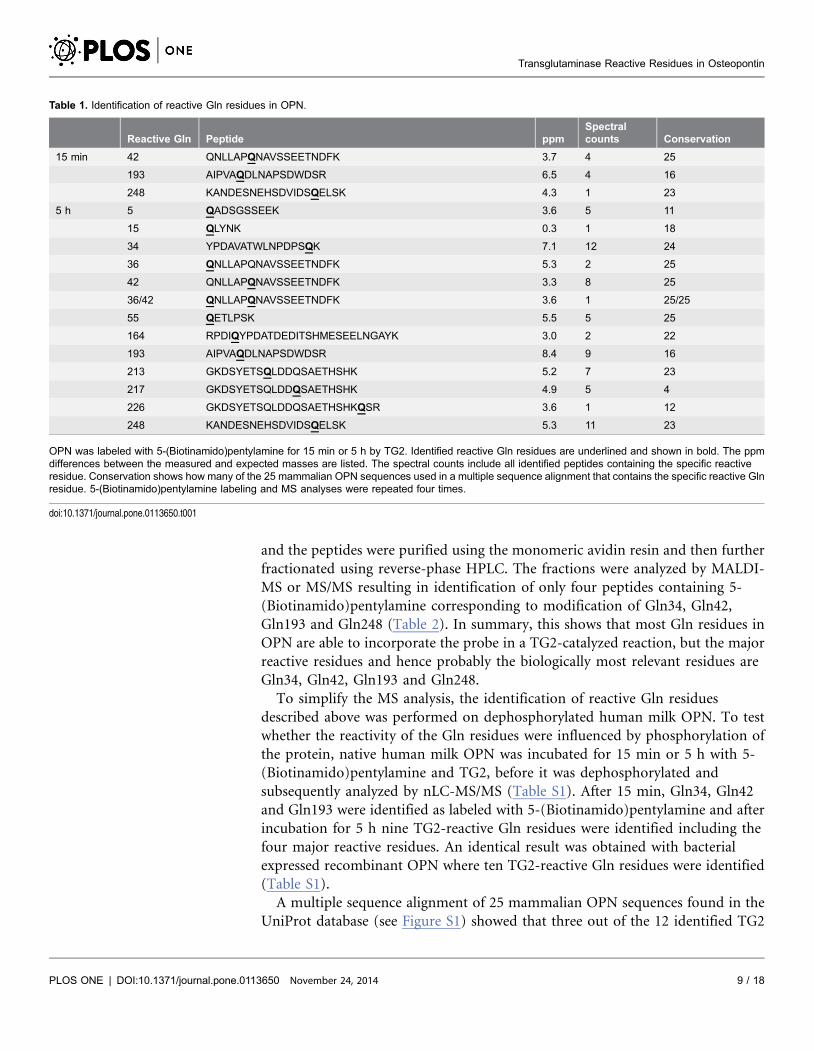
Using this approach, we identified 12 Gln residues in OPN to be reactive in the
transamidation reaction catalyzed by TG2 (Table 1). Of all the Gln residues in
OPN only Gln84 and Gln103, which are located in a large acidic fragment not
cleavable by trypsin, were not observed in this study. The peptides identified as
containing reactive Gln residues are listed in Table 1.
Spectral counts (listed in Table 1) depend on both the ionization efficiency and
the concentration of the analyzed peptides. It is therefore not an exact quantitative
measure of peptide abundance but it can provide a rough indication of the
peptide abundance and consequently the residue reactivity. None of the labeled
Gln residues stood out in the spectral count numbers clearly marking them as
major TG2 reactive residues after 5 h incubation with TG2, however, Gln34,
Gln42, Gln193 and Gln248 were observed with the highest numbers. In order to
get more information about which of the identified Gln residues that could be
considered as the major TG2 reactive residues, we used two different approaches.
Initially, OPN was incubated with 5-(Biotinamido)pentylamine and TG2 for only
15 min followed by nLC-MS/MS analysis as described above. After this short
incubation time, only Gln42, Gln193 and Gln248 were labelled with the probe,
indicating that these residues are major TG2 reactive sites in OPN (Table 1). In
another experiment, OPN was labeled with 5-(Biotinamido)pentylamine for 5 h
Figure 1. OPN contains TG2 reactive Gln and Lys residues. (A) Schematic representation of OPN showing the N- and C-terminal parts of the protein, thethrombin cleavage site and the distribution of potential TG2 reactive Gln and Lys residues. The previously identified TG2 reactive Gln residues (Gln34,Gln36) and the integrin binding RGD sequence are indicated. (B–C) Maxisorp plates were coated with full-length OPN (triangle), the N-terminal part of OPN(circle) or the C-terminal part of OPN (square) (3 mg/ml) and subsequently incubated with increasing concentrations of TG2 in the presence of the aminedonor 5-(Biotinamido)pentylamine (B) or the amine acceptor biotinyl-TVQQEL-OH (C). As negative control, wells not coated with OPN but incubated with 5-(Biotinamido)pentylamine or biotinyl-TVQQEL-OH and TG2 (5 mg/ml) are indicated with a cross. The samples were incubated for 3 h at 37˚C. For allexperiments data are expressed as mean ¡S.D. (n53). The experiments were repeated four times.
doi:10.1371/journal.pone.0113650.g001
Transglutaminase Reactive Residues in Osteopontin
PLOS ONE | DOI:10.1371/journal.pone.0113650 November 24, 2014 8 / 18
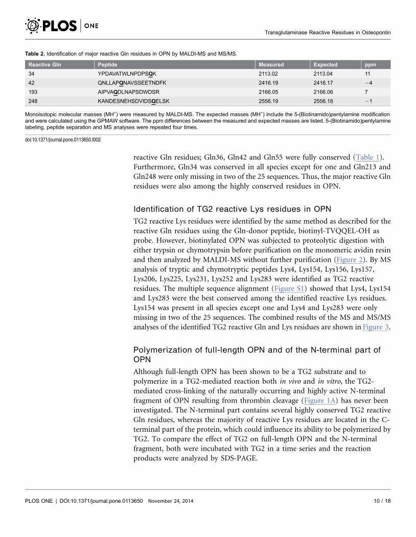
and the peptides were purified using the monomeric avidin resin and then further
fractionated using reverse-phase HPLC. The fractions were analyzed by MALDI-
MS or MS/MS resulting in identification of only four peptides containing 5-
(Biotinamido)pentylamine corresponding to modification of Gln34, Gln42,
Gln193 and Gln248 (Table 2). In summary, this shows that most Gln residues in
OPN are able to incorporate the probe in a TG2-catalyzed reaction, but the major
reactive residues and hence probably the biologically most relevant residues are
Gln34, Gln42, Gln193 and Gln248.
To simplify the MS analysis, the identification of reactive Gln residues
described above was performed on dephosphorylated human milk OPN. To test
whether the reactivity of the Gln residues were influenced by phosphorylation of
the protein, native human milk OPN was incubated for 15 min or 5 h with 5-
(Biotinamido)pentylamine and TG2, before it was dephosphorylated and
subsequently analyzed by nLC-MS/MS (Table S1). After 15 min, Gln34, Gln42
and Gln193 were identified as labeled with 5-(Biotinamido)pentylamine and after
incubation for 5 h nine TG2-reactive Gln residues were identified including the
four major reactive residues. An identical result was obtained with bacterial
expressed recombinant OPN where ten TG2-reactive Gln residues were identified
(Table S1).
A multiple sequence alignment of 25 mammalian OPN sequences found in the
UniProt database (see Figure S1) showed that three out of the 12 identified TG2
Table 1. Identification of reactive Gln residues in OPN.
Reactive Gln Peptide ppmSpectralcounts Conservation
15 min 42 QNLLAPQNAVSSEETNDFK 3.7 4 25
193 AIPVAQDLNAPSDWDSR 6.5 4 16
248 KANDESNEHSDVIDSQELSK 4.3 1 23
5 h 5 QADSGSSEEK 3.6 5 11
15 QLYNK 0.3 1 18
34 YPDAVATWLNPDPSQK 7.1 12 24
36 QNLLAPQNAVSSEETNDFK 5.3 2 25
42 QNLLAPQNAVSSEETNDFK 3.3 8 25
36/42 QNLLAPQNAVSSEETNDFK 3.6 1 25/25
55 QETLPSK 5.5 5 25
164 RPDIQYPDATDEDITSHMESEELNGAYK 3.0 2 22
193 AIPVAQDLNAPSDWDSR 8.4 9 16
213 GKDSYETSQLDDQSAETHSHK 5.2 7 23
217 GKDSYETSQLDDQSAETHSHK 4.9 5 4
226 GKDSYETSQLDDQSAETHSHKQSR 3.6 1 12
248 KANDESNEHSDVIDSQELSK 5.3 11 23
OPN was labeled with 5-(Biotinamido)pentylamine for 15 min or 5 h by TG2. Identified reactive Gln residues are underlined and shown in bold. The ppmdifferences between the measured and expected masses are listed. The spectral counts include all identified peptides containing the specific reactiveresidue. Conservation shows how many of the 25 mammalian OPN sequences used in a multiple sequence alignment that contains the specific reactive Glnresidue. 5-(Biotinamido)pentylamine labeling and MS analyses were repeated four times.
doi:10.1371/journal.pone.0113650.t001
Transglutaminase Reactive Residues in Osteopontin
PLOS ONE | DOI:10.1371/journal.pone.0113650 November 24, 2014 9 / 18
reactive Gln residues; Gln36, Gln42 and Gln55 were fully conserved (Table 1).
Furthermore, Gln34 was conserved in all species except for one and Gln213 and
Gln248 were only missing in two of the 25 sequences. Thus, the major reactive Gln
residues were also among the highly conserved residues in OPN.
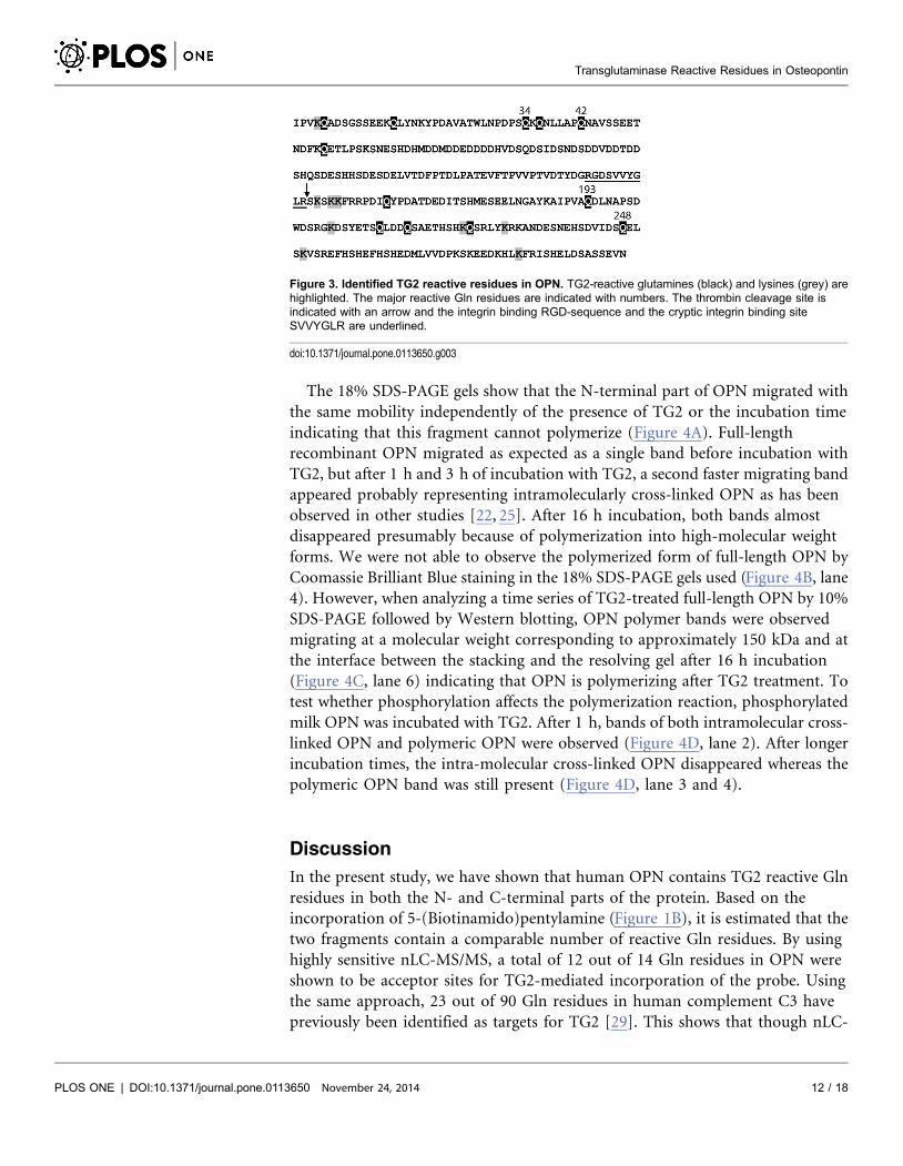
Identification of TG2 reactive Lys residues in OPN
TG2 reactive Lys residues were identified by the same method as described for the
reactive Gln residues using the Gln-donor peptide, biotinyl-TVQQEL-OH as
probe. However, biotinylated OPN was subjected to proteolytic digestion with
either trypsin or chymotrypsin before purification on the monomeric avidin resin
and then analyzed by MALDI-MS without further purification (Figure 2). By MS
analysis of tryptic and chymotryptic peptides Lys4, Lys154, Lys156, Lys157,
Lys206, Lys225, Lys231, Lys252 and Lys283 were identified as TG2 reactive
residues. The multiple sequence alignment (Figure S1) showed that Lys4, Lys154
and Lys283 were the best conserved among the identified reactive Lys residues.
Lys154 was present in all species except one and Lys4 and Lys283 were only
missing in two of the 25 sequences. The combined results of the MS and MS/MS
analyses of the identified TG2 reactive Gln and Lys residues are shown in Figure 3.
Polymerization of full-length OPN and of the N-terminal part of
OPN
Although full-length OPN has been shown to be a TG2 substrate and to
polymerize in a TG2-mediated reaction both in vivo and in vitro, the TG2-
mediated cross-linking of the naturally occurring and highly active N-terminal
fragment of OPN resulting from thrombin cleavage (Figure 1A) has never been
investigated. The N-terminal part contains several highly conserved TG2 reactive
Gln residues, whereas the majority of reactive Lys residues are located in the C-
terminal part of the protein, which could influence its ability to be polymerized by
TG2. To compare the effect of TG2 on full-length OPN and the N-terminal
fragment, both were incubated with TG2 in a time series and the reaction
products were analyzed by SDS-PAGE.
Table 2. Identification of major reactive Gln residues in OPN by MALDI-MS and MS/MS.
Reactive Gln Peptide Measured Expected ppm
34 YPDAVATWLNPDPSQK 2113.02 2113.04 11
42 QNLLAPQNAVSSEETNDFK 2416.19 2416.17 24
193 AIPVAQDLNAPSDWDSR 2166.05 2166.06 7
248 KANDESNEHSDVIDSQELSK 2556.19 2556.18 21
Monoisotopic molecular masses (MH+) were measured by MALDI-MS. The expected masses (MH+) include the 5-(Biotinamido)pentylamine modificationand were calculated using the GPMAW software. The ppm differences between the measured and expected masses are listed. 5-(Biotinamido)pentylaminelabeling, peptide separation and MS analyses were repeated four times.
doi:10.1371/journal.pone.0113650.t002
Transglutaminase Reactive Residues in Osteopontin
PLOS ONE | DOI:10.1371/journal.pone.0113650 November 24, 2014 10 / 18

Figure 2. Identification of TG2 reactive lysines by mass spectrometry. (A) MALDI-MS of tryptic and chymotryptic peptides. Peptides observed with amass corresponding to incorporation of biotin-TVQQEL are indicated with asterisks. (B) Table of tryptic and chymotryptic peptides containing biotin-TVQQEL. Location on the modified Lys residue in peptides containing more than one Lys was in some cases unambiguously achieved with theconsideration that modified Lys residues are not trypsin substrates. The labeled Lys residues have been underlined and are shown in bold if the residue wasunambiguously assigned. If the biotin label could not be assigned to a specific residue, the possible labeling sites are shown underlined and in italics.Monoisotopic molecular masses (MH+) were measured by MALDI-MS. The expected masses (MH+) include the biotinyl-TVQQEL-OH modification andwere calculated using the GPMAW software. The ppm differences between the measured and expected masses are listed. Conservation shows how manyof the 25 mammalian OPN sequences used in a multiple sequence alignment that contains the specific reactive Lys residue. Biotinyl-TVQQEL-OH labelingand MS analysis were repeated twice.
doi:10.1371/journal.pone.0113650.g002
Transglutaminase Reactive Residues in Osteopontin
PLOS ONE | DOI:10.1371/journal.pone.0113650 November 24, 2014 11 / 18
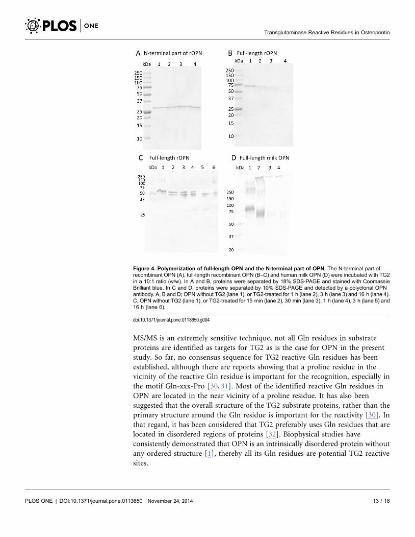
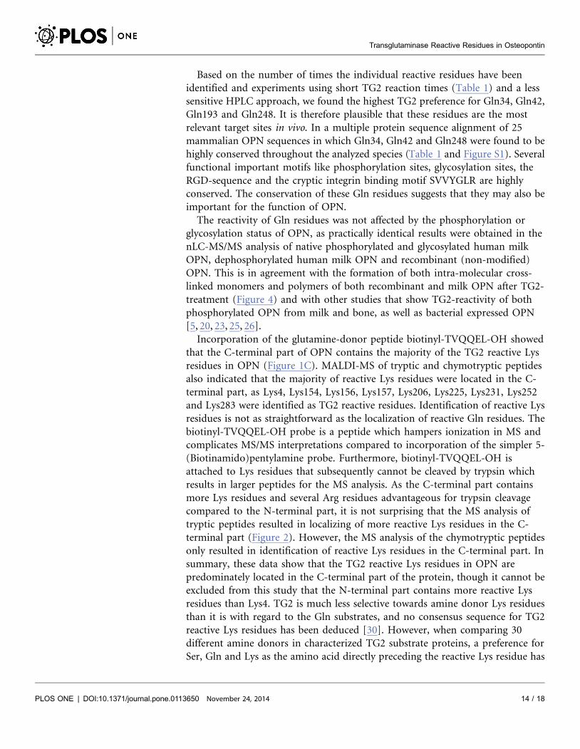
The 18% SDS-PAGE gels show that the N-terminal part of OPN migrated with
the same mobility independently of the presence of TG2 or the incubation time
indicating that this fragment cannot polymerize (Figure 4A). Full-length
recombinant OPN migrated as expected as a single band before incubation with
TG2, but after 1 h and 3 h of incubation with TG2, a second faster migrating band
appeared probably representing intramolecularly cross-linked OPN as has been
observed in other studies [22, 25]. After 16 h incubation, both bands almost
disappeared presumably because of polymerization into high-molecular weight
forms. We were not able to observe the polymerized form of full-length OPN by
Coomassie Brilliant Blue staining in the 18% SDS-PAGE gels used (Figure 4B, lane
4). However, when analyzing a time series of TG2-treated full-length OPN by 10%
SDS-PAGE followed by Western blotting, OPN polymer bands were observed
migrating at a molecular weight corresponding to approximately 150 kDa and at
the interface between the stacking and the resolving gel after 16 h incubation
(Figure 4C, lane 6) indicating that OPN is polymerizing after TG2 treatment. To
test whether phosphorylation affects the polymerization reaction, phosphorylated
milk OPN was incubated with TG2. After 1 h, bands of both intramolecular cross-
linked OPN and polymeric OPN were observed (Figure 4D, lane 2). After longer
incubation times, the intra-molecular cross-linked OPN disappeared whereas the
polymeric OPN band was still present (Figure 4D, lane 3 and 4).
Discussion
In the present study, we have shown that human OPN contains TG2 reactive Gln
residues in both the N- and C-terminal parts of the protein. Based on the
incorporation of 5-(Biotinamido)pentylamine (Figure 1B), it is estimated that the
two fragments contain a comparable number of reactive Gln residues. By using
highly sensitive nLC-MS/MS, a total of 12 out of 14 Gln residues in OPN were
shown to be acceptor sites for TG2-mediated incorporation of the probe. Using
the same approach, 23 out of 90 Gln residues in human complement C3 have
previously been identified as targets for TG2 [29]. This shows that though nLC-
Figure 3. Identified TG2 reactive residues in OPN. TG2-reactive glutamines (black) and lysines (grey) arehighlighted. The major reactive Gln residues are indicated with numbers. The thrombin cleavage site isindicated with an arrow and the integrin binding RGD-sequence and the cryptic integrin binding siteSVVYGLR are underlined.
doi:10.1371/journal.pone.0113650.g003
Transglutaminase Reactive Residues in Osteopontin
PLOS ONE | DOI:10.1371/journal.pone.0113650 November 24, 2014 12 / 18
MS/MS is an extremely sensitive technique, not all Gln residues in substrate
proteins are identified as targets for TG2 as is the case for OPN in the present
study. So far, no consensus sequence for TG2 reactive Gln residues has been
established, although there are reports showing that a proline residue in the
vicinity of the reactive Gln residue is important for the recognition, especially in
the motif Gln-xxx-Pro [30, 31]. Most of the identified reactive Gln residues in
OPN are located in the near vicinity of a proline residue. It has also been
suggested that the overall structure of the TG2 substrate proteins, rather than the
primary structure around the Gln residue is important for the reactivity [30]. In
that regard, it has been considered that TG2 preferably uses Gln residues that are
located in disordered regions of proteins [32]. Biophysical studies have
consistently demonstrated that OPN is an intrinsically disordered protein without
any ordered structure [1], thereby all its Gln residues are potential TG2 reactive
sites.
Figure 4. Polymerization of full-length OPN and the N-terminal part of OPN. The N-terminal part ofrecombinant OPN (A), full-length recombinant OPN (B–C) and human milk OPN (D) were incubated with TG2in a 10:1 ratio (w/w). In A and B, proteins were separated by 18% SDS-PAGE and stained with CoomassieBrilliant blue. In C and D, proteins were separated by 10% SDS-PAGE and detected by a polyclonal OPNantibody. A, B and D; OPN without TG2 (lane 1), or TG2-treated for 1 h (lane 2), 3 h (lane 3) and 16 h (lane 4).C, OPN without TG2 (lane 1), or TG2-treated for 15 min (lane 2), 30 min (lane 3), 1 h (lane 4), 3 h (lane 5) and16 h (lane 6).
doi:10.1371/journal.pone.0113650.g004
Transglutaminase Reactive Residues in Osteopontin
PLOS ONE | DOI:10.1371/journal.pone.0113650 November 24, 2014 13 / 18
Based on the number of times the individual reactive residues have been
identified and experiments using short TG2 reaction times (Table 1) and a less
sensitive HPLC approach, we found the highest TG2 preference for Gln34, Gln42,
Gln193 and Gln248. It is therefore plausible that these residues are the most
relevant target sites in vivo. In a multiple protein sequence alignment of 25
mammalian OPN sequences in which Gln34, Gln42 and Gln248 were found to be
highly conserved throughout the analyzed species (Table 1 and Figure S1). Several
functional important motifs like phosphorylation sites, glycosylation sites, the
RGD-sequence and the cryptic integrin binding motif SVVYGLR are highly
conserved. The conservation of these Gln residues suggests that they may also be
important for the function of OPN.
The reactivity of Gln residues was not affected by the phosphorylation or
glycosylation status of OPN, as practically identical results were obtained in the
nLC-MS/MS analysis of native phosphorylated and glycosylated human milk
OPN, dephosphorylated human milk OPN and recombinant (non-modified)
OPN. This is in agreement with the formation of both intra-molecular cross-
linked monomers and polymers of both recombinant and milk OPN after TG2-
treatment (Figure 4) and with other studies that show TG2-reactivity of both
phosphorylated OPN from milk and bone, as well as bacterial expressed OPN
[5, 20, 23, 25, 26].
Incorporation of the glutamine-donor peptide biotinyl-TVQQEL-OH showed
that the C-terminal part of OPN contains the majority of the TG2 reactive Lys
residues in OPN (Figure 1C). MALDI-MS of tryptic and chymotryptic peptides
also indicated that the majority of reactive Lys residues were located in the C-
terminal part, as Lys4, Lys154, Lys156, Lys157, Lys206, Lys225, Lys231, Lys252
and Lys283 were identified as TG2 reactive residues. Identification of reactive Lys
residues is not as straightforward as the localization of reactive Gln residues. The
biotinyl-TVQQEL-OH probe is a peptide which hampers ionization in MS and
complicates MS/MS interpretations compared to incorporation of the simpler 5-
(Biotinamido)pentylamine probe. Furthermore, biotinyl-TVQQEL-OH is
attached to Lys residues that subsequently cannot be cleaved by trypsin which
results in larger peptides for the MS analysis. As the C-terminal part contains
more Lys residues and several Arg residues advantageous for trypsin cleavage
compared to the N-terminal part, it is not surprising that the MS analysis of
tryptic peptides resulted in localizing of more reactive Lys residues in the C-
terminal part (Figure 2). However, the MS analysis of the chymotryptic peptides
only resulted in identification of reactive Lys residues in the C-terminal part. In
summary, these data show that the TG2 reactive Lys residues in OPN are
predominately located in the C-terminal part of the protein, though it cannot be
excluded from this study that the N-terminal part contains more reactive Lys
residues than Lys4. TG2 is much less selective towards amine donor Lys residues
than it is with regard to the Gln substrates, and no consensus sequence for TG2
reactive Lys residues has been deduced [30]. However, when comparing 30
different amine donors in characterized TG2 substrate proteins, a preference for
Ser, Gln and Lys as the amino acid directly preceding the reactive Lys residue has
Transglutaminase Reactive Residues in Osteopontin
PLOS ONE | DOI:10.1371/journal.pone.0113650 November 24, 2014 14 / 18
been suggested to enhance reactivity [33]. Lys154, Lys156 and Lys157 are located
in such sequences (Figure 3) and these residues were also present in five out of
twelve peptides containing the biotinyl-TVQQEL-OH probe (Figure 2).
Polymerization of OPN by TG2 is physiologically relevant as it alters the
functionality of the protein and especially its integrin binding properties [22–25].
Interestingly, OPN seems to form homo-polymer protein aggregates rather than
polymerizing in a heterotypic manner with other proteins [5]. By site-directed
mutagenesis it has been shown that especially Gln34 and/or Gln36 and to a lesser
extent Gln42 and Gln55 play important roles in TG2-mediated homo-
polymerization of OPN [25]. These Gln residues in the N-terminal part are
among the best conserved residues in OPN, as all 25 aligned sequences contained
Gln36, Gln42 and Gln55 whereas Gln34 is only missing in the African elephant
(Figure S1). In the present study, Gln34 and Gln42 are among the most TG2
reactive residues in OPN which also suggests an important role of these residues
for the TG2 regulated function of OPN. Our data suggests that Lys residues in the
C-terminal part most likely are the amine donors in the polymerization, as the N-
terminal fragment of OPN was unable to polymerize. This observation is
interesting, as the N-terminal fragment of OPN contains the integrin recognition
motifs and that specific cleavage by e.g. thrombin and plasmin modulates the
function of OPN and can lead to enhanced cell adhesion, migration and resistance
to apoptosis [7, 9, 34]. Cleaved OPN has been suggested to play a role in a variety
of biological processes such as cancer development [34], rheumatoid arthritis
[14], formation of renal calcium crystals [15] and stem cell retention in the bone
marrow niche [16]. Thus, the altered ability of the N-terminal part of OPN to
polymerize when compared to the full-length protein may be of importance in
vivo.
In the present study we have identified the Gln residues in OPN that are
substrates for TG2. The major reactive residues were shown to be Gln34, Gln42,
Gln193 and Gln248. OPN also contained several TG2 reactive Lys residues that
were predominately located in the C-terminal part of the protein. Polymerization
of OPN seemed to involve isopeptide bonds between residues located in the N-
terminal part (probably Gln residues) and the C-terminal part (probably Lys
residues). This can have important biological consequences for the activity of the
highly active N-terminal fragment as it is not able to polymerize in vivo.
Supporting Information
Figure S1. Alignment of mammalian OPN sequences. Sequence analyses were
performed on OPN from the following 25 mammalian species found in the
UniProt database (UniProt release 2014_06). The TG2-reactive glutamine (black)
and lysine (grey) residues identified in this study are highlighted and numbered.
Human, homo sapiens (P10451); rat, Rattus norvegicus (P08721); mouse, Mus
musculus (P10923); bovine, Bos tausrus (P31096); pig, Sus scrofa (P14287);
Rhesus monkey, Macaca mulatta, (F6V2X3); Chimpanzee, Pan troglodytes
Transglutaminase Reactive Residues in Osteopontin
PLOS ONE | DOI:10.1371/journal.pone.0113650 November 24, 2014 15 / 18

(H2RCV1); Lowland gorilla, Gorilla gorilla gorilla (G3QS39); Rabbit, Oryctolagus
cuniculus (P31097); Sheep, Ovis aries (Q9XSY9); horse, Equus caballus
(F7AYC1); cat, Felis catus (M3VZ83); dog, Canis familiaris (E2R161); african
elephant, Loxodonta africana (G3SYB7); golden hamster, Mesocricetus auratus
(Q0WX06); goat, Capra hircus (U5Y6U2); Little brown bat, Myotis lucifugus
(G1PFK5); David’s myotis, Myotis davidii (L5M309); Black flying fox, Pteropus
alecto (L5KPG6); Brandt’s bat, Myotis brandtii (S7MMZ5); Tasmanian devil,
Sarcophilus harrisii (G3VEM2); White-tufted-ear marmoset, Callithrix jacchus
(F7DN59); Thirteen-lined ground squirrel, Spermophilus tridecemlineatus
(I3MYE1); Duckbill platypus, Ornithorhynchus anatinus (F7EC46); Gray short-
tailed opossum, Monodelphis domestica (F7ANS8).
doi:10.1371/journal.pone.0113650.s001 (DOCX)
Table S1. Identification of reactive Gln residues in human milk OPN and
recombinant OPN. OPN was labeled with 5-(Biotinamido)pentylamine for
15 min or 5 h by TG2. Identified reactive Gln residues are underlined and shown
in bold. The ppm differences between the measured and expected masses are
listed.
doi:10.1371/journal.pone.0113650.s002 (DOCX)
Author Contributions
Conceived and designed the experiments: BC EDZ ESS. Performed the
experiments: BC EDZ MT MMC SK CS. Analyzed the data: BC EDZ CS MT
MMC JJE ESS. Contributed reagents/materials/analysis tools: CS JJE MMC SK CO
ESS. Wrote the paper: BC EDZ CS CO ESS.
References
1. Fisher LW, Torchia DA, Fohr B, Young MF, Fedarko NS (2001) Flexible structures of SIBLINGproteins, bone sialoprotein, and osteopontin. Biochem Biophys Res Commun 280: 460–465. doi:10.1006/bbrc.2000.4146.
2. Fisher LW, Fedarko NS (2003) Six genes expressed in bones and teeth encode the current members ofthe SIBLING family of proteins. Connect Tissue Res 44 Suppl 1: 33–40.
3. Schack L, Lange A, Kelsen J, Agnholt J, Christensen B, et al. (2009) Considerable variation in theconcentration of osteopontin in human milk, bovine milk, and infant formulas. J Dairy Sci 92: 5378–5385.doi: 10.3168/jds.2009–2360.
4. Christensen B, Petersen TE, Sørensen ES (2008) Post-translational modification and proteolyticprocessing of urinary osteopontin. Biochem J 411: 53–61. doi: 10.1042/BJ20071021.
5. Kaartinen MT, El-Maadawy S, Rasanen NH, McKee MD (2002) Tissue transglutaminase and itssubstrates in bone. J Bone Miner Res Off J Am Soc Bone Miner Res 17: 2161–2173. doi: 10.1359/jbmr.2002.17.12.2161.
6. Sodek J, Ganss B, McKee MD (2000) Osteopontin. Crit Rev Oral Biol Med Off Publ Am Assoc Oral Biol11: 279–303.
7. Christensen B, Schack L, Klaning E, Sørensen ES (2010) Osteopontin is cleaved at multiple sitesclose to its integrin-binding motifs in milk and is a novel substrate for plasmin and cathepsin D. J BiolChem 285: 7929–7937. doi: 10.1074/jbc.M109.075010.
Transglutaminase Reactive Residues in Osteopontin
PLOS ONE | DOI:10.1371/journal.pone.0113650 November 24, 2014 16 / 18
8. Prince CW, Dickie D, Krumdieck CL (1991) Osteopontin, a substrate for transglutaminase and factorXIII activity. Biochem Biophys Res Commun 177: 1205–1210.
9. Kazanecki CC, Uzwiak DJ, Denhardt DT (2007) Control of osteopontin signaling and function by post-translational phosphorylation and protein folding. J Cell Biochem 102: 912–924. doi: 10.1002/jcb.21558.
10. Christensen B, Kazanecki CC, Petersen TE, Rittling SR, Denhardt DT, et al. (2007) Cell type-specificpost-translational modifications of mouse osteopontin are associated with different adhesive properties.J Biol Chem 282: 19463–19472. doi: 10.1074/jbc.M703055200.
11. Kariya Y, Kanno M, Matsumoto-Morita K, Konno M, Yamaguchi Y, et al. (2014) Osteopontin O-glycosylation contributes to its phosphorylation and cell-adhesion properties. Biochem J 463: 93–102.doi: 10.1042/BJ20140060.
12. Agnihotri R, Crawford HC, Haro H, Matrisian LM, Havrda MC, et al. (2001) Osteopontin, a novelsubstrate for matrix metalloproteinase-3 (stromelysin-1) and matrix metalloproteinase-7 (matrilysin).J Biol Chem 276: 28261–28267. doi: 10.1074/jbc.M103608200.
13. Senger DR, Perruzzi CA, Gracey CF, Papadopoulos A, Tenen DG (1988) Secreted phosphoproteinsassociated with neoplastic transformation: close homology with plasma proteins cleaved during bloodcoagulation. Cancer Res 48: 5770–5774.
14. Yamamoto N, Sakai F, Kon S, Morimoto J, Kimura C, et al. (2003) Essential role of the cryptic epitopeSLAYGLR within osteopontin in a murine model of rheumatoid arthritis. J Clin Invest 112: 181–188. doi:10.1172/JCI17778.
15. Hamamoto S, Yasui T, Okada A, Hirose M, Matsui Y, et al. (2011) Crucial role of the cryptic epitopeSLAYGLR within osteopontin in renal crystal formation of mice. J Bone Miner Res Off J Am Soc BoneMiner Res 26: 2967–2977. doi: 10.1002/jbmr.495.
16. Grassinger J, Haylock DN, Storan MJ, Haines GO, Williams B, et al. (2009) Thrombin-cleavedosteopontin regulates hemopoietic stem and progenitor cell functions through interactions withalpha9beta1 and alpha4beta1 integrins. Blood 114: 49–59. doi: 10.1182/blood-2009-01-197988.
17. Lorand L, Graham RM (2003) Transglutaminases: crosslinking enzymes with pleiotropic functions. NatRev Mol Cell Biol 4: 140–156. doi: 10.1038/nrm1014.
18. Lai T-S, Greenberg CS (2013) TGM2 and implications for human disease: role of alternative splicing.Front Biosci Landmark Ed 18: 504–519.
19. Kaartinen MT, Murshed M, Karsenty G, McKee MD (2007) Osteopontin upregulation andpolymerization by transglutaminase 2 in calcified arteries of Matrix Gla protein-deficient mice.J Histochem Cytochem Off J Histochem Soc 55: 375–386. doi: 10.1369/jhc.6A7087.2006.
20. Kaartinen MT, Pirhonen A, Linnala-Kankkunen A, Maenpaa PH (1999) Cross-linking of osteopontinby tissue transglutaminase increases its collagen binding properties. J Biol Chem 274: 1729–1735.
21. Pedraza CE, Nikolcheva LG, Kaartinen MT, Barralet JE, McKee MD (2008) Osteopontin functions asan opsonin and facilitates phagocytosis by macrophages of hydroxyapatite-coated microspheres:implications for bone wound healing. Bone 43: 708–716. doi: 10.1016/j.bone.2008.06.010.
22. Forsprecher J, Wang Z, Goldberg HA, Kaartinen MT (2011) Transglutaminase-mediatedoligomerization promotes osteoblast adhesive properties of osteopontin and bone sialoprotein. CellAdhes Migr 5: 65–72. doi: 10.4161/cam.5.1.13369.
23. Higashikawa F, Eboshida A, Yokosaki Y (2007) Enhanced biological activity of polymeric osteopontin.FEBS Lett 581: 2697–2701. doi: 10.1016/j.febslet.2007.05.018.
24. Nishimichi N, Higashikawa F, Kinoh HH, Tateishi Y, Matsuda H, et al. (2009) Polymeric osteopontinemploys integrin alpha9beta1 as a receptor and attracts neutrophils by presenting a de novo binding site.J Biol Chem 284: 14769–14776. doi: 10.1074/jbc.M901515200.
25. Nishimichi N, Hayashita-Kinoh H, Chen C, Matsuda H, Sheppard D, et al. (2011) Osteopontinundergoes polymerization in vivo and gains chemotactic activity for neutrophils mediated by integrinalpha9beta1. J Biol Chem 286: 11170–11178. doi: 10.1074/jbc.M110.189258.
26. Sørensen ES, Rasmussen LK, Møller L, Jensen PH, Højrup P, et al. (1994) Localization oftransglutaminase-reactive glutamine residues in bovine osteopontin. Biochem J 304 (Pt 1): 13–16.
27. Christensen B, Nielsen MS, Haselmann KF, Petersen TE, Sørensen ES (2005) Post-translationallymodified residues of native human osteopontin are located in clusters: identification of 36
Transglutaminase Reactive Residues in Osteopontin
PLOS ONE | DOI:10.1371/journal.pone.0113650 November 24, 2014 17 / 18
phosphorylation and five O-glycosylation sites and their biological implications. Biochem J 390: 285–292. doi: 10.1042/BJ20050341.
28. Christensen B, Klaning E, Nielsen MS, Andersen MH, Sørensen ES (2012) C-terminal modification ofosteopontin inhibits interaction with the aVb3-integrin. J Biol Chem 287: 3788–3797. doi: 10.1074/jbc.M111.277996.
29. Nikolajsen CL, Scavenius C, Enghild JJ (2012) Human complement C3 is a substrate fortransglutaminases. A functional link between non-protease-based members of the coagulation andcomplement cascades. Biochemistry (Mosc) 51: 4735–4742. doi: 10.1021/bi3004022.
30. Esposito C, Caputo I (2005) Mammalian transglutaminases. Identification of substrates as a key tophysiological function and physiopathological relevance. FEBS J 272: 615–631. doi: 10.1111/j.1742-4658.2004.04476.x.
31. Sugimura Y, Hosono M, Wada F, Yoshimura T, Maki M, et al. (2006) Screening for the preferredsubstrate sequence of transglutaminase using a phage-displayed peptide library: identification of peptidesubstrates for TGASE 2 and Factor XIIIA. J Biol Chem 281: 17699–17706. doi: 10.1074/jbc.M513538200.
32. Csosz E, Bagossi P, Nagy Z, Dosztanyi Z, Simon I, et al. (2008) Substrate preference oftransglutaminase 2 revealed by logistic regression analysis and intrinsic disorder examination. J Mol Biol383: 390–402. doi: 10.1016/j.jmb.2008.08.026.
33. Grootjans JJ, Groenen PJ, de Jong WW (1995) Substrate requirements for transglutaminases.Influence of the amino acid residue preceding the amine donor lysine in a native protein. J Biol Chem270: 22855–22858.
34. Yamaguchi Y, Shao Z, Sharif S, Du X-Y, Myles T, et al. (2013) Thrombin-cleaved fragments ofosteopontin are overexpressed in malignant glial tumors and provide a molecular niche with survivaladvantage. J Biol Chem 288: 3097–3111. doi: 10.1074/jbc.M112.362954.
Transglutaminase Reactive Residues in Osteopontin
PLOS ONE | DOI:10.1371/journal.pone.0113650 November 24, 2014 18 / 18