transglutaminase transcription and antigen translocation in experimental renal scarring
TRANSCRIPT
Transglutaminase Transcription and Antigen Translocation inExperimental Renal Scarring
TIMOTHY S. JOHNSON,* N. JAMES SKILL,† A. MEGUID EL NAHAS,*SIMON D. OLDROYD,* GRAHAM L. THOMAS,* JULIE A. DOUTHWAITE,*JOHN L. HAYLOR,* and MARTIN GRIFFIN†
*Sheffield Kidney Institute, Northern General Hospital Trust, Sheffield, and†Department of Life Sciences,Nottingham Trent University, Nottingham, United Kingdom.
Abstract.It was recently demonstrated that renal tissue trans-glutaminase (tTg) protein and its catalytic product thee(g-glutamyl) lysine protein cross-link are significantly increasedin the subtotal (5/6) nephrectomy model (SNx) of renal fibrosisin rats. It was proposed that the enzyme had two importantphysiologic functions in disease development; one of stabiliz-ing the increased extracellular matrix (ECM) by protein cross-linking, the other in a novel form of tubular cell death. Thisstudy, using the same rat SNx model, demonstrates first byNorthern blotting that expression of tTg mRNA when com-pared with controls is increased by day 15 (170% increase,P , 0.05), then rises steadily, peaking at day 90 (1391%,P ,0.01), and remains elevated at 120 d (1205%,P , 0.05) whencompared with controls.In situ hybridization histochemistrydemonstrated that the tubular cells were the major site of the
additional tTg synthesis. Immunohistochemistry on cryostatsections revealed a sixfold increase (P , 0.001) in ECM-bound tTg antigen at 90-d post-SNx, whereasin situ transglu-taminase activity demonstrated by the incorporation of fluo-rescein cadaverine into cryostat sections indicated a 750%increase (P , 0.001) on day 90 in SNx animals. This increasedactivity was extracellular and predominantly found in the peri-tubular region. These results indicate that increased tTg genetranscription by tubular cells underlies the major changes inrenal tTg protein reported previously in SNx rats, and that thepresence of thee(g-glutamyl) lysine cross-links in the extra-cellular environment is the result of the extracellular action oftTg. These changes may be in response to tubular cell injuryduring the scarring process and are likely to contribute to theprogressive expansion of the ECM in renal fibrosis.
The progression of chronic renal insufficiency is characterizedby a relentless fibrosis of the kidney. This is typically de-scribed as an accumulation of the extracellular matrix (ECM)above normal requirements and a loss of specialized renal cellsand architecture. Increased synthesis and decreased breakdownof the renal ECM have been implicated in its expansion (1,2).The latter may be due to changes in the ECM-regulatingenzymes including a fall in renal matrix metalloproteinases(MMP) and plasminogens or an increase in their inhibitors(tissue inhibitors of matrix metalloproteinases and plasmino-gen activator inhibitors) (3–5). Other factors important in thepathogenesis of renal fibrosis that have thus far received littleattention are those contributing to the increased deposition andstabilization of the ECM, thereby increasing its resistance toenzymatic breakdown. Such a proposal has been put forward asa contributing factor in the development of other fibrogenicprocesses including lung and liver fibrosis as well as athero-sclerosis (6–10). We have recently described (11) the expres-
sion of tissue transglutaminase (tTg) in the 5/6 subtotal ne-phrectomy (SNx) model of renal fibrosis in which the cross-linking of the ECM by tTg and the formation of irreversiblee(g-glutamyl) lysine bonds may underlie its resistance tobreakdown. Furthermore, the intracellular consequence of hightTg and its product in the scarred kidney was hypothesized tobe a novel type of transglutaminase-mediated cell deathwhereby the cross-linking of intracellular proteins in the cellleads to its death. Transglutaminase-mediated cell death hassince been confirmed as a distinct biologic process in a hamsterfibrosarcoma cell line in response to dexamethasone inductionof tTg in the absence of any increase in apoptosis (12) and intTg transfected 3T3 fibroblasts after exposure to ionomycin(13). The ability of the enzyme to not only stabilize ECMproteins but also to be a key player in a novel cell death processrepresents a major step in improving our understanding of themechanisms of tissue fibrosis.
Transglutaminases are calcium-dependent enzymes that cat-alyze the posttranslation modification of proteins through anacyl transfer reaction between the carboxamide group of apeptide-bound glutaminyl residue and various primary amines(14). Covalent cross-links usinge(g-glutamyl) lysine bonds arestable and resistant to enzymatic, chemical, and mechanicaldisruption (14). Endopeptidases capable of hydrolyzing thee(g-glutamyl) lysine cross-links formed by transglutaminases(Tg) have not been described in vertebrates, and even lyso-somes do not contain enzymes capable of splitting thee(g-
Received January 28, 1999. Accepted April 18, 1999.Correspondence to Dr. Martin Griffin, Department of Life Sciences, Notting-ham Trent University, Clifton, Nottingham, NG11 8NS, United Kingdom.Phone: 144 (0)115 9486670; Fax:144 (0)115 9486636; E-mail:[email protected]
1046-6673/1010-2146Journal of the American Society of NephrologyCopyright © 1999 by the American Society of Nephrology
J Am Soc Nephrol 10: 2146–2157, 1999
glutamyl) lysine bonds (15–17). A number of Tg enzymes havebeen characterized that have distinct genes, structures, andphysiologic functions. Examples include the plasma factorXIIIa involved in cross-linking fibrin during wound healing(18) and the keratinocyte transglutaminase involved in theformation of the cornified envelope during terminal differen-tiation (19,20). tTg is the most widespread member of thisfamily and is present in many different cell types.
Recent evidence suggests that the tTg may have a number ofkey functions in cells. These include a role in programmed celldeath (21–23), cell adhesion (24), and interactions between thecell and its ECM via the cross-linking of proteins such asfibronectin, laminin, nidogen, and collagen types I, III, and IV(7,13,25–27). In addition, tTg has recently been demonstratedto play a role in the covalent stabilization of collagen fibrillarformation through cross-linking of the core fibrils (28).
Previous investigations in renal fibrosis have demonstratedthat the product of tTg,e(g-glutamyl) lysine cross-link, ispresent both in the extracellular space and in the cytoplasm oftubular cells during the scarring progress (11), whereas ele-vated tTg can only be found intracellularly. This raised thequestion of whether ECM components were being cross-linkedinternally before secretion or, as noted previously, whether thetTg antigen present in the matrix is difficult to detect becauseof occlusion of the epitope by standard fixation techniques(13). In addition, there is only circumstantial evidence thatelevated tTg cross-linking of ECM components affects theresistance of these proteins to the catalytic action of MMP.Although we demonstrated previously that enzyme activity isincreased by virtue of the presence of increased amounts of thee(g-glutamyl) lysine cross-link product in renal scarring, noclaim was made as to whether this increase in activity was aresult ofde novosynthesis of the enzyme, or, alternatively, theactivation of a latent proenzyme (29,30) as proposed previ-ously following wound healing in skin (31).
In this study, we demonstrate that induction of renal fibrosisin rats using the SNx model leads tode novosynthesis of tTgby the renal tubular cells. Moreover, by using cryostat ratherthan fixed tissue sections, we show that there is increasedamounts of tTg antigen present in the intercellular space of therenal tubules, which, by using a novelin situ activity method,we demonstrate to be active after progression of the fibroticdisease. In keeping with the importance of tTg in the stabili-zation of ECM proteins, we also demonstrate the increasedresistance of tTg cross-linked ECM proteins to the action ofMMP.
Materials and MethodsExperimental Animals and Protocol
Male Wistar rats (Sheffield University strain) of approximatelysimilar weight (350 to 400 g) and age (8 to 10 wk) were subjected toSNx by complete removal of the left kidney and upper and lower poleresections of the right kidney. Rats were housed two to four to a cageand were maintained at 20°C and 45% humidity on a 12-h light/darkcycle. They were allowed free access to standard rat chow (Labsure,March, Cambridge, United Kingdom) and tap water. At days 7, 15,30, 60, 90, and 120 post-SNx, experimental groups of five to six rats
were sacrificed and the remnant kidney was removed. At each timepoint, five to six control animals were also sacrificed that had beensubjected to a sham operation. Before sacrifice, renal function (serumcreatinine and creatinine clearance) and proteinuria were measured inall rats. Creatinine was measured by the standard autoanalyzer tech-nique and proteinuria by the biuret method. All procedures werecarried out under license according to regulations laid down by HerMajesty’s Government, United Kingdom (Animals Scientific Proce-dures Act, 1986).
Assessment of Renal ScarringAnalysis of renal scarring was determined on formal calcium-fixed,
paraffin-embedded sections (4mm) stained by hematoxylin and eosinor Masson’s trichrome. One of the authors (G.L.T.) scored the sever-ity of glomerulosclerosis and tubulointerstitial scarring, blinded totheir code, according to a previously described 4-point arbitrary score(11). An overall renal fibrosis score was determined by taking a meanof glomerulosclerosis and tubulointerstitial scores. Scores were attrib-uted as follows:
Glomerulosclerosis Score (0 to 3). 0, normal glomeruli; 1, pres-ence of mild segmental glomerulosclerosis affecting,25% of theglomerular tuft; 2, moderate segmental sclerosis affecting 25 to 50%of the tuft; 3, diffuse severe glomerulosclerosis affecting.50% of thetuft including glomeruli with total tuft obliteration, fibrosis, andobsolescence. A minimum of 25 glomeruli per kidney per remnantkidney was examined, and the mean of the glomerular scores wastaken to represent the severity of glomerulosclerosis for a given rat(11).
Tubulointerstitial Scarring (1 to 4). 0, normal tubules andinterstitium; 1, mild tubular atrophy and interstitial fibrosis; 2, mod-erate tubular atrophy and dilation with marked interstitial fibrosis; 3,end-stage kidney with extensive interstitial fibrosis and few remainingatrophic tubules. A score was given to each microscopic field viewedat a magnification of3200. A minimum of 10 fields was scored perkidney per remnant kidney, and the mean value was taken to representthe tubulointerstitial score for a given rat (11).
tTg mRNA LevelstTg mRNA levels were determined by Northern blot analysis. Total
RNA was extracted using Trizol™ (Life Technologies BRL, Paisley,United Kingdom) and quantified by both optical density at 260 nmand ultraviolet (UV) densitometry of the 18S rRNA subunit. Fifteenmicrograms of total RNA was then run on a 1.2% (wt/vol) agarose/4-morpholinepropanesulfonic acid/formaldehyde gel and then capil-lary-blotted on to Hybond-N (Amersham, Buckinghamshire, UnitedKingdom) and cross-linked with 70 mJ/cm2 UV radiation (UV cross-linker, Amersham). This was then probed with a32P-dCTP random-primed (Prime-A-Gene, Promega, United Kingdom) tTg-specificDNA probe corresponding to a 385-bpBamHI fragment of the com-plete mouse tTg sequence extending from bp 1240 to 1625 of thecoding region. This was then exposed to Biomax MS film for approx-imately 4 h. The resulting autoradiograph was then quantified byscanning densitometry using a Bio-Rad GS-690 densitometer andMolecular Analyst version 4 software. Transcript size was determinedby comparison to RNA molecular weight markers (Promega) usingthe same analysis package. Values were then corrected for loadingusing repeat probings with the housekeeping genes cyclophilin andb-actin. Regulation failure of housekeeping genes was by comparisonwith the 18S rRNA subunit on ethidium bromide-stained gels.
J Am Soc Nephrol 10: 2146–2157, 1999 Transglutaminase in Kidney Fibrosis 2147
tTg mRNA LocationtTg mRNA location was determined byin situ hybridization. Five-
micromolar sections of 10% neutral-buffered, formalin-fixed, paraf-fin-embedded tissue on aminopropyltriethoxysilane-coated slideswere rehydrated, treated with 5 mM levamisole, and 0.2N hydrochlo-ric acid for 20 min and then digested with 5mg/ml proteinase K(Sigma, Poole, United Kingdom) for 60 min at 37°C. They were thenpost-fixed in 1% paraformaldehyde, washed, and dehydrated. Thesewere then probed with sense and antisense riboprobe constructs to tTgconstructed using digoxigenin-labeled rUTP and generated from thesame tTg cDNA sequence used above using the riboprobe Gemini IIsystem (Promega). Hybridization was performed at 50°C for 18 h in50% (vol/vol) deionized formamide, 5 mM ethylenediaminetetra-acetic acid (EDTA), 10 mM NaH2PO4, 0.2 mg/ml herring sperm DNA(Promega), 0.1 mg/ml yeast tRNA (Sigma), 23 SSC, 13 Denhardt’ssolution, and 10% dextran sulfate containing 10 ng/ml digoxigenin-labeled riboprobe. After stringency washes, any nonduplexed RNAwas digested with 20mg/ml RNase A (Sigma) for 30 min at 37°C.Binding of the riboprobes was then revealed using alkaline phos-phatase-conjugated sheep anti-digoxigenin antibody (BoehringerMannheim, UK) diluted 1:500 (1.5 U/ml) in TBS containing 1%(vol/vol) normal sheep serum for 60 min at room temperature, fol-lowed by addition of a color substrate solution consisting of 4-ni-troblue tetrazolium chloride/5-bromo-4-chloro-3-indolyl phosphate.The color reaction was developed in the dark at 37°C and stoppedwith distilled water and mounted under aqueous mounting medium(60% [vol/vol] glycerol).
tTg Immunohistochemistry using Cryostat SectionsKidney tissue was snap-frozen in liquid nitrogen and then mounted
in OCT mounting media (Raymond Lamb, United Kingdom). Ten-micrometer cryostat sections were cut at212°C and placed onto lowiron clear glass slides (Raymond Lamb) and stored at220°C beforeuse.
All solutions before fixation were supplemented with the proteaseinhibitors 1 mM leupeptin, 1 mM benzamidine, 1 mM pepstatin, 1mM phenylmethylsulfonyl fluoride, and 10 mM EDTA. Sections werethawed by adding blocking media, washed, and then blocked for 1 hat room temperature with blocking media (3% bovine serum albumin,0.01% Triton X-100 in phosphate-buffered saline [PBS] at pH 7.4) towhich was added 5% (vol/vol) goat serum to absorb out any tTgreleased from damaged cells during hydration. Sections were thenwashed with PBS and either a 1:300 dilution of a mouse monoclonalanti-tTg antibody (CUB7042) (Stratech Scientific, Luton, UnitedKingdom) or mouse nonimmune serum (Dako, UK) applied to thesections and incubated overnight at 4°C. Sections were washed withPBS and then fixed with cold methanol (220°C) for 10 min. Thesewere then washed in PBS before addition of a 1:500 dilution of a goatanti-mouse Cy5 (indodicarbocyanine)-conjugated antibody (StratechScientific) and incubation for 1 h atroom temperature. Sections werethen washed in PBS and mounted with vector shield fluorescencemounting media (Vector Laboratories, Peterborough, United King-dom).
Sections were visualized using a Leica TCS NT confocal micro-scope (Leica DMRBE; Lasertechnik, Wetzlar, Germany) using a
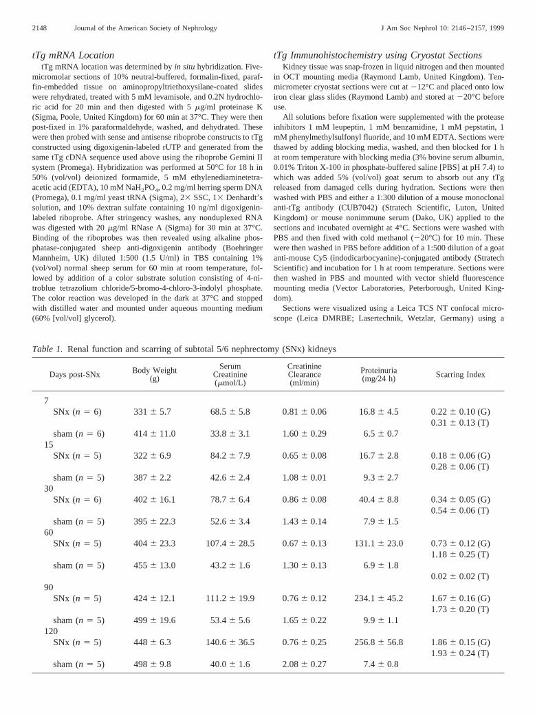
Table 1. Renal function and scarring of subtotal 5/6 nephrectomy (SNx) kidneys
Days post-SNx Body Weight(g)
SerumCreatinine(mmol/L)
CreatinineClearance(ml/min)
Proteinuria(mg/24 h) Scarring Index
7SNx (n 5 6) 3316 5.7 68.56 5.8 0.816 0.06 16.86 4.5 0.226 0.10 (G)
0.316 0.13 (T)sham (n 5 6) 4146 11.0 33.86 3.1 1.606 0.29 6.56 0.7
15SNx (n 5 5) 3226 6.9 84.26 7.9 0.656 0.08 16.76 2.8 0.186 0.06 (G)
0.286 0.06 (T)sham (n 5 5) 3876 2.2 42.66 2.4 1.086 0.01 9.36 2.7
30SNx (n 5 6) 4026 16.1 78.76 6.4 0.866 0.08 40.46 8.8 0.346 0.05 (G)
0.546 0.06 (T)sham (n 5 5) 3956 22.3 52.66 3.4 1.436 0.14 7.96 1.5
60SNx (n 5 5) 4046 23.3 107.46 28.5 0.676 0.13 131.16 23.0 0.736 0.12 (G)
1.186 0.25 (T)sham (n 5 5) 4556 13.0 43.26 1.6 1.306 0.13 6.96 1.8
0.026 0.02 (T)90
SNx (n 5 5) 4246 12.1 111.26 19.9 0.766 0.12 234.16 45.2 1.676 0.16 (G)1.736 0.20 (T)
sham (n 5 5) 4996 19.6 53.46 5.6 1.656 0.22 9.96 1.1120
SNx (n 5 5) 4486 6.3 140.66 36.5 0.766 0.25 256.86 56.8 1.866 0.15 (G)1.936 0.24 (T)
sham (n 5 5) 4986 9.8 40.06 1.6 2.086 0.27 7.46 0.8
2148 Journal of the American Society of Nephrology J Am Soc Nephrol 10: 2146–2157, 1999
Kr/AR laser (647 and 488 nm) for both Cy5 (optimal excitation 650nm) and autofluorescence. Computer imaging and analyses wereobtained at 665 and 530 nm for Cy5 and autofluorescence, respec-tively (Leica TCS NT, Lasertechnik).
Tg in Situ Activity AssayThe method is a modification of that used previously for detection
of in situ Tg activity of cells in live culture (13). Cryostat sectionswere prepared as described above. All solutions before fixation weresupplemented with the protease inhibitors as described above. Fiftymicroliters of 0.2 mM fluorescein cadaverine (32) (Molecular Probes,Leiden, The Netherlands), 50 mM Tris-HCl, pH 7.4, and either 10mM CaCl2 or 10 mM EDTA was pipetted on to a section andincubated for 1 h at 37°C in a humidity chamber. As additionalnegative controls to EDTA, some sections were also preincubated for5 min with 10 mM cystamine (Tg inhibitor) or with the inactivatingtTg antibody (CUB7402 purified IgG) dilution 1:50. To verify thatavailable endogenous Tg substrates were not a limiting factor, 50mgof tTg was included in the reaction buffer as a positive control.Sections were then washed in Tris-HCl, pH 7.4, and then fixed with220°C methanol for 10 min. The methanol was allowed to evaporateand the section was then blocked for 1 h at room temperature withantibody dilution buffer (3% bovine serum albumin, 0.01% TritonX-100 in PBS, pH 7.4) containing 5% (vol/vol) goat serum. After PBSwashes, the sections were probed with 50ml of a 1:50 dilution ofmouse anti-FITC monoclonal antibody (Sigma) at 4°C overnight.Sections were then washed again with PBS, and 50ml of a 1:500dilution of goat anti-mouse, Cy5-conjugated antibody (Stratech Sci-entific) was applied for 1 h at room temperature. Finally, sectionswere washed in PBS and mounted using vector shield fluorescencemounting media (Vector Laboratories).
Sections were visualized using confocal microscopy (LeicaDMRBE, Leica), using Kr/AR laser (647 and 488 nm) for both Cy5
(optimal excitation 650 nm) and FITC (optimum excitation 494 nm).Computer imaging and analyses were obtained at emission wave-lengths of 665 and 530 nm for Cy5 and FITC, respectively (Leica TCSNT). Antibody binding to the FITC was shown to quench emissionsfrom the FITC cadaverine conjugate such that emissions at 530 nmwere predominantly due to the autofluorescence of the tissue.
Measurement of the Effect of tTg Cross-Linking onMMP Activity
This was undertaken using a modification of the collagen fibrilassay of Werb and Burleigh (33). Briefly,3H-labeled collagen (NewEngland Nuclear, United Kingdom) with a specific activity of 925GBq/mmol was added to 1 mg/ml solution of purified collagen type 1(Sigma) in 0.01 M acetic acid, until the dpm were equivalent to 2500dpm perml. An equal volume of 40 mM Tris, 100 mM NaCl, 20 mMCaCl2, and 20 mM dithiothreitol was then added. This was supple-mented with either 10mg/ml tTg (Sigma), 10mg/ml tTg plus 100mg/ml fibronectin (Life Technologies BRL), or PBS. This was thenleft for 3 h at37°C for collagen fibril formation to occur, after whicha small volume was removed and centrifuged at 10,0003 g to pelletthe collagen fibril clot, and then the incorporation of labeled collagenwas determined. To the remaining fibril solution, semipurified andactivated MMP-1 was added to 1mg/ml and incubated for an addi-tional 16 h at 37°C. This was then centrifuged as before and theamount of solubilized collagen was determined. To verify that tTgwas not cross-linking MMP and thus reducing its activity, controlswere undertaken by adding the tTg inhibitor cystamine (34) beforeMMP addition. Inhibition of tTg activity was confirmed by the use ofthe [14C]-putrescine incorporation intoN1N
1-dimethylcasein assay forTg activity (29). MMP activity at the end of the assay was determinedby gelatinase zymography (35) and by assays using the fluorogenicMMP substrate Dnp-Pro-Leu-Gly-Cys(Me)-His-Ala-D-Arg-NH2
(Bachem Ltd., Essex, United Kingdom) (36).
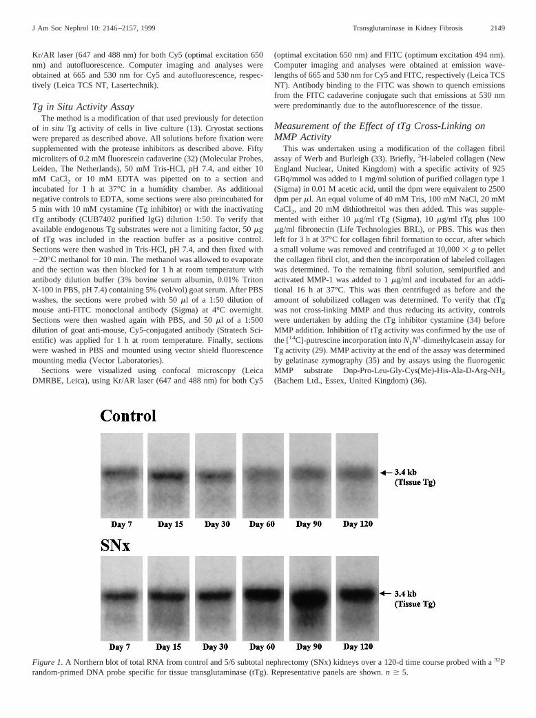
Figure 1.A Northern blot of total RNA from control and 5/6 subtotal nephrectomy (SNx) kidneys over a 120-d time course probed with a32Prandom-primed DNA probe specific for tissue transglutaminase (tTg). Representative panels are shown.n $ 5.
J Am Soc Nephrol 10: 2146–2157, 1999 Transglutaminase in Kidney Fibrosis 2149
Statistical AnalysesData analyses were performed using one-way ANOVA andt test.
P , 0.05 was taken as significant in both tests. All tests wereperformed on Microsoft Excel 97.
ResultsGeneral Observations
SNx rats demonstrated a steady increase in proteinuria andserum creatinine with time (Table 1), indicating progressiverenal insufficiency as documented previously (11) in our ear-lier studies using this model. All rats survived the SNx and theexperimental time course. The level of kidney fibrosis rosesteadily throughout the experimental time course showing se-vere scarring by day 90 post-SNx (Table 1). Mean tubular andglomerular scarring indices were comparable at each time point(r 5 0.95,P , 0.001). Both proteinuria and serum creatinineshowed a strong correlation with overall renal scarring (r 50.99, P , 0.0001, andr 5 0.82, P , 0.01, respectively).Creatinine clearance in SNx animals became progressivelylower than control animals with the development of renalscarring.
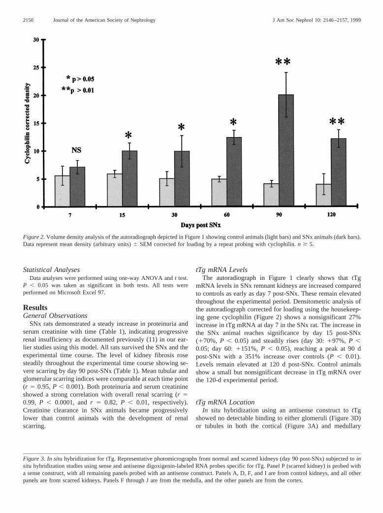
tTg mRNA LevelsThe autoradiograph in Figure 1 clearly shows that tTg
mRNA levels in SNx remnant kidneys are increased comparedto controls as early as day 7 post-SNx. These remain elevatedthroughout the experimental period. Densitometric analysis ofthe autoradiograph corrected for loading using the housekeep-ing gene cyclophilin (Figure 2) shows a nonsignificant 27%increase in tTg mRNA at day 7 in the SNx rat. The increase inthe SNx animal reaches significance by day 15 post-SNx(170%, P , 0.05) and steadily rises (day 30:197%, P ,0.05; day 60:1151%, P , 0.05), reaching a peak at 90 dpost-SNx with a 351% increase over controls (P , 0.01).Levels remain elevated at 120 d post-SNx. Control animalsshow a small but nonsignificant decrease in tTg mRNA overthe 120-d experimental period.
tTg mRNA LocationIn situ hybridization using an antisense construct to tTg
showed no detectable binding to either glomeruli (Figure 3D)or tubules in both the cortical (Figure 3A) and medullary
Figure 2.Volume density analysis of the autoradiograph depicted in Figure 1 showing control animals (light bars) and SNx animals (dark bars).Data represent mean density (arbitrary units)6 SEM corrected for loading by a repeat probing with cyclophilin.n $ 5.
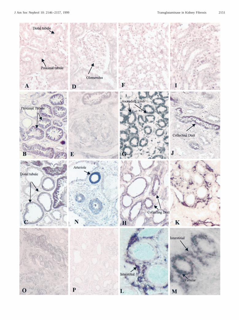
Figure 3. In situhybridization for tTg. Representative photomicrographs from normal and scarred kidneys (day 90 post-SNx) subjected toinsitu hybridization studies using sense and antisense digoxigenin-labeled RNA probes specific for tTg. Panel P (scarred kidney) is probed witha sense construct, with all remaining panels probed with an antisense construct. Panels A, D, F, and I are from control kidneys, and all otherpanels are from scarred kidneys. Panels F through J are from the medulla, and the other panels are from the cortex.
2150 Journal of the American Society of Nephrology J Am Soc Nephrol 10: 2146–2157, 1999
J Am Soc Nephrol 10: 2146–2157, 1999 Transglutaminase in Kidney Fibrosis 2151
(Figure 3, F and I) compartments within the developmentperiod used. In scarred kidneys (day 90 or later post-SNx),extensive staining was detected in the majority of proximaltubular cells (Figure 3B) and to a lesser extent in distal tubules(Figure 3C), the relative levels of which can be seen at lowermagnification (Figure 3O). No increase in the glomerular ex-pression of tTg was detectable in glomeruli in SNx kidneys(Figure 3E). Although staining was predominantly observed inthe cortex, there was also some increase in tTg mRNA detectedin the medulla, where pockets of what appear to be the ascend-ing limb of the loop of Henle and the occasional collecting ductdisplayed strong staining (Figure 3, G, H, and J). In somesections of scarred SNx kidneys, the identification of tubularsegments was made difficult by the degree of tubular atrophyand tubular dilation, a problem compounded by the nature ofthis technique.
tTg mRNA could also be found raised in some of the cellsconstituting the expanding tubular interstitium in terms of bothstain frequency and density, although this is best described assporadic rather than a uniform expression throughout the in-terstitium. Although this staining typically was seen in areaswhere elevated tubular expression was seen (Figure 3M), italso occurred independently of tubular expression (Figure 3, K
and L). We believe that these cells are fibroblasts, because asimilar staining pattern is obtained witha-smooth musclestaining as reported previously by us in this model (37). Wealso detected tTg mRNA in blood vessels (Figure 3N), but thisdid not appear to change from that detected in the controls.Probings performed at the same time on consecutive sectionsusing a sense probe to tTg failed to show any binding at all toSNx tissue.
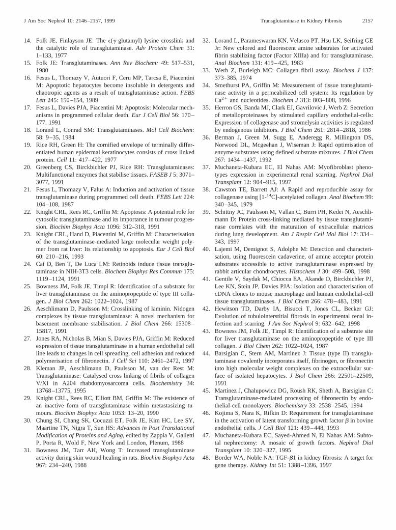
tTg Protein LocationImmunohistochemical analysis of tTg on blocked, unfixed
cryostat sections revealed areas of tissue where tTg is stronglybound to the renal architecture. Staining with a nonimmunemouse serum on SNx tissue demonstrates no significant stain-ing (Figure 4, A and D). When this was repeated on normalkidney tissue with a specific anti-tTg monoclonal antibody(CUB7402), there was widespread low-level staining (Figure4B), which when viewed at higher magnification (3400) isclearly in the extracellular environment (Figure 4E). An in-crease in the serum content of the blocking buffer to 10% from5% or the addition of fibronectin at 20mg/ml did not alter thisstaining, indicating that the interstitial staining is unlikely to befrom nonspecific binding of enzyme released during incubation
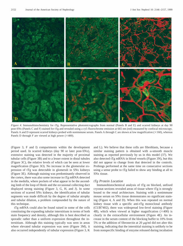
Figure 4. Immunohistochemistry for tTg. Representative photomicrographs from normal (Panels B and E) and scarred kidneys at day 90post-SNx (Panels C and F) stained for tTg and revealed using a cy5 fluorochrome emission at 665 nm (red) measured by confocal microscopy.Panels A and D represent scarred kidneys probed with nonimmune serum. Panels A through C are shown at low magnification (3160), whereasPanels D through F are viewed at high power (3400).
2152 Journal of the American Society of Nephrology J Am Soc Nephrol 10: 2146–2157, 1999
of sections since this would have been absorbed to the fi-bronectin in the serum. In scarred, 90-d post-SNx remnantkidneys, there is a large increase in the level of tTg antigenfound (Figure 4C) that is obviously in the expanding ECM(Figure 4F) when viewed at high magnification (3400). Semi-quantification of this increase in tTg antigen staining by mea-suring the emission intensity of the Cy5 label at 665 nmshowed a significant increase in the SNx compared with con-trols, rising from 196 6 to 1166 19 mV/mm2 (P , 0.001)(Figure 5).
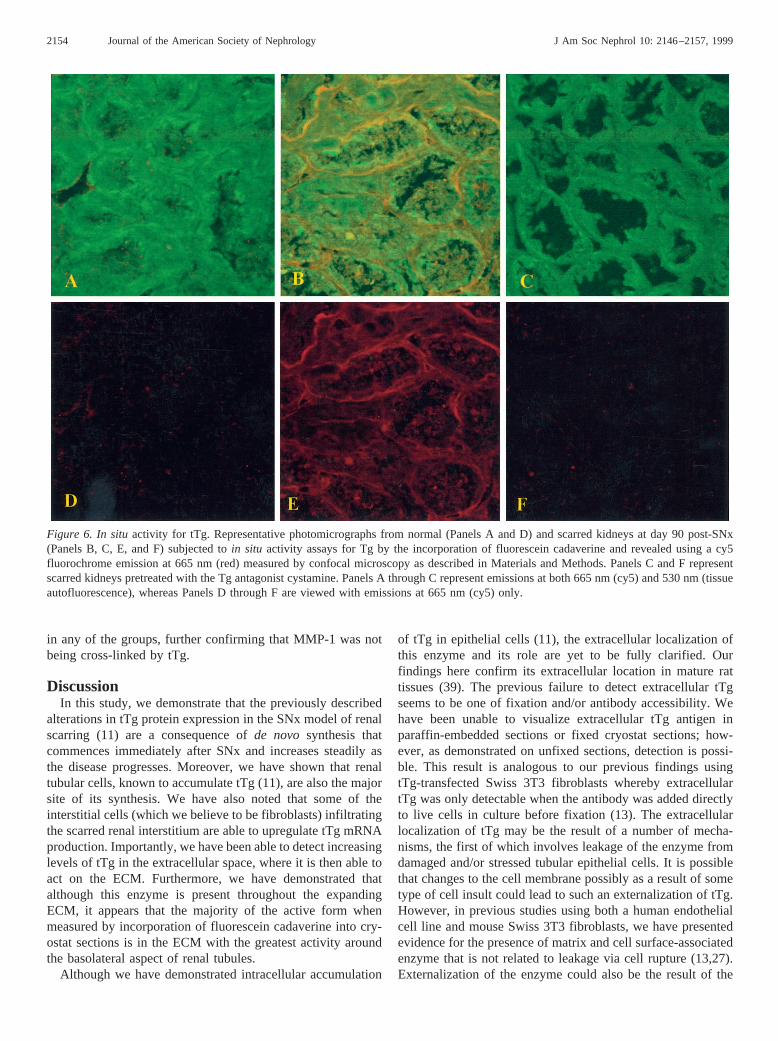
Measurement of tTg in Situ ActivityIn situ tTg activity assays revealed low incorporation of
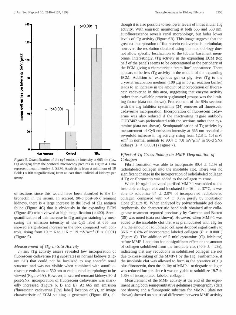
fluorescein cadaverine (tTg substrate) in normal kidneys (Fig-ure 6D) that could not be localized to any specific renalstructure and was not visible when combined with autofluo-rescence emissions at 530 nm to enable renal morphology to beviewed (Figure 6A). However, in scarred remnant kidneys 90-dpost-SNx, incorporation of fluorescein cadaverine was mark-edly increased (Figure 6, B and E). At 665 nm emission(fluorescein cadaverine [Cy5 label] location only), an imagecharacteristic of ECM staining is generated (Figure 6E), al-
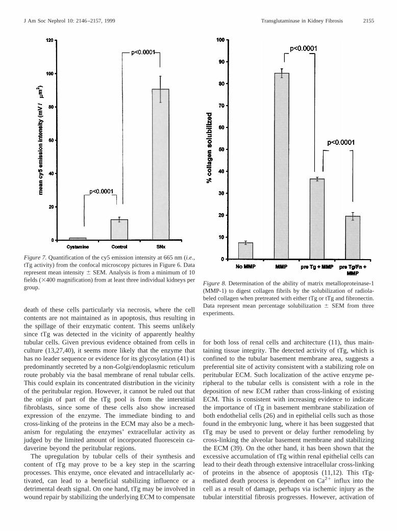
though it is also possible to see lower levels of intracellular tTgactivity. With emission monitoring at both 665 and 530 nm,autofluorescence reveals renal morphology, but hides lowerlevels of tTg activity (Figure 6B). This image suggests that thegreatest incorporation of fluorescein cadaverine is peritubular;however, the resolution obtained using this methodology doesnot allow specific localization to the tubular basement mem-brane. Interestingly, tTg activity in the expanding ECM (tophalf of the panel) seems to be concentrated at the periphery ofthe ECM giving a characteristic “tram line” appearance. Thereappears to be less tTg activity in the middle of the expandingECM. Addition of exogenous guinea pig liver tTg to thecryostat incubation medium (100mg in 50 ml reaction buffer)leads to an increase in the amount of incorporation of fluores-cein cadaverine in this area, suggesting that enzyme activityrather than available proteing-glutamyl groups was the limit-ing factor (data not shown). Pretreatment of the SNx sectionswith the tTg inhibitor cystamine (34) removes all fluoresceincadaverine incorporation. Incorporation of fluorescein cadav-erine was also reduced if the inactivating tTgase antibodyCUB7402 was preincubated with the sections rather than cys-tamine (data not shown). Semiquantification of Tg activity bymeasurement of Cy5 emission intensity at 665 nm revealed asevenfold increase in Tg activity rising from 12.36 1.4 mV/mm2 in normal animals to 90.46 7.8 mV/mm2 in 90-d SNxkidneys (P , 0.0001) (Figure 7).
Effect of Tg Cross-linking on MMP Degradation ofCollagen
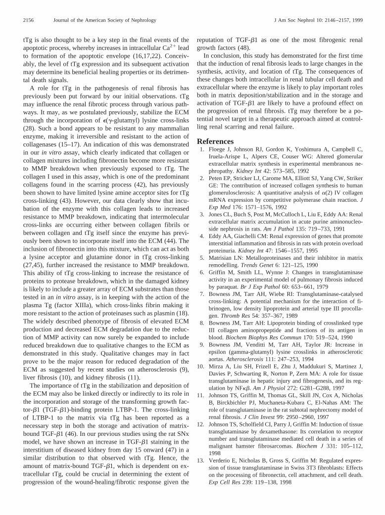
Fibril formation was able to incorporate 80.46 1.1% ofradiolabeled collagen into the insoluble clot. There was nosignificant change in the incorporation of radiolabeled collagenif Tg or fibronectin was added to the collagen mixture.
When 10mg/ml activated purified MMP-1 was added to theinsoluble collagen clot and incubated for 16 h at 37°C, it wasable to solubilize 846 2.0% of incorporated radiolabeledcollagen, compared with 7.46 0.7% purely by incubationalone (Figure 8). When analyzed by polyacrylamide gel elec-trophoresis, the characteristic band shift obtained after colla-genase treatment reported previously by Cawston and Barrett(38) was noted (data not shown). However, when MMP-1 wasadded to the insoluble clot that was preincubated with tTg for3 h, the amount of solubilized collagen dropped significantly to36.6 6 0.8% of incorporated labeled collagen (P , 0.0001)(Figure 8). The addition of 5 mM cystamine (tTg inhibitor)before MMP-1 addition had no significant effect on the amountof collagen solublized from the insoluble clot (40.96 4.2%),indicating that any reductions in solubilized collagen are notdue to cross-linking of the MMP-1 by the tTg. Furthermore, ifthe insoluble clot was allowed to form in the presence of tTgplus fibronectin, then the ability of MMP-1 to degrade collagenwas reduced further, since it was only able to solubilize 19.761.8% of incorporated labeled collagen.
Measurement of the MMP activity at the end of the exper-iment using both semiquantitative gelatinase zymography (datanot shown) and a fluorogenic substrate for MMP-1 (data notshown) showed no statistical difference between MMP activity
Figure 5.Quantification of the cy5 emission intensity at 665 nm (i.e.,tTg antigen) from the confocal microscopy pictures in Figure 4. Datarepresent mean intensity6 SEM. Analysis is from a minimum of 10fields (3160 magnification) from at least three individual kidneys pergroup.
J Am Soc Nephrol 10: 2146–2157, 1999 Transglutaminase in Kidney Fibrosis 2153
in any of the groups, further confirming that MMP-1 was notbeing cross-linked by tTg.
DiscussionIn this study, we demonstrate that the previously described
alterations in tTg protein expression in the SNx model of renalscarring (11) are a consequence ofde novosynthesis thatcommences immediately after SNx and increases steadily asthe disease progresses. Moreover, we have shown that renaltubular cells, known to accumulate tTg (11), are also the majorsite of its synthesis. We have also noted that some of theinterstitial cells (which we believe to be fibroblasts) infiltratingthe scarred renal interstitium are able to upregulate tTg mRNAproduction. Importantly, we have been able to detect increasinglevels of tTg in the extracellular space, where it is then able toact on the ECM. Furthermore, we have demonstrated thatalthough this enzyme is present throughout the expandingECM, it appears that the majority of the active form whenmeasured by incorporation of fluorescein cadaverine into cry-ostat sections is in the ECM with the greatest activity aroundthe basolateral aspect of renal tubules.
Although we have demonstrated intracellular accumulation
of tTg in epithelial cells (11), the extracellular localization ofthis enzyme and its role are yet to be fully clarified. Ourfindings here confirm its extracellular location in mature rattissues (39). The previous failure to detect extracellular tTgseems to be one of fixation and/or antibody accessibility. Wehave been unable to visualize extracellular tTg antigen inparaffin-embedded sections or fixed cryostat sections; how-ever, as demonstrated on unfixed sections, detection is possi-ble. This result is analogous to our previous findings usingtTg-transfected Swiss 3T3 fibroblasts whereby extracellulartTg was only detectable when the antibody was added directlyto live cells in culture before fixation (13). The extracellularlocalization of tTg may be the result of a number of mecha-nisms, the first of which involves leakage of the enzyme fromdamaged and/or stressed tubular epithelial cells. It is possiblethat changes to the cell membrane possibly as a result of sometype of cell insult could lead to such an externalization of tTg.However, in previous studies using both a human endothelialcell line and mouse Swiss 3T3 fibroblasts, we have presentedevidence for the presence of matrix and cell surface-associatedenzyme that is not related to leakage via cell rupture (13,27).Externalization of the enzyme could also be the result of the
Figure 6. In situactivity for tTg. Representative photomicrographs from normal (Panels A and D) and scarred kidneys at day 90 post-SNx(Panels B, C, E, and F) subjected toin situ activity assays for Tg by the incorporation of fluorescein cadaverine and revealed using a cy5fluorochrome emission at 665 nm (red) measured by confocal microscopy as described in Materials and Methods. Panels C and F representscarred kidneys pretreated with the Tg antagonist cystamine. Panels A through C represent emissions at both 665 nm (cy5) and 530 nm (tissueautofluorescence), whereas Panels D through F are viewed with emissions at 665 nm (cy5) only.
2154 Journal of the American Society of Nephrology J Am Soc Nephrol 10: 2146–2157, 1999
death of these cells particularly via necrosis, where the cellcontents are not maintained as in apoptosis, thus resulting inthe spillage of their enzymatic content. This seems unlikelysince tTg was detected in the vicinity of apparently healthytubular cells. Given previous evidence obtained from cells inculture (13,27,40), it seems more likely that the enzyme thathas no leader sequence or evidence for its glycosylation (41) ispredominantly secreted by a non-Golgi/endoplasmic reticulumroute probably via the basal membrane of renal tubular cells.This could explain its concentrated distribution in the vicinityof the peritubular region. However, it cannot be ruled out thatthe origin of part of the tTg pool is from the interstitialfibroblasts, since some of these cells also show increasedexpression of the enzyme. The immediate binding to andcross-linking of the proteins in the ECM may also be a mech-anism for regulating the enzymes’ extracellular activity asjudged by the limited amount of incorporated fluorescein ca-daverine beyond the peritubular regions.
The upregulation by tubular cells of their synthesis andcontent of tTg may prove to be a key step in the scarringprocesses. This enzyme, once elevated and intracellularly ac-tivated, can lead to a beneficial stabilizing influence or adetrimental death signal. On one hand, tTg may be involved inwound repair by stabilizing the underlying ECM to compensate
for both loss of renal cells and architecture (11), thus main-taining tissue integrity. The detected activity of tTg, which isconfined to the tubular basement membrane area, suggests apreferential site of activity consistent with a stabilizing role onperitubular ECM. Such localization of the active enzyme pe-ripheral to the tubular cells is consistent with a role in thedeposition of new ECM rather than cross-linking of existingECM. This is consistent with increasing evidence to indicatethe importance of tTg in basement membrane stabilization ofboth endothelial cells (26) and in epithelial cells such as thosefound in the embryonic lung, where it has been suggested thattTg may be used to prevent or delay further remodeling bycross-linking the alveolar basement membrane and stabilizingthe ECM (39). On the other hand, it has been shown that theexcessive accumulation of tTg within renal epithelial cells canlead to their death through extensive intracellular cross-linkingof proteins in the absence of apoptosis (11,12). This tTg-mediated death process is dependent on Ca21 influx into thecell as a result of damage, perhaps via ischemic injury as thetubular interstitial fibrosis progresses. However, activation of
Figure 7.Quantification of the cy5 emission intensity at 665 nm (i.e.,tTg activity) from the confocal microscopy pictures in Figure 6. Datarepresent mean intensity6 SEM. Analysis is from a minimum of 10fields (3400 magnification) from at least three individual kidneys pergroup.
Figure 8. Determination of the ability of matrix metalloproteinase-1(MMP-1) to digest collagen fibrils by the solubilization of radiola-beled collagen when pretreated with either tTg or tTg and fibronectin.Data represent mean percentage solubilization6 SEM from threeexperiments.
J Am Soc Nephrol 10: 2146–2157, 1999 Transglutaminase in Kidney Fibrosis 2155
tTg is also thought to be a key step in the final events of theapoptotic process, whereby increases in intracellular Ca21 leadto formation of the apoptotic envelope (16,17,22). Conceiv-ably, the level of tTg expression and its subsequent activationmay determine its beneficial healing properties or its detrimen-tal death signals.
A role for tTg in the pathogenesis of renal fibrosis haspreviously been put forward by our initial observations. tTgmay influence the renal fibrotic process through various path-ways. It may, as we postulated previously, stabilize the ECMthrough the incorporation ofe(g-glutamyl) lysine cross-links(28). Such a bond appears to be resistant to any mammalianenzyme, making it irreversible and resistant to the action ofcollagenases (15–17). An indication of this was demonstratedin our in vitro assay, which clearly indicated that collagen orcollagen mixtures including fibronectin become more resistantto MMP breakdown when previously exposed to tTg. Thecollagen I used in this assay, which is one of the predominantcollagens found in the scarring process (42), has previouslybeen shown to have limited lysine amine acceptor sites for tTgcross-linking (43). However, our data clearly show that incu-bation of the enzyme with this collagen leads to increasedresistance to MMP breakdown, indicating that intermolecularcross-links are occurring either between collagen fibrils orbetween collagen and tTg itself since the enzyme has previ-ously been shown to incorporate itself into the ECM (44). Theinclusion of fibronectin into this mixture, which can act as botha lysine acceptor and glutamine donor in tTg cross-linking(27,45), further increased the resistance to MMP breakdown.This ability of tTg cross-linking to increase the resistance ofproteins to protease breakdown, which in the damaged kidneyis likely to include a greater array of ECM substrates than thosetested in anin vitro assay, is in keeping with the action of theplasma Tg (factor XIIIa), which cross-links fibrin making itmore resistant to the action of proteinases such as plasmin (18).The widely described phenotype of fibrosis of elevated ECMproduction and decreased ECM degradation due to the reduc-tion of MMP activity can now surely be expanded to includereduced breakdown due to qualitative changes to the ECM asdemonstrated in this study. Qualitative changes may in factprove to be the major reason for reduced degradation of theECM as suggested by recent studies on atherosclerosis (9),liver fibrosis (10), and kidney fibrosis (11).
The importance of tTg in the stabilization and deposition ofthe ECM may also be linked directly or indirectly to its role inthe incorporation and storage of the transforming growth fac-tor-b1 (TGF-b1)-binding protein LTBP-1. The cross-linkingof LTBP-1 to the matrix via tTg has been reported as anecessary step in both the storage and activation of matrix-bound TGF-b1 (46). In our previous studies using the rat SNxmodel, we have shown an increase in TGF-b1 staining in theinterstitium of diseased kidney from day 15 onward (47) in asimilar distribution to that observed with tTg. Hence, theamount of matrix-bound TGF-b1, which is dependent on ex-tracellular tTg, could be crucial in determining the extent ofprogression of the wound-healing/fibrotic response given the
reputation of TGF-b1 as one of the most fibrogenic renalgrowth factors (48).
In conclusion, this study has demonstrated for the first timethat the induction of renal fibrosis leads to large changes in thesynthesis, activity, and location of tTg. The consequences ofthese changes both intracellular in renal tubular cell death andextracellular where the enzyme is likely to play important rolesboth in matrix deposition/stabilization and in the storage andactivation of TGF-b1 are likely to have a profound effect onthe progression of renal fibrosis. tTg may therefore be a po-tential novel target in a therapeutic approach aimed at control-ling renal scarring and renal failure.
References1. Floege J, Johnson RJ, Gordon K, Yoshimura A, Campbell C,
Iruela-Arispe L, Alpers CE, Couser WG: Altered glomerularextracellular matrix synthesis in experimental membranous ne-phropathy.Kidney Int42: 573–585, 1992
2. Peten EP, Stricker LJ, Carome MA, Elliott SJ, Yang CW, StrikerGE: The contribution of increased collagen synthesis to humanglomerulosclerosis: A quantitative analysis ofa(2) IV collagenmRNA expression by competitive polymerase chain reaction.JExp Med176: 1571–1576, 1992
3. Jones CL, Buch S, Post M, McCulloch L, Liu E, Eddy AA: Renalextracellular matrix accumulation in acute purine aminonucleo-side nephrosis in rats.Am J Pathol135: 719–733, 1991
4. Eddy AA, Giachelli CM: Renal expression of genes that promoteinterstitial inflammation and fibrosis in rats with protein overloadproteinuria.Kidney Int47: 1546–1557, 1995
5. Matrisian LN: Metalloproteinases and their inhibitor in matrixremodelling.Trends Genet6: 121–125, 1990
6. Griffin M, Smith LL, Wynne J: Changes in transglutaminaseactivity in an experimental model of pulmonary fibrosis inducedby paraquat.Br J Exp Pathol60: 653–661, 1979
7. Bowness JM, Tarr AH, Wiebe RI: Transglutaminase-catalysedcross-linking: A potential mechanism for the interaction of fi-brinogen, low density lipoprotein and arterial type III procolla-gen.Thromb Res54: 357–367, 1989
8. Bowness JM, Tarr AH: Lipoprotein binding of crosslinked typeIII collagen aminopropeptide and fractions of its antigen inblood.Biochem Biophys Res Commun170: 519–524, 1990
9. Bowness JM, Venditti M, Tarr AH, Taylor JR: Increase inepsilon (gamma-glutamyl) lysine crosslinks in atheroscleroticaortas.Atherosclerosis111: 247–253, 1994
10. Mirza A, Liu SH, Frizell E, Zhu J, Maddukuri S, Martinez J,Davies P, Schwarting R, Norton P, Zern MA: A role for tissuetransglutaminase in hepatic injury and fibrogenesis, and its reg-ulation by NFkb. Am J Physiol272: G281–G288, 1997
11. Johnson TS, Griffin M, Thomas GL, Skill JN, Cox A, NicholasB, Birckbichler PJ, Muchaneta-Kubara C, El-Nahas AM: Therole of transglutaminase in the rat subtotal nephrectomy model ofrenal fibrosis.J Clin Invest99: 2950–2960, 1997
12. Johnson TS, Scholfield CI, Parry J, Griffin M: Induction of tissuetransglutaminase by dexamethasone: Its correlation to receptornumber and transglutaminase mediated cell death in a series ofmalignant hamster fibrosarcomas.Biochem J331: 105–112,1998
13. Verderio E, Nicholas B, Gross S, Griffin M: Regulated expres-sion of tissue transglutaminase in Swiss 3T3 fibroblasts: Effectson the processing of fibronectin, cell attachment, and cell death.Exp Cell Res239: 119–138, 1998
2156 Journal of the American Society of Nephrology J Am Soc Nephrol 10: 2146–2157, 1999
14. Folk JE, Finlayson JE: Thee(g-glutamyl) lysine crosslink andthe catalytic role of transglutaminase.Adv Protein Chem31:1–133, 1977
15. Folk JE: Transglutaminases.Ann Rev Biochem:49: 517–531,1980
16. Fesus L, Thomazy V, Autuori F, Ceru MP, Tarcsa E, PiacentiniM: Apoptotic hepatocytes become insoluble in detergents andchaotropic agents as a result of transglutaminase action.FEBSLett 245: 150–154, 1989
17. Fesus L, Davies PJA, Piacentini M: Apoptosis: Molecular mech-anisms in programmed cellular death.Eur J Cell Biol 56: 170–177, 1991
18. Lorand L, Conrad SM: Transglutaminases.Mol Cell Biochem:58: 9–35, 1984
19. Rice RH, Green H: The cornified envelope of terminally differ-entiated human epidermal keratinocytes consists of cross linkedprotein.Cell 11: 417–422, 1977
20. Greenberg CS, Birckbichler PJ, Rice RH: Transglutaminases:Multifunctional enzymes that stabilise tissues.FASEB J5: 3071–3077, 1991
21. Fesus L, Thomazy V, Falus A: Induction and activation of tissuetransglutaminase during programmed cell death.FEBS Lett224:104–108, 1987
22. Knight CRL, Rees RC, Griffin M: Apoptosis: A potential role forcytosolic transglutaminase and its importance in tumour progres-sion.Biochim Biophys Acta1096: 312–318, 1991
23. Knight CRL, Hand D, Piacentini M, Griffin M: Characterisationof the transglutaminase-mediated large molecular weight poly-mer from rat liver: Its relationship to apoptosis.Eur J Cell Biol60: 210–216, 1993
24. Cai D, Ben T, De Luca LM: Retinoids induce tissue transglu-taminase in NIH-3T3 cells.Biochem Biophys Res Commun175:1119–1124, 1991
25. Bowness JM, Folk JE, Timpl R: Identification of a substrate forliver transglutaminase on the aminopropeptide of type III colla-gen.J Biol Chem262: 1022–1024, 1987
26. Aeschlimann D, Paulsson M: Crosslinking of laminin. Nidogencomplexes by tissue transglutaminase: A novel mechanism forbasement membrane stabilisation.J Biol Chem266: 15308–15817, 1991
27. Jones RA, Nicholas B, Mian S, Davies PJA, Griffin M: Reducedexpression of tissue transglutaminase in a human endothelial cellline leads to changes in cell spreading, cell adhesion and reducedpolymerisation of fibronectin.J Cell Sci110: 2461–2472, 1997
28. Kleman JP, Aeschlimann D, Paulsson M, van der Rest M:Transglutaminase: Catalysed cross linking of fibrils of collagenV/XI in A204 rhabdomyosarcoma cells.Biochemistry 34:13768–13775, 1995
29. Knight CRL, Rees RC, Elliott BM, Griffin M: The existence ofan inactive form of transglutaminase within metastasizing tu-mours.Biochim Biophys Acta1053: 13–20, 1990
30. Chung SI, Chang SK, Cocuzzi ET, Folk JE, Kim HC, Lee SY,Maartine TN, Nigra T, Sun HS:Advances in Post TranslationalModification of Proteins and Aging, edited by Zappia V, GallettiP, Porta R, Wold F, New York and London, Plenum, 1988
31. Bowness JM, Tarr AH, Wong T: Increased transglutaminaseactivity during skin wound healing in rats.Biochim Biophys Acta967: 234–240, 1988
32. Lorand L, Parameswaran KN, Velasco PT, Hsu LK, Seifring GEJr: New colored and fluorescent amine substrates for activatedfibrin stabilizing factor (Factor XIIIa) and for transglutaminase.Anal Biochem131: 419–425, 1983
33. Werb Z, Burleigh MC: Collagen fibril assay.Biochem J137:373–385, 1974
34. Smethurst PA, Griffin M: Measurement of tissue transglutami-nase activity in a permeabilized cell system: Its regulation byCa21 and nucleotides.Biochem J313: 803–808, 1996
35. Herron GS, Banda MJ, Clark EJ, Gavrilovic J, Werb Z: Secretionof metalloproteinases by stimulated capillary endothelial-cells:Expression of collagenase and stromelysin activities is regulatedby endogenous inhibitors.J Biol Chem261: 2814–2818, 1986
36. Berman J, Green M, Sugg E, Anderegg R, Millington DS,Norwood DL, Mcgeehan J, Wiseman J: Rapid optimisation ofenzyme substrates using defined substrate mixtures.J Biol Chem267: 1434–1437, 1992
37. Muchaneta-Kubara EC, El Nahas AM: Myofibroblast pheno-types expression in experimental renal scarring.Nephrol DialTransplant12: 904–915, 1997
38. Cawston TE, Barrett AJ: A Rapid and reproducible assay forcollagenase using [1-14C]-acetylated collagen.Anal Biochem99:340–345, 1979
39. Schittny JC, Paulsson M, Vallan C, Burri PH, Kedei N, Aeschli-mann D: Protein cross-linking mediated by tissue transglutami-nase correlates with the maturation of extracellular matricesduring lung development.Am J Respir Cell Mol Biol17: 334–343, 1997
40. Lajemi M, Demignot S, Adolphe M: Detection and characteri-sation, using fluorescein cadaverine, of amine acceptor proteinsubstrates accessible to active transglutaminase expressed byrabbit articular chondrocytes.Histochem J30: 499–508, 1998
41. Gentile V, Saydak M, Chiocca EA, Akande O, Birckbichler PJ,Lee KN, Stein JP, Davies PJA: Isolation and characterisation ofcDNA clones to mouse macrophage and human endothelial-celltissue transglutaminases.J Biol Chem266: 478–483, 1991
42. Hewitson TD, Darby IA, Bisucci T, Jones CL, Becker GJ:Evolution of tubulointerstitial fibrosis in experimental renal in-fection and scarring.J Am Soc Nephrol9: 632–642, 1998
43. Bowness JM, Folk JE, Timpl R: Identification of a substrate sitefor liver transglutaminase on the aminopropeptide of type IIIcollagen.J Biol Chem262: 1022–1024, 1987
44. Barsigian C, Stern AM, Martinez J: Tissue (type II) transglu-taminase covalently incorporates itself, fibrinogen, or fibronectininto high molecular weight complexes on the extracellular sur-face of isolated hepatocytes.J Biol Chem266: 22501–22509,1991
45. Martinez J, Chalupowicz DG, Roush RK, Sheth A, Barsigian C:Transglutaminase-mediated processing of fibronectin by endo-thelial-cell monolayers.Biochemistry33: 2538–2545, 1994
46. Kojima S, Nara K, Rifkin D: Requirement for transglutaminasein the activation of latent transforming growth factorb in bovineendothelial cells.J Cell Biol 121: 439–448, 1993
47. Muchaneta-Kubara EC, Sayed-Ahmed N, El Nahas AM: Subto-tal nephrectomy: A mosaic of growth factors.Nephrol DialTransplant10: 320–327, 1995
48. Border WA, Noble NA: TGF-b1 in kidney fibrosis: A target forgene therapy.Kidney Int51: 1388–1396, 1997
J Am Soc Nephrol 10: 2146–2157, 1999 Transglutaminase in Kidney Fibrosis 2157