oral / poster presentation - sbmu journals
TRANSCRIPT
The 5rd International Congress of Iranian Pediatric Nephrology Association
Journal of Pediatric Nephrology | Supplement 1 | Spring 2016
Oral / Poster Presentation
J Ped. Nephrology 2016 May. Supplement 1
http://journals.sbmu.ac.ir/jpn
The 5th International Congress of Iranian Pediatric
Nephrology Association
The forthcoming meeting of “5th international congress of Iranian pediatric nephrology association” offers a
unique opportunity to exchange new ideas in the field of pediatric nephrology with special attention to the
urinary tract infection, chronic kidney disease,renal transplantation,dialysis and obstructive uropathy. The
congress will be complemented by two satellite workshops.
We would be pleased and honored if you would accept our invitation at 2016 congress of ISPN and are
looking forward to seeing you in Shiraz, Iran.
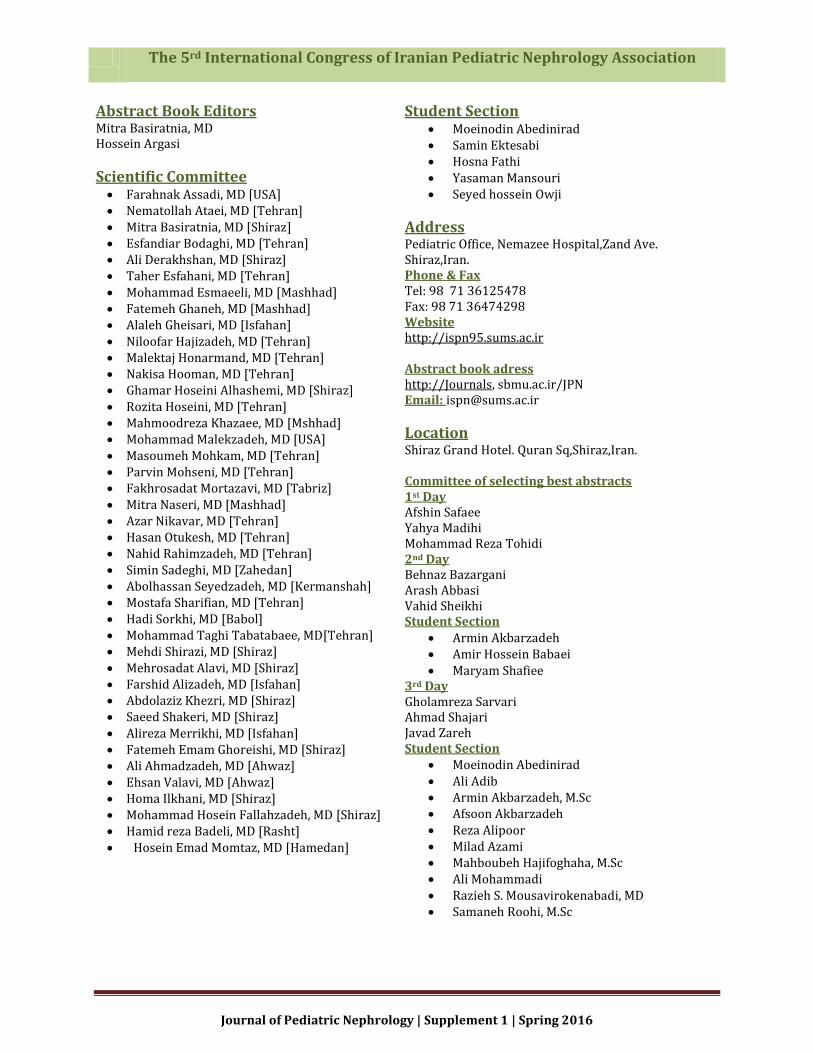
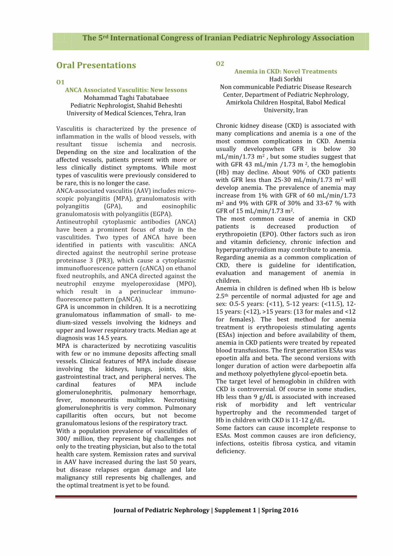
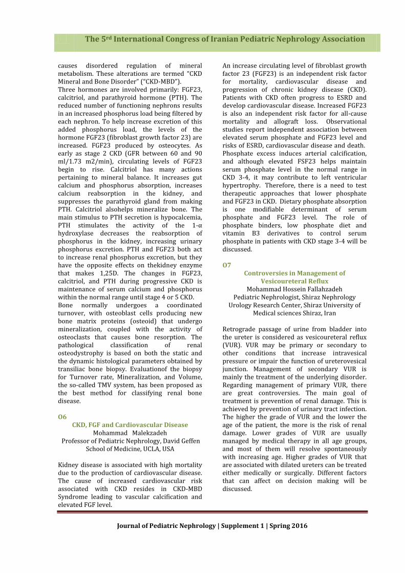
Congress chair Prof. Ghamar Hosseini Al-
Hashemi
Secretory General Prof. Ali Derakhshan
Secretary Executive Prof. Mitra Basiratnia
Congress President Ghamar Hosseini Alhashemi, MD
Secretary General Ali Derakhshan, MD Student Section Amir Hossein Babaei
Secretary Executive Mitra Basiratnia Student Section Fatemeh Zarei
Executive Committee Mitra Basiratnia Fatemeh Emamghoreishi Farhad Hanjani Ahmad Rezazadeh Sanaz Bahadori Niloofar Alishahi Hossein Mokhtari Maryam Farahmandi Marzieh Dashtpeyma Zahra Hemmat Mahboobeh Keshavarzi
The 5rd International Congress of Iranian Pediatric Nephrology Association
Journal of Pediatric Nephrology | Supplement 1 | Spring 2016
Abstract Book Editors Mitra Basiratnia, MD Hossein Argasi
Scientific Committee Farahnak Assadi, MD [USA] Nematollah Ataei, MD [Tehran] Mitra Basiratnia, MD [Shiraz] Esfandiar Bodaghi, MD [Tehran] Ali Derakhshan, MD [Shiraz] Taher Esfahani, MD [Tehran] Mohammad Esmaeeli, MD [Mashhad] Fatemeh Ghaneh, MD [Mashhad] Alaleh Gheisari, MD [Isfahan] Niloofar Hajizadeh, MD [Tehran] Malektaj Honarmand, MD [Tehran] Nakisa Hooman, MD [Tehran] Ghamar Hoseini Alhashemi, MD [Shiraz] Rozita Hoseini, MD [Tehran] Mahmoodreza Khazaee, MD [Mshhad] Mohammad Malekzadeh, MD [USA] Masoumeh Mohkam, MD [Tehran] Parvin Mohseni, MD [Tehran] Fakhrosadat Mortazavi, MD [Tabriz] Mitra Naseri, MD [Mashhad] Azar Nikavar, MD [Tehran] Hasan Otukesh, MD [Tehran] Nahid Rahimzadeh, MD [Tehran] Simin Sadeghi, MD [Zahedan] Abolhassan Seyedzadeh, MD [Kermanshah] Mostafa Sharifian, MD [Tehran] Hadi Sorkhi, MD [Babol] Mohammad Taghi Tabatabaee, MD[Tehran] Mehdi Shirazi, MD [Shiraz] Mehrosadat Alavi, MD [Shiraz] Farshid Alizadeh, MD [Isfahan] Abdolaziz Khezri, MD [Shiraz] Saeed Shakeri, MD [Shiraz] Alireza Merrikhi, MD [Isfahan] Fatemeh Emam Ghoreishi, MD [Shiraz] Ali Ahmadzadeh, MD [Ahwaz] Ehsan Valavi, MD [Ahwaz] Homa Ilkhani, MD [Shiraz] Mohammad Hosein Fallahzadeh, MD [Shiraz] Hamid reza Badeli, MD [Rasht] Hosein Emad Momtaz, MD [Hamedan]
Student Section
Moeinodin Abedinirad Samin Ektesabi Hosna Fathi Yasaman Mansouri Seyed hossein Owji
Address Pediatric Office, Nemazee Hospital,Zand Ave. Shiraz,Iran. Phone & Fax Tel: 98 71 36125478 Fax: 98 71 36474298 Website http://ispn95.sums.ac.ir Abstract book adress http://Journals, sbmu.ac.ir/JPN Email: [email protected]
Location Shiraz Grand Hotel. Quran Sq,Shiraz,Iran. Committee of selecting best abstracts 1st Day Afshin Safaee Yahya Madihi Mohammad Reza Tohidi 2nd Day Behnaz Bazargani Arash Abbasi Vahid Sheikhi Student Section
Armin Akbarzadeh Amir Hossein Babaei Maryam Shafiee
3rd Day Gholamreza Sarvari Ahmad Shajari Javad Zareh Student Section
Moeinodin Abedinirad Ali Adib Armin Akbarzadeh, M.Sc Afsoon Akbarzadeh Reza Alipoor Milad Azami Mahboubeh Hajifoghaha, M.Sc Ali Mohammadi Razieh S. Mousavirokenabadi, MD Samaneh Roohi, M.Sc

The 5rd International Congress of Iranian Pediatric Nephrology Association
Journal of Pediatric Nephrology | Supplement 1 | Spring 2016
Conference Topics Urinary tract infection
Reflux
Obstructive uropathy
Voiding dysfunction
Renal transplantation
Acute and chronic kidney disease
Hypertension
Hemodialysis
Peritoneal dialysis
Fluid and electrolyte
Glomerulonephritis
Keynote Lecturers Professor Lesley Rees MD FRCPCH Consultant Paediatric Nephrologist, Gt Ormond St Hospital for Children Renal Unit, London Professor of Paediatric Nephrology, University College London
Professor Kjell Tullus Professor of Paediatric Nephrology Consultant Paediatric Nephrologist, Gt Ormond St Hospital for Children Renal Unit, London
Professor Mohammad Malekzadeh Professor of pediatric nephrology David Geffen School of Medicine at UCLA,USA
Professor Farahnak Assadi Emeritus Professor Pediatrics and Director of Nephrology, Rush Children’s Hospital, Chicago, IL
Professor Oguz Soylemezoglu Professor of pediatric nephrology Gazi university, Ankara, Turkey
The 5rd International Congress of Iranian Pediatric Nephrology Association
Journal of Pediatric Nephrology | Supplement 1 | Spring 2016
Oral Presentations O1
ANCA Associated Vasculitis: New lessons Mohammad Taghi Tabatabaee
Pediatric Nephrologist, Shahid Beheshti University of Medical Sciences, Tehra, Iran
Vasculitis is characterized by the presence of inflammation in the walls of blood vessels, with resultant tissue ischemia and necrosis. Depending on the size and localization of the affected vessels, patients present with more or less clinically distinct symptoms. While most types of vasculitis were previously considered to be rare, this is no longer the case. ANCA-associated vasculitis (AAV) includes micro-scopic polyangiitis (MPA), granulomatosis with polyangiitis (GPA), and eosinophilic granulomatosis with polyangiitis (EGPA). Antineutrophil cytoplasmic antibodies (ANCA) have been a prominent focus of study in the vasculitides. Two types of ANCA have been identified in patients with vasculitis: ANCA directed against the neutrophil serine protease proteinase 3 (PR3), which cause a cytoplasmic immunofluorescence pattern (cANCA) on ethanol fixed neutrophils, and ANCA directed against the neutrophil enzyme myeloperoxidase (MPO), which result in a perinuclear immuno-fluorescence pattern (pANCA). GPA is uncommon in children. It is a necrotizing granulomatous inflammation of small- to me-dium-sized vessels involving the kidneys and upper and lower respiratory tracts. Median age at diagnosis was 14.5 years. MPA is characterized by necrotizing vasculitis with few or no immune deposits affecting small vessels. Clinical features of MPA include disease involving the kidneys, lungs, joints, skin, gastrointestinal tract, and peripheral nerves. The cardinal features of MPA include glomerulonephritis, pulmonary hemorrhage, fever, mononeuritis multiplex. Necrotising glomerulonephritis is very common. Pulmonary capillaritis often occurs, but not become granulomatous lesions of the respiratory tract. With a population prevalence of vasculitides of 300/ million, they represent big challenges not only to the treating physician, but also to the total health care system. Remission rates and survival in AAV have increased during the last 50 years, but disease relapses organ damage and late malignancy still represents big challenges, and the optimal treatment is yet to be found.
O2 Anemia in CKD: Novel Treatments
Hadi Sorkhi
Non communicable Pediatric Disease Research Center, Department of Pediatric Nephrology,
Amirkola Children Hospital, Babol Medical University, Iran
Chronic kidney disease (CKD) is associated with many complications and anemia is a one of the most common complications in CKD. Anemia usually developswhen GFR is below 30 mL/min/1.73 m2 , but some studies suggest that with GFR 43 mL/min /1.73 m 2, the hemoglobin (Hb) may decline. About 90% of CKD patients with GFR less than 25-30 mL/min/1.73 m2 will develop anemia. The prevalence of anemia may increase from 1% with GFR of 60 mL/min/1.73 m2 and 9% with GFR of 30% and 33-67 % with GFR of 15 mL/min/1.73 m2. The most common cause of anemia in CKD patients is decreased production of erythropoietin (EPO). Other factors such as iron and vitamin deficiency, chronic infection and hyperparathyroidism may contribute to anemia. Regarding anemia as a common complication of CKD, there is guideline for identification, evaluation and management of anemia in children. Anemia in children is defined when Hb is below 2.5th percentile of normal adjusted for age and sex: O.5-5 years: (<11), 5-12 years: (<11.5), 12-15 years: (<12), >15 years: (13 for males and <12 for females). The best method for anemia treatment is erythropoiesis stimulating agents (ESAs) injection and before availability of them, anemia in CKD patients were treated by repeated blood transfusions. The first generation ESAs was epoetin alfa and beta. The second versions with longer duration of action were darbepoetin alfa and methoxy polyethylene glycol-epoetin beta. The target level of hemoglobin in children with CKD is controversial. Of course in some studies, Hb less than 9 g/dL is associated with increased risk of morbidity and left ventricular hypertrophy and the recommended target of Hb in children with CKD is 11-12 g/dL. Some factors can cause incomplete response to ESAs. Most common causes are iron deficiency, infections, osteitis fibrosa cystica, and vitamin deficiency.

The 5rd International Congress of Iranian Pediatric Nephrology Association
Journal of Pediatric Nephrology | Supplement 1 | Spring 2016
O3 Automated Peritoneal Dialysis: Prescription
and Dose Adjustment Lesley Rees
Consultant Paediatric Nephrologist, Gt Ormond St Hospital for Children Renal Unit, London
University College London, UK
During a PD cycle, solute moves down the concentration gradient across the peritoneal membrane by diffusion and water byosmosis (ultrafiltration, UF). UF causes movement of solutes by convection, so that solutes may be carried across the membrane (solvent drag) even in the absence of a concentration gradient. Both solute and water transport is bidirectional. Diffusion Diffusion is affected by solute concentration between blood and dialysate and the molecular weight of the solute as diffusion is dependent on size. Diffusion is increased by increasing the fill volume until the intraperitoneal pressure is 18 cmH2O thereby increasing the volume for diffusion and recruitment of peritoneal surface area, and prolonging the osmotic difference. Transport of water (UF) Water moves from capillaries to peritoneal cavity down a pressure gradient. The force is osmotic pressure. Convective mass transfer This depends on the amount of fluid removed by UF and on membrane permeability. It contributes more to larger solute movement. Reabsorption of solute and water Is driven by loss of osmotic gradient and rising intraperitoneal pressure. It occurs via lymphatics under the diaphragm and lymphatics and blood vessels in the peritoneal cavity. Three types of pores affect water and solute transfer. The ultra-small pores (endothelial AQP-1 channels) are themost abundantand enable sodium-free water transport. The small pores allow diffusion of solutes and water. The large pores are in low numbers and facilitate convective mass transport and macromolecular leakage into the peritoneal cavity. O4
Biological Agents in Treatment of Glomerulonephritis
Nakysa Hooman Consultant Pediatric Nephrologist, Ali-Asghar Children Hospital, Iran University of Medical
Sciences
Current immunosuppression protocols in the management of glomerulonephritides remain unsatisfactory, especially in frequently relapsing or resistant to treatment. Adverse effects and toxicities associated with the use of these medications increase morbidity and mortality. Advances in our understanding of the immunopathogenesis of glomerulonephritis led to successfully implementation of biological agents in the treatment of immune-mediated glomerular diseases. Biological agents are produced using recombinant DNA technology in a living system. They can target specific immune cell types, cytokines or immune pathways involved in the pathogenesis of these disorders. These biological agents include Abatacept (block co-stimulation signaling of T-cell activation), BAFF inhibitors (block plasma cell), Rituximab (prevent B cell activation), Eculizumab (target complement pathway) and TNF-alpha inhibitors. These agents have a more directed and effective immunosuppression, with much more desirable side-effects. However, there have been few randomized controlled trials comparing biologic agents to conventional immunosuppression, and in many of them the reported side-effects have not been different. A wide variety of autoimmune disorders have been reported including systemic diseases (lupus, vasculitis, sarcoidosis, antiphospholipid syndrome and inflammatory myopathies) or organ-specific (interstitial lung disease, uveitis, optic neuritis, peripheral neuropathies, multiple sclerosis, psoriasis, inflammatory bowel disease and autoimmune hepatitis). Some of these side effects such as interstitial fibrosis with Rituximab or autoimmune renal disorders with anti-TNF-alpha blocker can be life threatening and led to death. Utilization of these biological agents should be balanced over the harm-benefit that could be achieved especially in diseases that are naturally frequently flare or resistant to conventional therapies. Long term monitoring is necessary for the occurrence of auto-immune disease secondary to these biological agents. O5
Bone Health in CKD and Dialysis Patients: New Insights
Mohammad Esmaeili Pediatric nephrologist, Mashhad University of
Medical Sciences, Mashhad, Iran
Childhood and adolescence period are the crucial times fordeveloping a healthy skeletal and vascular system; chronic kidney disease (CKD)
The 5rd International Congress of Iranian Pediatric Nephrology Association
Journal of Pediatric Nephrology | Supplement 1 | Spring 2016
causes disordered regulation of mineral metabolism. These alterations are termed “CKD Mineral and Bone Disorder” (“CKD-MBD”). Three hormones are involved primarily: FGF23, calcitriol, and parathyroid hormone (PTH). The reduced number of functioning nephrons results in an increased phosphorus load being filtered by each nephron. To help increase excretion of this added phosphorus load, the levels of the hormone FGF23 (fibroblast growth factor 23) are increased. FGF23 produced by osteocytes. As early as stage 2 CKD (GFR between 60 and 90 ml/1.73 m2/min), circulating levels of FGF23 begin to rise. Calcitriol has many actions pertaining to mineral balance. It increases gut calcium and phosphorus absorption, increases calcium reabsorption in the kidney, and suppresses the parathyroid gland from making PTH. Calcitriol alsohelps mineralize bone. The main stimulus to PTH secretion is hypocalcemia, PTH stimulates the activity of the 1-α hydroxylase decreases the reabsorption of phosphorus in the kidney, increasing urinary phosphorus excretion. PTH and FGF23 both act to increase renal phosphorus excretion, but they have the opposite effects on thekidney enzyme that makes 1,25D. The changes in FGF23, calcitriol, and PTH during progressive CKD is maintenance of serum calcium and phosphorus within the normal range until stage 4 or 5 CKD. Bone normally undergoes a coordinated turnover, with osteoblast cells producing new bone matrix proteins (osteoid) that undergo mineralization, coupled with the activity of osteoclasts that causes bone resorption. The pathological classification of renal osteodystrophy is based on both the static and the dynamic histological parameters obtained by transiliac bone biopsy. Evaluationof the biopsy for Turnover rate, Mineralization, and Volume, the so-called TMV system, has been proposed as the best method for classifying renal bone disease. O6
CKD, FGF and Cardiovascular Disease Mohammad Malekzadeh
Professor of Pediatric Nephrology, David Geffen School of Medicine, UCLA, USA
Kidney disease is associated with high mortality due to the production of cardiovascular disease. The cause of increased cardiovascular risk associated with CKD resides in CKD-MBD Syndrome leading to vascular calcification and elevated FGF level.
An increase circulating level of fibroblast growth factor 23 (FGF23) is an independent risk factor for mortality, cardiovascular disease and progression of chronic kidney disease (CKD). Patients with CKD often progress to ESRD and develop cardiovascular disease. Increased FGF23 is also an independent risk factor for all-cause mortality and allograft loss. Observational studies report independent association between elevated serum phosphate and FGF23 level and risks of ESRD, cardiovascular disease and death. Phosphate excess induces arterial calcification, and although elevated FSF23 helps maintain serum phosphate level in the normal range in CKD 3-4, it may contribute to left ventricular hypertrophy. Therefore, there is a need to test therapeutic approaches that lower phosphate and FGF23 in CKD. Dietary phosphate absorption is one modifiable determinant of serum phosphate and FGF23 level. The role of phosphate binders, low phosphate diet and vitamin B3 derivatives to control serum phosphate in patients with CKD stage 3-4 will be discussed. O7
Controversies in Management of Vesicoureteral Reflux
Mohammad Hossein Fallahzadeh Pediatric Nephrologist, Shiraz Nephrology
Urology Research Center, Shiraz University of Medical sciences Shiraz, Iran
Retrograde passage of urine from bladder into the ureter is considered as vesicoureteral reflux (VUR). VUR may be primary or secondary to other conditions that increase intravesical pressure or impair the function of ureterovesical junction. Management of secondary VUR is mainly the treatment of the underlying disorder. Regarding management of primary VUR, there are great controversies. The main goal of treatment is prevention of renal damage. This is achieved by prevention of urinary tract infection. The higher the grade of VUR and the lower the age of the patient, the more is the risk of renal damage. Lower grades of VUR are usually managed by medical therapy in all age groups, and most of them will resolve spontaneously with increasing age. Higher grades of VUR that are associated with dilated ureters can be treated either medically or surgically. Different factors that can affect on decision making will be discussed.
The 5rd International Congress of Iranian Pediatric Nephrology Association
Journal of Pediatric Nephrology | Supplement 1 | Spring 2016
O8 Current Knowledge in Recurrence of FSGS
Nahid Rahimzadeh Division of Pediatric Nephrology, Rasoul-e-Akram Hospital, Iran University of Medical
Sciences, Tehran, Iran
FSGS is the most frequently acquired condition leading to ESRD in children, and nearly 50% of affected patients will progress to ESRD over a 5 to 10-year period.Up to 55 % of patients develop recurrent disease after receiving a kidney transplant. Risk factors for recurrence include: younger age, rapid progression of original disease with development of end-stage renal failure within 3 years, mesangial hypercellularity of native kidney, caucasian race, history of previous graft failure due to recurrence, patients who have recurrence of FSGS in the first year after transplantation with rapid loss of their graft, collapsing variant of FSGS, living-related versus deceased-donor transplant. Standard medical care for the treatment of recurrent FSGS consists of administration of angiotensin-converting enzyme inhibitors and/or angiotensin receptor blockers alone or in combination with calcineurin inhibitors. Plasmapheresis have been used as the first-line treatment for FSGS and for recurrent FSGS after transplantation for nearly 20 years. Younger patients may be more responsive to therapy with plasmapheresis. Rituximab has been increasingly utilized as a therapy of last resort in cases of recurrent FSGS that are refractory to therapy. Novel treatments Abatacept a co-stimulatory inhibitor that targets B7-1 (CD80), is the most recent addition to the available options to treat patients with recurrent FSGS. Finally, there have been two recent reports in which infusion of allogeneic mesenchymal stem cells was used to successfully stabilize kidney function in children with recurrent FSGS. O9 Current Approach and Treatment in Children
with Nocturnal Enuresis Simin Sadeghi-bojd
Research Center for Children and Adolescents Health, Zahedan University of Medical Sciences,
Zahedan, IR Iran Nocturnal enuresis (NE), commonly known as “Bed Wetting”, is a disorder in which episodes of urinary incontinence (uncontrollable leakage of urine) occurs during sleep in children ≥5 years of age. NE can be present with or without lower
urinary tract (LUT) symptoms. When only NE is present, the disorder is referred to as monosymptomatic enuresis. In the presence of other symptoms, the disorder is referred to as nonmonosymptomatic enuresis. Typically additional symptom in patients with nonmonosymptomatic enuresis reflects LUT dysfunction. LUT symptoms include: LUT pain, increased voiding frequency (≥8 times/day), decreased voiding frequency (≤3 times/day), daytime incontinence, urgency, hesitancy, straining, weak or intermittent stream and spraying urinary stream. In this review, we examine the condition in detail, highlighting specific goals of the initial evaluation and treatment. Using current urologic and nephrologic reference textbooks, book chapters, Medline, journal articles, many aspects of NE were reviewed in order to describe NE and the current practices at our institution. The treatment of NE remains a challenge for many pediatricians and pediatric urologists. This likely stems from the multiple possible etiologies of the disorder. We have established a treatment algorithm at our institution, which we have found successful in the majority of our patients. This consists of starting patients on urotherapy, then offering both the enuresis alarm device and medication therapy as first line treatments, and finally adding anticholingerics for combination therapy. We will focus on the treatment strategies for primary monosymptomatic NE, as the treatment for secondary NE, involves treating the underlying stressor or medical condition causing the regression, and if no cause can be identified, these patients are treated in the same fashion as children with primary NE. O10
Evaluation of Neonates with Prenatal Hydronephrosis
Mahdi Shirazi Pediatric Urologist, Shiraz University of Medical
Sciences, Shiraz, Iran Nowadays with widespread use of ultrasound, antenatal hydronephrosis is diagnosed increasingly. It is identified in 1 to 4.5 % of all pregnancies. Despite of its prevalence there continues to be uncertainty regarding the postnatal management and follow up of neonates with prenatal hydronephrosis. The most common causes are UPJO, VUR, UVJO and PUV. Except of PUV (which early intervention is appropriate) and to a lesser extent VUR, most of the prenataly
The 5rd International Congress of Iranian Pediatric Nephrology Association
Journal of Pediatric Nephrology | Supplement 1 | Spring 2016
detected UPJO and UVJO tend to be resolved postnatally. But for preventing of permanent renal injury meticulous surveillance and expectant protocols are mandatory. In spite of ample previous studies, due to inadequacy of strict functional criterias, available radiologic modalities and biomarkers are not able to differentiate between progressive obstruction and spontaneously resolving types. In the following discussion I will review etiologies, diagnostic modalities and management of antenatal hydronephrosis, briefly. O11
Biomarkers of UPJO Farshid Alizadeh
Pediatric Urologist, Isfahan University of Medical Sciences, Isfahan, Iran
The effects of obstruction on renal function are the consequence of many factors that profoundly alter all components of glomerular function. Besides the acute effects on glomerular filtration rate and tubular function, a chronic obstruction induces tubular and interstitial injury that results from the activation of different pathways. The progression of tubulointerstitial injury leads to chronic renal damage characterized by tubular atrophy, inflammatory cell infiltration, and interstitial fibrosis. Obstructive nephropathy is an evolving disease inwhich the renal damage continues even after relief of the obstruction. In particular, it has been demonstrated that the time of relief is the most important factor in predicting long-term renal function deterioration. In this setting, the EGF/MCP-1 ratio, urinary NGAL, and urinary KIM-1 are useful early biomarkers of progressive renal damage and could have a potential role in predicting the long-term renal outcome. Other potentially useful markers are CA19-9, cystatin C, TGF β1, LDH, endothelin-1, MMPs, osteopontine and a number of other serum and urinary markers that might have prognostic significance in the management of urinary tract obstruction that will be reviewed briefly. O12 Pediatric Voiding Dysfunction: Evaluation and
Management Farshid Alizadeh
Ped Urologist, Isfahan University of Medical Sciences, Isfahan, Iran
Bladder dysfunction also referred to as voiding dysfunction, is a general term to describe
abnormalities in either the filling and/or emptying of the bladder. It is a common problem in children and constitutes up to 40 percent of pediatric urology clinic visits. In some children, bladder dysfunction is a component of bowel and bladder dysfunction, previously referred to as dysfunctional elimination, which involves abnormalities in both bladder and bowel emptying. Daytime urinary incontinence, a common feature of bladder dysfunction, can cause major stress in school-age children, and negatively impact a child's self-esteem. Thus, it is desirable to identify and treat affected school-age children as early as possible. The management of a child with bladder dysfunction is primarily directed at improving symptoms and avoiding renal damage. Therapeutic considerations include the underlying cause of bladder dysfunction, the age of the patient, symptom duration and severity, the motivation and attention span of the patient and family, and the presence of potential risk factors for renal injury such as recurrent urinary tract infections or vesicoureteral reflux. We will review the key points in diagnosis and management of this prevalent problem in children. O13
Fetal Programming Mohammad Malekzadeh
Professor of Pediatric Nephrology, David Geffen School of Medicine at UCLA, USA
Many “adult” diseases may in fact have their origin in fetal life. Experimental evidence and observational data suggest an increased risk of CKD for infants born prematurely or with IUGR. Low birth weight and IUGR result in low nephron number. The remaining nephrons undergo hypertrophy to maintain normal GFR. This results in glomerulosclerosis and enhanced decline in GFR leading to CKD and ESRD. 60% of nephrons are formed during the 3rd trimester. The entire complement nephrogenesis in human kidneys are completed by the 36 wks gestation. Acute kidney injury (AKI) will further compromise the nephrogenesis and reduction in the number of functional nephron leading to FSGS and CKD. Strategies to reduce or delay the onset of CKD will be discussed
The 5rd International Congress of Iranian Pediatric Nephrology Association
Journal of Pediatric Nephrology | Supplement 1 | Spring 2016
O14 Henoch– Schönlein Purpura Nephrıtıs:
Diagnosis and Management Oguz Soylemezoglu
Professor of Pediatric Nephrology, GaziUniversity, Ankara, Turkey
Henoch–Schönlein purpura (HSP) is the most frequently detected form of vasculitis in children. The incidence of HSP decreases with age, but the prevalence of the disease is not well established. Clinical symptoms A recent or simultaneous infection is reported in 33–66% of patients with HSP. Although any of the four major components of the syndrome (rash, joint pain, abdominal symptoms and renal disease) might precede the others, the renal symptoms are rarely first to develop. The risk factors for development of nephritis were age >8 years at onset, abdominal pain and recurrence of vasculitis. The prognosis is mostly dependent upon the severity of renal involvement. Nephritis is observed in about 30% of children with HSP. Renal damage eventually leads to chronic kidney disease in up to 20% of children with HSP nephritis in tertiary care centres, but in less than 5% of unselected patients with HSP, by 20 years after diagnosis. The most characteristic immunohistochemical finding consists of predominant glomerular deposits of IgA. HSP nephritis and IgA nephropathy are related diseases resulting from glomerular deposition of aberrantly glycosylated IgA1. Although both nephritides present with similar histological findings and IgA abnormalities, they display pathophysiological differences with important therapeutic implications. HSP nephritis is mainly characterized by acute episodes of glomerular inflammation with endocapillary and mesangial proliferation, fibrin deposits and epithelial crescents that can heal spontaneously or lead to chronic lesions. Treatment HSP nephritis in children was initially considered to be a rather benign disease for which only supportive treatment was necessary, as affected children mostly undergo spontaneous recovery. However, long-term follow-up studies showing delayed development of CKD in this population. The treatment policy changed and the use of steroids and immuno- suppressive treatments was recommended even in the absence of rapidly progressive glomerulonephritis. In light of the similarities between HSP nephritis and primary IgA nephritis, the KDIGO guidelines include
similar indications for the two diseases when their clinical features are similar. O15
Dilemma in Pediatric UTI Imaging Mostafa Sharifian MD
Professor of Pediatric Nephrology, Pediatric Nephrology Research Center (PNRC) and
Pediatric Infections Research Center (PIRC), Faculty of Medicine, Shahid Beheshti University
of Medical Sciences, Tehran, Iran There is controversy regarding whether imaging studies are needed after the first UTI and what kind of imaging is required. Based on the American Academy of Pediatrics (AAP) there is no need for imaging studies in children beyond six months of age for the first UTI, but we face many situations where the parents do not re-member any kind of urinary signs or symptoms and the child has multiple renal scars and hypertension and even hypertensive encephalopathy as the result of missing several episodes of UTIs. Regarding imaging modalities, ultrasound (U/S) is an operator-dependent method. Voiding cysto-urethrography (VCUG) has lower sensitivity in detecting a vesicoureteral reflux (VUR), as it is a static process and takes a picture of the urinary system once, at the time of exposure. However, it can show the detailed anatomy of the urinary tract. Direct radionuclide cystography (DRNC) however has higher sensitivity in detecting VUR but does not show the detailed anatomy. On the other hand VCUG and DRNC both need catheterization and have a significant radiation burden; many parents are unsatisfied and do not agree for catheterization and of course the phy-sician has radiation concerns for an adolescent girl even when there is recurrent UTIs and evidence of bladder dysfunction. Di-mercapto succinic acid scan (DMSA); has a high burden of isotope radiation, this is because National Institute for Health and Care Excellence (NICE) protocol recommends top down or bottom-up in recent years. Facing a pyelonephritis especially in a small child we suggest the U/S and if the results are abnormal, VCUG can be done and if U/S is normal, DMSA scan is needed to see whether renal injury has developed or not. However, if U/S is normal and the physician is concernedabout renal scars a DMSA scan should be done at the time of acute illness or six months later based on the patient and their family
The 5rd International Congress of Iranian Pediatric Nephrology Association
Journal of Pediatric Nephrology | Supplement 1 | Spring 2016
situation, if there is severe renal involvement, then VCUG, DRNC or MRU may be indicated. O16
Living Donor Kidney Transplantation: Risks and Outcome
Esfandiar Bodaghi Emeritus Professor in Pediatric Nephrology, Tehran University of Medical Sciences, Iran
Living donor kidney transplantation has more successful results statistically at present time; this is for multiple reasons: better selection so better quality of the graft, better experience of the specialized surgeons in this field; short duration of "cold ischemia", less periods of oliguria and the possibility of choosing an HLA identical dono; However the living donors run the risks before and after transplantation; for example the death about 3 over 10000 donors! On the other hand, we believed for long time that the risks of the occurrence of renal diseases and the need for dialysis did not exceed in donors; while the selection of the study population as well as the follow-up duration was not enough defendable. The recent studies from Norway and USA demonstrate the need for more investigations. The American study confirms the existence of higher risk of "kidney mortality"in kidney donors and try to inform the future kidney donors, While Europe, for example needs to review, the studies for kidney donors. The new information do change our attitude to inform the future kidney donors, and to find as soon as possible the "Biomarkers" to predict the risk of renal insufficiency. O17 mTOR Inhibitors in Kidney Transplantation:
pros & cons Rozita Hoseini
Ped Nephrologist, Ali Asghar Children Hospital, Iran University of Medical Sciences, Tehran, Iran
The mammalian target of the rapamycin (mTOR) inhibitors sirolimus and everolimus are increasingly being used in pediatric kidney transplantation. There are some beneficial effects in children with renal transplantation who receive these agents. Low-dose calcineurin inhibitor (CNI) and mTOR inhibitor after renal transplantation have been shown to result in good graft survival and a low rate of rejections. Regarding side effects of mTOR inhibitors, such as hyperlipidemia, delayed wound healing, growth impairment, decreased testosterone level
and proteinuria, the use of lower doses of these drugs especially in combination with CNI inhibitors are recommended. Side effects mainly occur if high doses are given and if treatment is not combined with CNI inhibitors. Treatment with mTOR inhibitors is also associated with a lower number of viral infections, especially cytomegalovirus. mTOR inhibitors reduce the risk of post-transplant lymphoproliferative disorders. mTOR inhibitors can safely be used in children after kidney transplantation. O18
Nutritional Management in Infants on PD Lesley Rees
Consultant Paediatric Nephrologist, Gt Ormond St Hospital for Children Renal Unit, London
University College London, UK
Perhaps the most challenging role in the management of infants with CKD is maintaining normal growth. This is exemplified by data from around the world showing that approximately 50% of children requiring renal replacement therapy before their 13th birthday have a final height below the normal range. Infants do particularly badly. Some of this is predetermined and cannot be influenced, such as poor growth in utero and premature delivery, both of which are common in infants with CKD. However, there are reasons for poor growth in infants that can be influenced, the most important of which is inadequate nutritional intake. Growth in infancy is predominantly dependent on nutrition and its impact exceeds that of growth hormone, which starts to take over from nutrition as a determinant of growth towards the end of the second year of life. Poor intake may be due to anorexia, which is common in CKD, and gastro-oesophageal reflux accompanied by recurrent vomiting. Loss of height standard deviation score can be as much as 0.6 SD per month. The vulnerability due to dependence on nutritional intake is compounded by a growth rate that is greater than at any other time of life, being as high as 25cm per year at birth, 18cm per year at 6 months of age, and 12 cm per year at 12 months of age. Decreased growth rates can potentially lead to irreversible loss of final height potential. Maintaining optimum nutrition to prevent growth failure is therefore vital, and intensive nutritional management during the infantile phase of growth can prevent or even reverse this decline.
The 5rd International Congress of Iranian Pediatric Nephrology Association
Journal of Pediatric Nephrology | Supplement 1 | Spring 2016
O19
Pathophysiology of Nephrolithiasis in Children
Hasan Otukesh Professor in Pediatric Nephrology, Ali Asghar Hospital, Iran University of Medical Sciences,
Tehran, Iran
Kidney stone development is thought to require the formation of crystals in the tubular fluid followed by crystal retention and accumulation in the kidney. Stone growth starts with the formation of crystals in supersaturated urine which then adhere to the urothelium, thus creating the nidus for subsequent stone growth. The biological processes that anchor crystals to the urothelium are incompletely understood. Till now three pathways are introduced for stone formation. The first pathway represents ‘free particle’ formation, either in the collection system of the kidney or along the nephron. The second pathway requires crystal nuclei to form in the lumen of a nephron at sites of cell injury, which results in crystal attachment and growth. Crystal attachment occurred at the opening of a duct of Bellini, and a plug of crystalline material projects into a minor calyx. The third pathway suggests that crystals in the urine can become attached to a site of exposed crystalline deposits of interstitial calcium phosphate following loss of the normal urothelial covering of the renal papilla. The pathophysiology of stone leads us to new levels of understanding and better treatment for nephrolithiasis in children. O20
Radionuclides in Nephrourology: Diagnostic Applications and Pitfalls
Mehrosadat Alavi Nuclear Medicine Department, Nemazee Hospital, Shiraz University of Medical Sciences, Shiraz, Iran
Renal scintigraphy provides important functional data to assist diagnosis and management of patients with genitourinary tract problems. Different radionuclide procedures evaluate cortical, perfusion and excretory function of the kidneys, perfusion, parenchymal transit time, and response to the Lasix. Quantitative studies are also possible by radionuclide methods such as measurement of total and differential glomerular filtration rate, effective renal plasma flow and split renal function. Maximizing the utility of the available studies requires a clear understanding of the clinical question, attention to quality
control, acquisition of the essential elements necessary to produce an informed interpretation, and production of a report that present a coherent impression that specifically addresses the clinical question and is supported by the data contained in the report. This lecture focuses on the evaluation of common clinical situations including prenatal hydronephrosis, renovascular hypertension, transplanted kidney, and urinary tract infection and discusses potential pitfalls and suggestions for future research. O21
Treatment of Lupus Nephritis Kj.Tullus
Professor of Paediatric Nephrology, Consultant Paediatric Nephrologist, Gt Ormond St Hospital
for Children Renal Unit,London, UK
Lupus nephritis (LN) is a very important manifestation of childhood lupus found in at least 50% of these children. Treatment should be based on a kidney biopsy and lupus nephritis of class III, IV, and V will need specific treatment. The treatment has over several decades mainly consisted of steroids, cyclophosphamide and azathioprine. This did, when it was introduced, dramatically improve the outcome for the children with LN but the mortality and morbidity is still significant. Ten to thirty per cent of the children will still in many countries have lost their kidney function after ten years. The treatment does also have significant side-effects with specific morbidity and even mortality. Modern treatment consists of mycophenolate, rituximab and an increasing number of other immunosuppressive drugs as e. g. belimumab. These treatments have improved the outcome but there are still a lot of improvements and developments to be done. I will in my talk give my personal advice on how to treat these children. O22
Recurrent UTI: Preventive Measures other than Antibiotics
Seyed Taher Esfahani Professor in Pediatric Nephrology, Tehran
University of Medical Sciences, Tehran, Iran
Recurrent UTI occurs in 10 to 30% of children with UTI and most of these recurrences are within 12 months of the primary infection. The risks for recurrence include: age of less than 6 months during the first UTI, anatomic abnormalities, and voiding dysfunction.
The 5rd International Congress of Iranian Pediatric Nephrology Association
Journal of Pediatric Nephrology | Supplement 1 | Spring 2016
Preventive measures other than prophylactic antibiotics include: 1-General measures (high fluid intake, complete bladder emptying, treating constipation, cleanliness, avoid bubble bath, avoid irritation from underclothes) 2-Correction of anatomical abnormalities (VUR, obstruction) 3-Treatment of voiding dysfunction 4-Circumcision 5-Preventing bacterial adhesion to the uroepithelial cells (Cranberry) 6- Use of probiotics 7-Vaccination Without a thorough knowledge of causality, it is not possible certainly to prescribe practices that will prevent the recurrence of UTI and there is little or no evidence to support any of these practices. A systematic review of five trials comparing the immunoactive agent Uro-Vaxom with placebo for the prevention of recurrent UTI claimed a benefit of Uro-Vaxom over placebo. These results should be considered cautiously, because the trial participants were adult women, the follow-up period was only 3 months, and details about key aspects of the design and conduct of this study are unclear. Probiotics have been studied in the context of the prevention of recurrent urinary tract infection. Most studies have been conducted in women and results are variable. One randomized, controlled trial in preterm infants found a trend toward a lower rate of UTI but a higher rate of bacterial sepsis in the probiotic group. Although differences were not significant, the possible risk of sepsis outweighs the possible benefit of these products for preterm infants. Until additional data become available, probiotics cannot be recommended for preterm or older children. O23
AKI Troponins Masoumeh Mohkam, MD
Professor of Pediatric Nephrology, Shahid Beheshti University of Medical Sciences, Tehran,
Iran
Acute kidney injury (AKI) is a common and serious condition in children.The incidence of AKI is estimated about 5–7%.The incidence of AKI in the intensive care unit (ICU) is even higher ,about 25% , and carries an overall mortality rate of 50–80%. AKI diagnosis currently depends on functional markers such as serum urea and creatinine measurements. Unfortunately, these
markersare delayed and unreliable indicators of AKI. In addition to aiding in the early diagnosis and prediction, AKI biomarkers should be highly specific for AKI, and enable the identification of AKI subtypes and etiologies. Biomarkers are also needed for: identifying the primary location of injury (proximal tubule, distal tubule, interstitium or vasculature); pinpointing the duration of kidney failure, identifying AKI etiologies, risk stratification and prognostication and monitoring the response to treatment. Indeed, understanding the early stress response of the kidney to acute injuries has revealed a number of potential biomarkers. Although NGAL is expressed only at very low levels in several human tissues, it is markedly induced in injured epithelial cells, including the kidney, colon, liver and lung. Urine and plasma levels of NGAL also represent early biomarkers of AKI in pediatric intensive care setting, being able to predict this complication about 2 days before the rise in serum creatinine. Several investigators have examined the role of NGAL as a predictive biomarker of contrast induced nephrotoxicity. In children undergoing cardiac surgery, early post-operative plasma troponin levels strongly correlated with duration and severity of AKI, length of hospital stay and mortality. The majority of AKI biomarkers have been measured in the urine. Urinary diagnostics have several advantages, including the non-invasive nature of sample collection. However, several disadvantages also exist, including the lack of sample from patients with severe oliguria, and potential changes in urinary biomarker concentration induced by hydration status and diuretic therapy. Other AKI biomarkers may include interleukin-18 (IL-18), kidney injury molecule-1 (KIM-1), cystatin C and liver-type fatty acid binding protein (L-FABP). In conclusion AKI biomarkers have been validated in multiple patient populations. O24
Updates on Urinary Tract Infection in Children
Ali Derakhshan, MD Shiraz Nephrology Urology Research Center,
Shiraz University of Medical Sciences, Shiraz-Iran Urinary tract infection (UTI) is one of the most common bacterial infections in children. Upper urinary tract infection (acute pyelonephritis) may lead to renal scarring, hypertension and end stage renal disease. Early and aggressive antibiotic therapy is necessary to prevent renal
The 5rd International Congress of Iranian Pediatric Nephrology Association
Journal of Pediatric Nephrology | Supplement 1 | Spring 2016
damage. Most infants and older children with UTI can be appropriately managed as outpatients. Usual indications for admission include: age<2 months, clinical urosepsis, immunocompromised patient, vomiting or inability to tolerate oral medication, lack of access or lack of adequate follow up and failure to respond to outpatient therapy. Empiric antibiotic therapy has to be started after appropriate urine collection in children with suspected urinary tract infection and a positive urinalysis particularly in those with high fever>39c, ill looking and toxic, known immune deficiency and known urologic abnormality. Duration of treatment is 3-5 days in lower UTI and 10-14 days in febrile UTI, and routine urine culture is not necessary during antibiotic therapy if the child has developed clinical response and the offending organism is susceptible to the prescribed antibiotic. The most controversial issue in the management of urinary tract infection is imaging studies and prophylaxis. Imaging studies are done for detection of abnormalities in genitourinary tract that may require further evaluation and/or management (obstructive uropathies or vesicoureteral reflux (VUR). American academy of pediatrics recommends renal and bladder ultrasonograghy (US) for all infants and children 2-24 months of age following their first febrile UTI but the NICE guideline has this recommendation for infants younger than 6 months and those with atypical UTI*. VCUG is advised in children with 2 or more febrile UTI and in those with a first UTI and abnormal US, high fever>39c and non Ecoli organism, poor growth, and hypertension. Renal DMSA scan is even a more controversial issue. AAP and NICE practice guidelines does not recommend antibiotic prophylaxis following first febrile UTI but may be considered in those with recurrent infection and those with dilated VUR. *Serious illness, poor urine flow, abdominal or bladder mass, elevated creatinine, septicemia, infection with an organism other than Ecoli and failure to respond to antibiotics within 48 hours. O25
UTI: Prophylaxis or not Kj. Tullus
Professor of Paediatric Nephrology, Consultant Paediatric Nephrologist, Gt Ormond St Hospital
for Children Renal Unit, London, UK
Febrile Urinary tract infections can, if not treated appropriately, cause scarring of the kidneys with
a risk for later problems with impaired kidney function, hypertension and complications during pregnancy. How important is post infectious renal scarring? The results are variable from studies from different parts of the world, most likely due to differences in the availability of treatment for these children. Long-term follow-up from a country with a well-developed system in treating these children, Sweden, show that it takes 40 year of monitoring for bilateral renal scarring to give some impairment of renal function and increase in blood pressure compared to controls.
What can be done to prevent renal scarring? Early diagnosis and treatment of the acute infection are most important. Many of children with recurrent UTI do also have functional bladder abnormalities and treating that reduces the number of infections. Children will high grade vesicoureteric reflux, grade III and IV, seem to benefit from long-term antibiotic prophylaxis. Studies have not been able to show any benefit of surgical correction of the VUR over that of prophylactic antibiotics. O26
Clinical Approach to the Child with Urlolithiasis
Farahnak Assadi Department of Pediatrics, Division of Nephrology,
Rush University Medical Sciences, Chicago, Illinois
This presentation discusses a current understanding of management of kidney stones. Strategies include a series of challenging, clinically oriented case studies focused on the patients’ symptoms and laboratory data and management as they present in clinical practice to promote quality, safety, and cost-effectiveness of patient care. A series of logical questioning from patient’s presentation is followed by a detailed explanation that reviews the most recent publications. This review will expand the clinical knowledge and experience of practicing nephrologists and other professionals involved in the care of children suffering from kidney stones to improve and sustain their quality of life. O27
Developmental Basis of Obesity Hypertension: Focus on Prevention
Farahnak Assadi Department of Pediatrics, Division of Nephrology, Rush University Medical Center, Chicago, Illinois
The 5rd International Congress of Iranian Pediatric Nephrology Association
Journal of Pediatric Nephrology | Supplement 1 | Spring 2016
Obesity hypertension (HTN) is a public health problem worldwide and its burden on our health care system is becoming enormous. Cardiovascular disease (CVD) is the most common important cause of morbidity and mortality of uncontrolled HTN. Uncontrolled HTN also contributes to the progression of CKD and stroke. Why do some people develop obesity hypertension? When does obesity HTN actually starts, and what causes obesity HTN? Both genetics and environment factors control our life. Epigenetics is the heritable changes in gene expression or cellular phenotype caused by mechanisms other than changes in the underlying DNA sequence. Drug use such as steroids, antidepressants, exposure to viruses, environment stress and chemicals, and lack of sleep can alter the genome that does not involve a change in the nucleotide sequence (Epigenetic causes of overeating and HTN). Further, hypertensive patients may have a genetically predetermined impairment in the renal ability to excrete sodium or “impaired natriuretic capacity” such as aldosterone synthesis gene (CYp11B2) mutations, angiotensin II type 1 receptor gene (ATI) polymorphism, endothelial nitric oxide synthesis gene (eNOS) mutations, and G-protein β3 subunit gene (GNβ3) variant. Primary physicians remain the cornerstone for early detection and identification of patients at risk including low birth weight infants, children with family history of HTN, CVD, chronic kidney disease (CKD), diabetes mellitus (DM), overweight children and those with metabolic syndrome, physical inactivity, high dietary fat and salt intake consumption, Low dietary potassium and calcium intake, and use of contraceptives. Dietary approach to prevent HTN (DASH) and life style interventions can effectively improve blood pressure control. Diet high in fruits and vegetables and low-fat dairy products lowers blood pressure (11 mmHg SBP and 5mmHg DBP) lower than traditional US diet, including more than a sodium-restricted diet. Ambulatory BP monitoring (ABPM) has been found to be a strong predictor of CVD events than office BP measurement ABPM can identify nocturnal dipping status in children at high risk for CVD. Urine microalbumin determination has also ben found to be a strong predictor future HTN in children with prehypertension.
O28 New Era forTreatment of Hyperkalemia
Abolhassan Seyedzadeh Professor of Pediatric Nephrology, Kermanshah
University of Medical Sciences, Kermanshah, Iran
The potassium concentration within human cells is approximately 140 meq/L, yet extracellular potassium concentration is normally 3.5 to 5.0 meq/L. Hyperkalemia is defined as a plasma potassium level of greater than 5.0 mmol/L. Mild hyperkalemia (>5.0 to 5.9 mmol/L) requires monitoring and avoidance of the high intake of potassium diet and drugs that may increase potassium levels. Greater degrees of hyperkalemia, potassium levels of 6.0 to 7.0 mmol/L (moderatehy perkalemia) and more than 7.0 mmol/L (marked hyperkalemia) may lead to cardiac arrhythmias and cardiac arrest, with fatal out comes. Medications that lower potassium levels, as opposed to shifting potassium into cells, have been limited to sodium polystyrene sulfonate (Kayexalate), which exchanges potassium for sodium, or the similar drug, calcium polystyrene sulfonate, which exchanges potassium for calcium. Loop diuretics, such as furosemide, may not work well in patients with chronic kidney disease. Therefore, it seems that new medications are needed. Patiromer FOS (for oral suspension) is a dry powder, binds potassium when mixed in small amounts of water. It exchanges potassium for calcium. Sodium zirconium cyclosilicate (ZS-9) trapspotassium preferentially (10 timesas much potassium as Kayexalate does). Considering design and power of studies introducing these drugs, the durability and side-effect profile of these agents over time remain unclear. Certainly, whether either or both of these agents will permit long-term administration of renoprotective and cardioprotective agents that block the RAAS will require more investigation. In addition, most studies didn’t address cases of markedly elevated levels of potassium (>6.5 mmol/L). However, both agents appear to offer some promise for the treatment of hyperkalemia in patients with chronic kidney and cardiac diseases.

The 5rd International Congress of Iranian Pediatric Nephrology Association
Journal of Pediatric Nephrology | Supplement 1 | Spring 2016
O29 History of Pediatric Nephrology
Ghamar Hosseini Alhashemi Emeritus Professor of Pediatric Nephrology,
Shiraz University of Medical Sciences, Shiraz, Iran During 1820-1950, pediatric scientists were interested in definition of glomerular diseases, fluid and electrolyte metabolism, and acid base disorders. The field of pediatric nephrology developed after the second world war with major advances in glucocorticoid therapy for nephrotic syndrome, renal biopsy, renal replacement therapy, renal transplantation, hypertension and glomerular injury due to immunologic factors. The term pediatric nephrology derives from Greek (nephron) and Latin (Ren) origin and used for the first time in a book entitled “Current problems in childhood nephrology” in 1963 written by Pierre Royer, Rene Habib, and Henri Mathiew. By establishment of American society of pediatric nephrology in 1969 and European society of pediatric nephrology in 1966 more advanced focused research and discoveries regarding kidney and its function conducted. In Iran clinical management of children with renal diseases was handled by pediatrician prior to 1965. Dr Ghamar Hoseini Alhashemi returned to Iran from USA in 1965 and with other colleagues Dr Majid Rasoulpour and Dr Sadegh Saberi treated children with kidney disease in Shiraz medical center. Professor Esfandiar Bodaghi who had been trained in France returned to Iran and began to work in Children’s hospital medical center. After the Islamic revolution the subspecialty centers were established by Dr Reza Malekzadeh, the minister of health and education including the pediatric nephrology department by Dr Ghamar Hosseini Alhashemi in Shiraz in 1990. From 1990 upto now, several Pediatric Nephrology Departments have been approved including Children’s hospital medical center in Tehran, Kermanshah Nephrology Department, Ali Asghar hospital in Tehran, Sheikh Hospital in Mashhad, and Mofid hospital in Tehran. All above centers have 2 years fellowship programs in order to train academic pediatric nephrologists. O30
Clinicopathological Features of Children's
Nephropathies
Abbas Madani
On the basis of etiology/pathogenesis, GN is classified into the following five pathogenic types,
each with specific disease entities: immune-complex GN, pauci-immune GN, antiglomerular basement membrane GN, monoclonal Ig GN, and C3 glomerulopathy. The pathogenesis-based classification forms the basis of the kidney biopsy report. The diagnosis consists of a primary and a secondary diagnosis. Guidelines for the report format, light microscopy, immunofluorescence microscopy, electron microscopy, and ancillary studies are also provided. Renal pathologists and nephrologists met on February 20, 2015, (Mayo Clinic/Renal Pathology Society Consensus) to establish an etiology/pathogenesis-based system for classification and diagnosis of GN, IMMUNE COMPLEXGN: Immune-complex GN is characterized by granular deposits of polyclonal Ig on IF or IHC. Complement is often co-deposited along with the Ig. The type and location of the immune deposits often point to the underlying etiology. Immune-complex GN includes specific disease entities, such as IgA nephropathy, lupus nephritis, and fibrillary GN, with the understanding that fibrillary GN may not represent as true immune–complex GN in the sense of antigen-antibody complexes. Immune-complex GN also includes GN resulting from infections and autoimmune diseases other than SLE Indeed, infections are an important cause of immune-complex GN in both developing and developed countries. Pauci–immune necrotizing and crescentic GN: Pauci–immune necrotizing and crescentic GN is characterized by negative or few Ig deposits on IF or IHC; 80%–90% of patien ts have serologic evidence of ANCA, and as such, this category has been referred to as ANCA-associated GN (ANCA GN) whereas the remaining patients are termed ANCA-negative GN. Pauci–immune necrotizing and crescentic GN is characterized by negative or few Ig deposits on IF or IHC; 80%–90% of patients have serologic evidence of ANCA, and as such, this category has been referred to as ANCA-associated GN (ANCA GN) whereas the remaining patients are termed ANCA-negative GN. The principal antigens targeted by ANCA include myeloperoxidase (MPO) and proteinase 3 (PR3). On the basis of the clinicopathologic findings ANCA GN is classified according to the Chapel Hill Consensus as microscopic polyangiitis, granulomatosis with polyangiitis, or eosinophilic granulomatosis with polyangiitis. The diagnosis of ANCA GN should include both the clinicopathologic phenotype and the ANCA specificity (e.g., MPO-ANCA microscopic polyangiitis). Cellular, fibrocellular, and fibrous crescents may be present depending
The 5rd International Congress of Iranian Pediatric Nephrology Association
Journal of Pediatric Nephrology | Supplement 1 | Spring 2016
on the stage of the disease process. The principal antigens targeted by ANCA include myeloperoxidase (MPO) and proteinase 3 (PR3). On the basis of the clinicopathologic findings ANCA GN is classified according to the Chapel Hill Consensus as microscopic polyangiitis, granulomatosis with polyangiitis, or eosinophilic granulomatosis with polyangiitisThe diagnosis of ANCA GN should include both the clinicopathologic phenotype and the ANCA specificity (e.g., MPO-ANCA microscopic polyangiitis) Cellular, fibrocellular, and fibrous crescents may be present depending on the stage of the disease process. Anti-GBM GN: Anti-GBM GN is characterized by linear deposits of Ig, most often IgG, and frequently, C3 along the GBM on IF or IHC, and it is confirmed by detection of circulating anti–GBM antibodies. The linear Ig staining characterizes this form of GN and contrasts with the granular deposits usually seen in immune-complex GN or smudgy deposits seen in fibrillary GN. Most active anti–GBM GN is characterized by a severe necrotizing and crescentic pattern; ≤25% of patients with anti-GBM GN also have circulating ANCA. Anti-GBM GN is characterized by linear deposits of Ig, most often IgG, and frequently, C3 along the GBM on IF or IHC, and it is confirmed by detection of circulating anti–GBM antibodies. The linear Ig staining characterizes this form of GN and contrasts with the granular deposits usually seen in immune-complex GN or smudgy deposits seen in fibrillary GN. Most active anti–GBM GN is characterized by a severe necrotizing and crescentic pattern; ≤25% of patients with anti-GBM GN also have circulating ANCA. Monoclonal Ig GN: Monoclonal Ig GN is characterized by monotypic Ig deposits in the glomeruli and/or along tubular basement membranes on IF or IHC. Monoclonal Ig GN is associated with an underlying monoclonal gammopathy/paraproteinemia in many but not all patients. Specific disease entities in this category that have diagnostic features by IF/IHC and EM include proliferative forms of monoclonal Ig deposition disease, immunotactoid GN, and rare patients of fibrillary GN with monoclonal Ig deposits. In the absence of these distinct patterns, GN with monotypic Ig glomerular deposits on IF/IHC and mesangial/capillary wall deposits on EM is labeled as proliferative GN with monoclonal Ig deposits. Although a membranoproliferative pattern is most common, other patterns, including mesangial proliferative, diffuse
proliferative, necrotizing and crescentic, or sclerosing, may be present. C3 glomerulopathy C3 glomerulopathy is characterized by the presence of dominant C3 deposits in the glomeruli with minimal or no Ig deposits on IF or IHC. C3 glomerulopathy is associated with abnormalities in regulation of the alternative pathway of complement. C3 glomerulopathy is further categorized as dense deposit disease or C3 GN on the basis of EM findings. The pattern of glomerular injury in C3 glomerulopathy is variable and can be mesangial proliferative, diffuse endocapillary proliferative, membranoproliferative, necrotizing and crescentic, or sclerosing GN The practical study in children’s medical center is discussed by the following slides.
Oral Posters OP1
Sildenafil for Treatment of Nephrogenic Diabetes Insipidus
Fateme Ghane Sharbaf1, Farahnak Assadi2 1Associate Professor of Pediatric Nephrology,
Department of Pediatrics, Mashhad University of Medical Sciences, Mashhad, Iran
2Professor of pediatric Nephrology, Department of Pediatrics, Rush University of Medical Center,
USA
Introduction: Congenital nephrogenic diabetes insipidus (NDI) characterized by inability to concentrate urine in response to arginine vasopressin (AVP), is caused by mutations in vasopressin receptor 2 (V2R) gene (90%) or mutations in the aquaporin 2 (AQP2) water channel (10%). Current conventional treatment regimen including adequate hydration, low sodium diet, hydrochlorothiazide (HCTZ) and nonsteroidal anti-inflammatory drugs (NSAIDs) can only partially control the NDI symptoms. Recent experimental studies have suggested that treatment with sildenafil citrate, a PDE5 inhibitor, may enhance cyclic adenosine monophosphate (cAMP)-mediated apical trafficking of AQP2 and may be effective in increasing water reabsorption in patients with congenital NDI. Methods: A 4-year old boy with x-linked NDI (12bp-deletion, delta R247-G250 at Xq28 position) resistant to conventional therapy (HCTZ-amiloride and indomethacin) treated with sildenafil citrate 2mg/kg/day for 10 days after a
The 5rd International Congress of Iranian Pediatric Nephrology Association
Journal of Pediatric Nephrology | Supplement 1 | Spring 2016
2-day washout period between the two treatment regimen. Aliquots of 24-hr urine collections before and after sildenafil treatment were analyzed for urine volume, osmolality and cAMP determination. Blood samples were also obtained for sodium and osmolality measurements. The primary endpoint was 24-hour urine volume after 10 days of sildenafil and conventional treatments. Results: Compared to conventional therapy, treatment with sildenafil resulted in significant reduction in 24-hr urine volume ( 1698 mL vs. 851 mL) and serum sodium (164 vs.148 mEq/L) and an increase in osmolality (101 vs.687 mOsm/L) and cAMP concentration (759 vs.1501 nmol/day). Patient tolerated sildenafil well and experienced no adverse effects. Conclusion: Sildenafil citrate should be considered as an alternative agent in treatment of x-linked NDI resistant to conventional therapy. OP2 Outcome of Immediate Use of the Permanent Peritoneal Dialysis Catheter in Children with
Acute and Chronic Renal Failure Ahmad-Ali Nikibakhsh1, Hashem
Mahmoodzadeh2, Mohamad Vali2, Ali Enashaei2 Abdolreza Asem2, and Zahra Yekta2
1Nephrology and Transplantation Research Center, Urmia University of Medical Sciences,
Iran 2Urmia University of Medical Sciences, Iran
Introduction: Peritoneal dialysis remains the only available option for patients which need immediate dialysis and it could be a bridge between end-stage renal failure (ESRD) and transplantation. There is a paucity of published experience of children with immediate use of permanent Tenckhoff Catheter for peritoneal dialysis from developing countries. In this study we report our experience on immediate use of permanent peritoneal access and continued peritoneal dialysis for a prolonged time. Methods: Fifty six patients were studied including 30 males and 26 females within the age range of 1 month to 14 years with mean age of 6.5 years in Urmia, North west, Iran. Results: No operative morbidity was seen. During a total of 499.5 continuous ambulatory peritoneal dialysis months, 16 patients had 28 episodes of peritonitis, which means an overall result of one episode per 17.8 months. There were 3 patients (5.35%) with catheter site leakage, 12 (21.4%) catheter obstructions (which led to omentectomy), 4 (7.2%) exit site infections
(2 patients in the early postoperative period and 2 patients during follow up). Death due to catheter related complications occurred in 1 per 56 patients and due to non-catheter related causes in 10 per 56 patients. Conclusion: Present results indicate that catheter-related complications were not higher than those previously reported and peritoneal dialysis could be initiated immediately after catheter implantation and could be a safe bridge between end-stage renal failure (ESRD) and transplantation. OP3
Evaluation of Prostaglandine1 Infusion on Urinary Calcium Excretion in Neonates with
Cngenital Heart Disease Khazaei Mahmood Reza1, Abtahi Saeed1, Akbari
Fatemeh2, Hosseini Afrooz2
1Department of Pediatrics, Mashhad Branch, Islamic Azad University, Mashhad, Iran
2General practitioner Introduction: Congenital heart disease is one of the most important life-threatening conditions in neonatal period. Administration of prostaglandin E1 to keep the ductus arteriosus open before corrective surgery is necessary. Despite the life-saving role of prostaglandin E1, numerous and dangerous side effects are considered; including the effect of prostaglandin E1 on generation of hypercalciuria. This study aimed to assess the effect of intravenous administration of prostaglandin E1 on urinary calcium excretion of newborns with congenital heart disease. Methods: Ten neonates with congenital heart disease related to patent ductus arteriosus were enrolled in this study. Three random urine samples; once before injection as well as 24 and 72 hours after prostaglandin E1 infusion were taken from each patient. Urine samples were examined for calcium, sodium and creatinine. Results: The calcium level and calcium-to-creatinine ratio of the third sample was higher in comparison to the second and first samples (p<0.05). The average of calcium-to-creatinine ratio in half of the patients in third sample was above the normal range (p<0.05). The sodium-to-creatinine ratio was higher than normal range (p<0.05). Conclusion: Increased urinary calcium excretion after PGE1 infusion might be suggestive of the role of prostaglandin E1 in generation of hypercalciuria in newborns with congenital heart disease and increased risk of kidney stone and nephrocalcinosis in future.
The 5rd International Congress of Iranian Pediatric Nephrology Association
Journal of Pediatric Nephrology | Supplement 1 | Spring 2016
OP4 Association of E-selectin with Hematological,
Hormonal Levels and Plasma Proteins in Children with End Stage Renal Disease
Rokhsareh Meamar1, 2, Mohammad Shafiei3, Amin Abedini3, Mohammad Reza Aghayeghazvini4,
Peyman Roomizadeh3, Shahram Taheri5, Alaleh Gheissari5
1Isfahan neurosciences Research center 2Department of medical Sciences, Islamic azad
University, Najafabad Branch, Isfahan 3Isfahan Kidney Diseases Research Center,
4Isfahan Center of Health Research, National Institute of Health Research, Tehran University of
Medical Sciences, Tehran, Iran 5Department of Pediatric Nephrology, Isfahan
University of Medical Sciences Introduction: Hypercoagulable state is a common serious problem in patients with end-stage renaldisease (ESRD). ESRD patients are in a condition of chronic inflammation. An increased level of E-selectin, “a key adhesion molecule that regulates leukocyte bindings to endothelium at damaged sites,” accompanies the higher risk of inflammation in ESRD patients. We aimed to investigate the possible correlation among E-selectin as an adhesion molecule, coagulation factors, and inflammatory factors in children with ESRD. Methods: Thirty-five children with ESRD who had been on regular dialysis treatmentwere registered in our study. Nineteen sex- and age-matched healthy volunteers were used as the controlgroup. Laboratory tests were requested for the evaluation of hematological and biochemical parameters, and parathyroid hormone (PTH), and for coagulation state; fibrinogen, protein C, and protein S were measured. The enzyme-linked immunosorbent assay (ELISA) (Biomerica, CA, and IDS, UK) for serum E-selectin assay was provided by R and D Systems (Abingdon, UK). Results: Hemoglobin (Hb), blood urea nitrogen (BUN), creatinine, calcium, PTH, triglyceride (TG) concentrations in serum as well as E-selectin showed significant difference between the two study groups. Serum E-selectin was significantly higher (P value = 0.033) in dialysis patients than in healthy subjects. E-selectin was positively correlated only with phosphorus in ESRD children (r = 0.398, P = 0.018). No association was found for other parameters. Conclusion: Although in our study circulating E-selectin concentration “as an inflammatory maker” isindependently positively associated
with limited blood markers, for better evaluation, well-designed cohortstudies should be examined in ESRD children. OP5
Can Duplex Doppler Ultrasound Predict the Complete Obstruction in Children with
Unilateral Ureteropelvic Junction Obstruction?
Ali Reza Merrikhi1, Alaleh Gheisari2, Maryam Riahinezhad3, Amir Hosseinsarrami4, Maryam
Farghadani5 1Associate Professor of Pediatric Nephrology, Isfahan University Of Medical Sciences Child Growth and Development Research Center,
EmamHosein Children hospital, Isfahan, Iran 2Professor of Pediatric Nephrology, Isfahan
University of Medical Sciences, Child Growth and Development Research Center, EmamHossein
Children Hospital, Isfahan, Iran 3Assistant Professor of Radiology, EmamHossein
Children Hospital, Isfahan, Iran 4Resident of Radiology, EmamHossein Children
Hospital, Isfahan, Iran 5Assistant Professor of Radiology, Alzahra
Hospital, Isfahan, Iran
Introduction: Duplex Doppler ultrasound is a safe and useful modality for evaluation of children with hydronephrosis. It may be used to improve the ability of conventional ultrasound in distinguishing between obstructive and non-obstructive hydronephrosis. Resistive index (RI) is the most valuable duplex index reflecting the renal obstructive conditions. Methods: In a prospective study between January 2014 and March 2015 children referred to radiology department of EmamHossein Children hospital (a tertiary center in Isfahan,Iran) for evaluation of unilateral hydronephrosis were enrolled, consecutively. The patients with the evidence of UPJO in gray-scale ultrasound were offered for supplementary Doppler study. In duplex Doppler study mean RI of arcuate arteries in upper, middle and lower parts of both kidneys of each patient were obtained. Then RI ratio and difference of RI between kidneys of each patient (dRI) were calculated and recorded. Voiding cystourethrogram was done for the exclusion of the cases with concomitant vesicoureteral reflux. In the next step, standard diuretic renal scintigraphy with Tc 99m diethylenetriaminepentaacetic acid (DTPA) was performed for the patients.
The 5rd International Congress of Iranian Pediatric Nephrology Association
Journal of Pediatric Nephrology | Supplement 1 | Spring 2016
Results: Of the 51 patients with primary diagnosis of UPJO in grey scale ultrasound, 27 were confirmed as UPJO by diuretic renal scintigraphy, and the others had various degrees of decrease in renal function and perfusion or had a normal scan. Patients with UPJO were 16 (59.3%) male and 11 (40.7) female aged 2 months to 9 years. The Rate of non-complete and complete UPJO was 85.2% and 14.8%, respectively. Mean RI in kidneys with complete UPJO was 0.77 ± 0.09 and in kidneys with non-complete UPJO was 0.68 ± 0.05 (p=0.009). For evaluating the ability of the indices in order to differentiate between the complete from non-complete UPJO, the area under the ROC curve for RI was 79.8% (95% CI 46.1, 100), for RI ratio was 90.8% (95% CI 77.9, 100.0) and for dRI was 92.4% (95% CI 79.4, 100.0). Conclusion: Duplex Doppler ultrasound (RI, RI ratio and dRI) can provide a non-ionizing convenient method for predicting complete UPJO and may be used for supporting the results of DPTA scan especially in challenging diuretic renograms. Larger studies are needed to validate our findings. OP6 Which Pediatric Patients with Vesicoureteral
Reflux Need Cystorethrography after Anti-reflux Surgery?
Hossein Emad Momtaz1, Habibolah Mousavi Bahar2
1Pediatric Nephrology Division-Besat Hospital- Hamadan University of Medical Sciences 2Department of Urology-Shahid Beheshti
Hospital-Hamadan University of Medical Sciences
Introduction: Vesicoureteral Reflux (VUR) is a common urinary problem in children. Postoperative imaging is one of the most controversial challenges of follow up for these patients. Methods: In this observational study 40 patients with primary VUR underwent antireflux surgery with Gil-vernet method. Clinical manifestations, urine culture, ultrasonograghy of urinary system and VCUG of all patients before and after surgery were carefully studied. Data collected by questionnaire and were analyzed by SPSS 18 software. Results: Thirty four patients were free of reflux after surgery and 6 patients showed variable degrees of reflux in postoperative VCUG. All 6 patients were in high risk group. In postoperative assessment 5 patients had positive urine culture. All 6 patients had variable degrees of
hydronephrosis. All of patients in low risk group and 77% of high risk group were completely improved, although 23% of high risk patients had reflux after surgery. Conclusion: This study showed that postoperative VCUG is not mandatory in all patients with reflux and it is better to be performed in the high risk group who has postoperative urinary tract infection or hydronephrosis. OP7
Nephrocalcinosis in Children: Its Effect on Renal Function and
Body Growth Zahra Pournasiri1, Abbas Madani2
1Pediatric Nephrologist, Shahid Beheshti University of Medical Sciences, Tehran, Iran
2Emeritus professors in pediatric nephrology, Teharanuniversity of medical sciences, Tehran,
Iran Introduction: Nephrocalsinosis (NC), is defined as tubulointerstitial calcification of the kidneys. NC is a complication of metabolic disturbances, underlying renal disorders, vitamin D excess, medications and prematurity. The body growth and renal function in children with NC have rarely been investigated. In this study, we aimed to assess the etiology of NC, retrospectively and to evaluate the growth and kidney function of patients with NC. Methods: This cross-sectional study performed on 30 patients with NC aged 2-27 years old who had been admitted or referred to Loghman Hakim Hospital between 2006 to 2013. The patients’ charts were Reviewed or age, gender, etiology of NC, clinical manifestations, GFR, Height and weight standard deviation scores at presentation and follow-up periods. Data analyzed by statistical tests with SPSS software version 18. Vesicoureteral reflux and urinary calculi in children may be interdependent but the presence of reflux may not be a decisive reason for nephrolithiasis. Results: Mean age at presentation was 2.2±2.5 (range: 0.1-9.7) years. Fourteen patients (47%) were male. Mean follow-up duration was 7.1±5.2 (range: 1.0-20.9) years. The most common symptoms were urinary tract infection (25%) and growth retardation (18%) . The etiology of NC included distal renal tubular acidosis (dRTA) in 34.5%, idiopathic hypercalciuria (IHC) in 17.2%, Bartter syndrome in 10.3% and unknown in 6.9%. Mean GFR was 75.6±29.1
The 5rd International Congress of Iranian Pediatric Nephrology Association
Journal of Pediatric Nephrology | Supplement 1 | Spring 2016
ml/min/1.73m2 at presentation and 105.7±21.9 ml/min/1.73m2 at follow-up. Four of 30 (14.3%) patients had hSDS<-2 at presentation that remained the same at the last follow up. Conclusion: The results of this study indicate that the etiology of nephrocalcinosis was almost similar to the other studies and in long term, nephrocalcinosis per se do not have significant influence on growth indices and GFR. OP8
Fanconi Syndrome: Case presentation Ali ahmadzadeh1, Arash Ahmadzadeh2
1Professor of Pediatrics, Nephrology Division, Abuzar Children`s Hospital, Ahvaz Jundishapur
University of Medical Sciences, Ahvaz, Iran 2Radiologist, Parsdarman Polyclinic, Valiasar
Blvd, Karaj, Iran Introduction: Fanconi syndrome (FS) is characterized by multiple defects in renal proximal tubular reabsorption: glucosuria, phosphaturia, generalized aminoaciduria, andbicarbonaturia. FS can be hereditary or acquired. The commonest cause of hereditary FS is cystinosis, but itmay also be seen with Wilson disease, hereditary fructose intolerance, galactosemia, Fanconi-Bickel syndrome, Lowesyndrome, Dent`s disease, mitochondrial cytopathies, tyrosinemiaand idiopathic FS. Case report: Here, we report 2 rare cases of primary FS. The first one was a neglected 10.5 year-old girl who referred due to severe respiratory failure (oxygen dependent) secondary to chest deformity andmuscle weakness. She was the first child and product of a consanguineous marriage. Her growth and development were relatively normal up to 12 months of age. At 2 years old, she was not able to walk due to severe weakness and limb fractures. She had received several injections of Vitamin D with the impression of rickets and admitted repeatedly for pneumonia. By the age of 7, she was candidate for surgical correction of the limb deformities but she was inoperable because of severe osteoporosis. She was treated as a case of refractory rickets and also received citrate solution for metabolic acidosis by endocrinologist with insignificant improvement. More investigations showed polycythemia (Hb:17.8 g/dl, RBC:6.3), biochemical and radiological evidences of advanced rickets, normal anion gap metabolic acidosis with hypokalemia,glucosuria and hyperphosphaturia. Eye examination and the othertests were normal. The 2nd case was her younger brother presenting
with FTT and marked rickets in infancy. Both were successfully treated as primary FS with better outcome in the second case. Conclusion: Although, cystinosis is the most common cause of hereditary FS, primary FS which is a rare disease should be considered in children with severe rickets and FTT. OP9
The Antibiotic Susceptibility Patterns of Uropathogenes in Children with Urinary Tract
Infection in Shiraz Gholamreza Pouladfar1, Mitra Basiratnia2,
Samaneh Zare3, Mojtaba Anvarinejad4, Jalal Mardaneh4
1Professor Alborzi Clinical Microbiology Research Center, Pediatric Department, Nemazee Hospital, School of Medicine, Shiraz University of Medical
Sciences, Shiraz, Iran 2Shiraz nephrology urology research center,
Shiraz University of Medical Sciences, Shiraz, Iran 3Student Research Committee, Shiraz University
of Medical Sciences, Shiraz, Iran 4Department of Microbiology, School of Medicine, Gonabad University of Medical
Sciences, Gonabad, Iran Introduction: Urinary tract infection (UTI) is one of the most common bacterial infections in children. This studyaimed to determine the frequency of bacteria that cause UTI and their antibiotic susceptibility to choose the best empirical treatment for children with UTI in Shiraz. Methods: In this prospective study, 202 children aged 2 month to 18 years old with UTI who referred to outpatient's clinics of Shiraz University of Medical Sciences, between August and November 2014, were enrolled. The evaluation of antibiotic susceptibility was performed by Kirby Bauer method. Patients’ data were collected from medical records and interview with parents. Results: UTI was significantly more prevalent in girls (70.3%) than boys. The most frequent microorganisms causing UTI were Escherichia coli, klebsiellaspp. Entrococcus species and Coagulase-negative Staphylococcus. There was a high rate of ESBL production among isolates of E. coli and Klebsiellaspp (69.2% and 50%, respectively). Overall, the lowest level of sensitivity was recorded for ampicillin (16.8%) and co-trimoxazole (17.8%) and the highest level of sensitivity forImipenem (90.1%) and Gentamicin (65.3%). The most effective antimicrobial therapy for patients with Gram-
The 5rd International Congress of Iranian Pediatric Nephrology Association
Journal of Pediatric Nephrology | Supplement 1 | Spring 2016
negativeand gram positive UTI were Colistin (98.8% susceptibility) and linezolid (100%), respectively and the least effective onesfor patients with gram-negative and gram positive UTI wereamoxicillin (16.2%) and Clindamycin (100%), respectively. Conclusion: The efficacy of third generation of Cephalosporins in treatment of children with UTI was reduced because of high rate of ESBL production. Intramuscular Gentamicin is the best candidate for outpatient treatment of UTI in children in Shiraz. OP10 Identification of Two New CTNS Mutations in Iranian Patients with Infantile Nephropathic
Cystinosis Majid Fardaei1, Forough Sadeghipour1, Mitra
Basiratnia2, Ali Derakhshan2
1Department of Medical Genetics, Shiraz University of Medical Sciences, Shiraz, Iran
2Department of Pediatric Nephrology, Namazi Hospital, Shiraz Nephrology Urology Research Center, Shiraz University of Medical Sciences,
Shiraz, Iran
Introduction: Nephropathic cystinosis, the most common cause of renal Fanconi syndrome, is a lysosomal transport disorder with an autosomal recessive inheritance pattern, resulting from different mutations in the CTNS gene located on chromosome 17p13. The CTNS gene contains 12 exons. The last 10 exons encode a lysosomaltransmembrane protein with 367-amino acids called cystinosin. This protein consists of 7 putative transmembrane domains (TM) and 2 lysosomal targeting motifs. Cystinosin dysfunction leads to deficient cystine transport and accumulation in cells of different organs particularly the kidney, cornea and thyroid. Without treatment, patients illustrate growth retardation, proximal renal tubular acidosis (Fanconi syndrome) at 6-12 months of age, renal failure at the end of the first decade of life and different nonrenal problems. Methods: In this study, PCR amplification and direct sequencing of coding regions of CTNS gene for 3 unrelated Iranian patients with infantile nephropathiccystinosis was performed. In order to confirm the novel missense mutation, ARMS PCR was performed for 100normal chromosomes. Results: We found 2 new mutations including one homozygous missense variant in one patient and one homozygous 24bp in-frame deletion that observed in two unrelated patients.
Conclusion: In this report, analysis of CTNS gene coding exons in 3 Iranian unrelated cystinosis patients, revealed 2 novel mutations that have not been previously reported. The first novel mutation is the missense mutation. This mutation changes the highly conserved Tyrosine at the TM2 domain of the protein to basic amino acid Histidine. The second novel mutation is the in-frame deletion identified in two unrelated patients that removes 8 of 21 amino acids from TM2 domain. Sequence analysis of the DNA surrounding deletion breakpoints revealed the presence of a 9-bp direct repeat at the both sides of breakpoints. These two short direct repeats provoke slipped strand mispairing (SSM) during DNA replication.
Poster Presentations P1
The Relationship between Mother and Neonate Health Conditions with First
Urination and Stool Passage in Iranian Neonates
Mojtaba Fazel1, Hossein Dalili2, Siamak Akbari3 1Assistant Professor of Pediatric Nephrology,
Valiasr Hospital, Imam Complex, Tehran University of Medical Sciences, Tehran, Iran
2Associate Professor of Pediatric Neonatology, Valiasr Hospital, Imam Complex, Tehran
University of Medical Sciences, Tehran, Iran 3Pediatrician, Valiasr Hospital, Imam Complex, Tehran University of Medical Sciences, Tehran,
Iran
Introduction: Some neonates have delayed first urine and stool passage. The main purpose of this study was to determine the relationship between first urination and defecation of newborns with mother and neonate’s health conditions. Methods: This descriptive cross-sectional study was carried out in Valiasr hospital of Tehran between March 2013 and October 2014. The time of meconium and first urine passage were recorded and the relationship between the time of the first urine and meconium passage and the pharmaceutical profile and the clinical history of mothers were assessed. Results: All of the 500 neonates passed the first urine in the first 24 hours of life. 490 (98%) neonates passed meconium in the first 48 hours of the life. Ten (2%) neonates didn’t pass the meconium in the first 48 hours of life: three neonates suffered from asphyxia, one had an
The 5rd International Congress of Iranian Pediatric Nephrology Association
Journal of Pediatric Nephrology | Supplement 1 | Spring 2016
imperforate anus and seven were pretern. There was a significant correlation between first urine and meconium passage and pregnancy age, birth weight, five-minute Apgar score, diabetes and Pre-eclampsia of the mother, Asphyxia of the infant, and severe acidosis in umbilical cord ABG (P<0.05). Significant correlation was observed between first defecation and pregnancy age, birth weight, five-minute Apgar score, asphyxia of the mother, receiving analgesic drugs and magnesium sulfate by mothers, hypermagnesaemia, hypercalcemia, hypocalcemia, birth asphyxia, severe acidosis in umbilical cord, and nutrition at first 24 hours of birth (P<0.05). Conclusion: This study showed that some parameters like prematurity, taking analgesic drugs and magnesium sulfate by mothers, hypermagnesaemia, hypercalcemia, and premature nutrition can cause the stool retention through affecting the gastrointestinal motility. P2 Mapping the Iranian’s Research Literature in
the Field of Urinary Tract Infection in Children in Scopus Database 2010-2015
Hossein Ghaed Amini1, Morteza Zare2, Zahra Saghafi3, Azam Bazrafshan2, Alireza Ghaed
Amini4, Mohammadreza Ghaed Amini5
1Medical Student, Neuroscience Research Center, Institute of Neuropharmacology, Kerman
University of Medical sciences, Kerman, Iran 2Neuroscience Research Center, Institute of Neuropharmacology, Kerman University of
Medical sciences, Kerman, Iran 3M.S of Midwifery, Faculty member, Rafsanjan University of Medical Science, Rafsanjan, Iran
4M.D Neuroscience Research Center, Institute of Neuropharmacology, Isfahan, Iran
5Medical Student, Neuroscience Research Center, Institute of Neuropharmacology Shahid Beheshti
University of Medical Sciences, Tehran, Iran
Introduction: Urinary tract infection is one of the most common fields for pediatrician research. This study aimed to provide research and collaboration overview of Iranian research efforts in the field of Urinary Tract Infection in Children during 2010-2015. Methods: This is a bibliometric study using the Scopus database as a data source, searching the affiliation address relevant to Urinary Tract Infection in Children and Iran was used as the search strategy. The subject and geographical overlay maps were also applied to visualize the network activities of the Iranian authors. Highly
cited articles (citations >10) were further explored to highlight the impact of research domains more specifically. Results: About 4567 articles were published by Iranian authors in Scopus database. The compound annual growth rate of Iranian publications was 0.08% during 2014-2015. Tehran University of Medical Sciences (823 articles), Shiraz University of Medical Sciences (324 articles) and Islamic Tabriz Medical University (234 articles) were the leading institutions in the field of Urinary Tract Infection in Children. Gram-positive cocci (70%), Gram-negative bacilli (23%), E. coli (10%), Candida spp. (8%) were the major research topics, accordingly. United States (9%), Netherlands (6%) and Canada (2.6%) were the most important collaborators of Iranian authors. Conclusion: Iranian’s research efforts in the field of Urinary Tract Infection in Children have been increased slightly over the last years. Yet joint multi-disciplinary collaborations are needed to cover the inadequately described areas of Urinary Tract Infection in Children in the country. P3
Prevention of Urinary Tract Infection with Oral Probiotics
Jalil Moshari1, Fatemeh Ghane sharbaf2, Mohammad Esmaeili2, Anoush Azarfar2, Mitra
Naseri2, Mahmood Maleknejad2, Hamid Ahanchian3
1Pediatric department, Medical faculty, Gonabad University of Medical Sciences, Gonabad, Iran
2Dr Sheikh Children Hospital, Pediatric
nephrology Department, Mashhad University of Medical Sciences, Mashhad, Iran
3Pediatric Immunologist, Department of Allergy and clinical Immunology, Ghaem Hospital, Mashhad University of Medical Sciences,
Mashhad, Iran
Introduction: Urinary Tract Infection (UTI) is a common infection in children. Diagnosis and treatment can prevent complications of infection in susceptible children. Recent studies demonstrate that probiotics against urogenital and urinary tract infections are effective. This randomized clinical trial evaluates the preventive effect of probiotics in children with recurrent UTI (RUTI). Methods: Between April 2014 to April 2015, fifty children with the history of RUTI were randomly assigned to receive concomitant probiotic and antibiotic (Lactobacillus
The 5rd International Congress of Iranian Pediatric Nephrology Association
Journal of Pediatric Nephrology | Supplement 1 | Spring 2016
acidophilus and bifidobacterium lactis) in addition to Cotrimoxazole, (group I) and prophylactic antibiotics alone (group II). Randomization was performed via using the random numerals table in a 1:1 manner. The urine examinations were done monthly for 8 months and the incidence of UTI was assessed in two groups. Results: There was 25 children (mean age: 66.8 ± 33.2 months) in group I and 25 children (age: 57.6 ± 22.8 months) in group II. Incidence of UTI reduced significantly in both groups after eight months of prophylaxis (P<0.001). In addition, the decrement in incidence of UTI was significantly higher in group I compared to group II (P=0.014). Conclusion: This study showed that the combination of antibiotic and probiotic are more effective in comparison to antibiotic alone for prevention of RUTI. P4
Prevalence of Voiding Dysfunction in Primary School Children in Semnan
Mojgan Mazaheri1, Maryam Kamali1
1Semnan University of Medical Sciences Introduction: Voiding dysfunction is a general term to describe abnormalities in either the filling and/or emptying of the bladder. It is a common problem in children and constitutes up to 40 percent of pediatric urology clinic visits. It can cause recurrent urinary tract infection, vesicoureteral reflux and dyselimination syndrome which may continue to adulthood and even in to pregnancy. Methods: in this cross-sectional study, primary school students studying in the academic year of 2014/15 were selected by classification and cluster multistage sampling. Data collection tools were demographic questionnaire and PLUTSS scale (pediatric lower urinary tract symptoms scores) with developed performance by international association of urinary incontinence in children, with the sensitivity of 91% and specificity of 73%. Children who, according to PLUTSS, gained abnormal scores, (6 ≤ in girls and ≤ 9 in boys), were studied by means of Uroflowmetric tool.The graphs were studied and classified. Stata-9.2 was used for analyzing data. Results: 1002 Individuals, 518female (51.7%) and 484 male (48.3%) with the average age of 9.26 ± 1.757 years were enrolled in the study.The average score of the PLUTSS questionnaire was 3.41 ± 2.543 and accordingly, 85 individuals (8.4%) had voiding dysfunction criteria and
among these individuals, 45 females (76.2%) and 23 males (88.4%) had referred for Uroflowmetry. Abnormal Uroflowmetric patterns were present in 45 individuals (70.6%), 32 (71.1%) females and 16 (69.6%) males. The highest prevalence of abnormal pattern was related to the staccato pattern (35.7%) and the lowest one was related to the obstructive patterns (2.3%). No meaningful relationship was found between age (p=0.822), sex (p=0.131), living with parents (p= 0.067), father`s education (p= 0.245), family income (p= 0.173), age of daily continence control (p= 0.211), age of defecation control (p= 0.810), and success at school performance (p= 0.790). But there was a significant relationship between the number of family members (p= 0.001), mother`s education (p=0.015), background of urinary dysfunction in the family (p= 0.024), family history of enuresis (p= 0.001), age of night time urinary continence (p= 0.001), and the history of urinary tract infection (p=0.001). Conclusion: According to the results of this study, prevalence of voiding dysfunction in primary school children in Semnan on the basis of PLUTTS scoring system and Uroflowmetry was 4.49%. Screening for urinary dysfunction at primary school children is suggested for timely diagnosis, treatment and prevention of various complications. P5
Measurement of Urinary Interleukin 8 as a Noninvasive Test for Diagnosis of Upper
Urinary Tract Infection Alireza Eskandarifar, MD
Assistant Professor of Pediatric Nephroloy, Department of Pdiatrics, Kurdistan University of
Medical Sciences, Sanandaj, Iran
Introduction: Fever is a common finding in children with urinary tract infection (UTI). Differentiation between upper and lower urinary tract infection is currently done on the basis of clinical findings, laboratory tests and imaging studies. The objective of this study was to assess the urinary level of IL-8 as a noninvasive marker for diagnosis of pyelonephritis. Methods: We measured urinary IL8 & Cr level in 50 children aged 1 to 60 months who referred to our clinic because of UTI. Children were divided into 2 groups: Group 1: children with history oflower UTI. Group2: children with history of upper UTI based on DMSA scan.
The 5rd International Congress of Iranian Pediatric Nephrology Association
Journal of Pediatric Nephrology | Supplement 1 | Spring 2016
Data were analyzed using statistical package for social sciences (SPSS) version 16. Results: The mean urine IL8/Cr ratio was 12.5 ±9.3 in group 1 and 181.7±90.1 in group 2. There was significant difference between urine IL8/Cr ratio in group 1 and group 2 (P<0.05). Conclusion: Our findings suggest that assessment of urine IL8/Cr ratio in UTI can be used as a noninvasive test to differentiate upper from lower UTI. P6
Is Vitamin D Supplementation Effective in Prevention of Recurrent Urinary Tract
Infections in Children? A Randomized Triple Masked Controlled Trial
Alireza Merrikhi1, Elahe Ziaei1, Armindokht Shahsanai1, Roya kelishadi1, Alaleh gheisari1
1Department of Pediatrics, Child Growth and Development Research Center, Research Institute for Primordial Prevention of Non-communicable Disease, Isfahan University of Medical Sciences,
Isfahan, Iran
Introduction: Urinary tract infection (UTI) is one of the most common infections in children. This study aimedto evaluate the impact of vitamin D supplementation in prevention of UTI in children. Methods: This randomized, triple-blind, placebo-controlledclinical trial was conducted in 2014 among 68 children and adolescents with recurrent UTI. They were randomly assigned into two groups, receiving either vitamin D (1000IU/daily) or placebo for six months.The serum concentration of vitamin D before and after thestudy and the frequency of UTI during study were recorded. Results: Overall 33 patients in the group receiving vitamin D and 32 in the placebo group completed the trial.The mean serum level of vitamin D had a significant increase in the intervention group (15.80± 8.7 vs. 20.56± 8.30 ng/mL, P<0.001) and significant decrease in the placebo group (20.43±13.28 vs.17.53±9.84ng/mL, P=0.04). During the trial, the frequency of UTI was not significantly different between the two groups (P=0.72). Both before and after the trial, the frequency of vitamin D deficiency, insufficiency and adequacy was not significantly different within and between groups (P>0.05). Conclusion: The findings of this trial revealed that vitamin D supplementation with the mentioned dose have nosignificant impact on prevention of recurrent UTI. Future studies with
higher doses of vitamin D and longer follow up period are suggested. P7 Evaluation of TNF-α-g308a polymorphism in
patients with nephrotic syndrome Simin Sadeghi –bojd1, Mohammad Hashemi2,
Mohsen taheri3, Mahrokh Firoozi4
1Department of Pediatrics, School of Medicine, Zahedan University of Medical Scinces, Zahedan,
Iran 2Department of Clinical Biochemistry, School of
Medicine, Zahedan University of Medical Scinces, Zahedan, Iran
3Genetics of Non Cmmunicable Disease Research Center, Zahedan University of Medical Scinces,
Zahedan, Iran 4Department of Pediatrics, School of Medicine,
Zahedan University of Medical Scinces, Zahedan, Iran
Introduction: Idiopathic nephrotic Syndrome (INS) is a disease in which immune system plays an important role and cytokines act as a potent immunomodulators. We have investigated association of TNF-α-G308A polymorphismwith INS and its effect on response to steroid. Methods: This case-control study was done on 168 patients withnephroticsyndrom who wereadmitted in pediatric department of Ali-ebnehAbitaleb Hospital, Zahedan, Iran. The control group consisted of 153 healthy children. They were analyzed for TNF-alpha gene polymorphisms by using polymerase chain reaction and restriction fragment length polymorphism and then its frequencies were compared beweentwo groups. Results: There was no difference between genotype and allele frequency in the control and children with NS sothere was no association at genotypic level (P = 0.98, OR = 1.00 95% CI = 0.62-1.65), as well as at allelic level (P = 0.73 , OR =0.92, 95% CI = 0.59-1.43) in patients and control group. We evaluated the effects of TNF-a gene polymorphisms on the response to steroid and no association were observed at genotypic (P=0.051, OR = 2.28 95% CI =1.03-5.04) and at allelic level (P=0.07, OR =1.93 95% CI = 0.97-3.87) in children with SSNS and SRNS . Coclusion: Our findings indicate TNF-α-G308A (db SNP ID rs1800629) is not associated with idiopathic nephrotic syndrome.
The 5rd International Congress of Iranian Pediatric Nephrology Association
Journal of Pediatric Nephrology | Supplement 1 | Spring 2016
P8 Ambulatory Blood Pressure and Heart Rate
Monitoring for Children With β–Thalassemia Major
Simin Sadeghi bojd1, Nour Mohamad Nouri1
1Department of Pediatrics, School of Medicine, Zahedan University of Medical Scinces, Zahedan,
Iran Introduction: The patients with major thalassemia need frequent blood transfusions with consequent tissue iron accumulation that can leads tocritical organ injury such as cardiovascular system and affects parameters like blood pressure and heart rate. This study conducted to evaluate the 24 hours blood pressure and heart rate of patients with β–Thalassemia Major. Methods: This descriptive study was performed in Zahedan.The study population consisted of 40 children with major thalassemia with EF greater than 50% and GFR more than 90 ml/min/1.73 m2. Blood pressure (BP) and heart rate (HR) were measured with Holter blood pressure device. Data were analyzed with descriptive statistical methods. Results: The population included 20 females and 20 males with the mean age of 13 years old. Seventy five percent of patients consumed Deferasirox. The data showed that mean hemoglobin level was 9.7 mg/dl. Mean 24-hours systolic, diastolic blood pressureand heart rate were 99.82, 61.52 mm-Hg and 84/min, respectively. Ambulatory blood pressure showed higher BP in patients with lower Hb. Ambulatory blood pressure parameters was lower in patients using Deferasirox. Mean 24-hours BP and HR had a reverse correlation with transfusion intervals. Masked hypertension was detected in 5% of thalassemic patients. Conclusion: Aprobable relationship might exist between blood pressureand Hb level, type of chelating agents and transfusion interval in major thalassemic children. Ambulatory blood pressure monitoring (ABPM) may help in early detection of masked hypertension. P9
Epidemiology of Chronic Kidney Disease in Children in the East of Iran
Fatemeh Ghane Sharbaf1, Mohammad Esmaeili1, Mitra Nasseri1, Anoush Azarfar1, Sarvari GH1
1Department of Pediatric Nephrology, Faculty of Medicine, Mashhad University of Medical
Sciences, Dr. Sheikh Hospital, Mashhad, Iran
Introduction: Chronic kidney disease (CKD) in children is a worldwide public health problem, with increasing incidence and prevalence, high costs, and poor outcome. The causes of CKD vary from one geographical area to another due to genetic and environmental factors. This study was performed to determine causes, first clinical presentation and deterioration rate in children with CKD. Methods: A retrospective analysis of 200 children (90 girls, 110 boys) with CKD was performed over a 10-year period (2005-2015). The following patients were excluded from the study: age less than 1 months and more than 18 years, glomerular filtration rate>60 ml/min/1.73 m2, unilateral nephrectomy with normal contralateral renal function, single solitary kidney, multi-cystic dysplastic kidney with normal function in contralateral kidney, and children with cancer/leukemia. Results: The mean age at diagnosis was 7.2 ± 2.6 years and the male to female ratio was 1.3:1, which had no significant changes during the period of study. The most common causes of CKD were congenital anomalies of the kidney and urinary tract (CAKUT, 54.7%), glomerulonephritis (17.9 %), hereditary nephropathy (13.4%) and multisystem disease (2.3%). The age at clinical onset of CKD was significantly lower in patients with CAKUT (2.3 years) in comparison with glomerulopathies (10.9 years). Pallor (45.3%), growth failure (42.6%) and hypertension ± neurologic symptoms (38.3 %) were the most common clinical presentations. Deterioration rate correlated with the cause of CKD and was more rapid in primary glomerulopathies (2.3 years) than in CAKUT (6.2 years). At the end of the study period, 23.2%of the patients were on conservative treatment, 32.6% on maintenance hemodialysis, 20.3% on peritoneal dialysis and 23.9% had functioning allografts. Conclusion: Congenital anomalies of the kidney and urinary tract were the main underlying causes of CKD and ESRD. These findings suggest that major efforts should be directed toward identification of the etiopathogenesis of congenital nephropathies to prevent the occurrence of the chronic renal failure. P10
Prevalence of Metabolic Abnormalities and Response to Medical Treatment in Infants
Younger than 3 Months with Urinary Stones Sarvari GH1. Ghane F2
The 5rd International Congress of Iranian Pediatric Nephrology Association
Journal of Pediatric Nephrology | Supplement 1 | Spring 2016
1Department of Pediatrics and Pediatric Nephrology, Dr. Sheik Hospital, Mashhad
University of Medical Sciences 2Departmen of Pediatric and Pediatric
Nephrology, Associate Professor of Pediatric Nephrology, Dr. Sheik Hospital. Mashhad University of Medical Sciences
Introduction: The prevalence of pediatric urinary tract stones in different parts of the world with regards to geographic, hereditary and racial backgrounds and drug consumption are different. The most common cause of stone formation in children is metabolic abnormalities. This study aimed to determine the prevalence of metabolic disorders causing urinary tract stones and its response to medical treatment in infants younger than 3 months. Methods: This cross-sectional study was carried out in 80 infants younger than 3 months during one year period. Urinary stones in children were diagnosed by an experienced sonographer. In all cases, urinalysis, urine culture, and random urine calcium, uric acid, creatinine, cystine and oxalate and a blood test to measure calcium, phosphorus, alkaline phosphatase, urea, creatinine, sodium, potassium, chloride and blood gas were performed. Results: The most common clinical symptoms which led to the diagnosis of kidney and urinary tract stones were as follows: Urine color changes to orange or pink (40%), agitation and irritability (32%) and urinary tract infection (11%), respectively. Metabolic abnormalities were found in 85% of the cases. Hyperuricosoria (75%), hypercalciuria (10%) and mixed (5%). There were distal tubular acidosis in 2 patients and cystinuria in one case. In urinalysis, 15% and 20% of the cases had pyuria, microscopic hematuria, respectively. Uric acid crystals were present in 65% of cases. In 90% of cases the Urine specific gravity was greater than 1025 and in most cases the urine PH was acidic and 85% of these infants responded to medical treatment. Conclusion: Hyperuricosuria was the most common metabolic abnormality in infants less than 3 months old with documented kidney stones, and the most common clinical manifestation changed in the urine color. Most cases of the urinary stones were resolved with medical treatment.
P11 Evaluation of the Relationship between Chronic Constipation and Incidence of
Urinary Tract Infection in Children Sarvari Gh1, Ghane F2, Partovi S3, Sheikhbahaie E4.
1Sheikh hospital, Mashhad University of Medical Sciences, Mashhad, Iran
2Department of Pediatric, Associate Professor of Pediatric Nephrology, Dr Sheik Hospital Mashhad
University of Medical Sciences. 3Department of Pediatrics, Professor of Pediatric
Gastreoenterology, Ghaem Hospital. Mashhad University of Medical Sciences.
4Master of Science in Nursing. Mashhad, Iran
Introduction: Urinary tract infection (UTI) is the second most common bacterial disease among children, if not diagnosed and remained untreated leads to serious complications such as; hypertension, chronic renal failure and renal scar. There are several risk factors for UTI including abnormalities of the urinary tract, infrequent urination, incomplete emptying of the bladder, or constipation. Our objectives were to describe relationship between chronic constipation and urinary tract infection in children. Methods: In this case-control study, 105 children between1-15 years old with functional chronic constipation were compared with 104 children without chronic constipation as control group. All demographic data of the experimental and control groups were the same, except history of constipation. We compared the incidence of UTI in both groups and their outcomes 6 months after treatment of constipation in the experimental group. The data was collected from a demographic questionnaire, clinical examination and laboratory data including serial urinalysis and urine culture. Ultra-sonography of kidney and urinary tract were done for all children with documented UTI. Results: Overall, the incidence of UTI in experimental and control groups were %13.3 and %6.7 respectively, and there was no statistically significant differences (p=0.17). But, the prevalence of UTI in patients with no anatomical abnormalities decreased to 3.8 % after treatment of constipation when compared with before treatment. Conclusion: Constipation is considered as a predisposing factor for urinary tract infection. Therefore, it is necessary to take a history for symptoms of UTI for any child with chronic

The 5rd International Congress of Iranian Pediatric Nephrology Association
Journal of Pediatric Nephrology | Supplement 1 | Spring 2016
constipation, and it is suggested to do the urinalysis and urine culture. P12
Comparison of Urinary Metabolic Profile in Childhood Urolithiasis in those with and
without Urinary Tract Infection (Sanandaj 2013 – 2014)
Alireza Eskandarifar Assistant Professorof Pediatric Nephrology,
Department of Pediatrics, Kurdistan University of Medical Sciences, Sanandaj, Iran
Introduction: Some situations may be associated with higher risk of urolithiasis. The aim of this study was to evaluatethe effect of urinary tract infection on urinary metabolic profileas a riskfactor for stone formation. Methods: In this case-control study, 222 children 6 months to 16 years old with nephrolithiasis in Sanandaj, Kurdistan, Iran in 2013 - 2014 were selected by random sampling and were evaluated. The cases were children with urinary stone and urinary tract infection and control group included children with urinary stones but without urinary tract infection. Data were analyzed using statistical package for social sciences (SPSS) version 16 (SPSS Inc. Chicago, IL). Results: The average ratio of calcium, magnesium, oxalate, cysteine, and uric acid to creatinine showed no significant differences between the two groups but the average ratio of citrate to creatinine was significantly lower in group of children with UTI and nephrolithiasis (P=0.01). Conclusion: Urinary tract infection does not have significant effect on the majority of urinary metabolites except for the urinary citrate which acts as a risk factor for stone formation in children. P13 Vitamin E as Adjuvant Treatment for Urinary
Tract Infection in Girls with Acute Pyelonephritis
Parsa Yousefichaijan1, Manijeh Kahbazi2, Sara Rasti3, Mohammad Rafiei4, Mojtaba Sharafkhah3
1Associate Professor of Pediatric Nephrology, Amirkabir Hospital, Department of Pediatric
Nephrology, Faculty of Medicine, University of Medical Sciences, Arak, Iran.
2Manijeh Kahbazi, Associate Professor of Pediatric Infection
3Medical Students, University of Medical Sciences, Arak, Iran
4Associate Professor of Biostatics, Arak, Iran
Introduction: Vitamin E is a fat-soluble vitamin that functions as an antioxidant. The aim of this study was to investigate the effects of vitamins E supplementation in combination with antibiotics for the treatment of girls with acute pyelonephritis. Methods: This double-blinded randomized controlled trial was conducted on 152 girls aged 5 to 12 years with the first acute pyelonephritis episode based on technetium Tc 99m dimercaptosuccinic acid (99mTc-DMSA). They were randomized to receive a 14-day treatment with only antibiotics (control group; n = 76) and 14-day treatment with supplements of vitamin E (intervention group; n = 76) in addition to the antibiotics. Patients’ clinical symptoms were monitored for 14 days and urine culture was performed 3 to 4 days and 7 to 10 days after the start of the treatment and its termination, respectively. All of the girls underwent DMSA scan 4 to 6 months after the treatment. Results: During the follow-up days, the mean frequency of fever (P = .01), urinary frequency (P = .001), urgency (P= .003), dribbling (P= .001), and urinary incontinence (P=.006) were significantly lower in the intervention group compared to the control group. There was no significant difference in the results of urine culture 3to 4 days after the start of treatment (P= .16) and 7 to 10 days after its termination (P= .37). There was also no significant difference between the results of DMSA scan 4 to 6 months after the start of treatment (P = .31). Conclusion: Vitamin E supplementation has a significant effect in ameliorating sign and symptoms of UTI. However, further studies are recommended to confirm these findings. P14
Suitable Intravenous Fluid for Preventing Dysnatremia in Children with Gastroenteritis:
A Randomized Controlled Trial Hamidreza badeli
Pediatrics Growth Disorders Research Center, 17 Shahrivar Hospital, School of Medicine, Guilan
University of Medical Sciences, Rasht, Iran Introduction: Gastroenteritis is one of the most common pediatric diseases that can lead to high mortality and morbidity in developing countries. selecting an appropriate intravenous fluid (IVF), is the main constituent of patients’ treatment. In the recent decade, administration of hypotonic fluids has been criticized by researches and there is no consensus on the ideal method of treatment.
The 5rd International Congress of Iranian Pediatric Nephrology Association
Journal of Pediatric Nephrology | Supplement 1 | Spring 2016
We aimed to assess a suitable IVF for preventing dysnatremia in children with gastroenteritis. Methods: This is a double blind randomized clinical trial that was conducted on 75 patients aged 6 months to 14 years that were admitted in 17thShahrivar Children’s Hospital in Rasht, North of Iran. Children were randomly assigned in two different groups. Group A received 20cc/kg 0.9% Isotonic saline as a bolus as needed, and 0.45% hypotonic saline as sum of maintenance fluid and volume deficit. Group B was treated with 20cc/kg 0.9% isotonic saline as a bolus and 0.9% isotonic saline with 20 meq/l kcl as sum of maintenance fluid and volume deficit. Blood and urine samples were taken at admission, 4 and 24 hours after commencing IV therapy. Data were analyzed by independent T-test, Mann Whitney U test, Friedmann test, chi- square and 2 tailed repeated measurements in the SPSS. P-value less than 0.05 indicated statistical significance. Results: Baseline hyponatremia and isonatremia were detected in 24(31.5%) and 51(67.1%) patients, respectively. Mean level of sodium between groups based on T0, T4 and T24 had no significant difference. No hypernatremia was noted by administering isotonic saline. Results showed that after 4 and 24 hours administering isotonic saline (group B), the mean plasma sodium differed significantly in baseline hyponatremic patients. However, no significant difference was noted after 4 and 24 hours administration of 0.45% hypotonic saline (group A). Conclusion: According to considerable effect of isotonic saline on hyponatremic patients, it seems that administering isotonic fluids regardless of the types of dysnatremia can be recommended to lessen clinicians’ conflicting decision making in selecting an appropriate fluid at the commence of treatment in patients with GE. P15
Efficacy of Sevelamer Hydrochloride in Hyperphosphatemia of Dialysis Patients: a
pilot study Mahmood Maleknejad1, Mitra Naseri1,
Mohammad Esmaeili1, Fatemeh Ghaneh1, Anoush Azarfar1
1Sheikh hospital, Mashhad University of Medical Sciences, Mashhad, Iran
Introduction: Hyperphosphatemia is a common metabolic complication of chronic kidney disease (CKD). Different medications have been used for control of hyperphosphatemia including Ca
containing phosphate binders such as calcium carbonate, calcium acetate and non calcium based phosphate binders such as Sevelamer hydrochloride. The aim of this study was to evaluate the efficacy of Sevelamer hydrochloride in reducing serum phosphorous level indialysis patients in a 3-months course of treatment. Methods: In a 7- month period, 10 children ≥ 5 years old with CKD and high serum phosphate (P≥5.5 mg/dl) who had received calcium carbonate at least for a 3 months period entered the study. Written consents were obtained from patients or their parents. At first cases didn’t receive any phosphate binder for 2 weeks (washout period), then Sevelamer hydrochloride was prescribed 1600-2400 mg/day in 2-3 divided doses with meal. Serum calcium, phosphorous, alkaline phosphataseand PTH level were checked in pre –intervention (washout period) and post intervention phasesand compared by paired T test and p value <0.05 was considered significant. Results: Six boys and 4 girls including 8 hemodialysis and 2 peritoneal dialysis cases enrolled in the study. Three cases were excluded from the study first one due to drug intolerance and the second and third one died with cardiovascular events. Final analysis were done on 7 cases who completed the study(9cases at the beginning and 7 cases at the end of the study) and revealed that short course therapy with Sevelamer is not effective in reducing serum phosphorous level. Conclusion: Short course treatment (3 months) with Sevelamer hydrochloride 1600-2400 mg/day is ineffective in returning serum phosphorous levels to normal values (≤5.5 mg/dl). Small sample size was the main limitation of this study and multi-center trials with higher number of cases are suggested. P16
Prevalence of Primary Enuresis in School aged Children in Mashhad
Mahmood Reza Khazaei1, Amin Allahzadeh2, Afrooz Hosseini2
1Department of Pediatrics, Mashhad Branch, Islamic Azad University, Mashhad, Iran
2General Practitioner
Introduction: Enuresis is defined as an involuntary and undesirable bedwetting beyond the age of anticipated control. Enuresis is an worldwide problem across all races and cultures. It a common problem among school aged children. The purpose of this study was to
The 5rd International Congress of Iranian Pediatric Nephrology Association
Journal of Pediatric Nephrology | Supplement 1 | Spring 2016
determine the prevalence of bedwetting in primary school aged children in Mashhad. Methods: After obtaining permission from ministry of education, this cross-sectional and questionnaire based descriptive study was done on primary school aged children in all seven districts of Mashhad by random cultures selection. Nocturnal enuresis was defined as "loss of urine occurring during sleep, at least once a month". The questionnaire consists of 10 questions about the outbreak, the number of bedwetting per week or month as well as family history. Personal information, bedwetting existence in student and also in their parents and siblings were recorded by parents at home by using a questionnaire. Participation was voluntary and their identities remained private. Results: a total of 1695 students consisting of 868 boys (50.6%) and 827 (49.4%) girls enrolled the study. The mean age of enuresis was 8.6 years old. The overall prevalence of enuresis was 8.6% and the prevalence rate, by exception of small increase rate in 8 years old, decreased with increasing age. Enuresis was notably more common in boys; male/female ratio equals 2.8/1. Children with and without enuresis had a family history of enuresis about 41.7% and5.4%, respectively. Enuretic boys had stronger correlation with positive family history than girls, 54.8% vs 25.4%. Conclusion: Our findings suggest that primary nocturnal enuresis is a common problem among students especially in boys and those with positive family history. P17 Evaluation of Ultrasonographic Parameters in
Children with Urinary Tract Infection Mahmood Reza Khazaei1, Narges Afzali2, Farnaz
Zolfaghari3, Afrooz Hosseini3
1Department of Pediatrics, Mashhad Branch, Islamic Azad University, Mashhad, Iran
2Department of Radiology, Mashhad Branch, Islamic Azad University, Mashhad, Iran
3General Practitioner
Introduction: Urinary tract infection is a common disease in infants and children and requires a standard urine sample with catheter or suprapubic aspiration as well as clean midstream sample for diagnosis. Most of the children with UTI suffer from bladder dysfunction and ultrasonography is a noninvasive method for evaluation of UTI in comparison with other invasive methods like urodynamic studies. The aim of this study was to
evaluate the ultrasonographic findings of urinary tract during UTI and post-treatment phase. Methods: 103 children (13 boys and 90 girls), aged 1 to 6 years with proven UTI enrolled the study. Ultrasonographic examinations were done for all patients during acute phase and 2 weeks after treatment of UTI. Maximum bladder volume, maximum bladder wall thickness and residual urine volume were measured and compared in both groups. Results: Sixty-six percent of patients were ≤ 30 monthsof age. E- Coli was the most frequent organism (89.3%) followed by Klebsiella (7.8%) and Proteus (2.9%). Bladder wall Thickness (BWT) became significantly thinner after treatment and the age, sex and urine culture had no significant effect on BWT. Maximum bladder volume, as well as residual urine volume were different significantly in two phases. Our study showed that only the age was correlated with residual urine volume (P<0.05). Conclusion: Ultrasonographic bladder examination is an effective and non-invasive method for supporting the diagnosis of UTI, and is a sensitive method for evaluation of the bladder function after treatment of UTI. P18
The Effect of Neurofeedback Therapy in Primary Enuretic Children
Mahmood Reza Khazaei1, Nasrin Aminifard2, Peyman Hashemian3
1Department of Pediatrics, Mashhad Branch, Islamic Azad University, Mashhad, Iran
2Department of Medicine, Mashhad Branch, Islamic Azad University, Mashhad, Iran
3Psychiatry, Psychiatry and Behavioral Sciences Research Center, Ibn-e-Sina Hospital, Faculty of
Medicine,Mashhad University of Medical Sciences, Mashhad, Iran
Introduction: Enuresis is one of the most common urinary problems in children. Classical treatments for primary enuretic children are conditioning (Bell and Pad method) and drug therapy. Neurofeedback is a kind of conditioning by changing amplitudes of brain waves. Methods: Three groups of 10 enuretic children were selected randomly. All three groups took imipramine. The first group also took neurofeedback sessions with protocol of enhancement of β/θ wave ratio in occipital zone. The second group took non-real neurofeedback sessions beside the drug. The thirdgroup just took the drug.
The 5rd International Congress of Iranian Pediatric Nephrology Association
Journal of Pediatric Nephrology | Supplement 1 | Spring 2016
Results: All three groups showed significant remission (P < 0.0001) after treatment and a three-month follow-up. Conclusion: Neurofeedback by this protocol was not more effective than single imipramine therapy.
P19 Use of Sedative Drugs for Pain Reduction
during VCUG in Children Anoush Azarfar1, Yalda Ravanshad2, Mohammad
Esmaeili1
1Department of Pediatrics, Faculty of Medicine, Mashhad University of Medical Sciences,
Mashhad, Iran 2Clinical Research Development Center, Ghaem
Hospital, School of Medicine, Mashhad University of Medical Sciences, Mashhad, Iran
Introduction: Imaging of the urinary tract system has a significant role in diagnosis of the genitourinary system disorders. Although technological development is towards less invasive approaches, however, some of the current methods are still invasive and annoying. Voiding cystoureterography (VCUG) is the best and most accurate method for diagnosis and grading of vesicoureteral reflux. Therefore, using sedative drugs with the least side effects is necessary and should be considered. The aim of present study is to determine the efficacy and risk of different types of sedation methods during VCUG. Methods: Doing a literature review on EMBASe, PubMed and Cochranc to evaluate existing works which discussed sedation in VCUG. Results: The sedation protocol includes non-pharmacological and pharmacological methods. None-pharmacological treatment includes the preparation before procedures, reassuring during and after the procedure, and presence of parents during painful procedure. Pharmacological methods (drug therapy) include oral midazolam (0.5 mg/kg) and intranasal use (0.2 mg/kg) five minutes before the procedure. Nitric oxide has also been used. Nitric oxide has a shorter recovery time versus midazolam, but a potential risk for deep Sedation. It may interfere with the child's voiding phase. Conclusions: Sedation reduces distress during VCUG. Midazolam is the most common drug that has been studied in the literature. It has been determined to be safe. The dose of 0.5-0.6mg/kg oral or 0.2mg/kg intranasal seems to be safe and effective.
P20 Clinical Course of Multicystic Dysplastic
Kidney in Children Abolhassan Seyedzadeh1, Mohammad reza
Tohidi1, Mohammad Saleh Seyedzadeh1
1Department of Pediatrics Nephrology, Kermanshah University of Medical Sciences,
Kermanshah, Iran Introduction: Multicystic dysplastic kidney (MDK) is a congenital renal cystic malformation most frequently diagnosed in children. Until the mid-1980s, the management of MDK was only nephrectomy. With improvements in fetal ultrasonography, nephrectomy has been replaced by clinical and ultrasound (US) follow-up. Some studies have shown that conservative management seems to be a safe option. This study aimed to describe the clinical course and ultrasound outcome inseries of patients with MDK on conservative management. Methods: A retrospective analysis of 42 cases with MDK between 1998 and 2012 was performed. The patients’ data including ultrasound reports associated urinary tract anomalies, and blood pressure that were collected from medical charts. Results: A total of 42 patients (26 boys and 16 girls) with unilateral MDK were investigated and followed up for a mean period of 42.6 months. MDK was detected on antenatal ultrasound in 29(69%) and postnataly in 13(31%) patients. The most frequent associated urological abnormality was vesicoureteral reflux (VUR) (n=12; 28.5%). Other associated abnormalities were: uretropelvic junction obstruction (n=3; 7.1%); ureterovesical junction obstruction (n=1; 2.4%); and bladder diverticulum (n=1; 2.4%). Follow up ultrasound revealed complete involution of MDK in 13(31%) patients. Blood pressure were within normal range in all children and no history of malignancy and none of them bunder went partial or total nephrectomy during follow up period. Conclusion: It is concluded that the natural history of MDK is benign; and serial US monitoring showed that the affected kidneys frequently involute with time. Therefore, no surgical approach for patients with MDK is advised but long term follow up is recommended. P21
Outcome of Children with Isolated Microscopic Hematuria without Renal Biopsy
The 5rd International Congress of Iranian Pediatric Nephrology Association
Journal of Pediatric Nephrology | Supplement 1 | Spring 2016
Abolhassan Seyedzadeh1, Mohammad reza Tohidi1, RahimpourAmiri1, Mohammad Saleh
Seyedzadeh1
1Department of Pediatric Nephrology, Kermanshah University of Medical Sciences,
Kermanshah, Iran Introduction: Hematuria (>5RBC/HPF) may be a transient finding or indicator of significant renal disorder. Isolated microscopic hematuria (IMH) is defined as microscopic hematuria in a healthy child with negative family history of significant renal disease. Since only a few population based studies have addressed the long-term outcome of this condition among children; we aimed to evaluate the outcome of children who referred due to IMH. Methods: This observational study was conducted on children who referred to pediatric nephrology clinic from 2002 to 2012 with IMH. The data included (CBC, BUN, Cr, Na, K, C3 C4, Ca/ Cr, and Serum IgA), renal biopsy, and mean time of follow up were collected from patients’ chart. Patients were reevaluated with laboratory and physical examination every 6-12 months. Renal biopsy was considered if hypertension [systolic and/or diastolic blood pressure > 95th percentile], persistent decrease in eGFR, persistent proteinuria, and decrease in complement [C3,C4] developed. Results: From 124 patient, 40 (32.3%) were girls and 84 (67.7%) were boys. The range of age was between 1 to 15 years (mean5.6 ± 2.4). Mean time of follow up was 14.3 months; 45.2% of patients were followed up less than 6 months; and 4% of patients had been followed up more than 4 years.The children were referred due to: routine checkup (66.1%); positive family history (21.8%) ; and UTI follow up (12.1%) . There was no hypertension, proteinuria, and decrease in eGFR during follow up period. Conclusion: We concluded that IMH is a benign condition at least in short terms follows up, however, long term follow up is strongly recommended. P22
Relationship between Low Birth Weight and Nephrotic Syndrome in children
Negin Rezavand1, Abolhassan Seyedzadeh2, Mohammad RezaTohidi2, Mohammad Saleh
Seyedzadeh2
1Department of Obstetric and Gynecology, Kermanshah University of Medical Sciences,
Kermanshah, Iran
2Department of Pediatric Nephrology, Kermanshah University of Medical Sciences,
Kermanshah, Iran
Introduction: Birth weight is one of the most important factors with significant influence on survival, growth and development. Many clinical studies have shown that low birth weight (LBW) is a risk factor for later renal diseases, due to reduction in glomerular number and development. Nephrotic syndrome is a chronic disease in children. In this study, we evaluated relationship between LBW and prevalence of nephrotic syndrome in children. Methods: In this case–control study we evaluated 70 patients with nephrotic syndrome who referred to nephrology clinic and Imam Raza hospital and 140 healthy age and sex matched children under 16 years and product of term delivery as a control group. Patient with renal dysfunction, genetic disorders, congenital heart disease and chronic pyelonephritis were excluded from the study. Data including age, gender, amount of proteinuria and birth weight were collected from patients’ records. Results: Forty one patients were male (%58.6) and 29 (41.4%) were female with mean age of (5.19 ± 3.24) years. The prevalence of LBW among children with nephrotic syndrome and control group was 12.9% and 6.4%, respectively. Conditional logistic regression test revealed that the prevalence of nephrotic syndrome in patient with LBW was two times more than those with normal birth weight. However, there was not statistically significant difference (P value =0.12). Conclusion: Although the result of this study didn’t show statistically significant association between nephrotic syndrome and LBW, but prevalence of nephrotic syndrome was twice more in case group .We recommend multicenter studies with larger sample size for better determination of the association between LBW and nephrotic syndrome. P23
A Randomized–Controlled Trial of the Effect of Hydrochlorothiazide on non-
Monosymptomatic Primary Nocturnal Enuresis and Idiopathic Hypercalciuria
Parsa Yousefichaijan1, Fatemeh Dorreh2, Maryam Mahdavimortazavi3, Mohammad Rafiei4
1Associate Professor of Pediatric Nephrology, Department of Pediatric Nephrology, Arak University of Medical Sciences, Arak, Iran
The 5rd International Congress of Iranian Pediatric Nephrology Association
Journal of Pediatric Nephrology | Supplement 1 | Spring 2016
2Associate Professor, Pediatrician, Department of Pediatrics, Arak University of Medical Sciences,
Arak, Iran 3Resident of General Pediatric Disease,
Department of Pediatrics, Arak University of Medical Sciences, Arak , Iran
4Associate Professor of Biostatistics, Arak University of Medical Sciences, Arak ,Iran
Introduction: Enuresis is a stressful condition affecting around 15% to 20% of five-year old children and up to 2% of young adults and hypercalciuria is considered as a risk factor for enuresis. This study was conducted to investigate the effect of hydrochlorothiazide on enuresis associated with hypercalciuria. Methods: This randomized controlled trial was conducted on 88 children with non-monosymptomatic enuresis and idiopathic hypercalciuria. They were randomly divided into the intervention group (received 1 mg/kg/day of hydrochlorothiazide for 3 months), and control group, to receive training and no intervention. Treatment compliance and response were reviewed monthly in each patient using a 30-day bed-wetting chart. Results: The mean bedwetting times in the first month was 14.477.06 and 12.617.57 in the intervention and control group, respectively (P =0 .23). The mean bedwetting in second month was 10.046.32 and 10.797.83 in the intervention, and control group, respectively (P =0 .62). The mean bedwetting in third month was 6.497.13, and 7.647.95 in the intervention and control groups, respectively (P = 0.59). There were no significant differences in mean bedwetting between the two groups. Conclusion: It was concluded that hydrochlorothiazide wasn’t better than training in control of bedwetting in children with non -monosymptomatic nocturnal enuresis. P24 Perinatal and Prenatal Factors for Prognosis
in Children with Nephrotic Syndrome Parsa Yousefichaijan1, Hassan Taherahmadi2,
Aziz Eghbali3, Mohammad Rafiei4, SimaTayebi5 1Associate Professor of Pediatric Nephrology Amirkabir Hospital, Department of Pediatric
Nephrology, Faculty of Medicine, University of Medical Sciences, Arak, Iran.
2Assistant Professor of Pediatrics, Amirkabir Hospital Department of Pediatric Nephrology,
Faculty of Medicine, University of Medical Sciences, Arak, Iran.
3Assistant Professor of Oncology and Hematology, Amirkabir Hospital, Department of
Pediatric Nephrology, Faculty of Medicine, University of Medical Sciences, Arak, Iran.
4Associate Professor of Biostatistics.1Amirkabir Hospital, Department of Pediatric Nephrology,
Faculty of Medicine, University of Medical Sciences, Arak, Iran.
5Student of Medicine, Amirkabir Hospital, Department of Pediatric Nephrology, Faculty of Medicine, University of Medical Sciences, Arak,
Iran.
Introduction: Most patients with steroid-sensitive nephrotic syndrome (SSNS) have frequent relapses (FR); this is considered as one of the main problems due to its association with a highincidence of complications. The aim of our study was to evaluate the different prenatal factors that mightbe associated with the occurrence of relapse in SSNS. Methods: This is a retrospective study of 184 patients with SSNS conducted at the Pediatric Nephrology Clinic in the Amirkabir Teaching Hospital. The patients were divided into three groups: mild (steroid responsive), moderate (frequent relapsing, steroid dependent), and severe (steroid resistant). Results: The age of patients was between one to 12 years; 91patients had mild (49%), 64 patients had moderate (35%), and 29 had severe (16%) and 98(53%) were male. In subgroup analysis by nephrotic syndrome subtype, low birth weight, maternal passive smoker, and maternal gestational hypertension and maternal pregestational DM was significantly associated with increased risk of moderate nephrotic syndrome. Very Low birth weight, SGA, and maternal overweight/obesity was significantly associated with severe nephrotic syndrome. Conclusion: The data suggest that prenatal factors may have an impact on the risk of moderate and severe nephrotic syndrome. Future studies should aim to determine if modification of these factors could reduce the risk of moderate and sever nephrotic syndrome. P25
Relationship between Vesicoureteral Reflux and Nephrolithiasis in Children with Urinary
Tract Infection Mohsen AkhavanSepahi1, 4, Rozita Hoseini2, Mohammad Reza Shokrollahi3, 4, Shahriar
Pormehr4
The 5rd International Congress of Iranian Pediatric Nephrology Association
Journal of Pediatric Nephrology | Supplement 1 | Spring 2016
1Department of Pediatric Nephrology, School of Medicine, Qom University of Medical Sciences and Health Services, Qom, Iran
2Department of Pediatric Nephrology, School of Medicine, Iran University of Medical
Sciences and Health Services, Tehran, Iran 3Department of Pediatric Infectious Disease,
School of Medicine, Qom University of Medical Sciences and Health Services, Qom,
Iran 4Pediatric Research Center, Qom University
of Medical Sciences, Qom, Iran
Introduction: Vesicoureteral reflux (VUR) may be suggested as a probable cause of nephrolithiasis in children. The present study aimed to assess the relationship betweenVUR and its related stasis with stone formation in children. Methods: This cross-sectional study was performed on 199 children diagnosed as nephrolithiasis on ultrasonography that were hospitalized in a referral children center in Iran between 2011 and 2013. The presence of VUR was assessed and graded by conventional voiding cystography (VCUG). Results: VCUG was normal in 83.9% of these children and 16.1% had VUR. From the last group1.5%,5.5%, 8.1% and 1%had grade I to IV, VUR, respectively.The most prevalent clinical manifestation was fever (45.2%) malaise (45.2%), and dysuria (19.6%). There was no correlation between the size of the stones and the presence of reflux and its grade. Also there was no correlation between the size of the stones and the degree of hematuria, pyuria, and urinary tract infection. Conclusion: Vesicoureteral reflux and urinary calculi in children may be inter dependent but the presence of reflux may not be a decisive reason for nephrolithiasis.
P26 Renal Complications of Sickle Cell Syndrome
in Children in Southern Provinces of Iran Sayed Yousef Mojtahedi 1, Mahdi Shahriari2,
Mohammad-Hossein Fallahzadeh2, 1Tehran University of Medical Sciences,
Department of Pediatrics, Bahrami children hospital, Tehran, Iran
2Shiraz University of Medical Sciences, Department of Pediatrics, Shiraz, Iran
Introduction: Sickle cell disease (SCD) is caused by inheritance of the sickle β globin gene, either
in the homozygous form (SS), heterozygous (SA) or Sickle β-thalassemia (Sβ) or even several less common hemoglobin variants. To best of our knowledge, frequencies of nephrologic complications are not well defined in subtypes of sickle cell syndrome especially in sickle thalassemia. Early detection and management of renal complications by appropriate methods can lead to prevention of end stage renal failure in adaulthood. Methods: The research study is a cross sectional analysis including all of the patients showing sickle cell syndromes in Hematology-Oncology Clinics affiliated to Shiraz University of Medical Sciences from 2012 to 2013. Ninety seven patients were enrolled in the study after taking informed consent. The intended criteria were: less than 18 years of age, having documented sickle cell anemia (SS), sickle trait (SA) or sickle-β thlassemia (Sβ). A random urine sample was taken from each of the patients for microalbuminuria, specific gravity, calcium, creatinine, urinalysis (U/A) and urine culture (U/C). A blood sample was sent CBC, creatinine, BUN and Cystatin C. A complete history and physical examination was then performed with a focus on urinary symptoms, edema and hypertension. Besides, Glomerular Filtration rate (GFR) was measured by four methods: Schwartz, Modified Schwartz, CystatinC and Filler’s formula. Results: 97 patients with mean age of 8.79 ±3.44 years (3-18 years) were enrolled in this study and were categorized into three groups: 30 had SS with mean age of 9.6±3.84 years; 31 patients were SA with mean age of 8.27±2.28 years (4-12 years); and36 patients had Sβ with mean age of 8.55±3.86 years. Male/female ratio was 5.5/4.5. Enuresis was present in 20 (22.9%) patients who were older than 5 years, in the SS group 8/26 (30.8%); in the SA group 7/29 (24.1%); and in the Sβ group 5/32 (15.6%). Mean age of those with enuresis was 7.76±2.18 years that was lower than those without enuresis which was 9.86±3.21 years (p=0.007). Significant proteinuria was not observed, but microalbuminuria was found in 14 patients (14.4%). Sixteen patients (16.5%) had hypercalciuria. Four methods were employed for comparing GFR. GFR was in normal range when calculated with creatinine clearance, but glomerular hyperfiltration was present in 56.7% of patients by Schwartz method, 3.1% of patients by modified Schwartz method, 7.2% by Filler’s method and 5.2% by cystatin C formula. Mean serum creatinine was in low normal range
The 5rd International Congress of Iranian Pediatric Nephrology Association
Journal of Pediatric Nephrology | Supplement 1 | Spring 2016
(0.504±0.139 mg/dl) but mean serum cystatin C (0.925±0.126 mg/dl) was higher than the normal range especially in Sβ patients (p=0.023). Conclussion: Renal complications are frequent in sickle cell syndrome, so pediatric nephrology consultation for early detection and management of these complications is critical for prevention of end stage renal failure in adulthood. Cystatin C levels inversely reflect GFR changes and may provide a useful marker of nephropathy in children with sickle cell syndromes. P27 Relationship between Microalbuminuria and Kidney Scars in Children with Vesicoureteral
Reflux Fakhrossadat Mortazavi1, Roya Zakeri1
1Tabriz University of Medical Sciences, Tabriz, Iran
Introduction: Vesicoureteral reflux (VUR) is one of the most common anomalies of the urinary tract system that may lead to renal parenchymal scars. Recent studies in adult show that microalbuminuria is an early marker of glomerular damage. However there is limited data in children. This study was conducted to evaluate the relationship between microalbuminuria and kidney scars, creatinine clearance and severity of reflux in children with VUR. Methods: Eighty seven children under 14 years old with VUR and urinary tract infection (UTI) who were admitted in Children’s Hospital of Tabriz/ Iran were enrolled. Urine microalbumin measurement and renal DMSA scan were performed in all the patients three months after treatment of UTI. Creatinine clearance was calculated by Schwartz formula. Microalbuminuria was defined as 30-300 mg microalbumin in 24 hour urine or microalbumin to creatinine ratio of 0.03-0.3 in random urine. Relationships between variables were assessed by SPSS software. Results: Average age of the patients was 4.49 2.64 years and 82.8% of patients were
female. Severity of reflux was mild in 23%, moderate in 33.3% and severe in 43.7% of the patients. DMSA scan was abnormal in 58 patients (66.6%). Microalbuminuria was detected in 19 patients (21.8%). With increasing grading of reflux the amount of microalbuminuria increased and the amount of creatinine clearance decreased but they were not statistically significant (P>0.05). Urinary microalbumin in patients with
scarred kidneys (33.32 28.69 mg) was
significantly higher than patients without scar (10.82 8.83 mg) (P=0.006). Frequency of
scarred kidneys in mild, moderate and severe grades of reflux were 50%, 62.1%, and 78.9%, respectively (P=0.07). Frequency of microalbuminuria was 31% in patients with scar while only 3.4% of patients without scar had microalbuminuria (P=0.003). There was not any significant difference in frequency of microalbuminuria and scarred kidneys between boys and girls (P>0.05). Conclusion: There was a significant positive correlation between microalbuminuria and presence of the scar in kidneys. So microalbuminuria may be considered as a marker for renal parenchymal damage. P28
Novel Urinary Biomarkers for Diagnosis of Acute Pyelonephritis in Children
Masoumeh Mohkam1, Mostafa Sharifian1, Reza Dalirani1, Nasrin Esfandiar1
1Pediatric Infectious Research Center, Shahid Beheshti University of Medical Sciences, Tehran-
Iran Introduction: Urinary tract infection (UTI) is one of the most common infections in children and if not treated properly is associated with a significant morbidity. We usually rely on clinical manifestations, laboratory findings and DMSA scintigraphy for differentiation of upper and lower UTI. Recent efforts have focused on the characterization of novel serum and urinary biomarkers for the early detection of acute pyelonephritis. In this study we try to highlight the diagnostic significance and thepractical aspects of these biomarkers. Methods: In a Quasi-experimental before-after study, we enrolled children who were admitted to Mofid Children’s Hospital due to pyelonephritis. Diagnosis of pyelonephritis was based on standard criteria. Glomerular filtration rate was calculated according to Schwartz formula and was in normal range in all of them. Fresh random urine samples were obtained on the admission time and at the 48-72th hour of treatment. Urine samples were tested for NAG, TNF-α, IL-8, and creatinine. The patients were treated with a same treatment protocol (intravenous ceftriaxone, 75 mg/kg, with or without amikacin). We also evaluated our patients with DMSA renography, voiding cystoureterography and biochemical studies. The findings before and after the treatment were
The 5rd International Congress of Iranian Pediatric Nephrology Association
Journal of Pediatric Nephrology | Supplement 1 | Spring 2016
compared in the patients using the Wilcoxon signed rank test. The Kruskal-Wallis test, 1-way analysis of variance, and post hoc analysis were used for comparisons and evaluation of correlations between groups. Continuous variables were expressed as mean ± standard deviation. Results: 324 children with pyelonephritis were evaluated. The mean age of the children was 34.4 ± 35.2 months and they were 20.9%boys and 72.1% girls. The mean levels of first urinary NAG/creatinine, TNF-α/creatinine and IL-8/creatinine ratio were 47.96±46.83, 0.0051± 0.0008 and 0.0527±0.0511, respectively. The post treatment values were 11.90±24.43, 0.0031±0.0005 and 0.185±0.208, respectively (P< 0.001, 0.03, 0.001). The sensitivity of urinary NAG/creatinine and TNF-α/creatininefor diagnosis of pyelonephritis were 73.6% and 91%, respectively. Conclusion: We concluded that urinary NAG, TNF-α and IL-8 may be considered as further criteria in the early diagnosis of acutepyelonephritis. P29
Beta Traces Protein as a GFR Marker in Children
Rama Naghshizadian Pediatric Nephrologist, Pediatric transplantation
and dialysis research center, Kurdestan University of Medical Sciences, Sanandaj, Iran
Introduction: Serum creatinine has some problems for estimation of glomerular filtration rate. So some surrogate GFR markers have been introduced for GFR estimation such as beta trace protein. The aim of our study was to estimate GFR by serum beta trace protein using three available equations and their comparison with DTPA GFR as gold standard and Schwartz estimated formula. Methods: The three BTP related GFR formulas were White formula (1): GFR=167.8×BTP-
0.758×creatinine-0.204, Poge formula (2): GFR=974.31×BTP-0.2594 ×creatinine-0.647 and Benlamri formula (3): GFR= 10^(1.902 + (0.9515×LOG(1/BTP)). They compared with Schwartz formula and DTPA GFR. Results: Twenty seven children were included in this study. All patients had Schwartz and DTPA GFR more than 50 cc/min/1.73m2. We showed that there was not any significant correlation between DTPA GFR and Schwartz estimated GFR (r=-0.1, P=0.5). There was not any association between GFR estimated by Poge and Benllamri
formulas and DTPA scan, in contrast there was significant correlation between DTPA GFR and White BTP formula estimated GFR (r=0.77, P=0.00). Conclusion: This study showed that GFR estimated by serum beta trace protein and White formula was more accurate than Schwartz formula in children with normal or mild reduced GFR. This result needs confirmation by further studies with larger sample size. P30
Etiologies of Urinary Tract Infection in Patients with and without Urological
Anomalies Mitra Naseri1, Niayesh Taffazoli2
1Dr.Sheikh children hospital, Mashhad University of Medical Sciences, Mashhad, Iran
2Mashhad faculty of medicine, Mashhad University of Medical Sciences, Mashhad, Iran
Introduction: Children with urological anomalies are more prone to develop urinary tract infection (UTI). Limited data exists regarding the etiologies of UTI in children with and without urological anomalies. This study was designed to find if there are different microorganisms responsible for UTI in children with urological anomalies. Methods: We prospectively evaluated the etiology of infection in 302 children with UTI. The patients categorized as VUR+ and VUR–, and with and without obstruction. Totally, 491 episodes of infections were assessed.Comparisons were done between groups based on etiologies of UTI. Chi square test used for data analysis and P value <0.05 considered significant. Results: VUR and obstruction were found in 41.4% and 3.3%, respectively. The etiologies of infections in VUR+ cases (221 episodes) were E-coli 78.7%, Klebsiella 9%, Proteus and Enterobacter each 2.7%, Enterococcus andCitrobacter each 2.25%, Staphylococcus coagulase positive 1.3%, Streptococcus group B 0.9%, Pseudomonas, Staphylococcus epidermidis, Staphylococcus saprophyticus, Morganellamorganii and Candida albicans each 0.45%. In VUR- cases the prevalence for these organismswere 78.5%, 7.4%, 1.85%, 4.1%, respectively. Pseudomonas, Enterococcus, Staphylococcus coagulase positive and negative each 1.5%, Staphylococcus epidermidis 1.1%, Citrobacter 0.75%, Staphylococcus saprophyticus and Streptococcus group an each 0.4%. (P>0.05 for all etiologies). Etiologies in cases with obstruction (16 episodes) were E-coli 81.2%, and Klebsiella, Citrobacter, and, Morganellamorganii
The 5rd International Congress of Iranian Pediatric Nephrology Association
Journal of Pediatric Nephrology | Supplement 1 | Spring 2016
each one Etiologies in cases with obstruction (16 episodes) were E-coli 81.2%, and Klebsiella, Citrobacter, and, Morganellamorganii each one episode (6.2%). In those without obstruction they were 80.4%, 7.15%, 1.25%, 0% respectively (P>0.05 for all). Conclusion: Etiologies of UTI in cases with and without urologicalanomalies were similar; however unusualmicroorganisms were more prevalent in those with urological anomalies. P31
Predisposing Factors for Urinary Tract Infection in Children: A Single Center Study
Niayesh Taffazoli1, Mitra Naseri2
1Mashhad Faculty of Medicine, Mashhad University of Medical Sciences, Mashhad, Iran
2Dr. Sheikh Children Hospital , Mashhad University of Medical Sciences, Mashhad, Iran
Introduction: Urological anomalies and dysfunctional voiding are main factors leading to development of urinary tract infection (UTI) in children. This study was conducted to determine the prevalence of these predisposing factors in children with UTI. Methods: During a 5 year period 407 children including 345 girls (84.8%) and 62 boys (15.2%) with UTI underwent imaging studies including kidney-bladder ultrasonography and VCUG to determine the underlying urological anomalies. Imaging studies were done in children with febrile UTI, boys and girls ≤5 years after the first UTI and girls >5 years if UTI recurred. In cases with moderate to severe hydronephrosis or hydroureteronephrosis, dynamic renal scan or IVP were done to rule out obstructive uropathies. Urodynamic evaluation was done in cases with repeated infection and no urological anomalies, those with symptoms or imaging findings suggestive of dysfunctional voiding and before surgery in cases with vesicoureteral reflux (VUR). Results: Enrolled cases aged 3 days to 17 years (median age; 20 months). Seventy five percent of cases aged ≤ 4 years. About half of the cases (47.5%) had urological anomalies including VUR (44.2%) or obstructive uropathies (3.7%), and urolithiasis was a rather common finding (14.5%). Dysfunctional voiding approved by UDS and neurogenic bladder mainly due to myelodysplasia were found in 14.3% of cases. Thirty eight patients (9.3%) had multiple factors leading to UTI. In about one third of the patients we didn’t find any factor responsible for UTI. Conclusion: Our study showed that urological anomalies are common finding in childhood UTI.
P32 Etiologies and Types of Urinary Tract
Infection in Children: A Comparison between Genders
Nooshin Tafazoli1, Mitra Naseri2
1Mashhad Faculty of Medicine, Mashhad University of Medical Sciences, Mashhad, Iran
2Dr Sheikh Children Hospitals, Mashhad University of Medical Sciences, Mashhad, Iran
Introduction: Limited data are available about the etiology and type of infection in children with urinary tract infections (UTI) based on gender. This study was conducted to find the etiologies and types of UTI in different genders. Methods: Six hundred forty nine episodes of UTI were assessed in 420 cases, aged 3 days to 17 years and 9 month (median age: 20 months). Results: There were 87.4% girls and 12.6 %boys in this study (F/M=6.9/1). The microorganisms were E-coli (78.73% ), Kelebsiella (7.25%), Enterobacter (3.1%), proteus (2%), Enterococcus (1.7%), Citrobacter (1.4%), Staphylococcus coagulase positive (1.4%), Staphylococcus epidermidis (1.1%), pseudomonas (0.9%), Staphylococcus coagulase negative (0.8%), Staphylococcus Saprophyticus (0.5%), streptococcus group B (0.3%), streptococcus group A(0.16%), Candida Albicans(0.16%), Morganella morganii (0.16%), Acintobacter (0.16%), and Shigella (0.16%). Among 649 episodes, 38.8% and 59.2%were febrile and afebrile infections, respectively. Febrile and afebrile UTI based on gender were 39.4% and 60.6% in girls and 51.6% and 48.4% in boys, respectively. Febrile infections were more prevalent in boys than girls (P=0.059). Prevalence of E-coli, Kelebsiella, Enterobacter, proteus, Enterococcus, Citrobacter, Staphylococcus coagulase positive, Staphylococcus epidermidis, pseudomonas aeruginosa, Staphylococcus coagulase negative, Staphylococcus saprophyticus, streptococcus group B in girls were 91.8%, 83%, 100%, 76.9 %, 81.8%, 66.6%, 77.7%, 85.7%, 66.6%, 80% , 66.6%, and 50%, respectively. There was no significant difference regarding microorganism causing UTI based on gender (P>0.05 for all, P=0.115 and 0.129 for Proteus and Enterobacter, respectively). Conclusion: Febrile infections are more common in boys. Infection with Entrobacter was more prevalent in girls, while Proteus infection was twice more common in boys.
The 5rd International Congress of Iranian Pediatric Nephrology Association
Journal of Pediatric Nephrology | Supplement 1 | Spring 2016
P33 Renal Scar in Children with Urinary Tract
Infection: Impact of Urological Anomalies and Neurogenic Bladder
Mitra Naseri1, Niayesh Taffazoli2
1Dr. Sheikh Children Hospital, Mashhad University of Medical Sciences, Mashhad, Iran
2Mashhad Faculty of Medicine, Mashhad University of Medical Sciences, Mashhad, Iran
Introduction: Urinary tract infection (UTI) can lead to renal scar in young children. Patients with urological anomalies are more prone for scar formation. We aimed to find the prevalence of scar formation in patients with UTI associated with urological abnormalities and neurogenic bladder. Methods: We prospectively evaluated 160 children including 136 girls (85%) and 24 boys (15%). TC99-DMSA scan was performed 4-6 months later in cases with VUR and febrile UTI, and if scar was reported in renal ultrasonography. Results: Totally 117 cases (73.1%) were assessed following febrile UTI and 7 subjects (4.3%) had a history of febrile infection. Renal scintigram was performed in 102 cases following the first UTI, 55 subjects after the second infection and in 3 patients after frequent infections. Of these cases 67.5% , 5% and 4.4% had VUR , neurogenic bladder and obstructive uropathies, respectively .The age at the first UTI was 3 days to 11 years (median: 15 months ) and renal scintigram were performed 4 months to 10 years (median: 1 year ) after first infection. Renal scars were reported in 54(33.8%) cases, including 42, 5 and 3 cases with VUR, obstruction, and neurogenic bladder, respectively. Renal scar was significantly more prevalent in cases with VUR (P=0.037) and those with urinary tract obstruction (P=0.032). There was no significant difference regarding scar formation between cases with and without neurogenic bladder (P=0.828). Conclusion: Patients with VUR and urinary tract obstruction are more prone to develop renal scar following UTI. P34
Predisposing Factors of Urinary Tract Infection in Children Regarding Gender and
Age at Presentation Niayesh Tafazoli1, Mitra Naseri2
1Mashhad Faculty of Medicine, Mashhad University of Medical Sciences, Mashhad, Iran
2 Dr Sheikh Children Hospitals, Mashhad University of Medical Sciences, Mashhad, Iran
Introduction: Urinary obstruction is more common in boys but voiding dysfunction is more in girls. Limited data are available about differences in age at presentation based on predisposing factors of UTI .We aimed to find whether predisposing factors of UTI are different based on gender and age at presentation. Methods: Four hundred seven children including 345 girls (84.8%) and 62 boys (15.2%) with UTI aged 3 days to 17 years and 9 months (median age; 20 months) were assessed. Kidney-bladder ultrasonography and VCUG were done in all. In cases with moderate to severe hydronephrosis or hydroureteronephrosis, dynamic renal scan or IVP were done to rule out obstructive uropathies. Urodynamic evaluation was done in cases with repeated infections and no urological anomalies, those with symptoms or imaging findings suggestive of dysfunctional voiding and before surgery in patients with VUR. Patients with and without predisposing factors were compared based on gender and age at presentation by Chi square and independent T tests, and P value <0.05 considered significant. Results: Totally we could find a predisposing factors in 261(64.1%) of the cases. Table 1 shows the results of the study. Conclusion: Patients with VUR presented with UTI earlier than those without, whereas cases with dysfunctional voiding were significantly older. Urinary obstruction was significantly more prevalent in boys, while dysfunctional voiding was significantly more common in girls. P35 Sensitivity and Specificity of Renal Ultrasound
In Predicting Vesico- Ureteral Reflux In Children With Urinary Tract Infection
Mitra Naseri1, Niayesh Taffazoli2
1Dr.Sheikh children Hospital, Mashhad University of Medical Sciences, Mashhad, Iran
2Mashhad Faculty of Medicine, Mashhad University of Medical Sciences, Mashhad, Iran
Introduction: Normal renal ultrasound (US) exclude to a large extend a diagnosis of high-grade VUR in children with urinary tract infection (UTI). This study was conducted to find the sensitivity and specificity of some renal sonography findings in children with UTI for predicting of VUR. Methods : The study population consisted of 414 patients, 85.1% girls and 14.9% boys aged 3 days
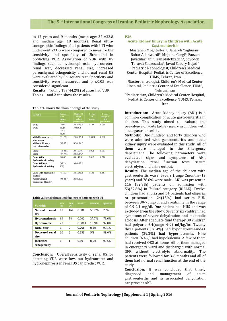
The 5rd International Congress of Iranian Pediatric Nephrology Association
Journal of Pediatric Nephrology | Supplement 1 | Spring 2016
to 17 years and 9 months (mean age: 32 ±33.8 and median age: 18 months). Renal ultra-sonographic findings of all patients with UTI who underwent VCUG were compared to measure the sensitivity and specificity of Ultrasound in predicting VUR. Association of VUR with US findings such as hydronephrosis, hydroureter, renal scar, decreased renal size, increased parenchymal echogenicity and normal renal US were evaluated by Chi square test. Specificity and sensitivity were measured, and p ≤0.05 was considered significant. Results: Totally 183(44.2%) of cases had VUR. Tables 1 and 2 can show the results. Table 1. shows the main findings of the study
Variable
Gender
% in
females-
males
Mean ±SD
of age
at first
presentation
P value
for
gender
P value
for age at
first
presentation
VUR+
VUR-
(42.6-
53.2)
(57.4-
46.8)
23.2±25.5
39±38.1
0.121
0.0001
With Urinary tract
obstruction
Without Urinary
tract obstruction
(2-12.9)
(98-87.1)
20.6±55.8
32.4±34.2
0.0001 0.218
Stone+
Stone -
(13-22.5)
(87-77.5)
24.1 ±29.7
33.3±34.7
0.051 0.057
Cases With
dysfunctional voiding
Cases Without
dysfunctional voiding
(0.8-0)
(99.2 -
100)
49 ±40.6
30.6±33.2
0.016 0.006
Cases with neurogenic
bladder
Cases without
neurogenic bladder
(6-11.3)
(94-88.7)
33.1±46.3
31.8±33.1
0.138 0.861
Table 2. Renal ultrasound findings of patients with UTI
Variable
VUR +(N)
VUR –(N)
P value
Sensitivity
Specificity
Normal renal
US
101 164 0.001 55.2 % 29%
Hydronephrosis 69 54 0.002 37.7% 76.6%
Hydroureter 20 5 0.0001 10.9% 97.8%
Renal scar 1 2 0.706 0.5% 99.1%
Decreased renal
size
10 6 0.133 5% 89.6%
Increased
echogenicity
1 1 0.89 0.5% 99.5%
Conclusion: Overall sensitivity of renal US for detecting VUR were low, but hydroureter and hydronephrosis in renal US can predict VUR.
P36 Acute Kidney Injury in Children with Acute
Gastroenteritis Mastaneh Moghtaderi1, Bahareh Yaghmaii1, Bahar Allahwerdi2, Mojtaba Gorgi3, Faezeh Javadilarijani1, Iran Malekzadeh3, Seyedeh Taravat Sadrosadat3, Javad Sabery Nejad³
1Pediatric Nephrologist, Children’s Medical Center Hospital, Pediatric Center of Excellence,
TUMS, Tehran, Iran 2Gasteroentrologist, Children’s Medical Center Hospital, Pediatric Center of Excellence, TUMS,
Tehran, Iran 3Pediatrician, Children’s Medical Center Hospital,
Pediatric Center of Excellence, TUMS, Tehran, Iran
Introduction: Acute kidney injury (AKI) is a common complication of acute gastroenteritis in children. This study aimed to evaluate the prevalence of acute kidney injury in children with acute gastroenteritis. Methods: One hundred and forty children who were admitted with gastroenteritis and acute kidney injury were evaluated in this study. All of them were managed in the Emergency department. The following parameters were evaluated: signs and symptoms of AKI, dehydration, renal function tests, serum electrolytes and urine output. Results: The median age of the children with gastroenteritis was2. 5years (range 2months–12 years) and 78.6% were male. AKI was present in 116 (82.9%) patients on admission with 53(37.8%) in ‘failure’ category (RIFLE). Twelve children had anuria and 54 patients had oliguria. At presentation, 24(15%) had serum BUN between 30-75mg/dl and creatinine in the range of 0.9-2.1 mg/dl. One patient had HUS and was excluded from the study. Seventy six children had symptoms of severe dehydration and metabolic acidosis. After adequate fluid therapy 30 children had polyuria 6.4(range 4-9) ml/kg/hr. Twenty three patients (16.4%) had hyponatremiaand41 patients (29.2%) had hypernatremia. Nine children (6.4%) had hypokalemia. A few of them had received ORS at home. All of them managed in emergency ward and discharged with normal GFR without electrolyte abnormality. The patients were followed for 3-6 months and all of them had normal renal function at the end of the study. Conclusion: It was concluded that timely diagnosed and management of acute gastroenteritis and its associated dehydration can prevent AKI.
The 5rd International Congress of Iranian Pediatric Nephrology Association
Journal of Pediatric Nephrology | Supplement 1 | Spring 2016
P37 Prevalence of Idiopathic Hypercalciuria in Children with Urinary symptoms in Bandar
Abbas, 2014 Maryam Esteghamati1, Kambiz ghasemi1, Marie
Nami2, Fatemeh Saburi3
1Pediatric Nephrologist, Department of Nephrology, Hormozgan University of Medical
Sciences (HUMS), Bandar Abbas, Iran. 2General Physician, Department of Nephrology,
Hormozgan University of Medical Sciences (HUMS), Bandar Abbas, Iran.
3Resident of Pediatric, Department of Nephrology, Hormozgan University of Medical
Sciences (HUMS), Bandar Abbas, Iran. Introduction: Idiopathic hypercalciuria (IH) is a disorder that can present with urinary symptoms. Its importance is due to high prevalence, recurrent urinary tract infection, stone formation, and different urinary symptoms. This study aimed to determine the prevalence of IH in children with urinary symptoms. Methods: This descriptive cross-sectional study was conducted on 321 children with different urinary symptoms aged between 2 months to 14 years old. Random morning urine sample was taken two times and assessed for Ca to creatinine ratio. Hypercalciuria was defined as urinary calcium/creatinineratio> 0.8 in infants below 6 months, > 0.6 in infants 6-12 months, and > 0.2 in children older than two years of age. Results: Among 321 children, 153 (47.7%) had IH. The mean age of the children with IH was 55.20±43.71 months. Prevalence of IH was 48.3% in children with urinary tract infection, 54.9% and 53.6% in children with microscopic and macroscopic hematuria, respectively. The prevalence of IH in children with dysuria, frequency, and kidney stone was 52.1%, 51.8%, and 49.1%, respectively. IH was present in 28.6% and 37.5% of children with nocturnal and diurnal urinary incontinence, respectively. There was no significant correlation between urinary symptoms and IH (P>0.05). Conclusion: Hypercalciuria can present with different urinary symptoms. Therefore, it is recommended to check urinary calcium excretion in children with unexplained urinary symptoms. P38 Therapeutic Effects of Zinc Supplementation
in Children with Urinary Tract Infection ParsaYousefichaijan
Department of Pediatric Nephrology, Arak University of Medical Sciences, Amir Kabir
Hospital, Arak, Iran Introduction: Urinary tract infection (UTI) is one of the most common diseases of the urinary tract system. This disease lacks characteristic signs and symptoms and occasionally occurs with non-specific symptoms such as weight loss, anorexia, and growth failure. Zinc improves host immune-system response to many infections and has a significant role in immune system integrity. The aim of this study was to evaluate the effect of zinc supplementation on treatment of UTI in children. Methods: In this clinical trial study, 200 children with UTI who were admitted to Amir Kabir hospital (Arak, Iran) were randomly divided into two control and case groups. Two groups were matched in age, gender, urine laboratory profiles, and clinical signs and symptoms such as fever, dysuria and frequency. The control group received only routine treatment of UTI. The case group, in addition to routine treatment of UTI, received oral zinc sulfate syrup. Results: Findings showed that dysuria and frequency in zinc-supplemented subjects recovered significantly faster than control subjects, while the abdominal pain persisted longer. There were no significant differences in termination of fever and negative urine culture at 48h and 7-10 days after treatment between two groups. Conclusion: Zinc supplementation has a significant effect in ameliorating severe dysuria and urinary frequency in UTI, but it is not recommended when abdominal pain is present. P39
Clinical Signs, Causes and Risk Factors of Pediatric Chronic Kidney Diseases due to
Obstructive Uropathy: A Hospital Based Case Control Study
Parsa Yousefichaijan1, Fatemeh Dorreh2, Hasan Taherahmadi3, Fakhreddin Shariatmadari4,
Yazdan Ghandi5, Saeed Alinejad6
1Associate Professor of Pediatrics Nephrology, Department of Pediatric Nephrology, Amir Kabir
Hospital, Arak University of Medical Sciences, Arak, Iran.
2 Associated Professor of Pediatric, Arak University of Medical Sciences, Arak, Iran.
3 Assistant Professor of Pediatric, Arak University of Medical Sciences, Arak, Iran.
4Assistant Professor of Pediatric Neurology, Arak University of Medical Sciences, Arak, Iran.
The 5rd International Congress of Iranian Pediatric Nephrology Association
Journal of Pediatric Nephrology | Supplement 1 | Spring 2016
5Assistant Professor of Pediatric Cardiology, Arak University of Medical Sciences, Arak, Iran. 6Assistant Professor ofneonatology ,Arak University of Medical Sciences, Arak, Iran.
Introduction: Obstructive uropathies (OU) are one of the most common causes of chronic kidney disease (CKD). Early in gestation, severe obstruction results in renal dysplasia. CKD is a major worldwide increasing health problem and often remains asymptomatic to reach end-stage renal disease (ESRD). The current study aimed to determine the epidemiologic characteristics and risk factors of CKD due to obstructive uropathy in patients younger than 11 years. Methods: In a hospital based case-control study, 179 cases with OU and CDK who had age less than 11 years were compared with 181 children without CKD. CKD was defined as renal injury and/or a glomerular filtration rate <60 mL/min/1.73 m2 for >3 mo. The information was collected using the questionnaire and clinical tests. Data analysis was performed by using chi-square, univariate and multivariate logistic regression. Results: Fetal hydronephrosis, fever, chills, and urinary tract infection were the most common clinical sign. The most common causes for referral of the CDK patients with OU were urinary tract infection (39.5%) and growth failure (12.9%). Ureteropelvic junction obstruction was the most common cause of CKD due to OU. Maternal age>35years olds, mother job, household income, growth failure, type of feeding in infancy (bottle feeding), percentile of maternal BMI>30kg/m2 (obese) during pregnancy, diagnosis of fetal hydronephrosis before 20 weeks of gestational age, and gestational age<37 weeks (preterm), chronic hypertension in mother during pregnancy, maternal exposer to smoking (passive smokers) were significant predictors of CKD in children with obstructive uropathy after controlling the effect of other variables. Conclusion: It was concluded that there were several preventable risk factors for CKD in patients with obstructive uropathies. Therefore, more attention is essential for early diagnosis and management of these risk factors to prevent CKD in children with obstructive uropathies. Further studies with larger sample size and considering more potential confounding variables may be needed.
P40 Comparison of Amiloride and
Hydrochlorothiazide for Treatment of Edema in Children with Nephrotic Syndrome
Soheila Mahdavynia1, Hasan Otukesh1, Rozita Hoseini1
1Pediatric Nephrology Department, Ali Asghar Hospital, Iran University of Medical Sciences,
Tehran, Iran
Introduction: Edema is a common clinical symptom in children with nephrotic syndrome. This study aimed to assess the effectiveness and safety of oral amiloride for treatment of edema in pediatric patients with idiopathic nephrotic syndrome. Methods: A randomized trial of amiloride vs hydrochlorthiazide was performed in 34 patients. The mean age was 4.3±0.7 years old. After a 2 weeks washout of diuretic therapy, nephrotic children with edema were randomized into two groups receiving amiloride and hydrochlorthiazide. The effect of diuretics was assessed by the amount of weight reduction and the measurement of urinary fractional excretion of sodium during the first three days of diuretic therapy. The primary end point was the decrease in body weight. Results: Thirty four patients were enrolled for this trial (17 patients in amiloride group and 17 patients in hydrochlorthiazide group). The mean weight loss during the first three days of diuretic therapy was 1.3±0.65 kg in amiloride group and1.19±0.4 kg in hydrochlorthiazide group (P=0.55). The mean of maximum urinary fractional excretion of sodium during the first three days of diuretic therapy was2.1±0.65% in amiloride group and 1.8±1% in hydrochlorthiazide group. Conclusion: There was not any difference between amiloride and hydrochlorthiazide in reducing weight and increasing urinary fractional excretion of sodium in children with nephrotic syndrome. P41
Awareness of Parents about Different Modalities for Renal Replacement Therapy: A
Pilot Survey Leila Ghafari1, Haiedeh Mamianloo2, Nakysa
Hooman3
1Register Nurse of Dialysis, Pediatric Nephrology, Ali-asghar Children hospital, Iran University of
Medical sciences 2 Faculty Member of Islamic Azad University,
Tehran Medical Branch
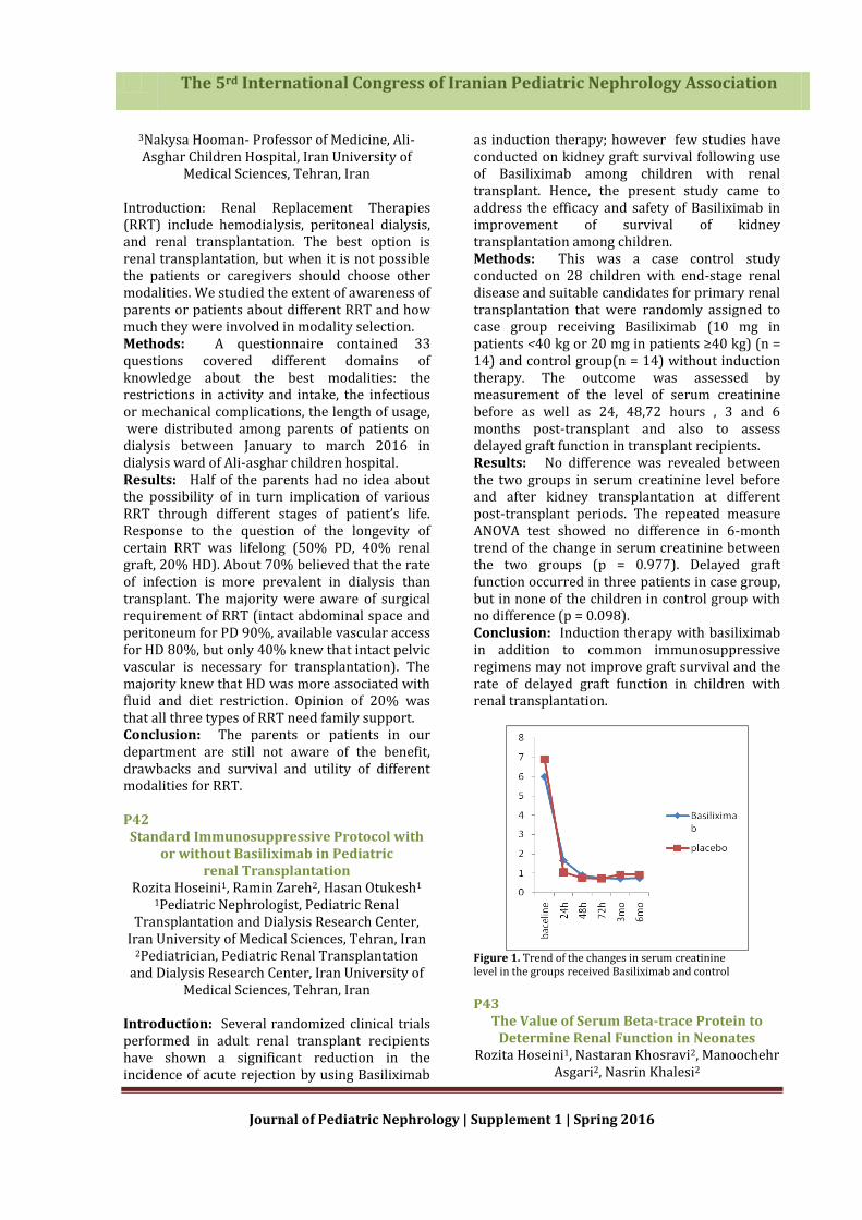
The 5rd International Congress of Iranian Pediatric Nephrology Association
Journal of Pediatric Nephrology | Supplement 1 | Spring 2016
3Nakysa Hooman- Professor of Medicine, Ali-Asghar Children Hospital, Iran University of
Medical Sciences, Tehran, Iran
Introduction: Renal Replacement Therapies (RRT) include hemodialysis, peritoneal dialysis, and renal transplantation. The best option is renal transplantation, but when it is not possible the patients or caregivers should choose other modalities. We studied the extent of awareness of parents or patients about different RRT and how much they were involved in modality selection. Methods: A questionnaire contained 33 questions covered different domains of knowledge about the best modalities: the restrictions in activity and intake, the infectious or mechanical complications, the length of usage, were distributed among parents of patients on dialysis between January to march 2016 in dialysis ward of Ali-asghar children hospital. Results: Half of the parents had no idea about the possibility of in turn implication of various RRT through different stages of patient’s life. Response to the question of the longevity of certain RRT was lifelong (50% PD, 40% renal graft, 20% HD). About 70% believed that the rate of infection is more prevalent in dialysis than transplant. The majority were aware of surgical requirement of RRT (intact abdominal space and peritoneum for PD 90%, available vascular access for HD 80%, but only 40% knew that intact pelvic vascular is necessary for transplantation). The majority knew that HD was more associated with fluid and diet restriction. Opinion of 20% was that all three types of RRT need family support. Conclusion: The parents or patients in our department are still not aware of the benefit, drawbacks and survival and utility of different modalities for RRT. P42
Standard Immunosuppressive Protocol with or without Basiliximab in Pediatric
renal Transplantation Rozita Hoseini1, Ramin Zareh2, Hasan Otukesh1
1Pediatric Nephrologist, Pediatric Renal Transplantation and Dialysis Research Center,
Iran University of Medical Sciences, Tehran, Iran 2Pediatrician, Pediatric Renal Transplantation
and Dialysis Research Center, Iran University of Medical Sciences, Tehran, Iran
Introduction: Several randomized clinical trials performed in adult renal transplant recipients have shown a significant reduction in the incidence of acute rejection by using Basiliximab
as induction therapy; however few studies have conducted on kidney graft survival following use of Basiliximab among children with renal transplant. Hence, the present study came to address the efficacy and safety of Basiliximab in improvement of survival of kidney transplantation among children. Methods: This was a case control study conducted on 28 children with end-stage renal disease and suitable candidates for primary renal transplantation that were randomly assigned to case group receiving Basiliximab (10 mg in patients <40 kg or 20 mg in patients ≥40 kg) (n = 14) and control group(n = 14) without induction therapy. The outcome was assessed by measurement of the level of serum creatinine before as well as 24, 48,72 hours , 3 and 6 months post-transplant and also to assess delayed graft function in transplant recipients. Results: No difference was revealed between the two groups in serum creatinine level before and after kidney transplantation at different post-transplant periods. The repeated measure ANOVA test showed no difference in 6-month trend of the change in serum creatinine between the two groups (p = 0.977). Delayed graft function occurred in three patients in case group, but in none of the children in control group with no difference (p = 0.098). Conclusion: Induction therapy with basiliximab in addition to common immunosuppressive regimens may not improve graft survival and the rate of delayed graft function in children with renal transplantation.
Figure 1. Trend of the changes in serum creatinine level in the groups received Basiliximab and control
P43
The Value of Serum Beta-trace Protein to Determine Renal Function in Neonates
Rozita Hoseini1, Nastaran Khosravi2, Manoochehr Asgari2, Nasrin Khalesi2
The 5rd International Congress of Iranian Pediatric Nephrology Association
Journal of Pediatric Nephrology | Supplement 1 | Spring 2016
1Pediatric Nephrologist, Pediatric Renal Transplantation and Dialysis Research Center,
Iran University of Medical Sciences, Tehran, Iran 2Neonatologist, Iran University of Medical
Science, Tehran, Iran Introduction: Compared to the conventional methods, serum beta-trace protein (BTP) has been shown to be more helpful for estimating glomerular filtration rate, however its value remained unclear in neonates. The present study aimed to investigate the normal range of serum BTP and its value to estimate glomerular filtration rate in term neonates. Methods: This cross-sectional study was conducted on 50 normal term neonates without underlying cardiovascular or renal disorders who were admitted to Ali Asghar hospital in 2013. Serum BTP was measured using an automated nephelometric immunoassay. The value of glomerular filtration rate was assessed by the creatinine based Schwartz equation. Results: The mean level of serum BTP was 0.41 ± 0.11 mg/l ranged 0.19 to 0.92 mg/l. There was a positive correlation between the inverse of BTP and eGFR measured by the Schwartz equation (r = 0.383, p = 0.006) and also between the inverse of BTP and the inverse of creatinine(r = 0.365, p = 0.009). Conclusion: Measurement of serum BTP can be a reliable tool for detecting renal function in neonates. We need more studies to design a suitable formula for GFR estimation based on serum BTP in neonates. P44
Renal Function in Children with Malignancy under Chemotherapy with Platinum
Afshin Ghalehgolab Behbahan1 , Azim Rezamand2,
Aydin Tabrizi3 , Roghaye Rahimi Asl4
1Associate Professor of Pediatric Nephrology, Pediatric Health Research Center, Tabriz
University of Medical Sciences, Tabriz, Iran. 2Associate Professor of Pediatric Hematology-Oncology, Tabriz Children’s Hospital, Tabriz
University of Medical Sciences. 3Pediatric Health Research Center, Tabriz
University of Medical Sciences. 4Tabriz Children’s Hospital, Tabriz University of
Medical Sciences Introduction: Chemotherapy is the most used modality of standard anti-cancer therapy worldwide. The platinum family is a common family of drugs that are generally known as broad-spectrum, highly effective anti-tumor
chemotherapeutic agents. However, these frequently used drugs may be accompanied by ominous adverse effects such as serious renal dysfunction. This study aimed to evaluate the effect of platinum drugs family on glomerular and tubular functions of the kidney in children. Methods: In a cross-sectional and descriptive/analytical study, we evaluated and followed 32 children with malignancies that were referred to Tabriz Children’s Hospital – Oncology Dept. in a period of one year (from September 2010 to September 2011) that were treated with platinum chemotherapeutics. All patients received the reno-protective regimen that includes: over hydration (as forced-alkaline diuresis), intra-venous Mg-sulfate supplementation and N-acetyl Cysteine infusion, from one day before to last day of platinum administration. Corrected glomerular filtration rate (as can be calculated by Schwartz formula: GFR=δL/Scr, serum Na+, K+, Ca2+, P, Mg2+, Clˉ, Uric acid and blood biochemical indices were measured for each case at three distinct time frames: before initiation of any treatment (as baseline), just after termination of treatment course (that usually lasted for 3 months), and 3 months later (during follow-up period). Additionally, results of urine and blood gas analyses were included in our investigation. Statistical analyzes of collected data was carried out by the SPSS 17.0 software using “Paired-sample t-test” and “Pearson Correlation”. Results: The results showed that during treatment course and then through follow-up period serum levels of sodium and creatinine rose, but on the contrary potassium, magnesium, albumin and GFR declined (p-value <0.05). On the opposite, changes of other parameters were statistically non-significant. Conclusion: We concluded that platinum drug family, especially cisplatin, can induce severe and even permanent adverse effects on patients’ kidney function; serum creatinine level increases as GFR decreases; moreover, detrimental effects on electrolyte regulatory systems (especially magnesium regulation) may occur. These events are somehow irreversible in most cases. However, more studies with larger sample size are needed in future to attain more reliable results. P45
Can We Recommend DMSA Scan as Replacement for Cystography in Children
with a First Episode of Acute Pyelonephritis?

The 5rd International Congress of Iranian Pediatric Nephrology Association
Journal of Pediatric Nephrology | Supplement 1 | Spring 2016
Neamatollah Ataei1, Arash abbasi1, Behnaz Bazargani1, Masteneh Moghtaderi1, Fatemeh Ataei1
1Pediatric Chronic Kidney Disease Research Center, Children’s Hospital Medical Center, Tehran
University of Medical Sciences, Iran Introduction: Urinary tract infection (UTI) is a common infection in infants and children. It may lead to irreversible changes in renal parenchyma and long-term complications. Early and definite diagnosis with using scintigraphy with technetium-99m - labeled dimercaptosuccinic acid (DMSA) scan and early treatment may decrease or prevent development of renal parenchymal lesions. DMSA scan has been advocated as a method of choice for the assessment of renal parenchymal involvement after first-time acute pyelonephritis (APN). The main aim of this study was to evaluate the usefulness of DMSA scan performed following a first episode of APN as a predictor of patient outcome, and to identify children at risk of vesicoureteral reflux (VUR). Methods: In a prospective study from November 2005 to March 2007, 102 children (93 girls and 9 boys, median age 2.92±2. 85 years, range 1 month to 12 years) with first-time APN were enrolled in the study. All children studied with DMSA scan and ultrasound (US) within 3 days of admission. Radionuclide cystography (RNC) orvoidingcystourethrography (VCUG) was performed in 100 children when urine culture became negative. The sensitivity and specificity of the DMSA scan for detecting renal VUR was investigated. Results: Renal lesions on the DMSA scan was found in 178 (88%) out of 203 renal units during the acute phase. DMSA scan results were abnormal in 155 kidneys (85.2%), without VUR and in 38 out of 40 kidneys (95%), with VUR (P >0.05). Normal DMSA scan was found in 25 renal units (12.3%) .A normal DMSA scan occurred in none of children with dilating VUR (grade 3 or 4).Reflux was found in 40 renal units (19.7%). Kidneys with moderate to severe reflux were more likely to have severe renal involvement (P=0.001). The sensitivity and specificity of abnormalities on DMSA renal scan for detecting the presence of VUR on voiding cystography were 92% (95% confidence interval [CI] = 70.03 to 97.8% ) and 11.8% [95% CI = 7.8% to 17.4%), respectively]. The positive predictive value (PPV) of abnormalities on DMSA renal scanning for detecting the presence of VUR was 12.8% (95% confidence interval [CI]=27% to 46%). The negative predictive value (NPV) of abnormalities
on DMSA renal scanning for detecting the presence of VUR on VCUG was 91.3% (95% confidence interval [CI] = 73.2% to 97.6%) .The likelihood ratios (LRs) positive for summarizing the utility of DMSA for ruling out VUR on VCUG was 1.04 (95% CI = 1. 02 to 1.64). Conclusion: High sensitivity and high NPV of DMSA scan for the diagnosis of VUR in children with a first febrile UTI indicates that a normal DMSA can exclude VUR, especially in patients with high-grade VUR. In children with a first episode of
febrile UTI, renal cortical scintigraphy with DMSA might be considered as a first line of imaging evaluation. P46
Chronic Neurological Complications in Children with Hemolytic Uremic Syndrome in
Ali Asghar and Mofid Hospital;2001-2015 Zafaranloo N1, Tavasoli A1, Otukesh H1, Hooman
N1, Hoseini R1
1Hazrat-e Ali Asghar Children Hospital, Iran University of Medical Sciences, Tehran-Iran
Introduction: Hemolytic Uremic Syndrome (HUS) is characterized by the triad ofmicroangiopathic hemolytic anemia, thrombocytopenia and Acute Kidney Injury (AKI). Most of the patients with HUS have Central Nervous System (CNS) manifestations. CNS involvement may be acute or proceed into chronic phase. We conducted this study to evaluate the chronic neurologic sequelae in children with HUS. Methods: This retrospective study was carried out on children with HUS who were admitted in two children hospitals between March 2001-March 2015.We examined patients for detection of the chronic neurological signs of HUS (6 months after the beginning of the disease). Results: There was 58 patients (Girls: 43.1%, Boys: 56.9%). the mean age was 43 months (5 months to14 years). CNS manifestations were seen in 53.4% of patients: 20.7% in acute phase, 13.8% in chronic phase and 19% in both phases. Atypical HUS (D-) and Typical HUS(D+)were seen in [55.2%] and [44.8%] respectively . In HUS (D+) patients, neurologic signs were more frequent in acute phase[50%][P-value=0.15], and 30% of them were accompanied with AKI. Whereas, chronic signs were more common in HUS(D-) [73.3%][P-value=0.15], and 70% had AKI. There was significant correlation between chronic CNS signs and hypertension [Pvalue=0.03]. In acute phase, seizure and coma respectively were the most common neurological

The 5rd International Congress of Iranian Pediatric Nephrology Association
Journal of Pediatric Nephrology | Supplement 1 | Spring 2016
signs. In chronic phase headache and seizure were the most common, respectively. Conclusion: Chronic neurological signs were more associated with atypical HUS and it was associated with hypertension. We should concentrate on neurological manifestations at follow up visits in patients with HUS. Prospective studies are recommended for further evaluation of neurological complications in children with HUS. P47
Correlation between H. Pylori Infection and the Onset and Relapse of Idiopathic Steroid
Sensitive Nephrotic Syndrome: A Pilot Study Ali Derakhshan1, Farrokh Farrokhnia2, Mitra
Basiratnia1, Mohammad Hossein Fallahzadeh1
1Shiraz Nephrology Urology Research Center, Shiraz University of Medical Sciences, Shiraz, Iran 2Yasuj University of Medical Sciences, Yasuj, Iran
Introduction: The main problem in the management of childhood idiopathic nephrotic syndrome is relapse of the disease which occurs in the majority of patients. This study aimed to evaluate the effect of H.pylori infection on the onset and relapse of nephrotic syndrome. Methods: This pilot study was carried out on a group of children who were either known cases or new cases of steroid responsive nephrotic syndrome .New cases of nephrotic syndrome were enrolled after their initial response to steroid. Age matched controls were selected from healthy children. Stool antigen was checked from the cases and controls in a qualified laboratory. SPSS soft ware and Chi-Square Test were used for statistical analysis. Results: Forty children in the case group and 41 children in the control group were studied. Mean age in the case and control group was 5.4 and 5.9 years, respectively. Stool antigen was positive in 4 of the cases and 7 of the controls (p=0.86). H.pylori infection was confirmed with upper GI endoscopy in 3 patients in whom endoscopy was performed. Conclusion: This pilot study does not support a positive role for H.pylori infection for initiation or relapse of idiopathic childhood nephrotic syndrome. P48 Using Small Animal SPECT in the Evaluation of
Recovery Process of Kidney in an Animal Model of Ischemia/Reperfusion Injury
Hossein Fatemikia1, Zahra Sedaghat1, Kaveh Tanha2
1Physiology Department, Medical School, Bushehr University of Medical Sciences, Bushehr,
Iran 2 Persian Gulf Nuclear Medicine Research Center, Bushehr University of Medical Sciences, Bushehr,
Iran
Introduction: Kidneys are susceptible to ischemia and reperfusion (I/R) injury in different clinical situations which may cause long term renal impairment. Single photon emission computed tomography (SPECT)is a technique for the long time measurement of the relative renal function. In this study we used small animal SPECTto assess the renal repair capacity of rats. Methods: Rat models of unilateral I/R were established by occlusion of the left renal pedicle with a bulldog clamp. At 24 hours, 1week and 3 weeks after I/R, 99mTc-DMSA was injected via tail vein and after 1 hour post-injection, the rats were scanned with gamma camera to assess the renal function. Results: Twenty four hour after reperfusion, the 99mTc-DMSA uptake in the injured kidney was about half of the normal kidney. After 1wk 55%, and after 3wks 70% of renal function was retrieved. Moreover, 3 weeks after ischemia very strong correlation between 99mTc-DMSA uptake and weight of dissected kidneys was observed. Conclusion: In summary, our study showed good potentials of 99mTc-DMSA scan by small animal SPECT as a promising non-invasive method for evaluation of kidney restoration after I/R injuries. P49
Characterizing the Development of Acute Lung Injury from Acute Kidney Injury by Comparing Rats Subjected to Bilateral or Unilateral Nephrectomy and Bilateral or Unilateral Renal Ischemia/Reperfusion Zynab Karimi1,2, Seyed Mohammad Owji3, Farzaneh Ketabchi1, Seyed Mostafa Shid
Moosavi1,2
1Department of Physiology, Shiraz University of Medical Sciences, Shiraz, Iran.
2Shiraz Nephro-Urology Research Center, Shiraz University of Medical Sciences, Shiraz, Iran.
3Department of Pathology, Shiraz University of Medical Sciences, Shiraz, Iran.
Introduction: Acute kidney injury (AKI) induces acute lung injury (ALI) via releasing injurious factors or impairing clearance of systemic
The 5rd International Congress of Iranian Pediatric Nephrology Association
Journal of Pediatric Nephrology | Supplement 1 | Spring 2016
mediators. To determine the links between AKI and ALI, different experimental models of renal ischemia with uremia (bilateral renal ischemia/reperfusion, BIR), renal ischemia without uremia (unilateral renal ischemia/reperfusion, UIR), uremia without renal ischemia (bilateral nephrectomy, BNX), and without uremia and renal ischemia (unilateral nephrectomy, UNX) were used in this study. Methods: Ninety male Sprague-Dawley rats were divided into 6 groups. Animals had 1-h bilateral or 2-h unilateral renal ischemia followed by 24-h reperfusion in the BIR and UIR groups, respectively, and 24-h period following bilateral or unilateral nephrectomy in the BNX and UNX groups, respectively. There were also sham and control groups with and without sham-operation, respectively. Results: Plasma malondialdehyde and nitric oxide were elevated by BIR more than UIR, but not changed by UNX and BNX. UIR slightly increased plasma creatinine, whereas BIR and BNX largely increased plasma creatinine, urea, K+ and osmolality and decreased arterial HCO3
-, pH andCO2. UNX did not affect lung, and UIR caused small rise in interstitial neutrophils and fall in PaO2/FiO2. Both BIR and BNX induced ALI with equal capillary leak and macrophages infiltration, while there were more prominent lung edema and vascular congestion following BNX but severe neutrophils infiltration and reductions of PaO2/FiO2 and airway resistance following BIR. Conclusion: Acutely accumulated systemic mediators following renal failure alone vary from those due to renal failure with ischemic-reperfused kidneys and consequently they induce ALI with distinct characteristics. P50
Outcome of Kidney Transplantation in Children in Khorasan
Mohammad Esmaeili1, Yalda Ravanshad2, Anoush Azarfar1
1Department of Pediatrics, Faculty of Medicine, Mashhad University of Medical Sciences,
Mashhad, Iran 2Clinical Research Development Center, Ghaem
Hospital, School of Medicine, Mashhad University of Medical Sciences, Mashhad, Iran
Introduction: The overall incidence of end-stage renal disease in children has been steadily increasing during the past 30 years. Children cannot tolerate chronic dialysis for a long period of time. Renal transplantation is the treatment of choice for end-stage renal disease in children. It
is associated with a number of complications which can be characterized by the time of appearance after renal transplantation (immediate, early, or late). We aimed to evaluate the outcome of children with renal transplantation in Khorasan Province, 1379 to 1391. Methods: A retrospective study was performed on 52 children under 18 years old with renal transplantations between 1379 -1391 in Mashhad. We evaluated the final outcomes of the patients with renal transplantations according to the type of the graft (living related donor, living unrelated donor, and deceased donor), gender, age of the donors and recipients, graft function, serum creatinine and the time of renal transplantation. This study was done according to the questionnaire which was filled by the patients and their parents. Results: Reflux nephropathy (n=18) was the most common underlying disease. Thirteen percent of the patients died. Early and late complications were reported in 19.5% and 5.8 %, respectively (acute and chronic rejection, infection, cardiovascular disease, malignancy, and pyelonephritis). The mean age of the patients was 16.3 4.3 ± years and 52% were boys and 48% were girls. In our study, 8(%15.4) patients received kidneys from living-related donors, 16 (%30.8) from living-nonrelated donors, and 28 (%53.8) from deceased donors. Mean duration of transplantation was 4.8 years. At the end of the first post-transplant year the mean GFR was 82 ±14 ml/min/1.73 m2. Conclusion: Early and late complications of renal transplantation were low in Khorasan province and it was associated with favorable patient and graft survival. P51
Management of Polyoma Virus Infection in Pediatric Renal Transplantation
Mostafa Sharifian1, Nasrin Esfandiar1
1Pediatric Nephrology Research Center, Pediatric Infectious Research Center, School of Medicine,
Shaheed Beheshti University of Medical Sciences, Tehran, Iran
Introduction: Viral infections are frequent complications in transplant recipients. Up to now10 viruses have been detected in polyomavirus family such as BK, JC, SV40, WU, KI and Merkel cell polyoma virus which is detected in Merkel cell carcinomas. BK virus nephropathy is an important cause of renal transplant dysfunction, especially in

The 5rd International Congress of Iranian Pediatric Nephrology Association
Journal of Pediatric Nephrology | Supplement 1 | Spring 2016
patients with high levels of immunosuppression. Up to 85 percent of adults have reportedly serologic evidence of exposure to the virus, suggesting the presence of asymptomatic, latent infection. Reactivation occurs in 10–68% of transplanted individuals. Herein we present our experience in BK Virus infection and nephropathy in renal transplant children. Methods: Between January 1985 and July 2015, 437 kidney transplantations were performed in children under 15 years in Labbafinejad Hospital, Tehran. Immunosuppressive medications consisted of Prednisolone, Cyclosporine A or Tacrolimus and Mycophenolate Mofetil. Thirty seven patients received Basiliximab as adjunct induction therapy. BK Virus was tested in 120 blood and urine samples from 80 transplant patients who were in regular follow-up by Polymerase Chain Reaction (PCR). Decoy cells were also tested in symptomatic patients. Results: BK Virus particles were detected in 21 transplant children (26%) of whom 4 patients had Decoy cells in pathologic examination of urine and a dramatic rise in plasma creatinine. Immunosuppressive medications were reduced as the first step of treatment in 4 patients with BKN, and it was effective in 2 patients evidenced by reduction of creatinine. Cidofovir was used for third patient who was partially effective leaving a plasma creatinine of 1.9 mg/dl. Conclusion: BK Virus nephropathy should be considered as a cause of allograft dysfunction in children with renal transplantation. P52
122 Cases of Minimal Change Disease Diagnosed by Electron Microscopy in Shiraz
University of Medical Sciences Seyed Mohammad Owji1, Seyed Hossein Owji2,
Hamide Jabedarbashy3
1Associate professor, Department of Pathology, Shiraz University of Medical Sciences, Shiraz, Iran
2Student Research Committee, Shiraz University of Medical Sciences, Shiraz, Iran
3Technician of Electron Microscopy, Department of Pathology, Shiraz University of Medical
Sciences, Shiraz, Iran
Introduction: Minimal Change Disease (MCD) is the most frequent cause of nephrotic syndrome in children, but may occur at any age. The course is characterized by remissions and exacerbation. The purpose of this study was to determine the specific ultra-structural features of MCD and finding the criteria to differentiate it from early
stage of Focal Segmental Glomerulosclerosis (FSGS) by electron microscopy (EM). Methods: A total of 122 cases of MCD were diagnosed in our center by electron microscopy (LEO 906) in pathology department during 15 years. The age ranged from 2 years up to 72 years, mostly between 3-8 years. The specimen for EM were fixed in Glutarldehyde 3% and after routine processing and preparing Resin blocks ultra-thin sections (90nm) were stained by Lead citrate and Uranium acetate and studied and photography by EM. Results: The glomeruli in most patients appeared normal by light microscopy except for few patients that revealed minor degrees of mesangial expansion with mild increase in mesangial cellularity. The main electron microscopic findings were effacement of total foot processes of visceral epithelial cells of glomeruli and the basement membrane of capillary wall being covered by sheets of cytoplasm, which was swollen with prominent organelles and frequently containing small cystic space with different size. From the surface of the cell membrane of podocytes numerous microvillous projections were protruded. The cytoplasm of proximal convoluted tubule presented focal vacuolization. No tubular atrophy or interstitial fibrosis was observed. Conclusion: Electron microscopic study is mandatory for diagnosis of MCD to differentiate it from early stage of focal segmental glomeroulosclerosis which has poor response to therapy. MCD can never be diagnosed by light microscopy alone. So EM plays an important role in diagnosis. P53
Evaluating the Effectiveness of Adding Magnesium Chloride to Conventional Protocol
of Citrate Alkali Therapy in Patient with Kidney Stone
Alaleh Gheissari1, Amin Ziaee2, Hassan Niroomand3
1Department of Pediatric Nephrology, Faculty of Medicine, Isfahan University of Medical Sciences,
Isfahan, Iran 2Faculty of Medicine, AJA University of Medical
Sciences, Tehran, Iran, 3Department of Urology, Faculty of Medicine, AJA
University of Medical Sciences, Tehran, Iran Introduction: Potassium citrate (K-Cit) is one of the therapeutic modalities broadly used in patients with Urolithiasis. However, in some cases with calcium oxalate (CaOx) stones

The 5rd International Congress of Iranian Pediatric Nephrology Association
Journal of Pediatric Nephrology | Supplement 1 | Spring 2016
significant response to alkaline therapy with K-Cit alone doesn’t occur. Considering the higher solubility of magnesium oxalate compared to calcium oxalate, a combination therapy with magnesium salts is crucial. There is scarce published data on the effect of magnesium chloride (Mg-Cl2) on urolithiasis in patients. Therefore, the goal of our study was to evaluate the effect of a combination of K-Cit - MgCl2 oral supplements, on urinary stone size. Methods: This study was performed on 70 asymptomatic urolithiasis cases. With the approval of the Ethics Committee of the Research Department of Isfahan University of Medical Sciences. The supplements included potassium citrate (K-Cit) and magnesium chloride (Mg-Cl2), purchased from MERCK Company, Germany. The patients were randomly divided in two groups. The urinary stone size by ultrasonography were measured in control group after prescribing K-Cit alone and the group was treated with combination of K-Cit and Mg-Cl2 for 4 weeks and also urinary parameters was measured in each group. Results: Hyperoxaluria and hypercalciuria were seen in 70% and 52% of patients, respectively. Initially the mean urinary stone size was measured in each group and there were no significant differences. But we found a significant decrease in urinary stone size in group which was treated with combination of K-Cit and Mg-Cl2 for 4 weeks in comparison with control group treated with K-Cit alone in the same duration of therapeutic course, p<0.05. All ultrasonography were performed by one radiologist and device. Soft stool was reported by 8 patients, but not severe enough to terminate the medications. Conclusion: Our results suggested that a combination of K-Cit and Mg-Cl2 chloride is more effective in decreasing urinary stone size than K-Cit alone. P54
Minirin versus Oxybutynin for Nocturnal Enuresis in Children in Bandar Abbas in 2014
Kambiz Ghasemi
Hormozgan University of Medical Sciences, Bandar Abbas, Iran
Introduction: Nocturnal enuresis is among the most common disorders in children. Several pharmacological and non-pharmacological treatments are available for nocturnal enuresis. Studies for reaching the best pharmacological treatment for this disorder are continuing. The aim of current study was to compare the efficacy
and safety of Minirin and oxybutynin for treatment of nocturnal enuresis in children in Bandar Abbas in 2014. Methods: This randomized controlled trial was done in 2014 on 66 children with nocturnal enuresis who were more than 5 years old. Patients were randomly assigned into 120 µg Minirin or 5 to 10 mg oxybutynin and were followed 1, 3, and 6 months later for treatment response. Study outcomes were frequency of nocturnal enuresis, urinary incontinency, urgency, and frequency. Data were analyzed using SPSS software. Results: There were no significant difference between two groups in sex, age, place of residence, and parents’ education (p<0.05). Nocturnal enuresis, incontinency, urgency, and frequency of nocturnal enuresis was significantly lower in minirin in comparison to oxybutynin group after one and 3 months (p<0.05). Also constipation and xerostomia were more frequent among oxybutynin group after 1, 3, and 6 months (p<0.01). Also blurred vision was more frequent among oxybutynin group after 3 months (p<0.01). After 6 months the frequency of nocturnal enuresis and its frequency was higher in oxybutynin group in comparison to minirin group (p<0.05). Conclusion: Our results showed that the prevalence of nocturnal enuresis is significantly lower in minirin group in comparison to oxybutynin group after one year. After 3 and 6 months no children in minirin group had nocturnal enuresis. We recommend using minirin for treatment of nocturnal enuresis. More studies are needed for assessment of long-term side effects of minirin and oxybutynin in treatment of nocturnal enuresis. P55
Modeling the Distribution of Urolithiasis Prevalence under Projected Climate Change
in Iran Ahmad Shajari1, Mohammad Mousaei Sanjerehei1
1Pediatric Nephrologist, Azad University of Yazd, Yazd, Iran
Introduction: Although studies support a positive correlation between temperature and stone risk, the precise relationship between these factors has not been elucidated. Methods: We modeled the current distribution of urolithiasis prevalence in Iran using 26 bioclimatic, climatic and topographic variables based on two multivariate linear regression
The 5rd International Congress of Iranian Pediatric Nephrology Association
Journal of Pediatric Nephrology | Supplement 1 | Spring 2016
models in geographical information system. The impact of climate change on the stone prevalence was predicted under the projections of GFDL-ESM2G, CCSM4 and HadGEM2- ES climate models by mid-century (2050). Results: Extraterrestrial radiation and isothermality in the first regression model and annual mean temperature, precipitation seasonality and isothermality in the second model were the significant (P < 0.01) predictors of urolithiasis prevalence. Both regression models provided good estimates of the stone prevalence (R2 > 0.9) and determined a mean urolithiasis prevalence of 6 %(range of 1.5-10.8 %) in Iran. The climate change under the projections of GFDL-ESM2G, CCSM4 and HadGEM2-ES models can, respectively, lead to an average increase of 5.7, 4.3 and 9 % in the urolithiasis prevalence based on the second regression model by 2050. Conclusion: The highest increase of the prevalence will occur in the west, northwest and southwest provinces of the country. Predicting the impact of climate change on climate-related diseases can be useful for effective preventive measures.
P56 The Miracle of Transplantation: A Case Report
Ali Derakhshan Shiraz Nephrology Urology Research Center,
Shiraz Universityof Medical Sciences, Shiraz, Iran
Introduction: Kidney transplantation is the optimal and life saving treatment in children with end stage kidney disease. The concept of being life saving is seen in this case report. Case report: N.L. a 7-year-old boy (weight= 19 kg, height= 112 Cm) with end-stage renal disease due to FSGS who was living in one the villages near Lamerd and was under hemodialysis in Lamerd, Fars province, Iran and had controlled hypertension with medication, presented with malaise, weakness and gradually became unconscious without any movement; since his parents and the neighbors assumed he was dead, they prepared a grave to bury his body. As they decided to carry him to the cemetery, his body started to move; therefore, he was transferred to the emergency department of the local hospital and subsequently was transferred to Shiraz Nemazee Hospital. Right in the depth of his parents’ despair, as the patient’s name was on the transplant waiting list, the transplant center called his father that night, and since the patient
was already hospitalized in the pediatric emergency ward, he received kidney transplant at the exact same night. Now, after twelve and a half years, the transplanted kidney functions normally with the patient’s weight and height being 63kg and 178Cm, respectively. Conclusion: Kidney transplantation is considered the best treatment for many children with end stage kidney disease. P57
A Case Report of Nephrotic Syndrome with Hemorrhagic Thrombosis of Central Nervous
System Hadi Sorkhi
Non Communicable Pediatric Disease Research Center, Department of Pediatric Nephrology,
Amirkola Children Hospital, Babol Medical University, Iran
Introduction: Nephrotic syndrome (NS) is defined by nephrotic range proteinuria, generalized edema, hypoalbuminemia and hyperlipidemia. It is a common pediatric nephrologic disease. The risk of stroke may be high especially in new cases and during relapses. Cerebral vein thrombosis is a rare complication of nephrotic syndrome that may present with headache, decreased level of consciousness and convulsion. We report a known case of NS with hemorrhagic thrombosis. Case report: An 8 year old boy with previous history of NS was admitted with generalized edema, vomiting, oliguria, and high grade fever. He had headache and decreased level of consciousness since 3 days ago. After admission, urinalysis showed 3+ proteins, blood urea nitrogen and creatinine were 20 mg/dL and 0.58 mg/dL, respectively. He had hemoconcentration (hemoglobin: 17g/dL, hematocrit: 52.5%). Serum sodium (136 mEq/L) and potassium (3.8 mEq/L) were normal. PT and PTT were in normal range. He did not have any increase in creatinine during admission and his headache was alleviated after two days of admission. His brain images revealed hemorrhagic thrombosis. The patient had no neurologic focal sign and there was not any abnormal neurological examination. The conservative management was done with correction of water and electrolytes. Conclusion: Because of the nonspecific signs and symptoms of central nervous system thrombosis in children with NS, the diagnosis may occur with delay. Cerebral vein thrombosis should be considered in patients with history of NS (especially in new cases and during relapses)
The 5rd International Congress of Iranian Pediatric Nephrology Association
Journal of Pediatric Nephrology | Supplement 1 | Spring 2016
and prevention of hemoconcentration is very important to decrease the risk of thrombosis. P58
Predictive Accuracy of Urinary Neutrophil Gelatinase Associated Lipocalin (NGAL) for
Renal Parenchymal Involvement in Children with Acute Pyelonephritis
Kambiz Ghasemi1, Maryam Esteghamati1, Sara Borzoo2, Erfan Parvaneh3, Samira Borzoo4
1Assistant Professor, Department of Pediatric Nephrology, Faculty of Medicine, Hormozgan University of Medical Sciences, Bandar Abbas,
Iran 2Pediatric Resident, Department of Pediatrics, Faculty of Medicine, Hormozgan University of
Medical Sciences, Bandar Abbas, Iran 3Medical Students, Department of Pediatrics,
Faculty of Medicine, Isfahan University of Medical Sciences, Isfahan, Iran
4M.Sc., Rehabilitation Administration, MPH Student of Health Policy Research Center, Shiraz
University of Medical Sciences, Shiraz, Iran
Introduction: Urinary tract infections (UTIs) are among the most prevalent infections in children. Early and accurate detection of renal parenchymal involvement in UTI is necessary for decision making and determining treatment strategies. The aim of this study was to assess the predictive accuracy of urinary neutrophil gelatinase-associated lipocalin (NGAL) for renal parenchymal involvement in children with acute pyelonephritis. Methods: This descriptive, cross-sectional study was conducted in 2014 on children who were admitted in children Hospital of Bandar Abbas, Hormozgan Province, Iran. The patients’ age ranged between two months to 14 years. Urine samples were taken for urinary NGAL, urine cultures, and urinalyses. Blood samples were collected for blood works including CBC, C-reactive protein (CRP) and erythrocyte sedimentation rate (ESR) tests. All patients underwent a dimercaptosuccinic acid (DMSA) scan as the gold standard test for diagnosis of acute pyelonephritis. SPSS software was used to analyze the data. Results: Among the participants in the study, 29 were male (32%), and 60 were female (68%). The mean age of the children who participated in the study was 2.99 ± 2.94 years. The results of the Kruskal-Wallis test showed a significant increase in the urinary NGAL level and CRP level, and higher DMSA scan grades (p < 0.001). The cutoff point of urinary NGAL > 5 mg/l, had the
negative predictive value (NPV) of 76.3%, the specificity of 97.83%, the positive predictive value (PPV) of 96.7%, and the sensitivity of 67.4%. Conclusion: Urinary NGAL is not sensitive enough for the prediction of renal parenchymal involvement, but it is a specific marker for detection of acute pyelonephritis. P59
Remote Per-conditioning Reduces the Oxidative Stress in the Renal
Ischemia/Reperfusion Injury: The short-time Effect
Zahra Sedaghat1, Mehri Kadkhodaee2, Behjat Seifi2
, HosseinFatemikia1 1Physiology Department, Medical School,
Bushehr University of Medical Sciences, Bushehr, Iran
2Physiology Department, Medical School, Tehran University of Medical Sciences, Tehran, Iran
Introduction: Renal ischemia/reperfusion (I/R) injury is a common clinical problem associated with significant mortality and morbidity. One newly described strategy to reduce this damage is remote per-conditioning (RPeC), in which short-time ischemia of a limb during renal ischemia reduces the I/R-induced kidney injury. However, its early-time effects and the oxidative status of renal tissue are still unknown. Methods: Male Rats were subjected to right nephrectomy and randomized into the shamoperated, IR and RPeC groups. According to different reperfusion time, each group was divided into 3 subgroups(3h, 6h and 24h). Renal ischemia was applied by clamping of the left renal pedicle. RPeC was performed by four cycles of 5-min IR of the left femoral artery right at the beginning of renal ischemia. Blood and kidney samples were collected at each reperfusion time point for measurement of creatinine (Cr) levels as well as assessment of malondialdehyde (MDA) and histopathological study. Results: Forty five min of renal ischemia followed by 3h, 6h and 24h reperfusion resulted in significant increases in Cr and MDA levels, and histological injury compared to the appropriate sham group. Three hours after reperfusion, these parameters were lower in the RPeC group compared to the 3h IR group, but they were not significant. However, these protective effects were significant after 6h and 24h in RPeC-treated animals compared to the respective IR groups. Conclusion: the protective effect of RPeC against renal I/R appears at the early time after
The 5rd International Congress of Iranian Pediatric Nephrology Association
Journal of Pediatric Nephrology | Supplement 1 | Spring 2016
reperfusion. This protection appears to be significant after 6h and is more pronounced in 24h after reperfusion. The mechanism of this protective strategy may involve decreased lipid peroxidation in the renal tissue. P60 Penile Tourniquet Injury Due to a Coil of Hair
Anoush Azarfar Department of Pediatrics, Faculty of Medicine,
Mashhad University of Medical Sciences, Mashhad, Iran
Introduction: Penile hair tourniquet syndrome (PHTS) is characterized by advanced penile strangulation caused by a coil of hair. This condition has several potential complications, such as necrosis of glans penis, urethrocutaneous fistulae and complete urethral transection. We report on a 4 years and 6 months old circumcised boy with a hair coil penile strangulation secondary to strands of hair being wrapped around the sulcus corona. Case report: The patient’s mother recognized an increasing swelling in glans penis and sulcus coronaris for 24 hours in his boy who presented to emergency department of Dr. Sheikh hospital with irritability during voiding for the last two weeks. He had no considerable past history of important disorder or surgery except circumcision under local anesthesia when he was 7 days old. On the examination the patient was irritable and there was a mild distal penile swelling, and erythematous and tender circumferential constriction at the coronal sulcus and a constriction ring of hair-thread like fibers at the sulcus of glans penis. It was removed with a pair of fine scissors and antibiotic was started. Conclusion: Hair penile strangulation is a common and potentially devastating form of penile tourniquet syndrome. Early diagnosis and management improve the prognosis of the patients. P61
Urinary Tract Infection among Children in Early Postoperative Period after Liver
Transplantation during one year, Shiraz Zahra Jafarpour1, Gholamreza Pouladfar1, Mohammad Firoozifar 2, Seyed Ali Malek
Hosseini2
1Professor Alborzi Clinical Microbiology Research Center, Nemazee Teaching Hospital, Shiraz University of Medical Sciences, Shiraz, Iran
2 Organ Transplant Research Center, Shiraz University of Medical Sciences, Shiraz, Iran
Introduction: Infectious complications remain an important problem in children undergoing liver transplantation.This study aimed to determine the prevalence ofurinary tract infection (UTI), respective risk factors, and frequency of isolates and antibacterial susceptibility among hospitalized liver transplant recipients. Methods: In this prospective study, 97 children who underwent liver transplantation in Nemazee hospital, southern Iranbetween October 2014 and October 2015,were enrolled. Results: UTIwas detected in 16patients(16.49%, 11 girls and 5 boys; mean age: 6.72 ± 4.74years).The patients with UTI had longer hospital stay (p=0.005), ICU stay (p= 0.002), longer duration of catheterization (p=0.02) and higher levels of direct bilirubin (P= 0.005) and blood urea nitrogen (0.002).Of the 28 microorganisms isolated from urine, 15(53.6%) were Gram- negative, 11(39.3%) were Gram-positive and 2(7.1%) were candidaspp.Enterococcus spp. were the most common bacterial isolates (11, 39.3%)and seven were Vancomycin Resistant (25%). The isolated gram negative bacteria were Klebsiellaspp. 5 (17.9%), Escherichia coli 3 (10.7%), Pseudomonas spp.3 (10.7%), Acinetobacter spp. 2 (7.1%), Entrobacterspp. 1(3.6%), and Alcaligenesis fecalis1(3.6%). The reported sensitivity rates of gram negative isolates to Amikacin were 33.3%, Ceftriaxone 26.7%,and ciprofloxacin and Nalidixic acid, each 13.3%. The susceptibility rates of gram positive isolates to Nitrofurantoin were 63.6%, tetracycline 54.5%, andvancomycin 18.2%. Conclusion: UTI is a common infection in hospitalized children in early post liver transplantation period. Multi-antibacterial resistance is a major concern. P62
Preventive Effect of L-Carnitine on Scar Formation in Children with Acute
Pyelonephritis Alaleh Gheissari1, Golnaz Vaseghi 2, Nahid Aslani3
1Professor of Pediatrics, Isfahan University of Medical Sciences, Isfahan Kidney Research
center, Pediatric Nephrologist. 2Isfahan University of Medical Sciences, Applied Physiology Research Center, Assistant Professor.
The 5rd International Congress of Iranian Pediatric Nephrology Association
Journal of Pediatric Nephrology | Supplement 1 | Spring 2016
3Isfahan University of Medical Sciences, Child Growth and Development Research Center,
Assistant Professor. Introduction: Urinary tract infection (UTI) is a clinical problem that frequently occurs in children. Several factors are responsible for renal tissue injury, morbidity and renal scarring after UTIs. The aim of this study was to evaluate the preventive effect of L-carnitine on renal scarring in children with acute pyelonephritis. Methods: A randomized simple double-blind clinical trial was conducted on 65 children aged 6 month to 10 years from July 2014 to February 2015. The inclusion criteria were positive urine culture, clinical findings of UTI, and 99mTc-dimercaptosuccinic acid (DMSA) scintigraphy-based evidence in favor of acute pyelonephritis. Patients were randomized into two groups to receive 7-day treatment with only antibiotics (control group; n=32) and 7-day treatment with L-carnitine (case group; n=33) during the acute phase of infection. 99mTc-DMSA scintigraphy performed for all children during the acute phase (in 2-7 days of hospitalization) and late phase. P value less than 0.05 was statistically significant. Results: We recruited 65 participants in the study. There was no significant difference between two groups in systolic blood pressure, diastolic blood pressure, the lab data including urine white blood cells (WBC) and serum ESR, and antibiogram profiles. The baseline DMSA was not significantly different in two groups, but worsening of kidney lesions was significantly more in control group after 6 months (P=0.012). Conclusion: Our study showed that L-carnitine significantly decreased renal scarring due to acute pyelonephritis. P63 Diffuse Pseudomonas Infection in a Boy with
Renal Transplantation after Cochlear Implantation
Ehsan Valavi1, Poorya Sharififard2
1Pediatric Nephrologist, Abuzar Children’s Hospital, Jundishapuruniversity of medical
sciences, Ahvaz, Iran. 2Faculty of medicine, Jundishapur University of
medical sciences, Ahvaz, Iran.
Introduction: Sensorineural hearing loss is common in children with chronic renal insufficiency. Cochlear implantation (CI) is performed routinely in children with profound sensorineural hearing loss. Children have an
increased risk of infections due to immunosuppressive therapy after renal transplantation. Implantation of a foreign body that communicates intracranially is associated with the risk of inner ear and intracranial infections especially in patients with recurrent otitis media. Case report: A four years old boy with congenital renal dysplasia and posterior urethral valve, who was successfully transplanted from a deceased donor, is presented. He was on peritoneal dialysis and had recurrent peritonitis with long term antimicrobial therapy. The boy had profound bilateral hearing loss before transplantation, most likely due to ototoxic antibiotic medication or an idiopathic congenital disease.The postoperative course was uneventful and the immunosuppression was with triple therapy consisting of prednisolone, tacrolimus, and MMF. At three months post-transplantation, he was without infection and rejection episode and with stable renal function. The boy still had a profound hearing loss so CI was performed and received prophylactic antibiotic (amoxicillin-clavulanic acid). After two weeks,his mothernoticedvesicularlesionsandinflammatorypapules in his left grointhatquicklytransformed intomultiple abscesses. The patientwas admittedwithfeverandleukopenia and treatment with piperacillin-tazobactam and drainage was started, and MMFand tacrolimus discontinued. The culture of their discharge revealed pseudomonas aeruginosa. After three weeks of treatment, the lesions generally improved. The immunosuppressive drugs were started gradually after resolving leukopenia. After two months there were no acute rejection and new lesions and the patient is nowon speech therapy program. Conclusion: This case was the first cochlear implantation in a child with renal transplantation in our region and the first report of diffuse pseudomonas skin lesions after CI in arenal transplant recipient. P64
Down-regulation of Tumor Necrosis Factor alpha (TNF-α) Involves in the Protective Effect
of Remote Per-conditioning During Renal Ischemia
Sedaghat Z1, 2, Kadkhodaee M2, Seifi B2,
Fatemikia H1 1Department of Physiology, School of Medicine,
Bushehr University of Medical Sciences, 2Department of Physiology, School of Medicine,
Tehran University of Medical Sciences.
The 5rd International Congress of Iranian Pediatric Nephrology Association
Journal of Pediatric Nephrology | Supplement 1 | Spring 2016
Introduction: Remote ischemic per-conditioning (RPEC) is a therapeutic intervention that has been recently demonstrated to reduce renal ischemia/reperfusion (I/R)injury. However, the underlying renalprotective mechanisms remain unclear. The aim of the present study was to consider the protective effect of RPEC on renal I/Rinjury in rats by evaluating renal functional indices andtumor necrosis factor alpha (TNF-α)protein expression. Methods: Rats were subjected to right nephrectomy and randomized as into a sham group (no additional intervention), a I/R group (subjected to 45 min left renal pedicle occlusion) and aRPEC group (subjected to four cycles of 5 min I/R of the left femoral artery administered at the beginning of renal ischemia). After 24 h, blood, urine and tissue samples were collected. Results: Compared with the sham group, I/R resulted in renal dysfunction, as evidenced by significantly lower creatinineclearance and blood urea nitrogen (BUN). This was accompanied by increased TNF-αprotein expression. In the RPEC group, renal function were significantly improved compared with theI/R group. Furthermore, compared with theI/R group, the RPEC group exhibited increases in TNF-α expression. Conclusion: RPEC appears to exert protective effects against renal I/R injury. This protection may be a consequence ofdown-regulation of TNF-α expression. A simpleapproach, RPEC may be a promising strategy for protectionagainst I/R injury in clinical practice. P65
Salt Intake and the Association with Blood Pressure in Young Iranian Children: First
Report from the Middle East and North Africa Roya Kelishadi1, 2, Alaleh Gheisari1, 2, 3, Narges
Zare1, 2, Sanam Farajian4, Keyvan Shariatinejad5
1Department of Pediatrics, Child Growth and Development Research Center, Isfahan University
of Medical Sciences, Isfahan, Iran, 2Department of Pediatrics, Faculty of Medicine, Isfahan University of Medical Sciences, Isfahan,
Iran 3Department of Pediatric Nephology, Isfahan
Kidney Diseases Research Center, Isfahan University of Medical Sciences, Isfahan, Iran,
4Department of Nutrition, Faculty of Nutrition and Food Sciences, Isfahan University of Medical
Sciences, Isfahan, Iran 5Department of Biostatistics, School of Nursing and Midwifery, Mashhad University of Medical
Sciences, Mashhad, Iran
Introduction: This study aimed to assess the salt intake of Iranian children, and to assess the correlation of urinary electrolytes excretion with blood pressure. Methods: This cross-sectional study was conducted in 2011-2012 among 3 to 10-year-old children, selected by multi-stage cluster sampling from urban and rural areas of Isfahan, Iran. The sodium (Na), potassium (K), and creatinine (Cr) were measured in a random sample of the children’s first morning fasting urine. Three-day averages of dietary intakes were analyzed by the Nutritionist-4 software. Results: The mean (SD) of urinary Na was 177.17 (28.68) mEq/day without significant difference according to gender and living area. The mean (SD) dietary intakes of Na and K were 2017.76 (117.94) and 1119.06 (76.03) mg/day, respectively. Children of urban and rural areas consumed similar sources of salty foods (bread, cheese, and snacks) and had low intake of vegetables. No significant association was documented between urinary electrolytes excretions and blood pressure. Conclusion: This study, which to the best of our knowledge is the first of its kind in the Middle East and North Africa region, revealed that Iranian young children consume a large amount of sodium and small amount of potassium. The non-significant associations of electrolyte excretions with blood pressure may be because of the very young age group of participants. Given the development of preference to salt taste from early childhood, and the tracking of risk factors of chronic diseases from this age, reducing salt intake of young children should be emphasized. P66
The Pattern of Antibiotic Resistance in Children with Urinary Tract Infection
Admitted in Rasoul-e-Akram and Ali Asghar Hospitals, 1992-2013
Rahimzadeh Nahid1, Aslani Sima2, Hosseini Rozita3
1Division of Pediatric Nephrology, Rasoul-e-Akram Hospital, Iran University of Medical
Sciences, Tehran, Iran 2General Practitioner
3Division of Pediatric Nephrology, Ali Asghar Children Hospital, Iran University of Medical
Sciences, Tehran, Iran Introduction: Urinary tract infectionis one of the most common bacterial infections in children and E. coli is considered as the main organism
The 5rd International Congress of Iranian Pediatric Nephrology Association
Journal of Pediatric Nephrology | Supplement 1 | Spring 2016
causing urinary tract infection.This study aimed to determine the prevalence of antibiotic-resistant among children with urinary tract infection between 1992-2013. Methods: In this study, From 1992 to 2013, allpatients with urinary tract infection between 2 month to 15 years of age were enrolled. Results: The age ranged between 2 months to 15 years with an average of 3.57 years old. Escherichia coli (E. coli) were the most common uropathogen in 253 patients (73.4%). Klebsiella in 57 patients (24.8%), Pseudomonas in 9 patients (2.6%), Proteusin7 patients (3%), Acinetobacter and Enterobacter each in 1 patient (0.3%), respectively. Most of the isolates were resistant to Ampicillin (64.5%), and Trimethoprim/Sulfamethoxazole (61.5%). Most of the strains were susceptible to Amikacin, Nitrofurantoin and Ciprofloxacin (90%, 84.5%, and 72%, respectively). During this period, a significant decrease in sensitivity was observed for Ampicillin, Cefotaxime and Nalidixic acid (p<0.001). Conclusion: These data suggest that Trimethoprim/Sulfamethoxazole may no longer be used as empirical treatment for community-acquired UTI. In order to preserve the susceptibility of fluoroquinolones for future, alternatives such as Nitrofurantoin should be considered.
P67
The Importance of White Coat Hypertension in Children
Leila Moghiseh1, Zahra Saffar 1, Haiedeh Mamianloo 2, Nakysa Hooman1
1Ali-Asghar children hospital, Iran University of Medical Sciences, Tehran, Iran
2Faculty Members of Islamic Azad University, Tehran Medical Branch, Tehran, Iran
Introduction: White coat hypertension (WCH) has been considered as a benign phenomenon in children without enough evidence. The aim of this study was to revaluate the renal and cardiovascular function in children with WCH in comparison to children with normal blood pressure. Methods: Between 2014 and 2015, eleven children WCH and 31 with normal blood pressure diagnosed by ambulatory blood pressure monitoring (ABPM) were enrolled in this study. WCH was defined as office blood pressure equal or more than 95th percentile for age, sex and height, but normal ABPM. Renal
function and cardiovascular function were assessed in all subjects. Results: There were no statistically significant differences between the age, BMI, eGFR, cIMT, LVMI between two groups. Although the frequency of thicker CMI and higher left ventricular mass index were higher in WCH group; because of small sample size, the difference was not statistically significant. However, 66% of children with WCH had poor sleep quality compared to 20% of children with normal blood pressure (P=0.01). Conclusion: This study revealed that children with WCH might be prone to cardiovascular dysfunction and had poor quality of sleep. However, larger multicenter studies are needed to evaluate the impact of WCH on renal and cardiovascular function, more precisely. P68
Calprotectin, Explores New Horizons in Approach to AKI
Mitra Basiratnia1, Masroor Ghassemof1
1Shiraz Nephrology Urology Research Center, Shiraz University of Medical Sciences, Shiraz Iran Introduction: AKI is a serious, common and occasionally under-recognized condition, affecting a large proportion of critically ill pediatric patients. To date, the diagnosis of AKI is made by serial measurement of Cr and BUN which are late markers of kidney injury. Here in we investigated whether urinary Calprotectin, could serve as a diagnostic biomarker in differentiating between prerenal and intrinsic acute kidney injury (AKI). Methods: The study constitutes a cross-sectional study including 75 children, between 3 months to 18 year old age, with typical history and definite diagnosis of AKI (30 pre-renal, 45 intrinsic renal) and 20 healthy controls. Assessment of urinary Calprotectin concentration was done by ELISA. Inclusion criteria were: The diagnosis of AKI, established by Acute Kidney Injury Network (AKIN) criteria; exclusion criteria were: any history of obstructive uropathy, malnutrition, renal transplantation, organ failure, urinary tract infection, and malignancy. Results: Calprotectin concentrations were not significantly different in healthy controls and pre-renal AKI. However, mean urinary Calprotectin was 36 times higher in intrinsic AKI than in pre-renal AKI and 44 times higher than in healthy controls. Receiver operating curve analysis revealed a high accuracy of Calprotectin in predicting intrinsic AKI. A cutoff level of 230
The 5rd International Congress of Iranian Pediatric Nephrology Association
Journal of Pediatric Nephrology | Supplement 1 | Spring 2016
ng/ml provided a sensitivity of 95.6%, a specificity of 100%, positive predictive value of 100% and a negative predictive value of 93.8%. The accuracy of measurement of urinary calprotectin/creatinine ratios was the same as urinary Calprotectin in diagnosing intrinsic AKI. Conclusion: Calprotectin could accurately diagnose intrinsic AKI and differentiate it from other etiologies of AKI. Calprotectin is an easily detectable, rapid, sensitive and specific biomarker of intrinsic AKI and it could serve as a reliable biomarker in approach to AKI and it explores new guidelines in Nephrology.
Students Section S1 Value of Electron Microscopy in Establishing
the Diagnosis of Early Stage of Renal Amyloidosis
Seyed hossein owji1, Seyed Mohammad Owji2, Hamideh Jabedarbashy3
1Student Research Committee, Shiraz University of Medical Sciences, Shiraz, Iran
2Associate Professor, Department of Pathology, Shiraz University of Medical Sciences, Shiraz, Iran 3Technician of Electron Microscopy, Department
of Pathology, Shiraz University of Medical Sciences, Shiraz, Iran
Introduction: Amyloid represents the tissue deposition of fibrils derived from amino-terminal region of light and heavy chains of homogenous immunoglobulin. Most patients with renal amyloidosis have proteinuria. The kidney is frequently affected in different types of amyloidosis therefore the kidney biopsy is a diagnostic method. Renal amyloidosis is the major cause of death so diagnosis at early stages is very important to search for any cause of amyloidosis and possible early treatment of primary disease. The purpose of this study was to determine the number of patients with normal light microscopy and negative Congo red staining but positive amyloid fibrils by electron microscopy (EM) in the kidney biopsies. Methods: A total of 32 cases of renal amyloidosis were diagnosed in our center by electron microscopy (LEO 906) in pathology department during 15 years. The specimen for EM were fixed in Glutarldehyde 3% and after routine processing and preparing Resin blocks ultra-thin sections (90nm) were stained by Lead citrate and
Uranium acetate and studied and photographed by EM. Result: Electron microscopy showed masses of fibrilar material in the mesangial area of glomeruli with narrowing of capillary lumen. The fibrils were non-branching with diameter of about 8-12 nm. In some patients with advanced disease the fibril deposition were also observed around the blood vessels or basement membrane of tubules. In 5 (15.6%) patients the Congored stain for amyloid were negative, while in 27 patients Congored stain were positive for amyloid. Conclusion: Regarding normal light microscopy in some patients with Amyloidosis, Electron microscopy is mandatory for diagnosis of early stage of amyloidosis in suspicious cases. S2 Study of the Anxiety, Stress, and Depression in
Patients with Chronic Kidney Disease Undergoing Hemodialysis in the Bandar Imam
Khomeini Rah-Zainab Hospital in 2015 Fatemeh Hashem Matouri1, Arman Jafari2
1Bachelor Student in Midwifery, Member of Student Research Committee, Faculty of Medical
Sciences, Shushtar, Iran 2Bachelor Student in Nursing, Member of Student Research Committee, Faculty of Medical Sciences,
Shushtar, Iran
Introduction: Chronic kidney disease (CKD) is a health problem and can lead to major changes in the patient's life style, which affects health status and quality of life. The present study aimed to evaluate the anxiety, stress and depression in patients with CKD undergoing hemodialysis in the Bandar Imam Khomeini Rah-Zainab Hospital. Methods: The study was a descriptive-analytical study. Forty three patients undergoing hemodialysis in the Bandar Imam Khomeini Rah-Zainab Hospital were selected by simple random sampling method. Demographic questionnaire, anxiety, stress, and depression (DASS 21) standard questionnaire were used to collect their data, respectively. Patients were survived by their complete satisfaction. For data analysis, Pearson correlation coefficient and one way ANOVA statistical methods were used. The data were statistically analyzed by the program SPSS23. Results: The mean age was 41.98 ± 9.71 years and 53% of them were male. Regarding anxiety, there was no anxiety in34.9% of the patients, 20.39% low, 23.3% moderate, and 20.9% were diametrical. Depression was low in 44.2%,
The 5rd International Congress of Iranian Pediatric Nephrology Association
Journal of Pediatric Nephrology | Supplement 1 | Spring 2016
moderate in 16.3% and the rest were normal. There was no correlation between depression and gender (r=0.17, p=0.26). There was a significant relationship between depression and financial situation (P=0.03). Thirty percent of the patients experienced stressful situations (20% female). Conclusion: Regarding relatively high rate of anxiety and depression in patients with CKD, appropriate interventions should be considered to improve their quality of life and health status. By providing care and supporting programs, family members and medical team scan help them to cope with the disease. S3 Bardet-Biedl Syndrome with End-Stage Renal Disease: A case Report of Rare Condition with
Congenital Renal Hypoplasia Ali Zhiani1, Kimia Aryan1,Mostafa Zare1, Tahereh
Charnaee2 , Leila Jouibary3,Akram sanagoo3
1Student Research Committee, Golestan University of Medical Sciences, Gorgan, Iran
2 Children’s Health Research Center, Golestan University of Medical Sciences, Gorgan, Iran 3 Educational Development Center, Golestan University of Medical Sciences, Gorgan, Iran
Introduction: End stage renal disease (ESRD) represents a clinical condition in which there is an irreversible loss of endogenous renal function. Bardet-Biedl syndrome (BBS) is a rare autosomal recessive disorder, characterized by clinical and genetic heterogeneity such as polydactyly, central obesity, mental retardation, hypogonadism, hypercholesterolemia, and vision loss. ESRD in BBS patients is the final stage of the disease and increase mortality of them. Our aim is to describe a 12 years Old Iranian boy who had BBS. Case report: The first presentation was fever and convulsions in age two. On physical examination the patient hadpolydactyly in both hands and both legs. Abdominal sonography showed renal hypoplasia. Undeveloped brain and cerebellum was showed in brain CT-scan. Other laboratory tests and physical examination were normal. At the age three he was hospitalized due to persistence high grade fever and seizure in the last few days. After several workups and based on his clinical presentation, the BBS was diagnosed. Also, mental retardation, respiratory distress, hypertension, difficulty in moving the eye, cerebral arachnoid cyst, reflux nephropathy and retarded bone age were diagnosed. After follow-up of the patient, at the age of 12, he was
diagnosed with ESRD and hemodialysis was started for him. Conclusion: Many associated minor features can be helpful in making a diagnosis and are important in the clinical management of BBS. Close follow-up for renal involvement in patients with BBS from an early age is highly recommended to postpone ESRD and so renal replacement therapy. This case exemplifies the need for multidisciplinary management in such cases. S4 The Relationship between Hemodialysis and
Dental Caries and Periodontal Health in Children
Sara Sadat Nabavizadeh1, Maryam Tangarpoor1
1Student Research Committee, Shiraz University of Medical Sciences, Shiraz, Iran
Introduction: Chronic kidney disease (CKD) is an increasing public health issue. Prevalence is estimated to be 1.3-1.7% among Iranian Children. Despite the fact that the hemodialysis therapy has increased the survival rate among the patients affected by renal disease, dental health among the patients undergoing hemodialysis has been found to be debilitated and gets worsened with the increased duration of hemodialysis. Thus, this study aimed to assess the relationship between hemodialysis and dental caries and periodontal health in children with CKD. Methods: This study was based on articles achieved from, Science direct, PubMed, Medline, Scopus and Google Scholar in chronological order of 2010-2016 with using standard and proper key words(Hemodialysis, Dental health, Duration of dialysis, Kidney diseases, and periodontal disease). In primary search 138 articles were obtained, and after reviewing the title and abstract 35 studies were selected and the full texts were examined. Eighteen articles were cross sectional and only 8 articles contained inclusion criteria (The patients without the presence of an additional infectious disease, Patients without seizures or nervous disorders, patients without drug dependency) and they reached the final stage of research. Results: Assessment of the 8 studies revealed that in patients undergoing hemodialysis VMI(mm/tooth), salivary urea, saliva pH and buffer capacity, plaque index (PI), probing pocket depth(PPD) are significantly higher, although the dental caries, decayed, missing, filled (DMF)were significantly lower.
The 5rd International Congress of Iranian Pediatric Nephrology Association
Journal of Pediatric Nephrology | Supplement 1 | Spring 2016
Conclusion: Generally patients with CKD suffer from dental and periodontal disorders. Due to Insufficient enamel development and mineralization and salivary characteristic, oral hygiene and periodontal status are in a poor condition, but because of increase in salivary urea level, pH and buffer capacity of saliva, there was a significant decrease in dental caries experience. S5
Vitamin D3 Supplementation Reduces Wet-nights in 7-15 years old Children with
Nocturnal Enuresis Rahmani E1, Eftekhari MH2, Fallahzadeh MH3,
Fararoii M4
1Department of Clinical Nutrition, School of Nutrition and Food Sciences, Shiraz University of
Medical Sciences, Shiraz, Iran. 2Departments of Clinical Nutrition, School of
Nutrition and Food Sciences, Shiraz University of Medical Sciences, Shiraz, Iran.
3Shiraz Nephrology Urology Research Center, Shiraz University of Medical Science, Shiraz, Iran.
4Department of Epidemiology, School of Health and Nutrition, Shiraz University of Medical
Sciences, Shiraz, Iran.
Introduction: Vitamin D deficiency is associated with nocturnal enuresis (NE) with an unknown mechanism. Recent studies showed that children with NE have low level of serum vitamin D and Vit D supplements decreased the incidence of NE. The renal action of vitamin D and its role in sleep disorder might be possible mechanism of Vit D for control of enuresis. In this study we examined the effect of vitamin D supplementation for treatment of NE in children. Methods: Participants were 90 children (mean age: 8.5 y) with NE who were selected from children referred to pediatric clinic of Imam Reza, Shiraz. Children with NE were randomly assigned into two groups. The first group received vitamin D capsules (1000 IU) and the second group received placebo for 2 months. Urine calcium/creatinine ratio and serum calcium were checked before the study. The number of wet-nights and the frequency of urination/night were reported each week during the study. Results: There were 38 children in treatment group (22 boys and 16 girls) and 37 children in control group (21 boys and 16 girls). There was no gender difference between two groups. After two months, the mean number of wet-nights/week significantly decreased in treatment
group in comparison with control group (p-value= 0.002). The reduction of the mean wet-nights/week was also significant in the intervention group before and after the study (p-value<0.001).The participants in treatment group had lower number of urination/night after supplementation and also lower numbers of urination in comparison with placebo group, too. Conclusion: It was concluded that Vitamin D might be a safe therapy for NE but further studies are needed to determine the appropriate dose of Vit D for control of NE in children. S6
Prevalence of FTT in Children with Chronic Kidney Disease in Hazrat Ali Asghar Hospital,
1393 -1394 Soheili Pour1, Nakysa Hooman2, Parnian
Ahmadvand3
1Pediatric Endocrinologist, Ali Asghar Hospital, Iran University of Medical Sciences, Tehran, Iran
2Pediatric Nephrologist, Ali Asghar Hospital, Iran University of Medical Sciences, Tehran, Iran
3Pediatric Resident, Ali Asghar Hospital, Iran University of Medical Sciences, Tehran, Iran
Introduction: Children with chronic kidney disease suffer from poor growth. The current study aimed to determine the prevalence of FTT in children with chronic kidney disease (CKD) in Hazrat Ali Asghar Hospital. Methods: In this cross-sectional study, the population consisted of 113 children between 2 to 16 years with CKD who referred to Hazrat Ali Asghar Hospital from Khordad 1393 to Tir 1394. The inclusion criteria were children with CKD without other underlying diseases (heart, lung and metabolic disease). The independent variables were age, gender, family history of renal disease, and the age at the time of diagnosis of CKD. Dependent variables were body mass index, height, weight, MUAC, skin fold thickness, Gomez index, and the intensity and the kind of FTT. The obtained information entered into the data gathering form (check list) and finally analysis was done by SPSS version 18. Results: One hundred thirteen children were examined. The mean age was 7.2 +4.4 years old. The 43.4% the children were girls. The mean age at the time of diagnosis of CKD was 4.3+3.3 years. The average of serum creatinine was 3.7+ 3.1 mg/dl, and the average amount of GFR was 36.6 +34.3 ml/min/1.73m2. Regarding the prevalence of FTT, 12.4% had mild FTT, 30.1 % moderate FTT and 11.5% had severe FTT. A meaningful relationship was observed between FTT and GFR
The 5rd International Congress of Iranian Pediatric Nephrology Association
Journal of Pediatric Nephrology | Supplement 1 | Spring 2016
(p=0.0001) and among a group of children without FTT, the amount of GFR was higher. There was also a meaningful relationship between creatinine and FTT (p=0.0001) and among those without FTT the serum creatinine was lower. The meaningful relationship between gender and MUAC was not observed (p>0.05). There was no association between age, gender and FTT (p>0.05). Conclusion: The results of the current study showed that half of the children with CKD had different degrees of growth retardation that was associated with the lower GFR. S7
Comparative Evaluation of Pre and Post Hemodialysis Hemoglobin Levels, Their Association with Quality of Life and the
Subsequent Difference in Erythropoietin Dosage in Patients with End-Stage Renal
Disease Mohammad Amin Fallahzadeh1, Mohammad
Mahdi Sagheb1, Mohammad Hosein Fallahzadeh1, Alieza Moaref 2, Banafsheh
Dormanesh3
1Shiraz Nephrourology Research Center, Shiraz University of Medical Sciences, Shiraz, Iran
2Department of Cardiology, Shiraz University of Medical Sciences, Shiraz, Iran.
3AJA University of Medical Sciences, Tehran, Iran
Introduction: Post dialysis hemoglobin (Hb) of hemodialysis (HD) patients is closer to real Hb level and can be a better reference for erythropoietin (EPO) dosage calculation. We measured the pre dialysis and post dialysis Hb concentrations of HD patients to calculate the decline in EPO dosage prescription and subsequent cost reduction using post dialysis Hb level as the reference. Additionally, the correlation between pre dialysis and post dialysis Hb and quality of life parameters was determined. Methods: In this cross-sectional study, we measured pre dialysis and post dialysis Hb of 52 HD patients. The adjusted EPO dosage using pre dialysis and post dialysis Hb was calculated. We computed the EPO dosage decline and estimated the cost reduction. In addition, we calculated the correlation of all 11 items of SF-36 questionnaire with pre dialysis and post dialysis Hb. Results: The mean Hb level rise after HD was 7.0±6.0%. Using the pre dialysis Hb, 34.6% of patients had a high Hb (>11.5g/dL); however, this percent increased to 55.8% using the post
dialysis Hb. The mean required EPO according to post dialysis Hb would be significantly lower comparing with the pre dialysis Hb (10947±6820 vs 12047±7542 U/week, P<0.001). Thus, using post dialysis Hb for EPO dosage calculation causes significant cost reduction: 17.57±11.00 vs 15.96±9.85 dollars/patient/week for pre dialysis and post dialysis Hb, respectively (P<0.001). This action causes saving of 83.72 dollars/patient/year. None of the items of SF-36 questionnaire had significant correlation with pre dialysis and post dialysis Hb. Conclusions: Using post dialysis Hb as the reference of EPO administration in HD patient's results in significant reduction in EPO dosage and cost. S8
The Frequency of Kidney Stones in Terms of Composition in Patients Referring to
Lithotripsy Center in Ilam, Western Iran SasanNikpay1, Kamran Moradi2, Milad Azami3,
Mashallah Babashahi4, Milad Borji5
1Student Laboratory, Student Research Committee, Ilam University of Medical Sciences,
Ilam, Iran 2Medical Student, Student Research Committee, Ilam University of Medical Sciences, Ilam, Iran
3Surgeon and specialist kidney and urinary tract, School of Medicine, Ilam University of Medical
Sciences, Ilam, Iran 4Pathologist, School of Medicine, Ilam University
of Medical Sciences, Ilam, Iran 5Nursing Graduate Student, Ilam University of
Medical Sciences, Ilam, Iran Introduction: Kidney stone is a common clinical disorder and the prevalence rate in Iran is higher than the global mean. Ilam province is also one of the areas that are located on the so-called stone belt. The purpose of this study was to estimate the frequency of different types of kidney stones in Ilam city in order to adopt appropriate strategies for planning and preventing of this disease. Methods: This descriptive analytical study was conductedon170patients referring to the Asia Lithotripsy Center in Ilam, Iran. Data were collected using bipartite questionnaire consisting of demographic data and type of stones .The first part was completed by interviewing the patients and the second part was filled after receiving the results of laboratory analysis of stones. Finally, obtained data was entered into SPSS version 16 and statistical analysis was performed.

The 5rd International Congress of Iranian Pediatric Nephrology Association
Journal of Pediatric Nephrology | Supplement 1 | Spring 2016
Results: Complete analysis of the stones indicated that the prevalence of kidney stones was 68% in males and 31.8% in females with the M/F ratio of 2: 1. There was no significant correlation between gender and type of stones (P=0.09).The highest prevalence of stones was between the ages of 31- 41 (33.7%) and also a significant relationship was found between age and type of stones (P=000). The frequency of stones composition was: calcium oxalate (61.2%), mixed (36.2%), uric acid (62%), and cystine (1.8%), respectively. Among mixed stones, calcium oxalate + uric acid (21.8%) and calciumoxalate+calciumphosphate (10.6%) stones had higher prevalence. Conclusion: In the present study, the calcium oxalate and uric acid+calcium oxalate stones were more prevalent. Considering the high prevalence of these stones, preventive measures should be considered in addition to medical treatment to reduce the risk of kidney stones. S9
Urine Evaluation in Children with Monosymptomatic Nocturnal Enuresis Fallahzadeh Mohammad Amin1, Rahmani
Elham2, Fallahzadeh Mohammad Hossein1, Basiratnia Mitra1
1Shiraz Nephrourology Urology Research Center, Shiraz University of Medical Sciences, Shiraz,
Iran 2Department of Nutrition, Shiraz University
of Medical Sciences, Shiraz, Iran. Introduction: Nocturnal enuresis (NE) is a common symptom in children. Most of the children with NE have no underlying organic cause. However, urine evaluations including urinalysis and urine culture is usually recommended. Therefore, we analyzed the results of urine evaluation in children presenting with monosymptomatic NE. Methods: In this cross-sectional study, 147 consecutive children referring to a nephrology clinic with monosymptomatic nocturnal enuresis and no suspected underlying organic cause for enuresis were included. After history taking and physical examination, urinalysis and urine culture were performed. Results: The patients had 6 to 14 years of age with mean age of 8.4 years. Male to female ratio was 1.5. The only positive urine culture was in a 6-year-old girl with 5-6 WBC/hpf in urinalysis. Abnormal findings of urinalysis included: pyuria (≥5 WBC/hpf) in 12 patients, hematuria (≥5 RBC/hpf) in 8 patients and proteinuria in 2
patients. No glucosuria was detected. All the samples had specific gravity of more than 1.014 except for one with 1.010. Conclusion: We may conclude that in children with monosymptomatic NE and normal physical examination, urine culture may not be indicated unless with abnormal urinalysis. S10
New Neurologic Findings in a Boy with Schimkeimmuno-Osseous Dysplasia: A Case
Report Mitra Basiratnia1, Soroor Inaloo2, Amir Hossein
Babaei3, Fateme Zarei3. 1Shiraz Nephrology-Urology Research Center,
Shiraz University of Medical Science, Shiraz, Iran 2Neonatal Research Center, Shiraz University of
Medical Science, Shiraz, Iran 3Student Research Committee, Shiraz University
of Medical Sciences, Shiraz, Iran
Introduction: Schimkeimmuno-osseous dysplasia (SIOD) is a rare autosomal recessive disease that caused by biallelic mutation in SMARCAL1 gene. Up to now, only three patient of SIOD was reported in Iran. Common findings in SIOD include; steroid resistance nephrotic syndrome due to focal segmental glomerulosclerosis (FSGS), progressive renal failure, spondyloepiphyseal dysplasia, cerebral infarction, and T-cell immunodeficiency. In this case report, we report a six year old boy with cerebral infarction and tremor of extremities. Case report: The proband was the second child of healthy consanguineous (3rd degree) parents. After birth, ventricular septal defect (VSD) and pulmonary hypertension was diagnosed and repaired. He had growth failure and recurrent respiratory infection in childhood. On physical examination, syndactyly of the second and third toes of the left foot, café au lait spots over the trunk, and right undescended testis were observed. After prescription of Rituximab at the age of 5.5 year old, he developed fever and oral aphthous lesion and 3 weeks later tremor of both hands and feet. Complete blood count test showed leukopenia. Triglyceride, cholesterol, LDL, and ESR were higher than the normal range. Brain MRI relieved a periventricular banding and capping infavour of micro vascular process. There were also bithalamic infarctions extending to the midbrain, infarction in the right inferior cerebellum, and a lacunar infarction in the left side deep frontal white matter. Brain MRA showed no arteriovenous malformation and no significant narrowing of arteries. Genetic analysis
The 5rd International Congress of Iranian Pediatric Nephrology Association
Journal of Pediatric Nephrology | Supplement 1 | Spring 2016
showed homozygote mutation of SMARCAL1 gene. Conclusion: SIOD is a rare disease that commonly causes cerebral infarction, renal dysfunction and hematologic malignancy. Tremor of extremities should be considered as a new neurologic finding due to cerebral infarction in patients with SIOD. S11
A Study on the Rate, Clinical Features and Etiology of Urolithiasis in Children Younger
than 15 years: Systematic Review Fathi F1, Dehnavi S1, Izadi N2
1Student Research Committee, Kermanshah University of Medical Sciences, Kermanshah, Iran
2Faculty of Health, Kermanshah University of Medical Sciences, Kermanshah, Iran
Introduction: Urinary stones are among the most common complaints in patients referring to nephrologist and urologists. Although Urolithiasis occurs less often in children than adults, but the incidence is increasing and it causes considerable morbidity and hematuria may be the only presenting sign. Metabolic derangements, infection, neurogenic bladder and urinary obstruction are the major risk factors of urolithiasis. The aim of this study was to determine the prevalence, clinical signs and risk factors of urolithiasis in children younger than 15 years old. Methods: In this systematic review study; cross-sectional articles published in databases including Iran Medex, SID, Medlib, PubMed, Scopus and Google scholar during the years 2002-2016 were reviewed. Keywords were "Urolithiasis", "Renal stone", "Children", "Hypercalciurian" and related words. All non-related studies were excluded. Results: In most studies the prevalence of urolithiasis in children younger than 15 years was relatively low but in a study fromMofid hospital, Tehran, the authors concluded that renal stone is common in pediatric patients. In almost all of the studies hematuria was the most important clinical sign but fever and dysuria was the commonest clinical features in Ahwaz study. Metabolic disturbances were most common cause of stone formation in children. Hyperuricosuria was the most common metabolic finding instead of hypercalciuria. Conclusion: It was concluded that all children with urolithiasis should be completely evaluated in terms of metabolic risk factors. Early diagnosis and management of renal stones in children is
necessary to prevent the development of renal failure. S12
Childhood Enuresis in Traditional Persian Medicine
Mohammad Mahdi Parviz, 1, 2,3,4,5, Zahra Parvizi3, 4
1Research Center for Traditional Medicine and History of Medicine, Shiraz University of Medical
Sciences, Shiraz, Iran
2Molecular Dermatology Research Center, Shiraz University of Medical Sciences, Shiraz, Iran
3Student Research Committee, Shiraz University of Medical Sciences, Shiraz, Iran
4Health Policy Research Center, Shiraz University of Medical Sciences, Shiraz, Iran
5Essence of Parsiyan Wisdom Institute, Traditional Medicine and Medicinal Plant
Incubator, Shiraz University of Medical Sciences, Shiraz, Iran.
Introduction: Enuresis is one of the most common problems in children and can cause many problems for mothers and also low quality of life in children. We decided to describe enuresis in children in traditional Persian medicine (TPM). Methods: This is a review article, using sources of TPM, including Tebb-e-Akbari, zakhireh-Kharazmshahi, Alaghraz-altabiat-va-mabahes-alalaniat, Al- ghanoon fi-al-teb, ect.to describe this condition in children.
Results: According to this study, the most common cause of enuresis in children in TPM are coldness and weakness of the bladder. Therefore, the treatment of this condition in TPM is warming the bladder with hot drugs. One of the most effective drugs is rubbing hot oil such as Liliumledebouri and Foeniculumvulgareoil along with Boswellia, Cyperus, Quercus and Punicagranatum on the site of the bladder, inferior of the abdomen. Also TPM recommends Golgh and (as one of traditional Persian drug component) as an effective drug, and eating dry foods, and Barbecueas the best food for these children and water limitation at night. Randomized clinical trials has revealed safety and efficacy of TPM advices in the treatment of enuresis. Good tolerability, lack of serious side-effects and drug interactions are the advantages of preparations Conclusion: TPM recommends simple advices, which are affordable, available, safe and cheap with minimal side effects, rather than different medical treatments and interventions for children with enuresis.
The 5rd International Congress of Iranian Pediatric Nephrology Association
Journal of Pediatric Nephrology | Supplement 1 | Spring 2016
S13 The Effect of Erythropoietin Against Motor
Dysfunctions Induced by Bilateral Renal Ischemia in Male Rats
Mahshid Tahamtan1, Mostafa Shid Moosavi2, Vahid Sheibani1, Mohammad Shabani1
1Neuroscience Research Center, Neuropharmacology Institute, Kerman University
of Medical Sciences, Kerman, Iran 2Physiology Department, Shiraz University of
Medical Sciences, Shiraz, Iran Introduction: Neurologic sequelae remain a common and destructive problem in patients with acute kidney injury (AKI). The objective of the present study was to evaluate the possible neuroprotective effect of erythropoietin (EPO) on motor impairments following bilateral renal ischemia (BIR) in two time points after reperfusion: short term (24h) and long term (1w). Methods: Male Wistar rats underwent BIR or sham surgery. EPO or saline administration was performed 30 min before surgery (1,000 IU/kg, i.p.). Explorative behaviors and motor function of the rats was evaluated by open field, rotarod and wire gripe tests. Results: Plasma concentrations of blood urea nitrogen (BUN) and creatinine (Cr) were significantly enhanced in BIR rats 24h after reperfusion. BIR group had only an increased level of BUN but not Cr 1w after reperfusion. Impairment of balance function by BIR was not reversed by EPO 24h after reperfusion, but counteracted 7 days after ischemia. Muscle strength had no significant differences between the groups. BIR group had a decrease in locomotor activity and EPO could not reverse this reduction in both time point of the experiment. Conclusion: Although EPO could not be offered as a potential neuroprotective agent in the treatment of motor dysfunctions induced by BIR, it could be effective against balance dysfunction 1w after ischemia. S14
Characterization of the Most Common Mutation in ctns Gene in Iranian Patients with
Infantile Nephropathic Cystinosis Forough Sadeghipour1, Majid Fardaei1, Mitra
Basiratnia2, Ali Derakhshan2 1Department of Medical Genetics, Shiraz
University of Medical Sciences, Shiraz, Iran 2Department of Pediatric Nephrology, Namazi
Hospital, Shiraz University of Medical Sciences, Shiraz, Iran
Introduction: Nephropathic Cystinosis is an inherited lysosomal transport disorder caused by mutations in the CTNS gene that encodes for a lysosomal membrane transporter, cystinosin. Dysfunction in this protein leads to cystine accumulation in cells of different organs. Cystinosis has a worldwide incidence between 1:100000 to 1:200000 live births; its frequency has not been studied in Iranian population. The most common mutation in Northern European population is the 57-kb deletion. Reports proving the 57-kb deletion mutation have not been observed so far in any of the Middle East studies, including Egypt, Iran, Turkey and Saudi Arabia. The aim of this study was analysis of CTNS mutations in 20 Iranian patients with infantile nephropathic cystinosis from 20 unrelated families. Methods: Mutation screening was performed by PCR amplification and sequencing of all 10 coding exons of CTNS gene in patients. Results: Among 20 patients with infantile nephropathic cystinosis from 20 unrelated families that were participated in this study, a previously reported splice site mutation, c.681G>A; E227E, was detected in 11 patients in homozygous state and in 2 patients in heterozygous state. Conclusion: Splice site mutation, c.681G>A, that comprises 60% (24 alleles) of the mutant alleles of all patients in this study, is the common mutation in the Middle East. According to former studies in the Middle East, this mutation is distributed with different frequencies in Iran (39.5%), Turkey (20%), Saudi Arabia (15.4%), and Egypt (7.7%) but has not been reported in none of the European and American populations up to now. Thus we suggest that c.681G>A is the most common mutation in Iran on the basis of this study and the previous study of cystinosis in southwestern of Iran and it can be called as a founder mutation in the Middle East with the maximum frequency in Iran.