observations on the histochemistry and ultrastructure of regenerating caudal epidermis of the...
TRANSCRIPT
Observations on the Histochemistry andUltrastructure of Regenerating Caudal Epidermis of theTuatara Sphenodon punctatus (Sphenodontida,Lepidosauria, Reptilia)Lorenzo Alibardi1 and Paul F.A. Maderson2*
1Dipartimento di Biologia evoluzionistica sperimentale, University of Bologna, Bologna 40126, Italy2Biology Department, Brooklyn College of CUNY, Brooklyn, New York 11210, USA
ABSTRACT Study of the histology, histochemistry, andfine structure of caudal epidermal regeneration in Sphe-nodon punctatus through restoration of a scaled form re-veals that the processes involved resemble those known inlizards. Following establishment of a wound epithelium(WE), subjacent scale neogenesis involves epidermaldowngrowths into the dermis. Although the process isextremely slow, and most new scales do not overlap, theirepidermal coverings reestablish epidermal generation(EG) formation. As in lizards, the flat, �-keratogenic, WEcells contain lipids as revealed by their affinity for SudanIII. A few mucous cells that store large PAS-positivemucus-like granules also occur in WE. During differenti-ation of WE cells, among the bundles of 70-nm tonofila-ments are many lamellar bodies (LBs) and mucous gran-ules (MGs) that discharge their contents into thecytoplasm and extracellular spaces producing a stronglyPAS-positive keratinized tissue. Richness of epidermallipids coexistent with mucus is a primitive characteristicfor amniote vertebrates, probably related to functions as abarrier to cutaneous water loss (CWL). As scale neogene-sis begins, beneath the superficial WE appear 3–5 layersof irregularly shaped cells. These contain tonofilamentbundles surrounded by small, round keratohyalin-likegranules (KHLGs) and a keratinized matrix with�-keratin packets and a 3–5-nm thick keratin granula-tion. This mixture of �- and �-keratogenic capacities re-sembles that seen in the innermost cells of a normaltuatara epidermal generation. As in the latter, but incontrast to both normal and regenerating lizard epider-mis, no definable shedding complex with interdigitatingclear layer and oberhautchen cells occurs ( Alibardi andMaderson, 2003). The tortuous boundaries, and merging�-keratin packets, identify subjacent keratinizing cells asprecursors of the typical stratified, squamous �-layer seenin long-term regenerated caudal skin wherein the entirevertical sequence of epidermal layers resembles that ofnormal scales. The sequence of events in caudal epidermalregeneration in S. punctatus resembles that documentedfor lizards. Observed differences between posttraumascale neogenesis and scale embryogenesis are responses tofunctional problems involved in, respectively, restoring, orforming, a barrier to CWL while accommodating rapidsomatic growth. J. Morphol. 256:134–145, 2003.© 2003 Wiley-Liss, Inc.
KEY WORDS: Reptilia; Lepidosauria; Sphenodon puncta-tus; skin; epidermis; tail regeneration
Following tail loss, the tuatara (Sphenodon punc-tatus) regenerates the organ in a manner similar towhat has been described in lizards (Bellairs andBryant, 1985). Little is known about caudal skinreplacement in S. punctatus beyond the fact thatregenerated tails are covered with scales that aresmall and irregularly placed, by comparison withthose covering the intact organ (Woodland, 1920;Byerly, 1925; Alibardi and Meyer-Rochow, 1989; Ali-bardi, 1990–1991).
The gross form of normal scaled integument ofSphenodon punctatus resembles that of lizards.Light (LM) and transmission electron microscopic(TEM) studies show that tuatara skin shedding in-volves periodic formation and loss of epidermal gen-erations similar to those of squamate reptiles. Thetwo major differences in generation structure be-tween tuatara and squamates—form of the mesostissues and absence of a shedding complex—are re-lated, respectively, to transitions from �- 3 �- andfrom �- 3 �-keratogenesis (Alibardi and Maderson,2003). An attempt to ascertain the evolutionary sig-nificance of these differences (loc. cit.) was con-strained by the limited number of stages of epider-mal differentiation available for study, and lack ofinformation concerning the status of animals withintheir shedding cycles.
In lizards, there are many similarities betweencaudal skin regeneration and posttrauma responsesto extirpation of large portions of integument (Mad-erson et al., 1978a,b). Following primary epithelial-ization of the traumatized region, a wound epithe-lium (WE) is laid down beneath and scale neogenesislater occurs. Eventually, regenerated caudal skin
Contract grant sponsor: Italian MURST 60% fund.
*Correspondence to: Dr. P.F.A. Maderson, 210 Axehandle Road,Quakertown, PA 18951-5904. E-mail: [email protected]
DOI: 10.1002/jmor.10076
JOURNAL OF MORPHOLOGY 256:134–145 (2003)
© 2003 WILEY-LISS, INC.
and/or the extirpated site reassume a scaled form.The extent to which restored tissues resemble theoriginal varies between species and/or the type oftrauma (Maderson, 1971; Maderson et al., 1978a,b).
One question concerning the posttrauma responseof lacertilian integument is the degree of similaritybetween scale embryogenesis and neogenesis. It haslong been known that the former involves a foldingof embryonic integument (Maderson, 1965), quitedifferent than the “carving-out” of new scales byepidermal pegs invading the dermal tissues duringthe latter (Maderson et al., 1978a). Those LM stud-ies revealed that, concomitant with either scale em-bryogenesis or neogenesis, the epidermis enters, orreenters, the shedding cycle (Maderson et al., 1998),although in the embryo the outermost componentsof the first epidermal generation differentiate be-neath a periderm, while scale neogenesis occurs be-neath a WE.
Results from recent studies of skin embryogenesis(Alibardi, 1996, 1997a,b, 1998a,b, 1999a, b; Alibardiand Thompson, 1999) and caudal skin restoration(Alibardi, 1994a,b, 1995, 1998c, 1999b, 2000a,b,2001; Alibardi et al., 2000) in lizards diminish thesignificance of differences previously described. Al-though the actual mechanisms responsible for scaleneogenesis are even less well understood than thoseinvolved in scale embryogenesis (Dhouailly andMaderson, 1984), new data reveal the following. Theinnermost layer of living cells of either the embry-onic periderm or of those lying beneath WE is func-tionally capable of assuming the role of the normalclear layer, the outer component of the squamateshedding complex (Maderson et al., 1998). This ca-pability permits differentiation of the inner compo-nent of the complex (the oberhautchen) and later theremainder of an epidermal generation. Of practicalimport for research is the fact that all TEM studiesreveal cytodifferentiative patterns of the six compo-nents of a lizard generation to be similar in embryos,intact, or restored adult tissues.
The renewal phase of the squamate shedding cy-cle, which lasts approximately 14 days (Maderson,1985), has been divided into five stages (Maderson etal., 1998) to aid description of EG formation. Here-tofore, because obtaining material representing thesequential events necessitated numerous longitudi-nal biopsies and/or systematic examination of largenumbers of museum specimens, the absence of liv-ing specimens and/or adequate representation incollections limited study of specific topics. We nowknow that study of regenerating tails can amelioratethese practical problems.
Availability of tuataras with regenerating tailspermitted TEM study of posttrauma response ofcaudal skin. Results permit comparison with thebetter-known lacertilian system, confirm and extendconclusions regarding normal epidermal generationmorphology in Sphenodon punctatus (Alibardi and
Maderson, 2003), and allow comparison betweenscale embryogenesis and neogenesis.
MATERIALS AND METHODS
From eggs collected in Stephens Island (Cook Strait, New Zea-land) by authority of the NZ Department of Conservation, speci-mens of tuatara (Sphenodon punctatus, Grey 1931), hatched un-der optimal laboratory conditions (Thompson, 1990), were usedfor tail regeneration studies. Animals were maintained at roomtemperature (17–24°C) with a normal daylight photoperiod. Tailsof three young animals (3 months old, 10–12 cm body length)were twisted at about 1/3rd distal to the cloaca to exploit thenatural skeletal weakness of the autotomy plane (Bellairs andBryant, 1985). Regenerating tissues were collected for study after3 months (first amputation), 8 months (second amputation on thesame animals), and 3 years (third amputation on the same ani-mals). A firm grip and twist of the tail yielded distal regeneratingtissues plus 2–3 mm of the native stump.
Regenerated tails at different stages, but of unknown age sinceloss, were taken from three other adults collected on StephensIsland. One male was 23 cm long and weighed 96 g; a larger malemeasured 42 cm and weighed 530 g; a female was 31 cm long andweighed 22 g.
Normal and regenerated caudal tissues were collected as smallpieces (1–3 mm long for TEM study), fixed in 2.5% glutaraldehydein Ringer’s, pH 7.4–7.6, postfixed in 1% OsO4 for 2 h, dehydratedin ethanol, and embedded in Epon. Semithick sections of skinfrom different levels were cut with an ultramicrotome andmounted. Some were stained with 0.5% toluidine blue and exam-ined by LM. After deplasticization with sodium ethoxide, othersections were stained with either a saturated solution of SudanIII in a 1:1 mixture of acetone and 95% ethanol to detect thepresence of lipids, or with PAS or alcian blue GN 1% (Troyer,1980) to detect mucoproteins. From selected areas, thin sectionsof normal and regenerating skin were collected on 200-meshcopper grids, stained with uranyl acetate and lead citrate accord-ing to routine techniques, and studied with a Philips CM 100electron microscope.
RESULTSGross Observations
A stump covered by an epidermal cap appeared30–50 days postamputation, depending on the spec-imen. A blastema with a thin, soft, dark gray epi-dermal covering became evident at 60–80 days post-amputation. It grew very slowly and at 5 monthspostamputation its black epidermis was still smoothtoward the apex, while small scales were discernibleadjacent to the original stump (Fig. 1). At 8 monthspostamputation, except for its apex, a regeneratingtail was covered with small neogenic scales (Fig. 2).At 10 months postamputation, three tails measured7.5, 3.5, and 6.5 mm. For two individuals, regener-ates collected 3 years later measured 26 and 22 mm.
Three Stephens Islands adults, chosen as having“advanced regenerates,” bore units 1.7, 8.7, and 10.4cm long. In two specimens the apical portions bore apliable, shiny, dark epidermis. More proximally,neogenic scales were smaller and more irregularthan original scales on the adjacent stump. On re-generates from both young and adult animals, pre-scaling epidermis is defined as that covering themost distal 1–4 mm, while scaling epidermis liesmore proximally (Alibardi, 1995).
135REGENERATING CAUDAL EPIDERMIS IN TUATARA
Most neogenic scales, including those present inregenerates 1 or more years old, overlapped little(Fig. 3) or not at all. Only on ventral scales did theouter surface overlap the hinge region (Maderson,1964) and a short portion of the next posterior scale.
Histology and Histochemistry ofRegenerating Epidermis
Prescaling epidermis comprises a superficial,stratified squamous, keratinized WE, a region offlattened, pre-keratinous cells, several layers of po-lygonal suprabasal cells, and a stratum germinati-vum whose cells are columnar (Fig. 4). Keratinizedand pre-keratinized components stain darkly withboth toluidine blue and eosin. While Sudan III andPAS-positive substances appear at greater concen-trations in keratinized layers, there are many posi-tive granules in pre-keratinized cells (Figs. 5, 6).PAS-positive material, confirmed as glycogen byTEM, is present in basal cells. Small irregularpatches of alcian blue-positive material are scat-tered throughout the WE. Between the germinallayer and the base of the WE are 4–10 living celllayers, depending on animal age, time of year, andtime postamputation. Mitoses occur in basal and thefirst 1–2 suprabasal cells.
Scaling epidermis begins approximately 4 mmproximal to the tail tip in regenerates 8–12 monthsof age or older. Most distally, epidermal infoldingsform symmetrical papillae (Fig. 7). More proximally,the latter are asymmetric and outer and inner scalesurfaces are recognizable (Fig. 8). Viewed with astereomicroscope, outer surfaces of the most proxi-mal scales seem to be bounded laterally by troughsof infolded epidermis (Fig. 9). The epidermis of suchscales is more complex than that covering the blast-ema (cf. Figs. 4 and 10). Beginning distally, andrunning proximally towards the intact skin of thestump, is a gradient of neogenic scales along whichepidermal coverings of outer surfaces show a se-quence of cytologies beneath the WE that reflect thenormal shedding cycle (Maderson et al., 1998).
Along the distal to proximal gradient describedabove, specific cytologies characterize living cells be-neath the keratinized WE. Most distally, a few con-spicuous pale cells are visible within whose vacuo-lated cytoplasm are large PAS- and toluidine blue-positive granules. These tinctorial propertiessuggest, respectively, a mucogenic potential andpresence of keratohyalin-like granules (KHLGs),characteristics confirmed by TEM (see below). Moreproximally, in regenerates 8 months postamputa-tion or older, appears another keratinized tissuethat stains heavily with hematoxylin or toluidineblue. An artifactual split may occur within this tis-sue and the overlying WE may be lost (Fig. 10). Theepidermis of the neogenic scale shown in Figure 10resembles that of a normal scale. The presumptive�-layer is strongly positive to toluidine blue andhematoxylin and slightly eosinophilic. The mature�-layer is chromophobic. The mature �-layer is tolu-idinophobic but strongly eosinophilic.
Ultrastructure of Prescaling Epidermis(Figs. 11–16)
Wound epithelium comprises several layers of ke-ratinized cells 0.05–0.5 �m in thickness (Fig. 11).More superficial cells are medium electron-denseand keratin filaments are difficult to discern. Deepercells show a typical �-keratin pattern of electron-lucent, 15–20 nm filaments and plasmalemmaehave a marginal layer 10–17 nm wide. Electron-pale, amorphous, or lamellate materials and desmo-somal remnants are widely distributed in extracel-lular domains.
Pre-keratinized cells are flat, have irregular sur-faces, and contain many, variously sized, electron-dense granules lying between bundles of tonofila-ments (Figs. 12–14). Smaller (0.2–0.3 to 0.1–0.2�m) granules are isolated or clustered into groups of2–6. At high magnification their contents appearfinely granulated, with lamellate regions with aninterlamellar periodicity of 6.0–6.5 to 8 nm (Fig.13): similar materials are discharged into extracel-
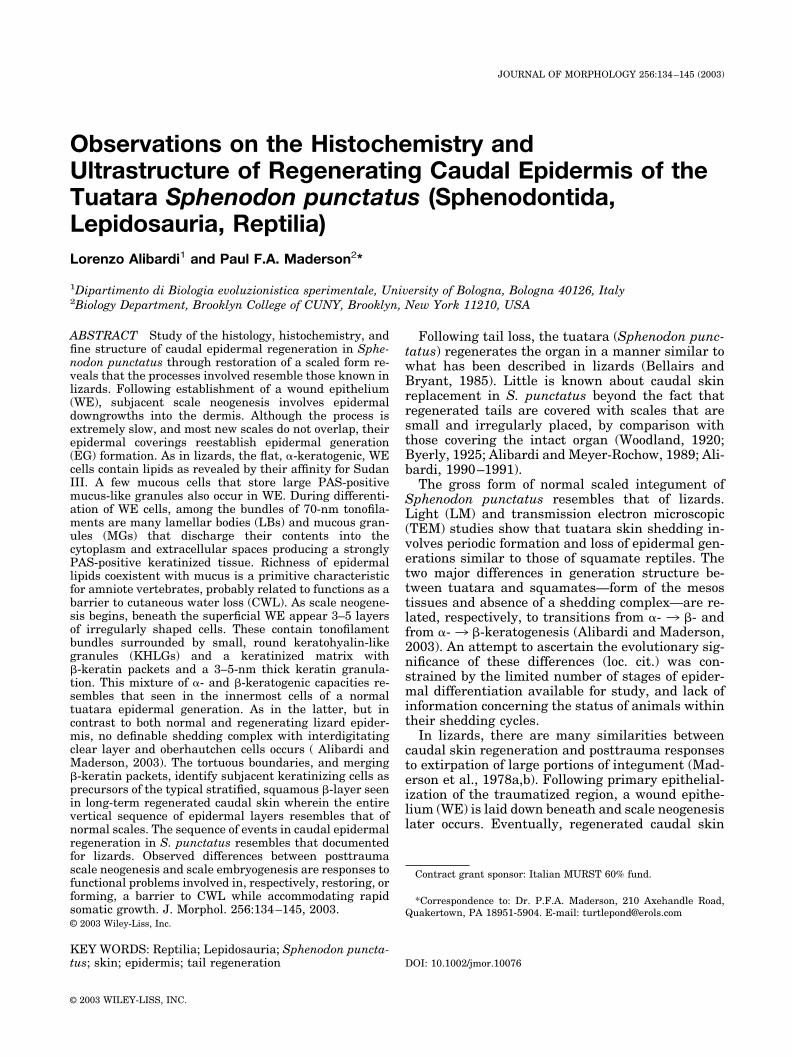
Fig. 1. Sphenodon punctatus, juvenile. Regenerating tail 5 months postamputation. Apex of regenerate is covered by smooth,prescaling epidermis. Scale neogenesis is beginning proximally (arrowhead). Bar � 5 mm.
Fig. 2. Sphenodon punctatus, juvenile. Regenerating tail 8 months postamputation. Plate-like, scarcely overlapping, neogenicscales cover most of proximal regenerate. Apex is still covered by smooth prescaling epidermis. Bar � 2.5 mm.
Fig. 3. Sphenodon punctatus, adult. Sagittal section of dorsal integument on regenerating tail of unknown age. Tail tip to right.Regenerated scale has recognizable outer (O) and inner (I) surfaces and hinge region (H). D, dermal core. Hematoxylin and eosin.Bar � 200 �m.
136 L. ALIBARDI AND P.F.A. MADERSON
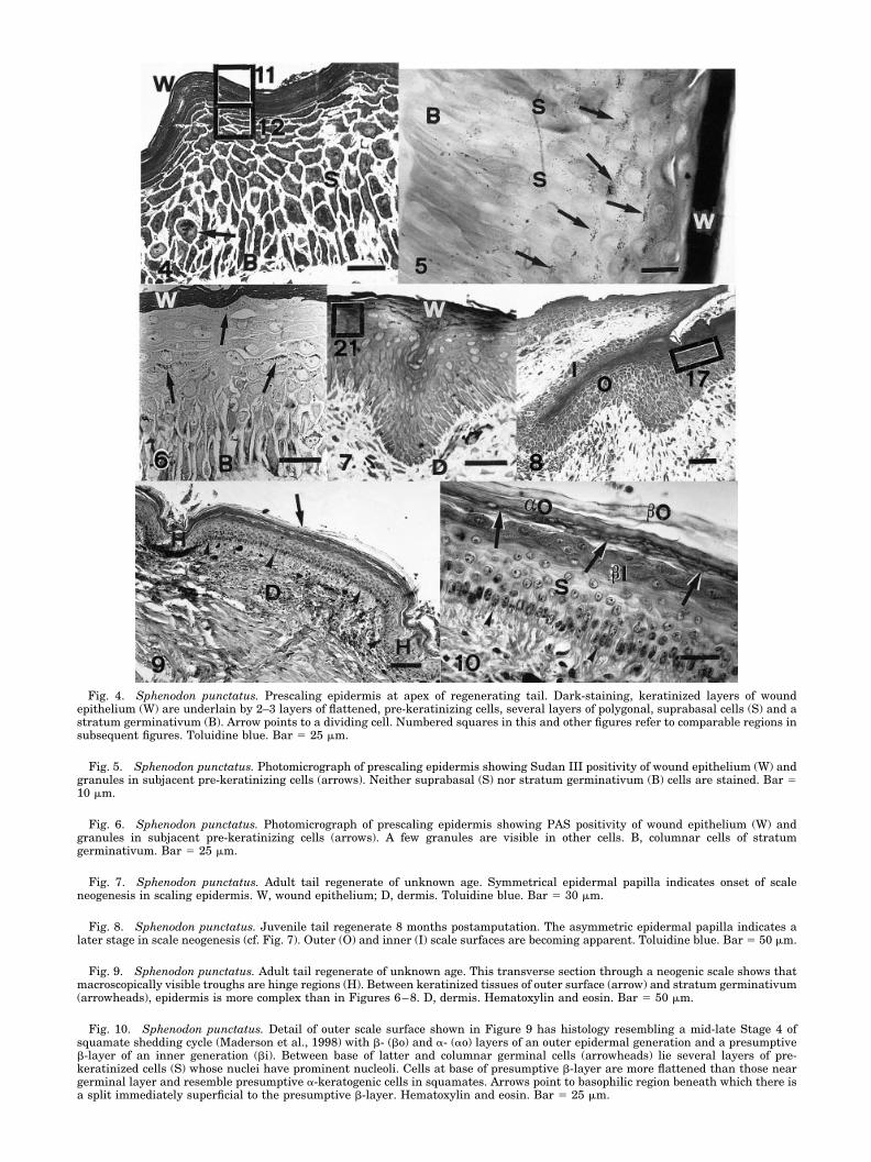
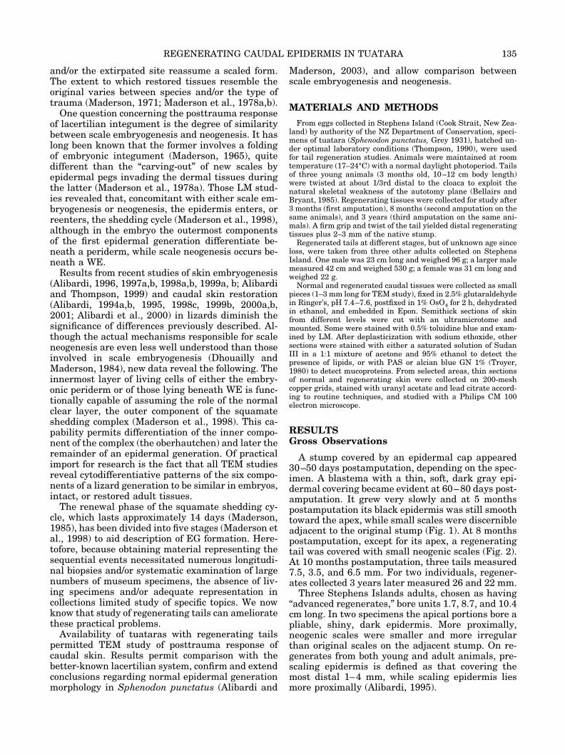
Fig. 4. Sphenodon punctatus. Prescaling epidermis at apex of regenerating tail. Dark-staining, keratinized layers of woundepithelium (W) are underlain by 2–3 layers of flattened, pre-keratinizing cells, several layers of polygonal, suprabasal cells (S) and astratum germinativum (B). Arrow points to a dividing cell. Numbered squares in this and other figures refer to comparable regions insubsequent figures. Toluidine blue. Bar � 25 �m.
Fig. 5. Sphenodon punctatus. Photomicrograph of prescaling epidermis showing Sudan III positivity of wound epithelium (W) andgranules in subjacent pre-keratinizing cells (arrows). Neither suprabasal (S) nor stratum germinativum (B) cells are stained. Bar �10 �m.
Fig. 6. Sphenodon punctatus. Photomicrograph of prescaling epidermis showing PAS positivity of wound epithelium (W) andgranules in subjacent pre-keratinizing cells (arrows). A few granules are visible in other cells. B, columnar cells of stratumgerminativum. Bar � 25 �m.
Fig. 7. Sphenodon punctatus. Adult tail regenerate of unknown age. Symmetrical epidermal papilla indicates onset of scaleneogenesis in scaling epidermis. W, wound epithelium; D, dermis. Toluidine blue. Bar � 30 �m.
Fig. 8. Sphenodon punctatus. Juvenile tail regenerate 8 months postamputation. The asymmetric epidermal papilla indicates alater stage in scale neogenesis (cf. Fig. 7). Outer (O) and inner (I) scale surfaces are becoming apparent. Toluidine blue. Bar � 50 �m.
Fig. 9. Sphenodon punctatus. Adult tail regenerate of unknown age. This transverse section through a neogenic scale shows thatmacroscopically visible troughs are hinge regions (H). Between keratinized tissues of outer surface (arrow) and stratum germinativum(arrowheads), epidermis is more complex than in Figures 6–8. D, dermis. Hematoxylin and eosin. Bar � 50 �m.
Fig. 10. Sphenodon punctatus. Detail of outer scale surface shown in Figure 9 has histology resembling a mid-late Stage 4 ofsquamate shedding cycle (Maderson et al., 1998) with �- (�o) and �- (�o) layers of an outer epidermal generation and a presumptive�-layer of an inner generation (�i). Between base of latter and columnar germinal cells (arrowheads) lie several layers of pre-keratinized cells (S) whose nuclei have prominent nucleoli. Cells at base of presumptive �-layer are more flattened than those neargerminal layer and resemble presumptive �-keratogenic cells in squamates. Arrows point to basophilic region beneath which there isa split immediately superficial to the presumptive �-layer. Hematoxylin and eosin. Bar � 25 �m.
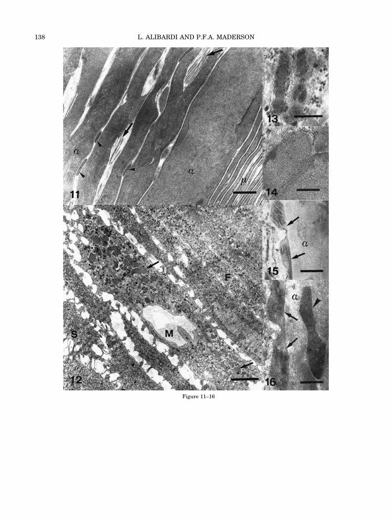
Figure 11–16
138 L. ALIBARDI AND P.F.A. MADERSON
lular domains (Figs. 15, 16). These features identifythe small granules as lamellar bodies (LBs) (Menonand Ghadially, 1997). Larger (0.3–0.8 to 0.5–1.0�m), irregular, or roundish granules are lesselectron-dense than LBs and contain a coarselygranular material (Figs. 12, 14). In some cells suchmucous granules (MGs) occupy extensive regions ofcytoplasm adjacent to enlarged ER vesicles andpolyribosomes. In such cells some granules werepositive for PAS, toluidine blue, Sudan III, and al-cian blue. Both LBs and MGs lie close to the Golgiapparatus in apparent continuity with electron-lucent blebbing vesicles containing amorphous ma-terial that concentrates progressively into densersecretory granules. Deposits of such condensed mu-cous materials (Matoltsy and Huszar, 1972; Ma-toltsy and Bednarz, 1975) occur between keratinizedcells (Fig. 16).
Germinal and suprabasal cells have organellestypical of undifferentiated amniote keratinocytes,especially widespread, but sparse, glycogen gran-ules. Melanocytes, Merkel cells, and possible phago-cytes are sometimes seen.
Ultrastructure of Scaling Epidermis (Figs.17–26)
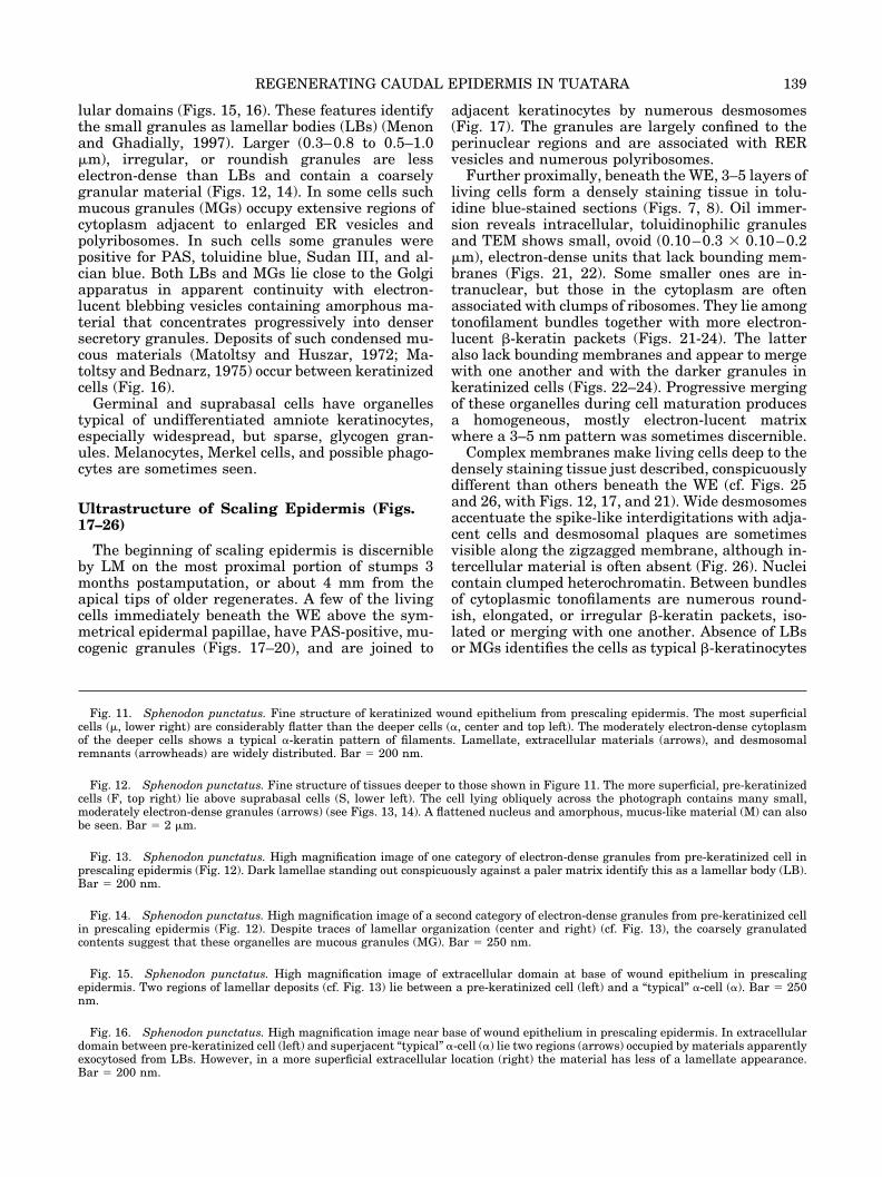
The beginning of scaling epidermis is discernibleby LM on the most proximal portion of stumps 3months postamputation, or about 4 mm from theapical tips of older regenerates. A few of the livingcells immediately beneath the WE above the sym-metrical epidermal papillae, have PAS-positive, mu-cogenic granules (Figs. 17–20), and are joined to
adjacent keratinocytes by numerous desmosomes(Fig. 17). The granules are largely confined to theperinuclear regions and are associated with RERvesicles and numerous polyribosomes.
Further proximally, beneath the WE, 3–5 layers ofliving cells form a densely staining tissue in tolu-idine blue-stained sections (Figs. 7, 8). Oil immer-sion reveals intracellular, toluidinophilic granulesand TEM shows small, ovoid (0.10–0.3 � 0.10–0.2�m), electron-dense units that lack bounding mem-branes (Figs. 21, 22). Some smaller ones are in-tranuclear, but those in the cytoplasm are oftenassociated with clumps of ribosomes. They lie amongtonofilament bundles together with more electron-lucent �-keratin packets (Figs. 21-24). The latteralso lack bounding membranes and appear to mergewith one another and with the darker granules inkeratinized cells (Figs. 22–24). Progressive mergingof these organelles during cell maturation producesa homogeneous, mostly electron-lucent matrixwhere a 3–5 nm pattern was sometimes discernible.
Complex membranes make living cells deep to thedensely staining tissue just described, conspicuouslydifferent than others beneath the WE (cf. Figs. 25and 26, with Figs. 12, 17, and 21). Wide desmosomesaccentuate the spike-like interdigitations with adja-cent cells and desmosomal plaques are sometimesvisible along the zigzagged membrane, although in-tercellular material is often absent (Fig. 26). Nucleicontain clumped heterochromatin. Between bundlesof cytoplasmic tonofilaments are numerous round-ish, elongated, or irregular �-keratin packets, iso-lated or merging with one another. Absence of LBsor MGs identifies the cells as typical �-keratinocytes
Fig. 11. Sphenodon punctatus. Fine structure of keratinized wound epithelium from prescaling epidermis. The most superficialcells (�, lower right) are considerably flatter than the deeper cells (�, center and top left). The moderately electron-dense cytoplasmof the deeper cells shows a typical �-keratin pattern of filaments. Lamellate, extracellular materials (arrows), and desmosomalremnants (arrowheads) are widely distributed. Bar � 200 nm.
Fig. 12. Sphenodon punctatus. Fine structure of tissues deeper to those shown in Figure 11. The more superficial, pre-keratinizedcells (F, top right) lie above suprabasal cells (S, lower left). The cell lying obliquely across the photograph contains many small,moderately electron-dense granules (arrows) (see Figs. 13, 14). A flattened nucleus and amorphous, mucus-like material (M) can alsobe seen. Bar � 2 �m.
Fig. 13. Sphenodon punctatus. High magnification image of one category of electron-dense granules from pre-keratinized cell inprescaling epidermis (Fig. 12). Dark lamellae standing out conspicuously against a paler matrix identify this as a lamellar body (LB).Bar � 200 nm.
Fig. 14. Sphenodon punctatus. High magnification image of a second category of electron-dense granules from pre-keratinized cellin prescaling epidermis (Fig. 12). Despite traces of lamellar organization (center and right) (cf. Fig. 13), the coarsely granulatedcontents suggest that these organelles are mucous granules (MG). Bar � 250 nm.
Fig. 15. Sphenodon punctatus. High magnification image of extracellular domain at base of wound epithelium in prescalingepidermis. Two regions of lamellar deposits (cf. Fig. 13) lie between a pre-keratinized cell (left) and a “typical” �-cell (�). Bar � 250nm.
Fig. 16. Sphenodon punctatus. High magnification image near base of wound epithelium in prescaling epidermis. In extracellulardomain between pre-keratinized cell (left) and superjacent “typical” �-cell (�) lie two regions (arrows) occupied by materials apparentlyexocytosed from LBs. However, in a more superficial extracellular location (right) the material has less of a lamellate appearance.Bar � 200 nm.
139REGENERATING CAUDAL EPIDERMIS IN TUATARA
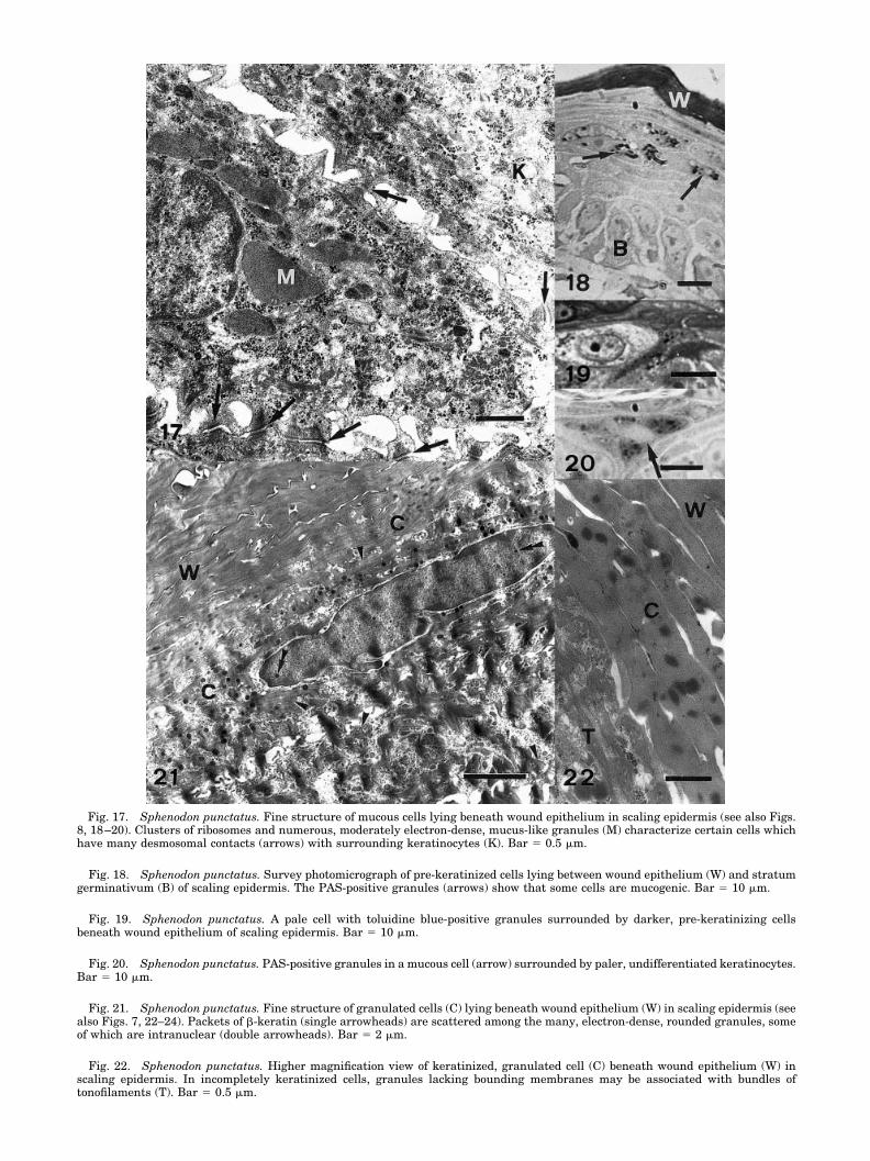
Fig. 17. Sphenodon punctatus. Fine structure of mucous cells lying beneath wound epithelium in scaling epidermis (see also Figs.8, 18–20). Clusters of ribosomes and numerous, moderately electron-dense, mucus-like granules (M) characterize certain cells whichhave many desmosomal contacts (arrows) with surrounding keratinocytes (K). Bar � 0.5 �m.
Fig. 18. Sphenodon punctatus. Survey photomicrograph of pre-keratinized cells lying between wound epithelium (W) and stratumgerminativum (B) of scaling epidermis. The PAS-positive granules (arrows) show that some cells are mucogenic. Bar � 10 �m.
Fig. 19. Sphenodon punctatus. A pale cell with toluidine blue-positive granules surrounded by darker, pre-keratinizing cellsbeneath wound epithelium of scaling epidermis. Bar � 10 �m.
Fig. 20. Sphenodon punctatus. PAS-positive granules in a mucous cell (arrow) surrounded by paler, undifferentiated keratinocytes.Bar � 10 �m.
Fig. 21. Sphenodon punctatus. Fine structure of granulated cells (C) lying beneath wound epithelium (W) in scaling epidermis (seealso Figs. 7, 22–24). Packets of �-keratin (single arrowheads) are scattered among the many, electron-dense, rounded granules, someof which are intranuclear (double arrowheads). Bar � 2 �m.
Fig. 22. Sphenodon punctatus. Higher magnification view of keratinized, granulated cell (C) beneath wound epithelium (W) inscaling epidermis. In incompletely keratinized cells, granules lacking bounding membranes may be associated with bundles oftonofilaments (T). Bar � 0.5 �m.
Fig. 23. Sphenodon punctatus. Very high magnification view of an incompletely keratinized, granulated cell lying beneath woundepithelium in scaling epidermis showing fusion of �-keratin packets (�), clusters of ribosomes and dense, rounded granules (arrows).N, electron-dense nucleus. Bar � 0.5 �m.
Fig. 24. Sphenodon punctatus. High magnification view of a nearly completely keratinized, granulated cell lying beneath woundepithelium in scaling epidermis. Patches of ribosomes are associated with keratohyalin-like granules (arrows). Bar � 0.5 �m.
Fig. 25. Sphenodon punctatus. Survey view of fine structure of two different cell types lying beneath wound epithelium in scalingepidermis. Most superficially (top left) is a granulated cell (G) where, once again, some of the characteristic organelles are intranuclear(double arrowheads). Three subjacent cells (S) are distinguished by exceptional membrane tortuosity (arrowheads), sparse granula-tion, and dense concentrations of �-keratin packets (arrows) in cytoplasm. Bar � 2 �m. Inset (bar � 100 nm): high magnification of�-keratin packet with 3–5 nm filaments. Density of these organelles permits identification of three deepest cells as presumptive �-cells.
Fig. 26. Sphenodon punctatus. High magnification view of membrane tortuosity between two presumptive �-cells in scalingepidermis (cf., Figs. 9, 10) showing associated desmosomes and dense masses of cytoskeletal filaments. Packets of �-keratin (�) showaggregated filaments in an electron-lucent matrix (cf., Fig. 25). Bar � 250 nm.
that form the �-layer when epidermal generationformation is reestablished on neogenic scales (Figs.9, 10).
DISCUSSIONGeneral Comments
Following tail loss in Sphenodon punctatus and inlizards, the exposed surface is covered by epidermalcells migrating from the wound edge and a WEcovers a slightly swollen, rounded blastema beforeelongation of a new tail begins. A major difference isthat while these processes take 7–14 days in lizards(Alibardi and Sala, 1983; Bellairs and Bryant, 1985),they take over a month in S. punctatus: a cone-shaped blastema does not appear until 2 monthspostamputation.
We confirm reports that the scalation pattern on aregenerated tail in Sphenodon punctatus is readilydistinguishable from the original because neogenicscales are smaller, more irregular in shape, andscarcely overlap. Comparably imperfect restorationof caudal skin occurs in many lizards (Maderson,1971). The present study does not reveal how longcaudal regeneration takes to be completed in S.punctatus, nor what percentage of tissue can be re-placed. The persistent apical WE implies thatgrowth and skin restoration must continue for atleast 1 year postamputation, a period of time far inexcess of the maximum of 8–10 weeks documentedfor lizards (Baranowitz et al., 1979).
The sequence of events involved in scale neogen-esis in Sphenodon punctatus follows that describedfor lizards. Symmetrical epidermal papillae pene-trate subjacent dermis, asymmetrization occurs,and, concomitant with scale shaping, the epidermisreenters the shedding cycle. This study does notpermit timing of these events and we know nothingof the cycle’s chronology in S. punctatus (Alibardiand Maderson, 2003). It is well established thatentry, or reentry, into the cycle accompanies bothscale embryogenesis (Alibardi 1994a,b, 1996,1997a,b, 1998a,b 1999a,b,; Alibardi and Thompson,1999; Dhouailly and Maderson, 1984), and neogen-esis during caudal regeneration and wound healing(Maderson et al., 1978a,b; Bellairs and Bryant,1985; Alibardi, 1995, 1998c, 1999b, 2000a,b 2001;Alibardi et al., 2000). Because any somatic growthincreases the surface area of all or part of the body(Maderson et al., 1998), we can offer a functionalexplanation for similarities and differences betweenscale embryogenesis and neogenesis.
Constraints on Embryogenesis andPosttrauma Restoration of Amniote Skin
Whether scaled or appendage-bearing, amnioteskin is geometrically patterned (Maderson, 1972;Maderson and Alibardi, 2000). Although the relativesizes of newly hatched or neonate amniotes vary
greatly, reflecting the percentage of in ovo, or inutero, life devoted to somatic growth sensu lato,such a topography helps explain why skin alwaysdevelops relatively late by comparison with otherorgan systems. In reptiles and birds, the presence of�-keratinized tissues in epidermis (Sawyer et al.,2000) reinforces this tendency for delayed develop-ment because of the difficulty of accommodating anincrease in surface areas of individual scales (Mad-erson et al., 1998). Thus, scale primordia form assymmetrical folds of the epidermal/dermal surface ofthe embryonic body after primary organogenesis oc-curs elsewhere (Maderson, 1965, 1985; Dhouaillyand Maderson, 1984; Alibardi, 1996, 1998a,b, 1999a,2000b). Such units, comprising a simple, unkera-tinized epidermis (periderm plus stratum germina-tivum) covering a dermal core, become asymmetricaland elongate as somatic growth continues. Becauseamniotic fluid protects the embryo physiologicallyand mechanically, cytodifferentiative events in theepidermis producing a barrier to cutaneous waterloss (CWL) housed in a mechanically strong, butflexible skin (Maderson et al., 1998; Maderson andAlibardi, 2000) can be delayed until just beforehatching or birth (Alibardi, 1996, 1998a,b, 1999a,2000b).
By contrast, functional priorities during post-trauma restoration of skin in tuatara and lizards arethe opposite of those obtaining during embryoniclife. Wounding or tail loss immediately exposedeeper tissues to possible desiccation. Restoration ofskin form, whether perfect or not (Maderson, 1971,1985), necessitates production of new cells, tissues,and organs and this takes time. If the organismcannot meet the challenge to its water economy, itwill die. Any mechanism(s) involved in skin restora-tion must operate within whatever anatomical con-straints emerge from the need to reduce CWL. Theseconsiderations explain 1) the form of the WE from itsinitiation through completion of scale neogenesis,and 2) why scale embryogenesis involves folding ofthe body surface, while neogenesis involves papillardowngrowth beneath a WE.
Initial Reepithelialization and Formation ofWound Epithelium
Cyclic epidermal generation formation is a lepido-saurian synapomorphy (Maderson and Alibardi,2000). Tuatara shares with lizards a common basisfor formation of a barrier to CWL — intra- andextracellular deposition of LB-derived lipidsthroughout �-keratogenic tissues (Alibardi andMaderson, 2003ˆ). The WE described here for Sphe-nodon punctatus resembles that described by LMand TEM for several lizards (Maderson, 1971, 1985;Maderson and Roth, 1972; Maderson et al., 1978a,b;Alibardi, 1994a, 1995, 1998c, 1999b, 2000a,b, 2001;Alibardi et al., 2000). However, an important differ-ence is that intra- and extracellular lipid materials
142 L. ALIBARDI AND P.F.A. MADERSON
evidenced by Sudan III staining and organelle ultra-structure (Figs. 5, 11, 13, 15, 16) coexist with abun-dant mucoid materials identified by strong PAS-positivity and the fine structure of MGs (Figs. 6, 12,14, 17, 18, 20). Such extensive mucogenic capacitiesin both immature and mature components of the WEconfirm interpretation of their presence throughoutthe �-keratogenic component of normal epidermis inS. punctatus (Alibardi and Maderson 2003, fig. 28), afeature primitive for lepidosaurs but shared withchelonia (Matoltsy and Huszar, 1972; Matoltsy andBednarz, 1975). The well-documented juxtaposition-ing of MGs and LBs in reptilian �-keratinocytes(Landmann, 1986) warrants further specific investi-gation because of the light that could be shed on theappearance of LBs with the origin of amniotes (Mad-erson and Alibardi, 2000). Coexpression of lipogenicLBs and MGs in �-keratogenic tissues is undoubt-edly associated with the emergence of a barrier toCWL in amniote evolution (Alibardi and Maderson,2003, fig. 28), an epidermal property whose restora-tion posttrauma is vital to the organism.
Within 72 h of Scotch tape stripping of body epi-dermis, a WE is laid down and rates of CWL arerestored to within 120% of normal values for intactskin (Maderson et al., 1978b). While no studies re-port rates of CWL from caudal tissues, we may inferthat the WE meets the first desideratum of caudalskin replacement — amelioration of diminished bar-rier efficacy.
All LM and TEM studies (loc. cit.) show that WEcytology is “looser” than that of the �-keratogenictissues of an intact epidermal generation, as indi-cated by more abundant intercellular spaces, espe-cially towards the organism–air interface. This sug-gests that WE form is a compromise between thephysiological requirement for a tissue to combatCWL and the developmental requirement to permitcontinuing expansion of subjacent tissues. A quan-titative comparison of proliferation rates and pat-terns of changes in cell adhesion molecules betweennormal epidermis and WE would be of great inter-est. Whatever the underlying causal mechanisms, arelative looseness of superficial keratinized tissuesis necessary to facilitate scale neogenesis.
Epidermal Cell Activity Over the Prescalingand Scaling Caudal Tissues
A regenerating tail is fundamentally a cone, thediameter of whose base is equal to that of the unin-jured stump. This implies that throughout elonga-tion the cross-sectional areas along the proximo–distal axis change continuously. All studies showthat the regenerate’s epidermis has two forms: overthe distal blastema there is only a WE, but proxi-mally, beneath a WE, there is a gradient of cytolo-gies reflecting reentry into the shedding cycle. Thisgradient, which begins at the margin of the blast-emal tissues and ends proximally against the stump
tissues, where a ring of completed new scales can berecognized, is also a gradient of neogenesis.
An obvious question concerns the possible causalrelationship between initiation of reentry into thecycle and the first recognizable event in shaping newscales, i.e., initiation of epidermal papillar down-growth into dermis (Fig. 7). Available data suggesttwo identical environmental requirements of epider-mal cells for participation in both events.
First, both require an enhanced level of germinalmitotic activity that is precisely patterned. The pa-pillae that carve out the shapes of incipient newscales represent the edges of the gradient ofproliferative/differentiative activity that spreadscentrifugally over the outer scale surface to producean epidermal generation (Maderson et al., 1998).Their penetration into the dermis is presumablyassisted by proteolytic activity similar to that occur-ring during the wound rejection reaction (Madersonand Roth, 1972; Maderson, 1985). Papillar growth inSphenodon punctatus (Fig. 8) is not accompanied bylacunar tissue hypertrophy, as it is in lizards (Mad-erson et al., 1978a; Maderson, 1985).
Second, a precondition for enhanced epidermalproliferation seems to be spatial stability of subja-cent regenerating dermal tissues. The immaturity ofthe latter that lack the characteristic complex archi-tecture (Maderson, 1985) facilitates papillar down-growth. The similarity between dermal tissuesaround such papillae (Figs. 7, 8) and those of embry-onic scales (Dhouailly and Maderson, 1984) is strik-ing. Lizard epidermal papillae grow down into aloose, mesenchymal dermis whose rate of cell prolif-eration is less than that of the epidermis. As a re-sult, the reticular and collagenous fibrils of the der-mis are distorted by the downpushing, and mature,dense dermal tissues do not form until after thepapillae have elongated and epidermal generationrestoration is completed (Alibardi, 1994a,b). LM ex-amination of very long-term posttrauma tissuesfrom both body and tail shows that collagen remod-eling continues, although neogenic scales are alwaysrecognizable (Maderson, 1985, pers. obs.).
A significant gap in our knowledge of lepidosau-rian skin regeneration was revealed by studies ofSphenodon punctatus. Intact epidermal tissuesproximal to the regenerating tail showed an anom-alous cytology, previously undescribed for this spe-cies, that might reflect an influence of juxtaposedregenerating tissues on normal cell dynamics (Ali-bardi and Maderson, 2003). Epidermal cycle statusat tail amputation does not influence the regenera-tive process in Anolis carolinensis (Maderson andLicht, 1968), but the question of whether caudal skinregeneration could affect juxtaposed normal tissueshas never been raised. For body skin, mode andtiming of reentry of epidermis of neogenic scales intoa normal shedding cycle depends upon the type oftrauma and cycle status of the tissue at the time oftrauma (Maderson et al., 1978a,b; Maderson, 1985).
143REGENERATING CAUDAL EPIDERMIS IN TUATARA
Although neogenic scales shed normally within amaximum of two cycles after extirpation of the orig-inal tissue, details of cellular activity at the periph-ery of the wound have not been followed in detailbeyond the initial wound rejection reaction (Mader-son and Roth, 1972). It is possible that the extremeduration of the caudal regenerative process in S.punctatus has a unique effect on more proximal tis-sues. However, further investigations of possible in-teractions between normal and regenerating caudalskin tissues in lizards are needed.
ACKNOWLEDGMENTS
Dr. Alibardi’s fellowship to stay in New Zealandcame from the NZ University Grant Committee inexchange with the Italian MAE. He thanks Dr. V.B.Meyer-Rochow (University of Waikato, Hamilton,NZ) for hosting him and for his important role ingetting a permit to work on the tuatara. We thankDrs. M. Thompson (University of Sydney, NSW,Australia), A. Cree (Victoria University, Wellington,NZ), and Mr. J. Govey (NZ Department of Conser-vation), who made possible the collection of speci-mens during a visit to Stephens Island. Mrs. Luci-ana Dipietrangelo’s photographic skills are greatlyappreciated.
LITERATURE CITED
Alibardi L. 1990–1991. Electron microscopic observations on themyelination of the long term regenerated caudal spinal cord inlizards and Sphenodon. Biol Struct Morphol 3:147–158.
Alibardi L. 1994a. Fine autoradiographical study on scale mor-phogenesis in the regenerating tail of lizards. Histol His-topathol 9:119–134.
Alibardi L. 1994b. Modification of the dermis during scale regen-eration in the lizard tail. Histol Histopathol 9:733–745.
Alibardi L. 1995. Electron microscopic analysis of the regenerat-ing scales in lizard. Boll Zool 62:109–120.
Alibardi L. 1996. Scale morphogenesis during embryonic devel-opment in the lizard Anolis carolinensis. J Anat 188:713–725.
Alibardi L. 1997a. Morphogenesis of the digital pad lamellae inthe embryo of the lizard Anolis lineatopus. J Zool 243:47–55.
Alibardi L. 1997b. Ultrastructural and autoradiographic analysisof setae development in the embryonic pad lamellae of thelizard Anolis lineatopus. Ann Sci Nat Zool (Paris) 18:51–61.
Alibardi L. 1998a. Glycogen distribution in relation to epidermalcell differentiation during embryonic scale morphogenesis inthe lizard Anolis lineatopus. Acta Zool 79:91–100.
Alibardi L. 1998b. Differentiation of the epidermis during scaleformation in embryos of lizard. J Anat 192:173–186.
Alibardi L. 1998c. Presence of acid phosphatase in the epidermisof regenerating tail of the lizard Podarcis muralis and its pos-sible role in the process of shedding and maturation. J Zool246:379–390.
Alibardi L. 1999a. Formation of large microornamentations indeveloping scales of agamine lizards. J Morphol 240:251–266.
Alibardi L. 1999b. Keratohyalin-like granules in embryos andregenerating epidermis of lizards and Sphenodon punctatus(Reptilia, Lepidosauria). Amphiba-Reptilia 20:11–23.
Alibardi L. 2000a. Ultrastructural localization of alpha keratinsin the regenerating epidermis of the lizard Podarcis muralisduring formation of the shedding layer. Tissue Cell 32:153–162.
Alibardi L. 2000b. Epidermal structure of normal and regenerat-ing skin of the agamine lizard Physignathus lesueurii (McCoy,1878) with emphasis on the formation of the shedding layer.Ann Sci Nat Zool (Paris) 21:27–36.
Alibardi L. 2001. Keratohyalin-like granules in lizard epidermis:evidence from cytochemical, autoradiographic and microanalyt-ical studies. J Morphol 284:4–79.
Alibardi L, Maderson PFA. 2003. Observations on the histochem-istry and ultrastructure of the epidermis of the tuatara Sphe-nodon punctatus (Sphenodontida, Lepidosauria, Reptilia): acontribution to an understanding of the lepidosaurian epider-mal generation and the evolutionary origin of the squamateshedding complex. J Morphol .
Alibardi L, Meyer-Rochow VB. 1989. Comparative fine structureof the axial skeleton inside the regenerated tail of some lizardspecies and the tuatara (Sphenodon punctatus). Gegenb MorphJahrb 135:705–716.
Alibardi L, Sala M. 1983. Distribuzione di sostanze d’importanzamorfogenetica in tessuti rigeneranti di Lacerta sicula, Triturusalpestris e Rana dalmatina. Atti Mem Acc Patav Sci Let Arti95:100–151.
Alibardi L, Thompson MB. 1999. Epidermal differentiation in thedeveloping scales of embryos of the Australian scincid lizardLampropholis guichenoti. J Morphol 241:139–152.
Alibardi L, Maurizii MG, Taddei C. 2000. Immunocytochemicaland electrophoretic distribution of cytokeratins in the regener-ating epidermis of the lizard Podarcis muralis. J Morphol 246:179–191.
Baranowitz SA, Maderson PFA, Connelly TG. 1979. Lizard andnewt tail regeneration: a qualitative study. J Exp Zool 210:17–38.
Bellairs AdA, Bryant SV. 1985. Autotomy and regeneration inreptiles. In: Gans C, Billett F, Maderson PFA, editors. Biologyof the Reptilia, vol 14. New York: John Wiley & Sons. p 523–598.
Byerly TC. 1925. Note on the partial regeneration of the caudalregion of Sphenodon punctatus. Anat Rec 30:61–66.
Dhouailly D, Maderson PFA. 1984. Ultrastructural observationson the embryonic development of the integument of Lacertamuralis. J Morphol 179:203–228.
Landmann L. 1986. Reptilian skin. In: Bereiter-Hahn J, MatoltsyAG, Richards KS, editors. Biology of the integument, vol. 2.Vertebrates. Berlin: Springer. p 150–187.
Maderson PFA. 1964. The skin of snakes and lizards. Br J Her-petol 3:151–154.
Maderson PFA. 1965. The embryonic development of the squa-mate epidermis. Acta Zool XLVI:275–295.
Maderson PFA. 1971. The regeneration of caudal epidermal spe-cialisations in Lygodactylus picturatus keniensis (Gekkonidae,Lacertilia). J Morphol 134:467–478.
Maderson PFA. 1972. When? Why? and How? Some speculationson the evolution of the vertebrate integument. Am Zool 12:159–171.
Maderson PFA. 1985. Some developmental problems of the rep-tilian integument. In: Gans C, Billett F, Maderson PFA, editors.Biology of the Reptilia, vol 14. New York: John Wiley & Sons. p523–598.
Maderson PFA, Alibardi L. 2000. The development of the saurop-sid integument: a contribution to the problem of the origin andevolution of feathers. Am Zool 40:513–529.
Maderson PFA, Licht P. 1968. Factors influencing rates of tailregeneration in the lizard Anolis carolinensis. Experientia 24:1083–1086.
Maderson PFA, Roth SI. 1972. A histological study of the earlystages of cutaneous wound healing in lizards in vivo and invitro. J Exp Zool 180:175–186.
Maderson PFA, Baranowitz SA, Roth SI. 1978a. A histologicalstudy of the long-term response to trauma of squamate integ-ument. J Morphol 157:121–136.
Maderson PFA, Zucker AH, Roth SI. 1978b. Epidermal regener-ation and percutaneous water loss following cellophane strip-ping of reptile epidermis. J Exp Zool 204:11–32.
144 L. ALIBARDI AND P.F.A. MADERSON

Maderson PFA, Rabinowitz T, Tandler B, Alibardi L. 1998. Ul-trastructural contributions to an understanding of the cellularmechanisms involved in lizard skin shedding with comments onthe function and evolution of a unique lepidosaurian phenom-enon. J Morphol 236:1–24.
Matoltsy AG, Bednarz JA. 1975. Lamellar bodies of the turtleepidermis. J Ultrastr Res 53:128–132.
Matoltsy AG, Huszar T. 1972. Keratinization of the reptilianepidermis: an ultrastructural study of the turtle skin. J Ultra-str Res 38:87–101.
Menon GK, Ghadially R. 1997. Morphology of lipid alterations inthe epidermis: a review. Microsc Res Tech 37:180–192.
Sawyer RH, Glenn T, French JO, Mays B, Shames RB, BarnesGL, Rhodes W, Ishikawa Y. 2000. The expression of �-keratinsin the epidermal appendages of reptiles and birds. Am Zool40:530–539.
Thompson MB. 1990. Incubation of eggs of tuatara, Sphenodonpunctatus. J Zool 222:303–318.
Troyer H. 1980. Principles and techniques of histochemistry.Boston: Little Brown.
Woodland WNF. 1920. Some observations on caudal autotomyand regeneration of the gecko (Hemidactylus flaviviridisRuppel), with note on Sphenodon. Q J Microsc Sci 65:63–100.
145REGENERATING CAUDAL EPIDERMIS IN TUATARA