proceedings correlation between ultrastructure and histochemistry of mammalian intrafusal muscle...
TRANSCRIPT

1PROCEEDINGS OF THE
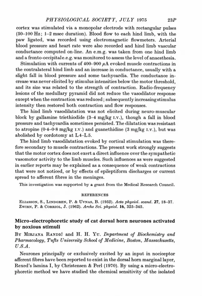
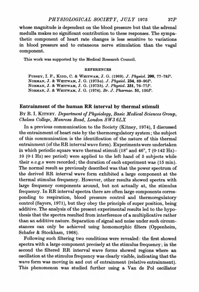
previously (Bergel 1961 a, b). The adapted nerve firing rate has beenrelated to the dimension changes of the sinus. An example of the responseto perfusion of the sinus at the animal's own arterial pressure is shown inthe figure. @ ~~~~~~~E: 200- _ .nJg.
- 0 MWPI R m-pr 1rao~~ ~~~~~~i
Fig. 1. Carotid sinus pouch perfused at the animal's own blood pressure.Records from the top down: electroneurogram (E.n.g.) from single carotidbaroreceptor fibre; carotid sinus pressure, mmHg (CSP); carotid sinlusdimension, mm (CSD) from ultrasound record; instantaneous firingfrequency (Hz) of baroreceptor fibre. The close similarity of the lowerthree traces is apparent.
REFERENCES
BERGEL, D. H. (1961a). J. Phyfseol. 156, 445-457.BERGEL, D. H. (1961cb). J. Physiol. 156, 458-469.BERTRAM, C. D. (1974). J. Phy8iol. 241, 85-87P.KousI:ANPouR, E. & KELSO, D. M. (1972). Circulation Re8. 31, 831-845.
A simple teaching film illustrating intestinal movementsBY BARBARA G. COLES and R. V. COXON. University Laboratory ofPhysiology, Parks Road, Oxford.
COMMUNICATIONS
Correlation between ultrastructure and histochemistry of mam-malian intrafusal muscle fibresBy R. W. BANKS, D. BARKER, D. W. HARKER and M. J. STACEY. ZoologyDepartment, Durham University, DurhamWe have devised a technique that allows for adjacent sections of the
same muscle spindle to be prepared for either histochemical or ultra-structural study. Muscle is frozen in iso-pentane cooled to - 1600 C andserial transverse sections cut in batches at about 15 ,um alternating withmuch thicker ones at about 60 ,um. Various histochemical techniques arethen applied to the thin sections, while the thick sections are processed for
16P

PHYSIOLOGICAL SOCIETY, JULY 1975
the observation of ultrastructure in both transverse and longitudinalsection. We have sectioned cat and rabbit peroneus longus, peroneusdigiti quinti and tenuissimus muscles, and the same peroneal and soleus
TABLE 1. Correlation of ultrastructure and histochemistry in cat, rabbit and ratintrafusal muscle fibres at two levels in the spindle: A, that adjacent to the area ofequatorial nucleation; B, that part of the juxta-equatorial region lying nearest tothe equator. Bag fibres designated 'bagL' and 'bag2' on the basis of their ATPasereactions following Ovalle & Smith (1972). Staining reactions: +, low; + +,medium;+ + +, high. Condition ofM line: 0, absent; M, present, dM, two faint parallel lines.
Level Fibre
Cat A Bag1Bag2Chain
B Bag1Bag2Chain
Rabbit A Bag1Bag2Chain
B Bag1Bag2Chain
Rat A Bag1Bag2Chain
B BagLBag2Chain
alkDiameter ATPase
MediumLargeSmallMediumLargeMedium
Medium/largeLargeSmall
MediumplargeLargeMedium
MediumLargeSmallMediumLargeMedium
+ +++ +.+++ +.
+ +++ +++++ +++
++++++
+++
+++++I++
+++++ +++
+ +++ ++++ +++
P'ase Glycogen M line
+ / + + 0/dM+ + + 0/dM++ +++ M
++++
+++++++++++
+
+++ 1+ ++++ ++I+++ +++++ +++ ++I++
0/dMMM
0/dM0/dMM0/dMMM
0/dM0/dMM0/dMMM
muscles of the rat, studying one spindle from each muscle. Histochemicalprofiles of intrafusal muscle fibres were determined with respect to acto-myosin ATPase after alkali pre-incubation (alk ATPase; Guth & Samaha,1971), phosphorylase (P'ase; Eranko & Palkama, 1961), and glycogen(PAS method). Ultrastructural observations have so far been restricted tonoting the M-line conditions.The results (Table 1) show that there may be variations in histochemical
profile along the length of all types of intrafusal muscle fibre, and that thebag2 fibres also show regional differences in ultrastructure.
REFERENCES
ERANKO, 0. & PALKAMA, A. (1961). J. Histochem. Cytochem. 9, 585.GuTr, L. & SA1IA, F. (1971). Expl Neurol. 28, 365-367.OVALLE, W. & SMITH, R. (1972). Can. J. Physiol. Pharmacol. 50, 195-202.
17P
1 8P PROCEEDINGS OF THE
The spontaneous activity of cortical neurones in sleep and wake-fulnessBY B. DELISLE BURNS and A. C. WEBB. National Institute for MedicalResearch, Mill Hill, London NW7 1AA
Interval distributions derived from the spontaneous activity of singlecortical neurones in the unrestrained cat can be described by log-normalcurves (Burns & Webb, 1975). This description has proved satisfactory forthose cells in visual, parietal and auditory cortex which fire faster than2.5/sec. Thus, for these neurones, two parameters - a model interval and ageometric standard deviation (GSD) - are sufficient to define the wholetemporal pattern of discharge. The same two parameters may be used todescribe the first part of the interval distribution of cells firing less fre-quently. We have tried to find out whether the values of these parametersvary systematically with gross changes in the animal's state of alertness.The spontaneous activity of single cortical neurones was recorded from
unrestrained cats (Burns, Stean & Webb, 1974) when the animals wereawake and when they were sleeping. A cat was said to be asleep when helay with his head supported, eyes shut and pinnae unresponsive tolaboratory noises. REM sleep was identified by jerky movements of theeyes and limbs. We have examined 70 trains of action potentials from 44neurones in visual, parietal and auditory cortex, and the results justify thefollowing conclusions:We have confirmed the finding of other workers (Evarts, 1964; Noda &
Adey, 1970) that sleep is invariably accompanied by a shortening of themodal interval. On average, the modal interval shortens by a factor offour when an animal falls into a quiet sleep. This coincides with an increaseof nearly 75 % in the size of the GSD. When an animal fell into REM sleep,these two parameters assumed mean values which were midway betweenthose found in the waking animal and those appropriate to quiet sleep.We were also able to assess the animal's state of arousal by examining a
train of only 200 action potentials derived from a single neurone in any ofthe three areas studied. Our results suggest that interval distributionswith modal intervals which are shorter than 19 msec are characteristic ofneural activity recorded from a sleeping animal. This rule offers a 91 %chance of successfully classifying a single interval distribution. A compo-site measure, the geometric coefficient of variation [log (GSD)/log (mode)],can also serve as an efficient 'test' of arousal. If one assumes that geo-metric coefficients of variation larger than 0'32 are diagnostic of recordstaken from animals which are asleep, one's chance of making an accurateclassification is 87 %. We were, however, unable to make any similardistinction between quiet and REM sleep.
18P
PHYSIOLOGICAL SOCIETY, JULY 1975
REFERENCES
BURNS, B. DELISLE & WEBB, A. C. (1975). J. Physiol. 248, 44-45P.BuRNS, B. DELISLE, STEAN, J. P. B. & WEBB, A. C. (1974). Electroenceph. dlin.
Neurophysiol. 36, 314-318.EVARTS, E. V. (1964). J. Neurophysiol. 27, 152-171.NODA, H. & ADEY, W. R. (1970). Brain Res. 18, 513-528.
Possible central cholinergic mechanism for the production ofcatechol convulsionsBY A. ANGEL and D. G. DEWHURST.* Department of Physiology, Universityof Sheffield S10 2TNOf the convulsant polyhydroxylic phenols, catechol has been shown to be
the most potent (Angel & Rogers, 1972). The convulsions occur after ad-ministration of catechol, either spontaneously, or in response to tactile orauditory stimuli, and characteristically consist of severe tremor and briefclonic jerking of the body musculature. The convulsions are entirelycentral in origin (Angel & Lemon, 1973), so studies relating to the pharma-cological mechanism of action of catechol were directed towards centralneurotransmission.The effects of various drugs on spontaneous convulsions were evaluated
either by measuring total integrated activity (Angel, 1970) of anaesthe-tized mice following catechol administration, before and after drugtreatment, or by looking for changes in the median convulsant dose, CD50,of drug-treated as compared to control groups of mice (Weil, 1952).Drugs which modify catecholaminergic transmission (reserpine, par-
gyline, iproniazid, pyrogallol, 6-hydroxydopamine, D-L propranolol,phenoxybenzamine, phentolamine, L-DOPA, and apomorphine) werefound to be ineffective in changing the convulsive response, with theexception of pyrogallol, which was found to significantly potentiate theeffects of catechol. Similarly drugs modifying 5-hydroxytryptaminetransmission (para-chlorophenylalanine, L-tryptophan, methysergide) andcerebral y-aminobutyric acid concentrations (GABA, amino-oxyaceticacid), had no consistent effect on the convulsions.On the other hand, drugs affecting cholinergic transmission were active
in modifying the response to catechol. Atropine and hyoscine significantlyreduced the intensity of the convulsions, while eserine and neostigminepotentiated both the intensity and duration of the convulsions. Mecamyl-amine, pempidine and hexamethonium were found to be less effective thanthe muscarinic-receptor blocking drugs, while atropine methyl nitrate, aperipheral muscarinic blocker, had no effect on the convulsions.
* M.R.C. Scholar.
19P

2PPROCEEDINGS OF THE
The results indicate that the catecholamines, 5-hydroxytryptamine, andy-aminobutyric acid have no significant role in the production of the con-vulsions. Pyrogallol potentiates the effects of catechol by competitivelyinhibiting catechol breakdown by the enzyme catechol-0-methyl trans-ferase. It appears that catechol has a central cholinergic mechanism ofaction, acting predominantly at muscarinic sites, possibly by increasingthe amount of transmitter released per nerve impulse, as at the neuro-muscular junction (Mogey & Young, 1949; Otsuka & Nonomura, 1963;Blaber & Gallagher, 1971).
REFERENCES
ANGEL, A. (1970). Br. J. Pharmacol. 39, 243P.ANGEL, A. & LEMON, R. N. (1973). Eleetroenceph. Clin. Neurophysiol. 34, 369-378.ANGEL, A. & ROGERS, K. J. (1972). Toxicol. appi. Pharmac. 21, 214-229.BLABER, L. C. & GALLAGHER, J. P. (1971). Neuropharmacology 10, 153-159.MOGEY, G. A. & YOUNG, P. A. (1949). Br. J. Pharmacol. 4, 359-365.OTSUKA, M. & NONOMURA, Y. (1963). J. Pharmac. exp. Ther. 140, 41-45.WEIL, C. S. (1952). Biometriy 8, 249-262.
Control of collateral sprouting in mechanosensory nerves ofsalamander skinBy E. COOPER, J. DIAMOND, LYNN MACINTYRE and C. TURNER. M.R.C.Group in Developmental Neurobiology, Department of Neurosciences,McMaster University, Hamilton, CanadaOur evidence from salamanders indicates that nerve section results in
collateral sprouting of adjacent nerves not because of 'products of degen-eration', or loss of function, but because fast axoplasmic transport is inter-rupted in the cut nerve (Aguilar, Bisby, Cooper & Diamond, 1973).Colchicine block of transport caused an identical sprouting of adjacent un-treated nerves, although the treated nerve behaved normally, and itsmechanosensory skin endings, we now know, are quantitatively unchangedin both number and function.
After partial denervation of the salamander hind limb, sprouting of theremaining nerves quantitatively makes up the loss of functional endings inthe skin. There is, however, an invisible frontier between their territorieswhich the 15th and 17th nerves will grow up to, but which they will notcross even if all the nerves beyond it are degenerating. The effect is not dueto a mechanical barrier in the skin or to a limited capacity of those nervesto sprout. It seems that the local stimulus which evokes collateral sprout-ing, which we suggest is produced by the target-tissue, has a selectivity ofaction. Surprisingly, skin rotation experiments suggest that the origin ofthis selectivity may not be in the skin itself.
After amputation the hind limb regenerates fully; the skin is then quite
20P
PHYSIOLOGICAL SOCIETY, JULY 1975 2P
indiscriminately innervated by the original three spinal nerves. If it ispresent, the mechanism underlying selectivity is ineffective in these condi-tions. Even in the normal limb a regenerating nerve can to some extentignore this regional specificity; guided into denervated foreign skin by wayof a degenerating nerve trunk, it makes normally functioning sensoryendings there, but it does not sprout beyond the limits of the presumedmechanical guidance. Apparently, regenerating and intact nerves havedifferent 'drives' with regard to sprouting. Additional evidence for thiscomes from seasonal studies. In winter, cut nerves will regenerate to pro-duce functional endings exactly as they do in summer; however, intactnerves often seem totally unable to sprout in response to section orcolchicine treatment of adjacent nerves. Perhaps the peripheral stimulusis absent.
REFERENCE
AGUILAR, C. E., BISBY, M. A., COOPER, E. & DIAMOND, J. (1973). J. Phy8iol. 234,449-464.
A role for a descending sympatho-inhibitory pathway in the ventralpart of the spinal cordBy J. H. COOTE* and A. SATO. Metropolitan Institute of Gerontology,35-2 Sakaecho, Itabashiku, Tokyo 173, JapanThe somatic afferent evoked reflex discharge into cardiac and renal
sympathetic nerves usually involves a long pathway ascending to, anddescending from, the brain stem (Sell, Erdelyi & Schaefer, 1958; Coote &Downman, 1966). A shorter spinal pathway can be demonstrated, but notreadily when the central nervous system is intact. This led Coote & Down-man (1966) to conclude that the spinal reflex pathways were normally in-hibited by a bulbospinal inhibitory system, which is tonically active. Inrecent years several inhibitory pathways on to sympathetic neurones havebeen identified (Illert & Seller, 1969; Illert & Gabriel, 1972; Coote & Mac-leod, 1974) but the functional role of some of them has not been ascer-tained. The present experiments were designed to examine the possibilitythat one of these descending inhibitory systems is responsible for blockingthe segmental pathway of the somato-cardiac reflex. In six cats anaes-thetized with chloralose, the only example of a somato-cardiac reflexresponse elicited by single or double shock, to an intercostal nerve of an
intensity 1-4-300 times the afferent fibre threshold, was acompact potentialoccurring after a mean latency of 56-0 msec, S.D. + 8-0 msec (n = 68). Theeffect on this cardiac reflex of small transverse cuts in the spinal cord at thelevel of C4 was examined. In all six animals an early somato-cardiac reflex
* Present address: Department of Physiology, The Medical School, Birmingham.
21P
PROCEEDINGS OF THE
appeared following a section made diagonally across the spinal cord fromthe contralateral dorsal horn to include the ventral part of the lateralfuniculus and/or the lateral part of the anterior funiculus. A hemi-sectionof the contralateral spinal cord or a section of dorsal columns performedsome hours previously had been without effect. The mean latency of theseearly reflex responses was 23 msec S.D. + 5 msec. The latency of the earlyresponse was little changed following a complete section of the spinalcord, but the long latency response was abolished.
Histological examination showed that the effective lesion was in theregion of the spinal cord which contains a known sympatho-inhibitorypathway which originates in the ventromedial reticular formation (Coote& Macleod, 1974, and unpublished observations), within the so-calledmedullary depressor region, as mapped by Alexander (1946).
It seems therefore that this is a major inhibitory system determiningwhether an afferent volley eliciting a response in cardiac nerves is routedover a long pathway to the brain stem or over a short spinal pathway. Sucha system may play a vital role in fashioning a widespread and patternedautonomic response.
REFERENCES
ALEXANDER. R. S. (1946). J. Neurophysiol. 9, 205-217.COOTE, J. H. & DowxNmAN, C. B. B. (1966). J. Physiol. 183, 714-729.CooTE, J. H. & MACLEOD, V. H. (1974). J. Physiol. 241, 453-475.ILLERT, M. & GABRIEL, M. (1972). Pflugers Arch. ges. Physiol. 335, 109-124.ILTERT, M. & SELLER, H. (1969). Pfliugers Arch. ges. Physiol. 313, 343-360.SELL, R., ERDELYI, A. & SCrAEFER, H. (1958). Pfiusgers Arch. ges. Physiol. 267,
566-581.
Hind limb vasodilatation evoked by stimulation of the motorcortexBy S. M. HILTON, K. M. SPYER and R. J. TIMMS. Department of Physiology,The Medical School, Birmingham, B15 2TJ
It has often been proposed that the motor cortex might initiate, via thepyramidal tract, the increased limb blood flow which occurs with exercise.Several investigators have reported that stimulation within the motor orpre-motor cortex can elicit vasodilatation in the limbs, mediated by inhibi-tion of sympathetic vasoconstrictor tone exerted via the pyramidal tract(e.g. Zwirn & Corriol, 1962), or by sympathetic cholinergic dilator fibres(Eliasson, Lindgren & Uvnas, 1952). We have reinvestigated these ques-tions, using carefully controlled electrical stimulation of the motor cortex.
In cats, lightly anaesthetized with a continuous infusion of 'Althesin'(Glaxo, alphaxalone-alphadolone), the 'hind limb area' of the motor
22P
PHYSIOLOGICAL SOCIETY, JULY 1975cortex was stimulated via a monopolar electrode with rectangular pulses(50-100 Hz; 1-2 msec duration). Blood flow to each hind limb, with thepaw ligated, was recorded using electromagnetic flowmeters. Arterialblood pressure and heart rate were also recorded and hind limb vascularconductance computed on-line. An e.m.g. was taken from one hind limband a fronto-occipital e.e.g. was monitored to assess the level of anaesthesia.
Stimulation with currents of 400-900 flA evoked muscle contractions inthe contralateral hind limb and an increase in conductance, usually with aslight fall in blood pressure and some tachycardia. The conductance in-crease was never elicited by stimulus intensities below the motor threshold,and its size was related to the strength of contraction. Radio-frequencylesions of the medullary pyramid did not reduce the vasodilator responseexcept when the contraction was reduced; subsequently increasing stimulusintensity then restored both contraction and flow responses.The hind limb vasodilatation was not elicited during neuro-muscular
block by gallamine triethiodide (3-4 mg/kg i.v.), though a fall in bloodpressure and tachycardia sometimes persisted. The dilatation was resistantto atropine (0.4-0.8 mg/kg i.v.) and guanethidine (3 mg/kg i.v.), but wasabolished by cordotomy at L 4-L 5.The hind limb vasodilatation evoked by cortical stimulation was there-
fore secondary to muscle contractions. The present work strongly suggeststhat the motor cortex does not exert a direct influence over the sympatheticvasomotor activity to the limb muscles. Such influences as were suggestedin earlier reports may be explained as a consequence of weak contractionsthat were not noticed, or by effects of epileptiform discharges or currentspread to afferent fibres in the meninges.
This investigation was supported by a grant from the Medical Research Council.
REFERENCES
ELIASSON, S., LINDGREN, P. & UVNAS, B. (1952). Acta physiol. scand. 27, 18-37.ZWIRN, P. & CORRIOL, J. (1962). Archs Sci. physiol. 16, 325-345.
Micro-electrophoretic study of cat dorsal horn neurones activatedby noxious stimuliBy MIRJANA RANDIC and H. H. Yu. Department of Biochemistry andPharmacology, Tufts University School of Medicine, Boston, Massachusetts,U.S.A.Neurones principally or exclusively excited by an input in nociceptor
afferent fibres have been reported to exist in the dorsal horn marginal layer,Rexed's lamina I, by Christensen & Perl (1970). By using a micro-electro-phoretic method we have studied the chemical sensitivity of the isolated
23P
24P PROCEEDINGS OF THE
cat dorsal horn neurones activated by noxious stimuli (at the level ofRexed's laminae I and II) to putative central neurotransmitter substancesacetylcholinee, L-glutamic acid, 5-hydroxytryptamine) and pain-producingpolypeptide, bradykinin.The experiments were performed on sixteen adults cats initially anes-
thetized by ether or Halothane. The brain was anaemically destroyed bybilateral occlusion of the common carotid and vertebral arteries. The spinalcord was transacted at the first cervical level. Thereafter, the animal wasartificially respired and immobilized by gallamine triethiodide or by aconstant infusion of succinylcholine chloride. A coccygeal dorsal root wasfreed and placed on a bipolar stimulating electrode distally and a bipolarrecording electrode centrally, leaving both peripheral and central connec-tions intact. This electrode arrangement served for electrical stimulationand recording of compound action potentials of different kinds of myeli-nated and unmyelinated afferent fibres. The activity of the dorsal hornneurones located in the sacral and coccygeal segments of the spinal cordwas recorded extracellularly through the central barrel of a multibarrelledglass micropipette filled with a solution of fast green (FCF, Matheson,Coleman and Bell) saturated in 3 M sodium chloride. The site of recordingwas marked by iontophoresis of a dye. Conventional micro-iontophoretictechnique was used to study the effects of the following substances:acetylcholine chloride (1 M, pH 4 0, Schwarz/Mann); bradykinin triacetatedehydrate (10 mm in distilled water or 165 mM-NaCl, Calbiochem.); L-glutamic acid (1 M, pH 8-0, Calbiochem); 5-hydroxytryptamine creatininesulphate (0*1 M, pH 4 3, Regis).
High-intensity mechanical stimuli were delivered to the skin of the tail(e.g. pressure from sharply pointed objects; squeezing a skin-fold betweentwo rigid surfaces).We have found that a majority of the cat dorsal horn neurones activated
by noxious stimuli were excited by L-glutamate (20-80 nA) and some byacetylcholine (40-80 nA). Bradykinin, applied either micro-electro-phoretically (50-200 nA) or intra-arterially (5-10 /zm), excited nociceptiveunits after a variable, but relatively long latent period (30-90 see). Excita-tion of polymodal nociceptors was also observed. 5-Hydroxytryptaminedepressed the firing of almost all units activated bynoxious stimuli, locatedin lamina I.These results suggest a possible chemical transmitter or modulator role
for L-glutamic acid, acetylcholine and 5-hydroxytryptamine at the level ofspinal neurones that could be excited exclusively or predominantly bynoxious stimuli.
This work was supported by PHS grant NS 11174-01 and NSF grant GB 37864.
24P

PHYSIOLOGICAL SOCIETY, JULY 1975
REFERENCE
CHRISTENSEN, B. N. & PERL, E. R. (1970). J. Neurophysiol. 33, 293-307.
Functional coupling between nerve terminals in teethBy B. MATTHEWS. Department of Physiology (Oral Biology), The MedicalSchool, University Walk, Bristol BS8 1TD
Action potentials can be recorded from the crown of a cat's lower caninetooth when chemical stimuli are applied to dentine and when single pulpalfibres in the inferior dental nerve are stimulated electrically. Recordingscan also be made from isolated nerve fibres during chemical or electricalstimulation of the tooth (Horiuchi & Matthews, 1974; Matthews, 1975).Some of the observations made with this preparation prompted the sug-gestion that action potentials might be propagated from one nerve toanother at their terminals and that several nerves might be coupledtogether to form a complex sensory unit in which stimulation of one fibrecaused a near synchronous discharge in all of them. The experimentsdescribed were carried out to test this possibility.
Single fibres dissected from the inferior dental nerve were stimulatedelectrically and simultaneous recordings were made from another inferiordental nerve strand, which included several pulpal nerves, and from thecanine tooth. Eight units have been isolated which, on stimulation, pro-duced a complex, all-or-none potential in the record from the tooth and anaction potential propagated away from the tooth in a fibre in the multi-unit strand. The conduction velocities of the units were between 16 and30 msec-1.One of these units also responded to the application of 2-5 molefl. NaCl to
the exposed dentine. This solution causes pain from human dentine(Anderson & Matthews, 1967) and is known to evoke a response fromnerves in cat's teeth (Horiuchi & Matthews, 1974). With the one unit thatshowed evidence of coupling and also responded to this stimulus, eachaction potential recorded from the isolated fibre was associated with acomplex action potential in the record from the tooth and an actionpotential propagated along the coupled fibre in the multiunit strand.
This work was supported by a grant from the Medical Research Council.
REFERENCES
ANDERSON, D. J. & MATiTws, B. (1967). Arch8 oral Biol. 12, 417-426.HoRiucm, H. & MATTHl;ws, B. (1974). J. Phymiol. 243, 797-829.MATTHEWS, B. (1975). J. Physiol. 245, 16P.
25P
2PPROCEEDINGS OF THE
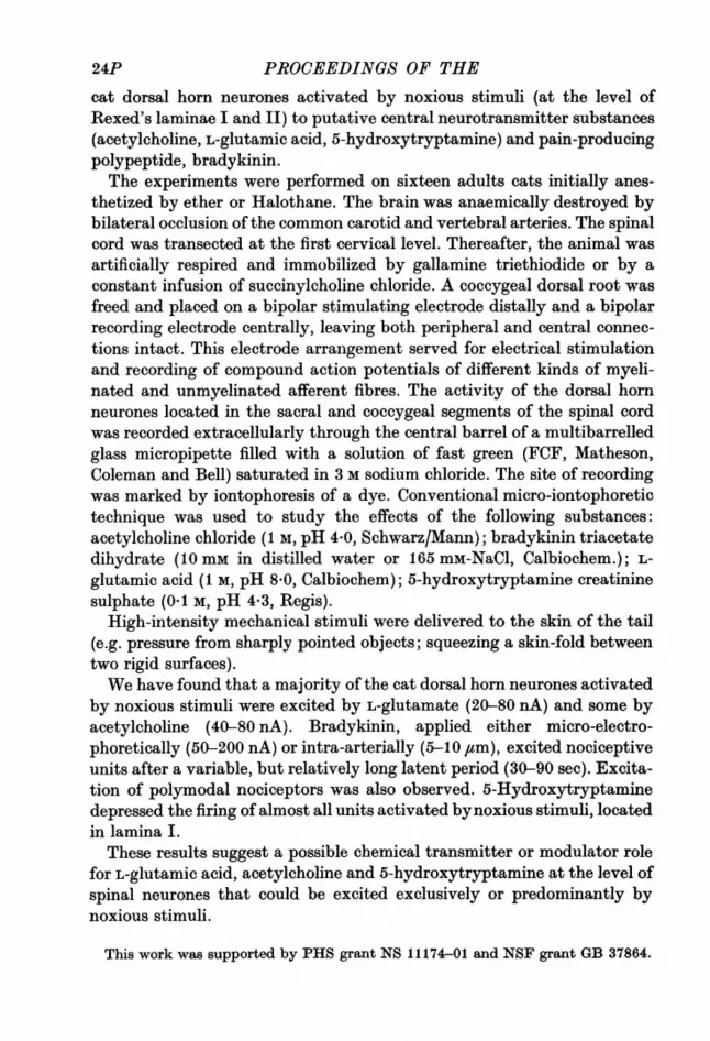
Backward placing in intact kittens and adult catsBY V. E. AMASSIAN. Department of Physiology, SUNY Downstate MedicalCenter, New York, U.S.A.When initiated by contact with the dorsal, radial or ulnar aspect of the
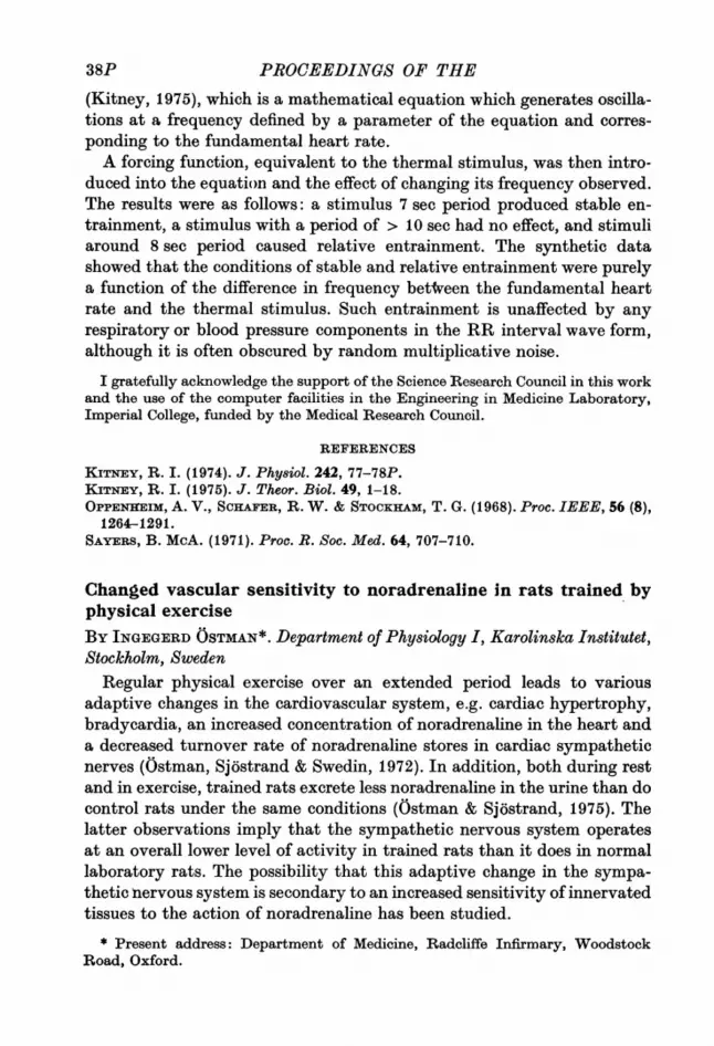
forelimb, contact placing (CP) has an initial lifting-withdrawal phase thatis produced by activation of flexors (Amassian, Ross, Wertenbaker &Weiner, 1972). Backward placing is readily elicited by gentle contact ofeither the ventral apect of the wrist or the back of the heel of an unsup-ported limb with the side or the edge of a solid. Unlike forward, medial or
Forward CP Backward CP
Landing contact
Photocell
G
..~....... .,....,..,
TA0-5 sec
Fig. 1. Forward and backward placing in an intact 47-day-old kitten. Con-tact of the hind paw with the front of the apparatus interrupts light beamsincident on a column of photocells. The altered output of the photocells isrestored by lifting-withdrawal of the paw. Subsequently, if the paw landson the solid, a circuit is completed through the kitten, the small currentbeing driven by dissimilar electrode potentials. The electromyogramswere each recorded with bipolar pins inserted percutaneously into thegastrocnemius (G) and tibialis anterior (TA) muscles; they were integratedwith a decay time constant of 10 msec. The overall gain for TA is approxi-mately five times that for G. At left, the dorsum of the digits contacts theapparatus. At right, contact of the back of the heel leads to backwardplacing, but the light beams are occluded by the protruding foot. Allrecords played back from tape through an ink-writing oscillograph.
lateral CP, the paw is usually only partially placed on top of the solid, thedigits protruding beyond the edge. Backward CP was elicited in adult catsand in kittens tested as early as 6-10 days old. In the intact kitten, thetibialis anterior is a prime mover in the initial phase of forward (dorsal) CPof the hind limb (Fig. 1. left), as was previously shown for the responsesrecorded in chronic spinal kittens by Forssberg, Grillner & Sjostr6m (1974).However, in backward CP, gastrocnemius is a prime mover and tibialisanterior is initially either silent or is only weakly activated with the ankleextensors (Fig. 1, right).
26P
PHYSIOLOGICAL SOCIETY, JULY 1975 27
Flexors are prime movers in stepping (Griliner, 1973), which is promi-nent in spinal kittens. In such preparations, the difficulty in elicitingplacing to tactile stimulation of the lateral aspect of the hind pawencountered by Forssberg et al. (1974), suggests the possibility that theresponse they readily obtained to dorsal stimulation started as a triggeredstep. Testing for backward CP provides an additional criterion which maybe useful in distinguishing the full tactile placing reaction from other motorresponses seen after lesions of the higher motor control systems.
This work was aided by USPHS Grant NS 11219.
REFERENCES
AMASSIAN, V. E., Ross, R., WERTENIBAKER, C. & WEINER, H. (1972). In Corti-cothalamic Projections and Sensorimotor Activities, ed. FRiGYEsi, T., Riwvix, E. &YAmuI, M. D., pp. 395-444. New York: Raven Press.
FORSSBERG, H., GRILLNER, S. & SJOsTROm, A. (1974). Acta physiol. scand. 92,114-120.
GRILLNER, S. (1973). In Control Of Posture and Locomotion, ed. STEIN, R. B., PEARn-SON, K. G., 5MmiT, R. S. & REDFORD, J. B., pp. 515-535. New York: PlenumPress.
Synthesis of microtubule protein in rat visual cortex during earlypost-natal life in relation to eye-openingBy J. R. CRONLY-DILLON and G. W. PERRY. Department of OphthalmicOptics, University of Manchester Institute of Science and Technology,Manchester M13 9PLIn several species the functional organization of the visual cortex can be
disrupted by visual deprivation during a certain 'critical period' in earlydevelopment (Hubel & Wiesel, 1970). Recent evidence implicating micro-tubules in axonal transport suggests the possibility that the turnover ofbrain tubulin may be critical in determining growth during early develop-ment of the visual cortex. We have therefore measured the rate of tubulinsynthesis during early life, paying particular attention to the periodimmediately preceding and following eye-opening.Rats were reared under normal conditions of illumination and then
killed at various times after birth, whereupon a plug of tissue was re-moved from the visual cortex in each hemisphere, care being taken toremove the white matter. The remaining plug of visual cortex grey matterwas used for the estimation of tubulin. Both the concentration of tubulinand its rate of synthesis were studied using a double labelling technique.Tritiated colchicine was used to assay tubulin and radioactive [14C]L-leu-cine injected intraventricularly 2 hr prior to death, to study rate ofsynthesis at different times post-natally.
27P
I9 PHY 252
28P ~~PROCEEDINGS OF THE
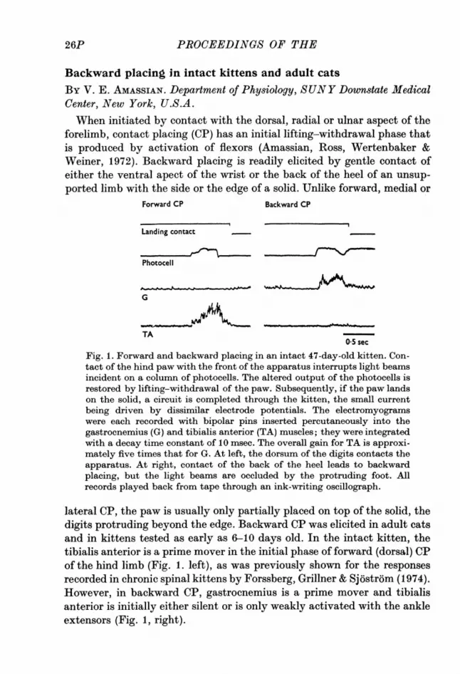
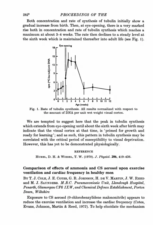
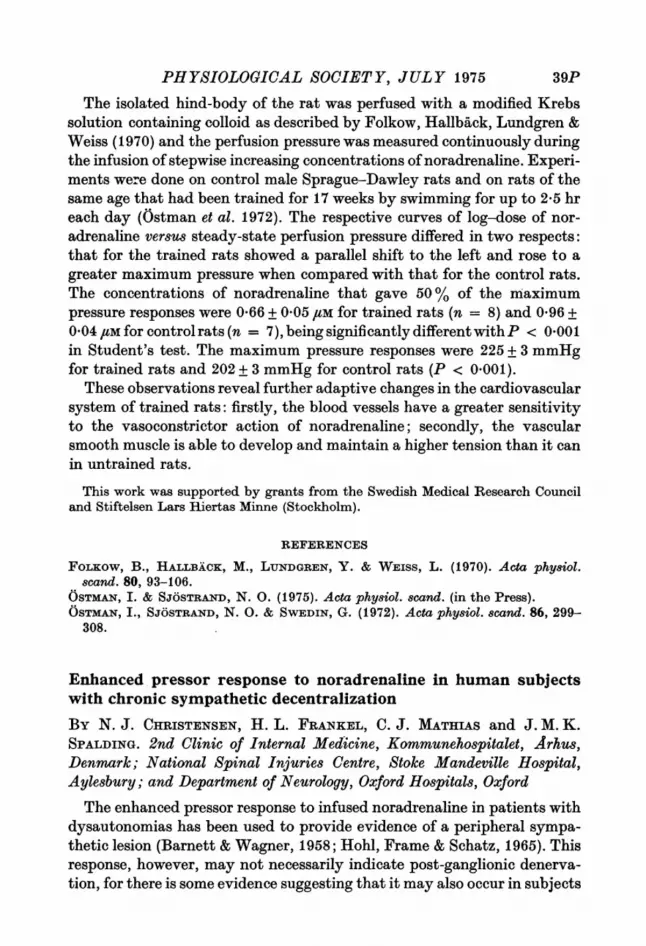
Both concentration and rate of synthesis of tubulin initially show agradual increase from birth. Then, at eye-opening, there is a very markedrise both in concentration and rate of tubulin synthesis which reaches amaximum at about 3-4 weeks. The rate then declines to a steady level atthe sixth week which is maintained thereafter into adult life (see Fig. 1).
IsEye opening
12
9
0.J
012 3 4 S 6 7 8 9 10 11 12Age (weeks)
Fig. 1. Rate of tubulin synthesis. All results normalized with respect tothe amount of DNA per unit wet weight visual cortex.
We are tempted to suggest here that the peak in tubulin synthesiswhich extends from eye-opening until about the sixth week after birth mayindicate that the visual cortex at that time, is 'primed for growth andready for learning'; and as such, this pattern in tubulin synthesis may becorrelated with the critical period of susceptibility to visual deprivation.However, this has yet to be demonstrated physiologically.
REFERENCE
HUBEL, D. H. & WIESEL, T. W. (1970). J. Physiol. 206, 419-436.
Comparison of effects of ammonia and CS aerosol upon exerciseventilation and cardiac frequency in healthy menBy T. J. COLE, J. E. COTEs, G. R. JOHNSON, H. DE V. MARTIN, J. W. REEDand M. J. SAUNDERS. M.R.C. Pneumoconiosis Unit, Llandough Hospital,Penarth, Glamorgan CF6 iX W, and Chemical Defence Establishment, PortonDown, Wiltshire
Exposure to CS aerosol (0-chlorobenzylidene malononitrile) appears toreduce the exercise ventilation and increase the cardiac frequency (Cotes,Evans, Johnson, Martin & Reed, 1972). To help elucidate the mechanism
28P

PHYSIOLOGICAL SOCIETY, JULY 1975
the response to a dosage of 0 4-44 mg m-3 has been compared with that toammonia in the dosage 50-340 mg m-3 (70-480 ppm). The 17 subjects andthe methods (including a progressive submaximal exercise test, Cotes,1972) were similar to those used previously.Measurements were made on three control days and during exposure to
CS and ammonia in two concentrations. The conditions were similar on alloccasions, except that during exposure, due to the ventilation of thechamber being turned off, the ambient temperature was on average24.00 C compared with 20.40 C on the control days.
Ventilation and cardiac frequency were interpolated to an oxygenuptake of 45 mmol min- and tidal volume to a respiratory frequency of20 min- ( fE45, fc., and VT20 respectively).
f04c was increased by exposure to CS by on average 6*0 min- (P < 0.05)from 101-3 to 107-3 min-; the increase was apparently related to dose.A similar increase was observed with ammonia.
VE45 was depressed during exposure to both gases on average by 1.5 1.min-' (P < 0-01) from 2541 to 23-6 1. min-. The change reflected a diminu-tion in the exercise tidal volume, the average VT20 diminishing from 1*34to 1-14 1. (P < 0.01). The reduction for CS was independent of dose but forammonia it was dose-related above a concentration of about 108 mg M-3(150 ppm). The respiratory frequency was increased slightly by exposureto CS. With ammonia it was reduced by exposure to low concentrationsand increased during exposure to higher ones.
These results confirm the earlier findings. They suggest that in thedosage used, the cardiorespiratory response to CS is small in relation to theassociated intense discomfort and not much different from that to am-monia in doses which cause only minimal symptoms. The increase incardiac frequency during exposure is that to be expected from the asso-ciated rise in ambient temperature (Miller & Martin, 1975). The reductionin ventilation volume may be due to the aerosols stimulating receptors inthe respiratory tract (e.g. Boushey, Richardson, Widdicombe & Wise,1974) and the changes in respiratory frequency to the operation of com-pensatory mechanisms.We are indebted to the Director and Dr F. W. Beswick, Medical Division, Chemi-
cal Defence Establishment, for provision of facilities and Messrs W. Hill, A. Kirkhamand R. G. White for other assistance.
REFERENCES
BousiEEy, H. A., RICIEARDSON, P. S., WIDDICOMBE, J. G. & WIsE, J. C. M. (1974).J. Physiol. 240, 153-175.
CoTEs, J. E. (1972). Br. J. Dis. Chest 66, 169-184.CoTEs, J. E., EvANs, L. R., JoimsoN, G. R., MARTiN H. DE V. & REED, J. W. (1972).
J. Physiol. 222, 77-78P.MILLER, G. J. & MARTIN H. DE V. (1975). Ergonomics 18 (in the Press).
19-2
29P

30P ~~~PAROCEEDINGS OF THE
Assessment of closure of lung units based on the pressure-volumecurve
BY JANE DAVIS. Physiological Flow Studies Unit, Imperial College, London,S. W. 7, D. H. GLAiSTER, Royal Air Force Institute of Aviation Medicine,Farnborough, Hampshire, and R. C. SCHROTER, Physiological Flow StudiesUnit, Imperial College, London, S. W. 7The deflation limb of the vital capacity pressure-volume curve in up-
right seated subjects and in excised lungs is sigmoid in shape. We haveshown earlier (Glaister, Milic-Emili, Schroter & Sudlow, 1972) that theupper portion of the curve may be closely represented by an exponentialfunction. The inflexion point, occurring at a distending transpulmonarypressure of approximately cmH2O, is associated with the onset of closureof lung units. This closure is either functional in form or real closure ofsmall terminal airways.We have studied the pressure volume curve at transpulmonary pressures
below the inflexion point and separated the effects of progressive closureof units from the elastic properties of individual lung units. If closure ofair spaces occurred simultaneously at residual volume then the pressure-volume curve would be exponential throughout its length. The ratio ofthe slopes of the actual and hypothetical pressure-volume curves at a
given low transpulmonary pressure thus indicates the proportion of unitsventilating at that pressure.
Deflation pressure-volume curves of excised dog and primate lungs andof seated normal subjects have been assessed in this way. It was found thatapproximately 70% of lung units closed when the transpulmonary pres-sure was reduced to 3 cmH2O below the inflexion point, the pressure atwhich closure began. This indicates that the majority of lung units closearound zero transpulmonary pressure. The rate of closure became muchless as the pressure was further reduced and even at residual volume (-5to -10 cmH2O) approximately 10 % of the units were still not closed.Units which have closed will resist any further reduction in size and willthus provide a degree of support for patent units making them stiffer andthus preventing their closure.
REFERENCE
GLAISTER, D. H., MILIC-EMILI, J., SCOEROTER, R. C. & SUDLOW, M. F. (1972). J.Physiol. 223, 24-25P.
30P

PHYSIOLOGICAL SOCIETY, JULY 1975
Influence of smooth muscle tone on intrapulmonary airway calibreBY T. P. CLAY, J. M. B. HUGHES and HAZEL A. JONES. Departments ofMedicine and Radiology, Royal Postgraduate Medical School, HammersmithHospital, London W12 OHS
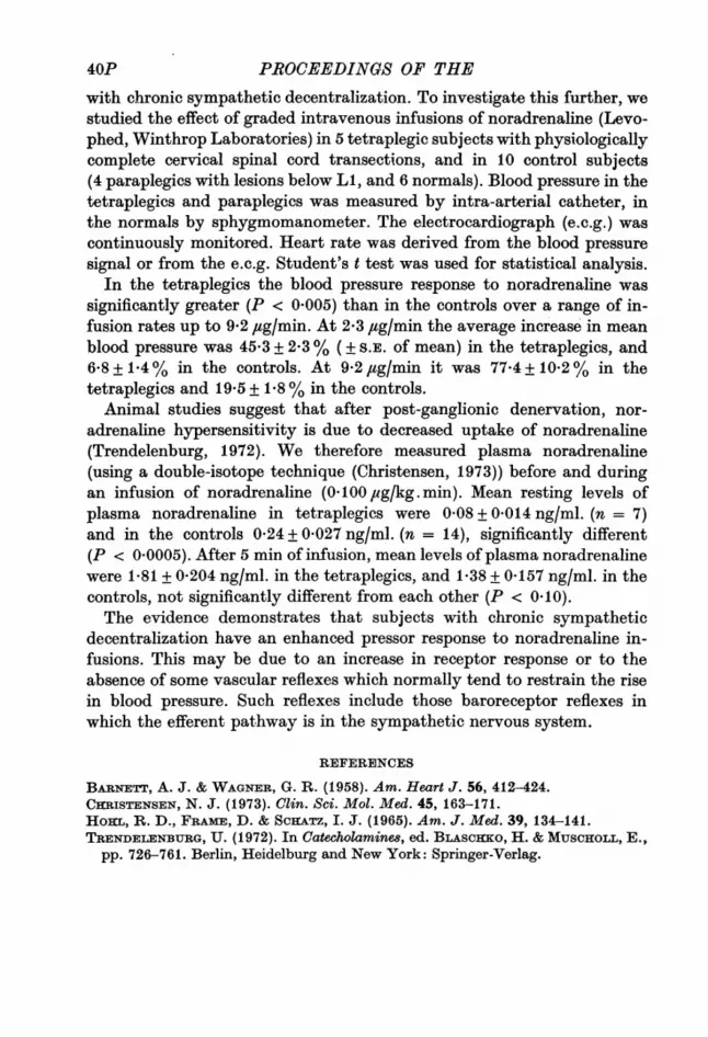
In the lung, airway expansion is linked to parenchymal expansion byvirtue of tissue attachments (Mead, Takashima & Leith, 1970). At constantlung volume the compliance of peribronchial tissues might be sufficientlylow to limit severely bronchodilation or constriction, especially at highlung volumes. Nevertheless, in excised lungs at constant volume a 20%narrowing of bronchial diameter occurred when intraluminal pressure waslowered by 15 cmH2O relative to pleural pressure (Hughes, Jones, Wilson,Grant & Pride, 1974). In the present study the competing effects of(a) mechanical interdependence and (b) changes of smooth muscle tone,upon intrapulmonary airway calibre have been studied.
100 x -- -- EDTA deflationo - - oEDTA inflationx 80 *'HistamineEo60
0 O EDTA
E * Histamine20 -
U 20
C 0 10 20 30 0 20 40 60 .80 1000coi Transpulmonary Lung volume I (% max.)
pressure (cmH20)
Fig. 1. Mean bronchial diameter as percent maximum after EDTA treat-ment plotted against (a) transpulmonary pressure and (b) cube root of lungvolume (as percentage maximum) after treatment with histamine andEDTA solutions. Vertical bars indicate S.E. mean for all airways measured(n = 52, diameter range 0-9-6-4 mm). Line of identity between diameterand volume I passes through EDTA points. Note almost parallel shiftsproduced by changes in smooth muscle tone.
Ten lower lobes from freshly excised dog lungs were degassed and filledwith solutions of isotonic saline, histamine (5 /ug/ml.) or isoprenaline(1O ug/ml.), and after twenty minutes emptied. Tantalum dust was in-sufflated into the airways and stereoscopic X-ray pairs taken to measurebronchial dimensions at several distending pressures during deflation andinflation. Lung volume changes were determined and absolute volumeobtained from the weight and water displacement. The procedure wasrepeated after degassing and filling the lung with 4 mM-EDTA solution.
31P

32PPROCEEDINGS OF THE
Fig. 1 shows airway diameter plotted against (a) lung distending pressureand (b) a linear function of volume, under conditions of high (histamine)and low (EDTA) airway tone. On inflation, changes of diameter occur atthe same pressure and volume (up to 35 %) between EDTA and histamine,and these changes persist up to full inflation. Smaller airways (< 2 mmi.d.) showed the larger differences. Isoprenaline had a similar effect toEDTA.We conclude that for the same lung volume the stiffness of peribronchial
tissue attachments is not sufficient to prevent 30-35% changes of intra-pulmonary airway calibre when smooth muscle tone is varied.
REFERENCES
HuGBEs, J. M. B., JoNEs, HAZEL, A., WILSON, A. G., GRANT, B. J. B. & PRIDE, N. B.(1974). J. apple. Phy8iol. 37, 684-694.
MEAD, J., TAiIsmMA, T. & LEITH, D. (1970). J. appi. Phys'iol. 28, 596-608.
An evaluation of rebreathing techniques used in the measurementof cardiac outputBY E. ZEIDIFARD. Department of Paediatrics and Neonatal Medicine,Hammersmith Hospital, London W12 OHSThe non-invasive carbon dioxide and nitrous oxide rebreathing methods
have often been used to determine cardiac output during exercise. The CO2rebreathing method estimates oxygenated mixed venous P002 (Pv o,) froma 10 to 12 sec rebreathing manoeuvre, and this is then inserted into the Fickequation for deriving cardiac output. In the N20 rebreathing method, thecardiac output is determined directly from the rate of absorption ofN20 bypulmonary blood during a 10 to 12 sec rebreathing period.A number of alternative techniques for performing these studies have
been proposed, but little systematic evaluation of them appears to havebeen performed. Two alternative methods for measuring PvC02 during re-breathing are an exponential analysis of rising PcO2 starting with a low gasconcentration (Defares method) and linear extrapolations from nearplateau levels (Denison method). In the N20 method various alternativecalculations have been used with varying degrees of correction for experi-mental errors.In a series of studies, the various methods for measuring Pv-02 and
cardiac output during exercise were initially compared. Plateau estimatesof Pivco2 and cardiac output were found to be more reproducible thancorresponding exponential estimates. Furthermore, for a range of re-breathing mixtures, a linear (Denison) as opposed to an exponential(Defares) extrapolation of PuCO2 also improved the reproducibility of the
32P

PHYSIOLOGICAL SOCIETY, JULY 1975
estimates. Investigation of various N20 rebreathing methods indicatedthat the reproducibility of cardiac output was improved when the expo-nential-like uptake of N20 by pulmonary blood was corrected for changesin the total rebreathing volume, and for a quantity of N20 dissolved inlung tissue.A comparison of the plateau CO2 with the modified N20 rebreathing
method indicated that the absolute value of cardiac output, and theirreproducibility, were similar in both methods. The two methods alsocompared favourably with direct Fick and dye dilution estimates ofcardiac output.
The separate effects of alternate-breath oscillations of PA,CO2during hypoxia on inspiration and expirationBY D. J. C. CUNNINGHAM and S. A. WARD.* The University Laboratory ofPhysiology, OxfordWe have examined the phase relations of the breath-by-breath reflex
responses of inspiration and expiration provoked by alternate inspirates ofhigh and low Pco, during hypoxia (Marsh, Lyen, McPherson, Pearson &Cunningham, 1973; Cunningham & Ward, 1975; cf. Wolff, 1975). Thirtyruns were performed at rest and 28 in mild exercise (H.R. 120 min-),each run comprising 40-150 breaths from which 13 respiratory outputvariables were obtained (inspiratory and expiratory times, volumes, flowsand accelerations).A prior, flow and the time for which it occurs may be regarded as the
primary variables ofinspiration and expiration. The patterns of alternatingresponse were therefore characterized in terms of mean inspiratory andexpiratory flows (VI and viE) and expiratory time (TE). Inspiratory time wasexcluded from the analysis because of its stability in the face of thePA00co2 oscillation.A variety of patterns was observed, one tending to predominate over
the others in a given experiment both at rest and during exercise.(1) A synergistic alternation of inspiratory and expiratory variables, in
which an inspiratory stimulation (VI increased) and an expiratory stimula-tion (either vtE increased or TE decreased, or both) occurred in the samebreath, and a depression of both in the next breath, et seq.
(2) An antagonistic alternation of inspiration and expiration, in whichan inspiratory stimulation and an expiratory depression occurred in thesame breath, and the converse in the next breath, et seq. This tended tostabilize ventilation.
* States of Guernsey Graduate Research Scholar.
33P
34P ~~P.ROCEEDINGS OF THE
(3) An alternation of inspiration alone.(4) An alternation of expiration alone.VT, I usually alternated more prominently than VT, E; the breath-by-
breath difference between them, which is AFRO, also alternated, indeedmore consistently than any other variable. Thus the passive relaxationvolume of the lung is accessible to the alternating signal.These results are interpreted in terms of the dynamic properties of the
carotid chemoreflex arc. In the cat either a stimulation of inspiration or adepression of expiration (increased T.) may be produced by afferent chemo-receptor volleys, depending upon the time of arrival of the volley at thebrainstem during the current respiratory cycle (Black & Torrance, 1967;Eldridge, 1972). Pattern 2 (antagonistic) could result if the alternatinginput to the brainstem arrived in phase with the respiratory cycle, andpattern 1 if the signal were out of phase by half a respiratory cycle.
It appears, therefore, that no single respiratory variable (e.g. ventila-tion) is the sole target for a changing drive under the conditions of ourexperiments. The ability to influence inspiration and expiration inde-pendently argues against an immutable linkage between them. Further-more, the observation that FRC alternates is scarcely consistent with thereturn of lung volume to FRC being important for the initiation ofinspiration.
REFERENCES
BLAcx, A. M. S. & TORRAwcE, R. W. (1967). J. Physiol. 189, 59-61P.CuNNINGHcAm, D. J. C. & WARD, S. A. (1975). J. Physiol. 251, 37P.ELDRIDGE, F. L. (1972). J. Physiol. 222, 297-318.MARSH~, R. H. K., LYEN, K. R., McPHERSON, G. A. D., PEARSON, S. B. & CUNNING.HAm, D. J. C. (1973). Resp. Physiol. 18, 80-91,
WOLFF, C. B. (1975). J. Physiol. 244, 63-64P.
Effect of airway anaesthesia on the ventilatory response to CO2 inman
By BRENDA CROSS, A. Guz and S. K. JAIN. Department of Medicine,Charing Cross Hospital Medical School, London W. 6An aerosol of 5% bupivacaine hydrochloride was used to anaesthetize
the airways of nine normal subjects. The method of administration,physical characteristics and effects on respiratory reflexes of the aerosolhave been described previously (Archer et al. 1975; Jamn, 1975). The coughreflex is abolished; the apnoeic response to lung inflation is severely re-duced as is the reflex broncho-constriction produced by airway surfaceirritation with citric acid. There were no consistent changes in restingfrequency, tidal volume or end-tidal PCO,(PET, C02).
34P
PHYSIOLOGICAL SOCIETY, JULY 1975
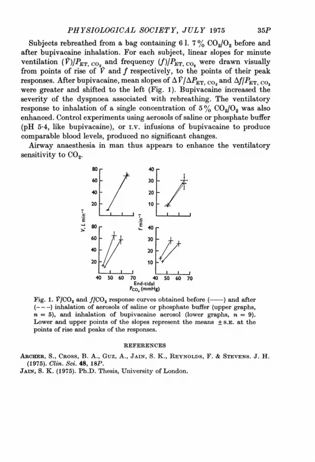
Subjects rebreathed from a bag containing 6 1. 7 % C02/02 before andafter bupivacaine inhalation. For each subject, linear slopes for minuteventilation (V)IPET co2 and frequency (V)/ ET, C02 were drawn visuallyfrom points of rise of f and f respectively, to the points of their peakresponses. After bupivacaine, mean slopes of A Jr/APET, C02 and Af/PET, Co2were greater and shifted to the left (Fig. 1). Bupivacaine increased theseverity of the dyspnoea associated with rebreathing. The ventilatoryresponse to inhalation of a single concentration of 5 % C02/02 was alsoenhanced. Control experiments using aerosols of saline or phosphate buffer(pH 5 4, like bupivacaine), or i.v. infusions of bupivacaine to producecomparable blood levels, produced no significant changes.Airway anaesthesia in man thus appears to enhance the ventilatory
sensitivity to CO2.
80 40-
60 - 30 -
40-20-
20 - 10 I
S TcE80 E 40
60 30 -±
40 // 20
201 10
40 50 60 70 40 50 60 70End-tidal
Pco2 (mmHg)Fig. 1. V/CO2 and f/CO2 response curves obtained before ( ) and after(---) inhalation of aerosols of saline or phosphate buffer (upper graphs,n = 5), and inhalation of bupivacaine aerosol (lower graphs, n = 9).Lower and upper points of the slopes represent the means + S.E. at thepoints of rise and peaks of the responses.
REFERENCES
ARCOIER, S., CROSS, B. A., Guz, A., JAIN, S. K., REYNOLDS, F. & STEVENS. J. H.(1,975). Clin. SC?. 48, 18P.
JAIN, S. K. (1975). Ph.D. Thesis, University of London.
35P
A6PPROCEEDINGS OF THE
The sympathetic contribution to increase in heart rate evoked bycutaneous nerve stimulation in the dogBY YOKO KUMAGAI,* J. NORMAN and J. G. WHITWAM. Department ofAnaesthetics, Royal Postgraduate Medical School, Hammersmith Hospital,DuCane Road, London W12 OHSIn dogs an increase in heart rate evoked by stimulation of a cutaneous
nerve can be mediated by a cholinergic vagal pathway (Norman & Whit-wamr, 1974) and the magnitude of the response is related to the bloodpressure before stimulation of the peripheral nerve (Norman & Whitwam,1973a). However, stimulation of cutaneous nerves also evokes activity insympathetic nerves in dogs (Fussey, Kidd & Whitwam, 1969; Norman &Whitwam, 1973b) and the experiments reported here were concerned withthe role of the sympathetic nervous system in reflexly evoked increases inheart rate.Dogs in which anaesthesia was induced with methohexitone and main-
tained with chloralose were artificially ventilated and paralysed withsuxamethonium. The vagus nerves were exposed in the neck and acutaneous branch of the radial nerve was desheathed, cut and mounted onstimulating electrodes in a mineral oil pool. Arterial blood pressure, thee.c.g., heart rate and a respiratory wave form were recorded. Trains ofstimuli (intensity 40-60 V, duration 0 5 msec, frequency 20 Hz and trainduration 10 see), triggered by an R wave occurring in expiration, wereapplied to the radial nerve and the evoked changes in heart rate and bloodpressure were measured.
Before and after vagotomy radial nerve stimulation evoked comparableincreases in heart rate (averages 33 and 36 beats/min respectively) but themaximum heart rate was delayed from an average of 4.7 to 7*0 see aftervagotomy. Also after vagotomy the relation between resting blood pressureand heart rate showed a smaller change in heart rate in response to achange in blood pressure than in sympathectomized dogs (Norman &Whitwam, 1973a). The changes in the evoked increase in heart rate asblood pressure was altered were also smaller.
In dogs in which the vagus nerves were divided and the post-ganglionicnerves blocked with bretylium tosylate (10 mg kg-'), stimulation of theradial nerve evoked no change in heart rate, but a small rise in bloodpressure was seen which could be abolished either by adrenalectomy or byhexamethonium bromide (5 mg kg-').
It was concluded that stimulation of a cutaneous nerve can evoke in-creases in heart rate which are mediated by increased sympathetic activity
* British Oxygen Company Research Fellow.
36P
PH YSIOLOGICAL SOCIETY, JULY 1975
whose magnitude is dependent on the blood pressure but that the adrenalmedulla makes no significant contribution to these responses. The sympa-thetic component of heart rate changes is less sensitive to variationsin blood pressure and to cutaneous nerve stimulation than the vagalcomponent.
This work was supported by the Medical Research Council.
REFERENCES
FusSEY, I. F., KIDD, C. & WETWAM, J. G. (1969). J. Physiol. 200, 77-78P.NORMAN, J. & WHITWAM, J. G. (1973a). J. Physiol. 234, 89-90P.NORMAN, J. & WHITWAM, J. G. (1973b). J. Physiol. 231, 76-77P.NORMAN, J. & WHITWAM, J. G. (1974). Br. J. Pharmac. 51, 150P.
Entrainment of the human RR interval by thermal stimuliBy R. I. KITNEY. Department of Physiology, Basic Medical Sciences Group,Chelsea College, Manresa Road, London SW3 6LXIn a previous communication to the Society (Kitney, 1974), I discussed
the entrainment of heart rate by the thermoregulatory system; the subjectof this communication is the identification of the nature of this thermalentrainment (of theRR interval wave form). Experiments were undertakenin which periodic square wave thermal stimuli (180 and 460, 7 (0-142 Hz)-10 (0.1 Hz) sec period) were applied to the left hand of 5 subjects whiletheir e.c.g.s were recorded; the duration of each experiment was (15 min).The normal result as previously described was that the power spectrum ofthe derived RR interval wave form exhibited a large component at thethermal stimulus frequency. However, other results showed spectra withlarge frequency components around, but not actually at, the stimulusfrequency. In RR interval spectra there are often large components corres-ponding to respiration, blood pressure control and thermoregulatorycontrol (Sayers, 1971), but they obey the principle of super position, beingadditive. The analysis of the present experimental results led to the hypo-thesis that the spectra resulted from interference of a multiplicative ratherthan an additive nature. Separation of signal and noise under such circum-stances can only be achieved using homomorphic filters (Oppenheim,Schafer & Stockham, 1968).
Following such filtering two conditions were revealed: the first showedspectra with a large component precisely at the stimulus frequency; in thesecond the filtered RR interval wave forms showed regions where anoscillation at the stimulus frequency was clearly visible, indicating that thewave form was moving in and out of entrainment (relative entrainment).This phenomenon was studied further using a Van de Pol oscillator
37P
38P ~~PARO1CEEDINGS OF THE
(Kitney, 1975), which is a mathematical equation which generates oscilla-tions at a frequency defined by a parameter of the equation and corres-ponding to the fundamental heart rate.A forcing function, equivalent to the thermal stimulus, was then intro-
duced into the equation and the effect of changing its frequency observed.The results were as follows: a stimulus 7 sec period produced stable en-trainment, a stimulus with a period of > 10 sec had no effect, and stimuliaround 8 sec period caused relative entrainment. The synthetic datashowed that the conditions of stable and relative entrainment were purelya function of the difference in frequency between the fundamental heartrate and the thermal stimulus. Such entrainment is unaffected by anyrespiratory or blood pressure components in the RR interval wave form,although it is often obscured by random multiplicative noise.
I gratefully acknowledge the support of the Science Research Council in this workand the use of the computer facilities in the Engineering in Medicine Laboratory,Imperial College, funded by the Medical Research Council.
REFERENCES
KiTNEY, R. I. (1974). J. Physiol. 242, 77-78P.KITNEY, R. I. (1975). J. Theor. IBiol. 49, 1-18.OPPENiIEim, A. V., ScHAFER, R. W. & STOcKHEAM, T. G. (1968). Proc. IEEE, 56 (8),
1264-1291.SAYERS, B. McA. (1971). Proc. R. Soc. Med. 64, 707-710.
Changed vascular sensitivity to noradrenaline in rats trained byphysical exerciseBy INGEGERD OSTMAN*. Department of Physiology I, Karolinska, Institutet,Stockholm, SwedenRegular physical exercise over an extended period leads to various
adaptive changes in the cardiovascular system, e.g. cardiac hypertrophy,bradycardia, an increased concentration of noradrenaline in the heart anda decreased turnover rate of noradrenaline stores in cardiac sympatheticnerves (Ostman, Sj6strand & Swedin, 1972). In addition, both during restand in exercise, trained rats excrete less noradrenaline in the urine than docontrol rats under the same conditions (Ostman & Sj6strand, 1975). Thelatter observations imply that the sympathetic nervous system operatesat an overall lower level of activity in trained rats than it does in normallaboratory rats. The possibility that this adaptive change in the sympa-thetic nervous system is secondary to an increased sensitivity of innervatedtissues to the action of noradrenaline has been studied.
* Present address: Department of Medicine, Radcliffe Infirmary, WoodstockRoad, Oxford.
38P
PHYSIOLOGICAL SOCIETY, JULY 1975
The isolated hind-body of the rat was perfused with a modified Krebssolution containing colloid as described by Folkow, Hallback, Lundgren &Weiss (1970) and the perfusion pressure was measured continuously duringthe infusion of stepwise increasing concentrations ofnoradrenaline. Experi-ments were done on control male Sprague-Dawley rats and on rats of thesame age that had been trained for 17 weeks by swimming for up to 2-5 hreach day (Ostman et al. 1972). The respective curves of log-dose of nor-adrenaline versus steady-state perfusion pressure differed in two respects:that for the trained rats showed a parallel shift to the left and rose to agreater maximum pressure when compared with that for the control rats.The concentrations of noradrenaline that gave 50 % of the maximumpressure responses were 0*66 + 0 05 ,am for trained rats (n = 8) and 0-96 +0 04 ftM for controlrats (n = 7), being significantly differentwithP < 0 001in Student's test. The maximum pressure responses were 225 + 3 mmHgfor trained rats and 202 + 3 mmHg for control rats (P < 0.001).
These observations reveal further adaptive changes in the cardiovascularsystem of trained rats: firstly, the blood vessels have a greater sensitivityto the vasoconstrictor action of noradrenaline; secondly, the vascularsmooth muscle is able to develop and maintain a higher tension than it canin untrained rats.
This work was supported by grants from the Swedish Medical Research Counciland Stiftelsen Lars Hiertas Minne (Stockholm).
REFERENCESFoLKow, B., HALLBACK, M., LUNDGREN, Y. & WEIss, L. (1970). Acta physiol.
scand. 80, 93-106.OSTMAN, I. & SJOSTRAND, N. 0. (1975). Acta physiol. sand. (in the Press).OSTMAN, I., SJOSTRAND, N. 0. & SWEDIN, G. (1972). Acta physiol. Scand. 86, 299-
308.
Enhanced pressor response to noradrenaline in human subjectswith chronic sympathetic decentralizationBy N. J. CHRISTENSEN, H. L. FRANKEL, C. J. MATHIAS and J. M. K.SPALDING. 2nd Clinic of Internal Medicine, Kommunehospitalet, Arhus,Denmark; National Spinal Injuries Centre, Stoke Mandeville Hospital,Aylesbury; and Department of Neurology, Oxford Hospitals, OxfordThe enhanced pressor response to infused noradrenaline in patients with
dysautonomias has been used to provide evidence of a peripheral sympa-thetic lesion (Barnett & Wagner, 1958; Hohl, Frame & Schatz, 1965). Thisresponse, however, may not necessarily indicate post-ganglionic denerva-tion, for there is some evidence suggesting that it may also occur in subjects
39P
P PROCEEDINGS OF THE
with chronic sympathetic decentralization. To investigate this further, westudied the effect of graded intravenous infusions of noradrenaline (Levo-phed, Winthrop Laboratories) in 5 tetraplegic subjects with physiologicallycomplete cervical spinal cord transactions, and in 10 control subjects(4 paraplegics with lesions below LI, and 6 normals). Blood pressure in thetetraplegics and paraplegics was measured by intra-arterial catheter, inthe normals by sphygmomanometer. The electrocardiograph (e.c.g.) wascontinuously monitored. Heart rate was derived from the blood pressuresignal or from the e.c.g. Student's t test was used for statistical analysis.In the tetraplegics the blood pressure response to noradrenaline was
significantly greater (P < 0.005) than in the controls over a range of in-fusion rates up to 9-2 ,ug/min. At 2-3 ,ug/min the average increase in meanblood pressure was 45.3 + 2 3% ( + S.E. of mean) in the tetraplegics, and6-8+ 1.4% in the controls. At 9.2,tg/min it was 77-4+ 10.2% in thetetraplegics and 19-5 + 18 %/ in the controls.Animal studies suggest that after post-ganglionic denervation, nor-
adrenaline hypersensitivity is due to decreased uptake of noradrenaline(Trendelenburg, 1972). We therefore measured plasma noradrenaline(using a double-isotope technique (Christensen, 1973)) before and duringan infusion of noradrenaline (0.100 fig/kg.min). Mean resting levels ofplasma noradrenaline in tetraplegics were 0-08 + 0-014 ng/ml. (n = 7)and in the controls 0-24 + 0-027 ng/ml. (n = 14), significantly different(P < 0-0005). After 5 min of infusion, mean levels of plasma noradrenalinewere 1-81 + 0-204 ng/ml. in the tetraplegics, and 1-38 + 0-157 ng/ml. in thecontrols, not significantly different from each other (P < 0.10).The evidence demonstrates that subjects with chronic sympathetic
decentralization have an enhanced pressor response to noradrenaline in-fusions. This may be due to an increase in receptor response or to theabsence of some vascular reflexes which normally tend to restrain the risein blood pressure. Such reflexes include those baroreceptor reflexes inwhich the efferent pathway is in the sympathetic nervous system.
REFERENCES
BARNETT, A. J. & WAGNER, G. R. (1958). Am. Heart J. 56, 412-424.CERISTENSEN, N. J. (1973). Clin. Sci. Mol. Med. 45, 163-171.HouL, R. D., FRAME, D. & SCi:ATZ, I. J. (1965). Am. J. Med. 39, 134-141.TRENDELENBURG, U. (1972). In Catecholamine8, ed. BLAscinEo, H. & Musc]EOLI, E.,
pp. 726-761. Berlin, Heidelburg and New York: Springer-Verlag.
40P

PHYSIOLOGICAL SOCIETY, JULY 1975 41P
Retinotopic organization in visual cortex and superior colliculus ofthe golden hamsterBy C. BLAKEMORE and TIAO, YUN-CHENG.* Physiological Laboratory,University of Cambridge, Cambridge CB2 3EGWe have recorded activity from unit clusters and single units in the
visual cortex (twenty-two animals) and superior colliculus (four animals)of hamsters, anaesthetized with i.v. infusion of urethane and paralysedwith Flaxedil.
Ipsilateral 3
Contralateral
/ ~~~~~~~~~27Oioo ~~~~~~Inferior
V2 030 /0VI
/ 0~~~~
30 S~~~uperior -
5 4 3 2 1 0A
Superior
Schneider's (1969) architectonic divisions of posterior cortex. B, left visualcortex, showing representation of cartographic, axis-vertical coordinates ofa hemisphere centre on the eyes (inferior and ipsilateral directionsnegative). C, projection of left superior colliculus on to horizontal stereo-taxic plane showing retinotopic organization. All dimensions in millimetres.Stereotaxic zero is the middle of the lambda suture.
In the cortex (Fig. 1 B) there are two main visually responsive areas,V 1 (corresponding to cytoarchitectural 17) and, more laterally, V 2 (area18a), with a retinotopic reversal at the border. The projection to V 1 islargely binocular and extends from 100 ipsilateral to 900 contralateral,
* British Council Research Visitor on leave from Institute of Biophysics,Academia Sinica, Peking, The People's Republic of China.

4PROCEEDINGS OF THE
from 400 down to 600 up. A larger area of visual field, virtually the entirecontralateral retina, is mapped on to the tectum (Fig. 1 C), but only asmall region around the area centralis representation has binocular input.The differences in the area of field represented and the magnification in
cortex and colliculus may partly account for the contrasting effects oflesions in the two areas (Schneider, 1969).
This work was supported by a grant from the M.R.C.
REFERENCE
SCHNEIDER, G. E. (1969). Science, N.Y. 163, 895-902.
Visual signals in the catBY B. G. CLELAND, W. R. LEVICK and W. A. H. RUSHTON. John CurtinSchool, Canberra, Australia, A.C.T. 2601A green flash of strength 0, falling upon a red steady background of
strength 0, elicits from rods a signal S that is transmitted to the connectorganglion, and generates N extra spikes. We have counted N after variousflashes, and deduced the size of the ganglion input S.
.c 50 () Red bkgraM 40 - .g 0-1 scot tdZ.40 A
30B
3, t _ ~~~~Extra&20 1 / ike
10
3o = area
stimulus
-3 -21 0 1Log flash Intensity (scot td sac)
Fig. 1
Flashes fell entirely upon the centre of the ganglion's receptive field,illuminating a circle of diameter either 0.460 (white circles, Fig. 1) or0.230 (black circles). Since white circles lie 0-6 (= log 4) to the left ofblack, quadrupling the area and quartering the flash intensity leaves Nunchanged (Ricco's Law). To deduce S (lower staircase) we assume (a) equalN values imply equal S values, (b) with fixed retinal illumination, S4 (from0.46°) = 4S, (from 0.230). In lower staircase all 'risers' are 0-6 high(assumption b); 'treads' are as wide as 'treads' above, namely 0-6 (Ricco,
42P

PHYSIOLOGICAL SOCIETY, JULY 1975assumption a). Hence stair gradient is 1 0, S oc 0, and N is linear withlog S.
Repeating with different 0-values confirms
1OaN = bS = 0 OD
(Alpern, Rushton & Torii, 1970),The formula holds over a thousandfold range.
REFERENCE
ALPERN, M., RUSHTON, W. A. H. & ToRII, S. (1970). J. Physiol. 206, 209-227.
Response of cat visual cortical cells to kinetic contours and staticnoiseBy P. HAMMOND and D. M. MACKAY. Research Department of Communi-cation, University of Keele, StaffordshireWith the moving black/white bars and straight edges commonly used
as stimuli for visual cortical units it is difficult to distinguish sensitivityto orientation from sensitivity to direction of motion, since the effectivecomponent of stimulus movement is always perpendicular to edgeorientation. Using movable patterns of static visual noise, however,one can test for sensitivity to motion without presenting an orientedluminance contour. At the boundary between two areas of noise in relativemotion one can also generate a 'kinetic contour' whose orientation andlocation can be changed independently of the direction of noise motion.We have already found qualitatively (Hammond & MacKay, 1975)
that such moving noise fields, or noise bars moving in the optimal direc-tion against stationary backgrounds of noise of identical texture, excitecomplex cells in area 17 of the lightly anaesthetized, paralysed cat whensimple cells in the same orientation column show no response. (We classifiedthese cells by such criteria as field size; presence or absence of discrete'on' and 'off' areas; sensitivity to bar length; preferred velocity; direc-tional bias; sharpness of orientational tuning; spontaneous firing rateand pattern of discharge to movement; and recording depth.) We nowreport a quantitative study of cell responses in visual cortex, showingthat although most simple cells are indifferent to noise fields drifting intheir preferred direction, or to noise bars moving on stationary noise fields,many complex cells show directionally biased responses to such stimuliwhich in some cases are stronger than those to optimally oriented light ordark bars of comparable dimensions moving against the same noisebackgrounds. All of 48 complex cells, compared with only one out of 44simple cells, showed some responsiveness to moving noise.
43P
PPROCEEDINGS OF THE
The few simple-type and complex-type hypercomplex cells so farencountered resemble their simple or complex counterparts in theserespects.
These findings appear to raise difficulties for current concepts of'hierarchic feature extraction' by simple and complex cells.
REFERENCEHAMMOND, P. & MACKAY, D. M. (1975). Expl Brain Res. 22, 427-430.
The application of thermography to the detection of energy meta-bolism of the brain in the new-born infantBY K. W. CROSS, J. K. STOTHERS, RUTH M. WARNER and R. W. WOOD-ROUGH. Department of Physiology, The London Hospital Medical College,Turner Street, London El 2AD, and Department of Medical Electronics,St Bartholomew's Hospital, West Smithfield, London, E.C. 1We have recently reported that the aural temperature of the new-born
infant when measured by the zero gradient aural thermometer (Keatinge& Sloan, 1973) is, in the neutral environment, considerably higher thanthat of the oesophagus (Cross & Stratton, 1974). Cross, Stothers & Stratton(1975) have also reported that after the infant has been fed this differentialtemperature becomes even greater, and concluded that this was morelikely to be because brain blood flow decreased than that a further increaseof brain energy metabolism occurred.We have now started a different approach towards estimating heat
output from the brain by examining thermographically the vertex of theinfant while it is in an incubator at a neutral environment (Aga Thermo-vision 680 Medical). The posterior portion of the anterior fontanelle showsitself to be considerably warmer than the surrounding skull-covered areasof the brain. Once again, after feeding there is a sharp rise of temperature(particularly in the posterior part of the vertex), and in a baby thoughtto be brain-damaged there was unilateral difference of temperature. Thedamaged part of the brain appearing to be less thermally responsive thanthe normal hemisphere. Our impression is that this method of examiningthe baby's brain, provided that the very strictest environmental condi-tions are maintained, provides a new and exciting insight into brain heatproduction and may have very considerable applications, not only inconsidering infant physiology, but also for the early detection of brainabnormalities. We feel that we are even nearer than we were with theaural thermometer in obtaining an index of brain heat output.
44P

PH'YSIOLOGICAL SOCIETY, JULY 1975
REFERENCES
CRoss, K. W., STOTHERS, J. K., STRATTON, D. (1975). J. Physiol. 250, 15-16P.CROSS, K. W. & STRATTON, D. (1974). Lancet, ii, 1179-1180.KEATINGE, W. R., SLOAN, R. E. G. (1973). J. Physiol. 234, 8-9P.
Afferent sympathetic nerve fibres with aortic endingsBY M. PAGANI. Cardiovascular Research Institute, University of Milan,and C.N.R., MilanoUsing anaesthetized and vagotomized spinal cats we have recently
shown that stretching the wall of the thoracic aorta induces reflex changesin the activity of single efferent sympathetic fibres in the left third thoracicramus communicans (T 3) which is known to contribute to the innerva-tion of the heart (Pagani, Schwartz, Banks, Lombardi & Malliani, 1974).
In this preparation the same stimulus was found to induce reflex in-creases in heart rate, myocardial contractility and arterial blood pressure,thus suggesting the existence of positive feed-back mechanisms in theneural control of the circulation (Lioy, Malliani, Pagani, Recordati& Schwartz, 1974).The present paper summarizes an electrophysiological investigation
into the properties of afferent sympathetic fibres which are likely tomediate these reflexes.Experiments were performed on anaesthetized, paralysed cats under
artificial ventilation. Details of the methods used for recording single unitactivity (T 3-T 6) and haemodynamic variables have been describedpreviously (Pagani et al. 1974).We recorded the activity of 21 single units, the endings of which were
found to be localized in the aortic wall. 18 fibres were spontaneouslyactive at a mean systolic blood pressure of 134 + 5 (s.E.) mmHg, givingnot more than a single impulse per cardiac cycle. Conduction velocitiessuggested that the fibres involved were mainly small myelinated (A delta)ones and that the unmyelinated (C) fibres were few.The activity in the fibres was markedly increased by raising aortic blood
pressure, either by occluding the descending aorta with a snare or injectingpressor drugs intravenously. Maximal activity was attained while arterialpressure was rising and was related to the extent and rate of such a rise.During this maximal excitation the units could give more than a singleimpulse per cardiac cycle. Induced hypotension caused a decrease orsuppression of the impulse activity.
Five units were studied at the end of the experiment, after the animalhad been killed. A saline-filled latex balloon was introduced into the aorta
45P

PROCEEDINGS OF THE
through the opened left ventricle in order to apply graded increases inpressure to the aortic segment where the endings were located. Theendings responded to the various increases in pressure (ranging from0-50 to 0-300 mmHg) with a burst of activity. For any given pessurestep, after a short latent period the firing rate reached a maximumfrequency, which rapidly declined toward much lower values (adaptation).In the range of pressures studied, the relationship between pressure andthe peak frequency was approximately linear, the threshold beingbetween 75 and 100 mmHg. The role of these mechanoreceptors insustaining a high sympathetic efferent activity through spinal excita-tory mechanisms seems worth studying under various physiological andpathological conditions (e.g. strenuous exercise, arterial hypertensionand hyperdynamic heart).
REFERENCES
Lioy, F., MALLIANI, A., PAGANI, M., RECORDATI, G. & SCHWARTZ, P. J. (1974).Circulation Res. 34, 78-84.
PAGANI, M., SCHWARTZ, P. J., BANKS, R., LOMBARDI, F. & MALLIANI, A. (1974).Brain Res. 68, 215-225.
Human blood platelet aggregation induced by dopamine,5-hydroxytryptamine and analoguesBy D. J. BOULLIN, A. R. GREEN and R. P. J. GRIMES. M.R.C. Unit andUniversity Department of Clinical Pharmacology, Radcliffe Infirmary,Oxford OX2 6HE
5-Hydroxytryptamine (5-HT) induced platelet aggregation has beenknown for some years (Mitchell & Sharp, 1964) and is associated with thetransport of the amine into platelets (Baumgartner & Born, 1969). Asdopamine is similarly transported (Boullin & O'Brien, 1970), we havecompared the effects of dopamine, 5-HT and some analogues on plateletaggregation.
Platelet-rich plasma (PRP) was prepared and aggregation studied asdescribed previously (Boullin, Grahame-Smith, Grimes & Woods, 1975).20-50 IUM dopamine induced aggregation in 15 out of 18 subjects of eithersex aged 17-35. The responses were similar to those normally seen with5-HT, being transient and reversible. In some instances dopamine pro-duced irreversible responses as recently described for 5-HT (Besterman &Gillett, 1974; Boullin et al. 1975). Dopamine responses were more difficultto evoke than 5-HT, being most frequently observed 0 5-1 hr after bloodcollection; occasionally subjects responded only to 5-HT.When 20-50 /tM dopamine and 5-HT were applied successively to the
same sample of PRP at 5 min intervals, each drug evoked aggregation,
46P
PHYSIOLOGICAL SOCIETY, JULY 1975
irrespective of the order of application. Also each of a series of applicationsof dopamine produced aggregation. In contrast, in the case of 5-HT, onlythe first of a series of doses produced aggregation.Dopamine aggregation was specifically blocked by 10 /tM Haloperidol
or Spiperone, without affecting 5-HT responses, while the effects of 5-HTwere blocked by lysergic acid diethylamide or methysergide (10 #UM).Of three dopamine analogues tested, only ac-methyl N,N-dimethyl
dopamine produced transient reversible aggregation; N,N-dimethyldopamine produced irreversible aggregation while x-methyl dopamine didnot aggregate but blocked dopamine responses.
Several 5-HT analogues also produced aggregation, with the followingorder of potency (5-HT = 100): 5-methoxytryptamine (80); 5,6-dihy-droxytryptamine (56); 5-methoxydimethyl tryptamine (34). Like 5-HT,all these substances produced aggregation only on the first applicationto PRP.As with the parent compounds, aggregation by dopamine and 5-HT
analogues was only blocked by specific antagonists.The data indicate that dopamine and 5-HT induce aggregation possibly
by stimulation of separate receptors.
REFERENCESBAUMMGARTNER, H. R. & BORN, G. V. R. (1969). J. Physiol. 201, 409-423.BESTERMAN, E. M. M. & GILLET, M. P. T. (1973). Nature, New Biol. 241, 223-224.BOULIN, D. J., GRArAME-SMITH, D. G., GRIMES, R. P. J. & WOODS, H. F. (1975).
Br. J. Pharmacy. 53, 121-125.BOULLIN, D. J. & O'BRIEN, R. A. (1970). Br. J. Pharmac. 39, 779-788.MITCHELL, J. R. A. & SHARP, A. A. (1964). Br. J. Haemat. 10, 78-93.
The effect of cooling nucleus interpositus of the cerebellum inrhesus monkeys on the tracking of a visual targetBY J. F. STEIN and J. WATTAM-BELL*. Department of Physiology, SouthParks Road, OxfordThe cerebellum contributes to the precision of voluntary movements by
adjusting the magnitude and timing of muscular contraction. Interferencewith the cerebellar output nuclei therefore would be expected to lead todisorders of movement. Brooks, Kozlovskaya, Atkin, Horvath & Uno(1973) found that cooling the dentate nucleus did have this effect. However,cooling the interpositus nucleus did not. This is strange since n. inter-positus receives from the paravermal regions of the cerebellar cortex wherethe spino-cerebellar tracts bringing feed-back from the limbs terminate,
* M.R.C. Scholar.
47P

48P PROCEEDINGS OF THE
and it, in turn, projects indirectly to the spinal cord via the red nucleus,and to the motor cortex via the ventrolateral nucleus of the thalamus.Neurones here change their frequency of discharge in relation to voluntarymovements (Thach, 1968; Pauls, Soye & Stein, 1974).In the course of studies on cerebellar neurones we have been training
monkeys to perform a visual tracking task. The monkeys move a joystickto follow a target which can be moved anywhere on an oscilloscope screen(Pauls et al. 1974). The animals can thus be led to make any movementswe choose and their errors in so doing recorded and analysed. It seemedworth while to re-examine the effects of cooling n. interpositus duringthis versatile task.We used a liquid-cooled probe whose tip could be cooled whilst the stem
was kept automatically at 370 C by a servocontrolled heating coil. Theprobes were implanted stereotaxically under general anaesthesia and theirlocations confirmed histologically afterwards. A continuous plot of averagepositional error was obtained before, during and after cooling by rectifyingand integrating the instantaneous difference between the positions of thetarget and of the monkey's hand (Pt-Pm).
During cooling of n. interpositus this error almost doubled. As coolingtests proceeded the animals learnt to compensate for their disability duringcooling to some extent, but their errors were still substantial after 2 monthsof testing. As the method we have employed to quantify the error mixesthe effects of misjudging amplitude and timing, we are computing thechanges in gain and phase at different frequencies of sinusoidal movement,which occur during cooling.
REFERENCES
THACH, W. T. (1968). J. Neurophysiol. 31, 785-797.PAULS, A. R., SOYE, I. & STEIN, J. F. (1974). J. Physiol. 242, 64P.BROOKS, V. B., KOZLOVSKAYA, A., ATKIN, A., HORVATH, E. E. & UNO (1973).
J. Neurophysiol. 36, 974.
The visual acuity of the catBy K. B. J. FRANKLIN, S. G. JACOBSON and W. I. MCDONALD. TheInstitute of Neurology, Queen Square, London, W.C.1
48P
PH YSIOLOGICAL SOCIETY, 'JULY 1975 4P
Mechanisms for processing of odour concentration in salamanderolfactory bulb neurones
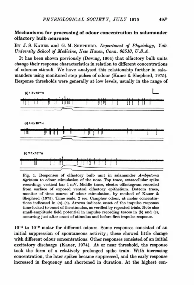
By J. S. KAUER and G. M. SHEPHERD. Department of Physiology, YaleUniversity School of Medicine, New Haven, Conn. 06510, U.S.A.
It has been shown previously (Doving, 1964) that olfactory bulb unitschange their response characteristics in relation to different concentrationsof odorous stimuli. We have analysed this relationship further in sala-manders using monitored step pulses of odour (Kauer & Shepherd, 1975).Response thresholds were generally at low levels, usually in the range of
(a) 1-2x10⁻⁸m L
(b) 44x1I0-11mA. ILAIk In a,
(c) 9-7x I1O m
Fig. 1. Responses of olfactory bulb unit in salamander Ambyatomatigrinum to odour stimulation of the nose. Top trace, extracellular spikerecording; vertical bar 1 mV. Middle trace, electro-olfactogram, recordedfrom surface of exposed ventral olfactory epithelium. Bottom trace,monitor of time course of odour stimulation, by method of Kauer &Shepherd (1975). Time scale, 2 sec. Camphor odour, at molar concentra-tions indicated in (a)-(c). Arrows indicate onset of the impulse responsetime-locked to onset of the stimulus, as verified by repeated trials. Note alsosmall-amplitude field potential in impulse recording traces in (b) and (c),occurring just after onset of stimulus and before first impulse response.
10O4 to 10O9 molar for different odours. Some responses consisted of aninitial suppression of spontaneous activity; these showed little changewith different odour concentrations. Other responses consisted of an initialexcitatory discharge (Kauer, 1974). At or near threshold, the responsetook the form of a relatively prolonged spike train. With increasingconcentration, the later spikes became suppressed, and the early responseincreased in frequency and shortened in duration. At the highest con-
49P
PDPROCEEDINGS OF THE
centrations the initial discharge consisted of only one or two spikes. Thesepatterns are illustrated in Fig. 1.The results of this study suggest that concentration information is
processed by an interplay of excitatory and inhibitory pathways feedinginto or located within the olfactory bulb. The intrinsic bulbar pathwaysare organized into two main layers - glomerular and external plexiform -differentially accessible to activation by olfactory nerve and olfactorytract stimulation. Preliminary experiments suggest that unitary andsummed potentials evoked through these fibres can be selectively sup-pressed during different phases of naturally evoked activity. This mayprovide evidence regarding the synaptic organization underlying theprocessing of information about odour concentration.
REFERENCESD0vING, K. B. (1964). Acta physiol. 8cand. 60, 150-163.KAUER, J. S. (1974). J. Physiol. 243, 695-715.KAUER, J. S. & SHEPHERD, G. M. (1975). Brain Re8. 85, 108-113.
Visual and taste neurones in the lateral hypothalamus and sub-stantia innominata: modulation of responsiveness by hungerBY M. J. BURTON, F. MORA and E. T. ROLLS. Department of ExperimentalPsychology, Oxford University, South Parks Road, Oxford OX1 3UDSome neurones in the monkey lateral hypothalamus and substantia
innominata respond only to the sight and/or taste of food (see Rolls, 1975).The responses to the sight of food appear to be visual in that the units donot respond in relation to motor movements, or during grasping the foodand feeding in the dark, or in other control tests, so that the responses donot appear to be motor or due to salivation, or to anticipation of food.The greatest response of the visual neurones is to the sight of the animal'smost preferred food. As these units are in the lateral hypothalamus andsubstantia innominata, which are involved in the control of food intake,we varied the hunger of the animals to determine whether this affectedthe firing rates of the units. At the start of the experiments the animals werehungry in that they had not eaten for 12 hr, and were willing to reach forfood, for example for 20 % glucose or saturated sucrose solution. Satietywas induced by feeding the animals 2 ml. of trial solution, until the animalbecame satiated and refused the solution. It was found that the meanspontaneous firing rate was unaffected by changes of hunger. But the sightand/or taste of food produced the largest response of the units (as measuredby a change of rate from the mean spontaneous firing rate) if the animalwas hungry, and ceased to produce a response if the animal was satiated.
50P

PHYSIOLOGICAL SOCIETY, JULY 1975 p
These results were found in two squirrel monkeys and one rhesus monkey,in 6 units which responded to the sight of food, in 3 units which respondedto both the sight and taste of food, and in 1 unit which responded to thetaste of food. In control experiments on units recorded in the globus palli-dus which fired in relation to swallowing, no effect of alterations of hungeron the response of the units to swallowing was found. It is suggested thatthe units in the lateral hypothalamus and the substantia innominata whichonly respond to the sight and/or taste of food if the animal is hungry areinvolved in the regulation of food intake by altering the response of theanimal to the sight and taste of food.
REFERENCE
ROLLS, E. T. (1975). The Brain and Reward. Oxford: Pergamon.
The role of the locus coeruleus in the acquisition of a conditionedavoidance response
By T. J. CROw and S. WENDLANDT. Division of Physiology and Pharma-cology, National Institute for Medical Research, Mill Hill, N. W. 7; Divisionof Psychiatry, Clinical Research Centre, Watford Road, Harrow, Middlesex,HAl 3UJ
Tests of the hypothesis (Crow, 1968; Kety, 1972) that the adrenergicinnervation of the cerebral cortex is necessary for establishing the neuralchanges involved in learning have given conflicting results (Anlezark,Crow & Greenway, 1973; Amaral & Foss, 1975). Stein, Belluzi & Wise(1975) report that animals injected s.c. with the dopamine-fi-hydroxy-lase inhibitor diethyldithiocarbamate (DDC) show evidence of immediateacquisition of a passive avoidance response but fail to show long-termretention. In these experiments we have attempted to demonstrate thatthis specific deficit of learning capacity may be a consequence of impairedtransmission in the coerulocortical adrenergic system. Seventy-n-ine femaleSprague-Dawley rats (initial weight 150 g) were divided into an untreatedcontrol group, a group injected bilaterally with 6-OR-dopamine (16 ,tgin 4#1d. over 4 min) just anterior to the locus coeruleus, a group with burrholes, and a group injected. s.c. with DDC 300 mg/kg between the firstand second trials of the training procedure. The time taken for each animalto step off a platform onto the electrifiable grid floor of the test chamberwas recorded in each of 5 test sessions. The first two trials were separatedby 1 hr. On the third trial (1 min after the second) rats were shocked as theystepped down, and on the fourth (1 min later) and fifth (3 days later)trials the effect of this experience on the animals' subsequent step-downtime was tested.
51P
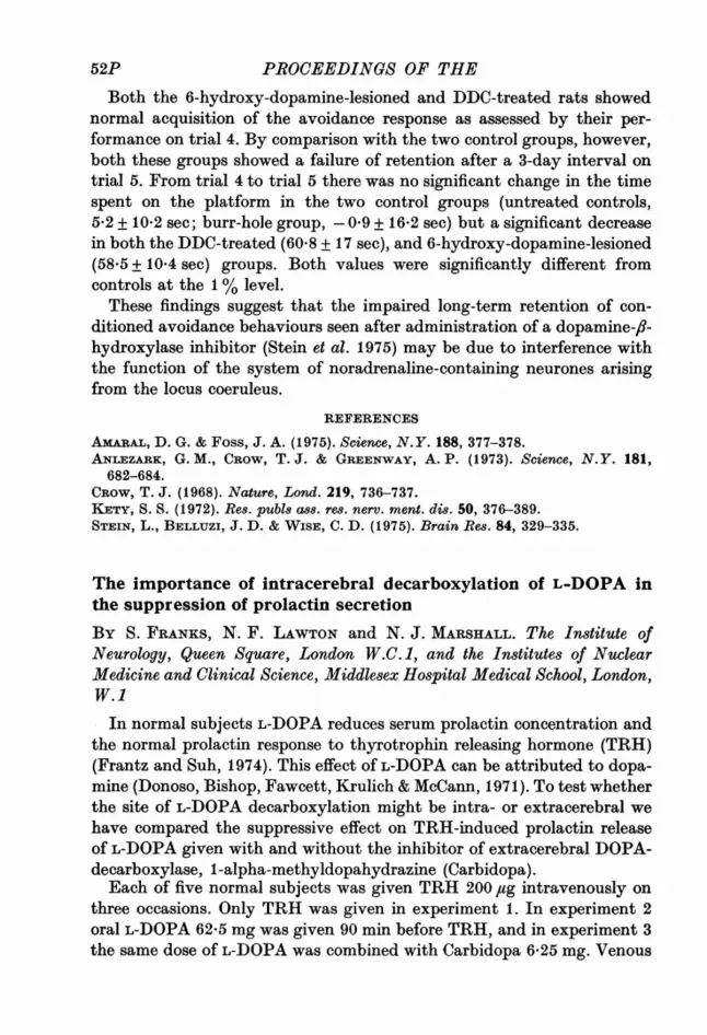
PROCEEDINGS OF THE
Both the 6-hydroxy-dopamine-lesioned and DDC-treated rats showednormal acquisition of the avoidance response as assessed by their per-formance on trial 4. By comparison with the two control groups, however,both these groups showed a failure of retention after a 3-day interval ontrial 5. From trial 4 to trial 5 there was no significant change in the timespent on the platform in the two control groups (untreated controls,5-2 + 10*2 see; burr-hole group,- 09 + 16-2 see) but a significant decreasein both the DDC-treated (60.8 + 17 see), and 6-hydroxy-dopamine-lesioned(58.5 + 10*4 sec) groups. Both values were significantly different fromcontrols at the 1 % level.These findings suggest that the impaired long-term retention of con-
ditioned avoidance behaviours seen after administration of a dopamine-fi-hydroxylase inhibitor (Stein et al. 1975) may be due to interference withthe function of the system of noradrenaline-containing neurones arisingfrom the locus coeruleus.
REFERENCES
AxARAL, D. G. & Foss, J. A. (1975). Science, N.Y. 188, 377-378.ANLEZARK, G. M., CROW, T. J. & GREENWAY, A. P. (1973). Science, N.Y. 181,
682-684.CROW, T. J. (1968). Nature, Lond. 219, 736-737.KETY, S. S. (1972). Res. pubis ass. res. nerv. ment. dis. 50, 376-389.STEIN, L., BELLUZI, J. D. & WISE, C. D. (1975). Brain Res. 84, 329-335.
The importance of intracerebral decarboxylation of L-DOPA inthe suppression of prolactin secretion
BY S. FRANKS, N. F. LAWTON and N. J. MARSHALL. The Institute ofNeurology, Queen Square, London W.C. 1, and the Institutes of NuclearMedicine and Clinical Science, Middlesex Hospital Medical School, London,W.1
In normal subjects L-DOPA reduces serum prolactin concentration andthe normal prolactin response to thyrotrophin releasing hormone (TRH)(Frantz and Suh, 1974). This effect of L-DOPA can be attributed to dopa-mine (Donoso, Bishop, Fawcett, Krulich & McCann, 1971). To test whetherthe site of L-DOPA decarboxylation might be intra- or extracerebral wehave compared the suppressive effect on TRH-induced prolactin releaseof L-DOPA given with and without the inhibitor of extracerebral DOPA-decarboxylase, 1-alpha-methyldopahydrazine (Carbidopa).Each of five normal subjects was given TRH 200 /ug intravenously on
three occasions. Only TRH was given in experiment 1. In experiment 2oral L-DOPA 62-5 mg was given 90 min before TRH, and in experiment 3the same dose of L-DOPA was combined with Carbidopa 6-25 mg. Venous
52P

PHYSIOLOGICAL SOCIETY, JULY 1975
blood was collected for serum prolactin and thyroid stimulating hormone(TSH) assay prior to TRH and at 20, 30 and 60 min thereafter. Radio-immunoassay of serum prolactin was performed by the method of Sinha,Selby, Lewis & Vanderlaan (1973) and of TSH by a modification of themethod of Odell, Wilbur & Paul (1965).
20
10
1 2 3 4 5Subject number
Fig. 1. The effect of L-DOPA 62-5 mg and of L-DOPA 62-5 mg withCarbidopa 6-25 mg on TRH-induced prolactin release in five subjects.Prolactin is the increment above the basal value 20 min after TRH injec-tion. The increment after TRH alone is shown by the open columns, afterpretreatment with L-DOPA by the hatched columns, and after L-DOPAwith Carbidopa by the stippled columns.
The mean fall in basal prolactin after L-DOPA alone was 1-2 ng/ml.(range 05-2-4 ng/ml.) and after L-DOPA with Carbidopa 1.0 ng/ml.(range 0-1-2-2 ng/ml.). Fig. 1 shows that in 4 subjects the increase inprolactin concentration after TRH was reduced by L-DOPA alone andfurther reduced by L-DOPA with Carbidopa. In subject 5 L-DOPA wasonly effective combined with Carbidopa. There was no consistent effecton TSH release.
Consistent potentiation by an extracerebral DOPA-decarboxylase in-hibitor of the L-DOPA effect on TRH-induced prolactin release indicatesthe importance of intracerebral DOPA-decarboxylase in the mechanismof this response.
Materials for radioimmunoassay were a gift from the National Institute ofArthritis, Metabolism and Digestive Diseases.
REFERENCES
DoNoso, A. O., BIsHoP, W., FAWCETT, C. P., KRULICH, L. & MCCANN, S. M.(1971). Endocrinology 89, 774-784.
FRANTZ, A. G. & SUH, H. K. (1974). In Advances in Neurology, vol. 5, pp. 447-456.New York: Raven Press.
53P
APPPROCEEDINGS OF THEODELL, W. D., WILBUR, J. F. & PAUL, W. E. (1965). J. din. Endocr. Metab. 25,
1179-1188.SINEA, Y. N., SELBY, F. W., LEWIS, U. J. & VANDERLAAN, W. P. (1973). J. din.
Endoer. Metab. 36, 509-516.
Is prolactin a fluid and electrolyte regulating hormone in man?BY E. N. COLE,* D. EVERED,t D. F. HORROBIN,t M. S. MANKU,t J. P.MTABAJT,t B. A. NASSAR.t * Tenovus Institute, Welsh National School ofMedicine, Cardiff. t University of Newcastle upon Tyne Medical School.
Prolactin causes renal sodium, potassium and water retention wheninjected or infused into cats, rats, sheep and humans (Lockett, 1965;Horrobin, Lloyd, Lipton, Burstyn, Durkin & Muiruri, 1971; Burstyn,Horrobin & Manku, 1972; Horrobin, 1973). In rats a low sodium dietincreases prolactin secretion (Relkin, 1973) and in man plasma prolactinlevels are influenced by plasma osmolality in the same way as is anti-diuretic hormone (ADH). These findings indicate that prolactin couldplay a part in human fluid and electrolyte metabolism but gives noindication that it does.A woman of 41 presented with extremely severe premenstrual syndrome
which was associated with monthly weight gain and loss of 5-8 kg. Thestory was confirmed by admission to a metabolic ward during the lutealphase of one menstrual cycle when marked premenstrual thirst and reduc-tion in renal sodium, water and potassium excretion were noted. Sinceconventional diuretics had proved ineffective treatment with lithium car-bonate (Sletton & Gershon, 1966) was attempted. Initially there wasa considerable improvement but after about 6 months the therapeuticeffect wore off and fluid and electrolyte retention became almost assevere as before. The treatment was then changed to the prolactin sup-pressing drug, bromocriptine, one 2-5 mg tablet each evening during thelast 3 weeks of each menstrual cycle. The patient has now been on thistherapy for almost a year and seems to be completely relieved of all signsand symptoms.
In the untreated state mean follicular phase plasma prolactin level was5-25 + 0-85 (S.E. of mean) ng/ml. while in the luteal phase it was10*52+0.88ng/mJ. During lithium therapy luteal phase levels rose to21-75 + 1-43 ng/ml. We have preliminary evidence that lithium inhibitsprolactin actions on rat kidneys and arterioles in a competitive fashion.While on bromocriptine luteal plasma prolactin levels were 6-94 + 0-86 ng/ml. Although bromocriptine is a dopamine agonist and might be expectedto have renal effects in its own right we suggest that this patient may bethe first example of a prolactin-induced fluid and electrolyte disorder.
54P
PHYSIOLOGICAL SOCIETY, JULY 1975
REFERENCESBURSTYN, P. G., HORROBIN, D. F. & MANKU, M. S. (1972). J. Endocr. 55, 369-376.HORROBIN, D. F., LLOYD, I. J., LIPTON, A., BURSTYN, P. G., DuIJKIN, N. & MumuI,K. L. (1971). Lancet ii, 352-354.
HORROBIN, D. F. (1973). Proladin: Physiology and Clinical Significance. Lancaster:Medical and Technical Publishing Co.
LOCKETT, M. F. (1965). J. Physiol. 181, 192-199.RELKIN, R. (1973). Neuronendocrinology 11, 240-247.SLETTON, I. W. & GERSHON, S. (1966). Compr. Psychiat. 7, 197.
Osmoreception and thirst in the dogBY D. J. RAMSAY, BARBARA J. ROLLS and R. J. WOOD. University Labora-tory of Physiology and Department of Experimental Psychology, Universityof OxfordHypothalamic osmoreceptors may be involved in the control of drinking
in the dog (Andersson, 1972) as in the rat (Blass & Epstein, 1971). In thisstudy, a 24 hr period of water deprivation in 8 dogs raised plasma osmo-lality from 297-5 + 0 9 to 309-6 + 0 9 m-osmole/kg. H20 (P < 0 001). Theeffect of removing the central osmotic stimulus on deprivation induceddrinking was investigated.
Eight dogs prepared with unilateral carotid loops were deprived ofwater for 24 hr. They were infused through the carotid loop with distilledwater for 5 min at 0-6 ml./kg. min, a rate designed to reduce the osmolalityin the cerebral circulation to pre-deprivation levels. The intake of waterduring a further 5 min of infusion was measured. Water intakes werevariable and were not significantly less than those following control in-fusions of 0.15 M-NaCl. The ineffectiveness of this procedure may reflecta non-uniform distribution of the infusate in the cerebral circulation. Totest this, second carotid loops were prepared on all dogs and the experi-ment repeated, infusing the same dose bilaterally. This reduced the jugularblood osmolality to pre-deprivation levels without measurably affectingsystemic blood, and produced a marked and significant reduction in waterintake (Fig. 1 a). Infusion of water to the deprived dogs at different ratesshows the effect is dose related (R = 0-56, N = 24, P < 0.01).
Further evidence for the importance of osmolality in thirst was obtainedby infusing 0 3 M, 0-45 M and 0-6 M-NaCl solutions at a rate of 0-6 ml./kg. min for 10 min into the eight dogs via both carotid loops when fluidreplete. A graded stimulation of drinking was obtained with increasingthe osmolality of the saline (R = 0 59, N = 32, P = < 0.001) (Fig. lb).The suggestion that central osmoreceptors may play an important role
in the mediation of thirst receives support from these results.We are grateful to the M.R.C. for financial support.
55P