nuchal glands: a novel defensive system in snakes
TRANSCRIPT
1 23
ChemoecologyEvolutionary, Mechanistic andEnvironmental Approaches toChemically-Mediated Interaction ISSN 0937-7409Volume 22Number 3 Chemoecology (2012) 22:187-198DOI 10.1007/s00049-011-0086-2
Nuchal glands: a novel defensive system insnakes
Akira Mori, Gordon M. Burghardt,Alan H. Savitzky, Kathleen A. Roberts,Deborah A. Hutchinson & RichardC. Goris

1 23
Your article is protected by copyright and all
rights are held exclusively by Springer Basel
AG. This e-offprint is for personal use only
and shall not be self-archived in electronic
repositories. If you wish to self-archive your
work, please use the accepted author’s
version for posting to your own website or
your institution’s repository. You may further
deposit the accepted author’s version on
a funder’s repository at a funder’s request,
provided it is not made publicly available until
12 months after publication.

REVIEW
Nuchal glands: a novel defensive system in snakes
Akira Mori • Gordon M. Burghardt •
Alan H. Savitzky • Kathleen A. Roberts •
Deborah A. Hutchinson • Richard C. Goris
Received: 16 April 2011 / Accepted: 13 July 2011 / Published online: 2 August 2011
� Springer Basel AG 2011
Abstract Of the various chemical defensive adaptations
of vertebrates, nuchal glands are among the most unusual.
First described in a Japanese natricine snake, Rhabdophis
tigrinus, in 1935, these organs are embedded under the skin
of the neck region as a series of paired glands that have
neither lumina nor ducts. The major chemical components
of the glandular fluid are bufadienolides, which are car-
diotonic steroids also found in the skin secretion of toads.
Here we review early studies of nuchal glands and briefly
introduce our recent findings on the sequestration of
bufadienolides from consumed toads and the maternal
provisioning of those sequestered compounds. We sum-
marize behavioral studies associated with the antipredator
function of the nuchal glands, which have been conducted
during our more than decade-long collaboration. Results of
preliminary analyses on the possible costs of toad-eating
and on the ultrastructure of the nuchal glands are also
presented. Finally, we discuss the evolutionary origin of
the nuchal glands and suggest future directions designed to
understand the biological importance of these novel ver-
tebrate organs, which have evolved in a limited number of
snake species.
Keywords Rhabdophis tigrinus � Bufo �Dietary sequestration � Antipredator behavior �Bufadienolide � Natricine snakes
Introduction
Chemical defense is a widespread strategy for organisms to
avoid predation. Selection pressure has shaped defensive
organs, coupled with morphologically and behaviorally
coordinated features, to enhance their efficacy in chemical
defense. Thus, we see a variety of defensive organs that
have a unique suite of chemically, morphologically, and
behaviorally integrated mechanisms. Nuchal glands are
unusual organs of chemical defense that occur only in
several species of specialized vertebrates, specifically
snakes. In this review, we first summarize early studies of
the nuchal glands. We then introduce a hypothesis that we
have proposed and tested concerning the derivation of the
chemical compounds of the glands. We also describe
functional and evolutionary aspects of the nuchal glands,
along with their behavioral and ecological correlates.
Finally, we discuss future directions designed to increase
A. Mori (&)
Department of Zoology, Graduate School of Science,
Kyoto University, Sakyo, Kyoto 606-8502, Japan
e-mail: [email protected]
G. M. Burghardt
Departments of Psychology and Ecology and Evolutionary
Biology, University of Tennessee, Knoxville,
TN 37996-0900, USA
A. H. Savitzky � K. A. Roberts � D. A. Hutchinson
Department of Biological Sciences,
Old Dominion University, Norfolk, VA 23529-0266, USA
R. C. Goris
Hatsuyama 1-7-13, Miyamae, Kawasaki,
Kanagawa 216-0026, Japan
Present Address:K. A. Roberts
Department of Natural Resources and Environmental Sciences,
Alabama A & M University, Normal, AL 35762, USA
Present Address:D. A. Hutchinson
Department of Biology, Coastal Carolina University,
Conway, SC 29528, USA
Chemoecology (2012) 22:187–198
DOI 10.1007/s00049-011-0086-2 CHEMOECOLOGY
123
Author's personal copy
our understanding of the function and evolution of these
unique vertebrate organs.
Discovery and early studies
The nuchal glands were first described in 1935 as integu-
mentary glands in a Japanese colubrid snake, Rhabdophis
tigrinus tigrinus (Nakamura 1935). The organs are
embedded under the skin of the neck region as a series of
paired glands that have neither lumina nor ducts. A yel-
lowish, opaque fluid with a disagreeable odor spews out
when pressure breaks the cell membranes and ruptures the
glands’ connective tissue capsules (Nakamura 1935). The
discharged fluid is almost neutral in reaction and contains
abundant small granules, isolated nuclei of the gland cells,
blood, lymphatic cells, and pigment cells (Nakamura
1935). Three years later, Smith (1938) reported similar
organs from nine additional species of natricine snakes in
three genera (Rhabdophis, Macropisthodon, and Balano-
phis), among 44 species of Colubridae and Elapidae
examined. He divided the species containing nuchal glands
into two groups, those having glands of sacculated form
(seven species) and those with the non-sacculated form
(three species: Table 1). In addition, he described glands
extending the entire length of the body in two of the nine
species (R. nuchalis and M. plumbicolor) and collectively
called the glands ‘‘nucho-dorsal glands’’. He also reported
the absence of the glands in four congeneric species
(Table 1).
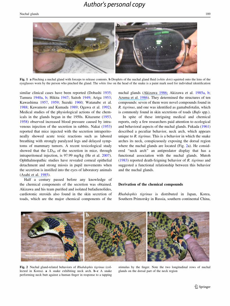
Nakamura (1935) surmised a protective function of the
nuchal glands on the basis of his own aversive experience.
When he decapitated an individual of R. tigrinus, yellow
droplets that sprayed from the nuchal region on which the
blades of the scissors were pressed splashed into his eyes,
causing acute pain (Fig. 1). The first clinical note of ocular
distress caused by unknown ‘‘skin secretions’’ of R. tigri-
nus predates his finding (Shibuya 1923), and a series of
Table 1 Occurrence of nuchal (nucho-dorsal) glands among species in the three natricine genera in which the glands have been reported
Species Glands Gland type Gland site Source
Rhabdophis callichromus P S N Smith (1938)
R. himalayanus P S N Smith (1938)
R. leonardi P – WBb Jiang and Zhao (1983)
R. nigrocinctus P S N Smith (1938)
R. nuchalis P S WB Smith (1938); Jiang and Zhao (1983)
R. pentasupralabialis P – WBb Zhao (1995)
R. subminiatus P S N Smith (1938)
R. tigrinus P S N Nakamura (1935); Smith (1938)
R. murudensis A/Pa – N Smith (1938); Stuebing and Lian (2002)
R. adleri A – – Zhao (1997)
R. chrysargos A – – Smith (1938)
R. spilogaster A – – Smith (1938)
R. swinhonis A – – Mao and Chang (1999)
R. angelii U – –
R. auriculatus U – –
R. barbouri U – –
R. chrysargoides U – –
R. conspicillatus U – –
R. lineatus U – –
Macropisthodon flaviceps P US N Smith (1938)
M. plumbicolor P S WB Smith (1938)
M. rhodomelas P US N Smith (1938)
M. rudis A – – Smith (1938)
Balanophis ceylonensis P US N Smith (1938)
Taxonomy of snakes follows David and Ineich (1999)
A absent, N neck region, P present, S sacculated form, U unknown, US unsacculated form, WB whole bodya Smith (1938) did not find the glands, whereas Stuebing and Lian (2002) confirmed the presence of the glandsb Based on the description of R. nuchalis that was formerly considered the same species
188 A. Mori et al.
123
Author's personal copy
similar clinical cases have been reported (Dobashi 1935;
Tamura 1940a, b; Hikita 1947; Saitoh 1949; Ariga 1953;
Kawashima 1957, 1959; Suzuki 1960; Watanabe et al.
1988; Kawamoto and Kumada 1989; Ogawa et al. 1992).
Medical studies of the physiological actions of the chem-
icals in the glands began in the 1950s. Kitazume (1953,
1958) observed increased blood pressure caused by intra-
venous injection of the secretion in rabbits. Nakai (1953)
reported that mice injected with the secretion intraperito-
neally showed acute toxic reactions such as labored
breathing with strongly paralyzed legs and delayed symp-
toms of mammary tumors. A recent toxicological study
showed that the LD50 of the secretion in mice, through
intraperitoneal injection, is 97.99 mg/kg (He et al. 2007).
Ophthalmopathic studies have revealed corneal epithelial
detachment and strong miosis in pupil movements when
the secretion is instilled into the eyes of laboratory animals
(Asahi et al. 1985).
Half a century passed before any knowledge of
the chemical components of the secretion was obtained.
Akizawa and his team purified and isolated bufadienolides,
cardiotonic steroids also found in the skin secretion of
toads, which are the major chemical components of the
nuchal glands (Akizawa 1986; Akizawa et al. 1985a, b;
Azuma et al. 1986). They determined the structures of ten
compounds: seven of them were novel compounds found in
R. tigrinus, and one was identified as gamabufotalin, which
is commonly found in skin secretions of toads (Bufo spp.).
In spite of these intriguing medical and chemical
reports, only a few researchers paid attention to ecological
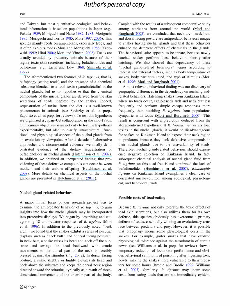
and behavioral aspects of the nuchal glands. Fukada (1961)
described a peculiar behavior, neck arch, which appears
unique to R. tigrinus. This is a behavior in which the snake
arches its neck, conspicuously exposing the dorsal region
where the nuchal glands are located (Fig. 2a). He consid-
ered ‘‘neck arch’’ an antipredator display that has a
functional association with the nuchal glands. Mutoh
(1983) reported death-feigning behavior of R. tigrinus and
suggested a functional relationship between this behavior
and the nuchal glands.
Derivation of the chemical compounds
Rhabdophis tigrinus is distributed in Japan, Korea,
Southern Primorsky in Russia, southern continental China,
Fig. 2 Nuchal gland-related behaviors of Rhabdophis tigrinus (col-
lected in Korea). a A snake exhibiting neck arch. b–c A snake
performing neck butt against a human finger in response to a tapping
stimulus by the finger. Note the two longitudinal rows of nuchal
glands on the dorsal part of the neck region
Fig. 1 a Pinching a nuchal gland with forceps to release contents. b Droplets of the nuchal gland fluid (white dots) squirted onto the lens of the
eyeglasses worn by the person who pinched the gland. The white line on the head of the snake is a paint mark used for individual identification
Nuchal glands 189
123
Author's personal copy
and Taiwan, but most quantitative ecological and behav-
ioral information is based on populations in Japan (e.g.,
Fukada 1959; Moriguchi and Naito 1982, 1983; Moriguchi
1985; Moriguchi and Toriba 1985; Mori 1997, 2006). This
species mainly feeds on amphibians, especially frogs, and
it often exploits toads (Mori and Moriguchi 1988; Kado-
waki 1992; Hirai 2004; Mori and Vincent 2008). Toads are
usually avoided by predatory animals because of their
highly toxic skin secretions, including bufadienolides and
bufotoxins (e.g., Licht and Low 1968; Shimada et al.
1977).
The aforementioned two features of R. tigrinus, that is,
bufophagy (eating toads) and the presence of a chemical
substance identical to a toad toxin (gamabufotalin) in the
nuchal glands, led us to hypothesize that the chemical
compounds of the nuchal glands are derived from the skin
secretions of toads ingested by the snakes. Indeed,
sequestration of toxins from the diet is a well-known
phenomenon in animals (see Savitzky et al. in prep.,
Saporito et al. in prep. for reviews). To test this hypothesis
we organized a Japan–US collaboration in the mid-1990s.
Our primary objectives were not only to test the hypothesis
experimentally, but also to clarify ultrastructural, func-
tional, and physiological aspects of the nuchal glands from
an evolutionary viewpoint. After a decade of alternative
approaches and circumstantial evidence, we finally dem-
onstrated evidence of the dietary sequestration of
bufadienolides in nuchal glands (Hutchinson et al. 2007).
In addition, we obtained an unexpected finding, that pro-
visioning of these defensive compounds can occur between
mothers and their unborn offspring (Hutchinson et al.
2008). More details on chemical aspects of the nuchal
glands are presented in Hutchinson et al. (2011).
Nuchal gland-related behaviors
A major initial focus of our research project was to
examine the antipredator behavior of R. tigrinus, to gain
insights into how the nuchal glands may be incorporated
into protective displays. We began by describing and cat-
egorizing 18 antipredator responses of R. tigrinus (Mori
et al. 1996). In addition to the previously noted ‘‘neck
arch’’, we found that the snakes exhibit a series of peculiar
displays such as ‘‘neck butt’’ and ‘‘dorsal facing posture’’.
In neck butt, a snake raises its head and neck off the sub-
strate and swings the head backward with erratic
movements so the dorsal part of the neck is forcibly
pressed against the stimulus (Fig. 2b, c). In dorsal facing
posture, a snake slightly or highly elevates its head and
neck above the substrate and keeps the dorsal neck region
directed toward the stimulus, typically as a result of three-
dimensional movements of the anterior part of the body.
Coupled with the results of a subsequent comparative study
among natricines from around the world (Mori and
Burghardt 2008), we concluded that neck arch, neck butt,
and dorsal facing posture are antipredator behaviors unique
to snakes having nuchal glands and that these behaviors
enhance the deterrent effects of chemicals in the glands.
The behavioral suite appears to be innate, because newly
hatched snakes perform these behaviors shortly after
hatching. We also showed that dependency of these
‘‘nuchal gland-related behaviors’’ varies according to
internal and external factors, such as body temperature of
snakes, body part stimulated, and type of stimulus (Mori
et al. 1996; Mori and Burghardt 2001).
A most relevant behavioral finding was our discovery of
geographic differences in the dependency on nuchal gland-
related behaviors. Hatchling snakes from Kinkasan Island,
where no toads occur, exhibit neck arch and neck butt less
frequently and perform simple escape responses more
frequently than hatchling R. tigrinus from populations
sympatric with toads (Mori and Burghardt 2000). This
result is congruent with a prediction deduced from the
aforementioned hypothesis: If R. tigrinus sequesters toad
toxins in the nuchal glands, it would be disadvantageous
for snakes on Kinkasan Island to expose their neck region
to predators because they lack defensive compounds in
their nuchal glands due to the unavailability of toads.
Therefore, nuchal gland-related behaviors should experi-
ence negative selection on Kinkasan Island. In fact,
subsequent chemical analysis of nuchal gland fluid from
R. tigrinus on this toad-free island confirmed the lack of
bufadienolides (Hutchinson et al. 2007). Rhabdophis
tigrinus on Kinkasan Island exemplifies a clear case of
correlated microevolution among ecological, physiologi-
cal, and behavioral traits.
Possible costs of toad-eating
Because R. tigrinus not only tolerates the toxic effects of
toad skin secretions, but also utilizes them for its own
defense, this species obviously has overcome a primary
defense of toads, essentially winning an evolutionary arms
race between predators and prey. However, it is possible
that bufophagy incurs some physiological costs in the
snakes. For example, garter snakes that have evolved
physiological tolerance against the tetrodotoxin of certain
newts (see Williams et al. in prep. for review) show a
temporary reduction of locomotor performance and obvi-
ous behavioral symptoms of poisoning after ingesting toxic
newts, making the snakes more vulnerable to their preda-
tors for some hours (Brodie and Brodie 1999; Williams
et al. 2003). Similarly, R. tigrinus may incur some
costs from eating toads that are not immediately evident.
190 A. Mori et al.
123
Author's personal copy
To explore this possibility, we conducted preliminary tests
to identify short-term effects of bufophagy on locomotor
performance. We also compared the survivorship of juve-
nile snakes reared on different diets to examine the long-
term effects of bufophagy.
Possible short-term effects of bufophagy were examined
by comparing crawling and swimming performances.
Crawling speeds of ten wild-caught snakes (body mass
3.1–125 g, five males and five females) were measured
immediately before, and 1, 2, and 3 days after the snakes
ate a prey item (4.5–35.1% of snake body mass). Each
snake was fed approximately the same mass of either toads
(Anaxyrus terrestris or A. woodhousei, formerly members
of the genus Bufo), frogs (Acris crepitans, Hyla squirella,
Lithobates palustris, or L. sphenocephala), or fish (Pim-
ephales promelas or Poecilia reticulata) every 4 days in
counterbalanced order (repeated measures design). In each
trial, a snake was gently moved from its home cage and
introduced onto a track (2 m length 9 0.3 m width) with
artificial floor covering (Astroturf) at an air temperature of
24 ± 1�C. The snake was then encouraged to traverse the
track by gently tapping the tail tip or the substrate just
behind the tail. The duration of the crawl (1.5 m for adults
and 1 m for juveniles) was measured with a stopwatch, and
the crawling speed was calculated. Trials for each snake
were repeated three times with 3-min intervals, and the
average speed of each snake was compared among food
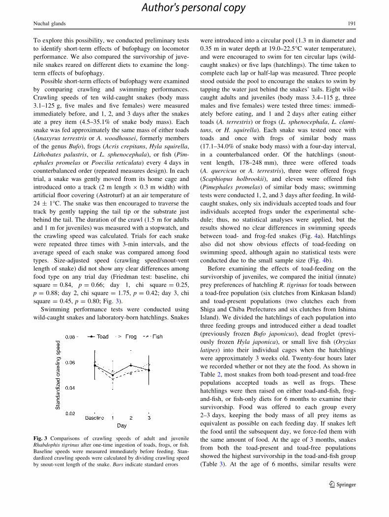
types. Size-adjusted speed (crawling speed/snout-vent
length of snake) did not show any clear differences among
food type on any trial day (Friedman test: baseline, chi
square = 0.84, p = 0.66; day 1, chi square = 0.25,
p = 0.88; day 2, chi square = 1.75, p = 0.42; day 3, chi
square = 0.45, p = 0.80; Fig. 3).
Swimming performance tests were conducted using
wild-caught snakes and laboratory-born hatchlings. Snakes
were introduced into a circular pool (1.3 m in diameter and
0.35 m in water depth at 19.0–22.5�C water temperature),
and were encouraged to swim for ten circular laps (wild-
caught snakes) or five laps (hatchlings). The time taken to
complete each lap or half-lap was measured. Three people
stood outside the pool to encourage the snakes to swim by
tapping the water just behind the snakes’ tails. Eight wild-
caught adults and juveniles (body mass 3.4–115 g, three
males and five females) were tested three times: immedi-
ately before eating, and 1 and 2 days after eating either
toads (A. terrestris) or frogs (L. sphenocephala, L. clami-
tans, or H. squirella). Each snake was tested once with
toads and once with frogs of similar body mass
(17.1–34.0% of snake body mass) with a four-day interval,
in a counterbalanced order. Of the hatchlings (snout-
vent length, 178–248 mm), three were offered toads
(A. quercicus or A. terrestris), three were offered frogs
(Scaphiopus holbrookii), and eleven were offered fish
(Pimephales promelas) of similar body mass; swimming
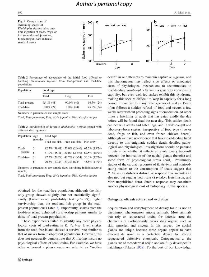
tests were conducted 1, 2, and 3 days after feeding. In wild-
caught snakes, only six individuals accepted toads and four
individuals accepted frogs under the experimental sche-
dule; thus, no statistical analyses were applied, but the
results showed no clear differences in swimming speeds
between toad- and frog-fed snakes (Fig. 4a). Hatchlings
also did not show obvious effects of toad-feeding on
swimming speed, although again no statistical tests were
conducted due to the small sample size (Fig. 4b).
Before examining the effects of toad-feeding on the
survivorship of juveniles, we compared the initial (innate)
prey preferences of hatchling R. tigrinus for toads between
a toad-free population (six clutches from Kinkasan Island)
and toad-present populations (two clutches each from
Shiga and Chiba Prefectures and six clutches from Ishima
Island). We divided the hatchlings of each population into
three feeding groups and introduced either a dead toadlet
(previously frozen Bufo japonicus), dead froglet (previ-
ously frozen Hyla japonica), or small live fish (Oryzias
latipes) into their individual cages when the hatchlings
were approximately 3 weeks old. Twenty-four hours later
we recorded whether or not they ate the food. As shown in
Table 2, most snakes from both toad-present and toad-free
populations accepted toads as well as frogs. These
hatchlings were then raised on either toad-and-fish, frog-
and-fish, or fish-only diets for 6 months to examine their
survivorship. Food was offered to each group every
2–3 days, keeping the body mass of all prey items as
equivalent as possible on each feeding day. If snakes left
the food until the subsequent day, we force-fed them with
the same amount of food. At the age of 3 months, snakes
from both the toad-present and toad-free populations
showed the highest survivorship in the toad-and-fish group
(Table 3). At the age of 6 months, similar results were
Fig. 3 Comparisons of crawling speeds of adult and juvenile
Rhabdophis tigrinus after one-time ingestion of toads, frogs, or fish.
Baseline speeds were measured immediately before feeding. Stan-
dardized crawling speeds were calculated by dividing crawling speed
by snout-vent length of the snake. Bars indicate standard errors
Nuchal glands 191
123
Author's personal copy
obtained for the toad-free population, although the fish-
only group showed slightly, but not statistically signifi-
cantly (Fisher exact probability test: p [ 0.9), higher
survivorship than the toad-and-fish group in the toad-
present populations (Table 3). Importantly, snakes from the
toad-free island exhibited survivorship patterns similar to
those of toad-present populations.
These experiments failed to identify any clear physio-
logical costs of toad-eating in R. tigrinus. Even snakes
from the toad-free island showed a survival rate similar to
that of snakes from toad-present populations. However, this
does not necessarily demonstrate that R. tigrinus incurs no
physiological effects of toad toxins. For example, we have
often witnessed a phenomenon we refer to as ‘‘sudden
death’’ in our attempts to maintain captive R. tigrinus, and
this phenomenon may reflect side effects or associated
costs of physiological mechanisms to accommodate to
toad-feeding. Rhabdophis tigrinus is generally voracious in
captivity, but even well-fed snakes exhibit this syndrome,
making this species difficult to keep in captivity for a long
period, in contrast to many other species of snakes. Death
often follows a sudden refusal of food and occurs a few
weeks later without preceding signs of emaciation. At other
times a hatchling or adult that has eaten avidly the day
before will be found dead the next day. This sudden death
can occur in adults and hatchlings, and in wild-caught and
laboratory-born snakes, irrespective of food type (live or
dead, frogs or fish, and even frozen chicken hearts).
Although we have no evidence that links toad-feeding habit
directly to this enigmatic sudden death, detailed patho-
logical and physiological investigations should be pursued
to determine whether it reflects an evolutionary trade-off
between the innovation of the nuchal glands (benefit) and
some form of physiological stress (cost). Preliminary
studies of the cardiac responses of R. tigrinus and nontoad-
eating snakes to the consumption of toads suggest that
R. tigrinus exhibits a distinctive response that includes an
elevated but regular heart rate (Savitzky, Hutchinson, and
Mori unpublished data). Such a response may constitute
another physiological cost of bufophagy in this species.
Ontogeny, ultrastructure, and evolution
Sequestration and redeployment of dietary toxin is not an
uncommon phenomenon among animals. Most animals
that rely on sequestered toxins for defense store the
chemicals in evolutionarily pre-existing organs, such as
skin, muscles, and viscera. In this respect, the nuchal
glands are unique because these organs appear to have
evolved de novo as a protective device for storing
sequestered defensive chemicals. Ontogenetically, the
glands are of mesodermal origin and are fully developed in
hatchlings (Fukada 1958). To the best of our knowledge,
Fig. 4 Comparisons of
swimming speeds of
Rhabdophis tigrinus after one-
time ingestion of toads, frogs, or
fish (a adults and juveniles,
b hatchlings). Bars indicate
standard errors
Table 2 Percentage of acceptance of the initial food offered to
hatching Rhabdophis tigrinus from toad-present and toad-free
populations
Population Food type
Toad Frog Fish
Toad-present 95.1% (41) 90.0% (40) 16.7% (24)
Toad-free 100% (24) 100% (24) 45.8% (24)
Numbers in parentheses are sample sizes
Toad, Bufo japonicus; Frog, Hyla japonica; Fish, Oryzias latipes
Table 3 Survivorship of juvenile Rhabdophis tigrinus reared with
different diet regimens
Population Age
(month)
Food type
Toad and fish Frog and fish Fish only
Toad-
present
3 92.7% (38/41) 50.0% (20/40) 62.5% (15/24)
6 61.0% (25/41) 50.0% (20/40) 62.5% (15/24)
Toad-free 3 87.5% (21/24) 41.7% (10/24) 50.0% (12/24)
6 70.8% (17/24) 33.3% (8/24) 45.8% (11/24)
Numbers in parentheses are sample sizes (surviving individuals/total
sample)
Toad, Bufo japonicus; Frog, Hyla japonica; Fish, Oryzias latipes
192 A. Mori et al.
123
Author's personal copy
the primary secretory tissue of all other skin glands of
terrestrial vertebrates is ectodermal in origin. Thus, the
nuchal glands are quite distinctive from an ontogenetic and
evolutionary viewpoint.
Our examinations of microstructure and cellular mor-
phology by light microscopy confirmed the characteristics
of the glands noted by Nakamura (1935), including the lack
of lumina and ducts and the holocrine nature of the glands
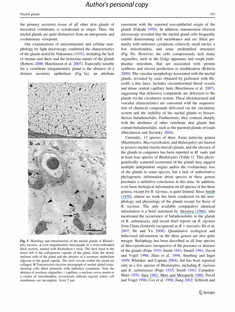
(Roberts 2000; Hutchinson et al. 2007). Especially notable
for a vertebrate integumentary gland is the absence of a
distinct secretory epithelium (Fig. 5a), an attribute
consistent with the reported non-epithelial origin of the
glands (Fukada 1958). In addition, transmission electron
microscopy revealed that the nuchal gland cells frequently
exhibit deteriorating cell membranes and are filled pri-
marily with indistinct cytoplasm, relatively small nuclei, a
few mitochondria, and some unidentified structures
(Fig. 5b). However, the cells conspicuously lack many
organelles, such as the Golgi apparatus and rough endo-
plasmic reticulum, that are associated with protein
synthesis and steroid production or modification (Roberts
2000). The vascular morphology associated with the nuchal
glands, revealed by casts obtained by perfusion with Mi-
crofil, a fine latex, includes circumferential blood vessels
and dense central capillary beds (Hutchinson et al. 2007),
suggesting that defensive compounds are delivered to the
glands via the circulatory system. These ultrastructural and
vascular characteristics are consistent with the sequestra-
tion of chemical compounds delivered via the circulatory
system and the inability of the nuchal glands to biosyn-
thesize bufadienolides. Furthermore, they contrast sharply
with the attributes of other vertebrate skin glands that
contain bufadienolides, such as the parotoid glands of toads
(Hutchinson and Savitzky 2004).
Currently, 13 species of three Asian natricine genera
(Rhabdophis, Macropisthodon, and Balanophis) are known
to possess nuchal (nucho-dorsal) glands, and the absence of
the glands in congeners has been reported in M. rudis and
at least four species of Rhabdophis (Table 1). This phylo-
genetically scattered occurrence of the glands may suggest
multiple independent origins and/or the evolutionary loss
of the glands in some species, but a lack of authoritative
phylogenetic information about species in these genera
precludes a definitive conclusion at this time. In addition,
even basic biological information on all species of the three
genera, except for R. tigrinus, is quite limited. Since Smith
(1938), almost no work has been conducted on the mor-
phology and physiology of the glands except for those of
R. tigrinus. The only available comparative chemical
information is a brief statement by Akizawa (1986), who
mentioned the occurrence of bufadienolides in the glands
of R. subminiatus, and recent brief reports on R. tigrinus
from China (formerly recognized as R. t. lateralis: He et al.
2007; He and Yu 2008). Quantitative ecological and
behavioral information on the three genera are also quite
meager. Bufophagy has been described in all four species
of Macropisthodon, irrespective of the presence or absence
of the glands (Pope 1935; Smith 1943; Daniel 1983; David
and Vogel 1996; Zhao et al. 1998; Stuebing and Inger
1999; Whitaker and Captain 2004), but has been reported
only in a few species of Rhabdophis, including R. tigrinus
and R. subminiatus (Pope 1935; Smith 1943; Campden-
Main 1970; Sura 1981; Mori and Moriguchi 1988; David
and Vogel 1996; Cox et al. 1998; Jiang 2002; Schleich and
Fig. 5 Histology and ultrastructure of the nuchal glands of Rhabdo-phis tigrinus. a Low-magnification micrograph of a resin-embedded
thick section, stained with Richardson’s stain. The dark band in the
lower left is the collagenous capsule of the gland. Note the nearly
uniform cells of the gland and the absence of a secretory epithelium
adjacent to the gland capsule. The dark streaks within the gland are
collagen. b Transmission electron micrograph of nuchal gland tissue,
showing cells filled primarily with indistinct cytoplasm. Note the
absence of secretory organelles. c capillary, n nucleus, arrow points to
a cluster of mitochondria; arrowheads indicate regions where cell
membranes are incomplete. Scale 5 lm
Nuchal glands 193
123
Author's personal copy
Kastle 2002; Whitaker and Captain 2004). Nuchal gland-
related behaviors have been confirmed only for R. tigrinus
and R. subminiatus. Neck flattening behavior, a display that
often accompanies the nuchal gland-related behaviors
(Mori and Burghardt 2008), is known in all four species of
Macropisthodon (Pope 1935; Smith 1943; Taylor 1965;
Daniel 1983; Tweedie 1983; Lue et al. 1999; Stuebing and
Inger 1999; Whitaker and Captain 2004; Baker and Lim
2008) and in at least four species of Rhabdophis, including
species that either possess or lack the glands (R. submini-
atus: Schleich and Kastle 2002; Whitaker and Captain
2004; Mori and Burghardt 2008, R. tigrinus: Mori et al.
1996; Mori and Burghardt 2008, R. murudensis: Stuebing
and Lian 2002, R. swinhonis: Lue et al. 1999). To formu-
late a working hypothesis regarding the origin and
evolution of the nuchal glands, comparative studies on
ecology, behavior, physiology, chemistry, and phylogeny
are required.
Whatever the evolutionary origin of the nuchal glands, it
is almost certain that exploitation of toads predated the
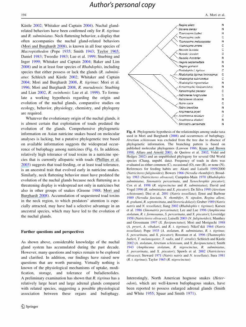
evolution of the glands. Comprehensive phylogenetic
information on Asian natricine snakes based on molecular
analyses is lacking, but a putative phylogenetic tree based
on available information suggests the widespread occur-
rence of bufophagy among natricines (Fig. 6). In addition,
relatively high tolerance of toad toxins in a natricine spe-
cies that is currently allopatric with toads (Phillips et al.
2003) suggests that toad-feeding, or at least toad tolerance,
is an ancestral trait that evolved early in natricine snakes.
Similarly, neck flattening behavior must have predated the
evolution of the nuchal glands because neck flattening as a
threatening display is widespread not only in natricines but
also in other groups of snakes (Greene 1988; Mori and
Burghardt 2008). Accumulation of sequestered toad toxins
in the neck region, to which predators’ attention is espe-
cially attracted, may have had a selective advantage in an
ancestral species, which may have led to the evolution of
the nuchal glands.
Further questions and perspectives
As shown above, considerable knowledge of the nuchal
gland system has accumulated during the past decade.
However, many questions and topics remain to be explored
and clarified. In addition, our findings have raised new
questions that are worth pursuing. Virtually nothing is
known of the physiological mechanisms of uptake, modi-
fication, storage, and tolerance of bufadienolides.
A preliminary examination has shown that R. tigrinus has a
relatively large heart and large adrenal glands compared
with related species, suggesting a possible physiological
association between these organs and bufophagy.
Interestingly, North American hognose snakes (Heter-
odon), which are well-known bufophagous snakes, have
been reported to possess enlarged adrenal glands (Smith
and White 1955; Spaur and Smith 1971).
Fig. 6 Phylogenetic hypothesis of the relationships among snake taxa
used in Mori and Burghardt (2008) and occurrences of bufophagy.
Atretium schistosum was excluded from the tree due to absence of
phylogenetic information. The branching pattern is based on
published molecular phylogenies (Lawson 1986; Kraus and Brown
1998; Alfaro and Arnold 2001; de Queiroz et al. 2002; Vidal and
Hedges 2002) and an unpublished phylogeny for several Old World
species (Chang, unpubl. data). Frequency of toads in diets was
evaluated as either common (C), occasional (O), rare (R), or none (N).
References for feeding habits are: Akani and Luiselli 1999/2000
(Natriciteres fuliginoides); Bowers 1966 (Nerodia rhombifer); Broad-
ley 1983 (Natriciteres olivacea); Campden-Main 1970 (Rhabdophissubminiatus, Sinonatrix percarinata, and Xenochrophis piscator);
Cox et al. 1998 (R. nigrocinctus and R. subminiatus); David and
Vogel 1996 (R. subminiatus and X. piscator); De Silva 1990 (Atretiumschistosum); Disi et al. 2001 (Natrix tessellata); Ernst and Barbour
1989 (Nerodia fasciata, N. rhombifer, N. sipedon, Regina alleni,R. grahami, R. septemvittata, and Storeria dekayi); Gruber 1989 (Natrixnatrix and N. tessellata); Jiang 2002 (Rhabdophis t. tigrinus); Karsen
et al. 1986 (Sinonatrix percarinata); Lee and Lue 1996 (Amphiesmastolatum, R. t. formosanus, S. percarinata, and X. piscator); Loveridge
1958 (Natriciteres olivacea); Luiselli 2003 (N. fuliginoides); Manthey
and Grossmann 1997 (X. flavipunctatus); Mori and Moriguchi 1988
(A. pryeri, A. vibakari, and R. t. tigrinus); Nikol’skii 1964 (Natrixtessellata); Pope 1935 (A. stolatum, R. subminiatus, R. t. tigrinus;
S. percarinata, and X. piscator); Rossman et al. 1996 (Thamnophisbutleri, T. melanogaster, T. radix, and T. sirtalis); Schleich and Kastle
2002 (A. stolatum, Atretium schistosum, and X. flavipunctatus); Smith
1943 (Amphiesma stolatum, R. nigrocinctus, R. subminiatus,
S. percarinata, and X. piscator); Spawls et al. 2002 (Natriciteresolivacea); Steward 1971 (Natrix natrix and N. tessellata); Sura 1981
(R. t. tigrinus); Taylor 1965 (R. nigrocinctus)
194 A. Mori et al.
123
Author's personal copy

The vasculature of the nuchal glands should be exam-
ined with more specimens because our observations to date
are limited. If the highly developed blood vessels reflect
delivery of bufadienolides to the glands via the circulatory
system, it is expected that blood plasma will contain sim-
ilar chemical compounds to those that are stored in the
nuchal glands; we have obtained a preliminary result sup-
porting this mechanism (Hutchinson et al. unpublished
data). Furthermore, clarification of the function of the
unknown organelles found in the cells of the nuchal glands
is necessary. Because discharge of the glandular fluid
involves destruction of the cells in the glands, those cells
must be regenerated if the snake is to use the organs again.
Alternatively, each nuchal gland may be an ‘‘expendable’’
organ that can be used only once in the snake’s lifetime.
Although the functional role of the nuchal glands as an
antipredator device is beyond doubt, the effectiveness of
the discharged fluid and the characteristic displays, such as
neck arch, to deter predators, as well as the role that the
discharged fluid plays in the process of deterrence, have not
been demonstrated. Direct field observations on the inter-
action between snakes and their predators are virtually
lacking (Mori et al. 2009). We suggest two scenarios, not
mutually exclusive, concerning the process of predator
deterrence. One possibility is that predators, such as raptors
(Tanaka and Mori 2000), that bite the neck region of the
snake would release the snake because of the unpalatability
of the fluid discharged by the biting force. The other pos-
sibility is that predators that restrain the snake by holding it
with their claws or feet, or by gripping it with their beaks,
exert pressure on the nuchal glands, which causes the
glandular fluid to squirt into the predator’s eyes. Presum-
ably, the predator would then release the snake as the result
of the irritation caused by the nuchal gland fluid. Staged
encounter tests with predators are necessary to clarify the
actual interactions.
The provisioning of sequestered chemical compounds
by mothers to their offspring (Hutchinson et al. 2008,
2011) implies the occurrence of indirect maternal care by
female snakes. Dams with large quantities of bufadieno-
lides in their nuchal gland fluid produce offspring with
large quantities of these compounds, but dams that lack
bufadienolides, or possess small quantities of them, pro-
duce hatchlings that lack these defensive compounds
(Hutchinson et al. 2008). Newly hatched snakes, which
appear in the wild around the end of August, may not be
able to consume juvenile toads due to gape limitation. That
is, the juvenile toads that metamorphosed the previous
spring may already have grown too large for hatchling
snakes to eat. If this is the case, juvenile snakes would not
be able to obtain bufadienolides from ingested toads until
the subsequent spring, when newly metamorphosed toad-
lets become available. Thus, maternal provisioning would
be an essential route to arm the hatchlings with toxic
compounds until they are able to consume toads them-
selves. Therefore, it is predicted that females (or gravid
females) are more likely to feed on toads than males (or
non-gravid females) because of the need for a sufficient
amount of sequestered compounds for provisioning
(Hutchinson et al. 2008). However, we do not know the
quantity of bufadienolides required for deterrence of pre-
dators by hatchlings or the delivery route of sequestered
chemical compounds from dams to offspring. Although
several routes appear to be involved in the transfer of
toxins to embryos (Hutchinson et al. 2008), it is not known
whether the provisioned toxins must be freshly ingested or
whether they can be mobilized from stores in the nuchal
glands or elsewhere in the female’s tissues. Behavioral
studies both in the field and laboratory, along with a
physiological investigation of the provisioning system in R.
tigrinus, may uncover a type of maternal care unique
among terrestrial vertebrates.
The possibility that individual snakes exhibit behavioral
adjustments according to the amount of toxins stored in the
nuchal glands is worthy of exploration. If snakes are able to
recognize how toxic they are based on the amount of
bufadienolides accumulated in their glands, they may
modify their antipredator responses accordingly. They may
rely more heavily on nuchal gland-related behaviors when
they possess a sufficient amount of toxins to deter preda-
tion than when they do not have enough. A preliminary
experiment addressing this question revealed that juvenile
snakes that have been fed toads exhibit neck arch more
frequently than those that have been reared on other types
of foods (Mori and Burghardt in prep.). This may imply
self-recognition of toxicity, whatever proximate mecha-
nism is involved, and, if so, would provide an interesting
case of a plastic behavioral response coordinated with
toxicity level.
An investigation of the antipredator behavior of addi-
tional species is necessary to understand the evolution of the
unique displays associated with nuchal glands. It would be
worth examining the antipredator responses of species other
than R. tigrinus that possess nuchal glands, including those
species that have glands extending the entire length of the
body (nucho-dorsal glands). Additionally, the antipredator
displays of congeneric species and closely related outgroups
that lack nuchal glands should be studied to elucidate the
correlated evolution of morphology and behavior. To
implement these approaches, broad phylogenetic analyses of
Old World natricine snakes, including the three genera
containing species with nuchal glands (Rhabdophis, Mac-
ropisthodon, and Balanophis), are required. The
phylogenetically independent occurrence of death feigning
behavior in R. tigrinus, Heterodon nasicus, H. platirhinos,
Natrix natrix, and Hemachatus haemachatus (Greene 1988),
Nuchal glands 195
123
Author's personal copy
which are all toad-eaters (Steward 1971; Broadley 1983;
Ernst and Barbour 1989), suggests the possible, but uncer-
tain, functional association between bufophagy and death
feigning. A comparative study of these snakes may reveal
unexpected physiological and neuroethological links.
We have observed considerable individual variation in
the size of the nuchal glands, composition of bufadieno-
lides, and the propensity to perform nuchal gland-related
behaviors that are not readily explained by differences in
sex or body size of the snakes. Clarification of the physi-
ological, genetic, developmental, and functional factors
that underlie such individual variation would shed light on
the adaptive nature of the nuchal glands.
Overall, we believe that research on nuchal glands and
their related features is a fruitful avenue not only for
understanding these unique squamate glands, but also for
comprehending broad mechanisms of physiology, bio-
chemistry, and evolution of phenotypic traits that may be
applicable to many other vertebrates. We hope that this
review will stimulate researchers in various fields of biology
and chemistry to address the many outstanding questions and
will foster integrated collaborative approaches.
Acknowledgments We thank J. Placyk and J. M. Ray for their
assistance in conducting experiments, M. Toriba for his comments on
taxonomy of snakes, K. Isogawa, M. Motokawa, and K. Nishikawa for
obtaining literature, and many students and colleagues for their help in
collecting and keeping animals. We greatly appreciate J. Meinwald and
F. C. Schroeder for their invaluable help with the chemical analyses.
The first author especially wishes to thank the late A. Mutoh for his
invitation to the amazing world of Rhabdophis tigrinus. The original
idea of dietary sequestration was inspired during a discussion with M.
Hasegawa. This research was supported in part by Grants from the
Japan–US Cooperative Science Program (Japan Society for the Pro-
motion of Science: JSPS); a Grant from JSPS (Scientific Research C:
23570115); Grants for the twentyfirst Century COE Program (A14) and
the Global COE Program (A06) to Kyoto University; and grants from
the US National Science Foundation (IBN-0429223 and IOB-0519458
to AHS and J. Meinwald). This paper is based on a presentation in the
symposium ‘‘Sequestered Defensive Compounds in Tetrapod Verte-
brates: A Symposium in Memory of John W. Daly’’, held at the Sixth
World Congress of Herpetology in Manaus, Brazil, on 21 August 2008
and supported by NSF IOS-0813842.
References
Akani GC, Luiselli L (1999/2000) Aspects of the natural history of
Natriciteres (Serpentes, Colubridae) in Nigeria, with special
reference to N. variegata and N. fuliginoides. Herpetol Nat Hist
7:163–168
Akizawa T (1986) Studies on the activities of chemicals in the nuchal
glands of Rhabdophis tigrinus. Ochanomizu Med J 34:85–97 (in
Japanese)
Akizawa T, Yasuhara T, Kano R, Nakajima T (1985a) Novel
polyhydroxylated cardiac steroids in the nuchal glands of the
snake, Rhabdophis tigrinus. Biomed Res 6:437–441
Akizawa T, Yasuhara T, Azuma H, Nakajima T (1985b) Chemical
structures and biological activities of bufadienolides in the
nucho-dorsal glands of Japanese snake, Rhabdophis tigrinus.
J Pharmacobio Dyn 8:s–60 (abstract)
Alfaro ME, Arnold SJ (2001) Molecular systematics and evolution of
Regina and the thamnophiine snakes. Mol Phylogenet Evol
21:408–423
Ariga F (1953) A case of eye disturbance by Rhabdophis tigrinus. Jpn
Rev Clinic Ophthalmol 47:258–259 (in Japanese)
Asahi H, Kohtari Y, Chiba K, Mishima A (1985) Effect of the
nuchodorsal gland venom of the Yamakagashi snake on the eye.
Folia Ophthalmol Japonica 36:379–383 (in Japanese with
English abstract)
Azuma H, Sekizaki S, Akizawa T, Yasuhara T, Nakajima T (1986)
Activities of novel polyhydroxylated cardiotonic steroids puri-
fied from nuchal glands of the snake, Rhabdophis tigrinus.
J Pharm Pharmacol 38:388–390
Baker N, Lim K (2008) Wild animals of Singapore. Draco Publ Distr
and Nature Soc, Singapore
Bowers JH (1966) Food habits of the diamond-backed water snake,
Natrix rhombifera rhombifera, in Bowie and Red River Coun-
ties, Texas. Herpetologica 22:225–229
Broadley DG (1983) FitzSimons’ snakes of Southern Africa. Revised
edn. Delta Books, Johannesburg
Brodie ED III, Brodie ED Jr (1999) Costs of exploiting poisonous
prey: evolutionary trade-offs in a predator-prey arms race.
Evolution 53:626–631
Campden-Main SM (1970) A field guide to the snakes of South
Vietnam. Division of reptiles and amphibians. US Nat Mus,
Smithsonian Inst, Washington
Cox MJ, van Dijk PP, Nabhitabhata J, Thirakhupt K (1998) A
photographic guide to snakes and other reptiles of Peninsular
Malaysia, Singapore and Thailand. Ralph Curtis Books, Sanibel
Island
Daniel JC (1983) The book of Indian reptiles. Bombay Nat Hist Soc,
Bombay
David P, Ineich I (1999) Les serpents venimeux du monde:
systematique et repartition. Dumerilia, vol 3. Lab Reptilies
Amphibiens Mus Natn Hist Nat Paris, Paris
David P, Vogel G (1996) The snakes of Sumatra. An annotated
checklist and key with natural history notes. Edition Chimaira,
Frankfurt
de Queiroz A, Lawson R, Lemos-Espinal JA (2002) Phylogenetic
relationships of North American garter snakes (Thamnophis)
based on four mitochondrial genes: How much DNA sequence is
enough? Mol Phylogenet Evol 22:315–329
De Silva A (1990) Colour guide to the snakes of Sri Lanka. R & A
Publishing, Avon
Disi AM, Modry D, Necas P, Rifai L (2001) Amphibians and reptiles of
the Hashemite Kingdom of Jordan. Edition Chimaira, Frankfurt
Dobashi H (1935) Eye disturbance caused by a venomous snake.
Central J Ophthalmol 27:1175–1176 (in Japanese)
Ernst CH, Barbour RW (1989) Snakes of eastern North America.
George Mason Univ Press, Fairfax
Fukada H (1958) Embryological study on the integumental poison
gland in the nuchal region of Natrix tigrina tigrina. Bull Kyoto
Gakugei Univ Ser B No. 12:3–8, 2 pls
Fukada H (1959) Biological studies on the snakes. VI. Growth and
maturity of Natrix tigrina tigrina (Boie). Bull Kyoto Gakugei
Univ Ser B No. 15:25–41, 2 pls
Fukada H (1961) Peculiar habits of the Japanese snake, Natrix t.tigrina (Boie). Bull Kyoto Gakugei Univ Ser B No. 18:13–16, 2
pls
Greene HW (1988) Antipredator mechanisms in reptiles. In: Gans C,
Huey RB (eds) Biology of the Reptilia, vol 16. Ecology B.
Defense and life history. Alan R Liss Inc, New York, pp 1–152
Gruber U (1989) Die Schlangen Europas und rund ums Mittelmeer.
Kosmos, Stuttgart
196 A. Mori et al.
123
Author's personal copy

He QY, Yu XD (2008) Isolation and identification of a metallopro-
teinase RT-2 from nuchal gland venom of Rhabdophis tigrinuslateralis. Shanxi Daxue Xuebao Ziran Kexue Ban 31:108–113
He QY, Yu XD, Liu JP (2007) Study on venom from nuchal glands of
Rhabdophis tigrinus lateralis in China. Sichuan J Zool
26:255–257
Hikita A (1947) A case of eye disturbance caused by the secretions of
Rhabdophis tigrinus. Jpn Rev Clinic Ophthalmol 41:207–208 (in
Japanese)
Hirai T (2004) Dietary shifts of frog eating snakes in response to
seasonal changes in prey availability. J Herpetol 38:455–460
Hutchinson DA, Savitzky AH (2004) Vasculature of the parotoid
glands of four species of toads (Bufonidae: Bufo). J Morphol
260:247–254
Hutchinson DA, Mori A, Savitzky AH, Burghardt GM, Wu X,
Meinwald J, Schroeder FC (2007) Dietary sequestration of
defensive steroids in nuchal glands of the Asian snake Rhabdo-phis tigrinus. Proc Natl Acad Sci 104:2265–2270
Hutchinson DA, Savitzky AH, Mori A, Meinwald J, Schroeder FC
(2008) Maternal provisioning of sequestered defensive steroids
by the Asian snake Rhabdophis tigrinus. Chemoecology
18:181–190
Hutchinson DA, Savitzky AH, Mori A, Burghardt GM, Meinwald J,
Schroeder FC (2011) Chemical investigations of defensive
steroid sequestration by the Asian snake Rhabdophis tigrinus.Chemoecology. doi:10.1007/s00049-011-0078-2
Jiang YF (2002) Observation on behaviors of Rhabdophis tigrinuslateralis. Sichuan J Zool 21:29–31 (in Chinese)
Jiang Y, Zhao E (1983) Studies on amphibians and reptiles of Mt.
Gongga region, Sichuan, China. 3. A study of species-group
nuchalis, genus Rhabdophis. Acta Herpetol Sinica 2:59–62 (in
Chinese with English abstract)
Kadowaki S (1992) Food resource overlap between the two sympatric
Japanese snakes (Elaphe quadrivirgata and Rhabdophis tigrinus)
in the paddy fields. Jpn J Ecol 42:1–7 (in Japanese with English
Synopsis)
Karsen SJ, Lau MW, Bogadek A (1986) Hong Kong amphibians and
reptiles. Urban Council, Hong Kong
Kawamoto F, Kumada N (1989) A case report of eye-injury caused by
cervical gland venom of a snake, Rhabdophis tigrinus (Boie).
Jpn J Sanitary Zool 40:211–212 (in Japanese with English
abstract)
Kawashima J (1957) Disturbance of the eye by snake venom (Natrixtigrina). Ganka Rinsho Iho 50:837–839 (in Japanese)
Kawashima J (1959) Disturbance of the eye by snake venom (Natrixtigrina), II. Ganka Rinsho Iho 53:834–837 (in Japanese)
Kitazume Y (1953) On the increasing action of nuchal gland
inclusions of the snake, Natrix tigrina, upon the blood pressure
of the rabbit. Zool Mag 62:225–227 (in Japanese with English
abstract)
Kitazume Y (1958) Further studies on the effect of the nuchal gland
substance of Natrix tigrina upon the blood pressure of some
animals. Zool Mag 67:111–115 (in Japanese with English
abstract)
Kraus F, Brown WM (1998) Phylogenetic relationships of colubroid
snakes based on mitochondrial DNA sequences. Zool J Linn Soc
122:455–487
Lawson R (1986) Molecular systematics of some Old World natricine
snakes. In: Rocek Z (ed) Studies in herpetology, Proc Eur
Herpetol Meeting Prague 1985. Charles Univ, Prague,
pp 227–234
Lee WJ, Lue KY (1996) The preliminary study on the food habits of
snakes in Taiwan. Biol Bull Nat Taiwan Normal Univ
31:119–124 (in Chinese with English abstract)
Licht LE, Low B (1968) Cardiac response of snakes after ingestion of
toad parotoid venom. Copeia 1968:547–551
Loveridge A (1958) Revision of five African snake genera. Bull Mus
Comp Zool 119:1–198
Lue KY, Tu MC, Hsiang G (1999) Atlas of Taiwan amphibians and
reptiles. Nature Press, Taipei
Luiselli L (2003) Do snakes exhibit shifts in feeding ecology
associated with the presence or absence of potential competitors?
A case study from tropical Africa. Can J Zool 81:228–236
Manthey U, Grossmann W (1997) Amphibien & Reptilien Sudostas-
iens. Natur und Tier, Berlin
Mao JJ, Chang HW (1999) Notes on nuchal gland anatomic of
Rhabdophis tigrinus formosanus and Rhabdophis swinhonis(Natricinae: Squamata). J Nat Taiwan Mus 52:87–92
Mori A (1997) A comparison of predatory behavior of newly hatched
Rhabdophis tigrinus (Serpentes: Colubridae) on frogs and fish.
Jpn J Herpetol 17:39–45
Mori A (2006) Is headfirst ingestion essential in gape-limited
predators? Prey-handling behavior of the anurophagous snake
Rhabdophis tigrinus (Colubridae). Can J Zool 84:954–963
Mori A, Burghardt GM (2000) Does prey matter? Geographic
variation in antipredator responses of hatchlings of a Japanese
natricine snake, Rhabdophis tigrinus. J Comp Psychol
114:408–413
Mori A, Burghardt GM (2001) Temperature effects on anti-predator
behaviour in Rhabdophis tigrinus, a snake with toxic nuchal
glands. Ethology 107:795–811
Mori A, Burghardt GM (2008) Comparative experimental tests of
natricine antipredator displays, with special reference to the
apparently unique displays in the Asian genus, Rhabdophis.
J Ethol 26:61–68
Mori A, Moriguchi H (1988) Food habits of the snakes in Japan: a
critical review. Snake 20:98–113
Mori A, Vincent SE (2008) An integrative approach to specialization:
relationships among feeding morphology, mechanics, behaviour,
performance and diet in two syntopic snakes. J Zool Lond
275:47–56
Mori A, Layne D, Burghardt GM (1996) Description and preliminary
analysis of antipredator behavior of Rhabdophis tigrinus tigri-nus, a colubrid snake with nuchal glands. Jpn J Herpetol
16:94–107
Mori A, Konishi E, Izumi T (2009) A putative predatory attempt by
Meles meles on Rhabdophis tigrinus and a possible aversive
function of the nuchal glands. Bull Herpetol Soc Japan
2009:18–20 (in Japanese)
Moriguchi H (1985) Body size differences between two populations
of Rhabdophis tigrinus. Snake 17:140–143
Moriguchi H, Naito S (1982) Activities and food habits of Amphiesmavibakari (Boie) and Rhabdophis tigrinus (Boie). Snake
14:136–142 (in Japanese with English abstract)
Moriguchi H, Naito S (1983) Tracing of distance of movement of
Rhabdophis tigrinus (Boie) in a paddy field by mark-recapture
method. Snake 15:14–15
Moriguchi H, Toriba M (1985) Note on the number of eggs in oviduct
of Rhabdophis tigrinus (Boie). Snake 17:144–147
Mutoh A (1983) Death-feigning behavior of the Japanese colubrid
snake Rhabdophis tigrinus. Herpetologica 39:78–80
Nakai T (1953) Some actions of the secretion of the integumental
poison glands of Natrix tigrina on the white mouse. Zool Mag
62:27–30 (in Japanese with English abstract)
Nakamura K (1935) On a new integumental poison gland found in the
nuchal region of a snake, Natrix tigrina. Mem College Sci Kyoto
Imperial Univ Ser B 10:229–240, 1 pl
Nikol’skii AM (1964) Fauna of Russia and adjacent countries.
Reptiles, vol II. Ophidia. Israel Prog Sci Trans, Jerusalem
Ogawa H, Ohasi D, Iritani I, Kisimoto H, Nakamura Y, Oda M,
Tuduki H, Suzuki M (1992) Eye disturbance caused by
nuchodorsal gland venom from a Yamakagashi (Rhabdophis
Nuchal glands 197
123
Author's personal copy
tigrinus): 16th case report in Japan. Jpn J Toxicol 5:169–172 (in
Japanese with English abstract)
Phillips BL, Brown GP, Shine R (2003) Assessing the potential
impact of cane toads on Australian snakes. Conserv Biol
17:1738–1747
Pope CH (1935) The reptiles of China. Turtles, crocodilians, snakes,
lizards. Am Mus Nat Hist, New York
Roberts KA (2000) An ultrastructural survey of the nuchal glands of
Rhabdophis tigrinus (Serpentes: Colubridae). Unpublished mas-
ter thesis, Old Dominion University
Rossman DA, Ford NB, Seigel RA (1996) The garter snakes.
Evolution and ecology. Univ Oklahoma Press, Norman
Saitoh T (1949) Right eye disturbance caused by the secretions of
Rhabdophis tigrinus and left eye disturbance by undiluted nitric
acid. Jpn Rev Clinic Ophthalmol 43:336 (in Japanese)
Schleich HH, Kastle W (eds) (2002) Amphibians and reptiles of
Nepal. A R G Gantner, Ruggell
Shibuya H (1923) A case of corneitis caused by the skin secretions of
a snake. Jpn Rev Clinic Ophthalmol 18:368–369 (in Japanese)
Shimada K, Fujii Y, Yamashita E, Niizaki Y, Sato Y, Nambara T
(1977) Studies on cardiotonic steroids from the skin of Japanese
toad. Chem Pharm Bull (Tokyo) 25:714–730
Smith MA (1938) The nucho-dorsal glands of snakes. Proc Zool Soc
Lond Ser B 100:575–583
Smith MA (1943) The fauna of British India. Ceylon and Burma,
including the whole of the Indo-Chinese sub-region. Reptilia and
amphibia, vol III. Serpentes. Taylor and Francis, London
Smith HM, White FN (1955) Adrenal enlargement and its signifi-
cance in the hognose snakes (Heterodon). Herpetologica
11:137–144
Spaur RC, Smith HM (1971) Adrenal enlargement in the hognosed
snake, Heterodon platyrhinos. J Herpetol 5:197–199
Spawls S, Howell K, Drewes R, Ashe J (2002) A field guide to the
reptiles of east Africa. Academic Press, San Diego
Steward JW (1971) The snakes of Europe. Fairleigh Dickinson Univ
Press, Rutherford
Stuebing RB, Inger RF (1999) A field guide to the snakes of Borneo.
Nat Hist Publ (Borneo), Kota Kinabalu
Stuebing RB, Lian TF (2002) Notes on the fire-lipped keelback
Rhabdophis murudensis (Smith 1925) (Ophidia: Colubridae:
Natricinae) from northern Borneo. Raffles Bull Zool 50:227–230
Sura P (1981) Captive breeding of Elaphe rufodorsata and Rhabdo-phis tigrinus from the Korean People’s Democratic Republic. Br
Herpetol Soc Bull No. 3:20–24
Suzuki R (1960) Disturbance of the eye by snake venom (Natrixtigrina (Boie)). J Clinic Ophthalmol 14:1384–1387 (in Japanese
with English abstract)
Tamura K (1940a) Eye disturbance caused by the secretions of a
snake. Jpn Rev Clinic Ophthalmol 35:860–861 (in Japanese)
Tamura K (1940b) Eye disturbance caused by the secretions of a
snake II. Jpn Rev Clinic Ophthalmol 35:1191 (in Japanese)
Tanaka K, Mori A (2000) Literature survey on predators of snakes in
Japan. Curr Herpetol 19:97–111
Taylor EH (1965) The serpents of Thailand and adjacent waters. Univ
Kansas Sci Bull 45:609–1096
Tweedie MWF (1983) The snakes of Malaya, 3rd edn. Singapore
Natn Printer, Singapore
Vidal N, Hedges SB (2002) Higher-level relationships of caenophi-
dian snakes inferred from four nuclear and mitochondrial genes.
C R Biol 325:987–995
Watanabe S, Tsuneto S, Matsumoto Y (1988) Colubridae snake:
Japanese Yamakagashi (Rhabdophis tigrinus tigrinus). Jpn J
Acute Med 12:441–450 (in Japanese)
Whitaker R, Captain A (2004) Snakes of India, the field guide. Draco
Books, Chennai
Williams BL, Brodie ED Jr, Brodie ED III (2003) Coevolution of
deadly toxins and predator resistance: self-assessment of resis-
tance by garter snakes leads to behavioral rejection of toxic newt
prey. Herpetologica 59:155–163
Zhao EM (1995) Taxonomic status of some snake species and
subspecies. J Shuzhou Railw Teach Coll 12:36–39 (in Chinese
with English summary)
Zhao EM (1997) A new species of Rhabdophis (Serpentes: Colubri-
dae) from Hainan Island, China. Asiatic Herpetol Res 7:166–169
Zhao E, Meihua H, Yu Z (1998) Fauna Sinica, Reptilia, vol 3.
Squamata, Serpentes. Science Press, Beijing (in Chinese)
198 A. Mori et al.
123
Author's personal copy