non-invasive high-resolution tracking of human neuronal pathways: diffusion tensor imaging at 7t...
TRANSCRIPT

Non-invasive high-resolution tracking of human neuronal pathways: Diffusion Tensor Imaging at 7T with 1.2 mm isotropic voxel size
Ralf Lützkendorfa, Frank Hertela, Robin Heidemannb, Andreas Thielc, Michael Luchtmannd, Markus Plaumanna, Jörg Stadlere, Sebastian Baeckea, Johannes Bernardinga
aDepartment of Biometry and Medical Informatics, Otto-von-Guericke University Magdeburg; bSiemens Healthcare Sector, Erlangen, Germany; cInstitute for Information Technology, OFFIS Oldenburg; dDepartment of Neurosurgery, Otto-von-Guericke-University Magdeburg; eLeibniz
Institute for Neurobiology, Magdeburg
ABSTRACT
Diffusion tensor imaging (DTI) allows characterizing and exploiting diffusion anisotropy effects, thereby providing important details about tissue microstructure. A major application in neuroimaging is the so-called fiber tracking where neuronal connections between brain regions are determined non-invasively by DTI. Combining these neural pathways within the human brain with the localization of activated brain areas provided by functional MRI offers important information about functional connectivity of brain regions. However, DTI suffers from severe signal reduction due to the diffusion-weighting. Ultra-high field (UHF) magnetic resonance imaging (MRI) should therefore be advantageous to increase the intrinsic signal-to-noise ratio (SNR). This in turn enables to acquire high quality data with increased resolution, which is beneficial for tracking more complex fiber structures. However, UHF MRI imposes some difficulties mainly due to the larger B1 inhomogeneity compared to 3T MRI. We therefore optimized the parameters to perform DTI at a 7 Tesla whole body MR scanner equipped with a high performance gradient system and a 32-channel head receive coil. A Stesjkal Tanner spin-echo EPI sequence was used, to acquire 110 slices with an isotropic voxel-size of 1.2 mm covering the whole brain. 60 diffusion directions were scanned which allows calculating the principal direction components of the diffusion vector in each voxel. The results prove that DTI can be performed with high quality at UHF and that it is possible to explore the SNT benefit of the higher field strength. Combining UHF fMRI data with UHF DTI results will therefore be a major step towards better neuroimaging methods.
Keywords: MRI, DWI, DTI, 7T, high resolution, fiber tracking,
1. INTRODUCTION Diffusion-weighted imaging (DWI) is an important clinical technique to detect pathological changes in the white matter [1], such as ischemic stroke. Its extension, diffusion tensor imaging (DTI) supplies fiber tracking [2] algorithms with the necessary information to find major pathways of neurons in the white matter connecting different functional regions of the brain. Higher magnetic field strength such as 7 Tesla is expected to increase the signal-to-noise ratio (SNR) which can be used to achieve a higher spatial resolution in DWI. A higher resolution is a major step forward to analyze more
Medical Imaging 2013: Physics of Medical Imaging, edited by Robert M. Nishikawa, Bruce R. Whiting, Christoph Hoeschen, Proc. of SPIE Vol. 8668, 866846 · © 2013 SPIE · CCC code: 1605-7422/13/$18 · doi: 10.1117/12.2006764
Proc. of SPIE Vol. 8668 866846-1
Downloaded From: http://proceedings.spiedigitallibrary.org/ on 01/15/2014 Terms of Use: http://spiedl.org/terms

complex fiber structures, such as crossing or kissing fibers which are difficult to distinguish. Furthermore, DWI data with high isotropic resolution provides information about the grey matter, such as the radial anisotropy of the cortex. However, higher field strength also leads to severe signal inhomogeneities, partial signal losses, susceptibility artifacts and image blurring in DWI. The use of parallel imaging with head coil arrays with many elements is mandatory to address these problems.
2. METHODOLOGY Experiments were performed on a 7 Tesla whole-body MR scanner (MAGNETOM 7T, Siemens Healthcare, Germany) equipped with a gradient system achieving a maximum amplitude of 70 mT/m per axes and a slew rate of 200 T/m/s. For signal reception a 32-channel head coil array (Nova Medical, USA) [3] is used. 5 healthy volunteer were examined each in a single session acquiring 110 slices with 1.2 mm slice thickness and no gap to cover the whole brain. DWI was carried out with an optimized Stesjkal Tanner sequence scheme [4] with 60 diffusion directions [5] and a maximum b-value of 1000 s/mm2. Generalized auto calibrating partially parallel acquisition (GRAPPA) was used with an acceleration factor of 3 and 36 reference lines, a pixel bandwidth of 1510 Hz/Px and 6/8 phase partial Fourier. A base resolution of 184 and a FOV of 220 mm results in an isotropic voxel size of 1.2 mm. The echo time (TE) with 58 ms was set to the minimum value. The repetition time (TR) of 12700 ms resulted in an acquisition time of around 15 minutes. The DWI data was processed by the commercial software package Brainvoyager (http://www.brainvoyager.com). No averaging, no registration and no eddy current correction were carried out due to show the clear unchanged data. The Software package calculated the standard Tensor and processed the fractional anisotropy (FA), the direction encoded color map (DEC) and the Apparent Diffusion Coefficient (ADC).
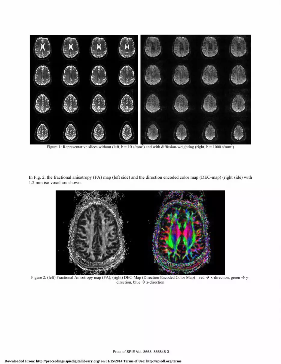
3. RESULTS Figure 1 shows the unprocessed diffusion raw data. The non-diffusion-weighted data is shown on the left side and the diffusion-weighted (b-value of 1000 s/mm2) data on the right side.
Proc. of SPIE Vol. 8668 866846-2
Downloaded From: http://proceedings.spiedigitallibrary.org/ on 01/15/2014 Terms of Use: http://spiedl.org/terms
!C,J
"1r-A
N. me z-v
(11. 5
frr-akJ,
o
Figure 1: Representative slices without (left, b = 10 s/mm2) and with diffusion-weighting (right, b = 1000 s/mm2)
In Fig. 2, the fractional anisotropy (FA) map (left side) and the direction encoded color map (DEC-map) (right side) with 1.2 mm iso voxel are shown.
Figure 2: (left) Fractional Anisotropy map (FA); (right) DEC-Map (Direction Encoded Color Map) – red x-direction, green y-
direction, blue z-direction
Proc. of SPIE Vol. 8668 866846-3
Downloaded From: http://proceedings.spiedigitallibrary.org/ on 01/15/2014 Terms of Use: http://spiedl.org/terms

A comparison between two different DWI sequences at 7T is shown in Fig. 3. The image on the left side was acquired with the standard DWI sequence, the same resolution but slice thickness is 4mm instead of 1.2 mm, a twice refocused Spin Echo EPI sequence. This sequence is used on MR scanners with lower field strength to compensate eddy currents. This is achieved by a certain gradient scheme and two refocusing pulses. In comparison, the Stesjkal Tanner sequence, a simple DWI sequence, has only a single refocusing pulse, enabling a much shorter TE. Even though eddy currents are not compensated, the shorter TE, in this special case from 92 ms to 58 ms, results in a significantly improved image quality. It turns out that eddy current are not a major problem with this gradient system, the major goal is to keep TE as short as possible.
Fig. 3: (left) Diffusion-weighted image (DWI) at 7T (b=800 s/mm2), diffusion-weighted SE-EPI sequences (twice refocused), 1,2 x 1,2 x 4 mm voxel size, TR=9000ms, TE=92ms (ISMRM, 2008); (right) similar slice from the actual dataset with optimized Stesjkal Tanner sequence (1.2 x 1.2 x 1.2 mm).
Proc. of SPIE Vol. 8668 866846-4
Downloaded From: http://proceedings.spiedigitallibrary.org/ on 01/15/2014 Terms of Use: http://spiedl.org/terms
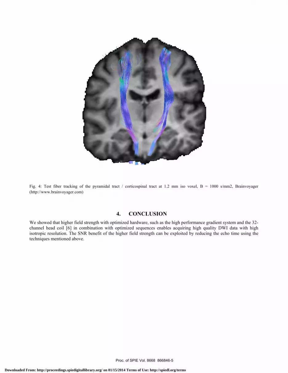
Fig. 4: Test fiber tracking of the pyramidal tract / corticospinal tract at 1.2 mm iso voxel, B = 1000 s/mm2, Brainvoyager (http://www.brainvoyager.com)
4. CONCLUSION We showed that higher field strength with optimized hardware, such as the high performance gradient system and the 32-channel head coil [6] in combination with optimized sequences enables acquiring high quality DWI data with high isotropic resolution. The SNR benefit of the higher field strength can be exploited by reducing the echo time using the techniques mentioned above.
Proc. of SPIE Vol. 8668 866846-5
Downloaded From: http://proceedings.spiedigitallibrary.org/ on 01/15/2014 Terms of Use: http://spiedl.org/terms

D (1.2mm iso)
Fig. 5: 1.7 mm versus 1.2 mm isotropic resolution (From the HBM conference 2012 [7]), 1.2mm has a more detailed structure compared to 1.7 mm, a profile through the sulcus at a certain region of the cortex from a slice acquired with 1.7 mm iso resolution does not show the cortex (A). The same position in the 1.2 mm iso voxel size image clearly shows up the cortex as two peaks (C). Compared to 1.7 mm isotropic voxel size and higher, the 1.2 mm isotropic voxel are a significant step towards resolving even very small fiber tracts. Due to the high isotropic resolution, anisotropy effects can be investigated not only in white matter, but also in grey matter, e.g. the radial anisotropy in the cortex. With the advanced techniques, DTI at 7T is not only feasible; it provides high-quality DWI data with 1.2 mm isotropic voxel resolution without averaging in an acceptable time. This would not be possible at lower field strength, such as 3 Tesla. However, even 15 minutes are too long for daily clinical routine. To reduce the scan time, fewer slices could be scanned or the diffusion directions can be reduced. Despite the relatively long scan time, this kind of high resolution DWI data can boost connection experiments by combining DTI with high-resolution fMRI at 7T or even higher. However, there are techniques that can further improve the image quality with focus on eddy current correction [8], zoomed techniques [9] or other sequences [10] that gain signal and increase the resolution. REFERENCE [1] Moseley, M. E., Cohen, Y., Kucharczyk, J., Mintorovitch, J., Asgari, H. S., Wendland, M. F., “Diffusion-weighted MR imaging of anisotropic water diffusion in cat central nervous system,” Radiology 176(2), 439–445 (1990). [2] Basser, P. J., Pajevic, S., Pierpaoli, C., Duda, J., Aldroubi, A., “In vivo fiber tractography using DT-MRI data,” MRM, 44(4), 625–632 (2004).
Proc. of SPIE Vol. 8668 866846-6
Downloaded From: http://proceedings.spiedigitallibrary.org/ on 01/15/2014 Terms of Use: http://spiedl.org/terms

[3] Luetzkendorf , R., “Diffusion weighted imaging at 3T and 7T: comparison of different phased array head coils,” Proc. 15th annual meeting of the Organization for Human Brain Mapping, San Francisco, S. F 275 AM (2009).
[4] Morelli, J.N., „ Evaluation of a Modified Stejskal-Tanner Diffusion Encoding Scheme, Permitting a Marked Reduction in TE, in Diffusion- Weighted Imaging of Stroke Patients at 3 T,” Investigative Radiology, 45, 29-35 (2010). [5] Jones, D.K., “Isotropic resolution diffusion tensor imaging with whole brain acquisition in a clinically acceptable time,” Human Brain Mapping, 15, 216-230 (2002). [6] Luetzkendorf, R, “In vivo DWI at 7T with a 70 mT/m gradient coil: 24- vs 32-channel head coil,” Proc. 17th annual meeting of the Organization for Human Brain Mapping, Québec, (2011).
[7] Luetzkendorf, R., Heidemann, R.M., Anwander, A., Stadler, J., Feiweier, T., Bernarding, J., “Diffusion-Weighted Imaging at 7Twith a High Performance Gradient System and a 32 Channel Head Coil: What is possible – Resolution versus Acquisition Time” Proc. 18th annual meeting of the Organization for Human Brain Mapping, Beijing, (2012). [8] Finsterbusch, J., “Eddy-current compensated diffusion weighting with a single refocusing RF pulse,” MRM, 61(3), 748–754 (2009).
[9] Heidemann, R.M., Anwander A., Eichner, C., Luetzkendorf, R., Feiweier, T., Knösche1, T.R., Bernarding, J., Turner, R., „ Isotropic Sub-Millimeter Diffusion MRI in Humans at 7T, “Proc. 17th annual meeting of the Organization for Human Brain Mapping, Québec, (2011). [10] Heidemann, R.M., Porter, D.A., Anwander, A., Feiweier, T., Heberlein, K., Knösche,T.R., Turner, R., “Diffusion imaging in humans at 7T using readout-segmented EPI and GRAPPA,” MRM 64 (1), 9–14 (2010).
Proc. of SPIE Vol. 8668 866846-7
Downloaded From: http://proceedings.spiedigitallibrary.org/ on 01/15/2014 Terms of Use: http://spiedl.org/terms