nitrite-cured cooked pork products - dtu orbit
TRANSCRIPT
General rights Copyright and moral rights for the publications made accessible in the public portal are retained by the authors and/or other copyright owners and it is a condition of accessing publications that users recognise and abide by the legal requirements associated with these rights.
Users may download and print one copy of any publication from the public portal for the purpose of private study or research.
You may not further distribute the material or use it for any profit-making activity or commercial gain
You may freely distribute the URL identifying the publication in the public portal If you believe that this document breaches copyright please contact us providing details, and we will remove access to the work immediately and investigate your claim.
Downloaded from orbit.dtu.dk on: Feb 11, 2022
Nitrite-cured cooked pork products – Characterisation of antioxidative andantimicrobial activities
Pedersen, Sabrine Tauber
Publication date:2018
Document VersionPublisher's PDF, also known as Version of record
Link back to DTU Orbit
Citation (APA):Pedersen, S. T. (2018). Nitrite-cured cooked pork products – Characterisation of antioxidative and antimicrobialactivities. Technical University of Denmark.
Nitrite-cured cooked pork products- Characterisation of antioxidative and antimicrobial activities
Sabrine Tauber PedersenPhD thesisMay 2018
Nitrite-cured cooked pork products
– Characterisation of antioxidative and antimicrobial activities
Sabrine Tauber Pedersen
PhD Thesis
Division of Food Technology
National Food Institute
Technical University of Denmark
May 2018
Title sheet
Title: Nitrite-cured cooked pork products – Characterisation of antioxidative and
antimicrobial activities
Author: Sabrine Tauber Pedersen
Affiliation: National Food Institute
Technical University of Denmark
Division of Food Technology
Research Group for Food Production Engineering
e-mail: [email protected]
Supervisors: Flemming Jessen, Senior Researcher, PhD
National Food Institute
Technical University of Denmark
Division of Food Technology
Research Group for Food Production Engineering
Lene Duedahl-Olesen, Associate Professor, PhD
National Food Institute
Technical University of Denmark
Research Group for Analytical Food Chemistry
Anette Granly Koch, Technical Manager, PhD
Danish Meat Research Institute
Technological Institute
DK-2630 Taastrup, Denmark
Caroline P. Baron, Head of Laboratory, PhD
BIOFAC A/S
DK-2770 Kastrup, Denmark
Funding: This project was financially supported by Norma and Frode S. Jacobsens Fund,
Danish Meat Research Institute and Technical University of Denmark
Preface
i
Preface
This thesis entitled “Nitrite-cured cooked pork products – Characterisation of antioxidative and antimicrobial
activities” presents the work conducted as part of my PhD study and was submitted in order to meet the
requirements for obtaining the PhD degree at the National Food Institute, Technical University of Denmark.
The work was conducted in the research groups of Food Production Engineering, Bioactives – Analysis and
Application and the former Protein and Quality at the division of Food Technology, National Food Institute,
Technical University of Denmark between the 15th of November 2014 and the 14
th of May 2018. The
chemical part of the experimental work was carried out at Technical University of Denmark, University of
Copenhagen, Aalborg University and Aarhus University while the microbiological investigations were
carried out by Danish Meat Research Institute. The project was supervised by Senior Researcher Flemming
Jessen (National Food Institute, Technical University of Denmark) as main supervisor and Associate
Professor Lene Duedahl-Olesen (National Food Institute, Technical University of Denmark), Caroline P.
Baron (former Associate Professor at National Food Institute, Technical University of Denmark, currently –
BIOFAC A/S) and Technical Manager Anette Granly Koch (Danish Meat Research Institute, Danish
Technological Institute) as co-supervisors.
The project was kindly funded by the Norma and Frode S. Jacobsens Fund, Danish Meat Research Institute
and Technical University of Denmark.
Acknowledgements
ii
Acknowledgements
The past three and a half year has been an intense experience and I would like to thank the many people
giving me the opportunity and strength to pursue a PhD.
First of all I would like to show gratitude to my DTU supervisors Flemming Jessen, Lene Duedahl-Olesen
and Caroline P. Baron for their time and experienced supervision. Especially, thanks to Flemming for
supervising me with great patience and supporting me wherever and whenever possible. A special thanks to
Lene for her caring nature, sharing my passion for the quality of teaching and being a great traveling partner.
Lastly to Caroline; I cannot express my appreciation for you always believing in me. I would also like to
thank the people from Danish Meat Research Institute including Anette Granly Koch, Flemming Hansen and
Anita Forslund for supervision and great assistance on all microbiological aspects in the project as well as
procurement of sample material.
For great collaboration and sharing of expertise and connections in field of mass spectrometry much
appreciation is also expressed to Post doc Cristian De Gobba and Associate Professor René Lametsch from
University of Copenhagen and Senior Researcher Emøke Bendixen and Laboratory Technician Dorte
Thomassen from Aarhus University. From the National Food Institute, Technical University of Denmark
thanks to Professor Jørn Smedsgaard and Senior Researcher Henrik Lauritz Frandsen for offering their great
experience and analytical minds for cracking of some of the challenges encountered in the project.
I would also like to thank my colleagues at the National Food Institute for contributing to creating a good
working environment − thanks for your company, your support and the fun times we shared. Furthermore, I
would like to thank the students I have had the privilege of supervising. It has been a great pleasure working
with you and you have provided me with valuable contributions and inspiration. A special thanks goes out to
fellow PhD student Maria Helbo Laub-Ekgreen not just for being co-founder of the building 227 social
committee but for always listening to me and pulling me back on the horse. I am so glad I got to meet you
and really appreciate that I was so fortunate as to share an office with you.
Completing the PhD project period had not been possible without the immense love and support of my
family and friends. To my dearest Bjørn thank you for encouraging and always believing in me. Thank you
for your endless love through the tough time. Your ability to take my mind of nitrite and make me laugh
means the world to me and I would not have made it this far without you.
____________________________
Sabrine Tauber Pedersen
Kgs. Lyngby, 2018
Abstract
iii
Abstract
Nitrite is a multifaceted additive contributing to colour and flavour formation as well as extending shelf-life
of processed meat products by ensuring oxidative and microbiological stability. It is generally agreed to be a
necessary additive especially for its anti-botulinum effect and despite massive research efforts, no true
alternative has been found. Application of nitrite in meat curing is however still receiving immense attention
for its role in formation of carcinogenic N-nitrosamines. The resulting public scepticism toward nitrite and
an industrial desire to lower nitrite addition has created a need for investigations of the existence, formation
and functional importance of antioxidative and antimicrobial compounds in nitrite-cured cooked meat
products in order to ultimately reduce nitrite addition. Consequently, the focus of this PhD has been on
characterising antioxidative and antimicrobial activities in a ≤10kDa aqueous fraction of nitrite-cured cooked
pork products (NCCPPs) and investigating the impact of processing, including the effects of amount of
added curing agents – nitrite and ascorbate.
Three different in vitro antioxidant activity assays were applied to ≤10kDa aqueous extracts of a selection of
nitrite-cured cooked commercial hams (Paper I) as well as model hams (Paper III) and sausages of varying
nitrite/ascorbate addition. A clear effect of curing on reducing power and 2,2′-azino-bis(3-
ethylbenzthiazoline-6-sulfonic acid) (ABTS) radical scavenging activity were evident for all samples. The
results showed these two types of in vitro antioxidant activity to be strongly connected with ascorbate,
however, whether the correlation was with added or residual ascorbate varied with sample categories.
Furthermore, the interplay between added ascorbate and added nitrite seemed to greatly affect the detected in
vitro antioxidant activity. This could be due to mutual reactions, leading to formation of reaction products of
increased or decreased antioxidative properties, compared to the individual reactants. Great attention should
also be paid to the added nitrite:ascorbate ratio (<1:2.3), in order to avoid conversion to pro-oxidant
activities when surpassing an unknown threshold concentration.
A storage experiment comprising the model hams were also conducted (Paper III). Reducing power activity
increased with extract concentrations at lower levels of nitrite/ascorbate addition but at higher addition levels
reducing power increased with extract concentration, only to decrease once extract concentration had
reached a certain level. Interestingly this changed during storage to reducing power activity increasing with
extract concentrations for all nitrite/ascorbate addition levels. The same development was shown for residual
ascorbic acid. Normalised ABTS radical scavenging activity increased throughout storage, while iron
chelating activity tended to increase with storage time in samples of higher nitrite/ascorbate addition. Thus,
nitrite/ascorbate addition, beyond a certain threshold concentration, could be affecting a time-depended
formation of active iron chelating component(s). No other connections between iron chelation and curing
were observed. Overall the results showed that addition of ≤150ppm nitrite and ≤600ppm ascorbate
constituted the tested levels, at which the best overall antioxidative response was obtained.
Abstract
iv
In the attempt to characterise the very complex ≤10kDa aqueous extracts the samples were subjected to
further fractionation using size exclusion chromatography (among others Paper II). It was clear from this
characterisation that processing had an impact on chromatographic peaks that coincides with fractions
displaying antioxidant activity. In addition to containing residual amounts of additives and active species
hereof, the extracts were generally found to constitute a very complex mixture of peptides. Through the
characterisation of the extracts, it became clear that the in vitro antioxidant activities had to originate from a
highly diverse selection of compounds, and that small peptides and certain amino acids e.g. tyrosine,
tryptophan, histidine, proline and cysteine may have been of great importance for the in vitro antioxidative
properties measured in the tested NCCPPs. Other methods including liquid chromatography-mass
spectrometry were also employed but it was not possible to obtain a full molecular characterisation of the
antioxidative origin. Yet, as part of the extract characterisation the samples were examined for content of S-
nitrosated and C-nitrated peptides, yet such peptides were not found. It was speculated that the lack of
detectable 3-nitrotyrosine (3NT) might be due to the presence of strong antioxidants or degradation during
sample preparation, however, spiking experiments indicated that 3NT might have been degraded by
compounds not transferring to the aqueous ≤10kDa fraction during dialysis.
Regardless of differences in product type and processing condition, including type and amount of additives,
of the tested NCCPPs no growth inhibitory activities were detected. A potential explanation could be that
antimicrobial activities in NCCPPs are in fact related to the curing process, but that the active compounds
might somehow be associated with the meat matrix and thus, could not be measured in the aqueous extracts.
Alternatively, the tested NCCPP fractions could contain antimicrobial compounds but they were merely
tested in too low concentrations to generate a response.
This study has emphasized the importance of the established curing agents – nitrite and ascorbate – for the
oxidative stability of cured meat products but has also pointed out, that other compounds such as yet
unidentified reaction products of curing agents and meat constituents, as wells as (modified) peptides and
amino acids may also contribute to this property. This does, however, need further investigation and a full
molecular characterisation for future utilisation in the processed meat industry.
Resumé
v
Resumé
Nitrit er et multifunktionel tilsætningsstof, der foruden at bidrage til farve- og smagsdannelse også forlænger
holdbarheden af forarbejdede kødprodukter ved at sikre en vis oxidativ og mikrobiel stabilitet. Nitrit er
alment anerkendt som et nødvendigt tilsætningsstof i særdeleshed for dets evne til at hæmme Clostridium
botulinum, og trods megen søgen er der endnu ikke fundet et reelt alternativ. Anvendelse af nitrit til saltning
af kød møder dog stadig stor bevågenhed på grund af dets rolle i dannelsen af kræftfremkaldende N-
nitrosaminer. Den resulterende forbrugerskepsis mod nitrit og et generelt ønske fra industrien om brug af
mindre nitrit, har skabt et behov for undersøgelser af eksistensen, dannelsen samt den funktionelle betydning
af antioxidative og antimikrobielle stoffer i varmebehandlede nitritsaltede produkter for med tiden at kunne
reducere nitrittilsætningen. Fokus for denne ph.d. har derfor været at karakterisere antioxidative og
antimikrobielle aktiviteter i en ≤10kDa vandig fraktion af varmebehandlede nitritsaltede svinekødsprodukter
(VNSSP) samt at undersøge effekten af forarbejdning herunder effekten af ændringer i mængden af tilsat
nitrit og askorbat.
≤10kDa vandige ekstrakter af et udvalg af kommercielle varmebehandlede nitritsaltede skinker (Paper I)
samt model skinker (Paper III) og pølser med varierende nitrit/askorbat tilsætning blev undersøgt med tre
forskellige in vitro assays til vurdering af antioxidant aktivitet. Der sås en tydelig effekt af nitritsaltningen på
reducing power og 2,2'-azino-bis (3-ethylbenzthiazolin-6-sulfonsyre) (ABTS) radikal scavenging aktivitet
for alle prøver. Resultaterne viste en tydelig sammenhæng mellem disse to typer af in vitro antioxidant
aktivitet og askorbat, men hvorvidt den observerede sammenhæng var i forhold til tilsat eller residual
askorbat varierede mellem prøvetyperne. Samspillet mellem askorbat og nitrit lod i høj grad til at påvirke den
detekterede in vitro antioxidant aktivitet. Dette kan skyldes deres indbyrdes reaktioner, som kunne lede til
reaktionsprodukter af øget eller mindsket aktivitet sammenlignet med de respektive reaktanter. Man bør
være særligt opmærksom på den anvendte nitrit:askorbat ratio (<1:2,3), da denne ved overskridelse af en
endnu ukendt græseværdi påvirker omslag til prooxidative aktiviteter.
Der blev også udført et lagringsforsøg med model skinkerne (Paper III). I starten af lagringsperioden steg
reducing power aktiviteten med ekstraktkoncentrationen for prøver tilsat lavere niveauer af nitrit/askorbat
mens den ved højere tilsætningsniveauer steg med ekstraktkoncentrationen, indtil den nåede et vist punkt,
hvorefter den faldt igen. Dette ændrede sig dog igennem lagringsperioden til, at reducing power aktiviteten
steg med ekstraktkoncentrationen for alle nitrit/askorbat tilsætningsniveauer. Den samme tendens blev
observeret for residual askorbat. Normaliseret ABTS radikal scavenging aktivitet steg igennem hele
lagringsperioden, mens jern kelerings-aktivitet tendenserede til at stige over lagringsperioden men kun for
prøver med højere nitrit/askorbat tilsætningsniveauer. Dette kunne tyde på, at der ved nitrit/askorbat
tilsætning over en vis størrelse sker en tidsafhængig dannelse af aktive jern-kelerende stoffer. Der blev ikke
observeret yderligere sammenhænge mellem jern-kelering og nitritsaltning. Overordnet viste resultaterne, at

Resumé
vi
tilsætning af ≤150ppm nitrit og ≤ 600ppm askorbat udgjorde det testkoncentrationsniveau, hvor det bedste
samlede antioxidative respons blev opnået.
I forsøget på at karakterisere de yderst komplekse ≤10kDa vandige ekstrakter, blev prøverne yderligere
fraktioneret ved størrelseskromatografi (bl.a. Paper II). Ved denne karakterisering blev det tydeligt at
forarbejdning havde en effekt på kromatografiske peaks, der var sammenfaldende med fraktioner, som
udviste antioxidant aktivitet. Udover at indeholde små mængder af tilsætningsstoffer og aktive
reaktionsprodukter heraf blev det fastslået, at ekstrakterne udgjorde en meget kompleks blanding af peptider.
Gennem karakteriseringen af ekstrakterne stod det klart, at in vitro antioxidantaktiviteterne måtte stamme fra
en samling af meget forskelligartede forbindelser, og at små peptider og visse aminosyrer, f.eks. tyrosin,
tryptophan, histidin, prolin og cystein kan have været af stor betydning for de in vitro antioxidative
egenskaber målt i de testede VNSSP'er. Der blev også brugt andre metoder til karakteriseringen, herunder
væskekromatografi-massespektrometri, men det var ikke muligt at opnå en fuld molekylær karakterisering af
ophavet af den målte antioxidant aktivitet. Som et led i karakterisering blev prøverne undersøgt for indhold
af S-nitrosylerede og C-nitrerede peptider, men sådanne peptider blev ikke detekteret. Det blev overvejet om
manglen på 3-nitrotyrosin (3NT) kunne skyldes tilstedeværelse af stærke antioxidanter eller nedbrydning
under prøveforberedelsen, men tilsætningsforsøg indikerede at 3NT kan være blevet nedbrudt af stoffer, der
ikke er overført til den vandige ≤10kDa fraktion under dialysen.
Uafhængig af forskelle i produkttype og forarbejdning, herunder type og mængde af tilsætningsstoffer, blev
der ikke fundet nogle bakterievæksthæmmende aktiviteter. En potentiel forklaring kunne være, at skønt
eventuelle antimikrobielle aktiviteter i VNSSP'er er forbundet med nitrit-saltning, så er de aktive
komponenter på ukendt vis fysisk associerede med kødmatricen og kunne derfor ikke måles i de vandige
ekstrakter. Alternativt var de aktive komponenter blot til stede i for lave koncentrationer til at generere et
respons.
Dette studie understreger betydningen af nitrit og askorbat for den oxidative stabilitet af nitrit-saltede
kødprodukter men påpeger samtidig, at andre komponenter, så som endnu uidentificerede reaktionsprodukter
mellem nitrit/askorbat og kødkomponenter, samt (modificerede) peptider og aminosyrer, også kan have
bidraget til denne stabilitet. Yderligere undersøgelser og en fuld molekylær karakterisering er dog
nødvendig, før denne nye viden kan tages i brug i industrien.
List of abbreviations
vii
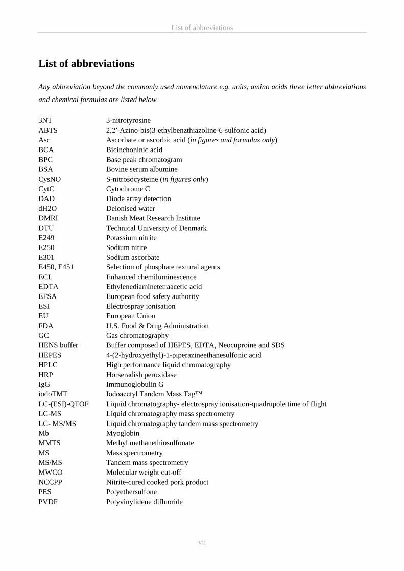
List of abbreviations
Any abbreviation beyond the commonly used nomenclature e.g. units, amino acids three letter abbreviations
and chemical formulas are listed below
3NT 3-nitrotyrosine
ABTS 2,2′-Azino-bis(3-ethylbenzthiazoline-6-sulfonic acid)
Asc Ascorbate or ascorbic acid (in figures and formulas only)
BCA Bicinchoninic acid
BPC Base peak chromatogram
BSA Bovine serum albumine
CysNO S-nitrosocysteine (in figures only)
CytC Cytochrome C
DAD Diode array detection
dH2O Deionised water
DMRI Danish Meat Research Institute
DTU Technical University of Denmark
E249 Potassium nitrite
E250 Sodium nitite
E301 Sodium ascorbate
E450, E451 Selection of phosphate textural agents
ECL Enhanced chemiluminescence
EDTA Ethylenediaminetetraacetic acid
EFSA European food safety authority
ESI Electrospray ionisation
EU European Union
FDA U.S. Food & Drug Administration
GC Gas chromatography
HENS buffer Buffer composed of HEPES, EDTA, Neocuproine and SDS
HEPES 4-(2-hydroxyethyl)-1-piperazineethanesulfonic acid
HPLC High performance liquid chromatography
HRP Horseradish peroxidase
IgG Immunoglobulin G
iodoTMT Iodoacetyl Tandem Mass Tag™
LC-(ESI)-QTOF Liquid chromatography- electrospray ionisation-quadrupole time of flight
LC-MS Liquid chromatography mass spectrometry
LC- MS/MS Liquid chromatography tandem mass spectrometry
Mb Myoglobin
MMTS Methyl methanethiosulfonate
MS Mass spectrometry
MS/MS Tandem mass spectrometry
MWCO Molecular weight cut-off
NCCPP Nitrite-cured cooked pork product
PES Polyethersulfone
PVDF Polyvinylidene difluoride
List of abbreviations
viii
QTOF Quadrupole time of flight
RNS Reactive nitrogen species
ROS Reactive oxygen species
RP-HPLC Reverse phase high performance liquid chromatography
SDS Sodium dodecyl sulphate
SDS-PAGE Sodium dodecyl sulphate polyacrylamide gel electrophoresis
SEC Size exclusion chromatography
SNO-RAC Resin assisted-capture of SNO-proteins
TBARS Thiobarbituric acid reactive substances
TBS Tris buffered saline
TBST-20 Tris buffered saline Tween®-20
UF Ultrafiltration
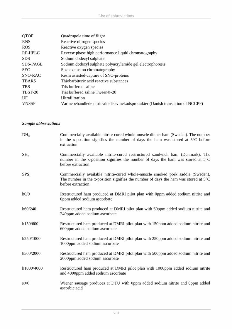
VNSSP Varmebehandlede nitritsaltede svinekødsprodukter (Danish translation of NCCPP)
Sample abbreviations
DHx Commercially available nitrite-cured whole-muscle dinner ham (Sweden). The number
in the x-position signifies the number of days the ham was stored at 5°C before
extraction
SHx Commercially available nitrite-cured restructured sandwich ham (Denmark). The
number in the x-position signifies the number of days the ham was stored at 5°C
before extraction
SPSx Commercially available nitrite-cured whole-muscle smoked pork saddle (Sweden).
The number in the x-position signifies the number of days the ham was stored at 5°C
before extraction
h0/0 Restructured ham produced at DMRI pilot plan with 0ppm added sodium nitrite and
0ppm added sodium ascorbate
h60/240 Restructured ham produced at DMRI pilot plan with 60ppm added sodium nitrite and
240ppm added sodium ascorbate
h150/600 Restructured ham produced at DMRI pilot plan with 150ppm added sodium nitrite and
600ppm added sodium ascorbate
h250/1000 Restructured ham produced at DMRI pilot plan with 250ppm added sodium nitrite and
1000ppm added sodium ascorbate
h500/2000 Restructured ham produced at DMRI pilot plan with 500ppm added sodium nitrite and
2000ppm added sodium ascorbate
h1000/4000 Restructured ham produced at DMRI pilot plan with 1000ppm added sodium nitrite
and 4000ppm added sodium ascorbate
s0/0 Wiener sausage produces at DTU with 0ppm added sodium nitrite and 0ppm added
ascorbic acid
List of abbreviations
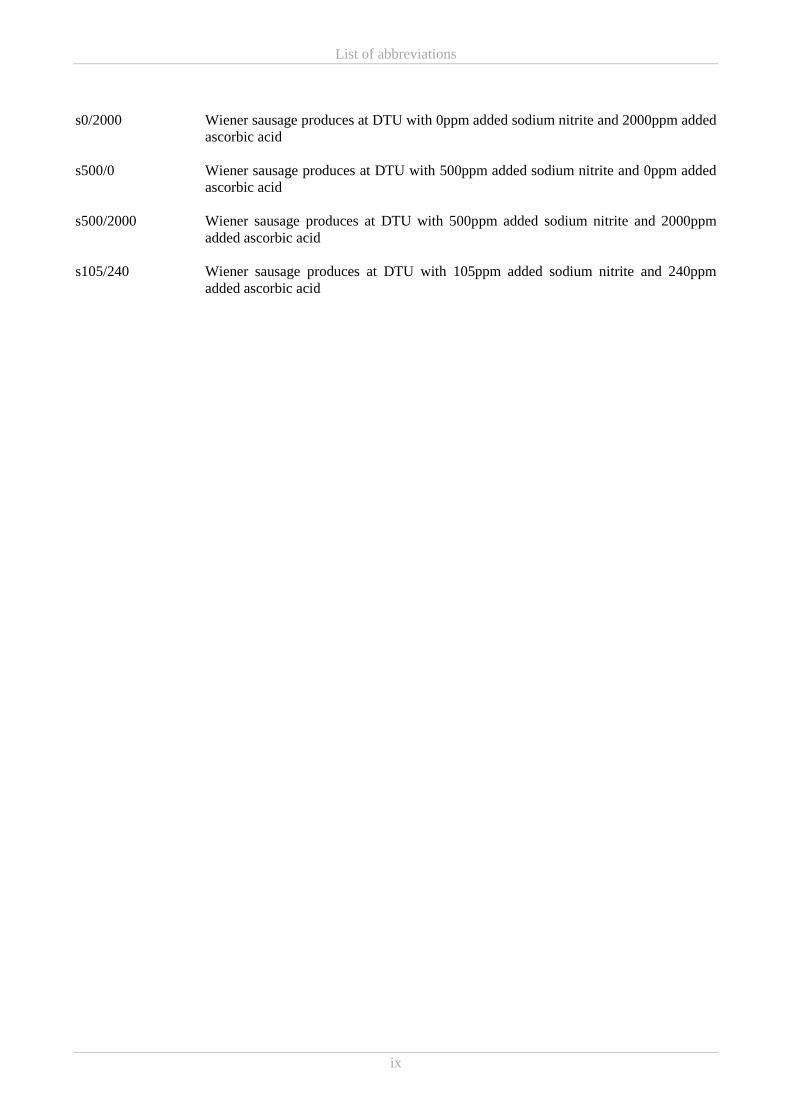
ix
s0/2000 Wiener sausage produces at DTU with 0ppm added sodium nitrite and 2000ppm added
ascorbic acid
s500/0 Wiener sausage produces at DTU with 500ppm added sodium nitrite and 0ppm added
ascorbic acid
s500/2000 Wiener sausage produces at DTU with 500ppm added sodium nitrite and 2000ppm
added ascorbic acid
s105/240 Wiener sausage produces at DTU with 105ppm added sodium nitrite and 240ppm
added ascorbic acid
List of manuscript
x
List of manuscript
Paper I:
Pedersen, S. T., Baron, C. P., De Gobba, C., Duedahl-Olesen, L., Koch, A. G. & Jessen, F. Antioxidative and
antimicrobial activity of extracts from nitrite-cured cooked pork products. Manuscript close to submission.
Paper II:
Pedersen, S. T., Duedahl-Olesen, L. & Jessen, F. Characterization of low molecular weight peptidic extracts
from nitrite-cured cooked pork products. Submitted to Journal of the Science of Food and Agriculture.
Paper III:
Pedersen, S. T., Duedahl-Olesen, L., Koch, A. G. & Jessen, F. Effect of storage and varying nitrite and
ascorbate concentrations on antioxidative and antimicrobial activity in low molecular weight extracts from
nitrite-cured cooked pork products.
Table of contents
xi
Table of Contents
Preface ................................................................................................................................................................ i
Acknowledgements ........................................................................................................................................... ii
Abstract ............................................................................................................................................................ iii
Resumé .............................................................................................................................................................. v
List of abbreviations ........................................................................................................................................ vii
List of manuscript .............................................................................................................................................. x
Introduction ................................................................................................................................................... 1 1.
Background .................................................................................................................................................... 3 2.
2.1 Nitrite-curing of meat .............................................................................................................................. 3
2.1.1 The origin of nitrite-curing ............................................................................................................... 3
2.1.2 Manufacturing of nitrite-cured meat ................................................................................................. 3
2.1.3 Legislation ........................................................................................................................................ 4
2.2 Nitrite-induced reactions in meat curing ................................................................................................. 5
2.2.1 What happens to nitrite once added to meat ..................................................................................... 5
2.2.2 Nitrite and the other curing agents ................................................................................................... 7
2.2.3 Nitrite and meat components ............................................................................................................ 7
2.3 Oxidation in meat .................................................................................................................................. 10
2.3.1 Lipid oxidation and antioxidant ...................................................................................................... 11
2.3.2 Protein oxidation and antioxidant ................................................................................................... 12
2.3.3 Curing agents and oxidative stability ............................................................................................. 13
2.4 Curing and antimicrobial activity .......................................................................................................... 16
2.5 Effects of storage and cooking of cured meat on nitrite and ascorbic acid ........................................... 17
2.6 Antioxidative and antimicrobial peptides .............................................................................................. 18
Materials, methods and methodological consideration ................................................................................ 21 3.
3.1 Sample material ..................................................................................................................................... 21
3.1.1 Commercial NCCPPs ..................................................................................................................... 21
3.1.2 Model hams .................................................................................................................................... 23
3.1.3 Model sausages ............................................................................................................................... 24
3.2 Methodological considerations .............................................................................................................. 25
3.2.1 Removal of potentially disturbing substances ................................................................................ 25
3.2.2 Application of in vitro antioxidant assays ...................................................................................... 25
3.2.3 Selection of microorganism for inhibition studies .......................................................................... 26
Table of contents
xii
3.2.4 Selection of suitable detection method and sample preparation strategy for 3-nitrotyrosine ......... 26
3.2.5 Developing a chromatographic method for ascorbic acid determination ....................................... 28
3.3 Methods ................................................................................................................................................. 30
3.3.1 Basic composition........................................................................................................................... 30
3.3.2 Sodium dodecyl sulphate polyacrylamide gel electrophoresis (SDS-PAGE) ................................ 31
3.3.3 Biotin switch ................................................................................................................................... 31
3.3.4 Liquid chromatography-mass spectrometry (LC-MS) ................................................................... 33
Results and Discussion ................................................................................................................................ 35 4.
4.1 Part A – Commercial NCCPPs .............................................................................................................. 35
4.1.1 Characterisation of antioxidative properties in commercial NCCPPs (Paper I and II) .................. 35
4.2 Part B – Model systems ......................................................................................................................... 40
4.2.1 Antioxidant activity in a ham model system (Paper III) ................................................................. 40
4.2.2 Antioxidant activity in a sausage model system ............................................................................. 43
4.2.3 SEC on extracts from model hams and sausages and antioxidant capacity of the SEC fractions .. 46
4.3 Part C – Antimicrobial activity .............................................................................................................. 54
4.3.1 Studies of bacterial growth inhibition by extracts of commercial and model hams ....................... 54
4.4 Part D – Characterisation of constituent components in aqueous ≤10kDa NCCPP extracts ................. 55
4.4.1 SDS-PAGE and biotin switch ......................................................................................................... 55
4.3.2 Mass Spectrometry (MS) ................................................................................................................ 60
4.5 Part E – Ascorbic acid and free amino acids in colourimetric in vitro assays ....................................... 66
4.5.1 BCA protein assay and ascorbic acid in NCCPPs .......................................................................... 66
4.5.2 In vitro reducing power and ABTS radical scavenging antioxidant activity of free amino acids .. 69
Summarising discussion and conclusion ..................................................................................................... 77 5.
Future work.................................................................................................................................................. 81 6.
References ................................................................................................................................................... 83 7.
8. Appendix ................................................................................................................................................... 101
Appendix A ............................................................................................................................................... 103
Appendix B ................................................................................................................................................ 105
Appendix C ................................................................................................................................................ 109
Paper I
Paper II
Paper III
Introduction
1
Chapter 1
Introduction 1.
The European processed meat market was valued at $103.55 billion in 2016 and was predicted a
considerable growth during the following years (Market Data Forecast, 2016) and of this market cured meat
accounts for the greatest share adding up to 30% (Mordor Intelligence, 2017; Zion Market Research, 2017).
The unique Danish lunch tradition of eating rye bread open sandwiches are contributing to a considerably
large intake of processed meat in the Danish population. In 2013 processed meat, which includes nitrite-
cured meat, was found to constitute almost 30% of the total meat (not including fish) consumed by the Danes
(Biltoft-Jensen et al., 2016). Nitrite-curing is a processing technique frequently used in the manufacture of
numerous types of processed meat products with the general purpose of extending shelf-life and creating an
appealing and microbiologically safe product. In this process nitrite has been pointed out to be a key-
responsible for the oxidative and microbial stability of such cured meat products. Despite all the many
effective and beneficial attributes of nitrite in regard to food preservation, nitrite is publically perceived as an
undesirable and even toxic food additive, the latter mainly referring to correlation with increased risk of
certain types of cancer among other caused by the formation of carcinogenic N-nitrosamines (Larsson,
Bergkvist, & Wolk, 2006; Larsson & Wolk, 2012; Santarelli, Pierre, & Corpet, 2008). Carcinogenic N-
nitrosamines may be formed from nitrite during meat processing but their formation can be limited by
lowering nitrite addition (Herrmann, Granby, & Duedahl-Olesen, 2015). Consequently, utilization of nitrites
in food is strictly controlled and limited by current EU legislations. Additionally, ever since the EU
harmonization concerning food additives in 1995, Denmark has been exempted from following the European
legislation concerning nitrite and has set lower limits for its addition to products to be sold in Denmark
(EFSA, 2017). In order to retain and preferably to further minimize nitrite addition to meat, it is necessary to
acquire further knowledge on the protective behaviour of nitrite and other additives in nitrite-cured products.
The ever growing public scepticism towards artificial food additives has led to an intensification of the
search for compounds of natural origin intended to replace unwanted E numbers like nitrite. Focus has to a
large extent been on peptides with antioxidative and antimicrobial properties. Such active peptides and also
free amino acids have been detected in many foods and food products (de Castro & Sato, 2015), however, it
would appear that none or remarkably few investigations have been performed on nitrite-cured cooked meat
products. Neither has it been possible to come up with a single non-protein alternative to cover all the
properties of the multifaceted nitrite (Sindelar & Milkowski, 2011). Simultaneously, there has been a strong
interest from the Danish meat industry to use the lowest possible amount of nitrite and to obtain better
knowledge about the metabolism of nitrite in meat products.
Introduction
2
The chemical reactions taking place during the nitrite-curing process are still not fully understood, however,
it was reported decades ago that nitrite reacts readily with proteins and that only a minor fraction reacts with
myoglobin to form the cured meat pigment (Cassens, Greaser, Ito, & Lee, 1979). Thus, a potential strategy
for optimizing nitrite addition could be to include investigations of nitrite-induced modification on
peptides/amino acids as such modified peptides have previously been found to possess antioxidant properties
(Chiueh & Rauhala, 1999; Kanner, 1979; Morrissey & Tichivangana, 1985).
Conclusively, there is a need and an emerging request for investigations of the existence, formation and
functional importance of antioxidative and antimicrobial compounds, possibly of protein-origin, occurring
during nitrite-curing of cooked meat products in order to ultimately reduce nitrite addition.
With this emerging need constituting the driving force behind the current PhD study the following three
working hypotheses were drafted using pork as the selected working matrix:
Hypothesis 1: During the production of cooked pork products with nitrite, antimicrobial and
antioxidant compounds are formed.
Hypothesis 2: There are optimal processing conditions for the formation of these antimicrobial and
antioxidant compounds.
Hypothesis 3: There are optimal storage conditions to retain the activity of antimicrobial and
antioxidant compounds in nitrite-cured pork products.
The work conducted in connection with answering the working hypothesis of this PhD study is documented
in the following chapters of this dissertation. After having outlined the motivation and objectives here in
Chapter 1, Chapter 2 begins with introducing the concept of nitrite-curing before and now. This is followed
by a brief review of the actions and reactions underlying the application of nitrite-curing in meat processing,
and finally the chapter finishes with a presentation of antioxidative and antimicrobial peptides. Chapter 3
describes the sample material, any relevant methodological considerations and applied experimental methods
not presented in enclosed the papers (Paper I-III). All results obtained during the PhD work are presented
and discussed in Chapter 4 and then collectively assessed in a summarising discussion and conclusion in
Chapter 5. As a final point, Chapter 6 presents any future work to be conducted.
Background
3
Chapter 2
Background 2.
The current chapter presents the background information of the PhD study, focusing on the fate and
antioxidative and antimicrobial behaviour of nitrite in meat curing. The elucidation of these topics will
include an outline of the underlying chemical reaction in order to provide a general overview of present
knowledge of nitrite’s complex chemistry. Before turning to this, a short introduction to the nitrite-curing
process is given.
2.1 Nitrite-curing of meat
2.1.1 The origin of nitrite-curing
Salting of meat is an ancient preservation technique dating back to Antiquity (Binkerd & Kolari, 1975). By
binding water, the salt decreases the water activity below levels required for microbial growth and thereby
protects meat and fish from spoilage. In the 19th century it became evident that certain salts were more
effective in preserving meat. Saltpetre was acknowledged as the activity enhancing salt contaminant which
also caused the meat to retain an appealing reddish pink colour (Binkerd & Kolari, 1975; Honikel, 2008).
The growing understanding of the chemical role of nitrite in the curing process eventually led industry to
fully or partially replacing saltpetre/nitrate with nitrite in curing salts and brines for different technological
reasons e.g. shorter curing time, increased production capacity and better control of colour formation and
colour uniformity (Binkerd & Kolari, 1975; Honikel, 2008). Nowadays, nitrate is rarely used, except for a
few special products requiring a slow curing process, and in products not subjected to heat treatment early
after manufacturing (Binkerd & Kolari, 1975; Honikel, 2008; Sebranek & Bacus, 2007).
2.1.2 Manufacturing of nitrite-cured meat
Today meat curing consists of adding salt containing the curing agents nitrite (or nitrate) and ascorbic acid
(or ascorbate) and other ingredients such as sugar and spices to fresh meat in order to create a unique flavour
and colour. Yet, the primary purpose of nitrite-curing is still to prolong shelf-life by preventing
microbiological spoilage, growth of pathogens and oxidative degradation.
Nitrite-curing is used for an immense variety of processed meat products and although a larger number of
methods for the addition of the curing salts exist, they are all modifications or combinations of wet-curing
and dry-curing. Wet-curing entails pumping, injecting or immersing fresh meat with/into brines consisting of
Background
4
curing salt dissolved in water before further downstream processing like boiling and smoking. The
differences in the processing technologies applied for production of wet-cured (cooked) products, mainly
depend on the size of the meat parts used for product manufacture. Regarding larger whole-muscle products
an even distribution of the curing brine after injection is ensured by either leaving the product to rest or
subjecting it to physical impact e.g. by tumbling (Heinz & Hautzinger, 2007). Afterwards, the meat is
prepared for cooking by restricting it to the desired product shape using e.g. string, elastic nets or moulds and
is then heated and/or smoked. Wet-cured (cooked) products may also be produced from smaller pieces of
meat that are assembled to larger entities. Such reconstituted products may be further subcategorised
(Pearson & Gillett, 1996) but for the sake of this thesis the general term restructured products will be used to
describe any product made from smaller pieces meat (smaller muscles or muscle parts). After cutting and
trimming the meat pieces are injected or if to small tumbled with the curing brine before stuffing into casings
or moulds and cooking to an internal temperature of 70°C (72°C) (Heinz & Hautzinger, 2007). Many
techniques exist for binding the meat together some of which include addition of gelatine, creating a surface-
protein matrix or utilising heat-induced coagulation of liquefied muscle protein released during tumbling
(Heinz & Hautzinger, 2007; Pearson & Gillett, 1996). Meanwhile, as the name imply, dry-curing entails
adding the curing ingredients without addition of water (Pearson & Gillett, 1996). This is done by rubbing
fresh meat with dry-curing salt and then piling the meat e.g. in barrels, vats or on shelves (Heinz &
Hautzinger, 2007; Pearson & Gillett, 1996). The curing salts will draw moisture from the meat to create a
“brine” which facilitates diffusion of the curing ingredients into the meat (Pearson & Gillett, 1996).
Manufacture of some dry-cured products, such as the whole-meat/muscle Spanish jamón curado (however,
sometimes produced without nitrite/nitrate), will often also included production steps of drying and
maturation. The products only subjected to dry-curing are most often intended for consumption in raw state
but may be submitted to further downstream processing e.g. smoking.
Slightly overlooked above, products produced from coarsely ground or minced meat may also be nitrite-
cured. Within the category of ground or minced meat products, sausages may be perceived as the most
important literally comprising hundreds of different products (Pearson & Gillett, 1996). The processing of
the individual sausage products are very diverse but generally speaking all ingredients, including curing,
taste and textural agents, are mixed to a “batter”, stuffed in casing and subjected to further processing such as
heating, smoking, drying or fermentation.
2.1.3 Legislation
Soon after the discovery of nitrite rather than nitrate as the active curing agent, nitrite was integrated in the
meat product manufacturing. However, nitrite in itself is a rather toxic compound compared to nitrate and
Background
5
early misuse ended up having fatal consequences. Consequently, some of the first legal actions regarding the
use of nitrite in meat curing were implemented (Honikel, 2008). Today, the use of nitrite and nitrate as food
additives are strictly controlled and limited by current EU legislations. The innate toxicity of nitrite, the
general notion that nitrite does not remain unchanged in the product during processing (Honikel, 2008), and
the later discovery of carcinogenic N-nitrosamines in cured meat, contributes immensely to the establishment
of these limits. Whether the limits should be set on added or residual nitrite has been recurrently debated but
is set in amended EU legislation as “added amount” due to the nature of the antimicrobial effect of nitrite
(Adler-Nissen, Ekgreen, & Risum, 2014; European Union, 2006; Merino, Örnemark, & Toldrá, 2017). This
will be further discussed in Chapter 2.4. As already mentioned Denmark are exempted from following the
European legislation concerning nitrite and has set lower limits for its addition to products to be sold in
Denmark (EFSA, 2017). While EU legislation generally sets limits on the use of nitrite (E249, E250;
expressed as sodium nitrite) in meat products at 150mg/kg and in heat-treated processed meat 100–
150mg/kg, Denmark sets a general maximum level of only 60mg/kg (European Union, 2008, 2011, 2015).
Product specific deviations especially regarding traditional products exist and can be found in Commission
Regulation (EU) No 1129/2011 (EU limits - (European Union, 2011)) and Commission Decision (EU)
2015/826 (Danish national limits - (European Union, 2015)).
2.2 Nitrite-induced reactions in meat curing
Processed meat is a highly complex matrix and the many possible oxidation states of nitrogen (Honikel,
2008), and thereby also nitrite, result in a complex web of reactions occurring between the reactive nitrite,
meat constituents and other additives during manufacture, processing and storage.
As a consequence of the unique multifaceted function of nitrite in cured meat, in regard to cured colour,
cured flavour, flavour protection (as an antioxidant), and as an antimicrobial agent, nitrite has been
extensively investigated. However, the ways by which nitrite achieves all these functions are not completely
understood within all the above mentioned aspects (Sebranek, 2009). Depending on the conditions of the
reaction environment nitrite is by several different mechanisms converted to nitric oxide which is essential
for weaving of the reaction web underlying meat curing (Sebranek, 2009).
2.2.1 What happens to nitrite once added to meat
As it was expressed by Cassens et al. (1979) not only changes to colour, flavour and shelf-life, but also the
fact that added nitrite disappear, is proof that nitrite reacts when added to meat (Cassens et al., 1979). Once
nitrite salts are added to meat the nitrite will dissolve in the water phase. In the weakly acidic (pH 5.5-6.0)
conditions of meat 99% of the nitrite will exist as an anion (NO2−) available to react with H
+ to form nitrous
Background
6
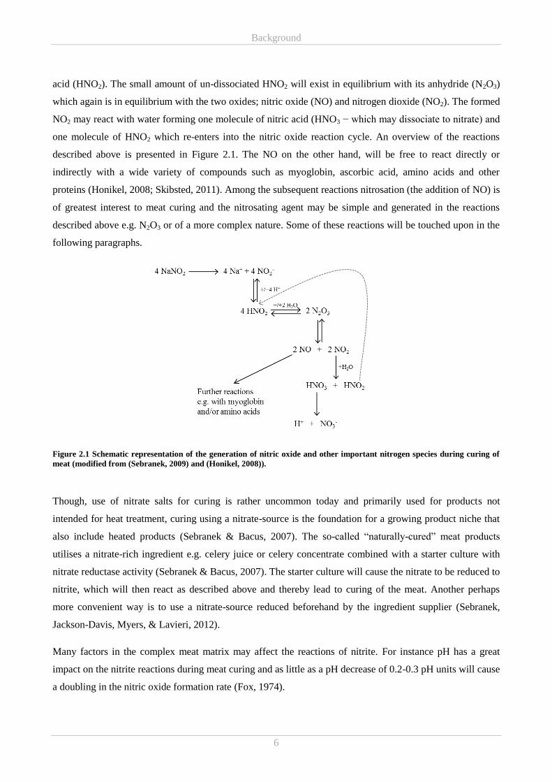
acid (HNO2). The small amount of un-dissociated HNO2 will exist in equilibrium with its anhydride (N2O3)
which again is in equilibrium with the two oxides; nitric oxide (NO) and nitrogen dioxide (NO2). The formed
NO2 may react with water forming one molecule of nitric acid (HNO3 − which may dissociate to nitrate) and
one molecule of HNO2 which re-enters into the nitric oxide reaction cycle. An overview of the reactions
described above is presented in Figure 2.1. The NO on the other hand, will be free to react directly or
indirectly with a wide variety of compounds such as myoglobin, ascorbic acid, amino acids and other
proteins (Honikel, 2008; Skibsted, 2011). Among the subsequent reactions nitrosation (the addition of NO) is
of greatest interest to meat curing and the nitrosating agent may be simple and generated in the reactions
described above e.g. N2O3 or of a more complex nature. Some of these reactions will be touched upon in the
following paragraphs.
Figure 2.1 Schematic representation of the generation of nitric oxide and other important nitrogen species during curing of
meat (modified from (Sebranek, 2009) and (Honikel, 2008)).
Though, use of nitrate salts for curing is rather uncommon today and primarily used for products not
intended for heat treatment, curing using a nitrate-source is the foundation for a growing product niche that
also include heated products (Sebranek & Bacus, 2007). The so-called “naturally-cured” meat products
utilises a nitrate-rich ingredient e.g. celery juice or celery concentrate combined with a starter culture with
nitrate reductase activity (Sebranek & Bacus, 2007). The starter culture will cause the nitrate to be reduced to
nitrite, which will then react as described above and thereby lead to curing of the meat. Another perhaps
more convenient way is to use a nitrate-source reduced beforehand by the ingredient supplier (Sebranek,
Jackson-Davis, Myers, & Lavieri, 2012).
Many factors in the complex meat matrix may affect the reactions of nitrite. For instance pH has a great
impact on the nitrite reactions during meat curing and as little as a pH decrease of 0.2-0.3 pH units will cause
a doubling in the nitric oxide formation rate (Fox, 1974).
Background
7
2.2.2 Nitrite and the other curing agents
Another very important factor governing nitrite/NO reactions during meat curing are reductants. This
includes endogenous reductants such as NADH and sulfhydryl groups, but more important the added
ascorbate or ascorbic acid which is added as a so-called cure-accelerator. They are so called for their central
role in reactions greatly contributing to the formation of NO. By accelerating the formation of NO the
addition of ascorbate/ascorbic acid will consequently lead to a faster turnover of added nitrite and a faster
development of cured meat characteristics. Oxidation of ascorbate/ascorbic acid in a redox reaction will yield
NO with a 1:2 stoichiometries (Møller & Skibsted, 2002; Skibsted, 2011):
Ascorbate+ N2O3 → dehydroascorbate + 2NO + H2O (Eq. 1)
Ascorbic acid+ 2HNO2 → dehydroascorbic acid + 2NO + 2H2O (Eq. 2)
Ascorbate/ascorbic acid may, however, also react with N2O3 binding the resulting NO (Skibsted, 2011)
which it seems to be capable of passing on to other meat ingredients (trans-nitrosation)(Honikel, 2008).
Several (Asc-NO) reaction intermediates have been suggested for any subsequent reactions e.g. for the
nitrosylation of myoglobin, but the exact nitrosating agents and reaction pathways (may include thiol
groups)(Izumi, Cassens, & Greaser, 1989; Skibsted, 2011) are not fully resolved.
All cured meats have been added sodium chloride in varying amounts (Sebranek, 2009) and according to
Sebranek & Fox (1985) the great importance of sodium chloride, as well as sodium nitrite for the distinctive
attributes of cured meat, the elimination of either would result in the product no longer being considered as
cured. This adds further to the complexity of the chemistry of nitrite in curing of meat as nitrous acid reacts
with the chloride ion
HNO2 + H+ + Cl
− → NOCl + H2O (Eq. 3)
forming nitrosyl chloride (NOCl) which is a stronger nitrosating agent than N2O3 (Skibsted, 2011). The
difference in reactivity means that formation of NOCl rather than N2O3 may have a number of consequences
on the curing/nitrosation chemistry, e.g. by affecting reaction rates and changing reaction specificity
(Sebranek & Fox, 1985).
2.2.3 Nitrite and meat components
Likely one of the most widely studied compounds related to nitrite-curing is the reaction product of nitrite (in
the form of NO) and the meat pigment myoglobin (denoted Mb in the following reaction schemes),
nitrosylmyoglobin. Upon addition of nitrite the meat quickly turns brown because the nitrite acts as a strong
Background
8
heme pigment oxidant while, nitrite itself is reduced and thereby further contributes to the generation of NO
(Sebranek, 2009):
NO2− + MbFe(II) (red myoglobin) → MbFe(III) (brown met myoglobin) + NO + OH
− (Eq. 4)
By reducing enzymes or chemical reactions with reducing agents like the added ascorbate/ascorbic acid
ferric iron is reduced back into ferrous iron (Honikel, 2008). NO may then react with myoglobin (Fe(II)),
coordinating to the iron, to form the characteristic cured meat colour, nitrosylmyoglobin (dark red). This is
also one of the mechanisms behind the assigning of ascorbate/ascorbic acid as a cure-accelerator
(Parthasarathy & Bryan, 2012). Heating of nitrosylmyoglobin-containing meat, like nitrite-cured cooked
pork products (NCCPPs), will cause a denaturation of the protein moiety of nitrosylmyoglobin, yet the NO-
porphyrin ring system remains. The latter is referred to as nitrosylhemochrome which is heat-stable and
responsible for the typical reddish-pink colour of NCCPPs (Honikel, 2008).
Nitric oxide may also bind directly with metmyoglobin (Fe(III)) followed by reduction to nitrosylmyoglobin
(Fe(II)) for cured colour formation (Pegg & Shahidi, 1997). Other paths involving metmyoglobin and nitrite
or HNO for the formation of nitrosylmyoglobin (Fe(II)) have also been suggested (Fox, 1966; Miranda et al.,
2003).
Proteins have a number of potential reaction sites for nitrite (Cassens et al., 1979). As muscles contain a
wide spectrum of proteins of different composition and functionality it is not surprising that a considerable
share of the nitrite added to meat has been found to be bound to proteins, other than myoglobin (Tricker &
Kubacki, 1992).
It has long been established that, of the amino acids constituting meat proteins, cysteine is most likely the
most reactive towards nitrite (Olsman & van Leeuwen, 1977). Nitrosation of the cysteine thiol by a
nitrosating agent e.g. N2O3 (S-nitrosation creating a nitrosothiol (–SNO) side chain) results in the formation
of S-nitrosocysteines. However, it appears that S-nitrosation might first occur after nitrosylation of
myoglobin (Sullivan & Sebranek, 2012). S-nitrosation of muscle proteins such as myosin has been proven to
occur but the extent of occurrence at pH levels equivalent to those found in meat has been disputed (Cassens
et al., 1979; Kubberød, Cassens, & Greaser, 1974). The varying levels of detected S-nitrosations in meat are
however, likely the result of the formation of –SNO being reversible. Consequently, meat proteins can serve
as nitrosating agents (trans-nitrosation) but also a general reservoir for NO and like any residual nitrite this is
part of a “NO generating pool” capable of supplying NO for the many reactions taking place during storage
and cooking of cured meats – even after ascorbate depletion (Skibsted, 2011). This reversibility of –SNO has
also been central to the more recently established importance of –SNO in cell signalling (Miersch & Mutus,
2005).
Background
9
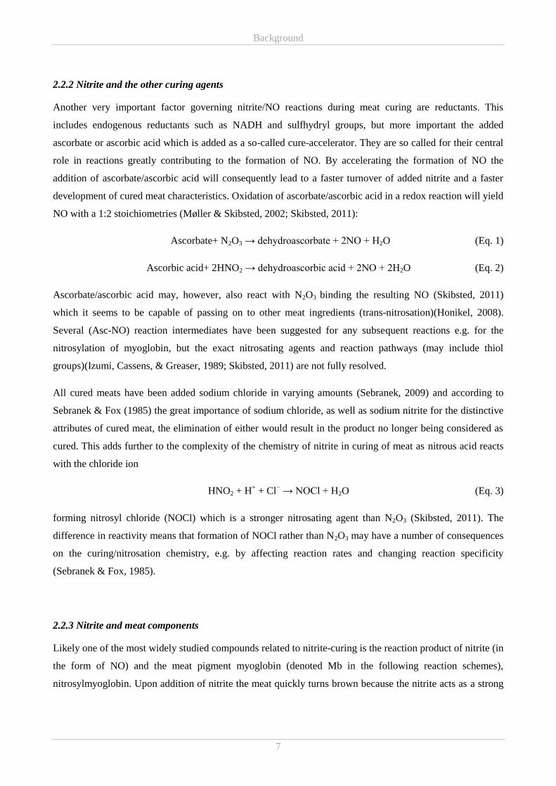
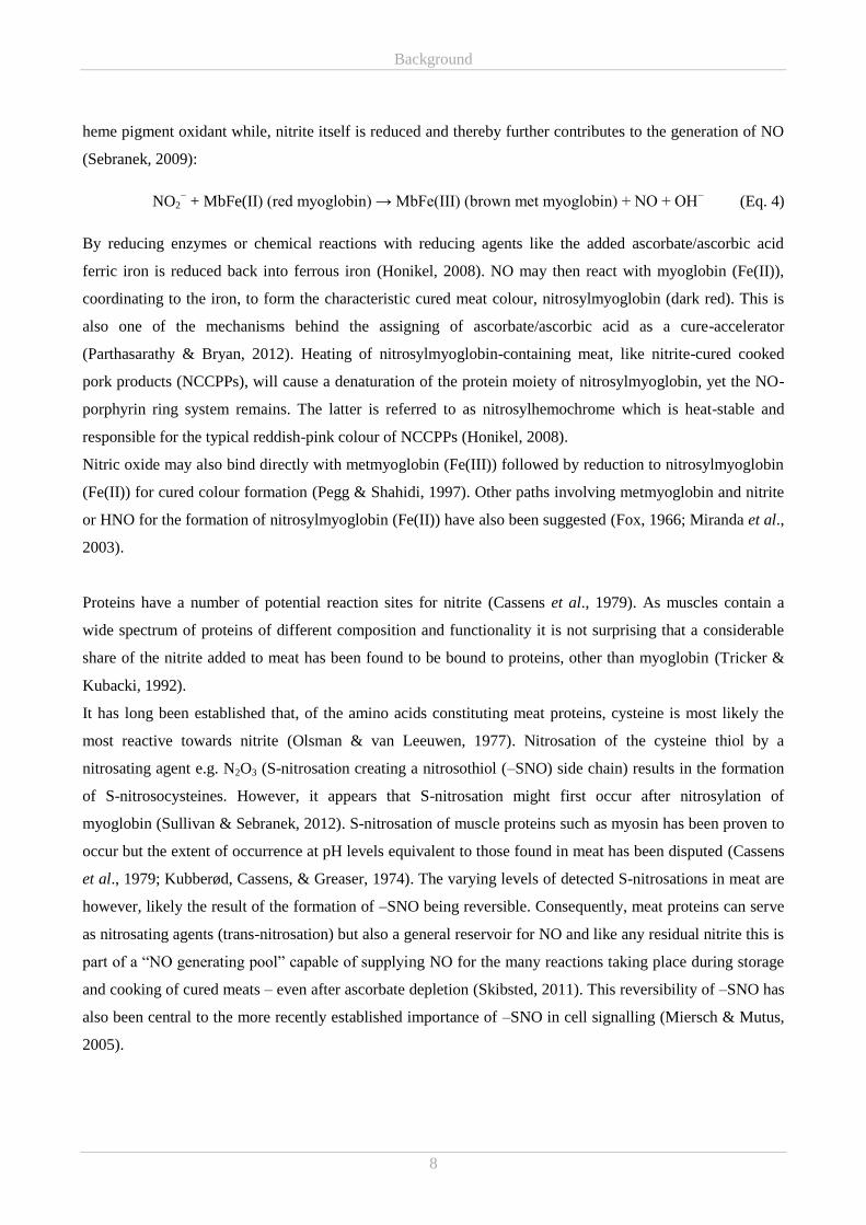
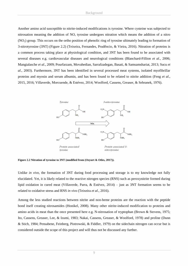
Another amino acid susceptible to nitrite-induced modifications is tyrosine. Where cysteine was subjected to
nitrosation meaning the addition of NO, tyrosine undergoes nitration which means the addition of a nitro
(NO2) group. This occurs on the ortho position of phenolic ring of tyrosine ultimately leading to formation of
3-nitrotyrosine (3NT) (Figure 2.2) (Teixeira, Fernandes, Prudêncio, & Vieira, 2016). Nitration of proteins is
a common process taking place at physiological condition, and 3NT has been found to be associated with
several diseases e.g. cardiovascular diseases and neurological conditions (Blanchard-Fillion et al., 2006;
Mangialasche et al., 2009; Pourfarzam, Movahedian, Sarrafzadegan, Basati, & Samsamshariat, 2013; Sucu et
al., 2003). Furthermore, 3NT has been identified in several processed meat systems, isolated myofibrillar
proteins and myosin and serum albumin, and has been found to be related to nitrite addition (Feng et al.,
2015, 2016; Villaverde, Morcuende, & Estévez, 2014; Woolford, Cassens, Greaser, & Sebranek, 1976).
Figure 2.2 Nitration of tyrosine to 3NT (modified from (Ozyurt & Otles, 2017)).
Unlike in vivo, the formation of 3NT during food processing and storage is to my knowledge not fully
elucidated. Yet, it is likely related to the reactive nitrogen species (RNS) such as peroxynitrite formed during
lipid oxidation in cured meat (Villaverde, Parra, & Estévez, 2014) – just as 3NT formation seems to be
related to oxidative stress and RNS in vivo (Teixeira et al., 2016).
Among the less studied reactions between nitrite and non-heme proteins are the reaction with the peptide
bond itself creating nitrosamides (Honikel, 2008). Many other nitrite-induced modification to proteins and
amino acids in meat than the once presented here e.g. N-nitrosation of tryptophan (Brown & Stevens, 1975;
Ito, Cassens, Greaser, Lee, & Izumi, 1983; Nakai, Cassens, Greaser, & Woolford, 1978) and proline (Dunn
& Stich, 1984; Pensabene, Feinberg, Piotrowski, & Fiddler, 1979) on the sidechain nitrogen can occur but is
considered outside the scope of this project and will thus not be discussed any further.
Background
10
Lipids, the other major macronutrient in meat (carbohydrate content is very low), may also react with nitrite.
It would appear that nitrite or derivatives hereof may react with the double bonds of unsaturated fatty acids
(or its derivatives) e.g. forming alkylnitrites (Goutefongea, Cassens, & Woolford, 1977; Honikel, 2008).
However, the knowledge regarding such reactions is relatively scarce.
The major cause of concern for consumptions of nitrite-cured meat is N-nitrosamines. The main examples of
N-nitrosamines found in some heat-treated cured products are N-nitrosodimethylamine and N-
nitrosopyrrolidine which are known to be carcinogenic, mutagenic and teratogenic in experimental animals
(Pegg & Shahidi, 2004). N-nitrosamines are formed by reaction of nitrosating agents with low molecular
weight amines (Parthasarathy & Bryan, 2012), but stable N-nitrosamines are principally formed from N-
nitrosation of secondary amines (Honikel, 2008). Prerequisite for N-nitrosamine formation in cured meat
products are heating at high temperature (>130°C) and naturally also availability of amines and nitrite
(Honikel, 2008). As heat-treated nitrite-cured products are made from fresh meat the amount of amines are
rather small (Honikel, 2008), and in the case of cooked hams the aimed core temperature is, as mentioned in
Chapter 2.1.2, approximately 70°C, thus the N-nitrosamine content of such products may be relatively
limited. N-nitrosamines may, however, be present in rubber nettings which may contaminate the edible part
of a cooked ham (Fiddler, Pensabene, Gates, & Adam, 1998). It has been established that ascorbate/ascorbic
acid is capable of inhibiting N-nitrosamine formation by reacting faster than secondary amines with the
nitrosating agent, e.g. N2O3, at the pH relevant for curing of meat (Skibsted, 2011). Basically this would
mean reducing N2O3 to NO (which is not a nitrosating agent) during ascorbate/ascorbic acid’s own oxidation
(Parthasarathy & Bryan, 2012). Thus, excess ascorbate added for facilitating curing may also act to prevent
potential N-nitrosamine formation and today this ability represents one of the strongest arguments for adding
ascorbate/ascorbic acid to cured meat.
While these are some of the major characterised chemical reactions describing the fate of nitrite during
nitrite-curing, there are many other reactions that can and do take place. For example nitrous oxide (N2O,
laughing gas) has been identified in the gases above curing mixtures.
2.3 Oxidation in meat
Oxidative reactions are central to ruining any food. Thus, a basic understanding of the oxidative activities
occurring in meat is essential for understanding the shelf-life prolonging effect of nitrite, in regard to
oxidative stability of cured meat products. The complexity of meat as a matrix makes it highly complicated
to cover all aspects of oxidative activities in meat and thus, it was chosen to focus on oxidation of the two
major meat constituents: lipids and proteins.
Background
11
2.3.1 Lipid oxidation and antioxidant
Oxidation of lipids is the main cause for deterioration of food quality and shelf-life, primarily manifested
through formation of objectionable off-flavour and odours – oxidative rancidity. Furthermore, lipid oxidation
impairs food quality by prompting loss of essential nutrients and changes in texture and colour, as a
consequence of reactions of lipid oxidation products with other food components. Lastly, lipid oxidation may
also result in formation of potentially toxic reaction products (Kanner, 1994; Velasco, Dobarganes, &
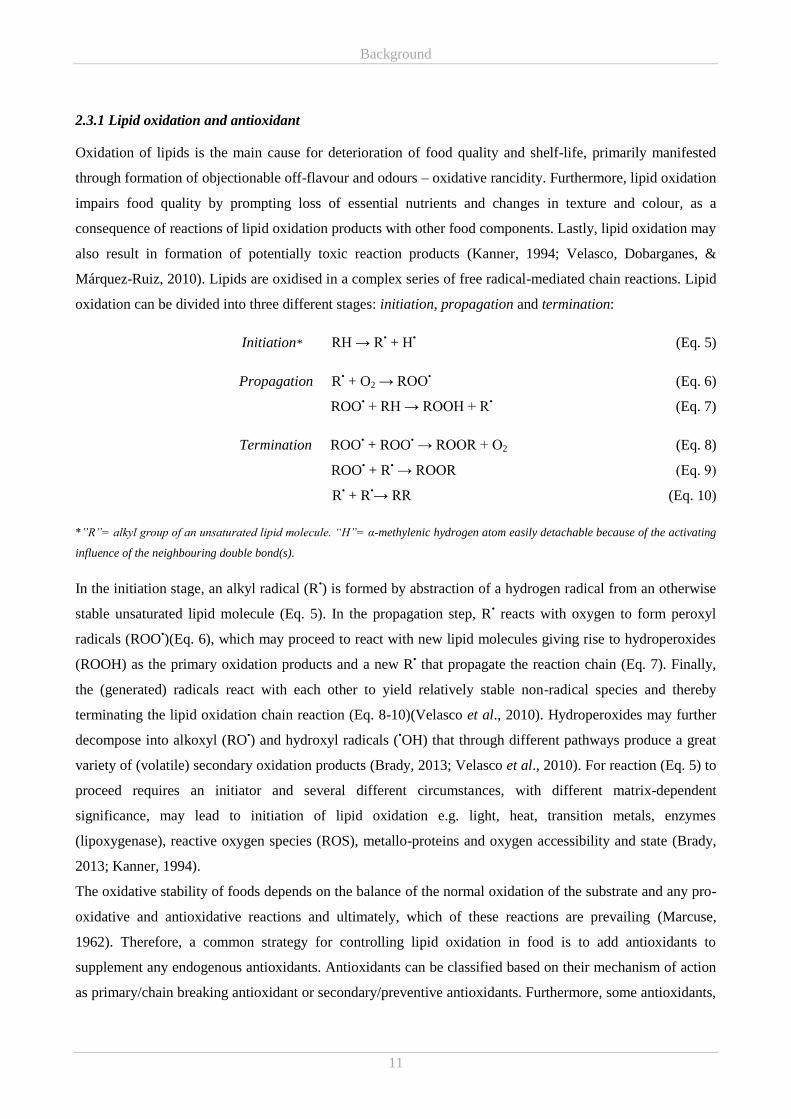
Márquez-Ruiz, 2010). Lipids are oxidised in a complex series of free radical-mediated chain reactions. Lipid
oxidation can be divided into three different stages: initiation, propagation and termination:
Initiation* RH → R• + H
• (Eq. 5)
Propagation R• + O2 → ROO
• (Eq. 6)
ROO• + RH → ROOH + R
• (Eq. 7)
Termination ROO• + ROO
• → ROOR + O2 (Eq. 8)
ROO• + R
• → ROOR (Eq. 9)
R• + R
•→ RR (Eq. 10)
*”R”= alkyl group of an unsaturated lipid molecule. “H”= α-methylenic hydrogen atom easily detachable because of the activating
influence of the neighbouring double bond(s).
In the initiation stage, an alkyl radical (R•) is formed by abstraction of a hydrogen radical from an otherwise
stable unsaturated lipid molecule (Eq. 5). In the propagation step, R• reacts with oxygen to form peroxyl
radicals (ROO•)(Eq. 6), which may proceed to react with new lipid molecules giving rise to hydroperoxides
(ROOH) as the primary oxidation products and a new R• that propagate the reaction chain (Eq. 7). Finally,
the (generated) radicals react with each other to yield relatively stable non-radical species and thereby
terminating the lipid oxidation chain reaction (Eq. 8-10)(Velasco et al., 2010). Hydroperoxides may further
decompose into alkoxyl (RO•) and hydroxyl radicals (
•OH) that through different pathways produce a great
variety of (volatile) secondary oxidation products (Brady, 2013; Velasco et al., 2010). For reaction (Eq. 5) to
proceed requires an initiator and several different circumstances, with different matrix-dependent
significance, may lead to initiation of lipid oxidation e.g. light, heat, transition metals, enzymes
(lipoxygenase), reactive oxygen species (ROS), metallo-proteins and oxygen accessibility and state (Brady,
2013; Kanner, 1994).
The oxidative stability of foods depends on the balance of the normal oxidation of the substrate and any pro-
oxidative and antioxidative reactions and ultimately, which of these reactions are prevailing (Marcuse,
1962). Therefore, a common strategy for controlling lipid oxidation in food is to add antioxidants to
supplement any endogenous antioxidants. Antioxidants can be classified based on their mechanism of action
as primary/chain breaking antioxidant or secondary/preventive antioxidants. Furthermore, some antioxidants,
Background
12
e.g. peptides and ascorbic acid, can be referred to as multi-functional as they exhibit both primary and
secondary antioxidant properties (Wanasundara & Shahidi, 2005). Primary antioxidants are capable of
reacting directly with free radicals converting them to more stable, non-radical products, e.g. by radical
scavenging or donating an electron (reducing properties). Thus, primary antioxidant may play a vital role in
delaying or inhibiting the initiation step or interrupt the propagation step. Secondary antioxidants exert their
antioxidant activity through various mechanisms to slow the rate of oxidation reactions and thus can be said
to limit lipid oxidation by indirect mechanisms. These indirect mechanisms include chelating of transition
metal ions, singlet oxygen quenching, oxygen scavenging, decomposing hydroperoxide to non-radical
species and regenerating primary antioxidant by H donation (Wanasundara & Shahidi, 2005).
2.3.2 Protein oxidation and antioxidant
While the consequences of lipid oxidation for food quality are (almost) immediately perceived by human
senses, oxidation of proteins results in more subtle changes with effects on meat texture and juiciness (Lund
& Baron, 2010).
Protein oxidation can be induced directly by different ROS, both including free radical and non-radical
species such as hydrogen peroxide. Furthermore, radical by-products of other oxidative processes, e.g. ROO•
from lipid oxidation, can also contribute to induction of protein oxidation (Lund & Baron, 2010; Soladoye,
Juárez, Aalhus, Shand, & Estévez, 2015). Generally, oxidation of proteins is believed to proceed through a
free radical chain reaction similar to that of lipid oxidation starting with ROS/radical-abstraction of hydrogen
from a protein molecule. However, due to the size and complex structure of proteins compared to lipids, free
radical-attacks on proteins will result in reaction pathways and reaction products which are much more
complex and of much higher variation. All mechanisms of protein oxidation have not been fully established,
but the basic chemical mechanisms involved in oxidative modification of proteins are continuously
becoming clearer. Detailed descriptions of the known chemical reactions related to protein oxidation will not
be giving here but elaborate reviews on the matter has been written Lund, Heinonen, Baron, & Estévez
(2011) and Soladoye et al. (2015).
Oxidative changes induced to proteins include protein fragmentation through cleavage of peptide bonds,
modification of amino acid side chains and formation of covalent intermolecular cross-linked protein
derivatives causing mild to severe protein damage. Globular proteins seem to be much more stable towards
protein oxidation than the non-globular proteins and of the myofibrillar proteins, myosin appear to be the
most susceptible to protein oxidation (Lund & Baron, 2010; Martinaud et al., 1997). The oxidation
susceptibility of amino acid also varies, and cysteine, methionine, tyrosine, tryptophan, phenylalanine,
histidine, proline, arginine and lysine have been described as particularly prone to oxidation (Lund et al.,
2011). Formation of protein carbonyl groups and protein hydroperoxides constitute the most general amino
Background
13
acid modification, while sulfoxide and sulfone formation from methionine and radical attack on the aromatic
ring in tryptophan and phenylalanine constitute examples of the more amino acid-specific oxidative
modifications (Lund & Baron, 2010). Protein intra- and inter-molecular cross-linking as a result of protein
oxidation are due to formation of disulphide and dityrosine. However, the latter has only been identified in
meat model systems and never directly in meat.
Controlling protein oxidation in meat at the processing end of the food chain is, like for lipid oxidation,
achieved by addition of antioxidants. Due to suspected connection between lipid oxidation products and
initiation of protein oxidation, inhibition of lipid oxidation is expected to prevent protein oxidation at least to
some extent. However, it seems that the classical lipid antioxidants strategies do not inevitably apply to
muscle proteins, as research have established that compounds capable of preventing lipid oxidation are not
always able to prevent protein oxidation (Lund et al., 2011). In a model system, it was shown that prevention
of protein oxidation, using a hydrophilic antioxidant, also had a protective effect on the lipids e.g. the
hydrophilic antioxidant Trolox (a vitamin E analogue) prevented oxidation of both protein and lipid
fractions. Yet, the lipophilic antioxidants tested were ineffective at preventing protein oxidation (Baron,
Berner, Skibsted, & Refsgaard, 2005). In addition to testing the classical antioxidants such as tocopherol,
multiple attempts have been made to control protein oxidation by plant phenolics with varying success (see
review Lund et al. (2011)).
With features such as the nature of the target, location of the site of attack together with the type of the
attacking species, having impact on the development of protein oxidation (Dean, Hunt, Grant, Yamamoto, &
Niki, 1991; Soladoye et al., 2015) and adding intricacy to the protein oxidation mechanisms (which is also
not fully elucidated), makes it difficult to describe the antioxidants kinetics and mechanisms for the
inhibition of protein oxidation. Thus, to date no systematic understanding on how proteins can be protected
from oxidation by antioxidant exists.
2.3.3 Curing agents and oxidative stability
Nitrite added to meat behaves as an oxidant (Skibsted, 2011). However, immense evidence points to the
addition of nitrite to reduce lipid oxidation in meat (Mac Donald, Gray, Kakuda, & Lee, 1980; Willemot,
Fillion-Delorme, & Wood, 1987), and several explanations in favour of nitrite as an active antioxidant has
been offered. These include iron chelation (Igene, Yamauchi, Pearson, Gray, & Aust, 1985) and stabilisation
of unsaturated fatty acid towards oxidation through formation of nitro-nitroso derivatives (Freybler et al.,
1993). Oxidation of NO and nitrite, to nitrite and nitrate, respectively, may also, in particular for the former,
be perceived as an antioxidant mechanism, as the sequestering of oxygen may contribute to the retardation of
lipid oxidation (Honikel, 2008). Yet, it generally seems to be NO that interferes with free radical
Background
14
intermediates in lipid oxidation. NO reacts rapidly with most radicals including the lipid derived alkyl (R•),
alkoxyl (RO•), and peroxyl (ROO
•) radicals, the latter being particularly fast (Carlsen, Møller, & Skibsted,
2005):
NO + ROO• → ROONO → RONO2 (Eq. 11)
This interaction will lead to formation of non-radical addition products, in effect breaking the radical chain
processes characteristic of oxidation of unsaturated lipids (Skibsted, 2011). NO may also react rapidly with
other radicals implicated in initiating lipid oxidation namely the hydroxyl radical and the superoxide radical
anion:
NO + •OH → HNO2 → H
+ + NO2
− (Eq. 12)
NO + O2• −
→ ONOO−
→ NO3− (Eq. 13)
While the interaction of NO with •OH (Eq. 12) may alter the course of oxidative reactions in a positive
manner, the formation of peroxynitrite (ONOO−) (Eq. 13) can have the quite opposite effects. Although,
ONOO− may deactivate by isomerisation to nitrate at meat pH, ONOO
− is a strong oxidant with the ability to
initiate both lipid and protein oxidation. Alternatively it may simply work to deplete the present antioxidant
pool (Brannan, Connolly, & Decker, 2001). Oxidative protein modifications induced by ONOO− includes the
nitration of tyrosine to 3NT and in that way linking ROS and RNS (oxidative and nitrosative stress).
Ascorbic acid is a well-known multifaceted antioxidant capable of acting as a scavenger of hydrophilic
radicals, an oxygen sequencer, and a reducing agent and as a secondary antioxidant, regenerating primary
antioxidant such as tocopherol (Mäkinen, Kähkönen, & Hopia, 2001; Niki, 1991). These properties enable
added ascorbate/ascorbic acid to have a direct antioxidant effect on lipids in cured meat, e.g. ascorbate may
react with oxygen forming dehydroascorbate and thus, reducing the amount of available oxygen for other
oxidative reactions (Honikel, 2008). Furthermore, ascorbate/ascorbic acid contribute to the oxidative stability
of cured meat through the reactions described above by enhancing the formation of NO (Eq. 1-2). This was
clearly demonstrated by Berardo et al. (2016) as addition of nitrite, ascorbate or combined addition in
increasing order resulted in a decrease in thiobarbituric acid reactive substance (TBARS) values.
It can be said that any antioxidant activity may only be a potential one as most antioxidant may potentially
convert to pro-oxidant activities under different circumstances (Marcuse, 1962). Pro-oxidative activities of
ascorbate/ascorbic acid involve generation of oxidation initiators/pro-oxidant by reducing Fe3+
to Fe2+
(Jacobsen, Adler-Nissen, & Meyer, 1999) and by means of Fenton chemistry generating hydroxyl radicals
(Skibsted, 2011; Villaverde, Parra, et al., 2014)
The antioxidant effect of nitrite addition to meat has primarily been directed towards lipid oxidation.
Though, there in recent years has been an increase in investigations on the effect of curing agents – nitrite
Background
15
and ascorbate – on protein oxidation in myofibrillar proteins and different meat products, the conclusions
have not been unanimous. Nitrite was both reported to behave as a prooxidant in fermented sausages
(Villaverde, Morcuende, et al., 2014) and have negligible or neither pro- nor antioxidative effects in isolated
myofibrillar proteins or raw porcine patties (Villaverde, Parra, et al., 2014; Vossen & De Smet, 2015). On
the other hand Feng et al. (2016) reported finding prooxidative as well as antioxidative effects of adding
nitrite to cooked sausages, depending on the amount of added sodium nitrite and what oxidative protein
modifications were measured. Berardo et al. (2016) also found nitrite to limit protein oxidation on one of the
two measured parameters, yet addition of ascorbate in combination with nitrite resulted in increased protein
oxidation. Contradictory, a clear antioxidant effect of combined nitrite and ascorbate addition on protein
oxidation was found by Villaverde, Morcuende, et al. (2014) and Villaverde, Parra, et al. (2014). Thus,
further clarification of the redox chemistry of nitrite and ascorbate and their interactions with meat proteins,
but also the interplay with lipid oxidation, is required to establish the technological importance of nitrite as
an antioxidant of protein oxidation in cured meat products.
While myoglobin has been found to be an important initiator of lipid and protein oxidation in meat (Baron &
Andersen, 2002; Lund et al., 2011), nitrosylmyoglobin from cured meat has been found to act as a lipid
antioxidant (Kanner, Ben-Gera, & Berman, 1980; Morrissey & Tichivangana, 1985; Møller, Sosniecki, &
Skibsted, 2002). Decades ago Morrissey & Tichivangana (1985) proposed that this property of
nitrosylmyoglobin was related to formation of a stable “myoglobin-NO” complex, formed upon heating of
meat, that blocked any catalytic activity of the heme-iron and also prevented the release of the heme-iron,
and lastly that nitrosylmyoglobin formed in meat curing acted as an antioxidant per se. Regarding the latter,
nitrosylmyoglobin(Fe(II)) should be considered as an antioxidant buffer as isotopic labelling studies have
indicated that this specie can regenerate active NO derived antioxidants even at low temperature (Andersen,
Saaby Johansen, Shek, & Skibsted, 1990). Accordingly, nitrosylmyoglobin(Fe(II)) appears to behave as an
antioxidant through dissociation, liberating NO for other antioxidant reactions (Skibsted, 2011):
MbFe(II)NO → MbFe(II) + NO (Eq. 14)
Additionally, nitrosylmyoglobin (Fe(II)) behaves antioxidatively through direct reaction with free radicals of
lipid oxidation such as peroxyl radicals (Kanner et al., 1980):
MbFe(II)NO + ROO• → non-radical products (Eq. 15)
Furthermore, it would appear that the heme centre of myoglobin may also act as an efficient scavenger of the
ONOO− oxidant (Carlsen et al., 2005). Although, nitrosylmyoglobin has been found to be important for
retaining the oxidative stability of cured meat, the meat pigment itself may also undergo oxidation leading to
discolouration and concomitant loss of lipid oxidative stability, as a result of depletion of the
Background
16
nitrosylmyoglobin (Fe(II)) antioxidant buffer as wells as the generation of the oxidation product
metmyoglobin (Fe(III)), resulting in prooxidative activities (Skibsted, 2011).
Returning to Morrissey & Tichivangana's (1985) suggestion of the antioxidant activity of the heat stabilised
“myoglobin-NO” complex being related to pacifying of the reactive iron, Wettasinghe & Shahidi (1997)
demonstrated that a preformed cooked cured-meat pigment (not referred to as nitrosylhemochrome by the
authors) acted as an active antioxidant that was more effective than nitrosylmyoglobin at selected
concentrations.
As it was described in Chapter 2.2.3 NO will not just associate with myoglobin coordinated to the central
iron, but can also bind covalently to amino acids of other proteins with one of these nitrite-induced
modifications being of particular interest for the oxidative stability of cured meat. Both S-nitrosoglutathione
and S-nitrosocysteine, where the nitrite-induced modification has occurred on a cysteine thiol (hydrogen has
been substituted for NO – S-nitrosation), have been found to have antioxidant properties (Chiueh & Rauhala,
1999; Kalyanaraman & Singh, 1998; Kanner, 1979). It is likely, that this effect may be related to S-
nitrosocysteine acting as a reducing agent and a nitric oxide donor (Sullivan & Sebranek, 2012), thus
contributing to the immense desirable and undesirable consequences of nitrite-curing.
2.4 Curing and antimicrobial activity
As it has already been implied, nitrite is not merely added for colour, flavour and antioxidant purposes, but
also as an important antimicrobial agent against both spoilage and pathogenic bacteria, commonly found in
meat products. However, the effectiveness of nitrite in controlling or preventing bacterial growth varies
between bacterial species, and nitrite has demonstrated markedly stronger antimicrobial properties against
Gram positive than Gram negative bacteria (Sindelar & Milkowski, 2012). Nevertheless, Pichner,
Hechelmann, Steinrueck, & Gareis ((Abstract) 2006) have reported reduced growth Escherichia coli (gram
negative) in salami with nitrite compared to a similar uncured product. Though, nitrite has been shown to
inhibit many different food pathogens e.g. Staphylococcus aureus, Bacillus cereus, E. coli, Listeria
monocytogenes and Salmonella (See review (Milkowski, Garg, Coughlin, & Bryan, 2010)) most research
have been focused on the powerful antimicrobial properties of nitrites towards Clostridium botulinum i.e.
vegetative cells, spores and toxin production (EFSA, 2003; Sindelar & Milkowski, 2012) due to severity of
intoxication by the botulinum toxin.
The specific inhibitory mechanisms of nitrite are not well known. Yet, it is likely that the mechanisms may
differ between different bacterial species (Sindelar & Milkowski, 2012) just as the antimicrobial
effectiveness differs between species. The marked pH dependence of the antimicrobial effectiveness of
nitrite (increased effectiveness with decreasing pH) suggest that the antimicrobial actions of nitrite is
Background
17
associated with the generation of NO or HNO2 (Sebranek, 2009) and thus are connected to the nitrite-curing
related reaction described in Chapter 2.2 and 2.3. However, confirming the statements above, the bacterial
NO tolerance varies, from NO acting as a metabolite for some bacteria to being toxic for others (Møller &
Skibsted, 2002). Another direct connection to the curing process is ascorbate/ascorbic acid. Besides taking
part in the reactions leading to formation of NO and HNO2, ascorbate addition has been found, together with
nitrite and NaCl, to reduce toxin production by proteolytic C. botulinum type A and B (Robinson, Gibson, &
Roberts, 1982).
Nitrites exert a concentration-dependent antimicrobial effect in cured meat products but the amount of nitrite
needed to inhibit C. botulinum can differ from product to product (EFSA, 2003). Yet, both added and
residual nitrite has been suggested as important for antimicrobial protection (EFSA, 2003; Sebranek, 2009).
EFSA advocates for added nitrite, rather than the residual amount, as contributing to the inhibitory activity
against C. botulinum and consequently states that “control of nitrite in cured meat products should be via the
input levels rather than the residual amounts” (EFSA, 2003). The believe that added nitrite rather than
residual is providing the inhibitory control during storage (Hustad et al., 1973), suggest the formation of
antimicrobial compounds as a result of nitrite-related reactions may be significant (Sindelar & Milkowski,
2012). This could well be nitrosated compounds, as S-nitrosocysteine has been found to act as an
anticlostridial agent, though less potent than nitrite, in minced turkey meat (Kanner & Juven, 1980). A
general antibacterial relevance of S-nitrosocysteine was further emphasized by Incze, Farkas, Mihályi, &
Zukál (1974). Finally, heat treatment by other means than thermal inactivation seem to affect
microbiological growth in cured meat as the results by Ashworth, Hargreaves, & Jarvis (1973) points to the
formation of an antimicrobial compound in pork when heated with nitrite.
2.5 Effects of storage and cooking of cured meat on nitrite and ascorbic acid
From the proceeding chapters it is very clear that nitrite derived compounds take part in a multitude of
reactions in meat curing consequently leading to a decrease in residual nitrite. Residual nitrite levels and thus
the generation of new nitrogen species capable of further reactions (Chapter 2.2 and 2.3) in cured meat are
affected by many factors. Some of these include originally amount of added nitrite, pH, storage and cooking,
meat characteristics, reducing substances, as previously mentioned, and transition metal ions (Lee, Cassens,
& Fennema, 1981). Furthermore, some of these factors are interlinked, e.g. that cooking can result in an
increase in low molecular weight metals and pH (Barbieri, Bergamaschi, Barbieri, & Franceschini, 2013;
Decker, Warner, Richards, & Shahidi, 2005), complicating the general picture.
The greatest loss of nitrite in cured cooked meat products occurs during production to the end of heat
treatment at which point nitrite may have been reduced to ≤35% of the originally added amount, and nitrite
Background
18
levels will continue to drop during any succeeding storage (Honikel, 2008; Hustad et al., 1973). The extent
of the decrease in nitrite during storage may depend both on storage temperature and meat pH as Hustad et
al. (1973) demonstrated nitrite depletion in wiener sausages to occur much slower at 7°C compared to 27°C,
while it was reported that higher pH slows down the rate of disappearance of nitrite (see review Honikel
(2008))(Nordin, 1969). An unequivocal time point in storage for complete nitrite depletion has not and most
likely cannot be determined due to the many influencing factors, however, the decline may well proceed for
more than three months (Honikel, 2008; Hustad et al., 1973).
Heat treatment will initiate protein structural changes, e.g. denaturation and aggregation and a cascade of
desirable as well as undesirable chemical reactions, e.g. development of desired flavours and lipid and
protein oxidation. Yet, meat processing, including heat treatment, in the presences of nitrite, will in addition
to the aforementioned yield RNS leading to consumption of nitrite. However, the circumstances for the heat
treatment greatly affect the nitrite turnover. The decreasing effect of increasing pH on nitrite depletion also
applies during heat treatment (see review Honikel (2008)). Furthermore, heating at higher temperatures will,
on a general basis, result in a greater loss of nitrite (Honikel, 2008). However, in a study of nitrite reaction
kinetics by Barbieri et al. (2013), smaller changes in heating temperature combined with changes in added
nitrite did not result in an equally simple residual nitrite/temperature correlation, but the rate of nitrite
disappearance was higher at test temperatures of 65°C or above. As previously mentioned the presences of
the reducing ascorbate/ascorbic acid will result in a faster turnover of added nitrite, however, heating will
slow down this effect (Honikel, 2008). This is accentuated even further by the addition of polyphosphates
(Gibson, Roberts, & Robinson, 1984). Finally it should be noted that ascorbate and ascorbic acid are rather
labile compounds, thus both storage and processing may lead to significant losses (Decker et al., 2005;
Klimczak & Gliszczyńska-Świgło, 2015).
2.6 Antioxidative and antimicrobial peptides
The biological tissue originally constituting any food contains several antioxidant systems to maintain an
optimal antioxidant/pro-oxidant balance, which includes free radical scavenger (e.g. ascorbate and
tocopherol), metal chelators (e.g. organic acids) and enzymes inactivating ROS (e.g. catalase and superoxide
dismutase) but also, non-enzymatic proteins contribute to the antioxidative reactions (Elias, Kellerby, &
Decker, 2008). However, these systems may be disturbed when the tissue is processed into food as different
processing steps may result in introduction of oxygen, removal and destruction of natural endogenous
antioxidants and increasing pro-oxidative factors (Elias et al., 2008). Thus, it is often necessary to consider
addition of antioxidant compounds, such as nitrite and ascorbate in the case of processed meat products.
However, proteins and more so peptides have great potential as antioxidant additives in food as they can
inhibit oxidative processes through multiple pathways such as free radical scavenging, metal chelating,
Background
19
acting as reducing agent and altering of the physical properties of a food system (Elias et al., 2008).
Consequently, there has, through the past few decades, been a massive interest in antioxidant (and
antimicrobial) peptides, among others, to find natural substitutes for the strictly regulated and potentially
health hazardous classic synthetic food additives. As for any other antioxidant, for a protein to be a good
antioxidant, in particular in regard to radical scavenging, it needs to be more oxidatively labile than
unsaturated fatty acids, and the resulting protein radical should be stable enough not to induce any further
oxidation.
Antioxidant peptides are generally short peptides that may be hidden and inactive in the sequence of a parent
protein but can be released and/or activated during food processing, by hydrolysis using commercial
enzymes or lastly during gastrointestinal digestion. It is approaching general perception that peptides have
considerably better antioxidant activities than their parent proteins and constituent amino acids (Elias et al.,
2008), yet the sequence and presence of certain amino acids are of great importance (Chen, Muramoto,
Yamauchi, & Nokihara, 1996). Histidine, tyrosine, tryptophan, methionine, cysteine, phenylalanine and
proline are some of the amino acids most frequently listed to enhance antioxidant activity when being part of
the peptide sequence (Di Bernardini et al., 2011), but also the presence of some of the remaining
hydrophobic amino acids are thought to enhance peptide antioxidant activities (Ren et al., 2008). However,
studies have also shown that certain amino acids may also be antioxidative on their own (Marcuse, 1962).
So far antioxidant peptides have been detected in many different food and food related matrixes including
matrixes of animal origin. This includes extracts and protein hydrolysates of porcine proteins/tissues
(Damgaard, Lametsch, & Otte, 2015; Saiga, Tanabe, & Nishimura, 2003), bovine tissues (Di Bernardini et
al., 2011), poultry tissues (Sacchetti, Mattia, Pittia, & Martino, 2008) and fish tissue (Farvin et al., 2014) but
also from dry-cured hams and sausages which have undergone processing intended protein breakdown
(Mora, Escudero, Fraser, Aristoy, & Toldrá, 2014; Sun et al., 2009; Zhu et al., 2013). However, to my
knowledge, the number of known antioxidant peptides extracted directly from untreated animal tissue is
rather limited and includes the two histidyl dipeptides carnosine (β-alanyl-L-histidine) and anserine (N-β-
alanyl-1-methyl-L-histidine) (Gil-Agustí, Esteve-Romero, & Carda-Broch, 2008) and glutathione (de Castro
& Sato, 2015).
Antimicrobial peptides are an essential part of the innate immune system and as such constitute an
organism's first line of defense against colonization by exogenous microorganisms (Zhang, Ross, & Blecha,
2000). Antimicrobial peptides are widely distributed in nature and more than 800 antimicrobial peptides have
been described in plants and animals (Boman, 2003). These peptides are generally short (<50 amino acids
and <10kDa) and composed primarily of cationic and hydrophobic amino acids, which are accounting for up
to 50% of the amino acid composition (Najafian & Babji, 2012). These features are central for one of the
primary antibacterial mechanisms of creating channels in the microbial cell membrane increasing
Background
20
permeability (Udenigwe & Howard, 2013). In addition to being naturally “omnipresent”, peptides with
antimicrobial activity have also been identified in several protein hydrolysates (Review: (de Castro & Sato,
2015)) and in dry-cured meat products (Castellano et al., 2016) undergoing extensive protein breakdown
during maturation and/or fermenting steps in the processing procedure. However, to my knowledge the
information on meat-protein related antimicrobial peptides, not submitted to intended protein hydrolysis, are
very limited. Furthermore, the research within food-derived antimicrobial peptides has mainly been oriented
towards their possible application in medicine and functional foods, rather than their potential as food-grade
bio-preservatives and natural role in food shelf-life.
Materials, methods and methodological consideration
21
Chapter 3
Materials, methods and methodological consideration 3.
The experimental work constituting the foundation for the current PhD study consisted of a broad variety of
methods ranging from advanced chromatographic and mass spectrometric methods to less apparatus
demanding methods e.g. dry matter determination. The majority of the specific methods employed during the
studies are described in the enclosed Papers. In consequence this chapter aims to present any remaining
information essential for the research results. Firstly, this includes a presentation of the sample material
which has been analysed. Secondly, a short discussion of the methodological consideration in regard to the
selection and development of experimental methods and thirdly, a brief description of any essential methods
not found in the Papers.
3.1 Sample material
The processed meat market is dominated by poultry but pork are second in line, and in Europe, consumption
of pork is reported to be the largest at 32.3 kg per capita (Mordor Intelligence, 2017; Zion Market Research,
2017) greatly contributed to by Denmark exporting pork meat for approximately 30 billion DKK in 2016
(Landbrug & Fødevarer, 2017). In Northern Europe nitrite-curing is mostly done in combination with
heating and/or smoking, and in Denmark the wet-curing techniques are distinctly the most commonly used.
Thus, it was decided to focus the investigations on wet-cured NCCPPs.
3.1.1 Commercial NCCPPs
It was decided to initiate the investigations of antioxidant and antimicrobial activity in NCCPPs on a
selection of commercial products. As much of the research in the field of antioxidant and antimicrobial
properties performed on non-dry-cured pork has been conducted in minced or comminute products, it was
decided to initially focus the investigations on products made up of larger pieces of meat such as hams.
A set of three different types NCCPPs were kindly donated to Danish Meat Research Institute (DMRI) from
undisclosed collaboration partners. The sample material comprised a sandwich ham (SH) from Denmark and
a boiled dinner ham (DH) as well as a smoked pork saddle (SPS) from Sweden. Pictures of the three
products can be found in Figure 3.1 while Table 3.1 contains product specifications.
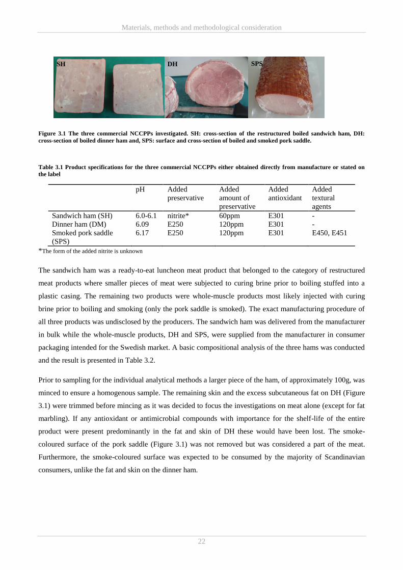
Materials, methods and methodological consideration
22
Figure 3.1 The three commercial NCCPPs investigated. SH: cross-section of the restructured boiled sandwich ham, DH:
cross-section of boiled dinner ham and, SPS: surface and cross-section of boiled and smoked pork saddle.
Table 3.1 Product specifications for the three commercial NCCPPs either obtained directly from manufacture or stated on
the label
pH Added
preservative
Added
amount of
preservative
Added
antioxidant
Added
textural
agents
Sandwich ham (SH) 6.0-6.1 nitrite* 60ppm E301 -
Dinner ham (DM) 6.09 E250 120ppm E301 -
Smoked pork saddle
(SPS)
6.17 E250 120ppm E301 E450, E451
*The form of the added nitrite is unknown
The sandwich ham was a ready-to-eat luncheon meat product that belonged to the category of restructured
meat products where smaller pieces of meat were subjected to curing brine prior to boiling stuffed into a
plastic casing. The remaining two products were whole-muscle products most likely injected with curing
brine prior to boiling and smoking (only the pork saddle is smoked). The exact manufacturing procedure of
all three products was undisclosed by the producers. The sandwich ham was delivered from the manufacturer
in bulk while the whole-muscle products, DH and SPS, were supplied from the manufacturer in consumer
packaging intended for the Swedish market. A basic compositional analysis of the three hams was conducted
and the result is presented in Table 3.2.
Prior to sampling for the individual analytical methods a larger piece of the ham, of approximately 100g, was
minced to ensure a homogenous sample. The remaining skin and the excess subcutaneous fat on DH (Figure
3.1) were trimmed before mincing as it was decided to focus the investigations on meat alone (except for fat
marbling). If any antioxidant or antimicrobial compounds with importance for the shelf-life of the entire
product were present predominantly in the fat and skin of DH these would have been lost. The smoke-
coloured surface of the pork saddle (Figure 3.1) was not removed but was considered a part of the meat.
Furthermore, the smoke-coloured surface was expected to be consumed by the majority of Scandinavian
consumers, unlike the fat and skin on the dinner ham.
SH DH SPS
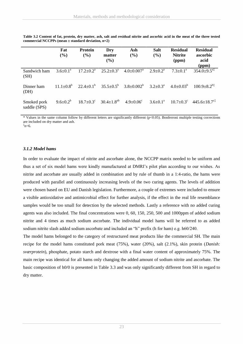
Materials, methods and methodological consideration
23
Table 3.2 Content of fat, protein, dry matter, ash, salt and residual nitrite and ascorbic acid in the meat of the three tested
commercial NCCPPs (mean ± standard deviation, n=2)
Fat
(%)
Protein
(%)
Dry
matter
(%)
Ash
(%)
Salt
(%)
Residual
Nitrite
(ppm)
Residual
ascorbic
acid
(ppm)
Sandwich ham
(SH)
3.6±0.1a 17.2±0.2
a 25.2±0.3
a 4.0±0.007
a 2.9±0.2
a 7.3±0.1
a 354.0±9.5
a‡
Dinner ham
(DH)
11.1±0.8b 22.4±0.1
b 35.5±0.5
b 3.8±0.002
b 3.2±0.3
a 4.0±0.03
b 100.9±8.2
b‡
Smoked pork
saddle (SPS)
9.6±0.2b 18.7±0.3
c 30.4±1.8
ab 4.9±0.06
c 3.6±0.1
a 10.7±0.3
c 445.6±18.7
c‡
* Values in the same column follow by different letters are significantly different (p<0.05). Bonferroni multiple testing corrections
are included on dry matter and ash. ‡n=6.
3.1.2 Model hams
In order to evaluate the impact of nitrite and ascorbate alone, the NCCPP matrix needed to be uniform and
thus a set of six model hams were kindly manufactured at DMRI’s pilot plan according to our wishes. As
nitrite and ascorbate are usually added in combination and by rule of thumb in a 1:4-ratio, the hams were
produced with parallel and continuously increasing levels of the two curing agents. The levels of addition
were chosen based on EU and Danish legislation. Furthermore, a couple of extremes were included to ensure
a visible antioxidative and antimicrobial effect for further analysis, if the effect in the real life resemblance
samples would be too small for detection by the selected methods. Lastly a reference with no added curing
agents was also included. The final concentrations were 0, 60, 150, 250, 500 and 1000ppm of added sodium
nitrite and 4 times as much sodium ascorbate. The individual model hams will be referred to as added
sodium nitrite slash added sodium ascorbate and included an “h” prefix (h for ham) e.g. h60/240.
The model hams belonged to the category of restructured meat products like the commercial SH. The main
recipe for the model hams constituted pork meat (75%), water (20%), salt (2.1%), skin protein (Danish:
sværprotein), phosphate, potato starch and dextrose with a final water content of approximately 75%. The
main recipe was identical for all hams only changing the added amount of sodium nitrite and ascorbate. The
basic composition of h0/0 is presented in Table 3.3 and was only significantly different from SH in regard to
dry matter.
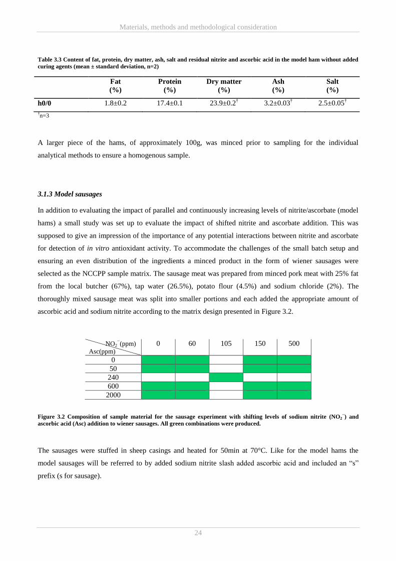
Materials, methods and methodological consideration
24
Table 3.3 Content of fat, protein, dry matter, ash, salt and residual nitrite and ascorbic acid in the model ham without added
curing agents (mean ± standard deviation, n=2)
Fat
(%)
Protein
(%)
Dry matter
(%)
Ash
(%)
Salt
(%)
h0/0 1.8±0.2 17.4±0.1 23.9±0.2† 3.2±0.03
† 2.5±0.05
†
†n=3
A larger piece of the hams, of approximately 100g, was minced prior to sampling for the individual
analytical methods to ensure a homogenous sample.
3.1.3 Model sausages
In addition to evaluating the impact of parallel and continuously increasing levels of nitrite/ascorbate (model
hams) a small study was set up to evaluate the impact of shifted nitrite and ascorbate addition. This was
supposed to give an impression of the importance of any potential interactions between nitrite and ascorbate
for detection of in vitro antioxidant activity. To accommodate the challenges of the small batch setup and
ensuring an even distribution of the ingredients a minced product in the form of wiener sausages were
selected as the NCCPP sample matrix. The sausage meat was prepared from minced pork meat with 25% fat
from the local butcher (67%), tap water (26.5%), potato flour (4.5%) and sodium chloride (2%). The
thoroughly mixed sausage meat was split into smaller portions and each added the appropriate amount of
ascorbic acid and sodium nitrite according to the matrix design presented in Figure 3.2.
NO2−(ppm)
Asc(ppm)
0 60 105 150 500
0
50
240
600
2000
Figure 3.2 Composition of sample material for the sausage experiment with shifting levels of sodium nitrite (NO2−) and
ascorbic acid (Asc) addition to wiener sausages. All green combinations were produced.
The sausages were stuffed in sheep casings and heated for 50min at 70°C. Like for the model hams the
model sausages will be referred to by added sodium nitrite slash added ascorbic acid and included an “s”
prefix (s for sausage).
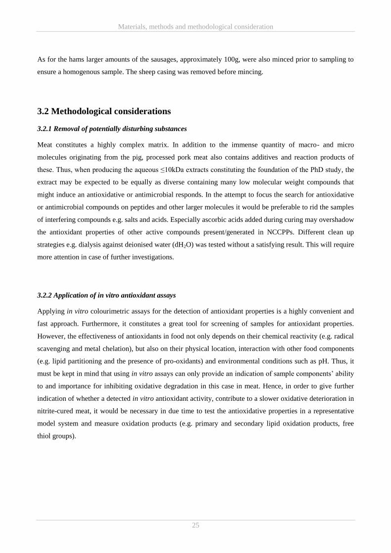
Materials, methods and methodological consideration
25
As for the hams larger amounts of the sausages, approximately 100g, were also minced prior to sampling to
ensure a homogenous sample. The sheep casing was removed before mincing.
3.2 Methodological considerations
3.2.1 Removal of potentially disturbing substances
Meat constitutes a highly complex matrix. In addition to the immense quantity of macro- and micro
molecules originating from the pig, processed pork meat also contains additives and reaction products of
these. Thus, when producing the aqueous ≤10kDa extracts constituting the foundation of the PhD study, the
extract may be expected to be equally as diverse containing many low molecular weight compounds that
might induce an antioxidative or antimicrobial responds. In the attempt to focus the search for antioxidative
or antimicrobial compounds on peptides and other larger molecules it would be preferable to rid the samples
of interfering compounds e.g. salts and acids. Especially ascorbic acids added during curing may overshadow
the antioxidant properties of other active compounds present/generated in NCCPPs. Different clean up
strategies e.g. dialysis against deionised water (dH2O) was tested without a satisfying result. This will require
more attention in case of further investigations.
3.2.2 Application of in vitro antioxidant assays
Applying in vitro colourimetric assays for the detection of antioxidant properties is a highly convenient and
fast approach. Furthermore, it constitutes a great tool for screening of samples for antioxidant properties.
However, the effectiveness of antioxidants in food not only depends on their chemical reactivity (e.g. radical
scavenging and metal chelation), but also on their physical location, interaction with other food components
(e.g. lipid partitioning and the presence of pro-oxidants) and environmental conditions such as pH. Thus, it
must be kept in mind that using in vitro assays can only provide an indication of sample components’ ability
to and importance for inhibiting oxidative degradation in this case in meat. Hence, in order to give further
indication of whether a detected in vitro antioxidant activity, contribute to a slower oxidative deterioration in
nitrite-cured meat, it would be necessary in due time to test the antioxidative properties in a representative
model system and measure oxidation products (e.g. primary and secondary lipid oxidation products, free
thiol groups).
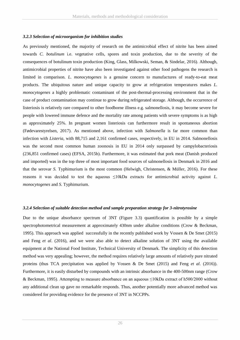
Materials, methods and methodological consideration
26
3.2.3 Selection of microorganism for inhibition studies
As previously mentioned, the majority of research on the antimicrobial effect of nitrite has been aimed
towards C. botulinum i.e. vegetative cells, spores and toxin production, due to the severity of the
consequences of botulinum toxin production (King, Glass, Milkowski, Seman, & Sindelar, 2016). Although,
antimicrobial properties of nitrite have also been investigated against other food pathogens the research is
limited in comparison. L. monocytogenes is a genuine concern to manufactures of ready-to-eat meat
products. The ubiquitous nature and unique capacity to grow at refrigeration temperatures makes L.
monocytogenes a highly problematic contaminant of the post-thermal-processing environment that in the
case of product contamination may continue to grow during refrigerated storage. Although, the occurrence of
listeriosis is relatively rare compared to other foodborne illness e.g. salmonellosis, it may become severe for
people with lowered immune defence and the mortality rate among patients with severe symptoms is as high
as approximately 25%. In pregnant women listeriosis can furthermore result in spontaneous abortion
(Fødevarestyrelsen, 2017). As mentioned above, infection with Salmonella is far more common than
infection with Listeria, with 88,715 and 2,161 confirmed cases, respectively, in EU in 2014. Salmonellosis
was the second most common human zoonosis in EU in 2014 only surpassed by campylobacteriosis
(236,851 confirmed cases) (EFSA, 2015b). Furthermore, it was estimated that pork meat (Danish produced
and imported) was in the top three of most important food sources of salmonellosis in Denmark in 2016 and
that the serovar S. Typhimurium is the most common (Helwigh, Christensen, & Müller, 2016). For these
reasons it was decided to test the aqueous ≤10kDa extracts for antimicrobial activity against L.
monocytogenes and S. Typhimurium.
3.2.4 Selection of suitable detection method and sample preparation strategy for 3-nitrotyrosine
Due to the unique absorbance spectrum of 3NT (Figure 3.3) quantification is possible by a simple
spectrophotometrical measurement at approximately 430nm under alkaline conditions (Crow & Beckman,
1995). This approach was applied successfully in the recently published work by Vossen & De Smet (2015)
and Feng et al. (2016), and we were also able to detect alkaline solution of 3NT using the available
equipment at the National Food Institute, Technical University of Denmark. The simplicity of this detection
method was very appealing; however, the method requires relatively large amounts of relatively pure nitrated
proteins (thus TCA precipitation was applied by Vossen & De Smet (2015) and Feng et al. (2016)).
Furthermore, it is easily disturbed by compounds with an intrinsic absorbance in the 400-500nm range (Crow
& Beckman, 1995). Attempting to measure absorbance on an aqueous ≤10kDa extract of h500/2000 without
any additional clean up gave no remarkable responds. Thus, another potentially more advanced method was
considered for providing evidence for the presence of 3NT in NCCPPs.
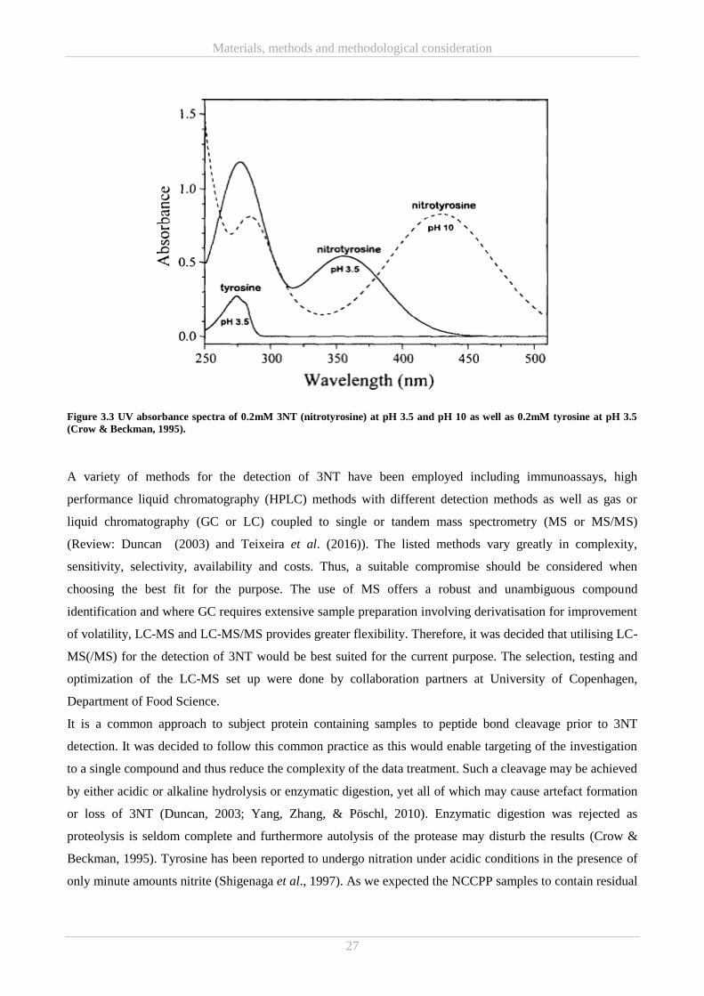
Materials, methods and methodological consideration
27
Figure 3.3 UV absorbance spectra of 0.2mM 3NT (nitrotyrosine) at pH 3.5 and pH 10 as well as 0.2mM tyrosine at pH 3.5
(Crow & Beckman, 1995).
A variety of methods for the detection of 3NT have been employed including immunoassays, high
performance liquid chromatography (HPLC) methods with different detection methods as well as gas or
liquid chromatography (GC or LC) coupled to single or tandem mass spectrometry (MS or MS/MS)
(Review: Duncan (2003) and Teixeira et al. (2016)). The listed methods vary greatly in complexity,
sensitivity, selectivity, availability and costs. Thus, a suitable compromise should be considered when
choosing the best fit for the purpose. The use of MS offers a robust and unambiguous compound
identification and where GC requires extensive sample preparation involving derivatisation for improvement
of volatility, LC-MS and LC-MS/MS provides greater flexibility. Therefore, it was decided that utilising LC-
MS(/MS) for the detection of 3NT would be best suited for the current purpose. The selection, testing and
optimization of the LC-MS set up were done by collaboration partners at University of Copenhagen,
Department of Food Science.
It is a common approach to subject protein containing samples to peptide bond cleavage prior to 3NT
detection. It was decided to follow this common practice as this would enable targeting of the investigation
to a single compound and thus reduce the complexity of the data treatment. Such a cleavage may be achieved
by either acidic or alkaline hydrolysis or enzymatic digestion, yet all of which may cause artefact formation
or loss of 3NT (Duncan, 2003; Yang, Zhang, & Pöschl, 2010). Enzymatic digestion was rejected as
proteolysis is seldom complete and furthermore autolysis of the protease may disturb the results (Crow &
Beckman, 1995). Tyrosine has been reported to undergo nitration under acidic conditions in the presence of
only minute amounts nitrite (Shigenaga et al., 1997). As we expected the NCCPP samples to contain residual
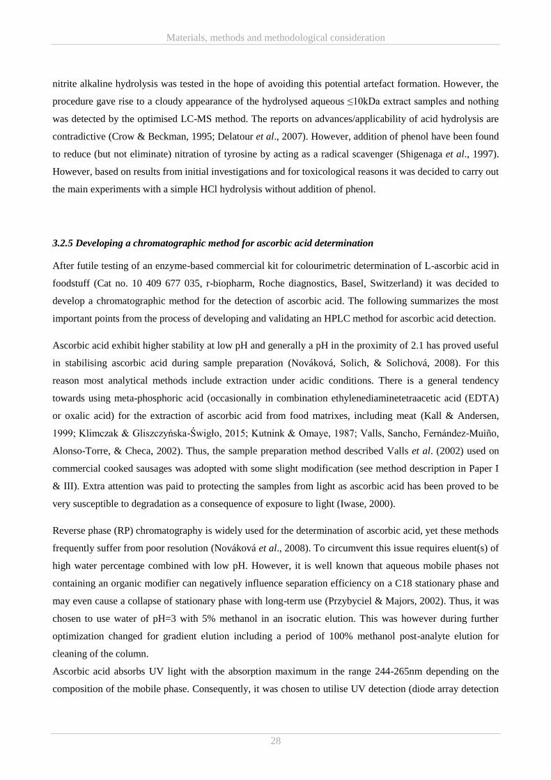
Materials, methods and methodological consideration
28
nitrite alkaline hydrolysis was tested in the hope of avoiding this potential artefact formation. However, the
procedure gave rise to a cloudy appearance of the hydrolysed aqueous ≤10kDa extract samples and nothing
was detected by the optimised LC-MS method. The reports on advances/applicability of acid hydrolysis are
contradictive (Crow & Beckman, 1995; Delatour et al., 2007). However, addition of phenol have been found
to reduce (but not eliminate) nitration of tyrosine by acting as a radical scavenger (Shigenaga et al., 1997).
However, based on results from initial investigations and for toxicological reasons it was decided to carry out
the main experiments with a simple HCl hydrolysis without addition of phenol.
3.2.5 Developing a chromatographic method for ascorbic acid determination
After futile testing of an enzyme-based commercial kit for colourimetric determination of L-ascorbic acid in
foodstuff (Cat no. 10 409 677 035, r-biopharm, Roche diagnostics, Basel, Switzerland) it was decided to
develop a chromatographic method for the detection of ascorbic acid. The following summarizes the most
important points from the process of developing and validating an HPLC method for ascorbic acid detection.
Ascorbic acid exhibit higher stability at low pH and generally a pH in the proximity of 2.1 has proved useful
in stabilising ascorbic acid during sample preparation (Nováková, Solich, & Solichová, 2008). For this
reason most analytical methods include extraction under acidic conditions. There is a general tendency
towards using meta-phosphoric acid (occasionally in combination ethylenediaminetetraacetic acid (EDTA)
or oxalic acid) for the extraction of ascorbic acid from food matrixes, including meat (Kall & Andersen,
1999; Klimczak & Gliszczyńska-Świgło, 2015; Kutnink & Omaye, 1987; Valls, Sancho, Fernández-Muiño,
Alonso-Torre, & Checa, 2002). Thus, the sample preparation method described Valls et al. (2002) used on
commercial cooked sausages was adopted with some slight modification (see method description in Paper I
& III). Extra attention was paid to protecting the samples from light as ascorbic acid has been proved to be
very susceptible to degradation as a consequence of exposure to light (Iwase, 2000).
Reverse phase (RP) chromatography is widely used for the determination of ascorbic acid, yet these methods
frequently suffer from poor resolution (Nováková et al., 2008). To circumvent this issue requires eluent(s) of
high water percentage combined with low pH. However, it is well known that aqueous mobile phases not
containing an organic modifier can negatively influence separation efficiency on a C18 stationary phase and
may even cause a collapse of stationary phase with long-term use (Przybyciel & Majors, 2002). Thus, it was
chosen to use water of pH=3 with 5% methanol in an isocratic elution. This was however during further
optimization changed for gradient elution including a period of 100% methanol post-analyte elution for
cleaning of the column.
Ascorbic acid absorbs UV light with the absorption maximum in the range 244-265nm depending on the
composition of the mobile phase. Consequently, it was chosen to utilise UV detection (diode array detection
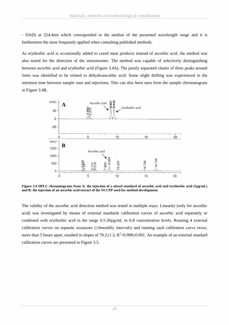
Materials, methods and methodological consideration
29
- DAD) at 254.4nm which corresponded to the median of the presented wavelength range and it is
furthermore the most frequently applied when consulting published methods.
As erythorbic acid is occasionally added to cured meat products instead of ascorbic acid, the method was
also tested for the detection of the stereoisomer. The method was capable of selectively distinguishing
between ascorbic acid and erythorbic acid (Figure 3.4A). The poorly separated cluster of three peaks around
5min was identified to be related to dehydroascorbic acid. Some slight drifting was experienced in the
retention time between sample runs and injections. This can also been seen from the sample chromatogram
in Figure 3.4B.
Figure 3.4 HPLC chromatograms from A: the injection of a mixed standard of ascorbic acid and erythorbic acid (5µg/mL)
and B: the injection of an ascorbic acid-extract of the NCCPP used for method development.
The validity of the ascorbic acid detection method was tested in multiple ways. Linearity (only for ascorbic
acid) was investigated by means of external standards calibration curves of ascorbic acid separately or
combined with erythorbic acid in the range 0.5-30µg/mL in 6-8 concentration levels. Running 4 external
calibration curves on separate occasions (≥4monthly intervals) and running each calibration curve twice,
more than 5 hours apart, resulted in slopes of 70.2±1.3, R2=0.998±0.001. An example of an external standard
calibration curves are presented in Figure 3.5.
Erythorbic acid
Ascorbic acid
Ascorbic acid
A
B
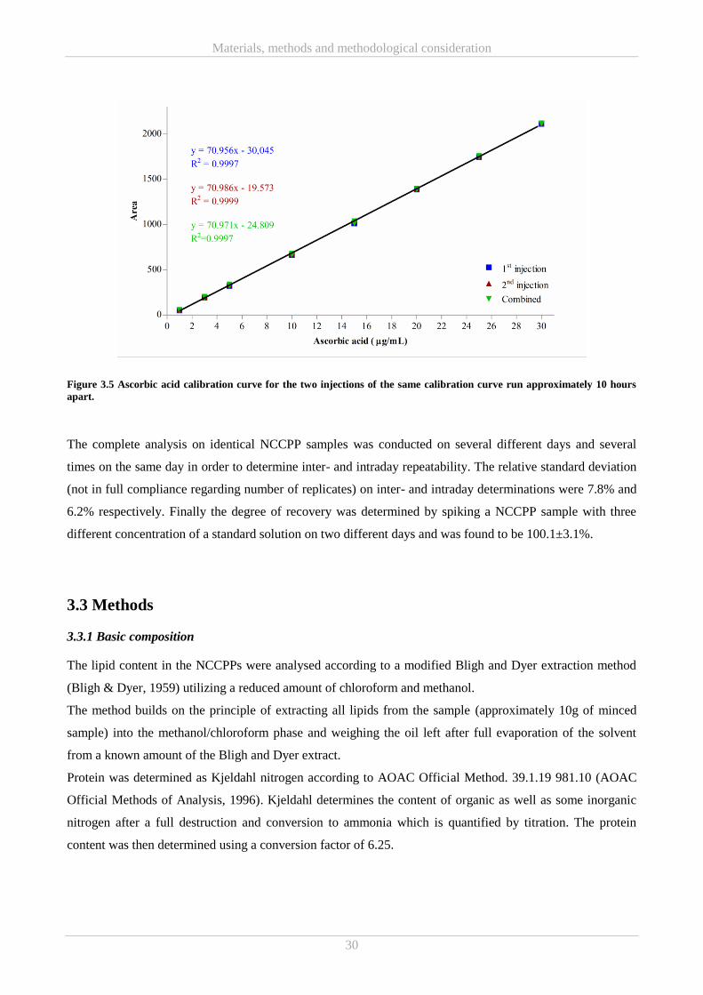
Materials, methods and methodological consideration
30
Figure 3.5 Ascorbic acid calibration curve for the two injections of the same calibration curve run approximately 10 hours
apart.
The complete analysis on identical NCCPP samples was conducted on several different days and several
times on the same day in order to determine inter- and intraday repeatability. The relative standard deviation
(not in full compliance regarding number of replicates) on inter- and intraday determinations were 7.8% and
6.2% respectively. Finally the degree of recovery was determined by spiking a NCCPP sample with three
different concentration of a standard solution on two different days and was found to be 100.1±3.1%.
3.3 Methods
3.3.1 Basic composition
The lipid content in the NCCPPs were analysed according to a modified Bligh and Dyer extraction method
(Bligh & Dyer, 1959) utilizing a reduced amount of chloroform and methanol.
The method builds on the principle of extracting all lipids from the sample (approximately 10g of minced
sample) into the methanol/chloroform phase and weighing the oil left after full evaporation of the solvent
from a known amount of the Bligh and Dyer extract.
Protein was determined as Kjeldahl nitrogen according to AOAC Official Method. 39.1.19 981.10 (AOAC
Official Methods of Analysis, 1996). Kjeldahl determines the content of organic as well as some inorganic
nitrogen after a full destruction and conversion to ammonia which is quantified by titration. The protein
content was then determined using a conversion factor of 6.25.
Materials, methods and methodological consideration
31
The dry matter content was determined by evaporation of all water at 105ºC for 24 hours. The dry matter
content is expressed in percent and calculated according to
%𝐷𝑟𝑦 𝑚𝑎𝑡𝑡𝑒𝑟 =
𝑚𝑎𝑠𝑠 𝑜𝑓 𝑑𝑟𝑦 𝑠𝑎𝑚𝑝𝑙𝑒 ∙ 100%
𝑚𝑎𝑠𝑠 𝑜𝑓 𝑤𝑒𝑡 𝑠𝑎𝑚𝑝𝑙𝑒
(Eq. 16)
The ash content was measured by burning the dried sample at 600ºC for approximately 24 hours. The
temperature was set at 100ºC for the first hour and afterwards increased to 600ºC. The ash content is reported
as
%𝐴𝑠ℎ =
𝑚𝑎𝑠𝑠 𝑜𝑓 𝑎𝑠ℎ ∙ 100%
𝑚𝑎𝑠𝑠 𝑜𝑓 𝑤𝑒𝑡 𝑠𝑎𝑚𝑝𝑙𝑒
(Eq. 17)
3.3.2 Sodium dodecyl sulphate polyacrylamide gel electrophoresis (SDS-PAGE)
Separation of the extract components prior to the intended immunoblotting for detection of S-nitrosated
peptides were conducted using the sodium dodecyl sulphate polyacrylamide gel electrophoresis (SDS-
PAGE) technique. Several minor modifications were implemented, such as using different sample buffers
(e.g. commercial buffers, laemmli buffer and urea buffer) as well as different gels and corresponding running
buffers (e.g. 16% tricine (tricine running buffer) and 10% BisTris gels (MOPS running buffer), NuPage,
Thermo Fisher Scientific, Waltham, MA, USA), but the overall procedure fits the method described by
Laemmli (1970). The main principle of SDS-PAGE is separation of a protein mixture according to protein
size (molecular weight) based on mobility in a polyacrylamide gel when applying an electrical field.
Separation solely based on molecular weight is achieved by boiling the protein with SDS causing
denaturation and masking of charges so that neither will have any influence on the migration in the gel.
Information about protein molecular weights can then be obtained by comparing the generated band pattern
with the pattern from a known standard.
3.3.3 Biotin switch
Two different procedures were tested for the detection of S-nitrosated peptides.
Initially detection of S-nitrosated peptides in the NCCPP aqueous ≤10kDa extracts was conducted using a
commercial S-Nitrosylated Protein Detection Kit from Cayman Chemical (Ann Arbor, MI, USA) based on
the principles of the biotin switch (see Chapter 4.4.1). The experiment was performed in indirect light and
according to manufacturer’s instructions with some modifications. Briefly, a fixed volume of the samples
were mixed 1:6 with Buffer A containing blocking reagent (thiol-blocking reagent) and incubated in the
Materials, methods and methodological consideration
32
dark, shaking at 300rpm for 30min at 7ºC. Afterwards, total protein was precipitated in 4 volumes of pre-
cooled acetone for 1 hour at -20ºC. Following centrifugation at 3000g for 10min at 4 ºC the pellet was re-
suspended in 500μL Buffer B containing reducing and labelling reagent (biotin constituted the label) and
further incubated in the dark for 1 hour at room temperature. Acetone precipitation was repeated as described
above and the resulting pellet was re-suspended in 100μL of MilliQ water. In addition to aqueous ≤10kDa
extracts from the samples SH0, DH0 and SPS0, h0/0 was also incorporated. MilliQ was included as negative
control while cysteine and S-nitrosated cysteine was included as positive control. The S-nitrosocysteine was
synthesised just prior to initiation of the assay by mixing equimolar concentration of cysteine and sodium
nitrite dissolved in dH2O and 0.1M HCl, respectively and incubating in the dark for 20-30min with gentle
agitation.
Three μL of the treated/labelled aqueous ≤10kDa extract samples and controls were dotted onto a
polyvinylidene difluoride (PVDF) membrane which was dried under a blow-dryer, changing the air above
the membrane from appropriate distance to limit heating. Free binding sites on the membrane were blocked
overnight at 5ºC in 2%(W/V) bovine serum albumin (BSA) in Tris-buffered saline (TBS; 20mM Tris-HCl,
0.8% NaCl, pH 8, diluted from 10x concentrated stock). The membrane was washed three times in TBS
before incubating for 1 hour in S-Nitrosylation Detection Reagent I (horseradish peroxidase (HRP)) from the
S-Nitrosylated Protein Detection kit diluted 1:75 in 2% BSA (W/V) in TBS. Again the membrane was
washed three times in TBS and reactive signals were visualized using an Enhanced chemiluminescence
(ECL) kit according to manufacturer’s instructions (Amersham ECL Prime Western Blotting Detection
Reagent, GE Healthcare Life Sciences, Chicago, IL, USA). Detection by substituting S-Nitrosylation
Detection Reagent I (HRP) with S-Nitrosylation Detection Reagent II (Fluorescein) was also tested.
Alternatively the PierceTM
S-Nitrosylation Western Blot kit from Thermo Fisher Scientific was applied. The
experiment was conducted according to manufacturer’s instructions with some modifications. In short,
100µL aqueous ≤10kDa extract sample or control were mixed with 100µL HENS buffer (buffer composed of
4-(2-hydroxyethyl)-1-piperazineethanesulfonic acid (HEPES), EDTA, neocuproine and SDS) and added 4µL
of 1M methyl methanethiosulfonate (MMTS), vortexed for 1min and incubated for 30min at room
temperature protected from light, in order to block free thiols. To remove excess MMTS proteins were
precipitated with 6 volumes of cold acetone for minimum 1 hour at -20°C. The samples were centrifuged at
10,000g for 10min at 4°C, acetone decanted and the pellet was left to dry for 10min before resuspension in
100µL HENS buffer. The resuspended samples were split in two and both added 1µL 20mM Iodoacetyl
Tandem Mass Tag™ (iodoTMT) labelling reagent and vortexed to mix. Two µL 1M sodium ascorbate were
added to all samples and positive controls but substituted for MilliQ in the negative control. The reaction was
allowed to proceed for 1-2 hours. S-nitrosoglutathione and reduced glutathione were used as positive and
negative controls, respectively.
Materials, methods and methodological consideration
33
The labelled samples were loaded onto a PVDF membrane as described above and free binding sites were
blocked by incubating in 5%(W/V) skimmed milk powder dissolved in TBS with Tween-20 (TBST-20) for 1
hour at room temperature. The membrane was then incubated for 1 hour with the primary antibody (anti-
TMT antibody diluted 1:1.000 in 5%(W/V) skimmed milk powder dissolved in TBST-20) and washed 5 x
5min in TBST-20. The membrane was then incubated with the secondary antibody (anti-mouse
immunoglobulin-G HRP-conjugated antibody (IgG-HRP) diluted 1:40,000 in 5%(W/V) skimmed milk
powder dissolved in TBST-20) at room temperature for 1 hour and subsequently washed 5 x 5min in TBST-
20. The reactive signals were visualised using an ECL kit according to manufacturer’s instructions
(Amersham ECL Prime Western Blotting Detection Reagent, GE Healthcare Life Sciences).
3.3.4 Liquid chromatography-mass spectrometry (LC-MS)
The following method(s) were developed in close collaboration with experienced colleagues from the
Research Group for Analytical Food Chemistry, at the National Food Institute.
Prior to LC-MS analysis the crude aqueous ≤10kDa extracts of SH0, DH0, SPS0 and h0/0 were subjected to
further fractionation by ultrafiltration (UF). Vivaspin6 3000Da molecular weight cut off (MWCO)
centrifugal concentrators (Sartorius, Göttingen, Germany) were rinsed with dH2O according to manufactures
description. Five mL of each aqueous ≤10kDa extract were transferred to the rinsed centrifugal concentrators
and centrifuged at 4000g (swing bucket) in short time intervals until approximately half the volume had
passed the polyethersulfone (PES) UF membrane. The fractions were collected and returned to -80ºC. The
following LC-MS analysis of the ham extracts was performed on Dionex UltiMate 3000 LC system (Thermo
Fisher Scientific) connected to a maXis electrospray ionisation-quadrupole time of flight (ESI-QTOF) mass
spectrometer from Bruker (Bruker, Billerica, MA, USA).
Initially, 5μL of the ≤3kDa extract fraction diluted 100x in dH2O were injected into the system and analysed
by flow injection with 50% B isocratic elution (A: 0.02% formic acid and 2.5mM NH4OH B: methanol).
Profiles were recorded using the mass range were 300-2900 m/z.
One μL of the aqueous ≤10kDa extracts, ≤3kDa extract fractions, and concentrate from the UF were
separated on a C8 column (Poroshell 120, 2.1x100mm, SB-C8 2.7μm, Agilent, Santa Clara, CA, USA) at
40ºC in gradient set-up. Eluents A: 0.02% formic acid and 2.5mM NH4OH and B: methanol running 0-1min
0% B, 1-3min 5% B, 3-10min 100% B, 10-12min 100% B, 12.1min 0% B and 12.1-14min 0% B at a
constant flow rate 0.3mL/min. The following MS analysis was conducted in the mass range 300-2900 m/z
with the following settings: end plate offset: 500V, capillary voltage: 4500V, nebulizer pressure: 2bar, drying
Materials, methods and methodological consideration
34
gas flow: 10L/min, dry gas temperature: 200ºC and scan frequency: 9Hz. Nitrogen was used for all gas
purposes.
To further investigate the low m/z ions, 1μL of the aqueous ≤10kDa ham extracts from h0/0 and SPS0 were
analysed in a different chromatographic setup: eluent A: 0.1% formic acid in water and eluent B: 0.1%
formic acid in acetonitrile, separation on C18 column (Poroshell 120, 2.1x100mm, SB-C18 2.7μm, Agilent)
and a gradient (0-1min 1% B, 1-3min 15%B, 3-6min 50%B, 6-9min 95% B, 9-10min 95% B, 10.1min 1% B
and 10.1-13min 1%B) at a constant flowrate of 0.4mL/min. The samples were detected in m/z range 50-
1000. Mass spectrometer setting were kept the same except from the scan frequency which was changed to
2Hz. Broadband fragmentation were then conducted at energies 20-40eV.
Results and Discussion
35
Chapter 4
Results and Discussion 4.
This Chapter is divided into five parts. Part A concerns antioxidative properties in commercial NCCPPs and
is a summary of the main results, discussion and conclusions presented in Paper I and II. Meanwhile Part B
concerns antioxidative properties in NCCPP model systems of varying nitrite and ascorbate addition and
among others include a summary of the main results, discussion and conclusions from Paper III. Part C
discusses the results from the investigations of antimicrobial activities (Paper I and III) as well as strategies
for potential further investigations.
A series of experimental strategies were tested for further characterisation of the aqueous ≤10kDa NCCPP
extracts, and the results and challenges from this are presented in Part D. Lastly, Part E addresses some
additional and supportive findings done during the PhD study.
4.1 Part A – Commercial NCCPPs
4.1.1 Characterisation of antioxidative properties in commercial NCCPPs (Paper I and II)
The characterisation of antioxidative properties in commercial NCCPPs were initiated by investigating
whether antioxidant activity could be identified in aqueous ≤10kDa extracts of different commercial
NCCPPs.
Using in vitro assays for iron chelating activity, reducing power and 2,2′-azino-bis(3-ethylbenzthiazoline-6-
sulfonic acid) (ABTS) radical scavenging activity, antioxidant activity was demonstrated for all three
aqueous ≤10kDa commercial NCCPP extracts in all assays with a single exception (Figure 4.1). The boiled
dinner ham, DH, was generally the NCCPP demonstrating the poorest antioxidative properties and was also
the one not demonstrating any iron chelating activity (Figure 4.1A).
With the exception of iron chelating, both reducing power and ABTS radical scavenging properties seemed
to be connected with curing by way of all the aqueous ≤10kDa nitrite-cured ham extracts displaying higher
antioxidant properties than the reference, h0/0 (Figure 4.1). However, the reducing power and ABTS radical
scavenging properties did not correlate with added nitrite/ascorbic acid but rather with residual
nitrite/ascorbic acid (Table 3.2 in Chapter 3.1.1).
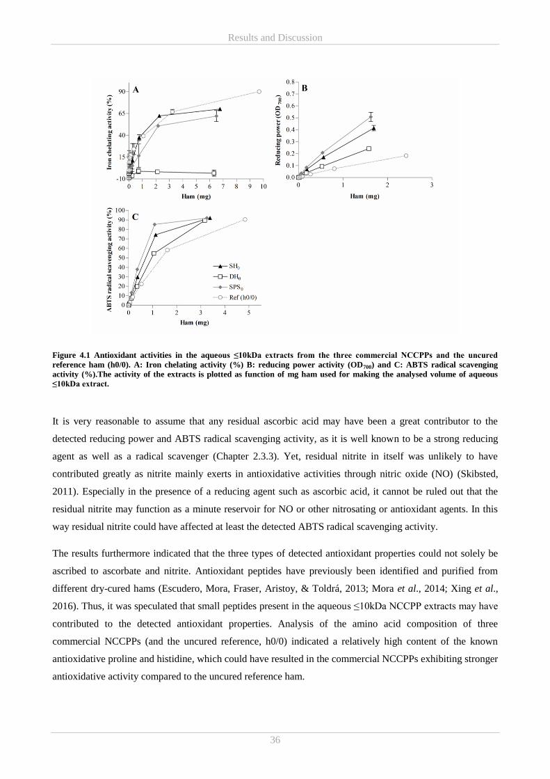
Results and Discussion
36
Figure 4.1 Antioxidant activities in the aqueous ≤10kDa extracts from the three commercial NCCPPs and the uncured
reference ham (h0/0). A: Iron chelating activity (%) B: reducing power activity (OD700) and C: ABTS radical scavenging
activity (%).The activity of the extracts is plotted as function of mg ham used for making the analysed volume of aqueous
≤10kDa extract.
It is very reasonable to assume that any residual ascorbic acid may have been a great contributor to the
detected reducing power and ABTS radical scavenging activity, as it is well known to be a strong reducing
agent as well as a radical scavenger (Chapter 2.3.3). Yet, residual nitrite in itself was unlikely to have
contributed greatly as nitrite mainly exerts in antioxidative activities through nitric oxide (NO) (Skibsted,
2011). Especially in the presence of a reducing agent such as ascorbic acid, it cannot be ruled out that the
residual nitrite may function as a minute reservoir for NO or other nitrosating or antioxidant agents. In this
way residual nitrite could have affected at least the detected ABTS radical scavenging activity.
The results furthermore indicated that the three types of detected antioxidant properties could not solely be
ascribed to ascorbate and nitrite. Antioxidant peptides have previously been identified and purified from
different dry-cured hams (Escudero, Mora, Fraser, Aristoy, & Toldrá, 2013; Mora et al., 2014; Xing et al.,
2016). Thus, it was speculated that small peptides present in the aqueous ≤10kDa NCCPP extracts may have
contributed to the detected antioxidant properties. Analysis of the amino acid composition of three
commercial NCCPPs (and the uncured reference, h0/0) indicated a relatively high content of the known
antioxidative proline and histidine, which could have resulted in the commercial NCCPPs exhibiting stronger
antioxidative activity compared to the uncured reference ham.
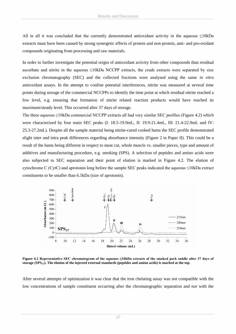
Results and Discussion
37
All in all it was concluded that the currently demonstrated antioxidant activity in the aqueous ≤10kDa
extracts must have been caused by strong synergetic effects of protein and non-protein, anti- and pro-oxidant
compounds originating from processing and raw materials.
In order to further investigate the potential origin of antioxidant activity from other compounds than residual
ascorbate and nitrite in the aqueous ≤10kDa NCCPP extracts, the crude extracts were separated by size
exclusion chromatography (SEC) and the collected fractions were analysed using the same in vitro
antioxidant assays. In the attempt to confine potential interferences, nitrite was measured at several time
points during storage of the commercial NCCPPs to identify the time point at which residual nitrite reached a
low level, e.g. ensuring that formation of nitrite related reaction products would have reached its
maximum/steady level. This occurred after 37 days of storage.
The three aqueous ≤10kDa commercial NCCPP extracts all had very similar SEC profiles (Figure 4.2) which
were characterized by four main SEC peaks (I: 18.5-19.9mL, II: 19.9-21.4mL, III: 21.4-22.9mL and IV:
25.3-27.2mL). Despite all the sample material being nitrite-cured cooked hams the SEC profile demonstrated
slight inter and intra peak differences regarding absorbance intensity (Figure 2 in Paper II). This could be a
result of the hams being different in respect to meat cut, whole muscle vs. smaller pieces, type and amount of
additives and manufacturing procedure, e.g. smoking (SPS). A selection of peptides and amino acids were
also subjected to SEC separation and their point of elution is marked in Figure 4.2. The elution of
cytochrome C (CytC) and aprotonin long before the sample SEC peaks indicated the aqueous ≤10kDa extract
constituents to be smaller than 6.5kDa (size of aprotonin).
Figure 4.2 Representative SEC chromatogram of the aquoues ≤10kDa extracts of the smoked pork saddle after 37 days of
storage (SPS37). The elution of the injected external standards (peptides and amino acids) is marked at the top.
After several attempts of optimization it was clear that the iron chelating assay was not compatible with the
low concentrations of sample constituent occurring after the chromatographic separation and nor with the
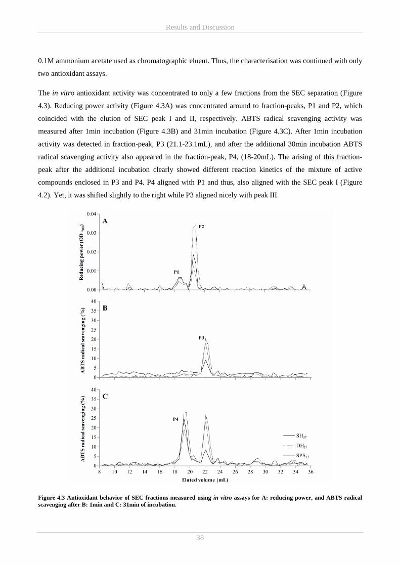
Results and Discussion
38
0.1M ammonium acetate used as chromatographic eluent. Thus, the characterisation was continued with only
two antioxidant assays.
The in vitro antioxidant activity was concentrated to only a few fractions from the SEC separation (Figure
4.3). Reducing power activity (Figure 4.3A) was concentrated around to fraction-peaks, P1 and P2, which
coincided with the elution of SEC peak I and II, respectively. ABTS radical scavenging activity was
measured after 1min incubation (Figure 4.3B) and 31min incubation (Figure 4.3C). After 1min incubation
activity was detected in fraction-peak, P3 (21.1-23.1mL), and after the additional 30min incubation ABTS
radical scavenging activity also appeared in the fraction-peak, P4, (18-20mL). The arising of this fraction-
peak after the additional incubation clearly showed different reaction kinetics of the mixture of active
compounds enclosed in P3 and P4. P4 aligned with P1 and thus, also aligned with the SEC peak I (Figure
4.2). Yet, it was shifted slightly to the right while P3 aligned nicely with peak III.
Figure 4.3 Antioxidant behavior of SEC fractions measured using in vitro assays for A: reducing power, and ABTS radical
scavenging after B: 1min and C: 31min of incubation.
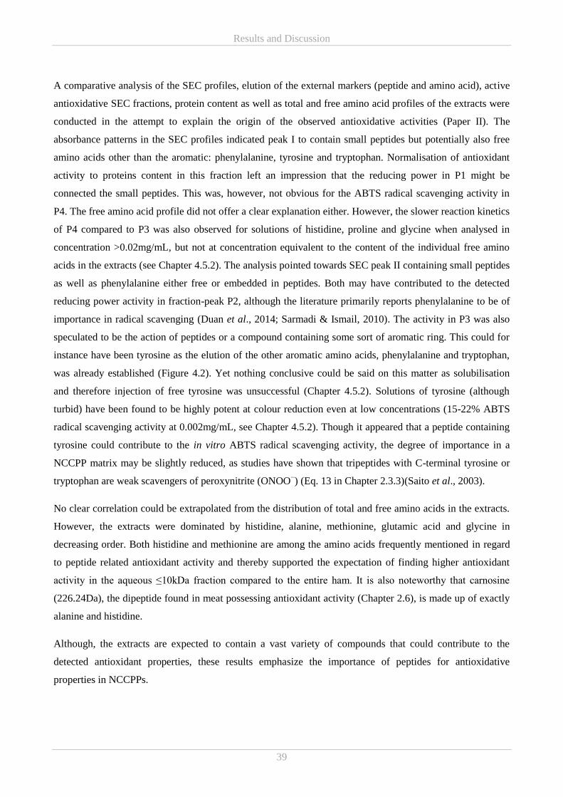
Results and Discussion
39
A comparative analysis of the SEC profiles, elution of the external markers (peptide and amino acid), active
antioxidative SEC fractions, protein content as well as total and free amino acid profiles of the extracts were
conducted in the attempt to explain the origin of the observed antioxidative activities (Paper II). The
absorbance patterns in the SEC profiles indicated peak I to contain small peptides but potentially also free
amino acids other than the aromatic: phenylalanine, tyrosine and tryptophan. Normalisation of antioxidant
activity to proteins content in this fraction left an impression that the reducing power in P1 might be
connected the small peptides. This was, however, not obvious for the ABTS radical scavenging activity in
P4. The free amino acid profile did not offer a clear explanation either. However, the slower reaction kinetics
of P4 compared to P3 was also observed for solutions of histidine, proline and glycine when analysed in
concentration >0.02mg/mL, but not at concentration equivalent to the content of the individual free amino
acids in the extracts (see Chapter 4.5.2). The analysis pointed towards SEC peak II containing small peptides
as well as phenylalanine either free or embedded in peptides. Both may have contributed to the detected
reducing power activity in fraction-peak P2, although the literature primarily reports phenylalanine to be of
importance in radical scavenging (Duan et al., 2014; Sarmadi & Ismail, 2010). The activity in P3 was also
speculated to be the action of peptides or a compound containing some sort of aromatic ring. This could for
instance have been tyrosine as the elution of the other aromatic amino acids, phenylalanine and tryptophan,
was already established (Figure 4.2). Yet nothing conclusive could be said on this matter as solubilisation
and therefore injection of free tyrosine was unsuccessful (Chapter 4.5.2). Solutions of tyrosine (although
turbid) have been found to be highly potent at colour reduction even at low concentrations (15-22% ABTS
radical scavenging activity at 0.002mg/mL, see Chapter 4.5.2). Though it appeared that a peptide containing
tyrosine could contribute to the in vitro ABTS radical scavenging activity, the degree of importance in a
NCCPP matrix may be slightly reduced, as studies have shown that tripeptides with C-terminal tyrosine or
tryptophan are weak scavengers of peroxynitrite (ONOO−) (Eq. 13 in Chapter 2.3.3)(Saito et al., 2003).
No clear correlation could be extrapolated from the distribution of total and free amino acids in the extracts.
However, the extracts were dominated by histidine, alanine, methionine, glutamic acid and glycine in
decreasing order. Both histidine and methionine are among the amino acids frequently mentioned in regard
to peptide related antioxidant activity and thereby supported the expectation of finding higher antioxidant
activity in the aqueous ≤10kDa fraction compared to the entire ham. It is also noteworthy that carnosine
(226.24Da), the dipeptide found in meat possessing antioxidant activity (Chapter 2.6), is made up of exactly
alanine and histidine.
Although, the extracts are expected to contain a vast variety of compounds that could contribute to the
detected antioxidant properties, these results emphasize the importance of peptides for antioxidative
properties in NCCPPs.
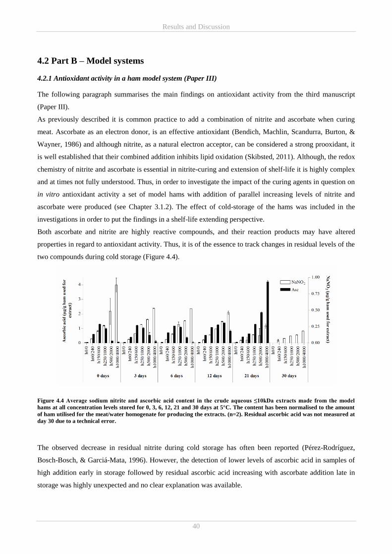
Results and Discussion
40
4.2 Part B – Model systems
4.2.1 Antioxidant activity in a ham model system (Paper III)
The following paragraph summarises the main findings on antioxidant activity from the third manuscript
(Paper III).
As previously described it is common practice to add a combination of nitrite and ascorbate when curing
meat. Ascorbate as an electron donor, is an effective antioxidant (Bendich, Machlin, Scandurra, Burton, &
Wayner, 1986) and although nitrite, as a natural electron acceptor, can be considered a strong prooxidant, it
is well established that their combined addition inhibits lipid oxidation (Skibsted, 2011). Although, the redox
chemistry of nitrite and ascorbate is essential in nitrite-curing and extension of shelf-life it is highly complex
and at times not fully understood. Thus, in order to investigate the impact of the curing agents in question on
in vitro antioxidant activity a set of model hams with addition of parallel increasing levels of nitrite and
ascorbate were produced (see Chapter 3.1.2). The effect of cold-storage of the hams was included in the
investigations in order to put the findings in a shelf-life extending perspective.
Both ascorbate and nitrite are highly reactive compounds, and their reaction products may have altered
properties in regard to antioxidant activity. Thus, it is of the essence to track changes in residual levels of the
two compounds during cold storage (Figure 4.4).
Figure 4.4 Average sodium nitrite and ascorbic acid content in the crude aqueous ≤10kDa extracts made from the model
hams at all concentration levels stored for 0, 3, 6, 12, 21 and 30 days at 5°C. The content has been normalised to the amount
of ham utilised for the meat/water homogenate for producing the extracts. (n=2). Residual ascorbic acid was not measured at
day 30 due to a technical error.
The observed decrease in residual nitrite during cold storage has often been reported (Pérez-Rodríguez,
Bosch-Bosch, & Garciá-Mata, 1996). However, the detection of lower levels of ascorbic acid in samples of
high addition early in storage followed by residual ascorbic acid increasing with ascorbate addition late in
storage was highly unexpected and no clear explanation was available.
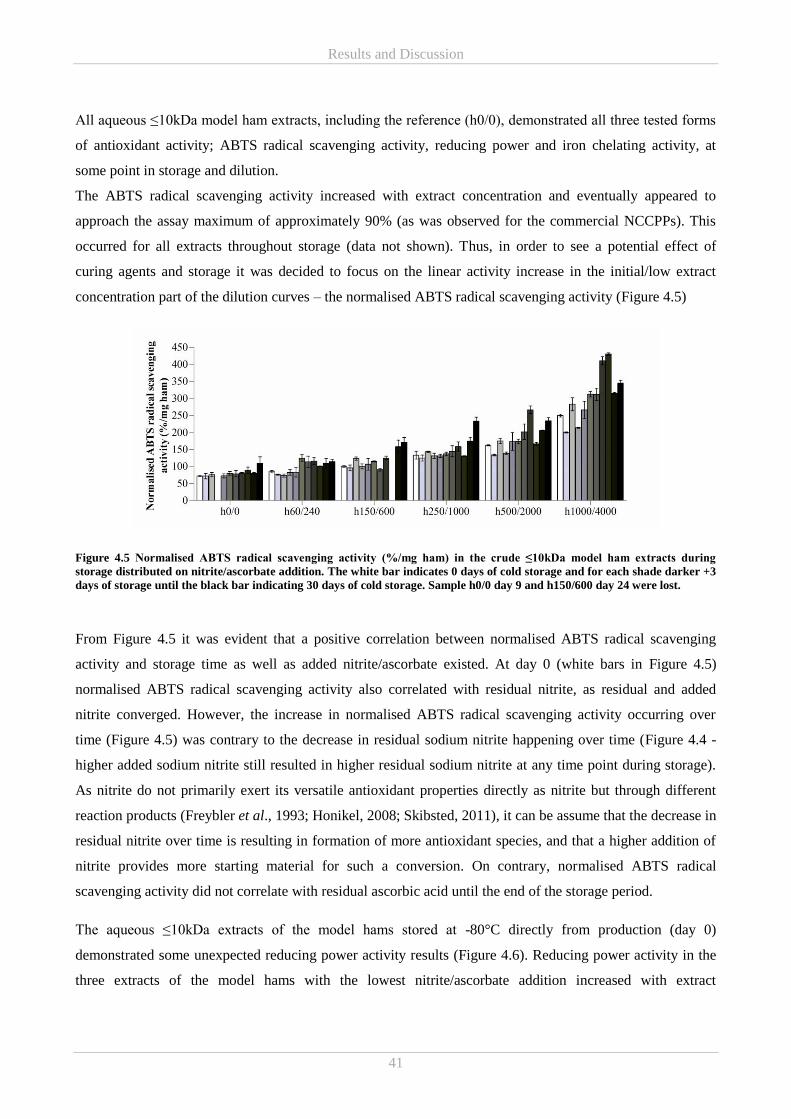
Results and Discussion
41
All aqueous ≤10kDa model ham extracts, including the reference (h0/0), demonstrated all three tested forms
of antioxidant activity; ABTS radical scavenging activity, reducing power and iron chelating activity, at
some point in storage and dilution.
The ABTS radical scavenging activity increased with extract concentration and eventually appeared to
approach the assay maximum of approximately 90% (as was observed for the commercial NCCPPs). This
occurred for all extracts throughout storage (data not shown). Thus, in order to see a potential effect of
curing agents and storage it was decided to focus on the linear activity increase in the initial/low extract
concentration part of the dilution curves – the normalised ABTS radical scavenging activity (Figure 4.5)
Figure 4.5 Normalised ABTS radical scavenging activity (%/mg ham) in the crude ≤10kDa model ham extracts during
storage distributed on nitrite/ascorbate addition. The white bar indicates 0 days of cold storage and for each shade darker +3
days of storage until the black bar indicating 30 days of cold storage. Sample h0/0 day 9 and h150/600 day 24 were lost.
From Figure 4.5 it was evident that a positive correlation between normalised ABTS radical scavenging
activity and storage time as well as added nitrite/ascorbate existed. At day 0 (white bars in Figure 4.5)
normalised ABTS radical scavenging activity also correlated with residual nitrite, as residual and added
nitrite converged. However, the increase in normalised ABTS radical scavenging activity occurring over
time (Figure 4.5) was contrary to the decrease in residual sodium nitrite happening over time (Figure 4.4 -
higher added sodium nitrite still resulted in higher residual sodium nitrite at any time point during storage).
As nitrite do not primarily exert its versatile antioxidant properties directly as nitrite but through different
reaction products (Freybler et al., 1993; Honikel, 2008; Skibsted, 2011), it can be assume that the decrease in
residual nitrite over time is resulting in formation of more antioxidant species, and that a higher addition of
nitrite provides more starting material for such a conversion. On contrary, normalised ABTS radical
scavenging activity did not correlate with residual ascorbic acid until the end of the storage period.
The aqueous ≤10kDa extracts of the model hams stored at -80°C directly from production (day 0)
demonstrated some unexpected reducing power activity results (Figure 4.6). Reducing power activity in the
three extracts of the model hams with the lowest nitrite/ascorbate addition increased with extract
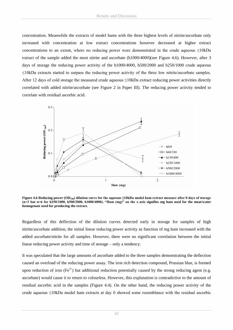
Results and Discussion
42
concentration. Meanwhile the extracts of model hams with the three highest levels of nitrite/ascorbate only
increased with concentration at low extract concentrations however decreased at higher extract
concentrations to an extent, where no reducing power were demonstrated in the crude aqueous ≤10kDa
extract of the sample added the most nitrite and ascorbate (h1000/4000)(see Figure 4.6). However, after 3
days of storage the reducing power activity of the h1000/4000, h500/2000 and h250/1000 crude aqueous
≤10kDa extracts started to surpass the reducing power activity of the three low nitrite/ascorbate samples.
After 12 days of cold storage the measured crude aqueous ≤10kDa extract reducing power activities directly
correlated with added nitrite/ascorbate (see Figure 2 in Paper III). The reducing power activity tended to
correlate with residual ascorbic acid.
Figure 4.6 Reducing power (OD700) dilution curve for the aqueous ≤10kDa model ham extract measure after 0 days of storage
(n=3 but n=6 for h250/1000, h500/2000, h1000/4000). “Ham (mg)” on the x-axis signifies mg ham used for the meat/water
homogenate used for producing the extract.
Regardless of this deflection of the dilution curves detected early in storage for samples of high
nitrite/ascorbate addition, the initial linear reducing power activity as function of mg ham increased with the
added ascorbate/nitrite for all samples. However, there were no significant correlation between the initial
linear reducing power activity and time of storage – only a tendency.
It was speculated that the large amounts of ascorbate added to the three samples demonstrating the deflection
caused an overload of the reducing power assay. The iron rich detection compound, Prussian blue, is formed
upon reduction of iron (Fe3+
) but additional reduction potentially caused by the strong reducing agent (e.g.
ascorbate) would cause it to return to colourless. However, this explanation is contradictive to the amount of
residual ascorbic acid in the samples (Figure 4.4). On the other hand, the reducing power activity of the
crude aqueous ≤10kDa model ham extracts at day 0 showed some resemblance with the residual ascorbic
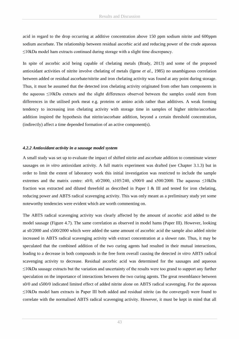
Results and Discussion
43
acid in regard to the drop occurring at additive concentration above 150 ppm sodium nitrite and 600ppm
sodium ascorbate. The relationship between residual ascorbic acid and reducing power of the crude aqueous
≤10kDa model ham extracts continued during storage with a slight time discrepancy.
In spite of ascorbic acid being capable of chelating metals (Brady, 2013) and some of the proposed
antioxidant activities of nitrite involve chelating of metals (Igene et al., 1985) no unambiguous correlation
between added or residual ascorbate/nitrite and iron chelating activity was found at any point during storage.
Thus, it must be assumed that the detected iron chelating activity originated from other ham components in
the aqueous ≤10kDa extracts and the slight differences observed between the samples could stem from
differences in the utilised pork meat e.g. proteins or amino acids rather than additives. A weak forming
tendency to increasing iron chelating activity with storage time in samples of higher nitrite/ascorbate
addition inspired the hypothesis that nitrite/ascorbate addition, beyond a certain threshold concentration,
(indirectly) affect a time depended formation of an active component(s).
4.2.2 Antioxidant activity in a sausage model system
A small study was set up to evaluate the impact of shifted nitrite and ascorbate addition to comminute wiener
sausages on in vitro antioxidant activity. A full matrix experiment was drafted (see Chapter 3.1.3) but in
order to limit the extent of laboratory work this initial investigation was restricted to include the sample
extremes and the matrix centre: s0/0, s0/2000, s105/240, s500/0 and s500/2000. The aqueous ≤10kDa
fraction was extracted and diluted threefold as described in Paper I & III and tested for iron chelating,
reducing power and ABTS radical scavenging activity. This was only meant as a preliminary study yet some
noteworthy tendencies were evident which are worth commenting on.
The ABTS radical scavenging activity was clearly affected by the amount of ascorbic acid added to the
model sausage (Figure 4.7). The same correlation as observed in model hams (Paper III). However, looking
at s0/2000 and s500/2000 which were added the same amount of ascorbic acid the sample also added nitrite
increased in ABTS radical scavenging activity with extract concentration at a slower rate. Thus, it may be
speculated that the combined addition of the two curing agents had resulted in their mutual interactions,
leading to a decrease in both compounds in the free form overall causing the detected in vitro ABTS radical
scavenging activity to decrease. Residual ascorbic acid was determined for the sausages and aqueous
≤10kDa sausage extracts but the variation and uncertainty of the results were too grand to support any further
speculation on the importance of interactions between the two curing agents. The great resemblance between
s0/0 and s500/0 indicated limited effect of added nitrite alone on ABTS radical scavenging. For the aqueous
≤10kDa model ham extracts in Paper III both added and residual nitrite (as the converged) were found to
correlate with the normalised ABTS radical scavenging activity. However, it must be kept in mind that all
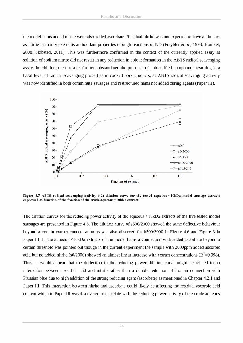
Results and Discussion
44
the model hams added nitrite were also added ascorbate. Residual nitrite was not expected to have an impact
as nitrite primarily exerts its antioxidant properties through reactions of NO (Freybler et al., 1993; Honikel,
2008; Skibsted, 2011). This was furthermore confirmed in the context of the currently applied assay as
solution of sodium nitrite did not result in any reduction in colour formation in the ABTS radical scavenging
assay. In addition, these results further substantiated the presence of unidentified compounds resulting in a
basal level of radical scavenging properties in cooked pork products, as ABTS radical scavenging activity
was now identified in both comminute sausages and restructured hams not added curing agents (Paper III).
Figure 4.7 ABTS radical scavenging activity (%) dilution curve for the tested aqueous ≤10kDa model sausage extracts
expressed as function of the fraction of the crude aqueous ≤10kDa extract.
The dilution curves for the reducing power activity of the aqueous ≤10kDa extracts of the five tested model
sausages are presented in Figure 4.8. The dilution curve of s500/2000 showed the same deflective behaviour
beyond a certain extract concentration as was also observed for h500/2000 in Figure 4.6 and Figure 3 in
Paper III. In the aqueous ≤10kDa extracts of the model hams a connection with added ascorbate beyond a
certain threshold was pointed out though in the current experiment the sample with 2000ppm added ascorbic
acid but no added nitrite (s0/2000) showed an almost linear increase with extract concentrations (R2=0.998).
Thus, it would appear that the deflection in the reducing power dilution curve might be related to an
interaction between ascorbic acid and nitrite rather than a double reduction of iron in connection with
Prussian blue due to high addition of the strong reducing agent (ascorbate) as mentioned in Chapter 4.2.1 and
Paper III. This interaction between nitrite and ascorbate could likely be affecting the residual ascorbic acid
content which in Paper III was discovered to correlate with the reducing power activity of the crude aqueous
Results and Discussion
45
≤10kDa extract. As mentioned above the measurement of residual ascorbic acid in the aqueous ≤10kDa
sausage extracts was erroneous and thus cannot support or reject the proposed explanation.
Figure 4.8 Reducing power (OD700) dilution curve for the tested aqueous ≤10kDa model sausage extracts expressed as
function of the fraction of the crude aqueous ≤10kDa extract.
Surprisingly s105/240 also deflected at high extract concentrations. This result is curious if assuming that
added ascorbic acid was the sole explanation as neither h60/240 nor h150/600 (with the same or more added
ascorbate) behaved in this manor (Figure 4.6 and Figure 3 in Paper III). The main difference between the
mentioned model hams and the model sausage is the added nitrite to added ascorbate/ascorbic acid ratios of
1:4 and 1:2.3 respectively. Thus, it would appear that along with some unknown threshold value the ratio is
essential for this deflecting behaviour. Furthermore, it would seem that the amount of nitrite is defining as
the deflection only occurs when nitrite is added but only when ascorbate is added too. This was emphasized
by the complete lack of OD700 signal in s500/0. This result shows that great attention should be paid to the
formulation of NCCPPs as slight chances in the amount of added curing agents may change the preservative
effect.
Like it was observed in the aqueous ≤10kDa extracts of the model hams, s0/0 also displayed some reducing
power activity (Figure 4.8). Thus, it would seem that cooked pork products in general may have a basic level
of reducing power activity just as was seen for ATBS radical scavenging activity. This could potentially
originate from the meat e.g. peptides and free amino acids. This is, however, largely overshadowed by the
ascorbate/nitrite chemistry in the current experimental setup.
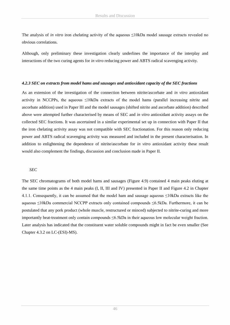
Results and Discussion
46
The analysis of in vitro iron chelating activity of the aqueous ≤10kDa model sausage extracts revealed no
obvious correlations.
Although, only preliminary these investigation clearly underlines the importance of the interplay and
interactions of the two curing agents for in vitro reducing power and ABTS radical scavenging activity.
4.2.3 SEC on extracts from model hams and sausages and antioxidant capacity of the SEC fractions
As an extension of the investigation of the connection between nitrite/ascorbate and in vitro antioxidant
activity in NCCPPs, the aqueous ≤10kDa extracts of the model hams (parallel increasing nitrite and
ascorbate addition) used in Paper III and the model sausages (shifted nitrite and ascorbate addition) described
above were attempted further characterised by means of SEC and in vitro antioxidant activity assays on the
collected SEC fractions. It was ascertained in a similar experimental set up in connection with Paper II that
the iron chelating activity assay was not compatible with SEC fractionation. For this reason only reducing
power and ABTS radical scavenging activity was measured and included in the present characterisation. In
addition to enlightening the dependence of nitrite/ascorbate for in vitro antioxidant activity these result
would also complement the findings, discussion and conclusion made in Paper II.
SEC
The SEC chromatograms of both model hams and sausages (Figure 4.9) contained 4 main peaks eluting at
the same time points as the 4 main peaks (I, II, III and IV) presented in Paper II and Figure 4.2 in Chapter
4.1.1. Consequently, it can be assumed that the model ham and sausage aqueous ≤10kDa extracts like the
aqueous ≤10kDa commercial NCCPP extracts only contained compounds ≤6.5kDa. Furthermore, it can be
postulated that any pork product (whole muscle, restructured or minced) subjected to nitrite-curing and more
importantly heat-treatment only contain compounds ≤6.5kDa in their aqueous low molecular weight fraction.
Later analysis has indicated that the constituent water soluble compounds might in fact be even smaller (See
Chapter 4.3.2 on LC-(ESI)-MS).
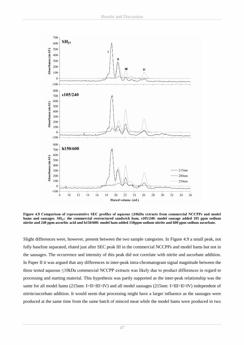
Results and Discussion
47
Figure 4.9 Comparison of representative SEC profiles of aqueous ≤10kDa extracts from commercial NCCPPs and model
hams and sausages. SH37: the commercial restructured sandwich ham, s105/240: model sausage added 105 ppm sodium
nitrite and 240 ppm ascorbic acid and h150/600: model ham added 150ppm sodium nitrite and 600 ppm sodium ascorbate.
Slight differences were, however, present between the two sample categories. In Figure 4.9 a small peak, not
fully baseline separated, eluted just after SEC peak III in the commercial NCCPPs and model hams but not in
the sausages. The occurrence and intensity of this peak did not correlate with nitrite and ascorbate addition.
In Paper II it was argued that any differences in inter-peak intra-chromatogram signal magnitude between the
three tested aqueous ≤10kDa commercial NCCPP extracts was likely due to product differences in regard to
processing and starting material. This hypothesis was partly supported as the inter-peak relationship was the
same for all model hams (215nm: I>II>III>IV) and all model sausages (215nm: I>III>II>IV) independent of
nitrite/ascorbate addition. It would seem that processing might have a larger influence as the sausages were
produced at the same time from the same batch of minced meat while the model hams were produced in two
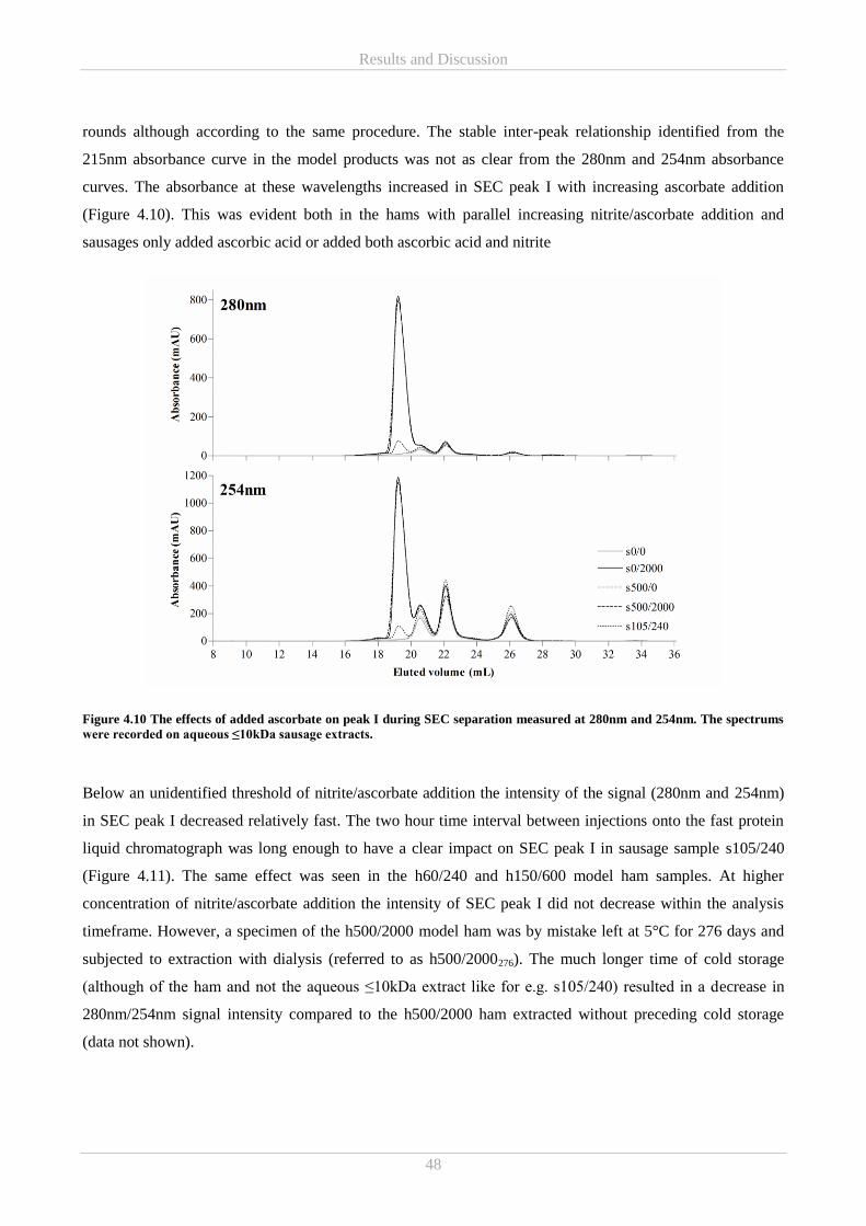
Results and Discussion
48
rounds although according to the same procedure. The stable inter-peak relationship identified from the
215nm absorbance curve in the model products was not as clear from the 280nm and 254nm absorbance
curves. The absorbance at these wavelengths increased in SEC peak I with increasing ascorbate addition
(Figure 4.10). This was evident both in the hams with parallel increasing nitrite/ascorbate addition and
sausages only added ascorbic acid or added both ascorbic acid and nitrite
Figure 4.10 The effects of added ascorbate on peak I during SEC separation measured at 280nm and 254nm. The spectrums
were recorded on aqueous ≤10kDa sausage extracts.
Below an unidentified threshold of nitrite/ascorbate addition the intensity of the signal (280nm and 254nm)
in SEC peak I decreased relatively fast. The two hour time interval between injections onto the fast protein
liquid chromatograph was long enough to have a clear impact on SEC peak I in sausage sample s105/240
(Figure 4.11). The same effect was seen in the h60/240 and h150/600 model ham samples. At higher
concentration of nitrite/ascorbate addition the intensity of SEC peak I did not decrease within the analysis
timeframe. However, a specimen of the h500/2000 model ham was by mistake left at 5°C for 276 days and
subjected to extraction with dialysis (referred to as h500/2000276). The much longer time of cold storage
(although of the ham and not the aqueous ≤10kDa extract like for e.g. s105/240) resulted in a decrease in
280nm/254nm signal intensity compared to the h500/2000 ham extracted without preceding cold storage
(data not shown).
Results and Discussion
49
Figure 4.11 Changes in absorption at 280nm in peak I during SEC fractionation of the aqueous ≤10kDa extract from model
sausage s105/240. The four injections were approximately 2 hours apart and the samples were stored at 5°C in the meantime.
The decrease was also visible at 254nm.
Ascorbic acid is a rather sensitive compound. Thus, the decrease in SEC peak I over time could be an
argument for the 280/254nm signal being largely induced by the labile ascorbate. Despite high levels of
uncertainties compromising the credibility of the results the aqueous ≤10kDa extract of s0/2000 were found
to contain more residual ascorbic acid than s500/2000 which also contained more residual ascorbic acid than
s105/240, all of which correlated with the 280/254nm signal intensity. However, the correlation was not
linear. On contrary the residual ascorbic acid content was in Chapter 4.2.1 Figure 4.4 found not to increase
continuously with nitrite/ascorbate addition and furthermore, residual ascorbic acid increased with cold
storage for extracts of h250/1000, h500/2000 and h1000/4000 (Paper III). Both arguments are opposing the
hypothesis of SEC peak I originating mainly from ascorbic acid. As the results point in opposite directions a
final conclusion on the importance of ascorbate for the 280/254nm signal cannot be drawn.
In the sausage sample with added nitrite but without added ascorbate (s500/0) a clear peak eluted after SEC
peak IV only absorbing at 215nm (Figure 4.12). This 215nm peak was also discovered in the remaining
sausage and ham samples added ≥500ppm nitrite. However, the addition of ascorbate appeared to result in a
lowering of the signal. Nitrite and nitrate has low-wavelength absorptivity and wavelengths between 205-
215nm are frequently used for their detection (Zacharis & Tzanavaras, 2012). Thus, this peak could be
associated with nitrite/nitrate. However, injections of nitrate solutions resulted in a signal responds at
approximately 19mL (data not shown). Yet, the reduction of the peak with ascorbate addition fits the general
notion that residual nitrite decreases faster in the presence of ascorbate (Izumi, 1992).
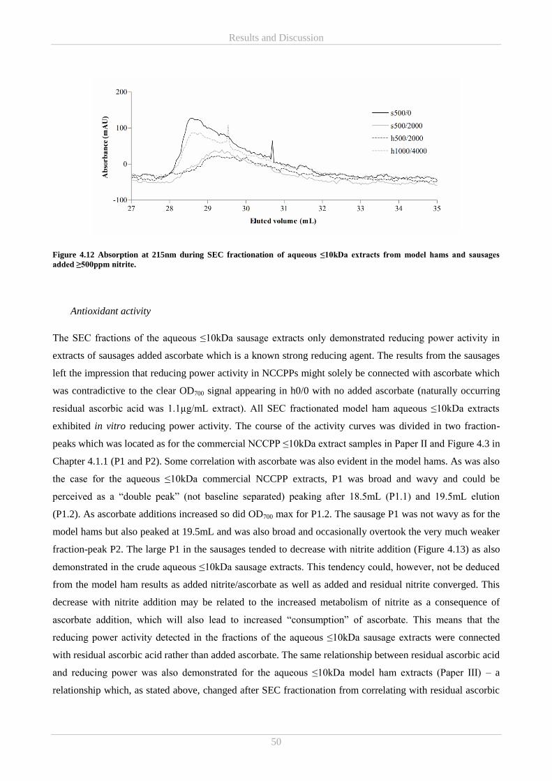
Results and Discussion
50
Figure 4.12 Absorption at 215nm during SEC fractionation of aqueous ≤10kDa extracts from model hams and sausages
added ≥500ppm nitrite.
Antioxidant activity
The SEC fractions of the aqueous ≤10kDa sausage extracts only demonstrated reducing power activity in
extracts of sausages added ascorbate which is a known strong reducing agent. The results from the sausages
left the impression that reducing power activity in NCCPPs might solely be connected with ascorbate which
was contradictive to the clear OD700 signal appearing in h0/0 with no added ascorbate (naturally occurring
residual ascorbic acid was 1.1µg/mL extract). All SEC fractionated model ham aqueous ≤10kDa extracts
exhibited in vitro reducing power activity. The course of the activity curves was divided in two fraction-
peaks which was located as for the commercial NCCPP ≤10kDa extract samples in Paper II and Figure 4.3 in
Chapter 4.1.1 (P1 and P2). Some correlation with ascorbate was also evident in the model hams. As was also
the case for the aqueous ≤10kDa commercial NCCPP extracts, P1 was broad and wavy and could be
perceived as a “double peak” (not baseline separated) peaking after 18.5mL (P1.1) and 19.5mL elution
(P1.2). As ascorbate additions increased so did OD700 max for P1.2. The sausage P1 was not wavy as for the
model hams but also peaked at 19.5mL and was also broad and occasionally overtook the very much weaker
fraction-peak P2. The large P1 in the sausages tended to decrease with nitrite addition (Figure 4.13) as also
demonstrated in the crude aqueous ≤10kDa sausage extracts. This tendency could, however, not be deduced
from the model ham results as added nitrite/ascorbate as well as added and residual nitrite converged. This
decrease with nitrite addition may be related to the increased metabolism of nitrite as a consequence of
ascorbate addition, which will also lead to increased “consumption” of ascorbate. This means that the
reducing power activity detected in the fractions of the aqueous ≤10kDa sausage extracts were connected
with residual ascorbic acid rather than added ascorbate. The same relationship between residual ascorbic acid
and reducing power was also demonstrated for the aqueous ≤10kDa model ham extracts (Paper III) – a
relationship which, as stated above, changed after SEC fractionation from correlating with residual ascorbic
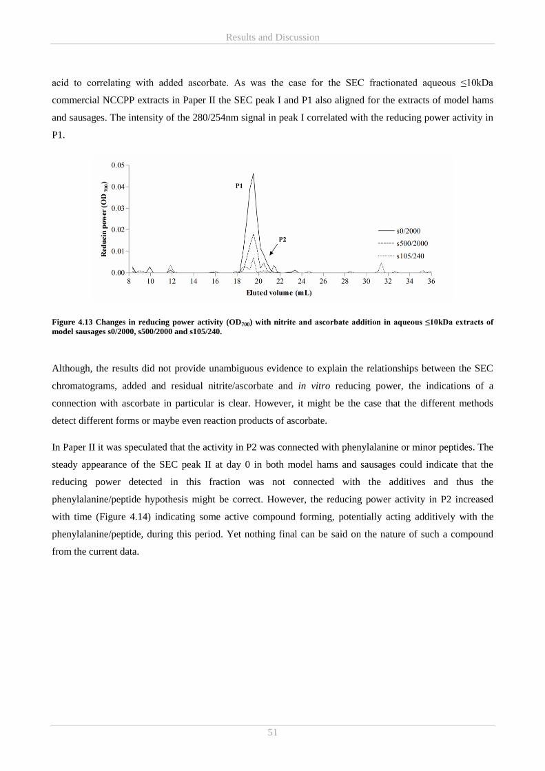
Results and Discussion
51
acid to correlating with added ascorbate. As was the case for the SEC fractionated aqueous ≤10kDa
commercial NCCPP extracts in Paper II the SEC peak I and P1 also aligned for the extracts of model hams
and sausages. The intensity of the 280/254nm signal in peak I correlated with the reducing power activity in
P1.
Figure 4.13 Changes in reducing power activity (OD700) with nitrite and ascorbate addition in aqueous ≤10kDa extracts of
model sausages s0/2000, s500/2000 and s105/240.
Although, the results did not provide unambiguous evidence to explain the relationships between the SEC
chromatograms, added and residual nitrite/ascorbate and in vitro reducing power, the indications of a
connection with ascorbate in particular is clear. However, it might be the case that the different methods
detect different forms or maybe even reaction products of ascorbate.
In Paper II it was speculated that the activity in P2 was connected with phenylalanine or minor peptides. The
steady appearance of the SEC peak II at day 0 in both model hams and sausages could indicate that the
reducing power detected in this fraction was not connected with the additives and thus the
phenylalanine/peptide hypothesis might be correct. However, the reducing power activity in P2 increased
with time (Figure 4.14) indicating some active compound forming, potentially acting additively with the
phenylalanine/peptide, during this period. Yet nothing final can be said on the nature of such a compound
from the current data.
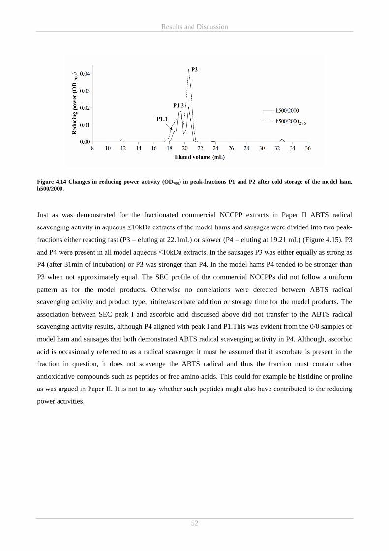
Results and Discussion
52
Figure 4.14 Changes in reducing power activity (OD700) in peak-fractions P1 and P2 after cold storage of the model ham,
h500/2000.
Just as was demonstrated for the fractionated commercial NCCPP extracts in Paper II ABTS radical
scavenging activity in aqueous ≤10kDa extracts of the model hams and sausages were divided into two peak-
fractions either reacting fast (P3 – eluting at 22.1mL) or slower (P4 – eluting at 19.21 mL) (Figure 4.15). P3
and P4 were present in all model aqueous ≤10kDa extracts. In the sausages P3 was either equally as strong as
P4 (after 31min of incubation) or P3 was stronger than P4. In the model hams P4 tended to be stronger than
P3 when not approximately equal. The SEC profile of the commercial NCCPPs did not follow a uniform
pattern as for the model products. Otherwise no correlations were detected between ABTS radical
scavenging activity and product type, nitrite/ascorbate addition or storage time for the model products. The
association between SEC peak I and ascorbic acid discussed above did not transfer to the ABTS radical
scavenging activity results, although P4 aligned with peak I and P1.This was evident from the 0/0 samples of
model ham and sausages that both demonstrated ABTS radical scavenging activity in P4. Although, ascorbic
acid is occasionally referred to as a radical scavenger it must be assumed that if ascorbate is present in the
fraction in question, it does not scavenge the ABTS radical and thus the fraction must contain other
antioxidative compounds such as peptides or free amino acids. This could for example be histidine or proline
as was argued in Paper II. It is not to say whether such peptides might also have contributed to the reducing
power activities.
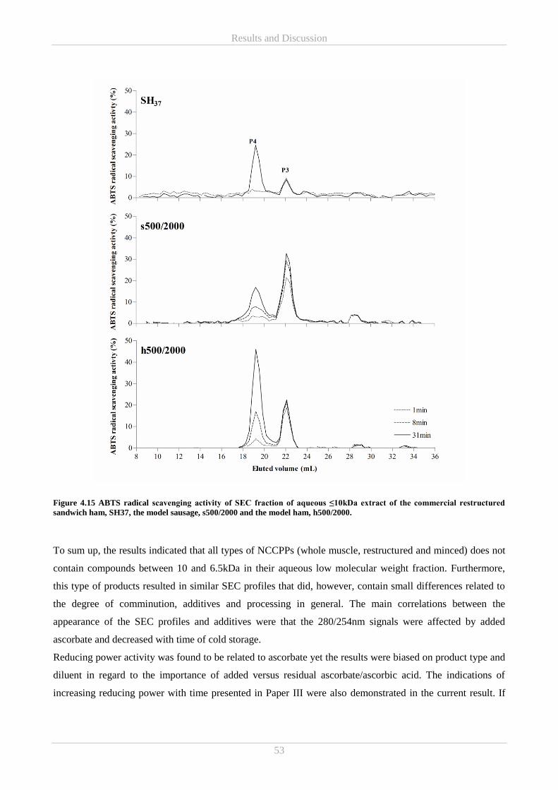
Results and Discussion
53
Figure 4.15 ABTS radical scavenging activity of SEC fraction of aqueous ≤10kDa extract of the commercial restructured
sandwich ham, SH37, the model sausage, s500/2000 and the model ham, h500/2000.
To sum up, the results indicated that all types of NCCPPs (whole muscle, restructured and minced) does not
contain compounds between 10 and 6.5kDa in their aqueous low molecular weight fraction. Furthermore,
this type of products resulted in similar SEC profiles that did, however, contain small differences related to
the degree of comminution, additives and processing in general. The main correlations between the
appearance of the SEC profiles and additives were that the 280/254nm signals were affected by added
ascorbate and decreased with time of cold storage.
Reducing power activity was found to be related to ascorbate yet the results were biased on product type and
diluent in regard to the importance of added versus residual ascorbate/ascorbic acid. The indications of
increasing reducing power with time presented in Paper III were also demonstrated in the current result. If
Results and Discussion
54
ascorbate is present in peak I/P1/P4 the chromatographic and colourimetric assays may not be responding
similarly to the same form of ascorbate. It is, however, still likely that these fractions-peaks as well as P2 and
P3 contain other antioxidant compounds that might, as argued in the papers, be of protein origin. This was
confirmed as evidence pointed towards the reducing power in P2 originating from phenylalanine. Unlike, at
previous occasions no correlation was found between nitrite/ascorbate and ABTS radical scavenging activity
after fractionation.
It would be interesting to carry out the same characterisation changing the samples to comminute sausages
with parallel increasing nitrite and ascorbate addition, and both hams and sausages of the full matrix
experimental design described in Chapter 3.1.3 in order to further establish the importance of product type.
4.3 Part C – Antimicrobial activity
The following paragraph summarises the results from the investigations of antimicrobial activity otherwise
presented in Paper I and Paper III and discusses potential further investigation strategies. All microbiological
investigations were conducted by DMRI.
4.3.1 Studies of bacterial growth inhibition by extracts of commercial and model hams
The aqueous ≤10kDa extracts of the three commercial NCCPPs as well as the model hams of parallel
increasing levels of added nitrite and ascorbate were tested for their growth inhibitory properties against L.
monocytogenes and S. Typhimurium. With the vast amount of literature in the field of inhibition of bacterial
growth by nitrite (Buchanan & Solberg, 1972; Hustad et al., 1973; Pichner, Hechelmann, Steinrueck, &
Gareis, 2006 (Abstract); Reddy, Lancaster, & Cornforth, 1983) including inhibition of L. monocytogenes and
S. Typhimurium (Hospital, Hierro, & Fernández, 2014; King et al., 2016), the expectations were rather high
and thus the aqueous ≤10kDa extracts were tested in three different concentrations. However, none of the
aqueous ≤10kDa extracts of either commercial NCCPPs or model ham origin showed any antimicrobial
properties, even at the highest tested concentration. Although, nitrite, active reaction product hereof and
ascorbic acid (reported to, to some extent, inhibit microbiological growth of e.g. L. monocytogenes
(Giannuzzi & Zaritzky, 1996; Valero, Carrasco, Pérez-Rodriguez, García-Gimeno, & Zurera, 2006)) were
expected to be present in the low molecular weight aqueous fraction it was decided to investigate if an
important antimicrobial agent had been excluded during the ≤10kDa dialysis. Yet, testing the supernatants of
the meat/water homogenates used for producing the aqueous ≤10kDa extracts gave the same result. The
absent antimicrobial response in the extracts but also in the homogenates may have been due to low test
concentrations of the active component(s) as a consequence of the 1:15 dilution of the ham in connection
Results and Discussion
55
with extraction. Thus, strategies for concentrating the aqueous ≤10kDa extracts could be explored. When
considering strategies for concentrating the samples, thoughts should be given to the consequences of
potentially harmful actions e.g. applying heat (above the cooking temperature) and exposure to air.
Optimizing the experimental design of the growth study focusing on limiting dilutions was considered,
however, it was concluded not to be possible for the time being.
In addition it would be highly relevant to conduct a study comparing bacterial growth of a complete nitrite-
cured cooked ham as well as the two aquatic fractions. Lastly, it would be interesting to see if the extracts are
capable of inhibiting other microorganism such as the highly relevant C. botulinum.
4.4 Part D – Characterisation of constituent components in aqueous ≤10kDa
NCCPP extracts
4.4.1 SDS-PAGE and biotin switch
One hypothesis central to the current PhD work is that in addition to the added preserving agents, nitrite and
ascorbate, antioxidant and antimicrobial peptides play an important role in the extended shelf-life of cured
meat products. Furthermore, that unique for nitrite-cured (cooked) meat products is antioxidant and
antimicrobial activities performed by nitrite-induced modified meat constituent and more specifically
nitrosated peptides.
As previously mentioned (Chapter 2.3.3) nitrosylmyoglobin and the somewhat smaller S-nitrosoglutathione
and S-nitrosocysteine has been found to have antioxidative (and antimicrobial) properties (Chiueh &
Rauhala, 1999; Incze et al., 1974; Kanner, 1979; Møller & Skibsted, 2002). Common for the last two are that
the nitrosation occurred on a thiol where the hydrogen has been substituted for NO – a so called S-
nitrosation.
A method known as the biotin-switch assay was developed by Jaffrey, Erdjument-Bromage, Ferris, Tempst,
& Snyder (2001) as a sulphur-based alternative to the many NO-based methods otherwise applied for the
detection of S-nitrosated proteins (Forrester, Foster, Benhar, & Stamler, 2009). This method involves three
sequential steps initiated by blockage of free thiols by MMTS followed by specific reduction of S-
nitrosothiols to free thiols by ascorbate, and lastly labelling the newly generated thiols with biotin.
Commercial assay kits build on this concept but slightly modified have been developed. By means of such
kits the ambition was to perform an initial characterisation of the aqueous ≤10kDa NCCPP extracts proven to
be antioxidative with focus on nitrosated peptides. By separating the extract components (post-biotin-switch)
by SDS-PAGE followed by immunoblotting for detection of the biotin-labelled peptides we would get an
impression of the extent and size of peptides in the aqueous ≤10kDa fraction to have undergone S-
Results and Discussion
56
nitrosation. After verification of the applicability of the method to the samples as wells as the presence of
nitrosothiols in the extracts, a deeper characterisation was to be conducted. The bands containing nitrosated
peptides could then for instance be further analysed by MS. However, the intent was to obtain such a
characterisation either by streptavidin pull down (Forrester et al., 2009) or by carrying out purification by
resin assisted-capture of SNO-proteins (SNO-RAC) (Thompson, Forrester, Moseley, & Foster, 2013)
directly on the extracts, both of which was to be followed by identification by MS. It would then be possible
to have the identified S-nitrosated peptides synthesised for individual analysis of in vitro antioxidant
activities.
It was at the outset not possible to produce a SDS-PAGE with visible bands from the aqueous ≤10kDa
extracts of SH0, DH0 and SPS0. Electrophoretic separation of small peptides is often problematic and despite
several efforts to optimise the electrophoresis procedure the result remained. Di Luccia et al. (2015) found
sarcoplasmic proteins of cooked hams and emulsion sausages to exhibit a very low extractability by a weak
phosphate buffer. As these experiments (Di Luccia et al., 2015) were also conducted on cooked cured pork
products it was concluded that the lack of visible bands in the current situation had also to, at least in part, be
due to low concentrations of extract protein caused by deep heat-induced conformational changes changing
the proteins solubility compared to raw pork (Di Luccia et al., 2015). As will be discussed in Chapter 4.5.1
the bicinchoninic acid (BCA) protein detection method applied in order to run comparable amounts of
protein later turned out to be highly sensitive to residual amounts of added ascorbate. Thus, the actual protein
concentrations loaded onto the initial electrophoresis gels were much lower than anticipated, confirming the
suspicion of low protein content as part of the explanation for lacking bands.
As consequence of the initially blank SDS-PAGE it was decided to attempt visualisation of S-nitrosated
proteins by means of a dot blot instead of the planned western blot. This would provide a yes/no answer for
the presences of S-nitrosated proteins in the aqueous ≤10kDa NCCPP extracts as well as give an opportunity
to optimise the procedure to the samples prior to a potential purification by streptavidin pulldown or SNO-
RAC. For this purpose an S-Nitrosylated protein detection kit from Cayman Chemical was acquired (item
no. 10006518, Cayman Chemical). This kit functioned very similarly to the original biotin switch with
blocking of free thiols utilising a compound undisclosed by manufacture, followed by reduction of –SNO
and labelling by biotin. For the exact procedure see Chapter 3.3.3.
The kit was initially tested on freshly synthesised S-nitrosocysteine (in the following figures denoted
CysNO). To accommodate any potential errors in this synthesis during testing of the kit, a cysteine sample
was also introduced in the biotin switch after the blocking stage. The cysteine thiol groups would then be
available for labelling just as any newly formed thiols reduced from –SNO. Both controls gave strong signals
when developed with chemiluminescence (HRP conjugate with Amersham ECL Prime detection reagent kit,
GE Healthcare) (Figure 4.16).
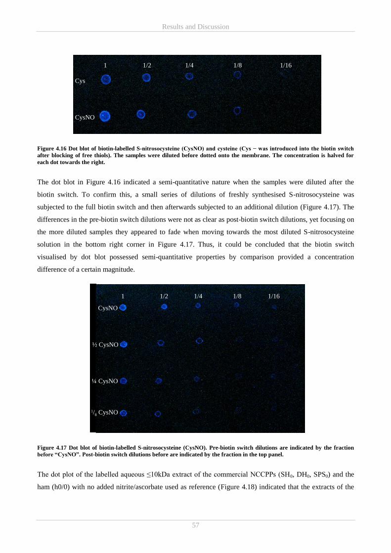
Results and Discussion
57
Figure 4.16 Dot blot of biotin-labelled S-nitrosocysteine (CysNO) and cysteine (Cys − was introduced into the biotin switch
after blocking of free thiols). The samples were diluted before dotted onto the membrane. The concentration is halved for
each dot towards the right.
The dot blot in Figure 4.16 indicated a semi-quantitative nature when the samples were diluted after the
biotin switch. To confirm this, a small series of dilutions of freshly synthesised S-nitrosocysteine was
subjected to the full biotin switch and then afterwards subjected to an additional dilution (Figure 4.17). The
differences in the pre-biotin switch dilutions were not as clear as post-biotin switch dilutions, yet focusing on
the more diluted samples they appeared to fade when moving towards the most diluted S-nitrosocysteine
solution in the bottom right corner in Figure 4.17. Thus, it could be concluded that the biotin switch
visualised by dot blot possessed semi-quantitative properties by comparison provided a concentration
difference of a certain magnitude.
Figure 4.17 Dot blot of biotin-labelled S-nitrosocysteine (CysNO). Pre-biotin switch dilutions are indicated by the fraction
before “CysNO”. Post-biotin switch dilutions before are indicated by the fraction in the top panel.
The dot plot of the labelled aqueous ≤10kDa extract of the commercial NCCPPs (SH0, DH0, SPS0) and the
ham (h0/0) with no added nitrite/ascorbate used as reference (Figure 4.18) indicated that the extracts of the
Cys
1 1/2 1/4 1/8 1/16
CysNO
CysNO
½ CysNO
¼ CysNO
1/8 CysNO
1 1/2 1/4 1/8 1/16
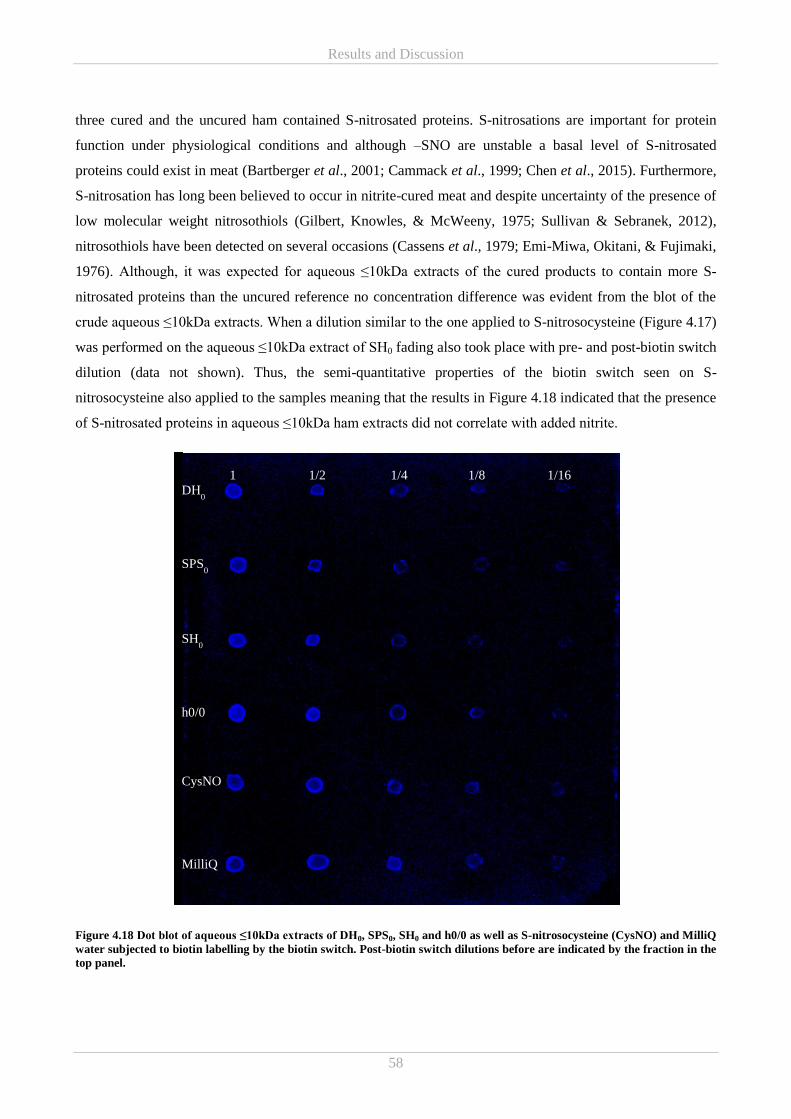
Results and Discussion
58
three cured and the uncured ham contained S-nitrosated proteins. S-nitrosations are important for protein
function under physiological conditions and although –SNO are unstable a basal level of S-nitrosated
proteins could exist in meat (Bartberger et al., 2001; Cammack et al., 1999; Chen et al., 2015). Furthermore,
S-nitrosation has long been believed to occur in nitrite-cured meat and despite uncertainty of the presence of
low molecular weight nitrosothiols (Gilbert, Knowles, & McWeeny, 1975; Sullivan & Sebranek, 2012),
nitrosothiols have been detected on several occasions (Cassens et al., 1979; Emi-Miwa, Okitani, & Fujimaki,
1976). Although, it was expected for aqueous ≤10kDa extracts of the cured products to contain more S-
nitrosated proteins than the uncured reference no concentration difference was evident from the blot of the
crude aqueous ≤10kDa extracts. When a dilution similar to the one applied to S-nitrosocysteine (Figure 4.17)
was performed on the aqueous ≤10kDa extract of SH0 fading also took place with pre- and post-biotin switch
dilution (data not shown). Thus, the semi-quantitative properties of the biotin switch seen on S-
nitrosocysteine also applied to the samples meaning that the results in Figure 4.18 indicated that the presence
of S-nitrosated proteins in aqueous ≤10kDa ham extracts did not correlate with added nitrite.
Figure 4.18 Dot blot of aqueous ≤10kDa extracts of DH0, SPS0, SH0 and h0/0 as well as S-nitrosocysteine (CysNO) and MilliQ
water subjected to biotin labelling by the biotin switch. Post-biotin switch dilutions before are indicated by the fraction in the
top panel.
1 1/2 1/4 1/8 1/16 DH
0
SPS0
SH0
h0/0
CysNO
MilliQ
Results and Discussion
59
However, in the dot blot in Figure 4.18 a clear signal was also detected from the MilliQ water used as
negative control in the biotin switch. Suspicions of a potential contamination of the MilliQ water was
dismissed as the same result occurred on repetition of the experiment with fresh MilliQ water as well as
dH2O (data not shown). In order to establish the origin of the water signal, blot development by means of a
fluorescein-conjugated streptavidin was also attempted, yet the signal remained. Thus, it was established that
the error was most likely associated with the labelling procedure and potentially due to insufficient removal
of excess biotin. This theory is slightly weakened as it is considered unlikely that water, with no binding
sites, would retain as much biotin as protein containing samples and thus, we would at least expect some
signal differences as was the case for samples and controls of different concentrations. Chen et al. (2015)
applied the same nitrosylation kit from Cayman Chemical on brain tissue and was able to produce a nice
differentiable dot blot. However, they reported nothing on the use of negative controls. Hence, this offers no
information on whether a signal in the negative control is a common problem. To my knowledge no other
data has been published on this problem.
In order to circumvent the challenges with the negative control an alternative S-nitrosation detection kit was
tested. The Pierce™ S-Nitrosylation Western Blot Kit from Thermo Fischer Scientific also applies the biotin
switch principles, yet labels with iodoTMT instead of biotin and detects by means of an antiTMT antibody
and a secondary goat anti-mouse IgG-HRP antibody. Furthermore, they recommend using reduced
glutathione and S-nitrosoglutathione as negative and positive control, respectively. For the exact procedure
see Chapter 3.3.3. However, the experiment was unsuccessful and resulted in a blank membrane for all
samples and control.
As it was not possible to produce a trustworthy result with the applied methods, it was considered too high-
risk to continue with an experimental strategy build on the biotin switch. However, even though it was not
possible to provide evidence for the presence of S-nitrosated proteins in aqueous ≤10kDa extracts of
commercial NCCPPs with the current experimental set up, it cannot be ruled out that such modified proteins
are in fact formed during nitrite-curing.
The SDS-PAGE of a NCCPP (meat/water homogenate) produced on much later occasion showed visible
bands in the water-soluble fraction (see Appendix A). The gel clearly emphasised that the NCCPPs
contained a very low concentration of water soluble proteins. Thus, the aqueous ≤10kDa fraction constituting
the foundation of this PhD study would be expected to contain even less protein capable of giving rise to an
electrophoretic protein pattern. As the result presented above did not give an impression of degree of sample
S-nitrosation, it is not possible to predict whether the limited amount of protein in the aqueous ≤10kDa
extracts would be sufficient for detecting S-nitrosated proteins by immunoblotting combined with the biotin
switch after electrophoretic separation.
Results and Discussion
60
Di Luccia et al. (2015) reported their myofibrillar protein extract to demonstrate an unexpected complex
electrophoretic protein pattern showing sarcoplasmic protein bands that co-migrated with several
myofibrillar components. The unusual composition of this fraction combined with the large amounts of
protein detected in the remaining fraction of meat/water homogenate (Appendix A) would make it highly
interesting to further investigate the fraction not soluble in water in regard to S-nitrosations.
4.3.2 Mass Spectrometry (MS)
As –SNO could not be detected by the biotin switch method an attempt to purify by streptavidin pulldown or
SNO-RAC was cancelled. Instead other approaches for characterisation of the antioxidative aqueous ≤10kDa
NCCPP extracts were considered.
A bottom-up proteomic technique commonly referred to as shotgun was applied to a SEC separated aqueous
≤10kDa extract sample in a collaboration between Aalborg and Aarhus University. This technique generally
comprises enzymatic digestion of a complex protein mixture followed by separation and identification of the
resulting peptides, using a combination of LC and MS (Zhang, Fonslow, Shan, Baek, & Yates, 2013).
Another, MS-based analysis strategy aiming at providing a broader picture and thus not confined to focusing
on protein was also attempted. Here, in addition to the crude aqueous ≤10kDa NCCPP extracts, a ≤3kDa
fractionation generated by UF (Vivaspin6 3000Da MWCO centrifugal concentrators − Sartorius) were also
analysed. These were then attempted analysed by direct flow injection and LC-MS on an ESI-QTOF
instrument. For details on the procedure see Chapter 3.3.4. Lastly, LC-MS were also applied in the search
for a specific modification (C-nitration) on proteins potentially occurring as a consequence of nitrite-curing
(Feng et al., 2016). Thus, a method for the detection of 3NT was developed in collaboration with University
of Copenhagen (Paper I).
Shotgun analysis (conducted by Aarhus and Aalborg University)
Shotgun analysis of selected SEC fractions (corresponding to SEC peak I, II, III and IV in Figure 4.19) of the
aqueous ≤10kDa SH0 extract revealed the extract to be a very complex mixture of peptides/proteins.
Although, the total protein content was low, the aqueous ≤10kDa extract contained peptides originating from
95 different proteins of pig origin across the tested SEC fractions (Appendix B). Even though, all the SEC
fractions contained peptides determined to be from the same six proteins, it was not the same peptides found
in all the SEC fractions.
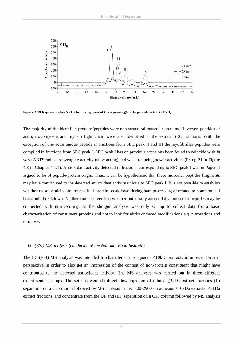
Results and Discussion
61
Figure 4.19 Representative SEC chromatograms of the aquoues ≤10kDa peptide extract of SH0.
The majority of the identified proteins/peptides were non-structural muscular proteins. However, peptides of
actin, tropomyosin and myosin light chain were also identified in the extract SEC fractions. With the
exception of one actin unique peptide in fractions from SEC peak II and III the myofibrillar peptides were
compiled in fractions from SEC peak I. SEC peak I has on previous occasions been found to coincide with in
vitro ABTS radical scavenging activity (slow acting) and weak reducing power activities (P4 og P1 in Figure
4.3 in Chapter 4.1.1). Antioxidant activity detected in fractions corresponding to SEC peak I was in Paper II
argued to be of peptide/protein origin. Thus, it can be hypothesised that these muscular peptides fragments
may have contributed to the detected antioxidant activity unique to SEC peak I. It is not possible to establish
whether these peptides are the result of protein breakdown during ham processing or related to common cell
household breakdown. Neither can it be verified whether potentially antioxidative muscular peptides may be
connected with nitrite-curing, as the shotgun analysis was only set up to collect data for a basic
characterisation of constituent proteins and not to look for nitrite-induced modifications e.g. nitrosations and
nitrations.
LC-(ESI)-MS analysis (conducted at the National Food Institute)
The LC-(ESI)-MS analysis was intended to characterise the aqueous ≤10kDa extracts in an even broader
perspective in order to also get an impression of the content of non-protein constituent that might have
contributed to the detected antioxidant activity. The MS analyses was carried out in three different
experimental set ups. The set ups were (I) direct flow injection of diluted ≤3kDa extract fractions (II)
separation on a C8 column followed by MS analysis in m/z 300-2900 on aqueous ≤10kDa extracts, ≤3kDa
extract fractions, and concentrate from the UF and (III) separation on a C18 column followed by MS analysis
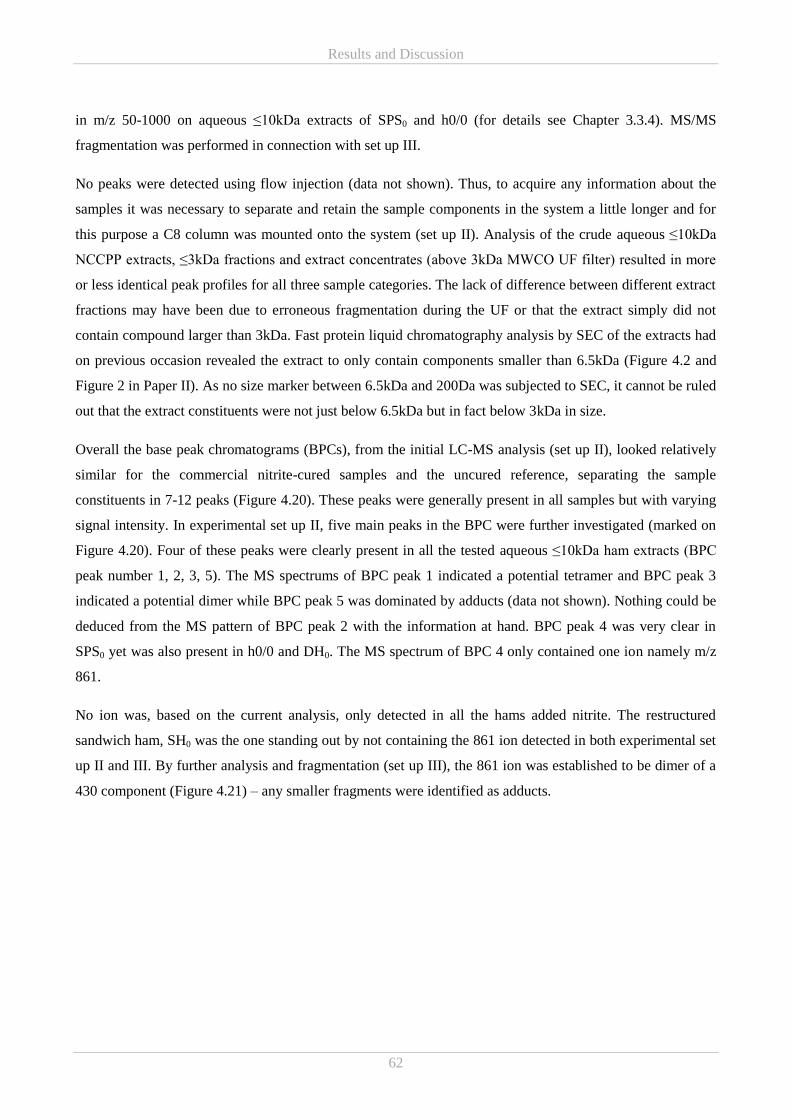
Results and Discussion
62
in m/z 50-1000 on aqueous ≤10kDa extracts of SPS0 and h0/0 (for details see Chapter 3.3.4). MS/MS
fragmentation was performed in connection with set up III.
No peaks were detected using flow injection (data not shown). Thus, to acquire any information about the
samples it was necessary to separate and retain the sample components in the system a little longer and for
this purpose a C8 column was mounted onto the system (set up II). Analysis of the crude aqueous ≤10kDa
NCCPP extracts, ≤3kDa fractions and extract concentrates (above 3kDa MWCO UF filter) resulted in more
or less identical peak profiles for all three sample categories. The lack of difference between different extract
fractions may have been due to erroneous fragmentation during the UF or that the extract simply did not
contain compound larger than 3kDa. Fast protein liquid chromatography analysis by SEC of the extracts had
on previous occasion revealed the extract to only contain components smaller than 6.5kDa (Figure 4.2 and
Figure 2 in Paper II). As no size marker between 6.5kDa and 200Da was subjected to SEC, it cannot be ruled
out that the extract constituents were not just below 6.5kDa but in fact below 3kDa in size.
Overall the base peak chromatograms (BPCs), from the initial LC-MS analysis (set up II), looked relatively
similar for the commercial nitrite-cured samples and the uncured reference, separating the sample
constituents in 7-12 peaks (Figure 4.20). These peaks were generally present in all samples but with varying
signal intensity. In experimental set up II, five main peaks in the BPC were further investigated (marked on
Figure 4.20). Four of these peaks were clearly present in all the tested aqueous ≤10kDa ham extracts (BPC
peak number 1, 2, 3, 5). The MS spectrums of BPC peak 1 indicated a potential tetramer and BPC peak 3
indicated a potential dimer while BPC peak 5 was dominated by adducts (data not shown). Nothing could be
deduced from the MS pattern of BPC peak 2 with the information at hand. BPC peak 4 was very clear in
SPS0 yet was also present in h0/0 and DH0. The MS spectrum of BPC 4 only contained one ion namely m/z
861.
No ion was, based on the current analysis, only detected in all the hams added nitrite. The restructured
sandwich ham, SH0 was the one standing out by not containing the 861 ion detected in both experimental set
up II and III. By further analysis and fragmentation (set up III), the 861 ion was established to be dimer of a
430 component (Figure 4.21) – any smaller fragments were identified as adducts.
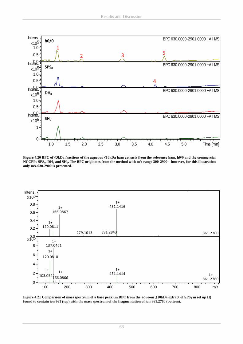
Results and Discussion
63
Figure 4.20 BPC of ≤3kDa fractions of the aqueous ≤10kDa ham extracts from the reference ham, h0/0 and the commercial
NCCPPs SPS0, DH0 and SH0. The BPC originates from the method with m/z range 300-2900 – however, for this illustration
only m/z 630-2900 is presented.
Figure 4.21 Comparison of mass spectrum of a base peak (in BPC from the aqueous ≤10kDa extract of SPS0 in set up II)
found to contain ion 861 (top) with the mass spectrum of the fragmentation of ion 861.2760 (bottom).
120.08111+
166.08671+
279.1013
431.14161+
861.2760
SHO FB_RA4_01_5678.d: +MS, 2.6min #317
137.04611+
431.14141+
861.27601+
SHO FB_RA4_01_5678.d: +bbCID MS, 20.0-40.0eV, 2.6min #3180.0
0.2
0.4
0.6
0.8
6x10
Intens.
0
2
4
6
8
5x10
100 200 300 400 500 600 700 800 m/z
-0 ppm low _RB5_01_5652.d: BPC 630.0000-2901.0000 +All MS
-SH0 low _RB4_01_5651.d: BPC 630.0000-2901.0000 +All MS
-SS0 low _RB3_01_5650.d: BPC 630.0000-2901.0000 +All MS
-T0 low _RB2_01_5649.d: BPC 630.0000-2901.0000 +All MS
0.0
0.5
1.0
5x10
Intens.
0.0
0.5
1.0
5x10
Intens.
0.0
0.5
1.0
5x10
Intens.
0
1
5x10
Intens.
1.0 1.5 2.0 2.5 3.0 3.5 4.0 4.5 5.0 Time [min]
4
3 2
1
h0/0
SPS0
DH0
SH0
5
1+
166.0866
1+
120.0810
1+
103.0544
391.2843
Results and Discussion
64
Despite the use of different analysis strategies including MS/MS fragmentation, different search engines and
a sample comparison taken notice of signal differences above a certain factor threshold, it was not possible to
identify many of extract compounds. The 431 ion were identified as either ononin or formononetin
(isoflavonoids potentially originating from feed - ononin from e.g. soybeans and formononetin from clover
or legumes in general (Budryn et al., 2018; Hloucalová et al., 2016)) with the most evidence pointing to
ononin. Another ion (m/z 177) discovered by the comparison of h0/0 and SPS0 (data from set up III) was
identified as ascorbic acid. As ascorbic acid (in the form of sodium ascorbate) was added to SPS0 in
connection with nitrite-curing and SPS0 was the commercial NCCPP found to contain the highest
concentration of residual ascorbic acid (Table 3.2 in Chapter 3.1.1), the current detection of its presence in
much higher concentrations in SPS0 vs h0/0 is considered correct.
The aqueous ≤10kDa extracts had previously been established to contain a wide variety of peptides yet the
current method was not developed to look selectively for peptides. In spite of this one noticeable ion, m/z
395.1558, was matched to a tri- and tetra-peptide. The tripeptides consisted of aspartic acid, phenylalanine
and asparagine in different order while the tetra peptides were composed of one aspartic acid, one
phenylalanine and two glycines, also in varying order. The presence of most of these amino acids in a
peptide have been associated with antioxidant properties (Carrasco-Castilla et al., 2012; Marcuse, 1962).
However, phenylalanine did not demonstrate high reducing power and ABTS radical scavenging activity
when tested as free amino acid in solution (see Chapter 4.5.2). Thus, it is difficult to establish the importance
of such. The remaining ions and their calculated gross formulas resulted in an immense list of potential
compound matches. These were generally complex structures often containing very condensed ring
structures. The result did not in any way point towards S-nitrosated peptides. However, a selection of these
hits contained buzz elements as pyrimidine, adenosine, purine as well as nitro in the systematic names
(Appendix C). Although, nitric oxide and related compounds have been found to interact with and modify
DNA, the resulting reaction products may to the best of my abilities be concluded to be far from the current
compound hits (Burney, Caulfield, Niles, Wishnok, & Tannenbaum, 1999). It has been established that
nitroso compounds from faecal matter after ingestion of nitrite-cured meat are capable of inducing in vitro
DNA damage (Joosen et al., 2009). Yet to my knowledge there are no published studies on NO-dependent
DNA modifications occurring post-mortem in nitrite-cured meat. However, if such modifications in fact do
occur, the reaction products would be expected to be similar to some of the ones found in vivo and in vitro.
Although, some information could be drawn from the MS analyses applied with the available resources,
further and more advanced investigation are required in order to fully characterise the aqueous ≤10kDa
extracts which is considered highly valuable for a full understanding of the antioxidative activities occurring
in NCCPPs
Results and Discussion
65
3-nitrotyrosine on LC- MS (conducted in collaboration with University of Copenhagen – Paper I)
As the previously applied narrow biotin switch and broader MS analyses described above were unsuccessful
in identifying nitrite related modification on proteins and peptides, an alternate approach was considered.
Sodium nitrite is, as has already been mentioned, generally regarded as an inhibitor of lipid oxidation.
However, its influence in regard to protein oxidation has not revealed a significant antioxidant effects.
In addition to ROS, RNS may also be present in meat systems subjected to nitrite-curing (Skibsted, 2011),
why initiation of both oxidative and nitrosative reactions may be expected to occur. However, the
manifestation of such reactions and the connection between oxidative and nitrosative reactions as well as
their consequences on cured meat products are not fully elucidated. Exposure of tyrosine to RNS such as
nitrogen dioxide and in particular peroxynitrite, leads to the formation of 3NT (Villaverde, Parra, et al.,
2014; Vossen & De Smet, 2015) which has long been used as a marker for nitrosative stress in vivo. Yet,
3NT has also been found in meat products (Feng et al., 2016; Vossen & De Smet, 2015) and has furthermore
been found to increase with nitrite addition (Villaverde, Morcuende, et al., 2014). Due to the potential
interlink between oxidative and nitrosative reactions, 3NT has been suggested as a potential marker for
protein oxidation in raw and cooked nitrite treated meat (Feng et al., 2015). Thus, investigating 3NT may
give an impression of the role of nitrite-induced peptide modifications in the redox state from another angle
than as active antioxidant compounds
In the current investigation of the presences of 3NT in the three commercial NCCPPs (SH0, DH0, SPS0) and
the reference with no added nitrite/ascorbate (h0/0), 3NT was found in neither the ≤10kDa fraction nor the
meat. If 3NT can in fact be considered a proper marker for protein oxidation the current results would
indicate limited protein oxidation potentially due to the presence of strong antioxidant compounds.
With the applied method detecting 3NT standard solution down to 0.5ng/mL it is odd that no 3NT was
detected at all, considering that a basal level of 3NT, most likely originating from physiological nitrosative
stress, is usually considered to be present in any muscle-based food (Villaverde, Parra, et al., 2014). The
absence of detectable 3NT may be partly explained by the addition of ascorbate as well as heat treatment as
both have been found to cause a decrease in 3NT (Feng et al., 2015; Villaverde, Parra, et al., 2014).
3NT spiking of the aqueous ≤10kDa extracts led to a recovery of approximately 80%. However, no 3NT was
recovered from spiking of the meat samples. This could indicate that 3NT formation may not just be
inhibited in the complete NCCPPs, it might even be degraded, and that the degradation reaction(s) only
occurs in the presences of unknown compounds >10kDa. Crow & Beckman (1995) pointed out that nitro-
tyrosine may be reduce to amino-tyrosine under the conditions of an acid hydrolysis and furthermore
indicated that such a reduction may be caused by thiol reductants. They also found that the conversion was
generally higher when nitro-tyrosine was added to a complex protein mixture prior to hydrolysis indicating
the mixture to contain compounds inducing the reduction (Crow & Beckman, 1995). Thus, it can be
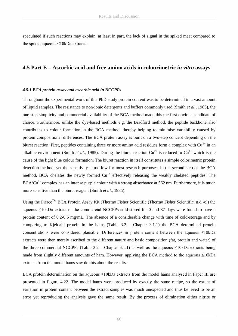
Results and Discussion
66
speculated if such reactions may explain, at least in part, the lack of signal in the spiked meat compared to
the spiked aqueous ≤10kDa extracts.
4.5 Part E – Ascorbic acid and free amino acids in colourimetric in vitro assays
4.5.1 BCA protein assay and ascorbic acid in NCCPPs
Throughout the experimental work of this PhD study protein content was to be determined in a vast amount
of liquid samples. The resistance to non-ionic detergents and buffers commonly used (Smith et al., 1985), the
one-step simplicity and commercial availability of the BCA method made this the first obvious candidate of
choice. Furthermore, unlike the dye-based methods e.g. the Bradford method, the peptide backbone also
contributes to colour formation in the BCA method, thereby helping to minimise variability caused by
protein compositional differences. The BCA protein assay is built on a two-step concept depending on the
biuret reaction. First, peptides containing three or more amino acid residues form a complex with Cu2+
in an
alkaline environment (Smith et al., 1985). During the biuret reaction Cu2+
is reduced to Cu1+
which is the
cause of the light blue colour formation. The biuret reaction in itself constitutes a simple colorimetric protein
detection method, yet the sensitivity is too low for most research purposes. In the second step of the BCA
method, BCA chelates the newly formed Cu1+
effectively releasing the weakly chelated peptides. The
BCA/Cu1+
complex has an intense purple colour with a strong absorbance at 562 nm. Furthermore, it is much
more sensitive than the biuret reagent (Smith et al., 1985).
Using the PierceTM
BCA Protein Assay Kit (Thermo Fisher Scientific (Thermo Fisher Scientific, n.d.-c)) the
aqueous ≤10kDa extract of the commercial NCCPPs cold-stored for 0 and 37 days were found to have a
protein content of 0.2-0.6 mg/mL. The absence of a considerable change with time of cold-storage and by
comparing to Kjeldahl protein in the hams (Table 3.2 – Chapter 3.1.1) the BCA determined protein
concentrations were considered plausible. Differences in protein content between the aqueous ≤10kDa
extracts were then merely ascribed to the different nature and basic composition (fat, protein and water) of
the three commercial NCCPPs (Table 3.2 – Chapter 3.1.1) as well as the aqueous ≤10kDa extracts being
made from slightly different amounts of ham. However, applying the BCA method to the aqueous ≤10kDa
extracts from the model hams saw doubts about the results.
BCA protein determination on the aqueous ≤10kDa extracts from the model hams analysed in Paper III are
presented in Figure 4.22. The model hams were produced by exactly the same recipe, so the extent of
variation in protein content between the extract samples was much unexpected and thus believed to be an
error yet reproducing the analysis gave the same result. By the process of elimination either nitrite or
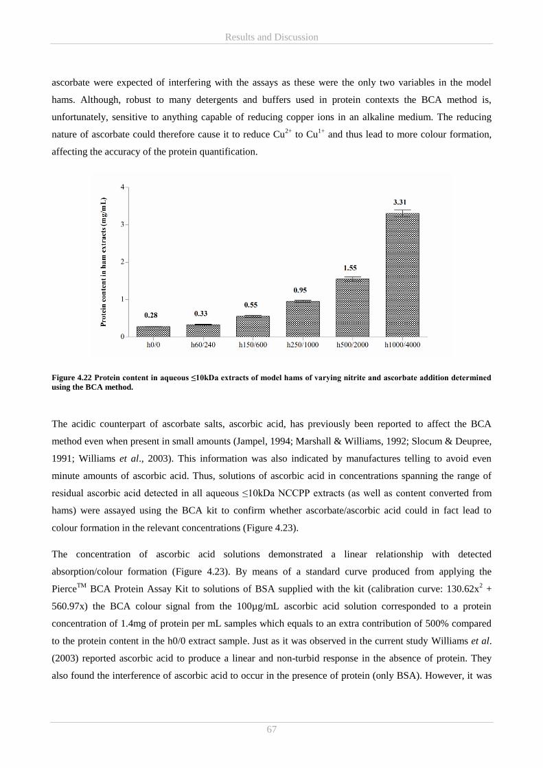
Results and Discussion
67
ascorbate were expected of interfering with the assays as these were the only two variables in the model
hams. Although, robust to many detergents and buffers used in protein contexts the BCA method is,
unfortunately, sensitive to anything capable of reducing copper ions in an alkaline medium. The reducing
nature of ascorbate could therefore cause it to reduce Cu2+
to Cu1+
and thus lead to more colour formation,
affecting the accuracy of the protein quantification.
Figure 4.22 Protein content in aqueous ≤10kDa extracts of model hams of varying nitrite and ascorbate addition determined
using the BCA method.
The acidic counterpart of ascorbate salts, ascorbic acid, has previously been reported to affect the BCA
method even when present in small amounts (Jampel, 1994; Marshall & Williams, 1992; Slocum & Deupree,
1991; Williams et al., 2003). This information was also indicated by manufactures telling to avoid even
minute amounts of ascorbic acid. Thus, solutions of ascorbic acid in concentrations spanning the range of
residual ascorbic acid detected in all aqueous ≤10kDa NCCPP extracts (as well as content converted from
hams) were assayed using the BCA kit to confirm whether ascorbate/ascorbic acid could in fact lead to
colour formation in the relevant concentrations (Figure 4.23).
The concentration of ascorbic acid solutions demonstrated a linear relationship with detected
absorption/colour formation (Figure 4.23). By means of a standard curve produced from applying the
PierceTM
BCA Protein Assay Kit to solutions of BSA supplied with the kit (calibration curve: 130.62x2 +
560.97x) the BCA colour signal from the 100µg/mL ascorbic acid solution corresponded to a protein
concentration of 1.4mg of protein per mL samples which equals to an extra contribution of 500% compared
to the protein content in the h0/0 extract sample. Just as it was observed in the current study Williams et al.
(2003) reported ascorbic acid to produce a linear and non-turbid response in the absence of protein. They
also found the interference of ascorbic acid to occur in the presence of protein (only BSA). However, it was
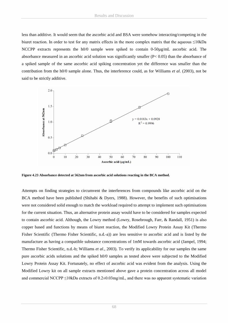
Results and Discussion
68
less than additive. It would seem that the ascorbic acid and BSA were somehow interacting/competing in the
biuret reaction. In order to test for any matrix effects in the more complex matrix that the aqueous ≤10kDa
NCCPP extracts represents the h0/0 sample were spiked to contain 0-50µg/mL ascorbic acid. The
absorbance measured in an ascorbic acid solution was significantly smaller (P< 0.05) than the absorbance of
a spiked sample of the same ascorbic acid spiking concentration yet the difference was smaller than the
contribution from the h0/0 sample alone. Thus, the interference could, as for Williams et al. (2003), not be
said to be strictly additive.
Figure 4.23 Absorbance detected at 562nm from ascorbic acid solutions reacting in the BCA method.
Attempts on finding strategies to circumvent the interferences from compounds like ascorbic acid on the
BCA method have been published (Shihabi & Dyers, 1988). However, the benefits of such optimisations
were not considered solid enough to match the workload required to attempt to implement such optimisations
for the current situation. Thus, an alternative protein assay would have to be considered for samples expected
to contain ascorbic acid. Although, the Lowry method (Lowry, Rosebrough, Farr, & Randall, 1951) is also
copper based and functions by means of biuret reaction, the Modified Lowry Protein Assay Kit (Thermo
Fisher Scientific (Thermo Fisher Scientific, n.d.-a)) are less sensitive to ascorbic acid and is listed by the
manufacture as having a compatible substance concentrations of 1mM towards ascorbic acid (Jampel, 1994;
Thermo Fisher Scientific, n.d.-b; Williams et al., 2003). To verify its applicability for our samples the same
pure ascorbic acids solutions and the spiked h0/0 samples as tested above were subjected to the Modified
Lowry Protein Assay Kit. Fortunately, no effect of ascorbic acid was evident from the analysis. Using the
Modified Lowry kit on all sample extracts mentioned above gave a protein concentration across all model
and commercial NCCPP ≤10kDa extracts of 0.2±0.03mg/mL, and there was no apparent systematic variation
Results and Discussion
69
related to added ascorbic acid. This level was also consistent with the levels found in the aqueous ≤10kDa
extract of h0/0 when using the BCA kit.
Though, the BCA method was deemed non-compatible for determining protein content in aqueous NCCPP
extracts high in ascorbic acid, the linearity of the colour development inspired the idea, that BCA could be
used to estimate ascorbic acid in this type of samples. A BCA-based method for determining ascorbic acid in
aqueous humor was developed by Shichi, Page, Sahouri, & Shin (1997). The referred method builds on two
key principles (I) eliminating protein-induced absorbance signal by TCA precipitation prior to BCA reaction
and (II) determining the ascorbic acid content by the absorbance difference between a sample where ascorbic
acid had been oxidised and an untreated sample. In order to apply this it would be necessary to first examine
if a TCA precipitation would be sufficient to rid the aqueous ≤10kDa NCCPP extracts of all protein/peptide-
induced signal that might interfere. For example, very small peptides will not precipitate but may contribute
to the BCA colour formation. Furthermore, the amino acids cysteine, cystine, tryptophan and tyrosine
(Wiechelman, Braun, & Fitzpatrick, 1988) are together with the peptide backbone greatly contributing to the
BCA colour formation. Hence, these four amino acids as free amino acids, if present, will not precipitate and
may thus contribute to the signal. Shichi et al. (1997) rejects the importance of other known BCA method
interferences (uric acid, glucose and gluthathione) by proving that they are not oxidised like ascorbic acid
under alkaline treatment. Any other reducing compounds present in the NCCPP extracts would at least have
to be tested in the same manor. After these minor investigations this method could well pose a good starting
point for the development of a simple ascorbic acid detection method for NCCPP extracts with lower
sensitivity but higher throughput compared to the HPLC method described in Chapter 3.2.5.
4.5.2 In vitro reducing power and ABTS radical scavenging antioxidant activity of free amino acids
A contribution from free amino acids (in addition to small peptides and non-peptidic compounds) to the
antioxidant activities measured in the low molecular weight aqueous NCCPP extracts was considered
reasonable in the current situation. Thus, it was desirable to be able to evaluate the contribution from free
amino acids in the applied assays (before and after SEC fractionation). Accordingly, 10 amino acids, either
previously reported to possess antioxidant properties (as free or embedded in peptides) or present in high
concentrations in the extracts, were selected for analysis. The 10 selected were histidine, methionine,
phenylalanine, tryptophan, cysteine, tyrosine, proline, glutamic acid, glycine, and alanine. The
concentrations of the free amino acid under investigation were adapted to the content determined in the
aqueous ≤10kDa NCCPP extracts (Paper II – Figure 4). Higher concentrations, up to 1mg/mL (the stock
used for making the remaining concentrations), were also tested for three reasons (I) in the matter at hand to
verify if a potentially absent antioxidant activity signal could merely be due to low concentration but also (II)
Results and Discussion
70
to evaluate if the concentration dependency on the interconversion between anti and pro-oxidant activities
previously reported (Marcuse, 1962) is relevant in the current experimental set up and (III) on a general basis
to investigate whether the antioxidant activities of free amino acids previously reported in the literature (by
means of many different methods) could be confirmed using the current colourimetric in vitro assays.
ABTS radical scavenging was measure after incubating for 1, 8 and 31min at room temperature to be able to
follow any kinetic differences between the different amino acids and solvents (Figure 4.24). Solutions of
cysteine, tryptophan and tyrosine showed the strongest ABTS radical scavenging activity and at 0.05mg/mL
these amino acids had reached the assay maximal capacity of approximately 93%. This maximum was
reached already within the first minute of incubation. At the lowest tested concentrations the decreasing
order of ABTS radical scavenging activity was cysteine (0.005mg/mL) > tryptophan (0.002mg/mL) >
tyrosine (0.002mg/mL). The magnitude of the activity increased with incubation time, but the internal order
remained. Furthermore, at this low concentration the activity was higher when the amino acid was dissolved
in water, compared to ammonium acetate. For cysteine and tryptophan the activity detected in the two
solvents was significantly different at all time points. The tyrosine solutions were turbid due to improper
solubilisation in the 1mg/mL stock solution. Any precipitate would result in an increased absorbance leading
to a decrease in the calculated colour reduction (ABTS radical scavenging activity). However, the turbidity
became less pronounced as the stock was diluted to the tested concentrations. Furthermore, as the strong
tyrosine solutions were capable of a full colour reduction (assay max) comparable to cysteine and
tryptophan, the turbidity is not considered problematic for the overall conclusion of tyrosine exhibiting
ABTS radical scavenging activity.
Antioxidant activity from solutions of tryptophan was established decades ago in systems of linoleic acid and
its methyl esters (Marcuse, 1962). Over time solution of tryptophan as well as tyrosine were also more
specifically found to act as radical scavengers in different in vitro assays e.g. ORAC-FL and an ABTS-based
assay different from the one applied in the current work (Hernández-Ledesma, Dávalos, Bartolomé, &
Amigo, 2005; Meucci & Mele, 1997). More recently, free tryptophan and tyrosine isolated from extracts of
egg yolk was also found to act as strong ABTS radical scavengers (Nimalaratne, Lopes-Lutz, Schieber, &
Wu, 2011). This scavenging ability of the aromatic tyrosine and tryptophan is believed to be connected to the
phenolic and indolylic groups, respectively, enabling these amino acids to act as proton/hydrogen donors to
electron deficient radicals and efficiently scavenge them. Also the aromatic structure of tyrosine is
considered a major contributor to its antioxidant activity (Carrasco-Castilla et al., 2012; Hernández-Ledesma
et al., 2005). The antioxidant capacity of tyrosine and tryptophan are most often reported to be very similar
as was also evident in the current results (Nimalaratne et al., 2011; Sharp, Becker, & Hettich, 2004).
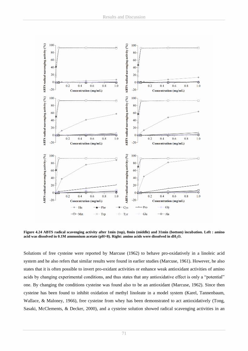
Results and Discussion
71
Figure 4.24 ABTS radical scavenging activity after 1min (top), 8min (middle) and 31min (bottom) incubation. Left : amino
acid was dissolved in 0.1M ammonium acetate (pH=8). Right: amino acids were dissolved in dH2O.
Solutions of free cysteine were reported by Marcuse (1962) to behave pro-oxidatively in a linoleic acid
system and he also refers that similar results were found in earlier studies (Marcuse, 1961). However, he also
states that it is often possible to invert pro-oxidant activities or enhance weak antioxidant activities of amino
acids by changing experimental conditions, and thus states that any antioxidative effect is only a “potential”
one. By changing the conditions cysteine was found also to be an antioxidant (Marcuse, 1962). Since then
cysteine has been found to inhibit oxidation of methyl linoleate in a model system (Karel, Tannenbaum,
Wallace, & Maloney, 1966), free cysteine from whey has been demonstrated to act antioxidatively (Tong,
Sasaki, McClements, & Decker, 2000), and a cysteine solution showed radical scavenging activities in an
Results and Discussion
72
ORAC-FL assay (Hernández-Ledesma et al., 2005). The potential for cysteine to act as a radical scavenger
lies in sulfhydryl-group (Meucci & Mele, 1997). Furthermore, Meucci & Mele (1997) found cysteine to
display antioxidant abilities at very low concentration in an ABTS-based assay, and that cysteine
demonstrated an inhibitory pattern quite similar to that of non-protein antioxidant like ascorbic acid and
trolox. In the current study cysteine showed similar behaviour to trolox, yet trolox demonstrated slightly
higher activity at 0.005mg/mL (data not shown). This could be due to solvent differences.
Histidine also demonstrated ABTS radical scavenging activity, however, the kinetic behaviour was
somewhat different as it reacted much slower but clearly increased during the incubation (Figure 4.24).
Within the tested timeframe (31min) the most concentrated solution approached the same activity (92% in
water and 88% in ammonium acetate –almost corresponding to assays max capacity) as cysteine, tryptophan
and tyrosine. Histidine is well known for its multifaceted antioxidant activities and like cysteine, tryptophan
and tyrosine the origin of histidine’s antioxidative properties lies in its sidechain structure as the imidazole
group enables histidine to scavenge radicals by proton/hydrogen donation (Chan, Decker, Lee, & Butterfield,
1994). Meucci & Mele (1997) did not see an effect of monomeric histidine on the ABTS radical, yet several
other studies have found free histidine to inhibit lipid oxidation and scavenge free radicals (Hernández-
Ledesma et al., 2005; Karel et al., 1966; Marcuse, 1962). This small inconsistency might be related to the
fact that histidine can act both as a hydrogen donor as well as an acceptor at physiological pH but also has a
tendency to invert to a pro-oxidant at high concentrations (Udenigwe & Aluko, 2011). Also for histidine the
water-based solution exhibited higher ABTS radical scavenging activity compared to the ammonium acetate
solution at the same concentration and time of incubation (significantly different for all time point at
concentration ≥0.1mg/mL, P<0.05).
The antioxidant activity of the individual amino acids is affected by a number of factors. One important
factor is the pH of the reaction environment as it may affect the optimum for the antioxidant activity of the
amino acids. This is not considered to be a problem as the assays applied to the sample were carried out in
exactly the same way as the assays applied on the amino acid solutions needed for the comparisons.
However, antioxidant capacity was found to decrease with decreasing pH in a linoleic acid model system
(Marcuse, 1962). If this is also the case in the aqueous and ammonium acetate environments, it would be
expected that the assays carried out in ammonium acetate (pH=8) would show higher antioxidant activity
than the same solution in assays carried out in dH2O. As this was not the case for any of the amino acids
mentioned above other factors may also be affecting the systems. In conclusion attention must be payed to
solvent and buffer differences when comparing to previous findings.
As for many antioxidants the concentration of the amino acids are essential for their degree of antioxidant
activity and at very high concentrations it might even turn into pro-oxidative activities (Marcuse, 1962).
From the current results it is believed that the highly active tryptophan, tyrosine, cysteine and histidine
Results and Discussion
73
contributed additively to the radical scavenging activities detected in the crude and SEC fractionated aqueous
≤10kDa NCCPP extracts and that the concentration of these as free amino acids is below the concentration
for conversion to pro-oxidative reactivity.
Among the remaining tested amino acid solutions only proline (water and ammonium acetate), glycine
(ammonium acetate) and methionine (water) in 1mg/mL demonstrated ABTS radical scavenging activity
above 5% after 31min incubation. Proline and methionine have previous found to scavenge free radicals
(Damgaard et al., 2015; Hernández-Ledesma et al., 2005). However, assuming a similar content of the
individual amino acids in aqueous ≤10kDa extracts of any NCCPP these amino acids are unlikely to
contribute to the overall radical scavenging antioxidant properties of such a product – at least on a short term
basis.
Of the last three tested amino acids showing <5% ABTS radical scavenging activity, phenylalanine was
selected for testing, due to previous reports of strong antioxidant properties (Carrasco-Castilla et al., 2012;
Di Bernardini et al., 2011) among others playing a positive role in the scavenging of H2O2 likely attributed to
by the aromatic ring (Udenigwe & Aluko, 2011) and for displaying in vitro radical scavenging activity
(ORAC-FL) in free form (Hernández-Ledesma et al., 2005). The unexpected absent result could be assay-
related, as Meucci & Mele (1997) also did not find free phenylalanine capable of inhibiting the ABTS
radical. Glutamic acid was the most dominant in the amino acid profile of the commercial NCCPPs (Paper I
– Figure 3), and it is also frequently mentioned in connection with antioxidant peptides but mainly in regards
to metal chelation (Saiga et al., 2003; Zhu et al., 2013). Glutamic acid may, however, donate its acidic
hydrogen near neutral pH and thereby potentially be able to scavenge free radicals.
Like for ABTS radical scavenging solutions of cysteine, tryptophan and tyrosine were also the ones standing
out in the reducing power assay (Figure 4.25). Cysteine demonstrated markedly higher reducing power
capacities than the other two amino acids in both water and ammonium acetate, however, OD700 was only
significantly different between the two solvents for tryptophan. The strong reducing power of cysteine is
again associated with the sulfhydryl-group (Udenigwe & Aluko, 2011). The minute reducing power activity
displayed by tyrosine is most likely to be caused by the turbidity of the solution, mentioned above. Based on
these results it is considered unlikely that any of the tested amino acids other than cysteine would contribute
noticeably to the overall in vitro reducing power, and that the contribution from free cysteine might be
limited due to low content in the aqueous ≤10kDa NCCPP extracts.
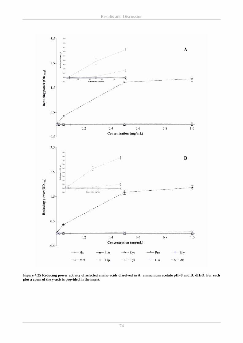
Results and Discussion
74
Figure 4.25 Reducing power activity of selected amino acids dissolved in A: ammonium acetate pH=8 and B: dH2O. For each
plot a zoom of the y-axis is provided in the insert.
Results and Discussion
75
In conclusion, it is believed that the tested free amino acids demonstrating ABTS radical scavenging and
reducing power activity contribute to the activities measured in the aqueous ≤10kDa NCCPP extracts and in
the SEC separated extract fractions although to a lesser extent due to dilution. However, the signals detected
in the current experiments do not add up to the full antioxidant capacities measured in the aqueous ≤10kDa
extracts. Thus, other compounds such as modified amino acids e.g. S-nitrosocysteine (Kanner, 1979),
peptides e.g. carnosine (Gil-Agustí et al., 2008), nitrite and ascorbate must also be contributing to varying
extent.

Results and Discussion
76
Hvid tekst
Summarising discussion and conclusion
77
Chapter 5
Summarising discussion and conclusion 5.
As the results of the current PhD work has been thoroughly discussed during their presentation in the
preceding parts of Chapter 4 and in the Paper I-III, the following aims to summarise the main findings by
addressing the interface between the studies mainly focusing on the three working hypothesis presented in
the introduction in Chapter 1.
Hypothesis 1 - During the production of cooked pork product with nitrite, antimicrobial and antioxidant
compounds are formed
Overall the current PhD originally set out to examine antimicrobial and antioxidative activities in NCCPPs,
and in order to do so in vitro measurements of these activities was carried out. Independently of differences
in the type and processing condition, including type and amount of additives, of the tested NCCPPs no
growth inhibitory activities were detected. Although, the lack of detected antimicrobial activities stands in
contrast to the literature emphasising a higher microbiological stability in nitrite-cured meat products,
(Myers et al., 2016; Osterbauer et al., 2017; Sindelar & Milkowski, 2012) the vast majority of the published
evidence seems to be based on inoculation studies or model studies of nitrite or similar antimicrobial agents
added to the growth medium rather than antimicrobial activity tests on isolated meat fractions. Thus, it may
be the case that antimicrobial activities in NCCPPs are related to the curing process but the active
compounds might somehow be associated with the meat matrix and thus could not be measured in the
aqueous extracts. Therefore, it must be stressed that although the two tested aqueous NCCPP fractions
(supernatant of meat/water homogenate and aqueous ≤10kDa fraction) demonstrated no antimicrobial
activities, a follow-up inoculation study is required in order to fully establish whether the result is in fact due
to a complete lack of antimicrobial activities in all the tested NCCPPs. Alternatively, as pointed out in
Chapter 4.3.1, the tested NCCPP fractions could contain antimicrobial compounds but they were merely
tested in too low concentrations to generate a response. In contrast to antimicrobial activities, in vitro
antioxidant activity was detected in the aqueous ≤10kDa extracts of all the different types of NCCPPs under
investigation. On a theoretical basis it is already known that the addition of nitrite and ascorbate in the
production of cooked pork products will lead to the presence of antioxidative species. Regarding NCCPPs
both heating in general and heating at higher temperature will lead to a decrease in residual inorganic nitrite
(Honikel, 2008), which could mean that more of the antioxidatively active NO is formed. The importance of
ascorbate was very clear from the current investigations, however, whether antioxidant activity correlated
with added or residual ascorbate varied between the sample categories, sample treatments (SEC) and storage.
Summarising discussion and conclusion
78
Furthermore, the interplay between added ascorbate and added nitrite seem to greatly affect the detected in
vitro activity. This could be due to mutual reactions leading to formation of known as well as unknown
reactions products of increased or decreased antioxidative properties, compared to the individual reactants.
When focusing on shelf-life extending additives it would appear that the circumstances under which
antioxidative activities take place in NCCPPs play a vital role for the type of mechanisms and potentially
also type of compounds exerting the antioxidative activities.
Decades ago it was reported that nitrite reacts readily with peptides (Cassens et al., 1979). Consequently, it
was initially strongly hypothesised that compounds with strong shelf-life extending properties formed during
production of cooked pork products with nitrite could very well be reaction products of protein origin. Yet,
the conducted experiments e.g. LC-MS and biotin switch could not aid in confirming the importance of
nitrite-induced modified peptides for the detected antioxidant activities. However, as the biotin switch did
not succeed in producing credible results (signal in negative control) and the LC-MS methods were not
optimised to search selectively for nitrosations and nitrations, S-nitosocysteine containing peptides formed in
reactions between nitrite and meat proteins and previously reported to be antioxidative (Kanner, 1979) may
have been formed and contributed to the in vitro antioxidant properties of the aqueous ≤10kDa extracts of the
tested NCCPPs. In addition to potentially containing active modified peptides, residual additives and active
species hereof the extracts were generally found to constitute very complex mixture of peptides/proteins.
Through the characterisation of the extracts it became clear that the in vitro antioxidant activities had to
originate from a highly diverse selection of compounds, and that small peptides and certain amino acids e.g.
tyrosine, tryptophan, histidine, proline and cysteine may have been of great importance for the in vitro
antioxidative properties measured in the tested NCCPPs. So in conclusion it can be said that antioxidant
activity in ≤10kDa NCCPP extracts was due to strong synergetic effects of protein and non-protein, anti- and
pro-oxidant compounds to a wide extent originating from processing.
Hypothesis 2 - There are optimal processing conditions for the formation of these antimicrobial and
antioxidant compounds
With active compounds generated during processing, attention should be paid to determining the optimal
condition for their formation. As lipid oxidation can be perceived as a self-perpetuating process, initiation of
oxidative reactions by any mechanism (e.g. metals or free radicals) may generate compounds that can initiate
new oxidative reactions or contribute to the elongation of ongoing reactions. Thus, all types of antioxidative
actions are of vital importance for the oxidative balance of the NCCPP and consequently the evaluation of
the optimal processing conditions for generation of an optimal antioxidative response should be made across
all three applied in vitro antioxidative assays. Although, antioxidant activity based on chelating of iron (and
other metals) is of general importance, such activity was not found to be related to curing. Thus, the iron
Summarising discussion and conclusion
79
chelating assay was not included in the estimation of optimal processing conditions concerning addition of
the two curing agents. As added nitrite/ascorbate to the model products correlated positive with normalised
ABTS radical scavenging activity and initial reducing power activity, but not with reducing power of the
crude aqueous ≤10kDa extracts, addition of ≤150ppm nitrite and ≤600ppm ascorbate constituted the test-
level, at which the best overall antioxidative response was obtained. In addition to indicating an optimal
curing agent addition level the results also show that in order to obtain a high degree of antioxidative
reactions without converting to pro-oxidative activities, the nitrite to ascorbate ratio should be considered
when producing NCCPPs. Solely adding ascorbate (on the expense of conventional nitrite-related effects on
colour and taste) could perhaps be a solution to achieve high antioxidative activities, as the reducing power
dilution curve for the aqueous ≤10kDa extracts from the sausage only added high levels of ascorbic acid
(s0/2000) did not deflect in the same manor as the samples added high levels of ascorbate as well as nitrite.
Ascorbate/ascorbic acid is considered a non-hazardous (EFSA, 2015a; FDA, 2018) additive and could thus
be added relatively unrestrained to NCCPPs (European Union, 2011). However, the literature states the
antioxidative properties may turn pro-oxidative under certain conditions (Haak, Raes, & De Smet, 2009;
Villaverde, Parra, et al., 2014). Furthermore, the antimicrobial properties accomplished with the addition of
nitrite – although not demonstrated in the current study – may likely be severely altered with this strategy.
For example ascorbic acid has been found to possess antimicrobial properties against a variety of bacteria
(Cross et al., 2003; Giannuzzi & Zaritzky, 1996; Juven & Kanner, 1986; Tabak, Armon, Rosenblat, Stermer,
& Neeman, 2003; Vilchèze, Hartman, Weinrick, & Jacobs, 2013). However, to the best of my knowledge no
newer research on anti-botulinal activities in meat as a result of ascorbate/ascorbic acid addition without
nitrite are available. Less recent studies reported of some reduction in spore germination in the presence of
ascorbic acid when tested in medium (Eller, Edwards, & Wynne, 1968), while no considerably different
effect on botulinal toxin formation was evident on ascorbate addition compared to combined ascorbate/nitrite
addition or nitrite alone to wiener sausages (Bowen, Cerveny, & Deibel, 1974). Thus, the suggested strategy
may be inferior to nitrite treatments in regard to inhibiting C. botulinum.
Once new or optimised methods for the detection of potential antimicrobial activities has been established, as
suggested in Chapter 4.3.1, it would be necessary to do a re-evaluation to set new levels required for the best
compromise between antioxidative and antimicrobial activities. Lastly, it must be kept in mind that matrix
effects may affect the oxidative (and microbiological) activities in the entire NCCPPs potentially changing
the suggestion for optimal processing conditions, in regard to curing agents, presented above.
Other aspects of processing than addition of curing agents were also found to affect the active antioxidant
aqueous ≤10kDa NCCPP extracts. When comparing the model hams and sausages a clear effect of
processing in the intensity of 215nm SEC peaks that aligned with the active antioxidant fraction-peaks was
evident. Since processing appeared to influence the protein portion of the SEC profile, it may be speculated
that different antioxidative peptides may be formed as a result of different processing methods. As the
Summarising discussion and conclusion
80
current results show that certain types of interventions associated with processing affect the measured in
vitro antioxidant activity in the aqueous ≤10kDa NCCPP extract, the next appropriate step would be to also
investigate the effect of other processing parameters such as cooking time and temperature.
Achieving a certain centre temperature as well as holding time at this temperature, is key to obtaining a
microbial safe product, but this will naturally also have other consequences on e.g. residual nitrite and
ascorbate. The supply of energy occurring during heat treatment may also act as an activation-energy for the
formation of so far unknown reaction products (e.g. between nitrite and meat proteins) with antioxidative and
antimicrobial properties. As different reactions, may require different activation energies in order to proceed,
the mixture of reaction products in a multi substrate system such as meat will vary with temperature. Also
the longer the heat is applied, the longer the heat/energy dependent reactions may proceed. Additionally
heating leads to an increase in lipid oxidation (Boles, 2010). Ergo, the process of determining optimal
processing conditions is not merely a matter of finding the optimal balance of antioxidative and antimicrobial
compounds but also to keep the level of “oxidation initiator”, e.g. free radicals, at a level where the newly
formed or already existing antioxidative compounds can keep them at bay (Baardseth, Bjerke, Aaby, &
Mielnik, 2005).
Hypothesis 3 - There are optimal storage conditions to retain the activity of antimicrobial and antioxidant
compounds in nitrite cured pork products
It was the ambition to investigate and establish the optimal storage conditions for retaining the activity of
identified antimicrobial and antioxidant compounds. The effects of storage temperature, as well as access to
oxidation initiators as light and oxygen were to be tested. So far, it was established that cold storage over
time resulted in an increase in normalised ABTS and partly in initial reducing power in the aqueous ≤10kDa
NCCPP extracts. Furthermore, the results showed a storage time (and additive concentration) dependent
formation of unidentified compounds contributing to reducing power and iron chelating activity. However,
during this time of cold storage oxidative species in the entire pork meat products will also have increased
hampering the potential of any new antioxidants requiring time to form. Combined this may still be a part of
the reason for the prolonged shelf-life of nitrite cured products, yet it does not provide evidence for any
recommendations for optimal storage conditions for retaining antimicrobial and antioxidant activity in
NCCPPs. Furthermore, regular refrigerated storage (short time for the ≤10kDa extracts of the lower level of
nitrite/ascorbate addition and longer for the hams, h500/2000276) resulted in a decrease in a SEC peak
expected of potentially representing ascorbic acid. As ascorbic acid was found to be of great importance for
the in vitro antioxidant activities optimised storage conditions for the entire hams will have to be determined.
Future work
81
Chapter 6
Future work 6.
The current PhD has established that antioxidant compounds are formed during the production of NCCPPs
but the nature of the compounds is multiple and occasionally inconclusive. Furthermore, the formation of
these compounds was affected by processing and storage. However, in order (for the industry) to fully draw
benefits and possibly apply the new knowledge obtained in this PhD work a complete characterisation of the
≤10kD extracts are required. By establishing the composition of the extracts, the compounds and
mechanisms behind the detected in vitro antioxidant activity can be investigated and ultimately contribute to
the optimisation of the nitrite-curing process.
With the many and rapid advancements in the field of MS it would be logic to turn to further exploration of
MS-based methods for extract characterisation and quantification. The next step would then be to conduct a
comprehensive analysis including testing of purified constituents individually and in different combinations,
in order to gain in-depth understanding of chemical interactions constituting the foundation of the detected in
vitro antioxidant activity. Should the aqueous ≤10kDa extract constituent characterisation lead to the
disclosure of until now unknown reaction product(s) of nitrite and meat constituents, possessing strong
antioxidant or even antimicrobial properties, several perspectives require further elucidation. First of all the
reaction kinetics and especially the concentration dependencies of the compound(s) needs to be investigated.
As frequently observed, antioxidative compounds may convert their reactivity from antioxidative to pro-
oxidative at higher concentrations. This information is essential if efforts are to be made for optimising the
production of NCCPPs for enhanced formation of such active compound(s) with the lowest possible addition
of nitrite. Simultaneous investigations of toxicity should be conducted, so that a search for preservatives less
harmful than nitrite are suspected of being, reaches its purpose. This is especially important considering that
other reaction products of nitrite and meat constituent namely some N-nitrosamines have been found to be
carcinogenic (Parthasarathy & Bryan, 2012).
A full characterisation would likely have to include alternative methods for further clean up and fractionation
of the extracts to be explored. As it was evident from the presented results that residual ascorbic acid are
greatly but not exclusively contributing to the detected in vitro antioxidant activity, methods for its removal
from the aqueous ≤10kDa extracts requires further attention. This would enable verification of the extent of
the antioxidant contribution from other extract constituents. If the ascorbic acid-free fraction were then found
to be equally important for in vitro antioxidant activity in aqueous ≤10kDa NCCPP extracts, a full
characterisation of extract composition would be even more prudent. New ways to pursue purification of
constituent compounds based on nitrite-induced modifications may also be considered. As part of this,
further optimisation of the SDS-PAGE (Appendix A) and re-attempting biotin switch immunoblotting could
Future work
82
be contemplated, as some samples unsuited for dot blotting may behave differently after electrophoretic
separation.
Other fractions than the aqueous ≤10kDa could certainly also have an impact on the prolonged oxidative and
microbial shelf-life of NCCPPs, and extending the investigations to include these would therefore also be
highly relevant. Ultimately, the antioxidative and antimicrobial findings would have to be tested and
optimised in genuine and different types of NCCPPs as indicated in Chapter 3.2.2.
On several occasions there appeared to be a correlation between ascorbic acid and certain types of in vitro
antioxidant activities, but one specific discovery especially contributed with new knowledge for the
understanding of antioxidative actions in NCCPPs. When measuring residual ascorbic acid in samples of
varying ascorbate addition during storage in connection with Paper III, the measured content behaved
different than expected (See Figure 4.4 in Chapter 4.2.1). The findings were supported by the behaviour of
the reducing power measurements as well as several measurement repetitions on aqueous ≤10kDa extracts
and the actual meat, however, a clear explanation could not be extrapolated. Thus, it is of great importance to
further investigate the cause of the observed drop to provided better understanding of the antioxidative
actions taking place in NCCPPs. Furthermore, it is worth considering testing other methods for the
determination of residual ascorbic acid in order to compare to the HPLC results. Especially, as the BCA
protein determination method in the preliminary investigations (Figure 4.22 − Chapter 4.5.1) seemed to
detect (residual) ascorbic acid to increase with ascorbate addition at day 0 as it was initially expected.
However, it must be emphasised that the BCA method in its current form was not thoroughly tested for its
application as an ascorbic acid detection method for NCCPP aqueous extracts.
Although, addition of nitrite to meat products has been extensively investigated in the current PhD and
through the past several decades the (partial) list above underlines that much is still to be explored. With the
many valid arguments for reducing nitrite addition it is clear that this topic still deserves a lot of attention.
References
83
References 7.
Adler-Nissen, J., Ekgreen, M. H., & Risum, J. (2014). Practical use of nitrite and basis for dosage in the
manufacture of meat products. Natinoal Food Institute, Technical University of Denmark.
Andersen, H. J., Saaby Johansen, H., Shek, C. K., & Skibsted, L. H. (1990). Nitric oxide exchange in
nitrosylmyoglobin. Zeitschrift Für Lebensmittel-Untersuchung Und -Forschung, 191, 293–298.
AOAC Official Methods of Analysis. (1996). Crude protein in meat. Block digestion method 39.1.19 981.10.
Ashworth, J., Hargreaves, L. L., & Jarvis, B. (1973). The production of an antimicrobial effect in pork heated
with sodium nitrite under simulated commercial pasteurization conditions. Journal of Food
Technology, 8(4), 477–484.
Barbieri, G., Bergamaschi, M., Barbieri, G., & Franceschini, M. (2013). Kinetics of nitrite evaluated in a
meat product. Meat Science, 93(2), 282–286.
Baron, C. P., Berner, L., Skibsted, L. H., & Refsgaard, H. H. F. (2005). Evaluation of activity of selected
antioxidants on proteins in solution and in emulsions. Free Radical Research, 39(7), 777–785.
Baron, C. P., & Andersen, H. J. (2002). Myoglobin-induced lipid oxidation. A review. Journal of
Agricultural and Food Chemistry, 50(14), 3887–3897.
Bartberger, M. D., Mannion, J. D., Powell, S. C., Stamler, J. S., Houk, K. N., & Toone, E. J. (2001). S−N
dissociation energies of S-nitrosothiols: On the origins of nitrosothiol decomposition rates. Journal of
the American Chemical Society, 123(36), 8868–8869.
Bendich, A., Machlin, L. J., Scandurra, O., Burton, G. W., & Wayner, D. D. M. (1986). The antioxidant role
of vitamin C. Advances in Free Radical Biology & Medicine, 2(2), 419–444.
Berardo, A., De Maere, H., Stavropoulou, D. A., Rysman, T., Leroy, F., & De Smet, S. (2016). Effect of
sodium ascorbate and sodium nitrite on protein and lipid oxidation in dry fermented sausages. Meat
Science, 121, 359–364.
Biltoft-Jensen, A. P., Kørup, K., Christensen, T., Eriksen, K., Ygil, K. H., & Fagt, S. (2016). Køds rolle i
kosten.
Binkerd, E. F., & Kolari, O. E. (1975). The history and use of nitrate and nitrite in the curing of meat. Food
and Cosmetics Toxicology, 13(6), 655–661.
Blanchard-Fillion, B., Prou, D., Polydoro, M., Spielberg, D., Tsika, E., Wang, Z., … Ischiropoulos, H.
References
84
(2006). Metabolism of 3-nitrotyrosine induces apoptotic death in dopaminergic cells. Journal of
Neuroscience, 26(23), 6124–6130.
Bligh, E. G., & Dyer, W. J. (1959). A rapid method for total lipid extraction and purification. Canadian
Journal of Biochemistry and Physiology, 37, 911–917.
Boles, J. A. (2010). Thermal Processing. In F. Toldrá (Ed.), Handbook of Meat Processing (1st ed., pp. 169–
183). Ames, IA, USA: Blackwell Publishing.
Boman, H. G. (2003). Antibacterial peptides: basic facts and emerging concepts. Journal of Internal
Medicine, 254(3), 197–215.
Bowen, V. G., Cerveny, J. G., & Deibel, R. H. (1974). Effect of sodium ascorbate and sodium nitrite on
toxin formation of Clostridium botulinum in wieners. Applied Microbiology, 27(3), 605–606.
Brady, J. W. (2013). Introductory food chemistry (1st ed.). Ithaca and London: Comstock Publishing
Associates.
Brannan, R. G., Connolly, B. J., & Decker, E. A. (2001). Peroxynitrite: a potential initiator of lipid oxidation
in food. Trends in Food Science & Technology, 12(5–6), 164–173.
Brown, T. B., & Stevens, M. F. G. (1975). Confirmation of the site of nitrosation in tryptophan derivatives
by α-Chymotrypsin. Journal of the Chemical Society Perkin Transactions I, (22), 2357–2360.
Buchanan, R. L., & Solberg, M. (1972). Interaction of sodium nitrate, oxygen and pH on growth
ofStaphylococcus aureus. Journal of Food Science, 37(1), 81–85.
Budryn, G., Gałązka-Czarnecka, I., Brzozowska, E., Grzelczyk, J., Mostowski, R., Żyżelewicz, D., … Pérez-
Sánchez, H. (2018). Evaluation of estrogenic activity of red clover (Trifolium pratense L .) sprouts
cultivated under different conditions by content of isoflavones, calorimetric study and molecular
modelling. Food Chemistry, 245, 324–336.
Burney, S., Caulfield, J. L., Niles, J. C., Wishnok, J. S., & Tannenbaum, S. R. (1999). The chemistry of
DNA damage from nitric oxide and peroxynitrite. Mutation Research, 424(1–2), 37–49.
Baardseth, P., Bjerke, F., Aaby, K., & Mielnik, M. (2005). A screening experiment to identify factors
causing rancidity during meat loaf production. European Food Research and Technology, 221(5), 653–
661. 7
Cammack, R., Joannou, C. L., Cui, X.-Y., Martinez, C. T., Maraj, S. R., & Hughes, M. N. (1999). Nitrite and
nitrosyl compounds in food preservation. Biochimica Et Biophysica Acta, 1411(2–3), 475–488.
References
85
Carlsen, C. U., Møller, J. K. S., & Skibsted, L. H. (2005). Heme-iron in lipid oxidation. Coordination
Chemistry Reviews, 249(3–4), 485–498.
Carrasco-Castilla, J., Hernández-Álvarez, A. J., Jiménez-Martínez, C., Jacinto-Hernández, C., Alaiz, M.,
Girón-Calle, J., … Dávila-Ortiz, G. (2012). Antioxidant and metal chelating activities of peptide
fractions from phaseolin and bean protein hydrolysates. Food Chemistry, 135(3), 1789–1795.
Cassens, R. G., Greaser, M. L., Ito, T., & Lee, M. (1979). Reactions of nitrite in meat. Food Technology,
33(7), 46–57.
Castellano, P., Mora, L., Escudero, E., Vignolo, G., Aznar, R., & Toldrá, F. (2016). Antilisterial peptides
from Spanish dry-cured hams: Purification and identification. Food Microbiology, 59, 133–141.
Chan, W. K. M., Decker, E. A., Lee, J. B., & Butterfield, D. A. (1994). EPR spin-trapping studies of the
hydroxyl radical scavenging activity of carnosine and related dipeptides. Journal of Agricultural and
Food Chemistry, 42(7), 1407–1410.
Chen, H.-M., Muramoto, K., Yamauchi, F., & Nokihara, K. (1996). Antioxidant activity of designed peptides
based on the antioxidative peptide isolated from digests of a soybean protein. Journal of Agricultural
and Food Chemistry, 44(9), 2619–2623.
Chen, L.-N., Shi, Q., Zhang, X.-M., Zhang, B.-Y., Lv, Y., Chen, C., … Dong, X.-P. (2015). Optimization of
the isolation and enrichment of S-nitrosylated proteins from brain tissues of rodents and humans with
various prion diseases for iTRAQ-based proteomics. International Journal of Molecular Medicine,
35(1), 125–134.
Chiueh, C. C., & Rauhala, P. (1999). The redox pathway of S -nitrosoglutathione, glutathione and nitric
oxide in cell to neuron communications. Free Radical Research, 31(6), 641–650.
Cross, J. B., Currier, R. P., Torraco, D. J., Vanderberg, L. A., Wagner, G. L., & Gladen, P. D. (2003). Killing
of Bacillus spores by aqueous dissolved oxygen, ascorbic acid, and copper ions. Applied and
Environmental Microbiology, 69(4), 2245–2252.
Crow, J. P., & Beckman, J. S. (1995). Quantitation of protein tyrosine, 3-nitrotyrosine, and 3-aminotyrosine
utilizing HPLC and iIntrinsic ultraviolet absorbance. Methods, 7(1), 116–120.
Damgaard, T., Lametsch, R., & Otte, J. (2015). Antioxidant capacity of hydrolyzed animal by-products and
relation to amino acid composition and peptide size distribution. Journal of Food Science and
Technology, 52(10), 6511–6519.
References
86
de Castro, R. J. S., & Sato, H. H. (2015). Biologically active peptides: Processes for their generation,
purification and identification and applications as natural additives in the food and pharmaceutical
industries. Food Research International, 74, 185–198.
Dean, R. T., Hunt, J. V., Grant, A. J., Yamamoto, Y., & Niki, E. (1991). Free radical damage to proteins:
The influence of the relative localization of radical generation, antioxidants, and target proteins. Free
Radical Biology & Medicine, 11(2), 161–168.
Decker, E. A., Warner, K., Richards, M. P., & Shahidi, F. (2005). Measuring antioxidant effectiveness in
food. Journal of Agricultural and Food Chemistry, 53(10), 4303–4310.
Delatour, T., Fenaille, F., Parisod, V., Richoz, J., Vuichoud, J., Mottier, P., & Buetler, T. (2007). A
comparative study of proteolysis methods for the measurement of 3-nitrotyrosine residues: Enzymatic
digestion versus hydrochloric acid-mediated hydrolysis. Journal of Chromatography B, 851(1–2), 268–
276.
Di Bernardini, R., Rai, D. K., Bolton, D., Kerry, J., O’Neill, E., Mullen, A. M., … Hayes, M. (2011).
Isolation, purification and characterization of antioxidant peptidic fractions from a bovine liver
sarcoplasmic protein thermolysin hydrolyzate. Peptides, 32(2), 388–400.
Di Luccia, A., la Gatta, B., Nicastro, A., Petrella, G., Lamacchia, C., & Picariello, G. (2015). Protein
modifications in cooked pork products investigated by a proteomic approach. Food Chemistry, 172,
447–455.
Duan, X., Ocen, D., Wu, F., Li, M., Yang, N., Xu, J., … Xu, X. (2014). Purification and characterization of a
natural antioxidant peptide from fertilized eggs. Food Research International, 56, 18–24.
Duncan, M. W. (2003). A review of approaches to the analysis of 3-nitrotyrosine. Amino Acids, 25(3–4),
351–361.
Dunn, B. P., & Stich, H. F. (1984). Determination of free and protein-bound N-nitrosoproline in nitrite-cured
meat products. Food and Chemical Toxicology, 22(8), 609–613.
EFSA. (2003). Opinion of the scientific panel on biological hazards on the request from the commission
related to the effects of nitrites/nitrates on the microbiological safety of meat products. EFSA Journal,
14, 1–31.
EFSA. (2015a). Scientific Opinion on the re-evaluation of ascorbic acid (E 300), sodium ascorbate (E 301)
and calcium ascorbate (E 302) as food additives. EFSA Journal, 13(5), 4087.
References
87
EFSA. (2015b). The European Union summary report on trends and sources of zoonoses, zoonotic agents
and food-borne outbreaks in 2014. EFSA Journal, 13(12), 4329.
EFSA. (2017). Scientific Opinion: Re‐evaluation of potassium nitrite (E 249) and sodium nitrite (E 250) as
food additives. EFSA Journal, 15(6), 4786.
Elias, R. J., Kellerby, S. S., & Decker, E. A. (2008). Antioxidant Activity of Proteins and Peptides. Critical
Reviews in Food Science and Nutrition, 48(5), 430–441.
Eller, C., Edwards, F. F., & Wynne, E. S. (1968). Sporicidal action of auto-oxidized ascorbic acid for
Clostridium. Applied Microbiology, 16(2), 349–354.
Emi-Miwa, M., Okitani, A., & Fujimaki, M. (1976). Comparison of the fate of nitrite added to whole meat,
meat fractions and model Systems. Agricultural and Biological Chemistry, 40(7), 1387–1392.
Escudero, E., Mora, L., Fraser, P. D., Aristoy, M.-C., & Toldrá, F. (2013). Identification of novel antioxidant
peptides generated in Spanish dry-cured ham. Food Chemistry, 138(2–3), 1282–1288.
European Union. (2006). Directive 2006/52/EC of the European Parliament and of the Council of 5 July
2006 amending Directive 95/2/EC on food additives other than colours and sweeteners and Directive
94/35/EC on sweeteners for use in foodstuffs. Official Journal of the European Union, L204/10.
European Union. (2008). Regulation (EC) No 1333/2008 of the European Parliament and of the Council of
16 December 2008 on food additives. Official Journal of the European Union, L354/16.
European Union. (2011). Commission Regulation (EU) No 1129/2011 of 11 November 2011 amending
Annex II to Regulation (EC) No 1333/2008 of the European Parliament and of the Council by
establishing a Union list of food additives. Official Journal of the European Union, L295/1.
European Union. (2015). Commission Decision (EU) 2015/826 of 22 May 2015 concerning national
provisions notified by Denmark on the addition of nitrite to certain meat products. Official Journal of
the European Union, L130/10.
Farvin, K. H. S., Andersen, L. L., Nielsen, H. H., Jacobsen, C., Jakobsen, G., Johansson, I., & Jessen, F.
(2014). Antioxidant activity of Cod (Gadus morhua) protein hydrolysates: In vitro assays and
evaluation in 5% fish oil-in-water emulsion. Food Chemistry, 149, 326–334.
FDA. (2018). CFR - Code of Federal Regulations Title 21 - Food and drugs Chapter I - Food and drug
administration depeartment of health and human services subchapter B -Food for human consumption
(continued) Part 182: substances generally recognised as safe. Retrieved March 22, 2018, from
References
88
https://www.ecfr.gov/cgi-bin/text-
idx?SID=6082cad3c61bf6145800cbb68691fd65&mc=true&tpl=/ecfrbrowse/Title21/21cfr182_main_02
.tpl
Feng, X., Li, C., Jia, X., Guo, Y., Lei, N., Hackman, R. M., … Zhou, G. (2016). Influence of sodium nitrite
on protein oxidation and nitrosation of sausages subjected to processing and storage. Meat Science,
116, 260–267.
Feng, X., Li, C., Ullah, N., Hackman, R. M., Chen, L., & Zhou, G. (2015). Potential biomarker of
myofibrillar protein oxidation in raw and cooked ham: 3-Nitrotyrosine formed by nitrosation. Journal
of Agricultural and Food Chemistry, 63(51), 10957–10964.
Fiddler, W., Pensabene, J. W., Gates, R. A., & Adam, R. (1998). Nitrosamine formation in processed hams
as related to reformulated elastic rubber netting. Journal of Food Science, 63(2), 276–278.
Forrester, M. T., Foster, M. W., Benhar, M., & Stamler, J. S. (2009). Detection of protein S-nitrosylation
with the biotin-switch technique. Free Radical Biology and Medicine, 46(2), 119–126.
Fox, J. B. (1966). The chemistry of meat pigments. Journal of Agricultural and Food Chemistry, 14(3), 207–
210.
Fox, J. B. (1974). Role of cure accelerators. In Proceedings of the Meat Industry Research Conference (pp.
17–21). Arlington, VA, USA: American Meat Institute Foundation.
Freybler, L. A., Gray, J. I., Asghar, A., Booren, A. M., Pearson, A. M., & Buckley, D. J. (1993). Nitrite
stabilization of lipids in cured pork. Meat Science, 33(1), 85–96.
Fødevarestyrelsen. (2017). Listeria. Retrieved July 25, 2017, from
https://www.foedevarestyrelsen.dk/Leksikon/Sider/Listeria.aspx
Giannuzzi, L., & Zaritzky, N. E. (1996). Effect of ascorbic acid in comparison to citric and lactic acid on
Listeria monocytogenes inhibition at refrigeration temperatures. LWT - Food Science and Technology,
29(3), 278–285.
Gibson, A. M., Roberts, T. A., & Robinson, A. (1984). Factors controlling the growth of Clostridium
botulinum types A and B in pasteurized cured meats VI. Nitrite monitoring during storage of
pasteurized pork slurries. Journal of Food Technology, 19(1), 29–44.
Gil-Agustí, M., Esteve-Romero, J., & Carda-Broch, S. (2008). Anserine and carnosine determination in meat
samples by pure micellar liquid chromatography. Journal of Chromatography A, 1189(1–2), 444–450.
References
89
Gilbert, J., Knowles, M. E., & McWeeny, D. J. (1975). Formation of C- and S- nitroso compounds and their
further reactions. Journal of the Science of Food and Agriculture, 26, 1785–1791.
Goutefongea, R., Cassens, R. G., & Woolford, G. (1977). Distribution of sodium nitrite in adipose tissue
during curing. Journal of Food Science, 42(6), 1637–1641.
Heinz, G., & Hautzinger, P. (2007). Meat processing technology for small- to medium-scale producers. Food
and Agriculture Organization of the United Nations (FAO). Retrieved from http://www.fao.org/3/a-
ai407e.pdf
Helwigh, B., Christensen, J., & Müller, L. (2016). Annual report on zoonoses in Denmark 2016. National
Food Institute, Technical University of Denmark.
Hernández-Ledesma, B., Dávalos, A., Bartolomé, B., & Amigo, L. (2005). Preparation of antioxidant
enzymatic hydrolysates from α-lactalbumin and β-lactoglobulin. Identification of active peptides by
HPLC-MS/MS. Journal of Agricultural and Food Chemistry, 53(3), 588–593.
Herrmann, S. S., Granby, K., & Duedahl-Olesen, L. (2015). Formation and mitigation of N-nitrosamines in
nitrite preserved cooked sausages. Food Chemistry, 174, 516–526.
Hloucalová, P., Skládanka, J., Horký, P., Klejdus, B., Pelikán, J., & Knotová, D. (2016). Determination of
phytoestrogen content in fresh-cut legume forage. Animals, 6(12), 43.
Honikel, K.-O. (2008). The use and control of nitrate and nitrite for the processing of meat products. Meat
Science, 78(1–2), 68–76.
Hospital, X. F., Hierro, E., & Fernández, M. (2014). Effect of reducing nitrate and nitrite added to dry
fermented sausages on the survival of Salmonella Typhimurium. Food Research International, 62,
410–415.
Hustad, G. O., Cerveny, J. G., Trenk, H., Deibel, R. H., Kautter, D. A., Fazio, T., … Kolari, O. E. (1973).
Effect of sodium nitrite and sodium nitrate on botulinal toxin production and nitrosamine formation in
wieners. Applied Microbiology, 26(1), 22–26.
Haak, L., Raes, K., & De Smet, S. (2009). Effect of plant phenolics, tocopherol and ascorbic acid on
oxidative stability of pork patties. Journal of the Science of Food and Agriculture, 89(8), 1360–1365.
Igene, J. O., Yamauchi, K., Pearson, A. M., Gray, J. I., & Aust, S. D. (1985). Mechanisms by which nitrite
inhibits the development of warmed-over flavour (WOF) in cured meat. Food Chemistry, 18(1), 1–18.
Incze, K., Farkas, J., Mihályi, V., & Zukál, E. (1974). Antibacterial effect of cysteine-nitrosothiol and

References
90
possible percursors thereof. Applied Microbiology, 27(1), 202–205.
Ito, T., Cassens, R. G., Greaser, M. L., Lee, M., & Izumi, K. (1983). Lability and reactivity of nonheme
protein-bound nitrite. Journal of Food Science, 48(4), 1204–1207.
Iwase, H. (2000). Use of an amino acid in the mobile phase for the determination of ascorbic acid in food by
high-performance liquid chromatography with electrochemical detection. Journal of Chromatography
A, 881(1–2), 317–326.
Izumi, K. (1992). Reaction of nitrite with ascorbic acid or ascorbic acid-2-derivatives. Journal of Food
Science, 57(5), 1066–1067.
Izumi, K., Cassens, R. G., & Greaser, M. L. (1989). Reaction of nitrite with ascorbic acid and its significant
role in nitrite-cured food. Meat Science, 26(2), 141–153.
Jacobsen, C., Adler-Nissen, J., & Meyer, A. S. (1999). Effect of ascorbic acid on iron release from the
emulsifier interface and on the oxidative flavor deterioration in fish oil enriched mayonnaise. Journal of
Agricultural and Food Chemistry, 47(12), 4917–4926.
Jaffrey, S. R., Erdjument-Bromage, H., Ferris, C. D., Tempst, P., & Snyder, S. H. (2001). Protein S-
nitrosylation: A physiological signal for neuronal nitric oxide. Nature Cell Biology, 3(2), 193–197.
Jampel, H. D. (1994). Determination of protein concentration in aqueous humor. Journal of Glaucoma, 3(1),
5–11.
Joosen, A. M. C. P., Kuhnle, G. G. C., Aspinall, S. M., Barrow, T. M., Lecommandeur, E., Azqueta, A., …
Bingham, S. A. (2009). Effect of processed and red meat on endogenous nitrosation and DNA damage.
Carcinogenesis, 30(8), 1402–1407.
Juven, B. J., & Kanner, J. (1986). Effect of ascorbic, isoascorbic and dehydroascorbic acids on the growth
and survival of Campylobacter-Jejuni. Journal of Applied Bacteriology, 61(4), 339–345.
Kall, M. A., & Andersen, C. (1999). Improved method for simultaneous determination of ascorbic acid and
dehydroascorbic acid, isoascorbic acid and dehydroisoascorbic acid in food and biological samples.
Journal of Chromatography B:, 730(1), 101–111.
Kalyanaraman, B., & Singh, R. J. (1998). S-nitrosoglutathione inhibits SOD/H2O2-dependent lipid
peroxidation. Faseb Journal, 12(5), A1008.
Kanner, J. (1979). S-nitrosocysteine (RSNO), an effective antioxidant in cured meat. Journal of the
American Oil Chemists’ Society, 56(2), 74–76.
References
91
Kanner, J. (1994). Oxidative processes in meat and meat products: Quality implications. Meat Science, 36(1–
2), 169–189.
Kanner, J., Ben-Gera, I., & Berman, S. (1980). Nitric-oxide myoglobin as an inhibitor of lipid oxidation.
Lipids, 15(11), 944–948.
Kanner, J., & Juven, B. J. (1980). S-nitrosocysteine as an antioxidant, color-developing, and anticlostridial
agent in comminuted turkey meat. Journal of Food Science, 45(5), 1105–1112.
Karel, M., Tannenbaum, S. R., Wallace, D. H., & Maloney, H. (1966). Autoxidation of methyl linoleate in
freeze-dried model systems. III. Effects of added amino acids. Journal of Food Science, 31(6), 892–
896.
King, A. M., Glass, K. A., Milkowski, A. L., Seman, D. L., & Sindelar, J. J. (2016). Modeling the impact of
ingoing sodium nitrite, sodium ascorbate, and residual nitrite concentrations on growth parameters of
Listeria monocytogenes in cooked, cured pork sausage. Journal of Food Protection, 79(2), 184–193.
Klimczak, I., & Gliszczyńska-Świgło, A. (2015). Comparison of UPLC and HPLC methods for
determination of vitamin C. Food Chemistry, 175, 100–105.
Kubberød, G., Cassens, R. G., & Greaser, M. L. (1974). Reaction of nitrite with sulfhydryl groups of myosin.
Journal of Food Science, 39(6), 1228–1230.
Kutnink, M. A., & Omaye, S. T. (1987). Determination of ascorbic acid, erythorbic acid, and uric acid in
cured meats by high performance liquid chromatography. Journal of Food Science, 52(1), 53–56.
Laemmli, U. K. (1970). Cleavage of Structural Proteins during the Assembly of the Head of Bacteriophage
T4. Nature, 227(5259), 680–685.
Landbrug & Fødevarer. (2017). Statistik 2016 - Svinekød. Landbrug og Fødevarer.
Larsson, S. C., Bergkvist, L., & Wolk, A. (2006). Processed meat consumption, dietary nitrosamines and
stomach cancer risk in a cohort of Swedish women. International Journal of Cancer, 119(4), 915–919.
Larsson, S. C., & Wolk, A. (2012). Red and processed meat consumption and risk of pancreatic cancer:
meta-analysis of prospective studies. British Journal of Cancer, 106(3), 603–607.
Lee, M., Cassens, R. G., & Fennema, O. R. (1981). Effect of metal ions on residual nitrite. Journal of Food
Processing and Preservation, 5(4), 191–205.
Lowry, O. H., Rosebrough, N. J., Farr, L., & Randall, R. J. (1951). Protein measurement with the Folin
References
92
phenol reagent. Journal of Biological Chemistry, 193(1), 265–275.
Lund, M. N., & Baron, C. P. (2010). Protein oxidation in foods and food quality. In L. H. Skibsted, J. Risbo,
& M. L. Andersen (Eds.), Chemical deterioration and physical instability of food and beverages (1st
ed., pp. 33–69). Cambridge, United Kingdom: Woodhead Publishing Limited.
Lund, M. N., Heinonen, M., Baron, C. P., & Estévez, M. (2011). Protein oxidation in muscle foods: A
review. Molecular Nutrition & Food Research, 55, 83–95.
Mac Donald, B., Gray, J. I., Kakuda, Y., & Lee, M. L. (1980). Role of nitrite in cured meat flavor: Chemical
analysis. Journal of Food Science, 45(4), 889–892.
Mangialasche, F., Polidori, M. C., Monastero, R., Ercolani, S., Camarda, C., Cecchetti, R., & Mecocci, P.
(2009). Biomarkers of oxidative and nitrosative damage in Alzheimer’s disease and mild cognitive
impairment. Ageing Research Reviews, 8(4), 285–305.
Marcuse, R. (1961). Zur Wirkung von Aminosäuren bei der Fett-Oxydation II: Untersuchungen mit Linoleat.
Fette, Seifen, Anstrichmittel, 63(10), 940–944.
Marcuse, R. (1962). The effect of some amino acids on the oxidation of linoleic acid and its methyl ester.
Journal of the American Oil Chemists Society, 39(2), 97–103.
Market Data Forecast. (2016). Europe processed meat market by processing technology (fresh-processed
meat, raw-cooked meat, pre-cooked meat, raw-fermented sausages, cured meat, dried meat), by meat
type (poultry, beef, pork and mutton), and by region - Industry analysis, size, share,. Retrieved March
14, 2018, from https://www.marketdataforecast.com/market-reports/europe-processed-meat-market-
867/
Marshall, T., & Williams, K. M. (1992). Drug interference in the 2,2′-bicinchoninic acid protein assay.
Biochemical Society Transactions, 20(1), 40S–40S.
Martinaud, A., Mercier, Y., Marinova, P., Tassy, C., Gatellier, P., & Renerre, M. (1997). Comparison of
oxidative processes on myofibrillar proteins from beef during maturation and by different model
oxidation systems. Journal of Agricultural and Food Chemistry, 45(7), 2481–2487.
Merino, L., Örnemark, U., & Toldrá, F. (2017). Analysis of nitrite and nitrate in foods: Overview of
chemical, regulatory and analytical aspects. Advances in Food and Nutrition Research, 81, 65–107.
Meucci, E., & Mele, M. C. (1997). Amino acids and plasma antioxidant capacity. Amino Acids, 12(3–4),
373–377.
References
93
Miersch, S., & Mutus, B. (2005). Protein S-nitrosation: Biochemistry and characterization of protein thiol–
NO interactions as cellular signals. Clinical Biochemistry, 38(9), 777–791.
Milkowski, A., Garg, H. K., Coughlin, J. R., & Bryan, N. S. (2010). Nutritional epidemiology in the context
of nitric oxide biology: A risk–benefit evaluation for dietary nitrite and nitrate. Nitric Oxide, 22(2),
110–119.
Miranda, K. M., Nims, R. W., Thomas, D. D., Espey, M. G., Citrin, D., Bartberger, M. D., … Wink, D. A.
(2003). Comparison of the reactivity of nitric oxide and nitroxyl with heme proteins - A chemical
discussion of the differential biological effects of these redox related products of NOS. Journal of
Inorganic Biochemistry, 93(1–2), 52–60.
Mora, L., Escudero, E., Fraser, P. D., Aristoy, M.-C., & Toldrá, F. (2014). Proteomic identification of
antioxidant peptides from 400 to 2500Da generated in Spanish dry-cured ham contained in a size-
exclusion chromatography fraction. Food Research International, 56, 68–76.
Mordor Intelligence. (2017). Europe Processed Meat Market - Growth, Trends and Forecasts (2018 - 2023).
Retrieved March 14, 2018, from https://www.mordorintelligence.com/industry-reports/europe-
processed-meat-market
Morrissey, P. A., & Tichivangana, J. Z. (1985). The antioxidant activities of nitrite and nitrosylmyoglobin in
cooked meats. Meat Science, 14(3), 175–190.
Myers, M. I., Sebranek, J. G., Dickson, J. S., Shaw, A. M., Tarté, R., Adams, K. R., & Neibuhr, S. (2016).
Implications of decreased nitrite concentrations on Clostridium perfringens outgrowth during cooling
of ready-to-eat meats. Journal of Food Protection, 79(1), 153–156.
Mäkinen, M., Kähkönen, M., & Hopia, A. (2001). Ascorbic acid and ascorbyl palmitate have only minor
effect on the formation and decomposition of methyl linoleate hydroperoxides. European Journal of
Lipid Science and Technology, 103(10), 683–687.
Møller, J. K. S., & Skibsted, L. H. (2002). Nitric oxide and myoglobins. Chemical Reviews, 102(4), 1167–
1178.
Møller, J. K. S., Sosniecki, L., & Skibsted, L. H. (2002). Effect of nitrosylmyoglobin and saturated fatty acid
anions on metmyoglobin-catalyzed oxidation of aqueous methyl linoleate emulsions. Biochimica et
Biophysica Acta, 1570(2), 129–134.
Najafian, L., & Babji, A. S. (2012). A review of fish-derived antioxidant and antimicrobial peptides: Their
production, assessment, and applications. Peptides, 33(1), 178–185.
References
94
Nakai, H., Cassens, R. G., Greaser, M. L., & Woolford, G. (1978). Significance of the reaction of nitrite with
tryptophan. Journal of Food Science, 43(6), 1857–1860.
Niki, E. (1991). Action of ascorbic acid as a radical scavenger of active and stable oxygen radicals. American
Journal of Clinical Nutrition, 54(6), 1119S–1124S.
Nimalaratne, C., Lopes-Lutz, D., Schieber, A., & Wu, J. (2011). Free aromatic amino acids in egg yolk show
antioxidant properties. Food Chemistry, 129(1), 155–161.
Nordin, H. R. (1969). The depletion of added sodium nitrite in ham. Canadian Institute of Food Technology
Journal, 2(2), 79–85.
Nováková, L., Solich, P., & Solichová, D. (2008). HPLC methods for simultaneous determination of
ascorbic and dehydroascorbic acids. TrAC Trends in Analytical Chemistry, 27(10), 942–958.
Olsman, W. J., & van Leeuwen, C. M. (1977). The assay of protein-bound nitrite in meat products.
Zeitschrift Für Lebensmittel-Untersuchung Und -Forschung, 164(4), 239–242.
Osterbauer, K. J., King, A. M., Seman, D. L., Milkowksi, A. L., Glass, K. A., & Sindelar, J. J. (2017).
Effects of nitrite and erythorbate on Clostridium perfringens growth during extended cooling of cured
ham. Journal of Food Protection, 80(10), 1697–1704.
Ozyurt, V. H., & Otles, S. (2017). Determination of 3-nitrotyrosine in food protein suspensions. Talanta,
171, 81–89.
Parthasarathy, D. K., & Bryan, N. S. (2012). Sodium nitrite: The “cure” for nitric oxide insufficiency. Meat
Science, 92(3), 274–279.
Pearson, A. M., & Gillett, T. A. (1996). Processed meats (3rd ed.). New York, NY, USA: Chapman & Hall.
Pegg, R. B., & Shahidi, F. (1997). Unraveling the chemical identity of meat pigments. Critical Reviews in
Food Science and Nutrition, 37(6), 561–589.
Pegg, R. B., & Shahidi, F. (2004). Nitrite curing of meat: The N-nitrosamine problem and nitrite alternatives
(1st ed.). Trumbull, CT, USA: Wiley-Blackwell.
Pensabene, J. W., Feinberg, J. I., Piotrowski, E. G., & Fiddler, W. (1979). Occurance and determination of
N-nitrosoproline and N-nitrosopyrrolidine in cured meat products. Journal of Food Science, 44(6),
1700–1702.
Pérez-Rodríguez, M. L., Bosch-Bosch, N., & Garciá-Mata, M. (1996). Monitoring nitrite and nitrate residues
References
95
in frankfurters during processing and storage. Meat Science, 44(1–2), 65–73.
Pichner, R., Hechelmann, H., Steinrueck, H., & Gareis, M. (2006). Shigatoxin producing Escherichia coli
(STEC) in conventionally and organically produced salami products. Fleischwirtschaft, 86(10), 112–
114.
Pourfarzam, M., Movahedian, A., Sarrafzadegan, N., Basati, G., & Samsamshariat, S. Z. (2013). Association
between plasma myeloperoxidase and free 3-nitrotyrosine levels in patients with coronary artery
disease. International Journal of Clinical Medicine, 4(3), 158–164.
Przybyciel, M., & Majors, R. E. (2002). Phase collapse in reversed-phase liquid chromatography. LC-GC
North America, 20(6), 516–523.
Reddy, D., Lancaster, J. R., & Cornforth, D. P. (1983). Nitrite inhibition of Clostridium botulinum: Electron
spin resonance detection of iron-nitric oxide complexes. Science, 221(4612), 769–770.
Ren, J., Zhao, M., Shi, J., Wang, J., Jiang, Y., Cui, C., … Xue, S. J. (2008). Purification and identification of
antioxidant peptides from grass carp muscle hydrolysates by consecutive chromatography and
electrospray ionization-mass spectrometry. Food Chemistry, 108(2), 727–736.
Robinson, A., Gibson, A. M., & Roberts, T. A. (1982). Factors controlling the growth of Clostridium
botulinum types A and B in pasteurized, cured meats V. Prediction of toxin production. Journal of
Food Technology, 17(6), 727–744.
Sacchetti, G., Mattia, C. D., Pittia, P., & Martino, G. (2008). Application of a radical scavenging activity test
to measure the total antioxidant activity of poultry meat. Meat Science, 80, 1081–1085.
Saiga, A., Tanabe, S., & Nishimura, T. (2003). Antioxidant activity of peptides obtained from porcine
myofibrillar proteins by protease treatment. Journal of Agricultural and Food Chemistry, 51(12),
3661–3667.
Saito, K., Jin, D.-H., Ogawa, T., Muramoto, K., Hatakeyama, E., Yasuhara, T., & Nokihara, K. (2003).
Antioxidative properties of tripeptide libraries prepared by the combinatorial chemistry. Journal of
Agricultural and Food Chemistry, 51(12), 3668–3674.
Santarelli, R., Pierre, F., & Corpet, D. (2008). Processed meat and colorectal cancer: A review of
epidemiologic and experimental evidence. Nutrition and Cancer, 60(2), 131–144.
Sarmadi, B. H., & Ismail, A. (2010). Antioxidative peptides from food proteins: A review. Peptides, 31(10),
1949–1956.
References
96
Sebranek, J. G. (2009). Basic curing ingredients. In R. Tarte (Ed.), Ingredients in Meat Products (3rd ed., pp.
1–23). New York, NY: Springer New York.
Sebranek, J. G., & Bacus, J. N. (2007). Cured meat products without direct addition of nitrate or nitrite: what
are the issues? Meat Science, 77(1), 136–147.
Sebranek, J. G., & Fox, J. B. (1985). A review of nitrite and chloride chemistry: Interactions and
implications for cured meats. Journal of the Science of Food and Agriculture, 36(11), 1169–1182.
Sebranek, J. G., Jackson-Davis, A. L., Myers, K. L., & Lavieri, N. A. (2012). Beyond celery and starter
culture: Advances in natural/organic curing processes in the United States. Meat Science, 92(3), 267–
273.
Sharp, J. S., Becker, J. M., & Hettich, R. L. (2004). Analysis of protein solvent accessible surfaces by
photochemical oxidation and mass spectrometry. Analytical Chemistry, 76(3), 672–683.
Shichi, H., Page, T., Sahouri, M. J., & Shin, D. H. (1997). Microplate assay of ascorbic acid in aqueous
humor with bicinchoninic acid. Journal of Ocular Pharmacology and Therapeutics, 13(3), 201–206.
Shigenaga, M. K., Lee, H. H., Blount, B. C., Christen, S., Shigeno, E. T., Yip, H., & Ames, B. N. (1997).
Inflammation and NOx-induced nitration: Assay for 3-nitrotyrosine by HPLC with electrochemical
detection. Proceedings of the National Academy of Sciences, 94(7), 3211–3216.
Shihabi, Z. K., & Dyers, R. D. (1988). Protein analysis with bicinchoninic acid. Annals of Clinical and
Laboratory Science, 18(3), 235–239.
Sindelar, J. J., & Milkowski, A. L. (2011). Sodium nitrite in processed meat and poultry meats: A review of
curing and examining the risk benefit of its usee. AMSA White Paper Series, 3, 1–14.
Sindelar, J. J., & Milkowski, A. L. (2012). Human safety controversies surrounding nitrate and nitrite in the
diet. Nitric Oxide, 26(4), 259–266.
Skibsted, L. H. (2011). Nitric oxide and quality and safety of muscle based foods. Nitric Oxide, 24(4), 176–
183.
Slocum, T. L., & Deupree, J. D. (1991). Interference of biogenic amines with the measurement of proteins
using bicinchoninic acid. Analytical Biochemistry, 195(1), 14–17.
Smith, P. K., Krohn, R. I., Hermanson, G. T., Mallia, A. K., Gartner, F. H., Provenzano, M. D., … Klenk, D.
C. (1985). Measurement of protein using bicinchoninic acid. Analytical Biochemistry, 150(1), 76–85.
References
97
Soladoye, O. P., Juárez, M. L., Aalhus, J. L., Shand, P., & Estévez, M. (2015). Protein oxidation in processed
meat: Mechanisms and potential implications on human health. Comprehensive Reviews in Food
Science and Food Safety, 14(2), 106–122.
Sucu, N., Ünlü, A., Tamer, L., Aytacoglu, B., Ercan, B., Dikmengil, M., & Atik, U. (2003). 3-nitrotyrosine
in atherosclerotic blood vessels. Clinical Chemistry and Laboratory Medicine, 41(1), 23–25.
Sullivan, G. A., & Sebranek, J. G. (2012). Nitrosylation of myoglobin and nitrosation of cysteine by nitrite in
a model system simulating meat curing. Journal of Agricultural and Food Chemistry, 60(7), 1748–
1754.
Sun, W., Zhao, H., Zhao, Q., Zhao, M., Yang, B., Wu, N., & Qian, Y. (2009). Structural characteristics of
peptides extracted from Cantonese sausage during drying and their antioxidant activities. Innovative
Food Science & Emerging Technologies, 10(4), 558–563.
Tabak, M., Armon, R., Rosenblat, G., Stermer, E., & Neeman, I. (2003). Diverse effects of ascorbic acid and
palmitoyl ascorbate on Helicobacter pylori survival and growth. FEMS Microbiology Letters, 224(2),
247–253.
Teixeira, D., Fernandes, R., Prudêncio, C., & Vieira, M. (2016). 3-Nitrotyrosine quantification methods:
Current concepts and future challenges. Biochimie, 125, 1–11.
Thermo Fisher Scientific. (n.d.). Chemistry of Protein Assays. Retrieved June 1, 2017, from
https://www.thermofisher.com/dk/en/home/life-science/protein-biology/protein-biology-learning-
center/protein-biology-resource-library/pierce-protein-methods/chemistry-protein-assays.html
Thermo Fisher Scientific. (n.d.). Instructions - Modified Lowry Protein Assay Kit (23240). Retrieved March
3, 2018, from https://www.funakoshi.co.jp/data/datasheet/PCC/23240.pdf
Thermo Fisher Scientific. (n.d.). Pierce BCA Protein Assay Kit - Thermo Fisher Scientific. Retrieved
January 27, 2018, from https://www.thermofisher.com/order/catalog/product/23225
Thompson, J. W., Forrester, M. T., Moseley, M. A., & Foster, M. W. (2013). Solid-phase capture for the
detection and relative quantification of S-nitrosoproteins by mass spectrometry. Methods, 62(2), 130–
137.
Tong, L. M., Sasaki, S., McClements, D. J., & Decker, E. A. (2000). Antioxidant activity of whey in a
salmon oil emulsion. Journal of Food Science, 65(8), 1325–1329.
Tricker, A. R., & Kubacki, S. J. (1992). Review of the occurrence and formation of non-volatile N-nitroso
References
98
compounds in foods. Food Additives and Contaminants, 9(1), 39–69. Retrieved from
Udenigwe, C. C., & Howard, A. (2013). Meat proteome as source of functional biopeptides. Food Research
International, 54(1), 1021–1032.
Udenigwe, C. C., & Aluko, R. E. (2011). Chemometric analysis of the amino acid requirements of
antioxidant food protein hydrolysates. International Journal of Molecular Sciences, 12(12), 3148–
3161.
Valero, A., Carrasco, E., Pérez-Rodriguez, F., García-Gimeno, R. M., & Zurera, G. (2006). Growth/no
growth model of Listeria monocytogenes as a function of temperature, pH, citric acid and ascorbic acid.
European Food Research and Technology, 224(1), 91–100.
Valls, F., Sancho, M. T., Fernández-Muiño, M. A., Alonso-Torre, S., & Checa, M. A. (2002). High-pressure
liquid chromatographic determination of ascorbic acid in cooked sausages. Journal of Food Protection,
65(11), 1771–1774.
Velasco, J., Dobarganes, C., & Márquez-Ruiz, G. (2010). Oxidative rancidity in foods and food quality. In L.
H. Skibsted, J. Risbo, & M. L. Andersen (Eds.), Chemical deterioration and physical instability of food
and beverages (1st ed., pp. 3–32). Cambridge, United Kingdom: Woodhead Publishing Limited.
Vilchèze, C., Hartman, T., Weinrick, B., & Jacobs, W. R. (2013). Mycobacterium tuberculosis is
extraordinarily sensitive to killing by a vitamin C-induced Fenton reaction. Nature Communications, 4,
1881.
Villaverde, A., Morcuende, D., & Estévez, M. (2014). Effect of curing agents on the oxidative and
nitrosative damage to meat proteins during processing of fermented sausages. Journal of Food Science,
79(7), C1331–C1342.
Villaverde, A., Parra, V., & Estévez, M. (2014). Oxidative and nitrosative stress induced in myofibrillar
proteins by a hydroxyl-radical-generating system: Impact of nitrite and ascorbate. Journal of
Agricultural and Food Chemistry, 62(10), 2158–2164.
Vossen, E., & De Smet, S. (2015). Protein oxidation and protein nitration influenced by sodium nitrite in two
different meat model systems. Journal of Agricultural and Food Chemistry, 63(9), 2550–2556.
Wanasundara, P. K. J. P. D., & Shahidi, F. (2005). Antioxidants: Science, technology, and applications. In F.
Shahidi (Ed.), Bailey’s Industrial Oil and Fat Products (6th ed., pp. 431–489). Hoboken, NJ, USA:
John Wiley & Sons, Inc.
References
99
Wettasinghe, M., & Shahidi, F. (1997). Antioxidant activity of preformed cooked cured-meat pigment in a β-
carotene/linoleate model system. Food Chemistry, 58(3), 203–207.
Wiechelman, K. J., Braun, R. D., & Fitzpatrick, J. D. (1988). Investigation of the bicinchoninic acid protein
assay: Identification of the groups responsible for color formation. Analytical Biochemistry, 175(1),
231–237.
Willemot, C., Fillion-Delorme, N., & Wood, D. F. (1987). Effect of nitrite on lipid degradation during
storage of cooked pork meat. Canadian Institute of Food Science and Technology Journal, 20(2), 70–
74.
Williams, K. M., Arthur, S. J., Burrell, G., Kelly, F., Phillips, D. W., & Marshall, T. (2003). An evaluation of
protein assays for quantitative determination of drugs. Journal of Biochemical and Biophysical
Methods, 57(1), 45–55.
Woolford, G., Cassens, R. G., Greaser, M. L., & Sebranek, J. G. (1976). The fate of nitrite: Reaction with
protein. Journal of Food Science, 41(3), 585–588.
Xing, L.-J., Hu, Y.-Y., Hu, H.-Y., Ge, Q.-F., Zhou, G.-H., & Zhang, W.-G. (2016). Purification and
identification of antioxidative peptides from dry-cured Xuanwei ham. Food Chemistry, 194, 951–958.
Yang, H., Zhang, Y., & Pöschl, U. (2010). Quantification of nitrotyrosine in nitrated proteins. Analytical and
Bioanalytical Chemistry, 397(2), 879–886.
Zacharis, C. K., & Tzanavaras, P. D. (2012). Preservatives. In L. M. L. Nollet & F. Toldra (Eds.), Food
Analysis by HPLC (3rd ed., pp. 529–550). Boca Raton, FL, USA: CRC Press, Taylor and Francis
Group.
Zhang, G., Ross, C. R., & Blecha, F. (2000). Porcine antimicrobial peptides: New prospects for ancient
molecules of host defense. Veterinary Research, 31(3), 277–296. Retrieved from
Zhang, Y., Fonslow, B. R., Shan, B., Baek, M.-C., & Yates, J. R. (2013). Protein analysis by
shotgun/bottom-up proteomics. Chemical Reviews, 113(4), 2343–2394.
Zhu, C.-Z., Zhang, W.-G., Zhou, G.-H., Xu, X.-L., Kang, Z.-L., & Yin, Y. (2013). Isolation and
identification of antioxidant peptides from Jinhua ham. Journal of Agricultural and Food Chemistry,
61(6), 1265–1271.
Zion Market Research. (2017). Processed meat market (cured processed, uncured processed and others) by
meat type (poultry, pork, mutton, beef and other); by types (chilled processed meat, frozen processed
References
100
meat, canned/preserved meat and meat products and frozen processed red meat): Gl. Retrieved March
14, 2018, from https://www.zionmarketresearch.com/market-analysis/processed-meat-market
Appendix
101
Appendix
_______________________________________________________________________________________
Appendix A: SDS-PAGE
Appendix B: Shotgun analysis of selected SEC fractions of SH0
Appendix C: Selected ChemSpider hits for the m/z 431 ion
_______________________________________________________________________________________

Appendix
102
Hvid tekst
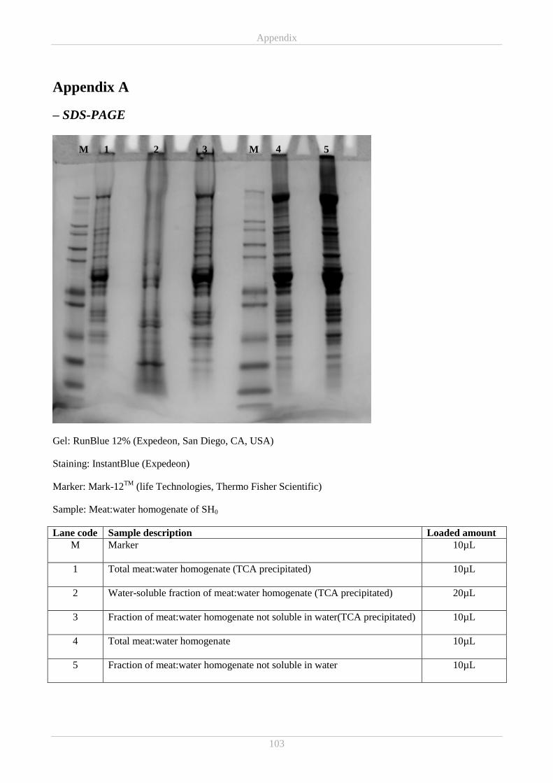
Appendix
103
Appendix A
– SDS-PAGE
Gel: RunBlue 12% (Expedeon, San Diego, CA, USA)
Staining: InstantBlue (Expedeon)
Marker: Mark-12TM
(life Technologies, Thermo Fisher Scientific)
Sample: Meat:water homogenate of SH0
Lane code Sample description Loaded amount
M Marker
10µL
1 Total meat:water homogenate (TCA precipitated)
10µL
2 Water-soluble fraction of meat:water homogenate (TCA precipitated)
20µL
3 Fraction of meat:water homogenate not soluble in water(TCA precipitated)
10µL
4 Total meat:water homogenate
10µL
5 Fraction of meat:water homogenate not soluble in water
10µL
M M 1 2 3 4 5

Appendix
104
Hvid tekst
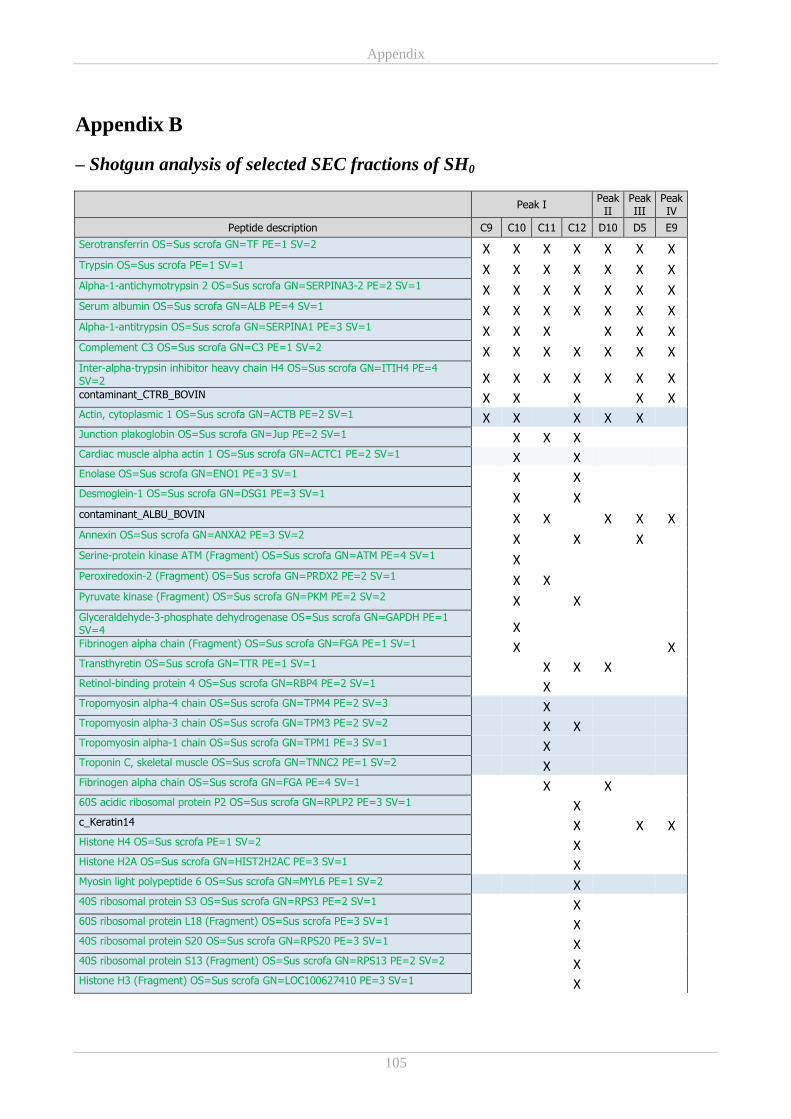
Appendix
105
Appendix B
– Shotgun analysis of selected SEC fractions of SH0
Peak I Peak
II Peak III
Peak IV
Peptide description C9 C10 C11 C12 D10 D5 E9
Serotransferrin OS=Sus scrofa GN=TF PE=1 SV=2 X X X X X X X Trypsin OS=Sus scrofa PE=1 SV=1 X X X X X X X Alpha-1-antichymotrypsin 2 OS=Sus scrofa GN=SERPINA3-2 PE=2 SV=1 X X X X X X X Serum albumin OS=Sus scrofa GN=ALB PE=4 SV=1 X X X X X X X Alpha-1-antitrypsin OS=Sus scrofa GN=SERPINA1 PE=3 SV=1 X X X
X X X
Complement C3 OS=Sus scrofa GN=C3 PE=1 SV=2 X X X X X X X Inter-alpha-trypsin inhibitor heavy chain H4 OS=Sus scrofa GN=ITIH4 PE=4 SV=2 X X X X X X X contaminant_CTRB_BOVIN X X
X
X X
Actin, cytoplasmic 1 OS=Sus scrofa GN=ACTB PE=2 SV=1 X X
X X X Junction plakoglobin OS=Sus scrofa GN=Jup PE=2 SV=1
X X X
Cardiac muscle alpha actin 1 OS=Sus scrofa GN=ACTC1 PE=2 SV=1
X
X
Enolase OS=Sus scrofa GN=ENO1 PE=3 SV=1
X
X
Desmoglein-1 OS=Sus scrofa GN=DSG1 PE=3 SV=1
X
X
contaminant_ALBU_BOVIN
X X
X X X
Annexin OS=Sus scrofa GN=ANXA2 PE=3 SV=2
X
X
X
Serine-protein kinase ATM (Fragment) OS=Sus scrofa GN=ATM PE=4 SV=1
X
Peroxiredoxin-2 (Fragment) OS=Sus scrofa GN=PRDX2 PE=2 SV=1
X X
Pyruvate kinase (Fragment) OS=Sus scrofa GN=PKM PE=2 SV=2
X
X
Glyceraldehyde-3-phosphate dehydrogenase OS=Sus scrofa GN=GAPDH PE=1 SV=4
X
Fibrinogen alpha chain (Fragment) OS=Sus scrofa GN=FGA PE=1 SV=1
X
X
Transthyretin OS=Sus scrofa GN=TTR PE=1 SV=1
X X X
Retinol-binding protein 4 OS=Sus scrofa GN=RBP4 PE=2 SV=1
X
Tropomyosin alpha-4 chain OS=Sus scrofa GN=TPM4 PE=2 SV=3
X
Tropomyosin alpha-3 chain OS=Sus scrofa GN=TPM3 PE=2 SV=2
X X
Tropomyosin alpha-1 chain OS=Sus scrofa GN=TPM1 PE=3 SV=1
X
Troponin C, skeletal muscle OS=Sus scrofa GN=TNNC2 PE=1 SV=2
X
Fibrinogen alpha chain OS=Sus scrofa GN=FGA PE=4 SV=1
X
X
60S acidic ribosomal protein P2 OS=Sus scrofa GN=RPLP2 PE=3 SV=1
X
c_Keratin14
X
X X
Histone H4 OS=Sus scrofa PE=1 SV=2
X
Histone H2A OS=Sus scrofa GN=HIST2H2AC PE=3 SV=1
X
Myosin light polypeptide 6 OS=Sus scrofa GN=MYL6 PE=1 SV=2
X
40S ribosomal protein S3 OS=Sus scrofa GN=RPS3 PE=2 SV=1
X
60S ribosomal protein L18 (Fragment) OS=Sus scrofa PE=3 SV=1
X
40S ribosomal protein S20 OS=Sus scrofa GN=RPS20 PE=3 SV=1
X
40S ribosomal protein S13 (Fragment) OS=Sus scrofa GN=RPS13 PE=2 SV=2
X
Histone H3 (Fragment) OS=Sus scrofa GN=LOC100627410 PE=3 SV=1
X
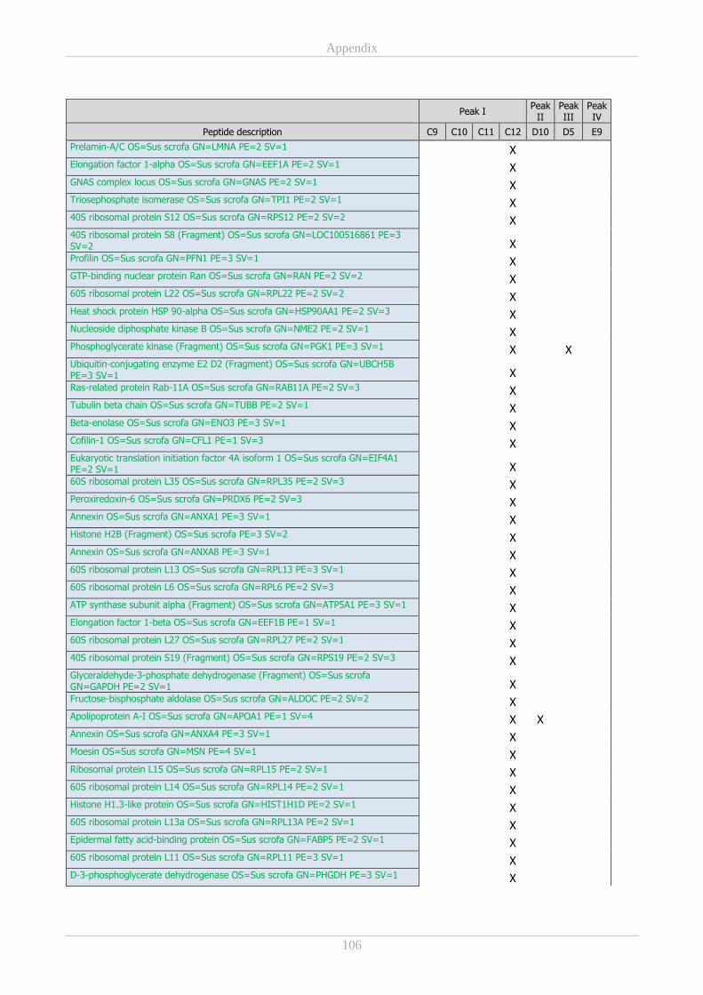
Appendix
106
Peak I Peak
II Peak III
Peak IV
Peptide description C9 C10 C11 C12 D10 D5 E9
Prelamin-A/C OS=Sus scrofa GN=LMNA PE=2 SV=1
X
Elongation factor 1-alpha OS=Sus scrofa GN=EEF1A PE=2 SV=1
X
GNAS complex locus OS=Sus scrofa GN=GNAS PE=2 SV=1
X
Triosephosphate isomerase OS=Sus scrofa GN=TPI1 PE=2 SV=1
X
40S ribosomal protein S12 OS=Sus scrofa GN=RPS12 PE=2 SV=2
X
40S ribosomal protein S8 (Fragment) OS=Sus scrofa GN=LOC100516861 PE=3 SV=2
X
Profilin OS=Sus scrofa GN=PFN1 PE=3 SV=1
X
GTP-binding nuclear protein Ran OS=Sus scrofa GN=RAN PE=2 SV=2
X
60S ribosomal protein L22 OS=Sus scrofa GN=RPL22 PE=2 SV=2
X
Heat shock protein HSP 90-alpha OS=Sus scrofa GN=HSP90AA1 PE=2 SV=3
X
Nucleoside diphosphate kinase B OS=Sus scrofa GN=NME2 PE=2 SV=1
X
Phosphoglycerate kinase (Fragment) OS=Sus scrofa GN=PGK1 PE=3 SV=1
X
X
Ubiquitin-conjugating enzyme E2 D2 (Fragment) OS=Sus scrofa GN=UBCH5B PE=3 SV=1
X
Ras-related protein Rab-11A OS=Sus scrofa GN=RAB11A PE=2 SV=3
X
Tubulin beta chain OS=Sus scrofa GN=TUBB PE=2 SV=1
X
Beta-enolase OS=Sus scrofa GN=ENO3 PE=3 SV=1
X
Cofilin-1 OS=Sus scrofa GN=CFL1 PE=1 SV=3
X
Eukaryotic translation initiation factor 4A isoform 1 OS=Sus scrofa GN=EIF4A1 PE=2 SV=1
X
60S ribosomal protein L35 OS=Sus scrofa GN=RPL35 PE=2 SV=3
X
Peroxiredoxin-6 OS=Sus scrofa GN=PRDX6 PE=2 SV=3
X
Annexin OS=Sus scrofa GN=ANXA1 PE=3 SV=1
X
Histone H2B (Fragment) OS=Sus scrofa PE=3 SV=2
X
Annexin OS=Sus scrofa GN=ANXA8 PE=3 SV=1
X
60S ribosomal protein L13 OS=Sus scrofa GN=RPL13 PE=3 SV=1
X
60S ribosomal protein L6 OS=Sus scrofa GN=RPL6 PE=2 SV=3
X
ATP synthase subunit alpha (Fragment) OS=Sus scrofa GN=ATP5A1 PE=3 SV=1
X
Elongation factor 1-beta OS=Sus scrofa GN=EEF1B PE=1 SV=1
X
60S ribosomal protein L27 OS=Sus scrofa GN=RPL27 PE=2 SV=1
X
40S ribosomal protein S19 (Fragment) OS=Sus scrofa GN=RPS19 PE=2 SV=3
X
Glyceraldehyde-3-phosphate dehydrogenase (Fragment) OS=Sus scrofa GN=GAPDH PE=2 SV=1
X
Fructose-bisphosphate aldolase OS=Sus scrofa GN=ALDOC PE=2 SV=2
X
Apolipoprotein A-I OS=Sus scrofa GN=APOA1 PE=1 SV=4
X X
Annexin OS=Sus scrofa GN=ANXA4 PE=3 SV=1
X
Moesin OS=Sus scrofa GN=MSN PE=4 SV=1
X
Ribosomal protein L15 OS=Sus scrofa GN=RPL15 PE=2 SV=1
X
60S ribosomal protein L14 OS=Sus scrofa GN=RPL14 PE=2 SV=1
X
Histone H1.3-like protein OS=Sus scrofa GN=HIST1H1D PE=2 SV=1
X
60S ribosomal protein L13a OS=Sus scrofa GN=RPL13A PE=2 SV=1
X
Epidermal fatty acid-binding protein OS=Sus scrofa GN=FABP5 PE=2 SV=1
X
60S ribosomal protein L11 OS=Sus scrofa GN=RPL11 PE=3 SV=1
X
D-3-phosphoglycerate dehydrogenase OS=Sus scrofa GN=PHGDH PE=3 SV=1
X
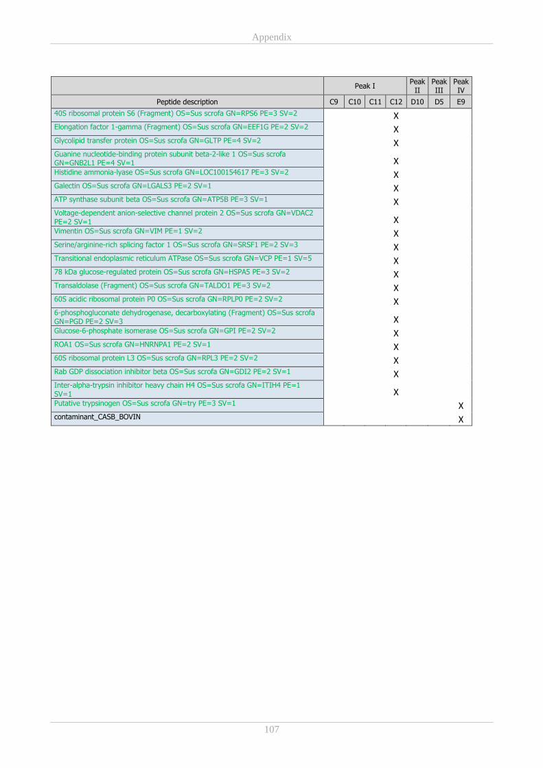
Appendix
107
Peak I Peak
II Peak III
Peak IV
Peptide description C9 C10 C11 C12 D10 D5 E9
40S ribosomal protein S6 (Fragment) OS=Sus scrofa GN=RPS6 PE=3 SV=2
X
Elongation factor 1-gamma (Fragment) OS=Sus scrofa GN=EEF1G PE=2 SV=2
X
Glycolipid transfer protein OS=Sus scrofa GN=GLTP PE=4 SV=2
X
Guanine nucleotide-binding protein subunit beta-2-like 1 OS=Sus scrofa GN=GNB2L1 PE=4 SV=1
X
Histidine ammonia-lyase OS=Sus scrofa GN=LOC100154617 PE=3 SV=2
X
Galectin OS=Sus scrofa GN=LGALS3 PE=2 SV=1
X
ATP synthase subunit beta OS=Sus scrofa GN=ATP5B PE=3 SV=1
X
Voltage-dependent anion-selective channel protein 2 OS=Sus scrofa GN=VDAC2 PE=2 SV=1
X
Vimentin OS=Sus scrofa GN=VIM PE=1 SV=2
X
Serine/arginine-rich splicing factor 1 OS=Sus scrofa GN=SRSF1 PE=2 SV=3
X
Transitional endoplasmic reticulum ATPase OS=Sus scrofa GN=VCP PE=1 SV=5
X
78 kDa glucose-regulated protein OS=Sus scrofa GN=HSPA5 PE=3 SV=2
X
Transaldolase (Fragment) OS=Sus scrofa GN=TALDO1 PE=3 SV=2
X
60S acidic ribosomal protein P0 OS=Sus scrofa GN=RPLP0 PE=2 SV=2
X
6-phosphogluconate dehydrogenase, decarboxylating (Fragment) OS=Sus scrofa GN=PGD PE=2 SV=3
X
Glucose-6-phosphate isomerase OS=Sus scrofa GN=GPI PE=2 SV=2
X
ROA1 OS=Sus scrofa GN=HNRNPA1 PE=2 SV=1
X
60S ribosomal protein L3 OS=Sus scrofa GN=RPL3 PE=2 SV=2
X
Rab GDP dissociation inhibitor beta OS=Sus scrofa GN=GDI2 PE=2 SV=1
X
Inter-alpha-trypsin inhibitor heavy chain H4 OS=Sus scrofa GN=ITIH4 PE=1 SV=1
X
Putative trypsinogen OS=Sus scrofa GN=try PE=3 SV=1
X
contaminant_CASB_BOVIN
X

Appendix
108
Hvid tekst
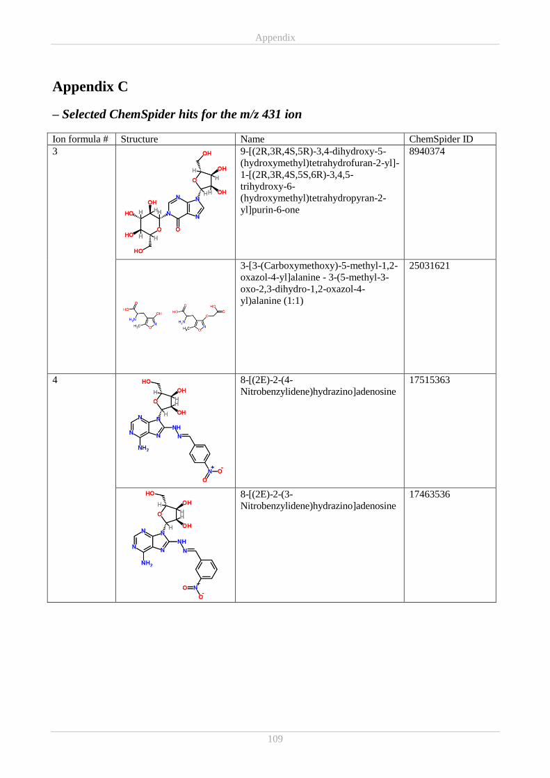
Appendix
109
Appendix C
– Selected ChemSpider hits for the m/z 431 ion
Ion formula # Structure Name ChemSpider ID
3
9-[(2R,3R,4S,5R)-3,4-dihydroxy-5-
(hydroxymethyl)tetrahydrofuran-2-yl]-
1-[(2R,3R,4S,5S,6R)-3,4,5-
trihydroxy-6-
(hydroxymethyl)tetrahydropyran-2-
yl]purin-6-one
8940374
3-[3-(Carboxymethoxy)-5-methyl-1,2-
oxazol-4-yl]alanine - 3-(5-methyl-3-
oxo-2,3-dihydro-1,2-oxazol-4-
yl)alanine (1:1)
25031621
4
8-[(2E)-2-(4-
Nitrobenzylidene)hydrazino]adenosine
17515363
8-[(2E)-2-(3-
Nitrobenzylidene)hydrazino]adenosine
17463536
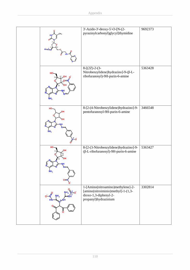
Appendix
110
3'-Azido-3'-deoxy-5'-O-[N-(2-
pyrazinylcarbonyl)glycyl]thymidine
9692373
8-[(2Z)-2-(3-
Nitrobenzylidene)hydrazino]-9-(β-L-
ribofuranosyl)-9H-purin-6-amine
5363428
8-[2-(4-Nitrobenzylidene)hydrazino]-9-
pentofuranosyl-9H-purin-6-amine
3466548
8-[2-(3-Nitrobenzylidene)hydrazino]-9-
(β-L-ribofuranosyl)-9H-purin-6-amine
5363427
1-[Amino(nitroamino)methylene]-2-
[amino(nitroiminio)methyl]-1-(1,3-
dioxo-1,3-diphenyl-2-
propanyl)hydrazinium
3302814
Appendix
111
8-[2-(3-Nitrobenzylidene)hydrazino]-
9-pentofuranosyl-9H-purin-6-amine
2970405
2-[(2E)-3-(Phenoxycarbonyl)-2-
triazen-1-yl]adenosine
9070535
9-(β-D-Arabinofuranosyl)-8-[(2E)-2-
(4-nitrobenzylidene)hydrazino]-9H-
purin-6-amine
17540217
8-[(2E)-2-(3-
Nitrobenzylidene)hydrazino]-9-
pentofuranosyl-9H-purin-6-amine
17540715
(2E)-2-[(2E)-3-(Phenoxycarbonyl)-2-
triazen-1-ylidene]-9-(D-
ribofuranosyl)-2,9-dihydro-1H-purin-
6-amine
24653191
Appendix
112
8-[(2E)-2-(4-
Nitrobenzylidene)hydrazino]-9-
pentofuranosyl-9H-purin-6-amine
29770815
8-[(3-Nitrobenzylidene)hydrazono]-8-
hydroadenosine
35319419
8-[2-(4-
Nitrobenzylidene)hydrazino]adenosine
35360694
6
(6E,6'E)-3,3'-(1,4-Phenylene)bis(5-
cyano-6-imino-2-oxo-3,6-dihydro-
1(2H)-pyrimidinecarboximidamide)
8608245
Paper I
_______________________________________________________________________________________
Pedersen, S. T., Baron, C. P., De Gobba, C., Duedahl-Olesen, L., Koch, A. G. &
Jessen, F. Antioxidative and antimicrobial activity of extracts from nitrite-cured
cooked pork products. Manuscript close to submission.
_______________________________________________________________________________________
Paper I
1
Title: Antioxidative and antimicrobial activity of extracts from nitrite-cured cooked pork products 1
Author: Sabrine Tauber Pedersena*
, Caroline P. Barona, Cristian De Gobba
b, Lene Duedahl-2
Olesena, Anette Granly Koch
c & Flemming Jessen
a. 3
aNational Food Institute, Technical University of Denmark, Søltofts Plads Building 227, DK-2800 4
Kongens Lyngby, Denmark 5
bUniversity of Copnhagen, Department of Food Science, Ingredient and Dairy Technology, 6
Rolighedsvej 26, DK-1958 Frederiksberg C, Denmark 7
cDanish Meat Research Institute, Gregersensvej 9, DK-2630 Taastrup, Denmark 8
9
Running title: Antioxidative and antimicrobial activity of extracts from cured hams 10
11
*Corresponding author: 12
Sabrine Tauber Pedersen 13
Postal address: 14
National Food Institute 15
Søltofts Plads Building 227, 1st floor, room 142 16
DK-2800 Kgs. Lyngby 17
E-mail & phone number: 18
+45 45252691 20
21
Declarations of interest: none 22
Paper I
2
Abstract 23
The role of nitrite for oxidative and microbial stability of cured meats has been extensively studied 24
yet focused mainly on nitrite itself. This study aimed to broaden the understanding of the role of 25
nitrite in cooked cured hams, by investigating in vitro antioxidative and antimicrobial properties. 26
Three nitrite-cured cooked hams were compared to a non-nitrite-cured cooked ham. Aqueous 27
≤10kDa extracts of the nitrite-cured hams demonstrated higher in vitro reducing power and ABTS 28
radical scavenging activities than the uncured, and activities appeared to correlate with residual 29
nitrite/ascorbate. Iron chelating activity was found in two nitrite-cured hams and in the uncured. 3-30
nitrotyrosine was neither detected in hams nor extracts. Antioxidant activity may be linked to 31
histidine-content. None of the extracts inhibited growth of Listeria monocytogenes or Salmonella 32
typhimurium. In conclusion, the increased antioxidant activity obtained by nitrite-curing is not 33
solely due to residual ascorbate but also to compounds formed during the processing. 34
35
Keywords: ham, nitrite-curing, antioxidant, in vitro assays, 3-nitrotyrosine, antimicrobial 36
37
38
39
40
41
42
43
Paper I
3
1 Introduction 44
Salting of meat is an ancient preservation technique. By binding water, the salt decreases the water 45
activity below levels required for microbial growth and thereby protects meat and fish from 46
spoilage. Through times it became evident that salts containing saltpetre (KNO3) contributed with 47
additional attributes in conservation, taste and colour, compared to pure salt (Binkerd & Kolari, 48
1975). Today meat curing consists of adding salt containing nitrite or nitrate and other ingredients 49
such as sugar and spices to fresh meat to create a unique flavour and colour. Curing is generally 50
done by either dry curing or wet curing or a combination. The resulting products may be intended 51
for consumption in raw state or may be submitted to further downstream processing like boiling and 52
smoking leading to an immense variety of processed cured meat products. Yet, the primary purpose 53
is still to prolong shelf-life by preventing microbiological spoilage, growth of pathogens and 54
oxidative degradation. 55
Indeed, nitrite is a strong oxidant but in combination with common meat additives like ascorbate 56
and salt they collectively possess the ability to reduce lipid oxidation (Honikel, 2008). For example, 57
Morrissey & Tichivangana (1985) found that levels as low as 20ppm nitrite was sufficient to 58
significantly inhibit lipid oxidation in fish, chicken, pork, and beef systems also when adding pro-59
oxidants. Also, Mac Donald, Gray, Kakuda & Lee (1980) found a significant reduction in 60
Thiobarbituric acid (TBA) values in pork when treated with any level of nitrite (50, 200, and 61
500ppm) compared to a non-nitrite treatment. 62
During processed meat manufacturing, a complex web of reaction is occurring between reactive 63
nitrite (species), meat constituents and additives and at the end of the heat treatment residual nitrite 64
is reduced to less than one third of the originally added amounts (Honikel, 2008). Furthermore, 65
residual nitrite declines even further during cold storage, occasionally to non-detectable levels 66
(Honikel, 2008). 67
Paper I
4
The main reactivity of nitrite is exerted through nitric oxide (NO) which reacts with a wide variety 68
of molecules such as myoglobin, ascorbic acid or amino acids and other proteins (Honikel, 2008). 69
The most studied reaction related to nitrite curing is the reaction of NO and myoglobin. 70
Yet, Tricker & Kubacki (1992) summarizes that only 5-15% of added nitrite is associated with 71
myoglobin while 5-10% is recovered as nitrite and 1-10% is oxidized to nitrate. Furthermore 1-5% 72
is thought to be lost as gasses and 1-5% is believed to be associated with lipids. Of the remaining 73
portion 5-15% may react with sulfhydryl groups, mostly in peptides and free amino acids, while as 74
much as 20-30% will bind to other proteins (Tricker & Kubacki, 1992). 75
Nitrite induced modification on proteins, peptides and amino acids mainly entail the addition of 76
NO. Examples of such are the S-nitrosylation of the cysteine thiol, the C-nitrosation of tyrosine, or 77
the N-nitrosation of amines among others leading to formation of nitrosamines. With the exemption 78
of N-nitrosamines, the existence and understanding of the properties of these modified peptides are 79
to our knowledge relatively limited in regard to food. However, amino acids and small proteins 80
modified by nitrite like S-nitrosocystein, and S-nitrosoglutathione have previously been found to 81
possess antioxidant properties (Kanner, 1979) (Chiueh & Rauhala, 1999) (Morrissey & 82
Tichivangana, 1985). 83
Within the last few decades, increasing research in antioxidant and antimicrobial peptides from 84
different food and food related matrixes, has led to the discovery of the naturally occurring 85
antioxidant dipeptides, carnosine and anserine. But also small active peptides in hydrolysates of 86
different animal tissue and animal based products has been identified (Review: Di Bernardini et al., 87
2011a) (Gil-Agustí, Esteve-Romero & Carda-Broch, 2008). However, to our knowledge, the 88
existence and extent of antioxidant activities from nitrite wet-cured cooked ham, beyond the direct 89
impact of nitrite, has not been fully investigated. 90
Paper I
5
Thus, it can be hypothesised that compounds with antioxidative and antimicrobial properties are 91
formed during nitrite curing and that these compounds might contribute to the mechanisms by 92
which nitrite exerts its preservation properties. Furthermore, it can be speculated that some of these 93
compounds may be low molecular weight compounds such as peptides that to some extent have 94
been nitrosylated/nitrosated by nitrite. 95
Thus, we set out to investigate the nature of the antioxidative and antimicrobial activities generated 96
during nitrite-curing 97
98
2 Material and methods 99
2.1 Materials 100
Ascorbic acid, sodium nitrite, 3-nitro-L-tyrosine, 3-(2-Pyridyl)-5,6-diphenyl-1,2,4-triazine-4′,4′′-101
disulfonic acid sodium salt (ferrozine) and 2,2′-Azino-bis(3-ethylbenzothiazoline-6-sulfonic acid) 102
diammonium salt (ABTS), were from Sigma-Aldrich (St. Louis, MO, USA). 103
Ethylenediaminetetraacetic acid disodium dihydrate (EDTA) was from Merck Millipore (Billerica, 104
MA, USA) and (±)-6-Hydroxy-2,5,7,8-tetramethylchromane-2-carboxylic acid (Trolox) from Fluka 105
Chemika (Deisenhofen, Germany). All remaining chemicals and reagents used were of analytical 106
grade and acquired from VWR International (Radnor, PA, USA), Merck Millipore, Sigma-Aldrich 107
or Fluka Chemika. 108
2.2 Hams 109
The sample material comprised three different types of commercial nitrite-cured cooked pork 110
products: a sandwich ham (SH), a boiled dinner ham (DH) and a smoked pork saddle (SPS). 111
Pictures of the three products can be found in Figure 1. The sandwich ham was a ready-to-eat 112
luncheon meat product belonging to the category of restructured meat products where smaller 113
Paper I
6
pieces of meat were subjected to curing brine prior to boiling stuffed into a plastic casing. The 114
remaining two products were whole-muscle products most likely injected with curing brine prior to 115
boiling and smoking (only the pork saddle was smoked). The exact manufacturing procedures were 116
undisclosed by the producers yet the following details were provided: SH was added 60ppm nitrite 117
and unknown amounts of sodium ascorbate (E301) while DH and SPS was both added 120ppm 118
sodium nitrite (E250) and unknown amounts of sodium ascorbate (E301). Furthermore, SPS was 119
added phosphates (E450, E451). The sandwich ham, SH, was delivered from the manufacturer in 120
bulk. The whole-muscle products, DH and SPS, were supplied from the manufacturer in consumer 121
packaging. For the four days from production to arrival at the laboratory, the vacuum-packed 122
samples were stored at 5-8°C and afterwards moved to -80ºC until further analysis. 123
The samples were tested without any further heat-treatment. Prior to sampling for the individual 124
analytical methods a larger piece of the ham, of approximately 100g, were minced to ensure a 125
homogenous sample. The remaining skin and the excess subcutaneous fat on the dinner ham (Figure 126
1) were trimmed before mincing. The smoke-coloured surface of the pork saddle (Figure 1) was not 127
removed as it is expected to be consumed by the majority of Scandinavian consumers. 128
A reference ham prepared without added nitrite or ascorbic acid was included. The reference ham 129
was a boiled restructured ham made from pork, water, salt, pork skin protein, phosphates, potato 130
starch and dextrose. 131
132
2.3 Proximate composition analysis 133
2.3.1 Nitrite content 134
Extraction and measurement of nitrite in the cured and uncured cooked pork products was done 135
according to Hermann, Duedahl-Olesen & Granby (2015). In short, the detection was carried out 136
Paper I
7
using ion chromatography coupled to UV detection at 225nm (HPLC and Diode Array Detector, 137
series1100, Agilent Technologies, Santa Clara, CA, USA) with separation on a Dionex IonPacTM 138
AS11 RFICTM 250 x 4mm (Lot # 011-33-189A, product # 044076, serial #020071, Thermo 139
Scientific, Waltham, MA, USA). The samples were analysed in a random order. External 140
calibration was performed in the range 0.25-100µg/mL NaNO2. As the nature of the added nitrite 141
was unknown in SH all results are presented as ppm nitrite-ion by the relationship 1g nitrite ion = 142
1.50g NaNO2. 143
2.3.2 Ascorbic acid content 144
Residual ascorbic acid in the hams were determined using an Agilent 1100 series HPLC using a 145
G1315A DAD detector set at 254.4nm (Agilent Technology, Waldbronn, Germany). 146
Ascorbic acid was extracted by leaving 2.5g of minced ham and 10mL of 5%(W/V) meta-147
phosphoric acid, added 0.5mL of 0.1mg/mL EDTA solution, to stir for 2min at room temperature 148
protected from light. MilliQ was added to a total volume of 25mL. The mixture was centrifuged at 149
3000rpm for 5min and filtered through a Munktell quantitative filter paper grade 00H and re-filtered 150
through a 0.2µm syringe filter directly into an amber HPLC vial. Twenty µL was injected and 151
separated on a Prodigy 5u ODS3 100Å 250x4.6mm column (Phenomenex®, Torrance, CA, USA) 152
and eluted at a flow rate of 0.7mL/min. Mobile phase A was water adjusted to pH 3 and B was 153
methanol running a gradient of 5% B for the first 23min followed by a 10sec linear increase to 154
100% B which were kept for 4min and 50sec before again decreasing to 5% B in 10sec and kept at 155
this level until the end of the run (total of 35min). Quantification was done using an external 156
calibration curve of L-ascorbic acid dissolved in 1%(W/V) meta-phosphoric acid. 157
2.3.3 Amino acid profile (total and free) 158
The total and free amino acid content of the hams was determined according to the procedure 159
described by Farvin, Baron, Nielsen, Otte & Jacobsen (2010) with slight modifications. In short, the 160
Paper I
8
total amino acid content was analysed by subjecting approximately 30mg of ham to microwave-161
assisted acidic hydrolysis (6M HCl) for 1 hour at 110ºC and 500W followed by 10min of cooling. 162
The samples were diluted with 1M NaCO3 and filtered through a 0.2μm syringe filter (Q-max 163
PTFE, Ø13mm, Frisenette ApS, Knebel, Denmark) before derivatisation using the EZ:FaastTM
164
Amino Acid Analysis kit from Phenomenex® (Torrance, CA, USA). The derivatised samples were 165
analysed by LC-(APCI)-MS (Agilent 1100, Agilent Technology). Separation took place on a 166
reversed phase column (EZ:FaastTM
AAA-MS 250 x 3.0mm, Phenomenex®, Torrance, CA, USA) 167
running a gradient (mobile phase A was water and B was methanol, both containing 10 mM 168
ammonium formate) at 0.5mL/min. Total runtime was 26min. 169
Prior to derivation of free amino acid using the EZ:FaastTM
Amino Acid Analysis kit, 250μL of 170
TCA was added to 100μg of ham and left over night at 4ºC, centrifuged at 12,000rpm for 3min and 171
100μL hereof was mixed with 50μL of 1M NaCO3 which was then analysed as described above. 172
2.4 Extraction of low molecular weight compounds 173
Water soluble compounds were extracted from the hams by homogenising 94.9-101.5g of minced 174
ham in approximately 400mL of deionised water, dH2O, for 1min with an Ultra Turrax (T 25 Basic 175
ULTRA-TURRAX® mounted with S25N -18G Dispersing element, IKA®-WERKE GmbH &Co. 176
KG, Staufen, Germany) at 13,500rpm and subsequently adding up to a total volume 1.5L. 177
Extraction from the reference ham was done in the same manner however at a 1:10 ratio. An 178
aqueous ≤10kDa extract was collected by submerging five times 10cm dialysis membrane tubes 179
(Spectra/Por 6, MWCO 10,000Da, Spectrum Lab. Inc, CA, USA) containing 16.5mL dH2O 180
(corresponding to 50% membrane volume) into the meat/water homogenate. After 24 hours of 181
magnetic stirring at 5ºC, the dialysate in the 5 membranes were pooled and stored at -80ºC until 182
further analysis. Later the protein content was determined using the PierceTM
Modified Lowry 183
Protein Assay Kit in microplate format according to manufactures description (Thermo Scientific, 184
Paper I
9
PierceTM
) using Bovine Serum Albumin (BSA) (Ampules, 2 mg/ml) supplied with the kit as an 185
external standard. 186
2.5 Antioxidant activity 187
Potential antioxidant activities of the aqueous ≤10kDa extract were analyzed using three in vitro 188
assays measuring iron chelation activity, reducing power and ABTS-radical scavenging activity. In 189
order to investigate the concentration dependency of the antioxidant activities, the crude undiluted 190
≤10kDa extract were analyzed followed by sequential threefold dilutions. The assays were 191
conducted in triplicate and all measurements were performed using a spectrophotometer (Synergy 2 192
Multi-Mode Microplate Reader, BioTek® Instruments, Inc., Vermont, USA). The extracts were 193
thawed at 5ºC, over night. 194
Iron chelating activity was assayed by the microtiter plate format method described by Hermund et 195
al. (2015). EDTA (0.5mM) was used as a positive control and was subjected to the same threefold 196
dilutions as the extract samples. dH2O was utilized as blank. The iron chelating activity (%) is 197
defined and calculated as percentage of inhibited colour formation/relative decrease in absorbance 198
(562nm): 199
𝐹𝑒2+ 𝑐ℎ𝑒𝑙𝑎𝑡𝑖𝑛𝑔 𝑎𝑐𝑡𝑖𝑣𝑖𝑡𝑦 (%) =𝐴𝑏𝑠𝑏𝑙𝑎𝑛𝑘 − 𝐴𝑏𝑠𝑠𝑎𝑚𝑝𝑙𝑒
𝐴𝑏𝑠𝑏𝑙𝑎𝑛𝑘∙ 100%
The reducing power was determined according to the procedure described by Oyaizu (1986) with 200
some modifications and adjusted to microtiter plate detection. First, equal parts of ≤10kDa extract 201
sample (and dilutions), 0.2M phosphate buffer (pH 6.6) and 1% potassium ferricyanide were mixed 202
and incubated for 20 min at 50°C. Subsequently, 1 part of 10% TCA was also added. Then, 100μL 203
of the sample mixture was mixed with 100μL dH2O and 20 μL of 0.1% ferric chloride, and 204
incubated 10min at room temperature. The absorbance was measured at 700nm. Ascorbic acid 205
Paper I
10
(0.5mM) was used as positive control and was subjected to the same threefold dilution as the extract 206
samples and dH2O was used as blank. Results are expressed as 207
𝑂𝐷700 = 𝐴𝑏𝑠700𝑛𝑚(𝑆𝑎𝑚𝑝𝑙𝑒) − 𝐴𝑏𝑠700𝑛𝑚(𝑏𝑙𝑎𝑛𝑘)
The ABTS radical scavenging activity was determined as described by Gringer, Osman, Nielsen, 208
Undeland & Baron (2014). Trolox (2.5mM, in 96% ethanol) was used as a positive control and was 209
subjected to the same threefold dilutions as the samples. Ethanol (96%) and dH2O was included as 210
blank for the positive control and extract sample, respectively. The ABTS radical scavenging 211
activity (%) is expressed as the relative decrease in absorbance (734nm) as for iron chelating 212
activity. 213
2.6 Antimicrobial activity 214
Antimicrobial activity of the cured ham aqueous ≤10kDa extracts and the supernatant of the 215
meat/water homogenate were tested with an inoculation culture consisting of a mixture of Listeria 216
monocytogenes (DMRICC 3042) and Salmonella typhimurium (DMRICC 4984). A small quantity 217
of the purified cultures where each transferred to 10mL Brain-heart infusion (BHI) broth (Oxoid 218
CM1135, United Kingdom) and incubated for 20 ± 2 hours at 37°C. These were afterwards diluted 219
to 10-6
and mixed 1:1 (V/V) to constitute the inoculation culture. 220
Diluted (1:5 and 1:25) and undiluted extracts were mixed 1:1 with BHI broth and inoculated with 221
the inoculation culture. The initial concentration of each bacteria culture in the inoculated samples 222
was 44 cfu/ml ~ 1.6 log cfu/ml. A sample substituting extract with FKP (0.85% NaCl and 0.1% 223
pepton (Merck 1.07214)) was included as a control. The inoculated extract samples were incubated 224
at 17 ± 1°C and samples were taken after 0, 24, 48 and 72 hours. The total bacterial count was 225
determined on Oxford agar (oxoid CM 0856, United Kingdom) and SSI Enteric agar (34121, 226
Statens Serum Institute, Copenhagen Denmark) after 24-48 hours of incubation at 37°C. 227
Paper I
11
2.7 3-Nitrotyrosine on LC-MS 228
The content of 3-nitrotyrosine (3-NT) was determined in both ≤10kDa extracts and hams following 229
acidic hydrolysis in 6M HCl. 0.5mL of extract or 3-NT standard solutions were mixed 1:1 (V/V) 230
with HCl while approximately 30mg of freshly minced ham were added 1mL of HCl. The mixtures 231
were overlayed with nitrogen, closed and hydrolysed at 110°C. After 24 hours the samples were left 232
to cool protected from light. Extract samples and external standards were transferred directly to 233
HPLC vials while the hams samples were filtered through a 0.2μm syringe filter (Q-max PTFE , 234
Ø13mm, Frisenette ApS) into the HPLC vial. 3-NT was quantified by LC-MS/MS using an 235
UHPLC+ Ultimate 3000, mounted with a C18 column (Aeris Peptide XB-C18, 150 x 2.1mm, 236
1.7µm, 100Å, 40°C) coupled with a Q Exactive Biotech mass spectrometer (both ThermoFisher 237
Scientific). Buffer A was 0.1% formic acid (FA) in water, and buffer B was 0.1% FA in 80% 238
acetonitrile. 10µL of sample were injected. The flowrate was 0.25mL/min and the gradient 239
consisted of 100% A for 5min, followed by a linear increase from 0% to 25% B in 15min. 3-NT 240
was detected and quantified in PRM mode using 227.06 m/z as parent ion with a resolution of 241
17500 and AGC target of 2e5, max IT 64 ms. The reporter ion used for quantification was 181.06 242
m/z. 3-NT solutions of 0.5-500ng/mL (after hydrolysis) were used for external calibration. 243
2.8 Statistical analysis 244
Measurements were carried out in triplicate (unless stated otherwise). Results are presented as 245
average values ± the standard deviation. All statistical analyses were conducted using the GraphPad 246
Prism® software Ver. 4.03 or R version 3.3.0 with P < 0.05. Results were compared using one-way 247
ANOVA and t-test with Tukeys posttest. 248
249
250
251
Paper I
12
3 Results and discussion 252
3.1 Antioxidative activity 253
The current study set out to investigate whether antioxidant and antimicrobial activity could be 254
identified in aqueous ≤10kDa extracts of three different nitrite-cured cooked pork products. Using 255
in vitro assays for iron chelating activity, reducing power and ABTS radical scavenging activity, 256
antioxidant activity was demonstrated for all aqueous ≤10kDa extracts in all assays with a single 257
exception (Figure 2). 258
The ≤10kDa extracts of SH, SPS and the non-cured reference ham possessed a maximal iron 259
chelating activity of 70%, 62% and 90%, respectively, at the highest tested concentrations, whereas 260
the extract from DH showed no activity (Figure 2A). In comparison the positive control in the 261
tested concentration (0.5mM EDTA) demonstrated 95-99% iron chelating activity (data not shown). 262
The antioxidant activities may be viewed as the initial linear activity increase per mg ham and is 263
presented in Table 1. From the curves in Figure 2A the reference ham seems to have the highest 264
initial linear iron chelating activity increase which from the first 2-3 data points may be calculated 265
to 577±187 % iron chelating activity/mg ham. In comparison, SH and SPS had initial linear iron 266
chelating activity increase of 54±4 and 24±1% iron chelating activity/mg ham. 267
From the current results iron chelating activity did not appear to be connected with nitrite curing as 268
the reference ham, without added nitrite, showed iron chelating activity and an initial linear iron 269
chelating activity increase much higher than the cured SPS and SH while, as already mentioned, the 270
cured DH showed no activity. 271
The reducing power of the aqueous ≤10kDa extracts are presented in Figure 2B. All extract 272
displayed lower activity than the positive control, ascorbic acid (data not shown). SPS showed the 273
highest activity with an initial linear activity increase (Table 1), which was about 4 times higher 274
Paper I
13
than the initial linear activity increase of the reference ham, showing the lowest reducing power of 275
all the extracts. The DH extract again showed the lowest activity of the three nitrite-cured hams. 276
All ≤10kDa extracts, including the reference, in crude form showed similar maximal ABTS radical 277
scavenging activities of 90-92% (Figure 2C), which was at the same level as the positive control, 278
trolox (data not shown). Compared to the reference ham, the initial linear radical scavenging 279
activity increase of SPS was approximately 2.5 times higher (Table 1). 280
As oppose to iron chelating both reducing power and ABTS radical scavenging properties seem to 281
be connected with curing by way of all the nitrite-cured ham extracts displaying higher antioxidant 282
properties than the reference. However, the reducing power and ABTS radical scavenging 283
properties did not correlate with added nitrite/ascorbic acid but rather with residual nitrite/ascorbic 284
acid (Table 2). Although, this indicates a correlation between residual nitrite/ascorbic acid and 285
ABTS radical scavenging activity. 286
High diversity in laboratory and data treatment methodologies makes it highly problematic to 287
compare current findings with previously reported data. However, antioxidant peptides have been 288
identified and purified from different dry-cured hams (Xing et al., 216)(Escudero, Mora, Fraser, 289
Aristoy & Toldrá, 2013). Mora, Escudero, Fraser, Aristoy & Toldrá (2014) identified peptides of 290
400-2500Da and tested a selected few purified peptides for DPPH radical scavenging activity and 291
reducing power. ABTS and DPPH radical scavenging assays both describe a sample’s ability to 292
scavenging radicals but they are not directly comparable (de Casto & Sato, 2015). However, the 293
peptides, purified by Mora et al. (2014), which demonstrated the highest reducing power had a 294
reducing power absorbance (corresponding to OD700 in the current work) of approximately 1.5 at 295
0.2mg/mL while the majority of the purified peptides were below 0.5 at the same concentration. 296
Also Escudero et al. (2013) found even lower reducing power absorbance for their purified peptides 297
Paper I
14
at 0.2mg/mL. In the current study the total protein concentration was approximately 0.2mg/mL in 298
all extracts (SH~0.23mg/mL, DH~0.22mg/mL, SPS~0.19mg/mL and reference~0.23mg/mL), yet 299
they still gave rise to a reducing power antioxidant activity of 0.25-0.5. Thus, if the observed 300
antioxidant capacities could be related to a single or a few peptides these may be more powerful 301
reducing antioxidant on their own than the majority of the once tested in the referred studies. 302
Otherwise, the currently demonstrated reducing power activity may be caused by strong synergetic 303
effects of protein and non-protein, anti- and pro-oxidant compounds in the extracts. 304
Residual nitrite and small active nitrite-derived compounds e.g. NO, N2O3 and HNO2 can freely 305
pass the dialysis membrane and the presence of these might interfere with the assays because of 306
their ability to prevent oxidation of lipids (Skibsted, 2011). The residual nitrite (Table 2) in itself 307
was not considered an issue however, it cannot be ruled out that the nitrite may function as a minute 308
reservoir for NO or other nitrosating or antioxidant agents especially in the presence of a reducing 309
agent such as ascorbic acid (Table 2). Many mechanisms on how nitrite exerts its antioxidant 310
capabilities have been suggested through the past decades. The most predominant (no rank order) 311
constitutes (I) oxygen sequestering by oxidation of NO to NO2, (II) the formation of a stable 312
complex between heme-iron and nitrite preventing release of iron, (III) stabilisation of unsaturated 313
fatty acids, (IV) formation of nitroso- and nitrosyl compounds having antioxidant properties, like S-314
nitrosocystein and (V) chelating of iron and free radicals (Honikel, 2008)(Freybler et al., 1993) 315
(Skibsted, 2011). Especially the last mechanism of nitrite will have a direct impact on the used 316
antioxidant assay. Consequently, the detected antioxidant activity of the extracts would appear 317
higher than if the activity was only attributed to by other unknown extract constituents. In high 318
concentrations, nitrite behaves as a strong pro-oxidant. However, this effect requires free nitrite 319
concentrations to exceed the levels found in commercially cured meat products (Mac Donald, Gray 320
& Gibbins, 1980). 321
Paper I
15
In addition to nitrite, (small free) ions (metal ions or salt), present in plenty in meat, are also capable 322
of passing the dialysis membrane and might therefore also interfere with the antioxidant assays. As 323
the iron chelating assay is based on the principle of neutralising added iron pro-oxidant, the 324
presence of additional ferrous ions would result in the nitrite-cured ham extracts appearing as less 325
potent antioxidant pools. In order to correct for this error, in future studies, it would be necessary to 326
include a control containing, ≤10kDa extract samples and ferrozine but substituting FeCl2 with 327
dH2O. Furthermore, NaCl is often mentioned in regard to decreasing oxidative stability of muscle 328
foods. Some of the pro-oxidative mechanisms are related to the chloride ion altering iron’s 329
reactivity and the sodium ion causing displacement of iron from macromolecules (Lee, Mei & 330
Decker, 1997). Both of which may have affected the iron chelating assay. 331
Sodium ascorbate is a common additive in nitrite-cured pork products and was also added to the 332
three nitrite-cured hams: SH, DH, and SPS. Sodium ascorbate is added for its curing accelerating, 333
nitrosamine reducing and antioxidant properties (Sebranek, 2009). The ascorbate ion functions as a 334
reducing agent and even though ascorbic acid is very light and heat sensitive residual amount was 335
expected to be present in the hams. Ascorbic acid would be able to pass the dialysis membrane and 336
thus result in the aqueous ≤10kDa extracts exhibiting a higher reducing power activity. The order of 337
hams in regard to detected residual ascorbic acid (SPS>SH>DH>Reference) also matched the order 338
of the ham extracts in regard to reducing power (both the initial linear and at maximal tested extract 339
concentration). Thus, it must be assumed that residual ascorbic acid was a great contributor to the 340
detected reducing power yet a doubling in residual ascorbic acid did not correspond to a doubling in 341
reducing power. In addition, phosphates were also added to SPS. Some phosphates are known to 342
function as metal chelators and are as such expected to enhance the iron chelating activity signal. 343
However, no iron chelating was observed for the respective ≤10kDa extract (Figure 2). 344
345
Paper I
16
3.1.1 Amino acids composition of the hams 346
As indicated previously a potential contributor to the observed antioxidant activity could be 347
peptides and amino acids originating from the hams. In the field of bioactive peptides a lot of effort 348
has been invested in identifying amino acids of importance to activity. Either by isolation and 349
further characterisation of active peptides or simply testing free amino acids in solution, it has 350
become clear that some amino acids exhibit stronger, and different, antioxidant properties than 351
others (Meucci & Mele, 1997)(Marcuse, 1962)(de Castro & Sato, 2015). Histidine, tyrosine, 352
tryptophan, methionine, cysteine, phenylalanine and proline are some of the amino acids most 353
frequently listed to have antioxidant activity either as free amino acids or embedded in a peptide (Di 354
Bernardini et al., 2011b) but also lysine, arginine and glutamic acid are ascribed as antioxidative 355
(Broncano, Otte, Petrón, Parra & Timon, 2012). Many of these have been observed to be of 356
importance for metal chelation, including the acidic amino acids (Saiga, Tanabe & Nishimura 357
2003). However, the current results did not exactly reflect this effect as DH were found to contain 358
the most protein (data not shown) and thus amino acids yet demonstrated the lowest iron chelating 359
activity. 360
In order to evaluate the peptidic contribution to the measured antioxidant activity both the free and 361
total amino acid profile was determined. The content of free amino acids was, as expected, very 362
low: 3.14, 2.42 and 3.95mg amino acid/g DM in SH, DH and SPS respectively, and not 363
significantly different between the three nitrite-cured hams. Overall the total amino acid profiles 364
were not significantly different and all profiles were dominated by glutamic an aspartic acid (Figure 365
3) which matched the findings by Wilkinson, Lee, Purchas & Morel (2014) in both raw and cooked 366
(60 and 75ºC) pork muscle as well as data from the Danish food composition database (Frida Food 367
Data - http://frida.fooddata.dk/). Glutamic acid constituted on average 14.6% in the nitrite-cured 368
hams, and 15.4% and 14.4%, respectively for the two literature references. On the contrary the 369
Paper I
17
nitrite-cured hams of the current study contained relatively more proline, histidine, serine and 370
alanine and much less valine, isoleucine and threonine (Wilkinson et al., 2014)(Frida Food Data, 371
2016). The relative higher content of the known antioxidative proline and histidine could have 372
resulted in the tested commercial nitrite-cured hams exhibiting stronger antioxidative activity 373
compared to other untested commercial nitrite-cured cooked pork products as well as the uncured 374
reference ham. 375
Histidine has been found to possess antioxidant capacities as a radical scavenger, active oxygen 376
quencher and metal ion chelator (Sun et al., 2009). Thus, the relatively high content of histidine in 377
the nitrite-cured hams (6.1-9.8%) could have contributed to the observed ABTS radical scavenging 378
activity. Also the order of ABTS radical scavenging activity of extracts of the nitrite-cured hams 379
and the uncured reference ham illustrated in Figure 2 fits with the relative histidine content. 380
Phenylalanine and in particular tyrosine are often mentioned in connection with electron donors and 381
radical scavengers mainly due to their aromatic ring. Both amino acids were found to be present in 382
the hams but it is not possible to speculate on their importance based on the current data however it 383
cannot be ruled out that they might have contributed additively. Tryptophan has also been found to 384
be of relevance in regard to (ABTS) radical scavenging (Nimalaratne, Lopes-Lutz, Schieber &Wu, 385
2011), and also reduction of iron and chelating of metals (de Castro & Sato, 2015) however, from 386
the current data, no connection between the observed antioxidant activity and tryptophan content 387
could be extrapolated, and the same goes for cysteine. However, this limitation of the data does not 388
rule out that these amino acids might contribute to the antioxidant capacities. 389
3.2 3-Nitrotyrosine 390
Although sodium nitrite is highly researched for its importance in oxidative stability in meat newer 391
research hypothesize that nitrosative stress induced on proteins by reactive nitrogen species e.g. 392
peroxynitrite leads to the formation of specific compounds that might function as an alternative 393
Paper I
18
marker for protein oxidation. Such a compound is the nitrosylated amino acid 3-nitrotyrosine. Most 394
research supports this hypothesis in a food context (Feng et al., 2016) although Vossen & De Smet 395
(2015) based on their study rejected the hypothesis. In the current study 3-NT was found in neither 396
the ≤10kDa fraction nor the meat of any of the nitrite-cured cooked hams and neither in the 397
reference with no added nitrite/ascorbate. If 3-NT can in fact be considered a proper marker for 398
protein oxidation the current results would indicate limited protein oxidation potentially due to the 399
presence of strong antioxidant compounds. 400
A basal level of 3-NT, most likely originating from physiological nitrosative stress, is usually 401
considered to be present in any muscle-based food (Villaverde, Parra & Estévez, 2014). With the 402
current method detecting 3-NT standard solution down to 0.5ng/mL it is odd that no 3-NT was 403
detected at all. Although increasing nitrite addition has been found to increase nitrosation degree of 404
tyrosine, ascorbate has also been found to inhibit 3-NT formation in a dose-dependent manner 405
(Villaverde et al., 2014). Despite the limited information available about ascorbate addition to the 406
tested nitrite-cured hams this correlation might contribute to the explanation for the lack of basal 407
levels of 3-NT in the cured samples, yet it does not offer any explanation for the uncured reference 408
ham. Furthermore, heat treatment decreases the stability of nitro-amino acids and Feng et al., 2015 409
also found this to be the case for 3-NT although they still retrieved some 3-NT in their heated 410
myofibrillar protein samples. 3-NT spiking of both ≤10kDa extracts and hams in the current study 411
led to a recovery of approximately 80% in the extract yet no 3-NT was recovered from the ham 412
samples. This indicates that 3-NT formation may not just be inhibited in the complete hams, it 413
might even be degraded and that the degradation reaction(s) only occurs in the presences of 414
unknown compounds >10kDa. 415
416
417
Paper I
19
3.3 Antimicrobial activity 418
Nitrite possesses antimicrobial activity against a variety of spoilage bacteria and foodborne 419
pathogens common in meat products (Milkowski, Garg, Coughlin & Bryan, 2010). However, in the 420
current study neither growth of Listeria monocytogenes nor Salmonella typhimurium were inhibited 421
compared to the control by any of the nitrite-cured ham ≤10kDa extracts, see Figure 4. Bacterial 422
growth of either culture was very similar in all samples and followed the growth curve predicted 423
using CombasePredictor (http://www.combase.cc/) (data not shown). 424
The supernatant of the meat/water homogenate used for producing the ≤10kDa extracts neither 425
demonstrated any antimicrobial activity in the tested concentrations. 426
The required amount of nitrite for sufficient antimicrobial efficacy seems to vary from product to 427
product but much evidence points towards the in-going amount of nitrite rather than the residual 428
amount of nitrite in the product being of importance (C. botulinum) (EFSA, 2003). Thus, it must be 429
concluded that even though nitrite could freely pass the dialysis membrane it was present in too low 430
concentration to be of any antimicrobial significance. To our knowledge antimicrobial compounds 431
“natural” to nitrite-cured cooked pork has not been investigated. Yet, Castellano et al. (2016) 432
identified ten anti-listerial peptides in dry cured hams, most likely generated in the hydrolysis 433
occurring during ripening, while Wang (2003) extracted peptides of approximately 7kDa from 434
porcine blood inhibiting growth of Staphylococcus aureus and Escherichia coli. However, if any of 435
these or related compounds were at all present in the nitrite-cured hams, they have not been 436
extracted in adequate concentration to prompt an antimicrobial response. 437
438
4 Conclusion 439
Effect of nitrite curing on antioxidant activity were evident in two out of three of the applied in vitro 440
antioxidant activity assays performed on aqueous ≤10kDa extracts of nitrite-cured cooked hams. 441
Paper I
20
Nitrite curing increased or accelerated reducing power and ABTS radical scavenging activity 442
compared to the non-cured reference. Although reducing power and ABTS radical scavenging 443
activity correlated with residual rather than added nitrite/ascorbate, the increased antioxidative 444
activity obtained by nitrite-curing is not due to nitrite itself and neither can it be assigned solely to 445
residual ascorbate. In conclusion, compounds with relation to nitrite formed during curing and 446
cooking must be important contributors to the demonstrated antioxidant activity. Such antioxidant 447
compound might also inhibit protein oxidation although the lack of detectable 3-NT might also have 448
other explanations such as degradation. The, differences in antioxidant activity may, for one, be 449
linked to differences in histidine content. If nitrite curing also results in formation of antimicrobial 450
compounds the activity from such compounds could not be affirmed in the investigated aqueous 451
≤10kDa extracts. 452
Acknowledgements 453
This work was supported by a grant from Norma and Frode S. Jacobsens Fond. Thanks are 454
expressed to Flemming Hansen for contributing in idea generation, sample material and data 455
collection and for supervising in all microbiological matters. 456
457
Declarations of interest: none 458
459
6 References 460
Binkerd, E. F., & Kolari, O. E. (1975). The History and use of nitrate and nitrite in curing of meat. 461
Food Cosmetics Toxicology., 13, 655-661. 462
Paper I
21
Broncano, J. M., Otte, J., Petrón, M. J., Parra, V., & Timón, M. L. (2012). Isolation and 463
identification of low molecular weight antioxidant compounds from fermented “chorizo” sausages. 464
Meat Science, 90, 494-501 465
Castellano, P., Mora, L., Escudero, E., Vignolo, G., Aznar, R., & Toldrá, F. (2016). Antilisterial 466
peptides from spanish dry-cured hams: Purification and identification. Food Microbiology, 59, 133-467
141 468
Chiueh, C., & Rauhala, P. (1999). The Redox Pathway of S-Nitrosoglutathione, glutathione and 469
nitric oxide in cell to neuron communications. Free Radical Research, 31, 641-650 470
de Castro, R. J. S., & Sato, H. H.(2015). Biologically active peptides: Processes for their generation, 471
purification and identification and applications as natural additives in the food and pharmaceutical 472
industries. Food Research International, 74,185-198 473
Di Bernardini, R., Harnedy, P., Bolton, D., Kerry, J., O’Neill, E., Mullen, A. M., & Hayes, M. 474
(2011a). Antioxidant and antimicrobial peptidic hydrolysates from muscle protein sources and by-475
products. Food Chemistry, 124, 1296-1307 476
Di Bernardini, R., Rai, D. K., Bolton, D., Kerry, J., O’Neill, E., Mullen A. M., Harnedy, P., & 477
Hayes, M. (2011b). Isolation, purification and characterization of antioxidant peptidic fractions 478
from a bovine liver sarcoplasmic protein thermolysin hydrolysate. Peptides, 32, 388-400 479
EFSA (2003) Opinion of the Scientific Panel on Biological Hazards on the request from the 480
Commission related to the effects of Nitrites/Nitrates on the Microbiological Safety of Meat 481
Products. The EFSA Journal, 14, 1-31 482
Escudero, E., Mora, L., Fraser, P. D., Aristoy, M.-C., & Toldrá, F. (2013). Identification of novel 483
antioxidant peptides generated in Spanish dry-cured ham. Food Chemistry, 138, 1282-1288 484
Paper I
22
Farvin, K. H. S., Baron C. P., Nielsen, N. S., Otte, J., & Jacobsen, C. (2010). Antioxidant activity of 485
yoghurt peptides: Part 2 – Characterisation of peptide fractions. Food Chemistry, 123, 1090-1097 486
Feng, X., Li, C., Ullah, N., Hackman, R. M., Chen, L., & Zhou, G. (2015). Potential biomarker of 487
myofibrillar protein oxidation in raw and cooked ham: 3-nitrotyrosine formed by nitrosation. 488
Journal of Agricultural and Food Chemistry, 63, 10957-10964 489
Feng, X., Li, C., Jia, X., Guo, Y., Lei, N., Hackman, R. M., Chen, L., & Zhou, G. (2016). Influence 490
of sodium nitrite on protein oxidation and nitrosation of sausages subjected to processing and 491
storage. Meat Science, 116, 260-267 492
Freybler, L. A., Gray, J. I., Asghar, A., Booren, A. M., Pearson, A. M., & Buckley, D.J. (1993). 493
Nitrite stabilization of lipids in cured pork. Meat Science, 33, 85-96 494
Frida Food Data (http://frida.fooddata.dk), version 3, 2016, National Food Institute, Technical 495
University of Denmark. Consulted 14th
of December 2016 and 6th
of September 2017 496
Gil-Agustí, M., Esteve-Romero, J., & Carda-Broch, S. (2008). Anserine and carnosine 497
determination in meat samples by pure micellar liquid chromatography. Journal of 498
Chromatography A, 1189, 444–450 499
Gringer, N., Osman, A., Nielsen, H. H., Undeland, I., & Baron C. P. (2014). Chemical 500
characterization, antioxidant and enzymatic activity of brines from Scandinavian marinated herring 501
products. Journal of Food Processing and Technology, 5, 346 502
Hermann, S. S., Duedahl-Olesen, L., & Granby, K. (2015). Occurrence of volatile and non-volatile 503
N-nitrosamines in processed meat products and the role of heat treatment. Food Control, 48, 163-504
169 505
Paper I
23
Hermund, D. B., Yeşiltaş¸ B., Honold, P., Jónsdóttir, R., Kristinsson, H. G., & Jacobsen, C. (2015). 506
Characterisation and antioxidant evaluation of Icelandic F. vesiculosus extracts in vitro and in fish-507
oil-enriched milk and mayonnaise. Journal of Functional Foods, 19, 828-841 508
Honikel, K.-O. (2008). The use and control of nitrate and nitrite for processing of meat products. 509
Meat Science, 78, 68-76 510
Kanner, J. (1979). S-nitrosocysteine (RSNO), an effective antioxidant in cured meat. Journal of the 511
American Oil Chemists Society, 56, 74-76 512
Lee, S. K., Mei, L., & Decker, E. A. (1997). Influence of sodium chloride on antioxidant enzyme 513
activity and lipid oxidation in frozen ground pork. Meat Science, 46, 349-355 514
Mac Donald, B., Gray, J. I., & Gibbins, L. N. (1980). Role of nitrite in cured meat flavor: 515
Antioxidant role of nitrite. Journal of Food Science, 45, 893-897 516
Mac Donald, B., Gray, J. I., Kakuda, Y., & Lee, L. (1980). Role of nitrite in cured meat flavor: 517
Chemical analysis. Journal of Food Science, 45, 889-892 518
Marcuse, R. (1962). The effect of some amino acids on the oxidation of linoleic acid and its methyl 519
ester. Journal of the American Oil Chemists Society, 39, 97-103 520
Meucci, E., & Mele, M. C. (1997). Amino acids and plasma antioxidant capacity. Amino Acids, 12, 521
373-377 522
Milkowski, A., Garg, H. K., Coughlin, J. R., & Bryan, N. S. (2010). Nutritional epidemiology in the 523
context of nitric oxide biology: A risk–benefit evaluation for dietary nitrite and nitrate. Nitric Oxide, 524
22, 110–119 525
Paper I
24
Mora, L., Escudero, E., Fraser, P. D., Aristoy, M-C., & Toldrá, F. (2014). Proteomic identification 526
of antioxidant peptides from 400 to 2500 Da generated in Spanish dry-cured ham contained in a 527
size-exclusion chromatography fraction. Food Research International, 56, 68–76 528
Morrissey, P.A., & Tichivangana, J. Z. (1985). The antioxidant activities of nitrite and 529
nitrosylmyoglbin in cooked meats. Meat Science, 14, 175-190 530
Nimalaratne, C., Lopes-Lutz, D., Schieber, A., & Wu, J. (2011). Free aromatic amino acids in egg 531
yolk show antioxidant properties. Food Chemistry, 129, 155–161 532
Oyaizu M (1986). Studies on products of browning reaction. Antioxidative activities of products of 533
browning reaction prepared from glucosamine. Japanese Journal of Nutrition and Dietetics, 44, 534
307- 315 535
Saiga, A., Tanabe, S., & Nishimura, T. (2003). Antioxidant activity of peptides obtained from 536
porcine myofibrillar proteins by protease treatment. Journal of Agricultural and Food Chemistry, 537
51, 3661-3667 538
Sebranek, J. G. (2009). Basic curing ingredients. In R. Tarte, (Ed.), Ingredients in meat products 539
(pp 1-24). New York: Springer Science + Business media LLC 540
Skibsted, L. H. (2011). Nitric oxide and quality and safety of muscle based foods. Nitric oxide, 24, 541
176-183 542
Sun, W., Zhao, H., Zhao, Q., Zhao, M., Yang, B., Wu, N., & Qian, Y. (2009). Structural 543
characteristics of peptides extracted from Cantonese sausage during drying and their antioxidant 544
activities. Innovative Food Science and Emerging Technologies, 10, 558–563 545
Paper I
25
Tricker, A. R., & Kubacki, S. J. (1992). Review of occurrence and formation of non-volatile N-546
nitroso compounds in food. Food Additives and Contaminants, 9, 39-69 547
Villaverde, A., Parra, V., & Estévez, M. (2014). Oxidative and nitrosative stress induced in 548
myofibrillar proteins by a hydroxyl-radical-generating system: Impact of nitrite and ascorbate. 549
Journal of Agricultural and Food Chemistry, 62, 2158-2164 550
Vossen, E., & De Smet, S. (2015). Protein oxidation and protein nitration influenced by sodium 551
nitrite in two different meat model systems. Journal of Agricultural and Food Chemistry, 63, 2550-552
2556 553
Wang, F. S. (2003). Effect of antimicrobial proteins from porcine leukocytes on Staphylococcus 554
aureus and Escherichia coli in comminuted meats. Meat Science, 65, 615-621 555
Wilkinson, B.H.P., Lee, E., Purchas, R.W., & Morel, P.C.H. (2014). The retention and recovery of 556
amino acids from pork longissimus muscle following cooking to either 60 °C or 75 °C. Meat 557
Science, 96, 361-365 558
Xing, L.-J., Hu, Y.-Y., Hu, H.-Y., Ge, Q.-F., Zhou, G.-H., & Zhang, W.-G. (2016). Purification and 559
identification of antioxidative peptides from dry-cured Xuanwei ham. Food Chemistry, 194, 951-560
958 561
562
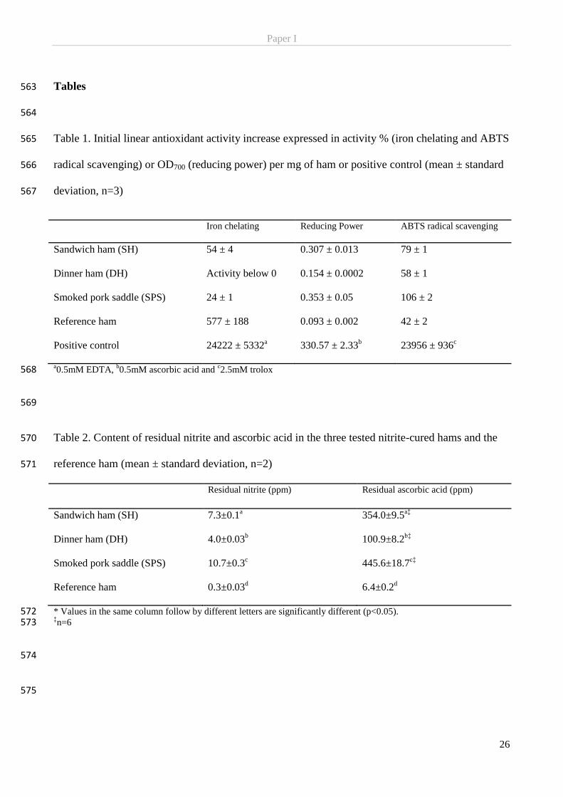
Paper I
26
Tables 563
564
Table 1. Initial linear antioxidant activity increase expressed in activity % (iron chelating and ABTS 565
radical scavenging) or OD700 (reducing power) per mg of ham or positive control (mean ± standard 566
deviation, n=3) 567
Iron chelating Reducing Power ABTS radical scavenging
Sandwich ham (SH)
54 ± 4 0.307 ± 0.013 79 ± 1
Dinner ham (DH)
Activity below 0 0.154 ± 0.0002 58 ± 1
Smoked pork saddle (SPS)
24 ± 1 0.353 ± 0.05 106 ± 2
Reference ham
577 ± 188 0.093 ± 0.002 42 ± 2
Positive control
24222 ± 5332a 330.57 ± 2.33
b 23956 ± 936
c
a0.5mM EDTA,
b0.5mM ascorbic acid and
c2.5mM trolox 568
569
Table 2. Content of residual nitrite and ascorbic acid in the three tested nitrite-cured hams and the 570
reference ham (mean ± standard deviation, n=2) 571
Residual nitrite (ppm) Residual ascorbic acid (ppm)
Sandwich ham (SH) 7.3±0.1a 354.0±9.5
a‡
Dinner ham (DH) 4.0±0.03b 100.9±8.2
b‡
Smoked pork saddle (SPS) 10.7±0.3c 445.6±18.7
c‡
Reference ham 0.3±0.03d 6.4±0.2
d
* Values in the same column follow by different letters are significantly different (p<0.05). 572 ‡n=6 573
574
575
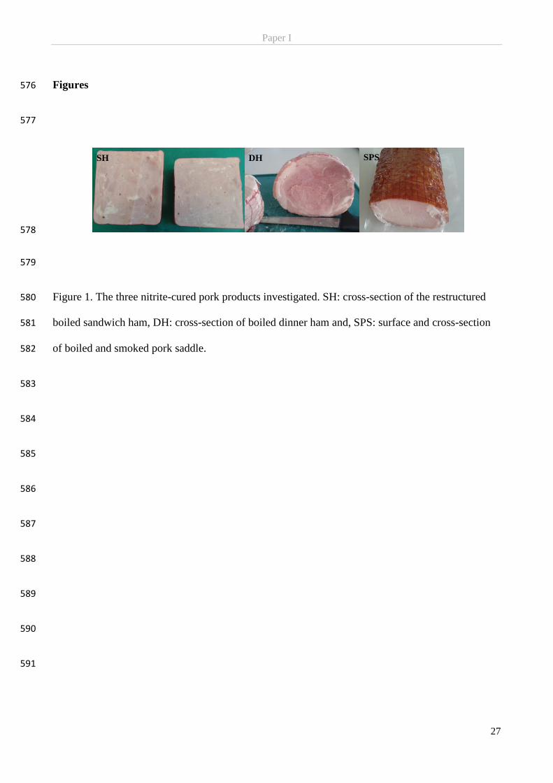
Paper I
27
Figures 576
577
578
579
Figure 1. The three nitrite-cured pork products investigated. SH: cross-section of the restructured 580
boiled sandwich ham, DH: cross-section of boiled dinner ham and, SPS: surface and cross-section 581
of boiled and smoked pork saddle. 582
583
584
585
586
587
588
589
590
591
SH DH SPS
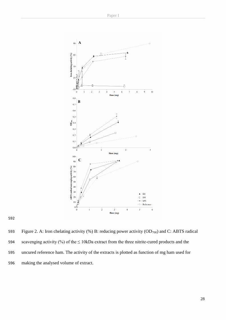
Paper I
28
592
Figure 2. A: Iron chelating activity (%) B: reducing power activity (OD700) and C: ABTS radical 593
scavenging activity (%) of the ≤ 10kDa extract from the three nitrite-cured products and the 594
uncured reference ham. The activity of the extracts is plotted as function of mg ham used for 595
making the analysed volume of extract. 596

Paper I
29
597
Figure 3. The relative content of the total amino acids in % in the sandwich ham (SH), the dinner 598
ham (DH), the smoked pork saddle (SPS) and the uncured reference ham (Reference). 599
600
601
Figure 4. Log10 transformed bacterial count from the growth inhibition study (study of 602
antimicrobial activity of ham extract) on Listeria monocytogenes and Salmonella typhimurium after 603
0, 24, 48 and 72 hours incubation. Control indicates ham extract being substituted for equivalent 604
amounts FKP. 605
606
Paper II
_______________________________________________________________________________________
Pedersen, S. T., Duedahl-Olesen, L. & Jessen, F. Characterization of low molecular
weight peptidic extracts from nitrite-cured cooked pork products. Submitted to
Journal of the Science of Food and Agriculture.
_______________________________________________________________________________________
For Peer Review
Characterization of low molecular weight peptidic extracts
from nitrite-cured cooked pork products
Journal: Journal of the Science of Food and Agriculture
Manuscript ID Draft
Wiley - Manuscript type: Research Article
Date Submitted by the Author: n/a
Complete List of Authors: Pedersen, Sabrine; Technical University of Denmark, National Food Institute Duedahl-Olesen, Lene; Technical University of Denmark, National Food Institute Jessen, Flemming; Technical University of Denmark, National Food Institute;
Key Words: ham, ABTS radical scavenging, reducing power, amino acids, nitrite-curing, FPLC
Journal of the Science of Food and Agriculture
For Peer Review
�
Title: Characterization of low molecular weight peptidic extracts from nitrite-cured cooked pork ��
products ��
Authors: Sabrine Tauber Pedersena, Lene Duedahl-Olesena & Flemming Jessena. ��
a National Food Institute, Technical University of Denmark, Søltofts Plads Building 227, DK-2800 ��
Kongens Lyngby, Denmark ��
��
Running title: Characterization of origin of antioxidant activity in extracts from cured hams ��
�
*Corresponding author: �
Flemming Jessen ���
Postal address: ���
National Food Institute, ���
Technical University of Denmark ���
Søltofts Plads Building 221, ground floor, room 050 ���
DK-2800 Kgs. Lyngby ���
E-mail: ���
��
Declaration of conflict of interest: none ��
Page 1 of 30
Journal of the Science of Food and Agriculture
123456789101112131415161718192021222324252627282930313233343536373839404142434445464748495051525354555657585960
For Peer Review
�
Abstract ���
BACKGROUND: Nitrite-cured cooked pork products constitute an immense variety of products ���
but a common characteristic is a long shelf-life attributed to by the curing process. In order to ���
contribute to a better understanding of the antioxidative mechanisms occurring in nitrite-cured ���
cooked pork products to ultimately optimize nitrite addition the aim of this study was to investigate ���
the contribution to antioxidant activity of other compounds than residual ascorbate and nitrite from ���
aqueous ≤10kDa extracts of cold-stored nitrite-cured cooked pork products. ���
RESULTS: By means of size exclusion chromatography (SEC), in vitro antioxidant assays and ���
constituent amino acids analysis it was demonstrated that the extracts exhibited antioxidant activity ��
in the form of reducing power and ABTS radical scavenging activity (with different kinetics). It was ��
also demonstrated that this activity was concentrated to only a few fractions from the SEC ���
separation. The SEC results indicated the antioxidant activity to originate from compounds <6.5kDa ���
and could include small peptides and free amino acids e.g. the aromatic phenylalanine and tyrosine. ���
Furthermore, the extracts were high in the antioxidant amino acids histidine and methionine. ���
CONCLUSION: The antioxidant activity did neither stem from residual nitrite nor ascorbate but it ���
could be associated with nitrite-related compounds formed during curing and heating. This shows ���
that we need to expand our perception of the cause of oxidative stability in nitrite-cured cooked ���
hams to include more than just nitrite and ascorbate. ���
���
Keywords: ham, ABTS radical scavenging, reducing power, amino acids, nitrite-curing, FPLC ��
����
� ����
Page 2 of 30
Journal of the Science of Food and Agriculture
123456789101112131415161718192021222324252627282930313233343536373839404142434445464748495051525354555657585960
For Peer Review
�
Introduction ���
Curing of meat is a well-established procedure for adding flavor and prolonging shelf-life of meat ���
products. In its general form meat curing consists of adding sodium chloride and salts of nitrite (or ���
nitrate) and ascorbate to raw meat, however, many curing processes also involves addition of spices ���
and further downstream processing as cooking and smoking. The chemistry of nitrite and ascorbate ���
in processed meat is complex and at times not fully understood. However, ascorbate as an electron ���
donor, is an effective antioxidant that through its relatively unreactive oxidized form ��
(dehydroascorbic acid) can terminate propagation of free radical reactions1. Chloride is in many ��
biological systems considered an unspecific prooxidant and although nitrite, as a natural electron ���
acceptor, can be considered a strong prooxidant several mechanisms have been suggested in favor ���
of nitrite as an active antioxidant2. However, it is well established that their combined addition ���
inhibits lipid oxidation2. ���
In a recent study aqueous low molecular weight extracts (≤10kDa) of nitrite-cured cooked hams ���
were demonstrated to exhibit in vitro antioxidant activity3. The origin of the antioxidant activity ���
was not fully elucidated and could as such originate from residues of the added antioxidants. ���
Activity originating directly from residual nitrite was rejected as the antioxidant activity of nitrite ���
primarily is exerted through other chemical forms e.g. NO. However, besides residual ascorbate the ��
results also indicated that compounds, potentially related to nitrite, formed during processing ��
(cooking and curing) could have contributed to the detected activity. Furthermore, certain peptides ���
or free amino acids may also have added to the detected antioxidant activity3. ���
Many peptides of 2-20 amino acids in length have been found to exhibit a range of bioactivities ���
including antioxidant activity4. Several amino acids such as histidine, methionine, tryptophan, ���
tyrosine and cysteine have generally been accepted as possessing antioxidant properties, a capacity ���
demonstrated in free form and/or enclosed in peptides5–7. Small antioxidant peptides have ���
Page 3 of 30
Journal of the Science of Food and Agriculture
123456789101112131415161718192021222324252627282930313233343536373839404142434445464748495051525354555657585960
For Peer Review
�
previously been identified in animal based foods. The most frequently mentioned are the ���
antioxidative endogenous histidyl dipeptides, carnosine and anserine which have been identified in ���
many different meats including pork meat8. However, other small peptides displaying antioxidant ��
properties have also been found in fresh pork ham9, porcine myofibrillar proteins7, sausages10 and ��
cured hams11,12. Most attempts to characterize and identify antioxidant peptides has been in raw ���
sausages or hams subjected to a drying or ripening step characterized by extensive protein ���
hydrolysis by endogenous enzymes or microorganisms or alternatively after enzymatic treatment on ���
the final product (or extracts hereof) – all resulting in the presence of immense amounts of small ���
peptides (and free amino acids). In cooked nitrite-cured hams this profound protein breakdown do ���
not occur. Thus, in order to contribute to a better understanding of the antioxidative mechanism ���
occurring in nitrite-cured cooked pork products, the aim of this study was to investigate the ���
potential origin of antioxidant activity from other compounds than residual ascorbate and nitrite. It ���
was hypothesized that aqueous ≤10kDa extracts of nitrite-cured cooked hams contains compounds ��
of peptide origin contributing to antioxidant activity. ��
Materials and method ��
Materials ��
For nitrite determination and antioxidant assays sodium nitrite, ascorbic acid, and 2,2′-Azino-bis(3-��
ethylbenzothiazoline-6-sulfonic acid) diammonium salt (ABTS), were acquired from Sigma-��
Aldrich (St. Louis, MO, USA). Ethylenediaminetetraacetic acid disodium dihydrate (EDTA) were ��
from Merck Millipore (Billerica, MA, USA) and (±)-6-Hydroxy-2,5,7,8-tetramethylchromane-2-��
carboxylic acid (Trolox) from Fluka Chemika (Deisenhofen, Germany). Cytochrome C from equine ��
heart (CytC), aprotinin from bovine lung, triglycine (Gly3), L-tryptophan (Trp) and L-histidine ��
(His), L-methionine (Met), L-tyrosine (Tyr), L-proline (Pro) and L-glutamic acid (Glu) were from �
Sigma-Aldrich while L-cysteine (Cys) and L-phenylalanine (Phe) were from Merck Millipore and �
Page 4 of 30
Journal of the Science of Food and Agriculture
123456789101112131415161718192021222324252627282930313233343536373839404142434445464748495051525354555657585960
For Peer Review
�
glycine (Gly) were supplied by AppliChem GmbH (Darmstadt, Germany). L-alanine (Ala) was ��
from Fluka Chemika. Remaining chemicals and reagents employed were of analytical grade and ��
acquired from VWR International (Radnor, PA, USA), Merck Millipore, Sigma-Aldrich or Fluka ��
Chemika. ��
Hams ��
The sample material comprised three different types of commercially available nitrite-cured cooked ��
pork products: a restructured sandwich ham (SH), a whole-muscle boiled dinner ham (DH) and a ��
whole-muscle smoked pork saddle (SPS). SH was added 60 ppm nitrite while DH and SPS were ��
added 120ppm nitrite. At the laboratory, four days after production, the pork products were cut into �
smaller pieces, vacuum-packed, stored at 5°C (from now on referred to as day 0) and samples were �
periodically taken and moved to -80ºC until further analysis. ����
The samples were tested without any further heat treatment. Any remaining skin and excess ����
subcutaneous fat on the boiled dinner ham were trimmed before mincing. The smoke-colored ����
surface of the pork saddle was not removed as it is expected to be eaten by the majority of ����
Scandinavian consumers, unlike the fat and skin on the dinner ham. ����
Extraction of water soluble ham components and preliminary fractionation ����
Water soluble compounds were extracted from the cured hams by homogenising 79.4-114.3g of ����
minced ham in 400mL of deionized water, dH2O, for 1min with an Ultra Turrax (T 25 Basic ����
ULTRA-TURRAX® mounted with S25N -18G dispersing element, IKA®-WERKE GmbH &Co. ���
KG, Staufen, Germany) at 13,500rpm and subsequently adding up to a total volume of 1.5L. An ���
aqueous ≤10kDa extract was collected by submerging five times 10cm dialysis membrane tubes ����
(Spectra/Por 6, MWCO 10,000Da, Spectrum Lab. Inc, CA, USA) containing 16.5mL dH2O ����
(corresponding to 50% membrane volume) into the meat/water homogenate. After 24h of magnetic ����
stirring at 5ºC, the dialysate (extract) in the 5 membranes were pooled and stored at -80ºC until ����
Page 5 of 30
Journal of the Science of Food and Agriculture
123456789101112131415161718192021222324252627282930313233343536373839404142434445464748495051525354555657585960
For Peer Review
�
further analysis. Later the protein content was determined using the PierceTM Modified Lowry ����
Protein Assay Kit in microplate format according to manufactures description (Thermo Scientific, ����
PierceTM) using Bovine Serum Albumin (BSA) (Albumin Standard Ampules, 2 mg/ml) supplied ����
with the kit as an external standard. ����
Size exclusion chromatography (SEC) ���
Size exclusion chromatography (SEC) was performed on the aqueous ≤10kDa extract on fast ���
performance liquid chromatography (FPLC) equipment (Äkta Purifier system with Frac 950 ����
collector, GE Healthcare Life Sciences, Little Chalfont, UK). In short, 100µL or 200µL of the ����
extract were injected three times onto a SuperdexTM peptide 10/300 GL column (GE Healtcare), ����
using a 100mM ammonium acetate, pH 8 as running buffer at a flow rate of 0.5mL/min. Eluting ����
compounds were detected at 215nm (peptide bonds), 254nm (phenylalanine) and 280nm (aromatic ����
ring e.g. tyrosine, tryptophan, phenylalanine). CytC (12.3kDa), Aprotinin (6.5kDa), Gly3 (189Da) ����
and the amino acids glycine (75Da), cysteine (121Da), histidine (155Da), phenylalanine (165Da) ����
and tryptophan (204Da) were used as external standards. Elution volumes and molecular weights of ����
standards are not fully related as the column possesses some hydrophobic affinity properties apart ���
from being a size exclusion column. For each extract-run, 94 fractions (SEC fractions) of 320µL ���
each were collected into a 96 well microplate and the plate was stored at 5ºC until the run for the ����
third injection, of the same extract, had finish. The content of each well were then pooled, in ����
Eppendorf tubes, with the corresponding well in the other two plates of the same sample and stored ����
at -20ºC until further analysis. ����
Chemical analyses ����
Nitrite ����
The day before analysis, approximate 100g of ham sample stored at -80ºC was thawed, minced two ����
times 5sec, and re-vacuum packed before being returned to -80ºC. Extraction and measurement of ����
Page 6 of 30
Journal of the Science of Food and Agriculture
123456789101112131415161718192021222324252627282930313233343536373839404142434445464748495051525354555657585960
For Peer Review
�
nitrite in the cured cooked pork products was done according to Herrmann, Duedahl-Olesen & ���
Granby (2015)13. The samples were analyzed in a random order in duplicate. External calibration ���
was performed in the range 0.25-100µg/mL NaNO2. As the nature of the added nitrite was unknown ����
in SH all results are presented as ppm nitrite-ion by the relationship 1g nitrite ion = 1.50g NaNO2. ����
Salt ����
The salt content of the pork extracts was determined through potentiometric titration of chlorine ����
ions/chloride using AgNO3 according to AOAC Official Method 976.18 (Salt (Chlorine as Sodium ����
Chloride) in Seafood. Potentiometric method, 2000). ����
Protein ����
The protein content of the collected SEC separated fractions were determined using the PierceTM ����
BCA Protein Assay Kit according to manufactures description for determination in microplates ���
(Thermo Scientific, PierceTM) using Bovine Serum Albumin (BSA) (Albumin Standard Ampules, 2 ���
mg/ml) supplied with the kit as an external standard. ����
Amino acid profile (total and free) ����
The total amino acid content was analyzed by subjecting 0.5mL of the aqueous ≤10kDa extract to ����
microwave-assisted acidic hydrolysis (6M HCl) for 1h at 110ºC and 500W followed by 10min of ����
cooling. The samples were diluted with 1M NaCO3 and filtered through a 0.2µm syringe filter (Q-����
max PTFE, Ø13mm, Frisenette ApS, Knebel, Denmark) before derivatization using the EZ:FaastTM ����
Amino Acid Analysis kit from Phenomenex® (Torrance, CA, USA). The derivatized samples were ����
analyzed by LC-(APCI)-MS (Agilent 1100, Agilent Technology) according to the procedure ����
described by Farvin, Baron, Nielsen, Otte & Jacobsen (2010)14. ���
Free amino acid content was determined by introducing the extracts at the derivation step and ���
otherwise proceeding as described above. The analysis was done in duplicate.�����
Page 7 of 30
Journal of the Science of Food and Agriculture
123456789101112131415161718192021222324252627282930313233343536373839404142434445464748495051525354555657585960

For Peer Review
�
����
Antioxidant assays ����
Potential antioxidant properties of the SEC fractions were analyzed using two in vitro assays for ����
antioxidant activities; reducing power and ABTS-radical scavenging. The assays were conducted in ����
triplicate and all measurements were performed using a Synergy 2 Multi-Mode Microplate Reader ����
(BioTek® Instruments, Inc., Winooski, VT, USA). Before analysis the SEC fractions were thawed ����
at 5ºC, overnight. ����
The reducing power was determined according to the procedure described by Oyaizu (1986)15 with ���
some modifications and adjusted to microplate detection. First, equal parts of each SEC fraction, ���
0.2M phosphate buffer (pH 6.6) and 1% potassium ferricyanide were mixed and incubated for ����
20min at 50°C. Subsequently, 1 part of 10% TCA was added. Then, 100µL of the sample mixture ����
was mixed with 100µL dH2O and 20µL of 0.1% ferric chloride, and incubated 10min at room ����
temperature. The absorbance was measured at 700nm. Ascorbic acid (0.5mM) was used as positive ����
control and dH2O and 100mM ammonium acetate were used as blanks. Results are expressed as ����
����� � �������� ����� � ��������� ���
The radical scavenging activity was determined according to the method described by Gringer, ����
Osman, Nielsen, Undeland & Baron (2014)16. Trolox (2.5mM, in 96% ethanol) was used as a ����
positive control. Ethanol (96%) and 100mM ammonium acetate were included as blanks for the ����
positive control and sample, respectively. The ABTS radical scavenging activity (%) is expressed as ���
the relative decrease in color/absorbance (734nm): ���
����� ��� ��� ������� ������ �!� ����"#$% � ���&$'#(
���"#$%) *++!
Statistical analysis ���
Page 8 of 30
Journal of the Science of Food and Agriculture
123456789101112131415161718192021222324252627282930313233343536373839404142434445464748495051525354555657585960
For Peer Review
�
Measurements were carried out in triplicate (unless it is stated otherwise). Results are presented as ���
mean ± standard deviation. All statistical analyses were conducted using the GraphPad Prism® ���
software Ver. 4.03 or R version 3.3.0 with P < 0.05. Results were compared using one-way ���
ANOVA with Tukeys posttest. ���
���
Results and discussion ���
Nitrite was measured at several time points during storage of the cured hams to identify the time ���
point at which residual nitrite reached a low level to limit any potential interference e.g. ensuring ��
that formation of nitrite related reaction products will have reached its maximum/steady level. ��
The residual nitrite concentration in the tested hams decreased over time, as was expected, and ���
reached a common low level after 37 days (Figure 1). The smoked pork saddle (SPS) had the ���
highest concentration of residual nitrite at 10.7±0.3ppm at day 0 followed by the sandwich ham ���
(SH) and boiled dinner ham (DH) at 7.3±0.1ppm and 4.0±0.03ppm, respectively. Cassens (1997)17 ���
reported residual nitrite levels of 7±1ppm and 4±1ppm in commercial sliced hams which was ���
similar to SH and DH. After end processing and heat treatment residual nitrite has been reported to ���
be reduced to ≤35% of the originally added amounts18, fitting with the current data (day 0). The ���
product difference in residual nitrite at day 0 did not correlate with added nitrite as DH and SPS ���
were added twice the amount of nitrite compared to SH. After the 37 days the residual nitrite ��
concentration reached a low level around 1-2ppm for all hams. Pérez-Rodríquez, Bosch-Bosch and ��
Garciá-Mata (1996)19 measured residual sodium nitrite in pork frankfurter sausages over time and ����
found residual sodium nitrite in a sausage (125ppm NaNO2, 500ppm Na-ascorbate, 2.2% NaCl) to ����
be fairly constant just below 7 ppm NaNO2 (equivalent <4.7 ppm NO2-ion) after 27 days of storage ����
at 3°C (NaNO2 content was extrapolated from Fig. 2 in Pérez-Rodríquez et al. (1996)19). Also in the ����
Page 9 of 30
Journal of the Science of Food and Agriculture
123456789101112131415161718192021222324252627282930313233343536373839404142434445464748495051525354555657585960
For Peer Review
�
lean as well as fat part of bacon (156ppm NaNO2, 500ppm ascorbate, 2.5% NaCl) residual sodium ����
nitrite were close to zero after 35 days at 2°C20. ����
From the hams stored at 5°C for 37 days, ≤10kDa aqueous extracts were produced and used for ����
characterization. The ≤10kDa extracts were produced from meat/water homogenates made from ����
slightly different amounts of meat. Yet, the protein content were very similar between the extracts, ���
(SPS: 0.17mg/mL, DH: 0.17mg/mL, SH: 0.16mg/mL) and did not correlate with the difference in ���
amount of meat used. Based on this the extracts were regarded to be comparable. However, due to ����
much lower absorbance signals from the ≤10kDa extract from DH compared to SH and SPS (data ����
not shown) when injecting 100µL into the FPLC it was decided to inject twice the volume of DH ����
making the signal magnitude of the absorbance profile for DH comparable to SH and SPS. The ����
extracts all had similar SEC profiles (Figure 2) according to the absorbance at 215nm, and were ����
characterized by four main peaks (I:18.5-19.9mL, II:19.9-21.4mL, III: 21.4-22.9mL and IV: 25.3-����
27.2mL), a shoulder eluting before peak I and a peak eluting after peak III only base-line separated ����
in SH (Figure 2 top panel). ����
Only minor signals were detected from aromatic rings absorbing at 280nm and these correlated ���
nicely with peak II-IV. The strongest signal at 280nm was achieved in peak III in DH, Figure 2 ���
middle panel. Aromatic rings were also detected at 254 nm (optimum phenylalanine). This signal ����
generally correlated well with the elution pattern from the 280nm signal yet gave rise to much ����
higher maximum absorbance in the range 15-500mAU compared to 2-90mAU at 280nm. ����
Although the SEC chromatograms show similar profiles between the three samples (non-����
quantitatively) the mutual relationship between the peak maximum differed between the hams ����
(Figure 2). As both DH and SPS are whole muscle products and were both added the same amount ����
of nitrite - 120ppm (information given by manufacture) corresponding to twice the amount added to ����
SH – they would be expected to be the most similar. However, if focusing on 254nm and 280nm ����
Page 10 of 30
Journal of the Science of Food and Agriculture
123456789101112131415161718192021222324252627282930313233343536373839404142434445464748495051525354555657585960
For Peer Review
�
curves the inter-peak relationship shows greater resemblance between SPS and SH. This is also ���
contradictive to the fact that the smoking of the SPS could potentially cause formation of aromatic ���
smoke components e.g. phenolic compounds and polycyclic aromatic hydrocarbons (PAH)21,22, ����
possibly increasing the aromatic absorbance signal compared to non-smoked hams. For example ����
Chen, Wang and Chiu (1996)23 detected 16 different PAHs in several meat products, including ����
smoked pork, at 254nm after separation on a C18 column. ����
CytC and aprotonin eluted before the sample constituents indicating the aqueous ham extracts only ����
contained compounds, detectable by the current FPLC method, that were smaller than 6.5kDa ����
(Figure 2). Thus, the presence of peptides with antioxidant properties in the extracts is not unlikely ����
as antioxidant peptides seldom contain more than 20 amino acids per molecule and have masses ����
below 6000Da4,24. For example Escudero et al., (2012)11 found a peptide fraction <1700Da from ���
dry cured ham to possess antioxidant activity while Li, Han & Chen (2008)25 indicated that peptides ���
of 500-1500Da exhibited stronger antioxidant activity than both smaller and larger peptides. Peak I ����
including the anterior shoulder, present in all three extracts, spanned the elution of Gly3 (189Da → ����
18.4mL), glycine (75Da → 19.3mL), and towards the end cysteine (121Da → 19.7mL) and ����
histidine (155Da → 19.9mL). The elution of the remaining two standards, phenylalanine (165Da) ����
and tryptophan (204Da), which both contain an aromatic ring, correlated with the sample elution of ����
peak II (280 and 254nm) and a minute 280nm-peak eluting at approximately 28.5mL (Figure 2). ����
The inconsistency in relation between the sizes of standards and their elution volumes is due to ����
column properties (see Materials and Methods) ����
The results from in vitro determination of reducing power and ABTS radical scavenging activity in ���
the collected SEC separated fractions are presented in Figure 3. Reducing power (Figure 3A) is for ���
all three extracts concentrated around two fraction-peaks, P1 and P2. DH showed the highest ����
reducing power activity of the three, in fraction-peak P2, and SPS the lowest. The higher signal ����
Page 11 of 30
Journal of the Science of Food and Agriculture
123456789101112131415161718192021222324252627282930313233343536373839404142434445464748495051525354555657585960
For Peer Review
�
from DH might have been due to the injection of 200µL of this extract to the FPLC which as such ����
created a different foundation for the collected SEC fractions compared to the remaining two where ����
only 100µL were injected. It is noteworthy that P1 fell within the range of peak I (including the ����
anterior shoulder) in the SEC chromatogram (Figure 2). Yet more notable that reducing power ����
fraction-peak, P2, perfectly aligned with chromatographic peak, II, with a strong signal at all three ����
wavelengths, but in particular 215nm and 254nm (Figure 2). This indicated the presence of peptide ����
bonds and aromatic rings among the eluting compounds in the antioxidant active (reducing power) ���
fraction-peak. Furthermore, P2 and peak II also coincided with the elution of the external ���
phenylalanine standard (Figure 2 top panel). Thus, it can be speculated that the reducing power ����
noted in the tested ham extracts may in full (or partly) have been caused by phenylalanine or ����
alternatively small peptides. As a result of the ring structure the aromatic amino acids ����
phenylalanine, tyrosine and tryptophan can act as proton donors for free radicals deficient in ����
electrons and efficiently scavenge them26. Broncano et al. (2012)10 for example suggested that a ����
tripeptide Phe-Gly-Gly with the N-terminal phenylalanine is capable of scavenging DPPH radicals. ����
Despite studies showing that antioxidants may display different efficiency in scavenging the ABTS ����
and the DPPH radical27 it would be expected, based on the above, that peak II would correlate with ����
a fraction-peak in the ABTS radical scavenging profile rather than with a reducing power fraction-���
peak. Thus, if the assumption that phenylalanine is contributing to the discovered reducing power ���
activity is valid, then the aromatic amino acids including phenylalanine may potentially also be able ����
to contribute to the reduction of iron. However, no reducing power activity could be detected for ����
free phenylalanine at concentrations ≤1mg/mL while tryptophan exhibited some activity ����
(OD700=0.06 at 1mg/mL) (supplementary data, Figure 1S). ����
Incubation time used for ABTS radical scavenging activity assays varies a lot between laboratories ����
and also the procedure differs slightly. Some studies apply 1-10min16,28,29 while others report up to ����
Page 12 of 30
Journal of the Science of Food and Agriculture
123456789101112131415161718192021222324252627282930313233343536373839404142434445464748495051525354555657585960
For Peer Review
�
1h incubation30. Thus ABTS radical scavenging activity was measured after 1min (Figure 3B) ����
incubation to detect a fast response and 31min (Figure 3C) incubation to look for any slower acting ����
activities. After 1min incubation activity was detected in fraction-peak, P3, at 21.1-23.1mL. After ���
additional 30min incubation the ABTS radical scavenging activity in P3 had only increased slightly ���
from 17.9 to 23.1% for SPS and 20.6 to 28.2 % for DH while it increased a bit more for SH from ���
9.2 to 24.6%. However, after the additional 30min incubation ABTS radical scavenging activity ���
also appeared in the fraction-peak P4. This clearly showed different reaction kinetics of the mixture ���
of active compounds enclosed in P3 and P4. Thus, it should be considered for future studies to ���
measure ABTS radical scavenging activity at more time points to gain more information on ���
antioxidant compound kinetics. Overall DH displayed the strongest ABTS radical scavenging ���
activity however as argued before this could be due to the larger injection volume. Aside from this ���
SH showed higher ABTS radical scavenging activity than SPS in P4 while the relation was reversed ���
in P3. ��
The position of ABTS radical scavenging activity fraction-peak P4 corresponded to reducing power ��
fraction-peak P1 (Figure 3A and C) and thus, also aligned with the large peak I (Figure 2), in the ���
SEC chromatogram yet slightly shifted to the right. At this point of elution the absorbance at 254nm ���
and 280nm was very limited and thus led to idea that the antioxidant activity in peak I/P1 could ���
involve the action of small peptides but also free amino acids (other than the aromatic). The longer ���
incubation time required for P4 was also evident especially for the free amino acids histidine, ���
proline and glycine when analyzed in concentration >0.02mg/mL but not at concentration ���
equivalent to the content of the individual free amino acids in the extracts (Figure 4 and ���
supplementary data, Figure 2S). ���
The ABTS radical scavenging activity detected after 1min incubation, P3, (Figure 3) aligned nicely ��
with peak III absorbing at both 215nm and 254nm/280nm (Figure 2). Therefore some of the ��
Page 13 of 30
Journal of the Science of Food and Agriculture
123456789101112131415161718192021222324252627282930313233343536373839404142434445464748495051525354555657585960
For Peer Review
�
detected activity may again have been the action of a peptide or a compound containing some sort ����
of aromatic ring. This could have been from the amino acids tyrosine as the elution of the other ����
aromatic amino acids phenylalanine and tryptophan was already established. Yet nothing conclusive ����
can be said on this matter as solubilization and therefore injection of tyrosine was unsuccessful. ����
However, solutions of tyrosine (although turbid) were found to be highly potent at color reduction ����
even at low concentrations (15-22% ABTS radical scavenging activity at 0.002mg/mL, see ����
supplementary data, Figure 2S). ����
In spite of the SEC separation of the extracts resulted in four main absorbance peaks, the ����
antioxidant activity was limited to three of these as described above. It is of no surprise that the ���
extract would contain compounds of no antioxidant activity. Yet it is relatively unexpected that ���
compounds giving rise to an absorbance peak at 254nm and 280nm (peak IV in Figure 2) would not ����
possess some antioxidant activity as the most common antioxidants from food matrixes contain ����
aromatic rings. However, it cannot be ruled out that a potential antioxidant activity from compounds ����
in this peak simply is not detectable by the two in vitro antioxidant assays applied. ����
The importance of nitrite for the oxidative stability of meat inspires the hypothesis that hams added ����
more nitrite would exhibit more antioxidant activity. However, comparing SH and SPS (same ����
volume used for SEC fractionation) this was not unambiguous substantiated. In the peak-fractions ����
P2 (reducing power) and P3 (ABTS radical scavenging 1min peak) the hypothesis of more added ����
nitrite being equivalent to more activity was supported. On contrary P4 (ABTS radical scavenging ���
31min peak) in SH showed stronger activity than SPS. Morrissey & Tichivangana (1985)31 also ���
demonstrated a significant reduction in lipid oxidation/TBA values at the addition of 20ppm nitrite ����
to pork (and other meat types) which further decreased to a 12-fold reduction at the addition of ����
200ppm. On contrary MacDonald, Gray & Gibbins (1980)32 found higher concentrations of nitrite ����
to cause more pronounced prooxidant effect on lipid oxidation, but the level of nitrite in commercial ����
Page 14 of 30
Journal of the Science of Food and Agriculture
123456789101112131415161718192021222324252627282930313233343536373839404142434445464748495051525354555657585960
For Peer Review
�
meat products was too low to exert this effect. Thus, the current data does not rule out the ����
importance of nitrite in regards to presence/creation of compounds possessing antioxidant activity ����
yet neither does it confirm its insignificance. ����
Ascorbic acid/ascorbate is a known antioxidant33, which was also added to all three hams, and as ����
such could have disturbed the measurements. Extracts, similar to the current ham extracts, however ���
produced from sausages of varying nitrite and ascorbate content were fractionated by SEC (see ���
supplementary data, Figure 3S). A massive peak protruded at 18.5-20.2 mL, which coincided with ����
peak I observed here, which did not materialized when ascorbic acid was not added and only ����
resulted in a minor peak when added in smaller amount (240ppm). However, as would be expected ����
from the ring structure of ascorbic acid the peak was due to high absorbance at 254 and 280nm. As ����
no such peak were evident in the current study it is considered unlikely that the detected reducing ����
power and ABTS radical scavenging is caused by residual amounts of the ascorbic acid added to the ����
hams. ����
The total amino acid profile was relatively similar between the three extracts and for 13 of the 19 ����
investigated amino acids no statistical differences were detected (Figure 4A). Comparison of the ���
distribution of the individual amino acids (percent of total amino acid) in the extract to the ���
distribution found in raw and cooked (60 and 75ºC) pork muscle34 and ham products in the Danish ����
food composition database (Frida Food Data - http://frida.fooddata.dk/35) revealed some striking ����
differences. The profiles from the literature were dominated by glutamic acid (glutamic acid + ����
glutamine) and aspartic acid (aspartic acid + asparagine) a result also found during previous ����
investigations of the three ham products used in the current study3. However, the extracts were ����
dominated by histidine, alanine, methionine, glutamic acid and glycine in decreasing order. Both ����
histidine and methionine are among the amino acids frequently mentioned in regard to peptide ����
related antioxidant activity and thereby supported the expectation of finding higher antioxidant ����
Page 15 of 30
Journal of the Science of Food and Agriculture
123456789101112131415161718192021222324252627282930313233343536373839404142434445464748495051525354555657585960
For Peer Review
�
activity in the ≤10kDa fraction compared to the entire ham. It is also noteworthy that carnosine ���
(226.24Da), a dipeptide found in meat possessing antioxidant activity, is made up of exactly alanine ���
and histidine. ����
The importance of aromatic amino acids in antioxidative peptides has been emphasized previously ����
yet the presence of other amino acids in the sequence e.g. glycine, cysteine and the acidic amino ����
acids may also be of great importance for antioxidant properties in a peptide26. However, from an ����
alignment of the sample and standard SEC chromatograms and the antioxidant activity profiles no ����
information can be drawn concerning small antioxidative peptides of this particular composition. ����
Although SH and SPS contains significantly more histidine and alanine no direct correlation can be ����
made from the content of the amino acids and the detected antioxidant activity due to the difference ����
in starting material for the assays. ���
Several amino acids have also been found to possess antioxidant properties as free amino acids36 ���
including histidine, tryptophan and cysteine5,6. The latter two showed as high ABTS radical ����
scavenging activity at 0.05mg/mL as the positive control (2.5mM Trolox) while cysteine also had ����
the strongest reducing power of the ten tested amino acids (supplementary data, Figure 2S). The ����
content of free amino acids was, as expected, very low: 0.0788, 0.0799 and 0.0684 mg amino acid/g ����
extract in SH, SPS and DH respectively. However, the amount of free amino acids accounted for ����
23-27% of the total amino acid composition of the extract. The free amino acid pool was more even ����
distributed between the individual amino acids compared to the total amino acids (Figure 4B). ����
Overall the free amino acids profiles were very similar but for seven amino acids the content was ����
significantly different in one ham extract compared to the remaining two. SPS contained higher ���
amounts of lysine, tyrosine and aspartic acid compared to SH and DH. DH contained lower ���
amounts of hydroxyproline, methionine and threonine, compared to in the remaining extracts while ����
SH contained lower amount of valine. Although alanine generally was dominating the free amino ����
Page 16 of 30
Journal of the Science of Food and Agriculture
123456789101112131415161718192021222324252627282930313233343536373839404142434445464748495051525354555657585960
For Peer Review
�
acid profile, alanine in the free form is seldom mentioned in regard to antioxidant activity but ����
mainly in connection with carnosine. Also, alanine did not demonstrate any reducing power or ����
ABTS radical scavenging activity in concentration ≤1mg/mL (supplementary data, Figure 2S). ����
However, alanine was by Karel et al. (1966)5 found to exhibit antioxidant activity. Thus, it cannot ����
be ruled out that alanine contributed to the peptide related antioxidant pool yet the extent of its ����
contribution is unclear. Consequently, it must be assumed that alanine together with the remaining ����
free amino acids, all jointly contributed to the observed antioxidant activity. ���
The fractions giving rise to the main peaks in the SEC chromatograms that also showed antioxidant ���
activity were pooled and the antioxidant activities were normalized to the protein content (Table 1). ���
The slight shift between P1 and P4 was disregarded and the two peak-fractions were pooled as one. ���
Based on protein content the rank order between the samples in P1 changed from DH showing a ���
slightly higher reducing power than SH with SH possessing double the�protein specific reducing ���
power of DH (Table 1). Considering the injected amount of protein (SPS: 17µg, DH: 34µg, SH: ���
16µg) it appeared that the changes in the previously similar OD700 of SH and DH almost matched ���
the difference in injected amount of protein. This could be an indication of the reducing power ���
detected in P1 being caused by proteins. The same correlation to protein content was not evident for ���
P2. However, the biuret reaction constituting the foundation for the BCA method is limited by not ��
detecting proteins smaller than tripeptides although studies have suggested that dipeptides also ��
affect the color formation37,38. Anyway, the method does not detect free amino acids making the ���
previously presented hypothesis of the activity originating from phenylalanine in P2 still applicable. ���
Although SPS had the highest content of free phenylalanine (Figure 4B) it was not significantly ���
different from the two remaining ham extracts. Taking this and any uncertainties introduced with ���
the normalization into consideration it must be noted that DH demonstrated almost twice the�protein ���
specific reducing power of SPS (double amount injected of DH). Also that the relationship between ���
Page 17 of 30
Journal of the Science of Food and Agriculture
123456789101112131415161718192021222324252627282930313233343536373839404142434445464748495051525354555657585960
For Peer Review
�
protein specific reducing power for SPS and DH (68/73=0.93) matched the relationship between ���
injected protein (16/17=0.94) ���
The protein specific ABTS radical scavenging activity in P4 was the highest in SH, very similar in ��
DH, however, only half in SPS (Table 1). Thus, a correlation between the ABTS radical scavenging ��
activity detected in P4 and protein in general is not obvious. Neither does anything in the free amino ����
acid profile seem to offer a clear explanation. Even though the free histidine content is highest in ����
SH (Figure 4B) the standard deviation on DH, and the fact that more was injected of this extract, ����
would lead to the expectation of DH showing the highest protein specific ABTS radical scavenging ����
activity if it was to be caused by free histidine. From the current data it is not possible to determine ����
if the detected activity originated from proteins or free amino acids. It can be speculated that the ����
activity in the SH and DH are related to protein content as they approach each other numerically ����
and that the higher protein specific ABTS radical scavenging activity in SPS may be related to ����
compounds not present in DH and SH that could contribute to ABTS radical scavenging activity. ���
As SPS is the only smoked ham it is sensible to assume that such antioxidant compounds could ���
originate from this particular processing step as several smoke phenols have been demonstrated to ����
be active antioxidants22. Furthermore, it can be speculated whether nitrogen oxides from the smoke ����
could have entered into some of the many reactions leading to the antioxidant effects of nitrite. ����
����
Conclusion ����
Conclusively, the characterization of the aqueous low molecular weight extracts (≤10kDa) from the ����
three nitrite-cured cooked pork products revealed that the detected antioxidant activity likely ����
originated from a broad variety of components. Components both natural to the meat e.g. peptides ����
and free amino acids, and components related to the processing e.g. smoke components and nitrite-���
Page 18 of 30
Journal of the Science of Food and Agriculture
123456789101112131415161718192021222324252627282930313233343536373839404142434445464748495051525354555657585960
For Peer Review
�
related compounds formed during curing and heating. However, further investigations are needed in ���
order to fully determine the extent of the antioxidant effect of the individual contributors - ����
knowledge needed to better understand the antioxidative mechanism in nitrite-cured cooked pork so ����
that nitrite addition may be optimized in the long term. ����
����
Funding ����
This work was supported by a grant from Norma and Frode S. Jacobsens Fond. ����
����
����
Page 19 of 30
Journal of the Science of Food and Agriculture
123456789101112131415161718192021222324252627282930313233343536373839404142434445464748495051525354555657585960
For Peer Review
�
References ���
1. Bendich A, Machlin LJ, Scandurra O, et al. The antioxidant role of vitamin C. Adv Free ���
Radic Biol Med 1986; 2: 419–444. ����
2. Skibsted LH. Nitric oxide and quality and safety of muscle based foods. Nitric Oxide 2011; ����
24: 176–183. ����
3. Pedersen ST, Baron CP, Gobba C De, et al. Antioxidative and antimicrobial activity of ����
extracts from nitrite-cured cooked pork products. Submitt to Food Chem. ����
4. Korhonen H, Pihlanto A. Bioactive peptides: Production and functionality. Int Dairy J 2006; ����
16: 945–960. ����
5. Karel M, Tannenbaum SR, Wallace DH, et al. Autoxidation of methyl linoleate in freeze-����
dried model systems. III. Effects of added amino acids. J Food Sci 1966; 31: 892–896. ���
6. Marcuse R. The effect of some amino acids on the oxidation of linoleic acid and its methyl ���
ester. J Am Oil Chem Soc 1962; 39: 97–103. ����
7. Saiga A, Tanabe S, Nishimura T. Antioxidant activity of peptides obtained from porcine ����
myofibrillar proteins by protease treatment. J Agric Food Chem 2003; 51: 3661–3667. ����
8. Mora L, Sentandreu MÁ, Toldrá F. Contents of creatine, creatinine and carnosine in porcine ����
muscles of different metabolic types. Meat Sci 2008; 79: 709–715. ����
9. Park SY, Chin KB. Antioxidant activities of pepsin hydrolysates of water- and salt-soluble ����
protein extracted from pork hams. Int J Food Sci Technol 2011; 46: 229–235. ����
10. Broncano JM, Otte J, Petrón MJ, et al. Isolation and identification of low molecular weight ����
antioxidant compounds from fermented ‘chorizo’ sausages. Meat Sci 2012; 90: 494–501. ���
Page 20 of 30
Journal of the Science of Food and Agriculture
123456789101112131415161718192021222324252627282930313233343536373839404142434445464748495051525354555657585960
For Peer Review
�
11. Escudero E, Aristoy M-C, Nishimura H, et al. Antihypertensive effect and antioxidant ���
activity of peptide fractions extracted from Spanish dry-cured ham. Meat Sci 2012; 91: 306–����
311. ����
12. Mora L, Escudero E, Toldrá F. Characterization of the peptide profile in Spanish Teruel, ����
Italian Parma and Belgian dry-cured hams and its potential bioactivity. Food Res Int 2016; ����
89: 638–646. ����
13. Herrmann SS, Duedahl-Olesen L, Granby K. Occurrence of volatile and non-volatile N-����
nitrosamines in processed meat products and the role of heat treatment. Food Control 2015; ����
48: 163–169. ����
14. Farvin KHS, Baron CP, Nielsen NS, et al. Antioxidant activity of yoghurt peptides: Part 2 – ���
Characterisation of peptide fractions. Food Chem 2010; 123: 1090–1097. ���
15. Oyaizu M. Studies on products of browning reaction. Antioxidative activities of products of ����
browning reaction prepared from glucosamine. Japanese J Nutr Diet 1986; 44: 307–315. ����
16. Gringer N, Osman A, Nielsen HH, et al. Chemical characterization, antioxidant and ����
enzymatic activity of brines from scandinavian marinated herring products. J Food Process ����
Technol; 5. Epub ahead of print 2014. DOI: 10.4172/2157-7110.1000346. ����
17. Cassens R. Residual nitrite in cured meat. Food Technol 1997; 51: 53–55. ����
18. Honikel K-O. The use and control of nitrate and nitrite for the processing of meat products. ����
Meat Sci 2008; 78: 68–76. ����
19. Pérez-Rodríguez ML, Bosch-Bosch N, Garciá-Mata M. Monitoring nitrite and nitrate ���
residues in frankfurters during processing and storage. Meat Sci 1996; 44: 65–73. ���
Page 21 of 30
Journal of the Science of Food and Agriculture
123456789101112131415161718192021222324252627282930313233343536373839404142434445464748495051525354555657585960
For Peer Review
�
20. Woolford G, Cassens RG. The fate of sodium nitrite in bacon. J Food Sci 1977; 42: 586–589. ����
21. Knowles ME, Gilbert J, McWeeny DJ. Phenols in smoked cured meats. Phenolic ����
composition of commercial liquid smoke preparations and derived bacon. J Sci Food Agric ����
1975; 26: 189–196. ����
22. Sikorski ZE, Kołakowski E. Smoking. In: Toldrá F (ed) Handbook of Meat Processing. ����
Oxford, UK: Wiley-Blackwell, pp. 231–245. ����
23. Chen BH, Wang CY, Chiu CP. Evaluation of analysis of polycyclic aromatic hydrocarbons ����
in meat products by liquid chromatography. J Agric Food Chem 1996; 44: 2244–2251. ����
24. Sun J, He H, Xie BJ. Novel antioxidant peptides from fermented mushroom Ganoderma ���
lucidum. J Agric Food Chem 2004; 52: 6646–6652. ���
25. Li X, Han L, Chen L. In vitro antioxidant activity of protein hydrolysates prepared from corn ���
gluten meal. J Sci Food Agric 2008; 88: 1660–1666. ���
26. Sarmadi BH, Ismail A. Antioxidative peptides from food proteins: A review. Peptides 2010; ���
31: 1949–1956. ���
27. de Castro RJS, Sato HH. Biologically active peptides: Processes for their generation, ���
purification and identification and applications as natural additives in the food and ���
pharmaceutical industries. Food Res Int 2015; 74: 185–198. ���
28. Floegel A, Kim D-O, Chung S-J, et al. Comparison of ABTS/DPPH assays to measure ���
antioxidant capacity in popular antioxidant-rich US foods. J Food Compos Anal 2011; 24: ��
1043–1048. ��
29. Re R, Pellegrini N, Proteggente A, et al. Antioxidant activity applying an improved ABTS ���
Page 22 of 30
Journal of the Science of Food and Agriculture
123456789101112131415161718192021222324252627282930313233343536373839404142434445464748495051525354555657585960
For Peer Review
�
radical cation decolorization assay. Free Radic Biol Med 1999; 26: 1231–1237. ���
30. Damgaard TD, Otte JAH, Meinert L, et al. Antioxidant capacity of hydrolyzed porcine ���
tissues. Food Sci Nutr 2014; 2: 282–288. ���
31. Morrissey PA, Tichivangana JZ. The antioxidant activities of nitrite and nitrosylmyoglobin ���
in cooked meats. Meat Sci 1985; 14: 175–190. ���
32. Mac Donald B, Gray JI, Gibbins LN. Role of nitrite in cured meat flavor: Antioxidant role of ���
nitrite. J Food Sci 1980; 45: 893–897. ���
33. Sebranek JG. Basic Curing Ingredients. In: Tarte R (ed) Ingredients in Meat Products. New ��
York, NY: Springer New York, pp. 1–23. ��
34. Wilkinson BHP, Lee E, Purchas RW, et al. The retention and recovery of amino acids from ����
pork longissimus muscle following cooking to either 60°C or 75°C. Meat Sci 2014; 96: 361–����
365. ����
35. National Food Institute. Frida Food Data, ver. 3http://frida.fooddata.dk (2016, accessed 29 ����
August 2017). ����
36. Mora L, Reig M, Toldrá F. Bioactive peptides generated from meat industry by-products. ����
Food Res Int 2014; 65: 344–349. ����
37. Wiechelman KJ, Braun RD, Fitzpatrick JD. Investigation of the bicinchoninic acid protein ����
assay: Identification of the groups responsible for color formation. Anal Biochem 1988; 175: ���
231–237. ���
38. ThermoFisher Scientific. Chemistry of Protein ����
Assayshttps://www.thermofisher.com/dk/en/home/life-science/protein-biology/protein-����
Page 23 of 30
Journal of the Science of Food and Agriculture
123456789101112131415161718192021222324252627282930313233343536373839404142434445464748495051525354555657585960
For Peer Review
�
biology-learning-center/protein-biology-resource-library/pierce-protein-methods/chemistry-����
protein-assays.html (accessed 1 June 2017). ����
����
����
Page 24 of 30
Journal of the Science of Food and Agriculture
123456789101112131415161718192021222324252627282930313233343536373839404142434445464748495051525354555657585960
For Peer Review
�
Figure caption ����
Figure 1. Decline in nitrite over time of storage measured in days in the sandwich ham (SH), the ����
boiled dinner ham (DH) and the smoked pork saddle (SPS). ���
���
Figure 2. Alligned representative SEC chromatograms of the aquoues ≤10kDa extracts of the three ����
nitrite-cured cooked ham products after 37 days of storage. For the sandwich ham, SH (top panel), ����
the dinner ham, DH (middle panel) and the smoked pork saddle, SPS (bottom panel) 100µL, 200µL ����
and 100µL extract was injected, respectively. The elution of the injected external standards ����
(peptides and amino acids) is marked on the top panel. ����
����
Figure 3. Antioxidant behavior of SEC fractions measure using in vitro assays for reducing power ����
(A), and ABTS radical scavenging after 1min (B) and 31min (C) incubation. ����
���
Figure 4. The content of the total (A) and free (B) amino acids in µg/mL extract in the ≤10kDa ���
aqueous extracts of the sandwich ham (SH), the one dinner ham (DH) and the smoked pork saddle ����
(SPS). ����
Page 25 of 30
Journal of the Science of Food and Agriculture
123456789101112131415161718192021222324252627282930313233343536373839404142434445464748495051525354555657585960
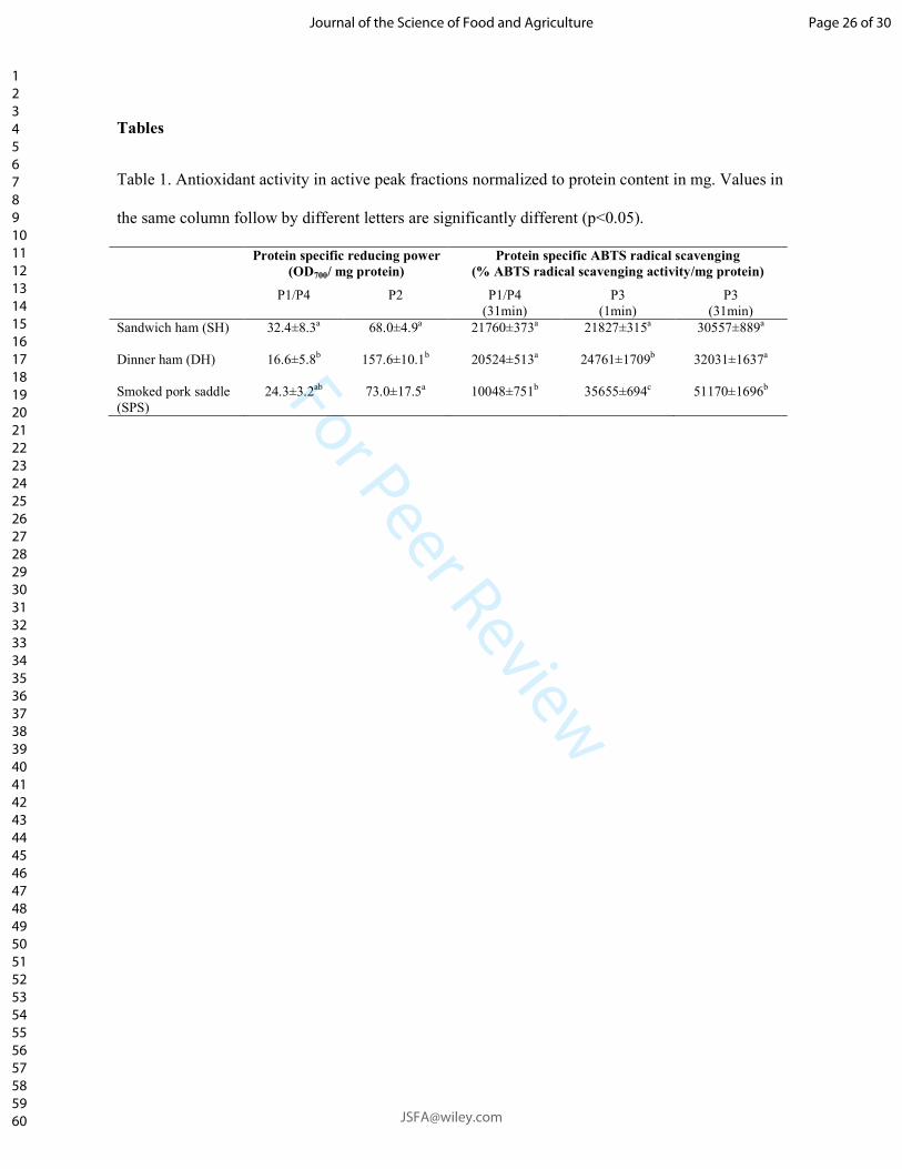
For Peer Review
Tables
Table 1. Antioxidant activity in active peak fractions normalized to protein content in mg. Values in
the same column follow by different letters are significantly different (p<0.05).
Protein specific reducing power
(OD700/ mg protein)
Protein specific ABTS radical scavenging
(% ABTS radical scavenging activity/mg protein)
P1/P4 P2 P1/P4
(31min)
P3
(1min)
P3
(31min)
Sandwich ham (SH)
32.4±8.3a 68.0±4.9a 21760±373a 21827±315a 30557±889a
Dinner ham (DH)
16.6±5.8b 157.6±10.1
b 20524±513
a 24761±1709
b 32031±1637
a
Smoked pork saddle
(SPS)
24.3±3.2ab 73.0±17.5a 10048±751b 35655±694c 51170±1696b
Page 26 of 30
Journal of the Science of Food and Agriculture
123456789101112131415161718192021222324252627282930313233343536373839404142434445464748495051525354555657585960
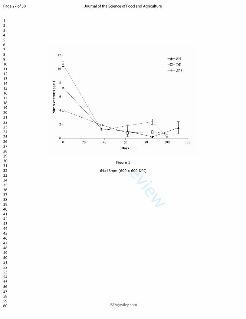
For Peer Review
Figure 1
64x46mm (600 x 600 DPI)
Page 27 of 30
Journal of the Science of Food and Agriculture
123456789101112131415161718192021222324252627282930313233343536373839404142434445464748495051525354555657585960
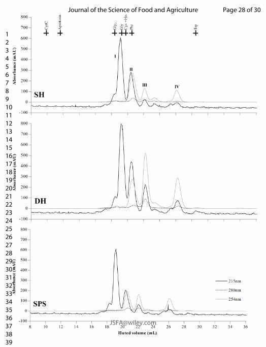
For Peer Review
Page 28 of 30
Journal of the Science of Food and Agriculture
123456789101112131415161718192021222324252627282930313233343536373839404142434445464748495051525354555657585960
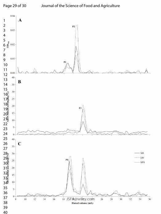
For Peer Review
Page 29 of 30
Journal of the Science of Food and Agriculture
123456789101112131415161718192021222324252627282930313233343536373839404142434445464748495051525354555657585960
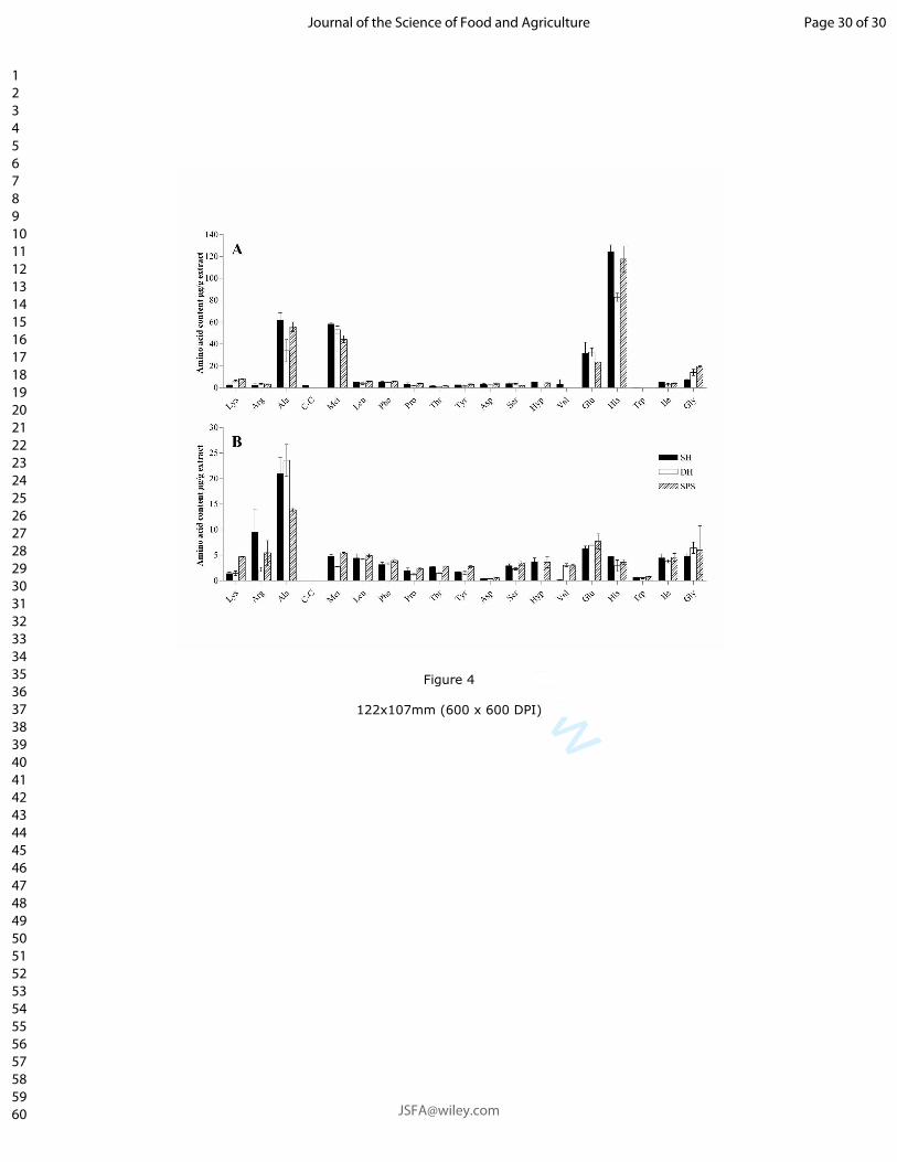
For Peer Review
Figure 4
122x107mm (600 x 600 DPI)
Page 30 of 30
Journal of the Science of Food and Agriculture
123456789101112131415161718192021222324252627282930313233343536373839404142434445464748495051525354555657585960
Paper III
_______________________________________________________________________________________
Pedersen, S. T., Duedahl-Olesen, L., Koch, A. G. & Jessen, F. Effect of storage and
varying nitrite and ascorbate concentrations on antioxidative and antimicrobial
activity in low molecular weight extracts from nitrite-cured cooked pork products
_______________________________________________________________________________________
Paper III
1
Title: Effect of storage and varying nitrite and ascorbate concentrations on antioxidative and antimicrobial 1
activity in low molecular weight extracts from nitrite-cured cooked pork products 2
Authors: Sabrine Tauber Pedersena, Lene Duedahl-Olesen
a, Anette Granly Koch
b and Flemming Jessen
a 3
aNational Food Institute, Technical University of Denmark, Søltofts Plads Building 227, DK-2800 Kongens 4
Lyngby, Denmark 5
bDanish Meat Research Institute, Gregersensvej 9, DK-2630 Taastrup, Denmark 6
7
8
Running title: Effect of nitrite on bioactivity in extracts from nitrite-cured hams 9
10
*Corresponding author: 11
Sabrine Tauber Pedersen 12
Postal address: 13
National Food Institute 14
Technical University of Denmark 15
Søltofts Plads Building 227, 1st floor, room 142 16
DK-2800 Kgs. Lyngby 17
E-mail: 18
20
Declarations of interest: none 21
22
Paper III
2
Abstract 23
For future optimisation of nitrite usage in meat curing this study focused on the influence of nitrite and 24
ascorbate concentrations on antioxidative and antimicrobial activities. Aqueous ≤10kDa extracts of nitrite-25
cured cooked hams produced with different nitrite and ascorbate concentrations were analysed by in vitro 26
assays. ABTS radical scavenging activity increased with added nitrite/ascorbate and with storage time while, 27
extract reducing power increased with storage time (more pronounced for higher nitrite/ascorbate addition) 28
and with added nitrite/ascorbate after 12days of cold storage. Iron chelating activity increased with storage 29
time for extracts of hams added ≥150ppm nitrite and ≥600ppm ascorbate. Antimicrobial activity was not 30
found. Finally, the ABTS activity increase seemed to be indirectly related to residual nitrite through 31
formation of NO and the exhibited reducing power appeared partly to be related to residual ascorbate. Iron 32
chelating activity may in the current situation originate from ham components or a compound formed during 33
storage. 34
35
36
37
Keywords: Cooked hams, curing, nitrite, ascorbate, antioxidant, in vitro assays 38
39
Paper III
3
1 Introduction 40
Preserving of meat by nitrite-curing is a widely used strategy to extent shelf-life as well as creating a 41
microbiologically safe and appealing product. However, utilization of nitrites in food is strictly controlled 42
and limited by current EU legislations. In order to optimise and preferably to further minimise nitrite 43
addition to meat, it is necessary to acquire further knowledge on the protective behaviour of nitrite and other 44
additives in nitrite-cured products. 45
Although nitrite, as a natural electron acceptor, can be considered a pro-oxidant its use in meat curing is also 46
for antioxidative purposes and nitrite has been pointed out to be a key-responsible not only for the 47
microbiological but also the oxidative stability of cured meat products. Nitrite exerts its antioxidant effect 48
through several different mechanism (Freybler et al., 1993). Yet, nitric oxide (NO) derived from the nitrite 49
ion is more and more accepted as the species interacting with free radicals of lipid oxidation (Skibsted, 50
2011). Furthermore, NO is essential in the reactions/reactivity of nitrite as it will react with a wide variety of 51
compounds like myoglobin, other proteins and amino acids. Ascorbate is commonly used in combination 52
with nitrite in meat curing. Ascorbate, as an electron donor, is in itself a strong antioxidant that through its 53
relatively unreactive oxidised form (dehydroascorbic acid) can terminate propagation of free radical 54
reactions (Bendich, Machlin, Scandurra, Burton, & Wayner, 1986). Nevertheless, ascorbate may in the 55
presence of metal ions also act as a pro-oxidant (Villaverde, Parra, & Estévez, 2014). 56
The reaction patterns of nitrite and ascorbate recurrently cross each other e.g. as ascorbate can accelerate the 57
curing process through formation of more NO (Møller & Skibsted, 2002; Skibsted, 2011). Furthermore, 58
ascorbate is consuming oxygen for its own oxidation and thereby limiting oxidation of NO and NO2ˉ 59
(Honikel, 2008) and also nitrite reaction products and ascorbate directly interacts forming NO-Asc reaction 60
intermediate (Izumi, Cassens, & Greaser, 1989). During cooking and storage, residual nitrite declines to 10-61
35% of the originally added amounts (Honikel, 2008; Hustad et al., 1973) which to a certain extent may be 62
explained by the type of reaction mentioned above. Although the redox chemistry of nitrite and ascorbate is 63
essential in nitrite-curing it is highly complex and at times not fully understood. 64
Paper III
4
In the recent years there has been an increased interest in natural antioxidative and antimicrobial compounds, 65
especially to replace unwanted E numbers and preservatives such as nitrite. This has led to an intensification 66
of the search for peptides with antioxidative and antimicrobial properties. Such peptides have been detected 67
in many foods and food products (de Castro & Sato, 2015) however to our knowledge none or remarkably 68
few investigations have been performed on nitrite-cured cooked meat products. Neither has it been possible 69
to come up with a single non-protein alternative to cover all the properties of the multifaceted nitrite 70
(Sindelar & Milkowski, 2011). 71
All this and a strong interest from the Danish meat industry to use the lowest possible amount of nitrite 72
requires better knowledge about the metabolism of nitrite in meat products. Therefore, it was decided to 73
investigate the effect of ingoing nitrite and ascorbate on antioxidant and antimicrobial activity in nitrite-74
cured cooked pork products. The levels required to obtain oxidative and microbiological stability in cured 75
meat products to a satisfying degree have previously been examined; however, the oxidative stability has to 76
our knowledge only been assessed by measurements of oxidation products. Thus, it is suggested that this 77
issue by means of antioxidant activity assessment could contribute with a new angle to this field of research. 78
In addition to expecting an effect of amounts of added nitrite/ascorbate it was supposed that cold storage of 79
the hams would result in a decrease in antioxidant capacity with time. 80
81
2 Materials and method 82
2.1 Materials 83
Ascorbic acid, sodium nitrite, 3-(2-Pyridyl)-5,6-diphenyl-1,2,4-triazine-4′,4′′-disulfonic acid sodium salt 84
(ferrozine) and 2,2′-Azino-bis(3-ethylbenzothiazoline-6-sulfonic acid) diammonium salt (ABTS), were 85
acquired from Sigma-Aldrich (St. Louis, MO, USA). Ethylenediaminetetraacetic acid disodium dihydrate 86
(EDTA) was from Merck Millipore (Billerica, MA, USA) and (±)-6-Hydroxy-2,5,7,8-tetramethylchromane-87
2-carboxylic acid (Trolox) from Fluka Chemika (Deisenhofen, Germany). All remaining chemicals and 88

Paper III
5
reagents employed were of analytical grade and acquired from VWR International (Radnor, PA, USA), 89
Merck Millipore, Sigma-Aldrich or Fluka Chemika. 90
2.2 Samples 91
Hams belonging to the category of restructured meat products where smaller pieces of meat are subjected to 92
curing brine prior to boiling stuffed into a plastic casing. The main recipe constituted 75% pork meat, 20% 93
water, 2.1% salt, skin protein (Danish: sværprotein), phosphate, potato starch and dextrose with a final water 94
content of approximately 75%. The main recipe was identical for all hams only changing the added amount 95
of sodium nitrite and ascorbate. The final concentrations were 0, 60, 150, 250, 500 and 1000ppm of added 96
sodium nitrite and 4 times as much ascorbate. The individual hams will be referred to as added sodium nitrite 97
slash added sodium ascorbate e.g. 60/240. The meat was vacuum packed and stored at -80°C until analysis. 98
A storage experiment was carried out were the sample were thawed and left at 5°C for a total of one month 99
periodically taking out samples and returning them to -80°C until further analysis. 100
2.3 Extraction of low molecular weight compounds 101
Water soluble compounds were extracted from the cured hams by homogenising approximately 70-100 g of 102
minced ham in approximately 400mL of deionised water, dH2O, for 1 minutes with an Ultra Turrax (T 25 103
Basic ULTRA-TURRAX® mounted with S25N -18G Dispersing element, IKA®-WERKE GmbH &Co. 104
KG, Staufen, Germany) at 13,500rpm and subsequently adding up to a total volume 1.5L. An aqueous 105
≤10kDa extract was collected by submerging three to five pierces of dialysis membrane tube of 10 cm each 106
(Spectra/Por 6, MWCO 10,000 Da, Spectrum Lab. Inc, CA, USA) containing 16.5mL dH2O (corresponding 107
to 50% membrane volume) into the meat/water homogenate. After 24 hours of magnetic stirring at 5ºC, the 108
dialysate in the membranes were pooled and stored at -80ºC until further analysis. Later the protein content 109
was determined colourimetrically using the PierceTM Modified Lowry Protein Assay Kit in microplate 110
format according to manufactures description (Thermo Scientific, PierceTM) using Bovine Serum Albumin 111
(BSA) (Albumin Standard Ampules, 2 mg/ml) supplied with the kit as an external standard. 112

Paper III
6
2.4 Nitrite content 113
The sodium nitrite content was determined in both the hams and the low molecular weight extracts hereof. 114
The day before analysis, approximate 50g of meat sample stored at -80ºC was thawed, minced for ten 115
seconds, and re-vacuum packed before being returned to -80 ºC. Extraction and measurement of nitrite in the 116
cured cooked hams was done according to the method for simultaneous measurement of nitrite and nitrate 117
described by Herrmann, Duedahl-Olesen, & Granby (2015). The detection was carried out using ion 118
chromatography coupled to UV detection at 225nm (HPLC and Diode Array Detector, series1100, Agilent 119
Technologies, Santa Clara, CA, USA) with separation on a Dionex IonPacTM AS11 RFICTM 250 x 4mm 120
(Thermo Scientific, Waltham, MA, USA). The extracts were diluted 1:1(V/V) in 1mM NaOH to obtain a pH 121
comparable to mixed minced ham and 1mM NaOH (1+10). The subsequent extraction, clean-up and analysis 122
were identical for the two sample types. The samples were analysed in a random order. 123
2.5 Ascorbic acid 124
Residual ascorbic acid in the hams and extracts was determined using an Agilent 1100 series HPLC using a 125
G1315A DAD detector set at 254.4nm (Agilent Technology, Waldbronn, Germany). 126
Ascorbic acid was extracted by leaving 2.5g of minced ham and 10mL of 5%(W/V) meta-phosphoric acid, 127
added 0.5mL of 0.1mg/mL EDTA solution, to stir for 2 minutes at room temperature protected from light. 128
MilliQ was added to a total volume of 25mL. The mixture was centrifuged at 3000rpm for 5 minutes and 129
filtered through a Munktell quantitative filter paper grade 00H and re-filtered through a 0.2µm syringe filter 130
directly into an amber HPLC vial. The extracts were merely mixed 1:1 with 1% (W/V) meta-phosphoric acid 131
and filtered through a 0.2µm syringe filter directly into an amber HPLC vial. Twenty µL sample was injected 132
and separated on a Prodigy 5u ODS3 100Å 250x4.6mm column (Phenomenex®, Torrance, CA, USA) and 133
eluted at a flow rate of 0.7mL/min. Mobile phase A was water adjusted to pH 3 and B was methanol running 134
a gradient of 5% B for the first 23 min followed by a 10second linear increase to 100% B which were kept 135
for 4min and 50 seconds before again decreasing to 5% B in 10 seconds and kept at this level until the end of 136
the run (total of 35 min). Quantification was done using an external calibration curve of L-ascorbic acid 137
dissolved in 1% (W/V) meta-phosphoric acid. The samples were analysed in a random order. 138
Paper III
7
2.6 Antioxidant assays 139
Potential antioxidant activities of the aqueous ≤10kDa extract were analysed using three in vitro assays 140
measuring iron chelation activity, reducing power and ABTS-radical scavenging activity. In order to 141
investigate the concentration dependency of the antioxidant activities, the crude undiluted ≤10kDa extract 142
were analysed followed by sequential threefold dilutions. The assays were conducted in triplicate and all 143
measurements were performed using a spectrophotometer (Synergy 2 Multi-Mode Microplate Reader, 144
BioTek® Instruments, Inc., Vermont, USA). The extracts were thawed at 5ºC, over night. 145
Iron chelating activity was assayed by the microtiter plate format method described by Hermund et al. 146
(2015). EDTA (0.5mM) was used as a positive control and was subjected to the same threefold dilutions as 147
the extract samples. dH2O was utilised as blank. The iron chelating activity (%) is defined and calculated as 148
percentage of inhibited colour formation/relative decrease in absorbance (562nm): 149
𝐹𝑒2+ 𝑐ℎ𝑒𝑙𝑎𝑡𝑖𝑛𝑔 𝑎𝑐𝑡𝑖𝑣𝑖𝑡𝑦 (%) =
𝐴𝑏𝑠𝑏𝑙𝑎𝑛𝑘 − 𝐴𝑏𝑠𝑠𝑎𝑚𝑝𝑙𝑒
𝐴𝑏𝑠𝑏𝑙𝑎𝑛𝑘∙ 100%
(1)
150
The reducing power was determined according to the procedure described by Oyaizu (1986) with some 151
modifications and adjusted to microtiter plate detection. First, equal parts of ≤10kDa extract sample (and 152
dilutions), 0.2M phosphate buffer (pH 6.6) and 1% potassium ferricyanide were mixed and incubated for 20 153
min at 50°C. Subsequently, 1 part of 10% TCA was also added. Then, 100μL of the sample mixture was 154
mixed with 100μL dH2O and 20 μL of 0.1% ferric chloride, and incubated 10 minutes at room temperature. 155
The absorbance was measured at 700nm. Ascorbic acid (0.5mM) was used as positive control and was 156
subjected to the same threefold dilution as the extract samples and dH2O was used as blank. Results are 157
expressed as 158
𝑂𝐷700 = 𝐴𝑏𝑠700𝑛𝑚(𝑆𝑎𝑚𝑝𝑙𝑒) − 𝐴𝑏𝑠700𝑛𝑚(𝑏𝑙𝑎𝑛𝑘) (2)
159
Paper III
8
The radical scavenging activity was determined according to the method described by Gringer, Osman, 160
Nielsen, Undeland, & Baron, (2014). Trolox (2.5mM, in 96% ethanol) was used as a positive control and 161
was subjected to the same threefold dilutions as the samples. Absorbance was measured after 8 minutes. 162
Ethanol (96%) and dH2O was included as blank for the positive control and extract sample, respectively. The 163
ABTS radical scavenging activity (%) for the individual dilution is expressed as the relative decrease in 164
absorbance (734nm) as for iron chelating activity 165
𝐴𝐵𝑇𝑆 𝑟𝑎𝑑𝑖𝑐𝑎𝑙 𝑠𝑐𝑎𝑣𝑒𝑛𝑔𝑖𝑛𝑔 𝑎𝑐𝑡𝑖𝑣𝑖𝑡𝑦 (%) =
𝐴𝑏𝑠𝑏𝑙𝑎𝑛𝑘 − 𝐴𝑏𝑠𝑠𝑎𝑚𝑝𝑙𝑒
𝐴𝑏𝑠𝑏𝑙𝑎𝑛𝑘∙ 100%
(3)
As all the crude ≤10kDa extracts and some of the dilutions with highest extract concentration reached the 166
assays maximal capacity around 90%, similar to the positive control (Trolox), the ABTS radical scavenging 167
activity of the extracts was expressed as normalised to ham (%/mg ham) and denoted as Normalised ABTS 168
radical scavenging activity. This was calculated as the initial linear slope from curves of ABTS radical 169
scavenging activity (%), as expressed in equation (3), versus mg ham constituting the basis for the individual 170
dilutions. 171
2.7 Antimicrobial activity 172
Antimicrobial activity of the cured ham aqueous ≤10kDa extracts and the supernatant of the meat/water 173
homogenate were tested with an inoculation culture consisting of a mixture of Listeria monocytogenes 174
(DMRICC 3042) and Salmonella typhimurium (DMRICC 4984). A small quantity of the purified cultures 175
where each transferred to 10mL Brain-heart infusion (BHI) broth (Oxoid CM1135, United Kingdom) and 176
incubated for 20 ± 2 hours at 37°C. These were afterwards diluted to 10-6 and mixed 1:1 (V/V) to constitute 177
the inoculation culture. 178
Diluted (1:5 and 1:25) and undiluted extracts were mixed 1:1 with BHI broth and subsequently inoculated 179
with the inoculation culture. The initial theoretical concentration of each bacteria culture in the inoculated 180
samples is 44 cfu/ml ~ 1.6 log cfu/ml. A sample substituting extract with FKP (0.85% NaCl (Merck 1.06404, 181
Germany) and 0.1% pepton (Merck 1.07214, Germany)) was included as a control. The inoculated extract 182
samples were incubated at 17 ± 1°C and samples were taken after 0, 24, 48 and 72 hours. The total bacterial 183
Paper III
9
count was determined on Oxford agar (oxoid CM 0856, United Kingdom) and SSI Enteric agar (34121, 184
Statens Serum Institute, Copenhagen Denmark) after 24-48 hours of incubation at 37°C. 185
2.8 Statistical analysis 186
All analyses were carried out in triplicate unless otherwise stated. Statistical analyses were conducted using 187
the GraphPad Prism® software Ver. 4.03 or R version 3.3.0 with P < 0.05. Results were compared using 188
one-way ANOVA and t-test with Tukeys posttest. Regression analyses were conducted in excel using the 189
regression function in the Data analysis toolbox. 190
3 Results 191
3.1 Antioxidant activity 192
Preliminary experiments (data not shown) had indicated a potential effect of storage time on the investigated 193
types of in vitro antioxidant activity in extract from cured hams. Thus, a storage experiment was set up to 194
evaluate the impact of time, as well as nitrite/ascorbate addition, on in vitro iron chelating activity, reducing 195
power and ABTS radical scavenging activity during 30 days of cold storage. The protein content of the 196
aqueous ≤10kDa extracts of the hams stored for 0-30 days and taken out with 3 days intervals was on 197
average 0.25±0.06 mg/mL and was not significantly different between hams of different nitrite/ascorbate 198
addition and neither between days of storage. 199
3.1.1 Iron chelating activity 200
The iron chelating activity of the crude ≤10kDa extracts ranged from approximately 36-88% with 250/1000 201
showing the highest average activity across time and 1000/4000 the lowest. In comparison the positive 202
control in the tested concentration (0.5mM EDTA) demonstrated approximately 95% iron chelating activity. 203
The average iron chelating activity across storage time of the remaining crude ≤10kDa extract arranged in 204
the order 0/0 > 60/240 > 500/2000 > 150/600. 205
Paper III
10
No apparent correlation between iron chelating activity in the crude ≤10kDa extracts and storage time was 206
evident across all nitrite/ascorbate concentrations (Figure 1). However, a significantly (regression analysis) 207
increasing iron chelating activity of the crude ≤10kDa extracts over time was found in sample 150/600, 208
250/1000, 500/2000 and 1000/4000. 209
3.1.2 Reducing power 210
Figure 2 shows the reducing power of the crude ≤10kDa extracts as a function of storage time. At day 0 the 211
order of reducing power activity was 150/600 > 60/240 > 0/0 > 250/1000 > 500/2000 > 1000/4000. The 212
analysis OD700 was lower in the extract samples compared to the positive control (0.5mM ascorbic acid) 213
which exhibited an OD700 of 0.72±0.05. Looking at the analytical dilution curves of the samples from day 0 214
(Figure 3) the reducing power activity increased continuously with the extract concentration for samples 0/0, 215
60/240 and 150/600 (Figure 3). In contrast the activity of the samples 250/1000, 500/2000 and 1000/4000 216
only increased with concentration at low extract concentrations however decreased at higher extract 217
concentrations to an extent where no reducing power were demonstrated in the crude ≤10kDa extract of the 218
sample added the most nitrite and ascorbate (1000/4000). However, after 3 days of storage the reducing 219
power activity of the 1000/4000, 500/2000 and 250/1000 crude ≤10kDa extracts started to surpass the 220
reducing power activity of the three low nitrite/ascorbate samples (Figure 2). And after 12 days of cold 221
storage the measured extract reducing power activities directly correlated with added nitrite/ascorbate. For 222
all samples there were a significant (regression analysis) positive correlation between reducing power and 223
storage time (Figure 2). 224
Regardless of the decrease in reducing power detected early in storage for samples of high extract 225
concentration and high nitrite/ascorbate addition the initial linear reducing power activity as function of mg 226
ham increased with the added nitrite/ascorbate for all samples (Figure 4). However, there were no significant 227
and consistently positive correlation between the initial linear reducing power activity and time of storage 228
(Figure 4). 229
Paper III
11
3.1.3 ABTS radical scavenging 230
The normalised ABTS radical scavenging activity increased significantly (regression analysis) for all 231
extracts both with storage time and with increased nitrite/ascorbate addition (Figure 5). As an example, it 232
may be mentioned that the ABTS radical scavenging activity at day 0 was approximately 3.5 times higher for 233
1000/4000 compared to 0/0. 234
3.2 Antimicrobial activity at day 0 235
In the tested concentrations, neither growth of Listeria monocytogenes nor Salmonella typhimurium were 236
inhibited by any of the day 0 ≤10kDa extracts compared to the control (ham extract substituted for equivalent 237
amounts diluent). This was the case after 0, 24, 48 and 72 hours of incubation. Testing the supernatants of 238
the meat/water homogenates used for producing the ≤10kDa extracts gave the same result. 239
3.3 Residual ascorbic acid and nitrite 240
Residual ascorbic acid was measured in the ≤10kDa extracts of hams stored for 0, 3, 6, 12, and 21 days at 241
5°C. This was normalised to the amount of ham utilised for the ham/water homogenate used for producing 242
the extracts and is presented in Figure 6. Residual ascorbic acid increased with added nitrite/ascorbate for the 243
samples 0/0, 60/240 and 150/600 and remained at the same level during the 21 days of storage (0/0: 0.06-244
0.07µg/g ham; 60/240: 0.4-0.7µg/g ham; 150/600:1.1-1.3µg/g ham) with the exception of slight drop for 245
sample 150/600 at day 21. At day 0 the residual ascorbic acid content in the three remaining extract samples 246
were lower than the 150/600 sample and decreased with increasing added nitrite/ascorbate. A similar pattern 247
was also evident for residual ascorbic acid in the hams at day 0 (data not shown). At day 12 extract samples 248
250/1000 and 500/2000 had increased to a level significantly different from the previous measurement days, 249
and now these two samples also demonstrated higher residual ascorbic acid content than the three samples 250
with the lowest added nitrite/ascorbate concentrations. At day 21 the residual ascorbic acid content correlated 251
with added nitrite/ascorbate for all samples. Detectable residual ascorbic acid increased approximately 55 252
times in extract 1000/4000 during the 21 days of cold storage 253
Paper III
12
Residual sodium nitrite was measured in the ≤10kDa extracts of hams stored for 0, 3, 6, 12, 21 and 30 days 254
at 5°C. This was also normalised to the amount of ham utilised for the ham/water homogenate used for 255
producing the extracts and is presented in Figure 7. Residual sodium nitrite correlated with added sodium 256
nitrite throughout storage yet the content generally decreased with time (no significant decrease was found 257
for 0/0 and 60/240 at any time points). Residual sodium nitrite was also measured in the hams at day 0 (data 258
not shown) and here residual and added sodium nitrite also correlated. The hams contained on average 259
approximately 18 times more nitrite per g than the extracts contained per mL – matching the ratio between 260
minced ham and total volume of 1.5L for the meat/water homogenate (Materials and Method). 261
4 Discussion 262
4.1 Reducing power 263
The surprising shape difference in the reducing power dilution curves of sample extract 0/0, 60/240 and 264
150/600 versus 250/1000, 500/2000 and 1000/4000 (Figure 3) at day 0 indicated a clear concentration 265
dependence. This furthermore indicated that at higher concentrations the active reducing components in the 266
≤10kDa extracts potentially resulted in an overload of the assay and that this was somehow related to 267
addition of higher concentrations of nitrite/ascorbate. The applied assay for determining in vitro reducing 268
power was based on the formation of pigment Prussian blue (FeIII
[FeIII
FeII(CN)6]3) that is formed upon 269
reduction of potassium ferricyanide (Fe3+
) to form potassium ferrocyanide (Fe2+
) by an antioxidant present in 270
the sample in the presence of excess ferric chloride (Fe3+
) (Wiberg, Wiberg, & Holleman, 2001). However, 271
the electrochromic nature of the Prussian blue pigment may cause it to return to colourless upon further 272
reduction of Fe3+
to Fe2+
. The large amounts of ascorbic acid added to the three samples in question could 273
have caused this double reduction however this explanation is contradictive to the amount of residual 274
ascorbic acid in the samples. On the other hand, the inter-sample pattern of reducing power activity of the 275
crude ≤10kDa extracts at day 0 showed some resemblance with the residual ascorbic acid in regard to the 276
drop occurring at additive concentration above 150/600. However, the residual ascorbic acid content of 277
sample 500/2000 and 1000/4000 were almost as low as 0/0 yet this did not transfer trough to the reducing 278
Paper III
13
power determined in crude 0/0. The relationship between residual ascorbic acid and reducing power of the 279
crude ≤10kDa extracts continued during storage with a slight time discrepancy. At day 12 the reducing 280
power activity increased with increasing nitrite/ascorbate addition while residual ascorbic acid did not 281
increase with added nitrite/ascorbate until day 21. However the full change to a residual/added ascorbic acid 282
correlation might have occurred just after day 12 (only 1000/4000 did not match the correlation at this point) 283
and then have become more pronounced from thereon as was also the case with the reducing power. 284
285
4.2 Ascorbic acid 286
Plant foodstuff e.g. fruits and vegetables are the primary source of dietary ascorbic acid. Nutrient data on 287
fresh pork meat rarely reports of any ascorbic acid content at all. However, ascorbic acid is found in all 288
living cells, plant as well as animal, which may explain the minute amounts found in the ham and extract of 289
the 0/0 sample (ham: approx. 13ppm (µg/g WW). Extracts: approx. 1µg/mL) (Belitz, Grosch, & Schieberle, 290
2004). Otherwise the detected residual ascorbic acid in the remaining samples must be assumed to originate 291
from the added sodium ascorbate. However, only approximately half of the sodium ascorbate added to the 292
hams was recovered as residual ascorbic acid (sample 60/240 and 150/600) which match the levels of 293
residual ascorbic acids found in other commercial processed meat products (Hidiroglou, Madere, & Behrens, 294
1998; Schüep & Keck, 1990; Valls, Sancho, Fernández-Muiño, Alonso-Torre, & Checa, 2002). Ascorbic 295
acid is readily (and reversible) oxidised to dehydroascorbic acid but may also be further degraded by opening 296
of the ring-structure leading to decrease or loss, respectively, of antioxidant activity. This oxidation and 297
degradation depends on a number of factors in particular oxygen partial pressure, pH, temperature and 298
presence of heavy metal ions e.g. Cu2+
and Fe3+
(Belitz et al., 2004). Essential in regard to the loss of 299
measurable ascorbic acid in the cured hams is ascorbic acid’s interactions/reactions with the added nitrite 300
leading to its oxidation but also formation of nitrosated reaction intermediates (capable of functioning as 301
nitrosating agents) – intermediates most likely not detected by the applied HPLC method [Reactions of 302
nitrite and ascorbate are reviewed in Skibsted (2011) and Honikel, (2008)]. The EDTA added to the samples 303
during sample preparation may have protected and stabilised any residual ascorbic acid from oxidizing but as 304
Paper III
14
a metal chelator it is not considered likely that it has had an impact on such intermediates or their formation. 305
The many ways in which ascorbic acid may be degraded or react with other ham components does however 306
not offer a clear explanation for the lower ascorbic acid detected in the three concentrated samples and not 307
the samples 0/0, 60/240 and 150/600 and neither does it provide an explanation for the increase over time. 308
4.3 Iron chelating activity 309
Iron chelating activity was detected in all tested ≤10kDa sample extracts throughout storage yet there did not 310
appear to be any correlation with amounts of nitrite/ascorbate initially added to the hams. This being in spite 311
of ascorbic acid being capable of chelating metals (Brady, 2013) and some of the proposed antioxidant 312
activities of nitrite involve chelating of metals (Igene, Yamauchi, Pearson, Gray, & Aust, 1985). A similar 313
lack of correlation also came from the investigations of a small selection of commercial cooked cured hams 314
of known nitrite addition concentration (Pedersen et al., n.d.). Furthermore, results did neither correlate with 315
residual amounts of sodium nitrite or ascorbic acid. Thus, it must be assumed that the detected activity 316
originated from other ham components in the extracts. Some phosphates are known to function as metal 317
chelators and could as such be expected to also contribute to the iron chelating activity signals (Igene et al., 318
1985) however the same amount of phosphates were added to all the hams. Therefore the slight differences 319
observed could stem from other sources potentially related to differences in the utilised pork meat e.g. 320
proteins or amino acids rather than additives. Throughout the storage period the activity of extracts 150/600, 321
250/1000, 500/2000 and 1000/4000 increased yet the lack of a direct correlation with amounts of 322
nitrite/ascorbate initially added to the hams remained. Yet as this tendency was not evident in the two 323
samples of lowest or no added nitrite/ascorbate, this inspired the hypothesis that nitrite/ascorbate addition 324
beyond a certain threshold concentration indirectly affect the time depended formation of the active 325
component(s). 326
4.4 ABTS radical scavenging 327
At day 0 the normalised ABTS radical scavenging activity correlated positively with the amount of added 328
nitrite/ascorbate (Figure 5). As added and residual sodium nitrite converged in the current study, the 329
Paper III
15
normalised ABTS radical scavenging activity also correlated with residual sodium nitrite. This correlation 330
was also reported in a previous study (Pedersen et al., n.d.). However, the increase in normalised ABTS 331
radical scavenging activity occurring over time (Figure 5) was contrary to the decrease in residual sodium 332
nitrite happening over time (higher added sodium nitrite still resulted in higher residual sodium nitrite at any 333
time point during storage). This type of decrease in residual nitrite during cold storage has often been 334
reported (Pérez-Rodríguez, Bosch-Bosch, & Garciá-Mata, 1996). As nitrite do not primarily exert its 335
versatile antioxidant properties directly as nitrite but through reaction products like NO and MbNO (Freybler 336
et al., 1993; Honikel, 2008; Skibsted, 2011) it can be assume that the decrease in residual nitrite over time is 337
resulting in formation of more antioxidant species and that a higher addition of nitrite provides more starting 338
material for such a conversion. Thus, the increase in normalised ABTS radical scavenging activity may be 339
linked with added as well as residual nitrite. The correlation between normalised ABST radical scavenging 340
and residual nitrite reported by Pedersen et al. (n.d.) does not fit with this explanation as residual and added 341
nitrite did not correlate. This might partly be due to the nature of the sample material as the hams in the 342
current study are more or less identical with the exception of differences in added amounts of 343
nitrite/ascorbate whereas the samples used by Pedersen et al. (n.d.) were very different (type of 344
processing/cooking, national origin, additives etc.) and lacking details on other additives like amount of 345
added ascorbate. These differences can have affected the conversion of nitrite to its active antioxidant 346
species e.g. higher addition of ascorbate result in faster turnover of nitrite (Izumi, 1992). 347
Ascorbic acid is generally regarded as a reducing agent but is also occasionally referred to as a radical 348
scavenger (Villaverde, Ventanas, & Estévez, 2014). However, the normalised ABTS radical scavenging 349
activity did not correlate with residual ascorbic acid until the end of the storage period. Furthermore, we have 350
not been able find literature on ascorbic acids ability to scavenge the ABTS radical. As 0/0 also 351
demonstrated an activity although not as high as the nitrite-cured samples, it can be speculated that the 352
curing agents might merely accelerate the ABTS radical scavenging properties naturally enclosed in the 353
hams. The same can be said regarding the initial reducing power activity. 354
Paper III
16
4. 5 Antimicrobial activity 355
It was decided to investigate whether the antimicrobial activities known from nitrite-cured products was 356
associated with the water-soluble ≤10kDa fraction and most importantly if it was related to the ingoing and 357
residual amount of additives and more specifically sodium nitrite and sodium ascorbate. Despite that nitrite-358
derived NO is known to be toxic towards L. monocytogenes (Cammack et al., 1999) no growth inhibitory 359
effect was evident from the extracts at any additive concentration. Several studies have emphasised the 360
importance of e.g. ingoing and residual nitrite as well as pH and storage temperature(Birzele, Djordjević, & 361
Krämer, 2005; King, Glass, Milkowski, Seman, & Sindelar, 2016; McClure, Kelly, & Roberts, 1991). A 362
relationship between ingoing nitrite and antimicrobial properties through NO makes great sense as more 363
added nitrite logically would give more starting material for NO formation. However, as no effects based on 364
current results can be assigned to ingoing nitrite it could also be considered whether the lack of response is 365
due to a combination of other parameters such as low residual nitrite (≤14.8µg/mL extract equivalent to 366
0.9µg/g ham used for the extract).Birzele et al. (2005) found L. monocytogenes inhibition to correlate with 367
concentration of nitrite in the applied curing salt however the residual nitrite were never below 19mg/kg 368
sample. So in the event that the residual nitrite in their study contributed to the inhibitory effect the lower 369
levels found in the current study may explain the absence of an antimicrobial effect. The lack of a response 370
may potentially also be connected to pH as McClure et al. (1991) found that much higher amounts of ingoing 371
nitrite were required at pH above 5.3 and the pH of the extracts were around 6. Hospital, Hierro, & 372
Fernández (2014) reported that 75ppm ingoing nitrite led to a reduction in Salmonella in raw minced pork 373
and sausages during storage however the effect of nitrite on Salmonella is a controversial issue and research 374
seems to point in opposite directions. Thus if any antimicrobial effect at all exists the lack of response in the 375
current study (also towards L. monocytogenes) must be due to low concentration of the active components in 376
the tested samples. 377
Paper III
17
Conclusion 378
It was expected that nitrite/ascorbate addition and time of cold storage would affect in vitro antioxidant 379
activity. All three tested types of antioxidant activity increased with storage time although not all 380
significantly and not for all samples. Collectively the different types of antioxidant activity are not solely 381
correlated with residual levels of ascorbate and nitrite despite correlations with added nitrite/ascorbate in the 382
case of normalised ABTS radical scavenging and initial linear reducing power activity. The results show that 383
other components than nitrite and ascorbate are of great importance for antioxidative activities in ≤10kDa 384
ham extracts. The identity and nature of these compounds need to be investigated in order to optimise and 385
preferably minimise the addition of additives to cured meat without needlessly compromising product 386
stability. The unexpected pattern detected in residual ascorbic acid during storage, which correlated with in 387
vitro reducing power, requires further investigations for an explanation. 388
389
Acknowledgment 390
Hams were kindly manufactured by Danish Meat Research Institute. Furthermore thanks are expressed to 391
Flemming Hansen and Anita Forslund for contributing to idea generation and supervision in all 392
microbiological matters. 393
This work was supported by a grant from Norma and Frode S. Jacobsens Fond. 394
References 395
Belitz, H.-D., Grosch, W., & Schieberle, P. (2004). Food Chemistry (3rd ed.). New York, NY: Springer 396
Berlin Heidelberg. 397
Bendich, A., Machlin, L. J., Scandurra, O., Burton, G. W., & Wayner, D. D. M. (1986). The antioxidant role 398
of vitamin C. Advances in Free Radical Biology & Medicine, 2(2), 419–444. 399
Birzele, B., Djordjević, S., & Krämer, J. (2005). A study of the role of different nitrite concentrations on 400
human pathogenic bacteria in fresh spreadable ham and onion sausage. Food Control, 16(8), 695–699. 401
Paper III
18
Brady, J. W. (2013). Introductory food chemistry (1st ed.). Comstock Pub. Associates. 402
Cammack, R., Joannou, C. L., Cui, X.-Y., Martinez, C. T., Maraj, S. R., & Hughes, M. N. (1999). Nitrite and 403
nitrosyl compounds in food preservation. Biochimica Et Biophysica Acta, 1411(2–3), 475–488. 404
de Castro, R. J. S., & Sato, H. H. (2015). Biologically active peptides: Processes for their generation, 405
purification and identification and applications as natural additives in the food and pharmaceutical 406
industries. Food Research International, 74, 185–198. 407
Freybler, L. A., Gray, J. I., Asghar, A., Booren, A. M., Pearson, A. M., & Buckley, D. J. (1993). Nitrite 408
stabilization of lipids in cured pork. Meat Science, 33(1), 85–96. 409
Gringer, N., Osman, A., Nielsen, H. H., Undeland, I., & Baron, C. P. (2014). Chemical characterization, 410
antioxidant and enzymatic activity of brines from scandinavian marinated herring products. Journal of 411
Food Processing & Technology, 5(346). 412
Hermund, D. B., Yeşiltaş, B., Honold, P., Jónsdóttir, R., Kristinsson, H. G., & Jacobsen, C. (2015). 413
Characterisation and antioxidant evaluation of Icelandic F. vesiculosus extracts in vitro and in fish-oil-414
enriched milk and mayonnaise. Journal of Functional Foods, 19, 828–841. 415
Herrmann, S. S., Duedahl-Olesen, L., & Granby, K. (2015). Occurrence of volatile and non-volatile N-416
nitrosamines in processed meat products and the role of heat treatment. Food Control, 48(Sp. Iss. SI), 417
163–169. 418
Hidiroglou, N., Madere, R., & Behrens, W. (1998). Electrochemical determination of ascorbic acid and 419
isoascorbic acid in ground meat and in processed foods by high pressure liquid chromatography. 420
Journal of Food Composition and Analysis, 11, 89–96. 421
Honikel, K.-O. (2008). The use and control of nitrate and nitrite for the processing of meat products. Meat 422
Science, 78(1–2), 68–76. 423
Hospital, X. F., Hierro, E., & Fernández, M. (2014). Effect of reducing nitrate and nitrite added to dry 424
fermented sausages on the survival of Salmonella Typhimurium. Food Research International, 62, 425
410–415. 426
Hustad, G. O., Cerveny, J. G., Trenk, H., Deibel, R. H., Kautter, D. A., Fazio, T., … Kolari, O. E. (1973). 427
Effect of sodium nitrite and sodium nitrate on botulinal toxin production and nitrosamine formation in 428
wieners. Applied Microbiology, 26(1), 22–26. 429
Igene, J. O., Yamauchi, K., Pearson, A. M., Gray, J. I., & Aust, S. D. (1985). Mechanisms by which nitrite 430
Paper III
19
inhibits the development of warmed-over flavour (WOF) in cured meat. Food Chemistry, 18(1), 1–18. 431
Izumi, K. (1992). Reaction of nitrite with ascorbic acid or ascorbic acid-2-derivatives. Journal of Food 432
Science, 57(5), 1066–1067. 433
Izumi, K., Cassens, R. G., & Greaser, M. L. (1989). Reaction of nitrite with ascorbic acid and its significant 434
role in nitrite-cured food. Meat Science, 26(2), 141–153. 435
King, A. M., Glass, K. A., Milkowski, A. L., Seman, D. L., & Sindelar, J. J. (2016). Modeling the impact of 436
ingoing sodium nitrite, sodium ascorbate, and residual nitrite concentrations on growth parameters of 437
Listeria monocytogenes in cooked, cured pork sausage. Journal of Food Protection, 79(2), 184–193. 438
McClure, P. J., Kelly, T. M., & Roberts, T. A. (1991). The effects of temperature, pH, sodium chloride and 439
sodium nitrite on the growth of Listeria monocytogenes. International Journal of Food Microbiology, 440
14(1), 77–91. 441
Møller, J. K. S., & Skibsted, L. H. (2002). Nitric oxide and myoglobins. Chemical Reviews, 102(4), 1167–442
1178. 443
Oyaizu, M. (1986). Studies on products of browning reaction. Antioxidative activities of products of 444
browning reaction prepared from glucosamine. The Japanese Journal of Nutrition and Dietetics, 44(6), 445
307–315. 446
Pedersen, S. T., Baron, C. P., Gobba, C. De, Duedahl-Olesen, L., Koch, A. G., & Jessen, F. (n.d.). 447
Antioxidative and antimicrobial activity of extracts from nitrite-cured cooked pork products. 448
Unpublished manuscript. 449
Pérez-Rodríguez, M. L., Bosch-Bosch, N., & Garciá-Mata, M. (1996). Monitoring nitrite and nitrate residues 450
in frankfurters during processing and storage. Meat Science, 44(1–2), 65–73. 451
Schüep, W., & Keck, E. (1990). Measurement of ascorbic acid and erythorbic acid in processed meat by 452
HPLC. Zeitschrift Für Lebensmittel-Untersuchung Und -Forschung, 191(4–5), 290–292. 453
Sindelar, J. J., & Milkowski, A. L. (2011). Sodium nitrite in processed meat and poultry meats: A review of 454
curing and examining the risk benefit of its usee. AMSA White Paper Series, 3, 1–14. 455
Skibsted, L. H. (2011). Nitric oxide and quality and safety of muscle based foods. Nitric Oxide, 24(4), 176–456
183. 457
Valls, F., Sancho, M. T., Fernández-Muiño, M. A., Alonso-Torre, S., & Checa, M. A. (2002). High-pressure 458
liquid chromatographic determination of ascorbic acid in cooked sausages. Journal of Food Protection, 459
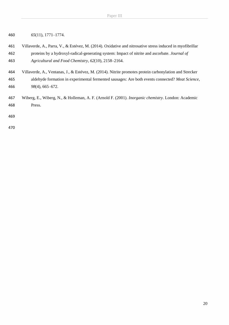
Paper III
20
65(11), 1771–1774. 460
Villaverde, A., Parra, V., & Estévez, M. (2014). Oxidative and nitrosative stress induced in myofibrillar 461
proteins by a hydroxyl-radical-generating system: Impact of nitrite and ascorbate. Journal of 462
Agricultural and Food Chemistry, 62(10), 2158–2164. 463
Villaverde, A., Ventanas, J., & Estévez, M. (2014). Nitrite promotes protein carbonylation and Strecker 464
aldehyde formation in experimental fermented sausages: Are both events connected? Meat Science, 465
98(4), 665–672. 466
Wiberg, E., Wiberg, N., & Holleman, A. F. (Arnold F. (2001). Inorganic chemistry. London: Academic 467
Press. 468
469
470
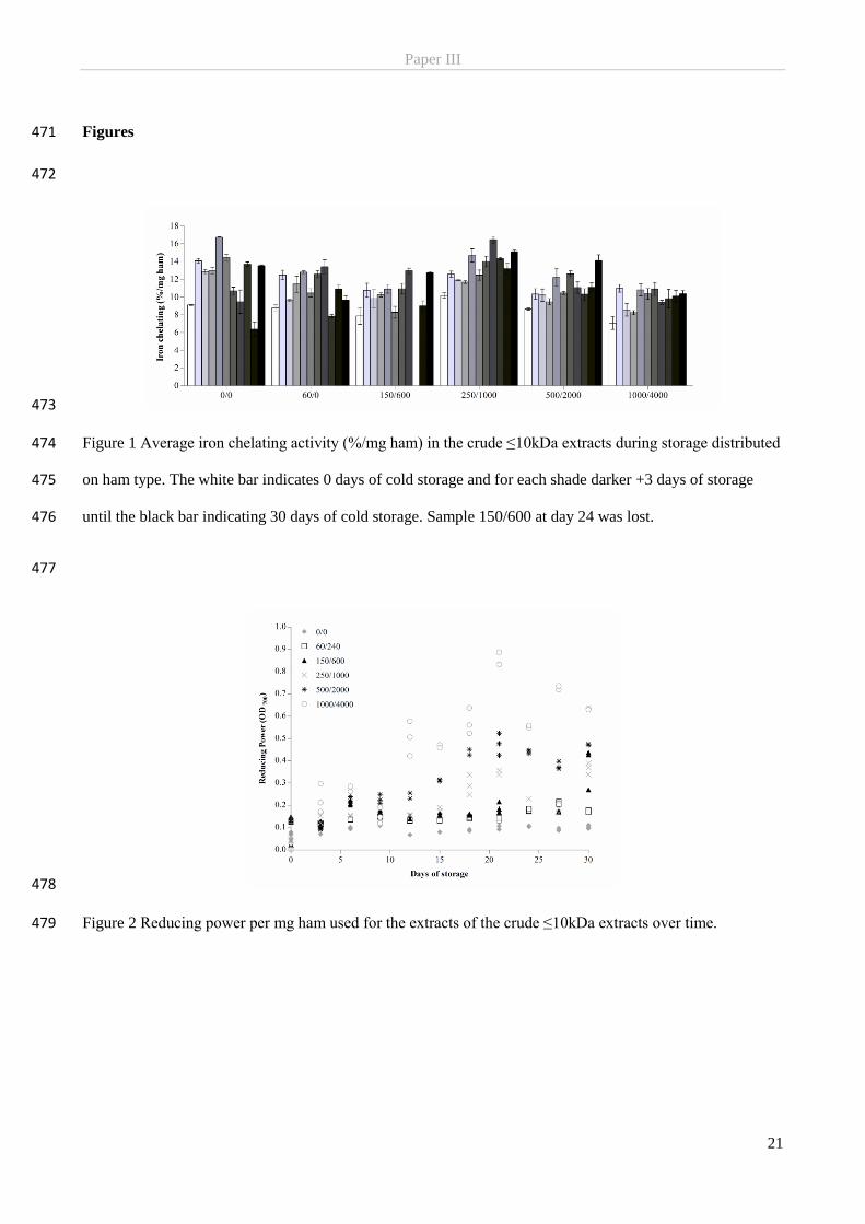
Paper III
21
Figures 471
472
473
Figure 1 Average iron chelating activity (%/mg ham) in the crude ≤10kDa extracts during storage distributed 474
on ham type. The white bar indicates 0 days of cold storage and for each shade darker +3 days of storage 475
until the black bar indicating 30 days of cold storage. Sample 150/600 at day 24 was lost. 476
477
478
Figure 2 Reducing power per mg ham used for the extracts of the crude ≤10kDa extracts over time. 479
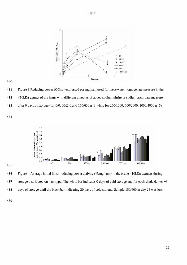
Paper III
22
480
Figure 3 Reducing power (OD700) expressed per mg ham used for meat/water homogenate measure in the 481
≤10kDa extract of the hams with different amounts of added sodium nitrite or sodium ascorbate measure 482
after 0 days of storage (for 0/0, 60/240 and 150/600 n=3 while for 250/1000, 500/2000, 1000/4000 n=6). 483
484
485
Figure 4 Average initial linear reducing power activity (%/mg ham) in the crude ≤10kDa extracts during 486
storage distributed on ham type. The white bar indicates 0 days of cold storage and for each shade darker +3 487
days of storage until the black bar indicating 30 days of cold storage. Sample 150/600 at day 24 was lost. 488
489
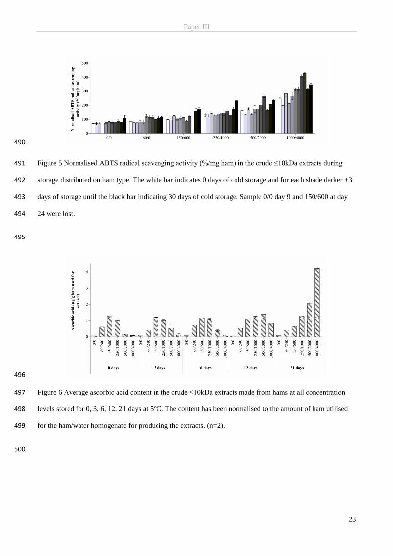
Paper III
23
490
Figure 5 Normalised ABTS radical scavenging activity (%/mg ham) in the crude ≤10kDa extracts during 491
storage distributed on ham type. The white bar indicates 0 days of cold storage and for each shade darker +3 492
days of storage until the black bar indicating 30 days of cold storage. Sample 0/0 day 9 and 150/600 at day 493
24 were lost. 494
495
496
Figure 6 Average ascorbic acid content in the crude ≤10kDa extracts made from hams at all concentration 497
levels stored for 0, 3, 6, 12, 21 days at 5°C. The content has been normalised to the amount of ham utilised 498
for the ham/water homogenate for producing the extracts. (n=2). 499
500
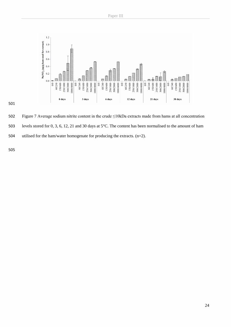
Paper III
24
501
Figure 7 Average sodium nitrite content in the crude ≤10kDa extracts made from hams at all concentration 502
levels stored for 0, 3, 6, 12, 21 and 30 days at 5°C. The content has been normalised to the amount of ham 503
utilised for the ham/water homogenate for producing the extracts. (n=2). 504
505

National Food InstituteTechnical University of DenmarkKemitorvetBuilding 2022800 Kgs. LyngbyDenmark
T: 35 88 70 00F: 35 88 70 01www.food.dtu.dk