neurons refine the caenorhabditis elegans body plan by directing axial patterning by wnts
TRANSCRIPT

Neurons Refine the Caenorhabditis elegans Body Plan byDirecting Axial Patterning by WntsKatarzyna Modzelewska1, Amara Lauritzen1¤a, Stefan Hasenoeder2,3¤b, Louise Brown4, John Georgiou4,
Nadeem Moghal1,2,3¤c*
1 Department of Oncological Sciences, Huntsman Cancer Institute, University of Utah, Salt Lake City, Utah, United States of America, 2 Campbell Family Cancer Research
Institute, Ontario Cancer Institute, Princess Margaret Hospital, University Health Network, Toronto, Ontario, Canada, 3 Department of Medical Biophysics, University of
Toronto, Toronto, Ontario, Canada, 4 Samuel Lunenfeld Research Institute, Mount Sinai Hospital, Toronto, Ontario, Canada
Abstract
Metazoans display remarkable conservation of gene families, including growth factors, yet somehow these genes are usedin different ways to generate tremendous morphological diversity. While variations in the magnitude and spatio-temporalaspects of signaling by a growth factor can generate different body patterns, how these signaling variations are organizedand coordinated during development is unclear. Basic body plans are organized by the end of gastrulation and are refinedas limbs, organs, and nervous systems co-develop. Despite their proximity to developing tissues, neurons are primarilythought to act after development, on behavior. Here, we show that in Caenorhabditis elegans, the axonal projections ofneurons regulate tissue progenitor responses to Wnts so that certain organs develop with the correct morphology at theright axial positions. We find that foreshortening of the posteriorly directed axons of the two canal-associated neurons(CANs) disrupts mid-body vulval morphology, and produces ectopic vulval tissue in the posterior epidermis, in a Wnt-dependent manner. We also provide evidence that suggests that the posterior CAN axons modulate the location andstrength of Wnt signaling along the anterior–posterior axis by employing a Ror family Wnt receptor to bind posteriorlyderived Wnts, and hence, refine their distributions. Surprisingly, despite high levels of Ror expression in many other cells,these cells cannot substitute for the CAN axons in patterning the epidermis, nor can cells expressing a secreted Wntinhibitor, SFRP-1. Thus, unmyelinated axon tracts are critical for patterning the C. elegans body. Our findings suggest thatthe evolution of neurons not only improved metazoans by increasing behavioral complexity, but also by expanding thediversity of developmental patterns generated by growth factors such as Wnts.
Citation: Modzelewska K, Lauritzen A, Hasenoeder S, Brown L, Georgiou J, et al. (2013) Neurons Refine the Caenorhabditis elegans Body Plan by Directing AxialPatterning by Wnts. PLoS Biol 11(1): e1001465. doi:10.1371/journal.pbio.1001465
Academic Editor: Julie Ahringer, University of Cambridge, United Kingdom
Received July 10, 2012; Accepted November 16, 2012; Published January 8, 2013
Copyright: � 2013 Modzelewska et al. This is an open-access article distributed under the terms of the Creative Commons Attribution License, which permitsunrestricted use, distribution, and reproduction in any medium, provided the original author and source are credited.
Funding: This research was supported by Public Health Services grant R01 GM073184 from the National Institutes of Health to NM and an Investigator Award (IA-006) from the Ontario Institute for Cancer Research to NM. This research was conducted with the support of the Ontario Institute for Cancer Research throughfunding provided by the Government of Ontario, and was funded in part by the Ontario Ministry of Health and Long Term Care. The views expressed do notnecessarily reflect those of the OMOHLTC. The funders had no role in study design, data collection and analysis, decision to publish, or preparation of themanuscript.
Competing Interests: The authors have declared that no competing interests exist.
Abbreviations: CAN, canal-associated neuron; HSN, hermaphrodite-specific neuron; P-Rvl, posterior-reversed vulval lineage; RNAi, RNA interference
* E-mail: [email protected]
¤a Current address: Graduate Program in Genetic Counseling, University of Utah School of Medicine, Salt Lake City, Utah, United States of America¤b Current address: Institute of Stem Cell Research, Helmholtz Zentrum Munchen, Neuherberg, Germany¤c Current address: Ontario Cancer Institute, Princess Margaret Hospital, University Health Network, Toronto, Ontario, Canada
Introduction
Metazoan body plans display great morphological diversity in
their overall organization and detailed patterns. Yet remarkably,
these plans are generated from a small number of conserved
growth factors that are reused many times during development to
refine the plans in distinct ways. Thus, determining how a limited
number of growth factors create different body patterns is key to
understanding this diversity. It is known that variations in the
magnitude and spatio-temporal aspects of signaling by growth
factors and their effectors generate different cellular responses and
body patterns [1–3]. However, the mechanisms by which a
developing animal organizes and coordinates the correct ampli-
tudes of signaling at the right times and places are not well
understood.
Wnts comprise one of the oldest families of growth factors, and
have conserved functions in organizing and refining body plans. In
many metazoan embryos, Wnts are initially expressed from one
pole, with their diffusion generating polarity and early patterning
of the primary body axis [4]. During and after gastrulation, Wnt
gradients, often derived from the posterior body, continue to
establish basic identities in the body plan, and then refine these
fates to create specific tissue and organ patterns [4–6]. As animals
develop, it becomes increasingly challenging to spatially organize
Wnt activity into the appropriate high and low signaling domains
throughout the body. Furthermore, this organization must be
coordinated between the multiple Wnts and other growth factors
that pattern tissues and organs. Perhaps as a reflection of these
challenges, numerous secreted and membrane-associated inhibi-
tors have been identified that help modulate Wnt gradient activity
PLOS Biology | www.plosbiology.org 1 January 2013 | Volume 11 | Issue 1 | e1001465

[7–10]. However, our knowledge of what a Wnt gradient must
look like to pattern a specific limb or organ is very limited, as is our
understanding of the distinct roles of the different antagonists.
In Caenorhabditis elegans, the generation of a vulva in the middle of
the anterior–posterior axis has become a paradigm for under-
standing how Wnt and EGF family growth factors generate
specific patterns at precise locations (reviewed in [11]) (Figure 1A).
Vulval organogenesis begins with the mid-body generation of six
vulval progenitors from 11 blast cells. These progenitors (P3.p–
P8.p) are specified by two posteriorly derived Wnt gradients (EGL-
20 and CWN-1 [orange and green, respectively, in Figure 1])
[7,12–15], with EGL-20 also polarizing some of the progenitors to
face towards the posterior (e.g., P5.p and P7.p) [16] (Figure 1B).
Later, mid-body-produced Wnts (LIN-44 and MOM-2 [blue in
Figure 1]) reverse P7.p polarity so that P5.p and P7.p face each
other and subsequently divide with mirror image symmetry
(Figure 1C and 1D) [16,17]. With the help of the posterior Wnts
(EGL-20 and CWN-1), centrally produced EGF (purple in
Figure 1) instructs P6.p to adopt a 1u fate, divide three times,
and form the vulval lumen that attaches to the uterus (Figure 1C
and 1D) [7,11,18–20]. In parallel, P6.p activates Notch signaling
in adjacent P5.p and P7.p to induce 2u fates, which, after three
rounds of division, create the symmetrical sides of the vulva that
attach the organ to the epidermis (Figure 1C and 1D). Insufficient
signaling alters vulval patterning and reduces the amount of vulval
tissue [11,21]. Excessive signaling also alters vulval patterning and,
if it occurs at certain axial positions, generates ectopic, non-
functional vulvae, which can interfere with normal positioning of
muscles and neurons that promote egg-laying [19,21–24].
To understand how growth factors such as Wnts generate
specific fates at precise positions, we looked for mutations that
affected placement of vulval tissue along the anterior–posterior
axis. We were intrigued by mutations in the vab-8 gene, which
affect vulval development through an unknown mechanism and
are primarily known for disrupting the migration and axon
outgrowth of a few neurons [25,26]. While nervous systems co-
develop with tissues and organs [6], with only rare exceptions,
their importance in refining body plans has been unexplored. In
flies, through unknown mechanisms, motor neurons contribute to
abdominal and flight muscle patterning [27,28], and in mammals,
by secreting VEGF, sensory nerves direct arterial patterning in
skin [29]. In addition, we previously discovered that in C. elegans,
motor neuron excitation stimulates vulval fate signaling [30].
Thus, we were interested in exploring the possibility that neurons
might refine patterning by widely used growth factors such as
Wnts.
Here we show that C. elegans has evolved a neuronal-based
mechanism to refine the amplitude and spatial signaling properties
of the posterior-derived Wnt gradients that pattern the epidermis.
Two canal-associated neurons (CANs), whose axons span the
anterior–posterior axis, ensure that a vulva is generated with the
correct morphology and only at the mid-body. When outgrowth of
the posterior CAN axon is severely shortened, Wnt signaling is
increased along the anterior–posterior axis, especially in the
posterior body. This deregulated signaling alters the symmetry of
the normal mid-body vulva, and causes ectopic vulval tissue to
form in the posterior epidermis. Finally, we provide evidence that
although the Ror/CAM-1 Wnt receptor is widely expressed, its
expression in the CAN axons is part of a unique Wnt-sequestration
mechanism that ultimately directs the locations and strength of
Wnt signaling necessary for proper epidermal patterning.
Results
Mutations Affecting Neuronal Migration and AxonOutgrowth Affect the Symmetry and Axial Position ofVulval Tissue
vab-8 encodes a long isoform, VAB-8L, and several short
isoforms collectively called VAB-8S [31]. These proteins act in a
few neurons to promote their posterior-directed migration and
axon outgrowth [31,32]. Both isoforms possess C-terminal coiled-
coil domains, with VAB-8L having an additional N-terminal
kinesin domain. In wild-type animals, only the three central vulval
progenitors (P5.p–P7.p) adopt vulval fates. However, in vab-
8(gm99) and vab-8(gm138) mutants, which lack both isoforms and
have anteriorly displaced neurons, the posterior P8.p progenitor
also acquired a vulval fate (Figure 2A). In the anterior epidermis of
wild-type animals, Wnt signaling is limiting in P3.p because of its
distance from the posterior Wnts; therefore, it becomes a
progenitor only 50% of the time (Figure 1B and 2B). vab-8
mutations did not cause ectopic vulval fates in anterior P3.p or
P4.p (n = 170), but they increased the frequency with which P3.p
became a vulval progenitor (Figure 2B). In the mid-body, at low
frequency, vab-8 mutations disrupted the mirror image symmetry
of the vulva. Initially, during the generation of the vulval
progenitors in wild-type animals, posterior-derived EGL-20/Wnt
causes P5.p and P7.p to polarize and face towards the posterior
(Figure 1B) [16]. Later, centrally produced MOM-2 and LIN-44
Wnts maintain P5.p polarity towards the posterior, but cause P7.p
to reorient and face towards the anterior (Figures 1C and 2C)
[16,17]. vab-8 mutations caused a ‘‘posterior-reversed vulval
lineage’’ (P-Rvl) phenotype, in which P7.p remained polarized
towards the posterior EGL-20/Wnt signal, so that its subsequent
cell divisions caused a second vulval invagination (Figure 2D).
To study how VAB-8-regulated cells affect epidermal develop-
ment, we used mutant backgrounds permitting sensitive quanti-
fication of the effects of vab-8 mutations. A loss-of-function egfr/let-
23(lf) mutation severely diminishes the 1u fate response in P6.p
such that fewer than the normal three progenitors adopt vulval
fates, causing an ‘‘underinduced’’ or ‘‘vulvaless’’ phenotype. This
mutant phenotype can be suppressed by activating the EGFR
pathway downstream of the receptor (e.g., by a loss-of-function
Author Summary
How a limited number of conserved growth factors such asWnts generate diverse bodies throughout the animalkingdom is a fundamental question in developmental andevolutionary biology. Diversity is thought to arise in partthrough variations in the strength and location of growthfactor signaling. How the signaling properties of growthfactors are precisely tuned at specific locations to generatedistinct tissue patterns is not well understood. Here, wepresent evidence that the axons of two specific neuronsthat span the anterior–posterior axis help pattern theepidermis of the nematode Caenorhabditis elegans. Whenthe posteriorly directed axons of these neurons fail togrow to their normal length, the symmetry of the mid-body vulva is altered, and additional vulval tissueinappropriately forms in the posterior epidermis. Wefurther present evidence that these neurons directepidermal patterning by binding and sequestering poste-riorly derived Wnts, thereby refining the strength andlocation of Wnt signaling along the anterior–posterior axis.We postulate that the evolution of neurons not onlyimproved animals by endowing them with complexbehaviors, but also by helping expand the diversity ofbody patterns generated by growth factors.
Axons Direct Patterning by Wnts
PLOS Biology | www.plosbiology.org 2 January 2013 | Volume 11 | Issue 1 | e1001465

Axons Direct Patterning by Wnts
PLOS Biology | www.plosbiology.org 3 January 2013 | Volume 11 | Issue 1 | e1001465

mutation in the Ras/LET-60 inhibitor gap-1 [33]) or by mutations
that increase signaling by the parallel Wnt pathway (e.g., a loss-of-
function mutation in the Wnt pathway inhibitor axin/pry-1 [19])
(Table 1). By counting total vulval progeny, the extent to which
any progenitor adopts a vulval fate can be quantified. vab-8(gm99)
and vab-8(gm138) mutations suppressed the underinduced pheno-
types of loss-of-function egf/lin-3 and egfr/let-23 alleles and of a
dominant-negative ras/let-60(dn) mutation (Table 1). Since in-
creasing EGFR activity above wild-type levels does not suppress
the ras/let-60(dn) mutation [30], VAB-8-regulated cells must
modulate a signal that is distinct from EGF. Based on the
posterior bias in the induction of ectopic vulval fates, the P-Rvl
phenotype (which can result from overactive EGL-20 signaling),
and the increased P3.p progenitor frequency (which is normally
regulated by EGL-20 and CWN-1), this signal(s) could include one
of the posteriorly enriched Wnts such as EGL-20 or CWN-1.
The CAN Neurons Regulate Epidermal PatterningIn contrast to mutations that disrupt both VAB-8 isoforms, the
vab-8(ev411) mutation, which only affects VAB-8L [31], did not
cause ectopic vulval fates in P8.p, did not increase the frequency of
P3.p becoming a vulval progenitor, and did not suppress the
underinduced phenotypes of egf/lin-3 or egfr/let-23 mutants
(Figure 2A and 2B; Table 1). Thus, VAB-8S is sufficient to inhibit
vulval fates. To identify cells whose position might influence
epidermal development, we examined promoter activity in the
smallest genomic fragment previously shown to encode functional
VAB-8S activity [31]. This promoter drove reporter expression in
eight head neurons and the pair of CANs (CANL and CANR)
(Figure 3A). To determine whether any of these neurons regulate
epidermal development, we used this promoter to restore VAB-8S
(Pvab-8s::vab-8s) to all of these cells in egfr/let-23(lf); vab-8(gm138)
double mutants. VAB-8S expression in these cells fully restored the
egfr/let-23(lf) vulvaless phenotype (Table S1), suggesting that the
positioning of one or more of these cells inhibits vulval fate
signaling in P6.p. We then focused on the CANs, since (1) of the
ten neurons, only the CAN axons span the entire anterior–
posterior axis, (2) vab-8 null mutations severely displace CAN cell
bodies and shorten posterior axons [26], and (3) the vab-8(ev411)
mutation, which does not affect epidermal development, does not
affect CAN cell body position, and only weakly affects CAN axon
outgrowth [26]. Since VAB-8 acts cell autonomously in the CANs
to promote their posterior migration and axon outgrowth [31,32],
we used a previously described CAN-specific enhancer [34] to
create an expression vector to specifically restore VAB-8S and
proper positioning only to the CANs. This vector drove YFP
expression only in the CANs, which was also the only site of co-
localization with vab-8s-driven DsRed2 (Figure 3B and 3C),
confirming the cell-specificity of this vector. When expressed from
the CAN-specific promoter (PCAN::vab-8s), but not the control
minimal pes-10 promoter, VAB-8S also restored full inhibition to
vulval fate signaling in P6.p in egfr/let-23(lf); vab-8(gm138) double
mutants (Figure 3D; Table S1). This transgene also fully
suppressed the other vab-8(gm138) epidermal phenotypes, includ-
ing the ectopic vulval fates at P8.p (Figure 3E), the increased
frequency of P3.p becoming a vulval progenitor (Figure 3F), and
the P-Rvl phenotype (0%, n = 64). Thus, VAB-8 acts in the CANs
to regulate epidermal development.
Since the CANs have been implicated in the control of osmotic
balance [35,36], we ruled out the CANs indirectly modulating
epidermal development through regulation of animal physiology.
Experiments involving direct osmotic stress, disruption of the
osmoregulatory system, and mutation of the osmotic-stress-
responsive p38 mapk/pmk-1 did not support this model (Text S1;
Tables S2 and S3; Figure S1).
The CANs Inhibit EGL-20/Wnt ActivitySince our genetic and phenotypic analyses suggested the CANs
inhibit Wnt activity, we directly examined whether CAN
displacement might increase Wnt activity in epidermal progenitors
[16]. An mCherry-based Wnt reporter has been described that
specifically reflects Wnt signaling in epidermal progenitors
beginning after their first division (Pn.px stage) and extending
through their second division (Pn.pxx) (Figure 1D) [16]. In axin/
pry-1 Wnt inhibitor mutants, both the frequency and intensity of
reporter activity was increased in these cells (Figure S2A–S2C).
Although vab-8(gm99) and vab-8(gm138) mutations did not increase
Wnt reporter intensity as dramatically as an axin/pry-1 mutation,
they did cause more animals to show reporter activity in P3.px,
P4.px, P6.px, and P8.x progeny (Figure 4A and 4B). These data
suggest that the CANs dampen Wnt signaling along much of the
anterior–posterior axis, and that deregulation of this signaling
might account for the epidermal patterning defects observed in
vab-8 mutants.
In wild-type animals, mutation or RNA interference (RNAi) of
individual Wnt genes did not reduce the normal frequency of
vulval fates (Table 2). However, mutation of egl-20/wnt or RNAi of
cwn-1/wnt or mom-2/wnt abrogated the ability of a vab-8(lf)
mutation to suppress the underinduced phenotype of egfr/let-
23(lf) mutants, suggesting one or more of these Wnts is a target of
the CANs (Table 2). Sensitivity to the levels of these Wnts was
specific, as mutations in the cwn-2 and lin-44 Wnt genes did not
cause comparable effects (Tables 2 and S4; Text S1). Individual
mutations in either egl-20/wnt or cwn-1/wnt also strongly reduced
ectopic vulval fates at P8.p, suggesting that one or more of these
Wnts are targeted by the CANs (Figure 2A).
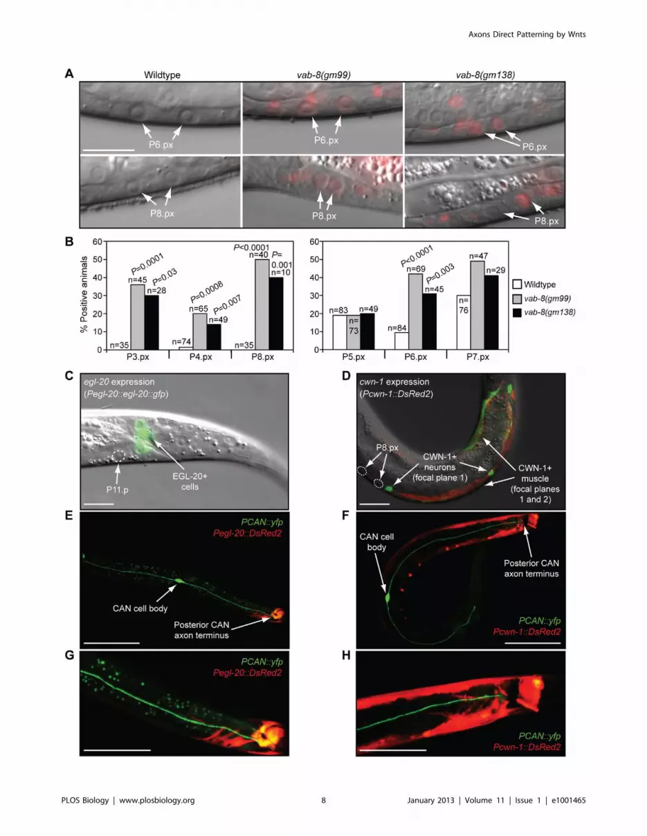
During vulval development, egl-20/wnt expression was detected
in four rectal cells (K, F, B, and U) just posterior to P11.p
(Figure 4C) [15]. cwn-1/wnt was strongly expressed in posterior
body-wall muscle and a subset of posterior motor neurons
(Figure 4D) [13,37]. These cells were posterior to the P8.px
progeny (Figure 4D), but weakly expressing neurons and muscle
Figure 1. Wnt signaling and epidermal patterning in C. elegans. (A) A wild-type C. elegans adult hermaphrodite. Scale bar is 100 mm. (B)During the L2 larval stage, LIN-3/EGF from pre-anchor cell/ventral uterine precursor cells (not shown) cooperates with a gradient of EGL-20/Wnt(orange) from rectal cells and CWN-1/Wnt (green) from posterior muscle and neurons to cause six epidermal cells to become vulval progenitors(P3.p–P8.p). 50% of the time, P3.p does not receive sufficient Wnt signaling and adopts the ‘‘F’’ fate (also known as the 4u fate) and fuses with ahypodermal syncytium called hyp7. EGL-20/Wnt also polarizes P5.p and P7.p so that they face posteriorly (horizontal arrows). The epidermal cellsnormally touch each other, but are drawn apart to facilitate depiction of muscle and neurons. (C) At the end of the L2 larval stage, anchor cell-produced MOM-2 and LIN-44 Wnts (blue) reorient P7.p towards the anterior (horizontal arrows). During the L3 larval stage, LIN-3/EGF (purple) fromthe anchor cell induces the 1u vulval fate in P6.p, which is facilitated by EGL-20 and CWN-1 Wnts. P5.p and P7.p adopt 2u vulval fates because of theactivation of LIN-12/Notch via a lateral signal from P6.p. (D) During the L3–L4 larval stages, vulval progenitor cells (Pn.p) divide to generate Pn.px cells,with P5.p–P7.p undergoing two additional rounds of cell division (to ultimately make Pn.pxxx cells). Because of the opposite polarities of P5.p andP7.p, their asymmetrically dividing progeny generate mirror image patterns. By the early L4 stage, a 22-cell vulva is generated. The Pn.px progeny ofP3.p, P4.p, and P8.p fuse with hyp7 (3u fate).doi:10.1371/journal.pbio.1001465.g001
Axons Direct Patterning by Wnts
PLOS Biology | www.plosbiology.org 4 January 2013 | Volume 11 | Issue 1 | e1001465

Figure 2. vab-8 mutations that affect neuronal cell body positioning and axon outgrowth cause epidermal patterning defects. (A)vab-8 mutations cause P8.p to adopt a vulval fate. In wild-type animals, P8.p divides once, but never forms vulval tissue. (B) vab-8 mutations increasethe frequency of P3.p becoming a vulval progenitor. 50% of the time, P3.p receives sufficient Wnt signaling to become a vulval progenitor and
Axons Direct Patterning by Wnts
PLOS Biology | www.plosbiology.org 5 January 2013 | Volume 11 | Issue 1 | e1001465

extended anteriorly to P2.p. Strikingly, the CANs also expressed
CWN-1/Wnt (Figure S3) [38], and their posterior axons traveled
parallel to the epidermal progenitors and the other CWN-1-
producing cells, and terminated at the sources of EGL-20/Wnt
(Figure 4E–4H). Thus, the CANs appear well-positioned to
regulate epidermal progenitor responses to these Wnts.
To further investigate the possibility that the CANs target EGL-
20/Wnt, we examined hermaphrodite-specific neuron (HSN)
migration in vab-8 mutants. The two HSNs are born in the
posterior of the embryo, close to the EGL-20/Wnt-producing
cells, and migrate to the mid-body position of the prospective
vulva [39] (Figure S4A and S4B). egl-20/wnt mutations prevent this
migration, while mutations in other Wnt genes have little effect
[40,41]. Conversely, increased EGL-20/Wnt activity causes
anterior overmigration of the HSNs [23], implying that an
EGL-20 gradient drives correct mid-body placement of the HSNs.
Consistent with prior reports [26,35,42], we found that in vab-8
mutants HSNs overmigrated anteriorly (Figure S4C), suggesting
EGL-20/Wnt activity is increased along the anterior–posterior
axis. This phenotype was enhanced by increasing EGL-20/Wnt
levels with an integrated transgenic array that carries additional
copies of the genomic egl-20 locus (Figure S4D and S4E) [23,40].
Since laser ablation of the CAN precursors also causes HSN
overmigration [35], the effect of vab-8 mutation on HSN migration
is likely mediated by the CANs. The integrated egl-20/wnt
transgenic array also cooperated with the vab-8(gm138) mutation
to increase the incidence of the P-Rvl phenotype (Figure 2C and
2D), and to cause an embryonic posterior P11 to P12 blast fate
transformation, which can occur when either EGF [43] or Wnt
signaling activity is abnormally high (Figure S4F and S4G). The
P11 fate transformation in vab-8 mutants is also at least partly
mediated by the CANs, since CAN-specific restoration of VAB-8S
significantly rescued this phenotype (Figure S4G).
Since elevated EGL-20/Wnt activity specifically causes a P-Rvl
phenotype, and increased activity of either EGL-20 or CWN-1/
Wnt could, in principle, account for the increased conversion of
P3.p into a vulval progenitor [7,14,16], we asked whether
elevation of one of these Wnts could also account for how vab-8
mutations suppress the underinduced phenotype of an egfr/let-
23(lf) mutation. When expressed from a heat-shock-regulated
transgene (Phs::egl-20), EGL-20/Wnt suppressed the egfr/let-23(lf)
mutation comparably to vab-8 mutations (Table 2). However, this
transgenic array did not induce ectopic vulval fates at P8.p in wild-
type animals (n = 52). This may be due to a lack of sufficiently high
EGL-20/Wnt expression in the posterior epidermis and/or a need
to increase signaling by additional Wnts such as CWN-1.
In agreement with the idea that Wnt signaling sufficient for the
induction of ectopic vulval fates is still limiting after heat shock of
just EGL-20, mutation of the intracellular Wnt inhibitor axin/pry-1
caused very high Wnt reporter activity in vulval progenitor
progeny, fully suppressed the underinduced phenotype of egfr/let-
23 mutants, and caused ectopic vulval fates (Table 1; Figure S2)
[19]. This high level of Wnt signaling also caused P9.p and P10.p
to become vulval progenitors, which, along with P8.p, formed
most of the ectopic vulval tissue (Figure S2D and S2E). Together,
our combined data suggest that EGL-20/Wnt is one CAN target,
and that additional Wnts, including CWN-1, may also be inhibited
by the CANs.
Anterior displacement of CAN cell bodies and foreshortening of
the posterior axons caused a gradual post-embryonic withering of
the posterior half of the animal [42], which placed some epidermal
progenitors closer to the anus, where EGL-20/Wnt is produced
(Figure S5A), and reduced posterior body volume (Figure S6A–
S6C). We ruled out these physical changes as being the major
mechanism by which the CANs regulate epidermal development
(see Text S1). In vab-8 mutants, Wnt reporter activity in vulval
divides once. (C) Upper panel depicts wild-type signaling by Wnts and EGF that promotes vulval development with mirror image symmetry. MOM-2and LIN-44 Wnts dominate over EGL-20/Wnt to polarize P7.p towards the anterior. Lower panel shows a wild-type 22-cell vulva with normal symmetryat the mid-L4 stage. (D) Upper panel depicts abnormal Wnt signaling in vab-8 mutants that causes the formation of vulval tissue with a P-Rvlphenotype. EGL-20/Wnt dominates over MOM-2 and LIN-44 Wnts, preventing P7.p from reorienting towards the anterior. Lower panel shows a P-Rvlvulva at the mid-L4 stage. In (C) and (D), EGL-20/Wnt was overexpressed from its native promoter with the muIs49 transgene. Scale bar is 10 mm.Colors depict Wnt signaling as in Figure 1. p-Values were calculated using a two-tailed Fisher’s exact test versus wild-type animals (A and B) or asotherwise indicated (A and D).doi:10.1371/journal.pbio.1001465.g002
Table 1. Mutations impairing CAN neuron migration andaxon outgrowth affect epidermal development.
GenotypeVulvalFatesa nb p-Valuec
Wild-type 3.00 47
gap-1(lf) 3.00 20
pry-1(lf) 3.42d 57
vab-8(gm99) 3.19 21
vab-8(gm138) 3.22 77
vab-8(ev411) 3.00 68
ceh-10(lf) 3.08 87
sfrp-1(lf) 3.00 53
let-23(lf) 0.52 24
let-23(lf); gap-1(lf) 4.00 40 ,0.00001 versus let-23(lf)
pry-1(lf); let-23(lf) 4.00 25 ,0.00001 versus let-23(lf)
let-23(lf); vab-8(gm99) 1.85 24 0.0002 versus let-23(lf)
let-23(lf); vab-8(gm138) 1.95 20 0.0001 versus let-23(lf)
let-23(lf); vab-8(ev411) 0.66 40 0.54 versus let-23(lf)
let-23(lf); ceh-10(lf) 0.64 21 0.65 versus let-23(lf)
let-23(lf); sfrp-1(lf) 0.05 21
lin-3(lf) 0.91 46
lin-3(lf); vab-8(gm99) 2.09 20 0.002 versus lin-3(lf)
lin-3(lf); vab-8(gm138) 2.12 46 ,0.00001 versus lin-3(lf)
lin-3(lf); vab-8(ev411) 0.80 27 0.64 versus lin-3(lf)
ceh-10(lf); lin-3(lf) 0.85 34 0.78 versus lin-3(lf)
let-60(dn)/+e 1.07 21
let-60(dn)/+; vab-8(gm99)e 2.30 20 0.0005 versus let-60(dn)
let-60(dn)/+; vab-8(gm138)e 2.48 20 0.00005 versus let-60(dn)
aVulval fates: number of vulval progenitor cells adopting vulval fates.bn: number of animals assayed.cp-Values were calculated using a two-tailed Student’s t test.dThis number of vulval fates is an approximation, since in this background it isdifficult to count the exact number of vulval cells descended from P9.p andP10.p.eThe let-60 mutation was linked to unc-24(lf) and balanced by dpy-20(lf).dn, dominant-negative; lf, loss-of-function.doi:10.1371/journal.pbio.1001465.t001
Axons Direct Patterning by Wnts
PLOS Biology | www.plosbiology.org 6 January 2013 | Volume 11 | Issue 1 | e1001465
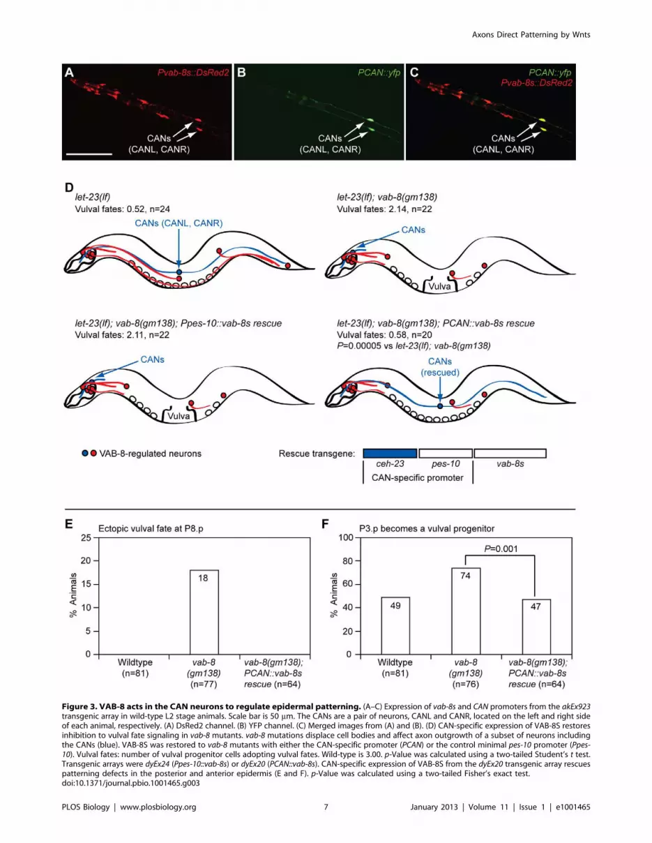
Figure 3. VAB-8 acts in the CAN neurons to regulate epidermal patterning. (A–C) Expression of vab-8s and CAN promoters from the akEx923transgenic array in wild-type L2 stage animals. Scale bar is 50 mm. The CANs are a pair of neurons, CANL and CANR, located on the left and right sideof each animal, respectively. (A) DsRed2 channel. (B) YFP channel. (C) Merged images from (A) and (B). (D) CAN-specific expression of VAB-8S restoresinhibition to vulval fate signaling in vab-8 mutants. vab-8 mutations displace cell bodies and affect axon outgrowth of a subset of neurons includingthe CANs (blue). VAB-8S was restored to vab-8 mutants with either the CAN-specific promoter (PCAN) or the control minimal pes-10 promoter (Ppes-10). Vulval fates: number of vulval progenitor cells adopting vulval fates. Wild-type is 3.00. p-Value was calculated using a two-tailed Student’s t test.Transgenic arrays were dyEx24 (Ppes-10::vab-8s) or dyEx20 (PCAN::vab-8s). CAN-specific expression of VAB-8S from the dyEx20 transgenic array rescuespatterning defects in the posterior and anterior epidermis (E and F). p-Value was calculated using a two-tailed Fisher’s exact test.doi:10.1371/journal.pbio.1001465.g003
Axons Direct Patterning by Wnts
PLOS Biology | www.plosbiology.org 7 January 2013 | Volume 11 | Issue 1 | e1001465
Axons Direct Patterning by Wnts
PLOS Biology | www.plosbiology.org 8 January 2013 | Volume 11 | Issue 1 | e1001465

progenitors did not strictly correlate with their distance from the
anus (Figure S5B); dpy-17(lf); dpy-20(lf) cuticular mutants had
normal CAN positioning and axon outgrowth, but even shorter
epidermal progenitor distances to the anus and smaller posterior
body volumes than vab-8 mutants (including vab-8 mutants with
ectopic vulval fates), yet did not show epidermal phenotypes
(Figures S5C–S5F, S6D, and S6E; Table S3); vab-8 mutations and
ablation of the CAN precursors promoted embryonic EGL-20/
Wnt-dependent HSN overmigration prior to any tail withering
(Figure S4B–S4E) [26,35,42]; and the CANs also prevented
overexpressed EGL-20/Wnt from causing embryonic P11 to P12
fate transformations, before tail withering occurred (Figure S4G).
The Posterior CAN Axon Inhibits Wnt ActivityWhile in wild-type animals the median CAN cell body position
along the anterior–posterior axis was near the P5.px progeny, and
most posterior CAN axons terminated near P11.p and the EGL-
20/Wnt-producing rectal cells (Figures 4E, 4G, 5A, and 5B), in
vab-8(gm138) mutants, both the CAN cell bodies and posterior
axon termini were severely displaced anteriorly (Figure 5A–5C).
Given the normal proximity of the posterior CAN axon terminus
to the EGL-20/Wnt-producing cells, the axons may secrete a
short-range signal that inhibits EGL-20 production. However,
severe mutation of vab-8 did not increase egl-20/wnt RNA levels
(Figure S7), and the vab-8(ev411) mutation, which mildly shifted
Figure 4. CAN neurons inhibit Wnt signaling in epidermal progenitors. (A) Pn.px stage animals showing syIs187 mCherry Wnt reporteractivity. Scale bar is 20 mm. (B) Quantification of reporter data. Reporter activity was lower in P5.px–P7.px cells than in P3.px, P4.px, and P8.px cells, sodata were collected using a higher brightness setting. p-Values were calculated using a two-tailed Fisher’s exact test versus wild-type animals. (C andD) Location of egl-20/wnt- and cwn-1/wnt-expressing cells relative to epidermal cells in L3, Pn.px stage animals. Scale bar is 10 mm. (C) muIs49[Pegl-20::egl-20::gfp] transgenic animal. (D) dyEx10[Pcwn-1::DsRed2] transgenic animal. To simultaneously visualize neurons and muscle, images were takenin different focal planes, differentially colored either green or red, and merged. (E–H) Location of Wnt-producing cells relative to CAN neurons. Onlyone CAN cell body is visible. (G and H) Blow-up of (E) and (F), respectively. Scale bars are 50 mm (E and F) and 25 mm (G and H). Transgenic arrays wereakEx906 (E and G) and akEx908 (F and H).doi:10.1371/journal.pbio.1001465.g004
Table 2. CAN displacement causes epidermal phenotypes that require signaling by specific Wnts and that resemble phenotypescaused by mutations in the Wnt-binding Ror/CAM-1 receptor.
Genotype Vulval Fatesa nb p-Valuec
Wild-type 3.00 47
egl-20(lf) 3.00 58
cwn-2(lf) 3.00 25
lin-44(lf) 3.00 20
Wild-type; vector RNAi 3.00 20
Wild-type; cwn-1 RNAi 3.00 20
Wild-type; mom-2 RNAi 3.00 20
let-23(lf); vab-8(gm138) 1.95 20 0.0001 versus let-23(lf)
let-23(lf); egl-20(lf); vab-8(gm138) 0.68 20 0.001 versus let-23(lf); vab-8(gm138)
lin-44(lf); let-23(lf); vab-8(gm138) 2.15 20 0.62 versus let-23(lf); vab-8(gm138)
let-23(lf); cwn-2(lf); vab-8(gm138) 1.30 25 0.07 versus let-23(lf); vab-8(gm138)
let-23(lf); vab-8(gm138); vector RNAi 1.45 20
let-23(lf); vab-8(gm138); cwn-1 RNAi 0.40 31 0.005 versus let-23(lf); vab-8(gm138); vector RNAi
let-23(lf); vab-8(gm138); mom-2 RNAi 0.63 23 0.04 versus let-23(lf); vab-8(gm138); vector RNAi
let-23(lf); syEx1024[Phs::egl-20] 0.13 20
let-23(lf); syEx1024[Phs::egl-20] heat shockd 1.70 20 ,0.00001 versus non-transgenic heat shock
Control non-transgenic siblinge 0.00 20
Control non-transgenic sibling heat shockd,e 0.05 20
cam-1(gm122) 3.04 51
cam-1(sa692) 3.03 74
cam-1(ks52) 3.00 62
lin-3(lf) 0.91 46
cam-1(gm122); lin-3(lf) 1.76 44 0.0004 versus lin-3(lf)
cam-1(sa692); lin-3(lf) 2.08 31 ,0.00001 versus lin-3(lf)
cam-1(ks52); lin-3(lf) 1.11 27 0.47 versus lin-3(lf)
aVulval fates: number of vulval progenitor cells adopting vulval fates. Wild-type is 3.00.bn: number of animals assayed.cp-Values were calculated using a two-tailed Student’s t test.dMixed-stage animals were heat-shocked at 33uC for 45 min and scored 16 h later.eControl animals were of the same genotype as the transgenic animals, but lacked the extrachromosomal transgenic array.lf, loss-of-function.doi:10.1371/journal.pbio.1001465.t002
Axons Direct Patterning by Wnts
PLOS Biology | www.plosbiology.org 9 January 2013 | Volume 11 | Issue 1 | e1001465

Axons Direct Patterning by Wnts
PLOS Biology | www.plosbiology.org 10 January 2013 | Volume 11 | Issue 1 | e1001465

the posterior CAN axon terminus away from the EGL-20/Wnt-
producing cells (Figure 5B and 5C), did not cause epidermal
phenotypes (Figure 2A and 2B; Table 1).
We next examined how a ceh-10 homeobox gene mutation that
has intermediate effects on posterior CAN axon outgrowth affects
epidermal development [35]. Although in ceh-10 mutants the
median CAN cell body position was anteriorly shifted to the head,
similarly as in vab-8(gm138) mutants (Figure 5A) [35], the posterior
CAN axon extended further than in vab-8 mutants, to a median
position of P4.px rather than P1.p (Figure 5B). Mutation of ceh-10
did not increase the frequency of P3.p becoming a vulval
progenitor (Figure 2B), and did not suppress the P6.p-based
underinduced phenotypes of egf/lin-3 or egfr/let-23 mutations
(Table 1). However, ceh-10 mutants had a low incidence of ectopic
vulval fates in P8.p (Figure 5D). These data suggest that a rarer
aspect of the ceh-10 mutant phenotype, such as severe shortening
of the posterior CAN axon rather than displacement of the cell
body, may promote vulval fate signaling along the anterior–
posterior axis.
To determine the relationship between the posterior CAN axon
terminus and vulval fate signaling, we examined both parameters
in individual ceh-10 mutants. In animals where the furthest
posterior CAN axon terminus of the pair of neurons was at or
posterior to P8.p progeny, P8.p did not adopt a vulval fate
(Figure 5E, e.g., animal 52). By contrast, in animals where the
furthest posterior CAN axon terminus was anterior to P8.p
progeny, P8.p acquired ectopic vulval fates, with some animals
also showing a P-Rvl phenotype in P7.p (Figure 5E, e.g., animal
79). We also identified rare ceh-10 mutants that demonstrated a
separation between tail withering and increased vulval fate
signaling. In animals 79, 74, and 45, the Wnt-responding P8.px
progenitors that gave rise to ectopic vulvae were further away from
the EGL-20/Wnt sources (.135 mm) than the non-Wnt-respond-
ing P8.px progenitors in animals not having ectopic vulval
induction (135 mm) (Figure S8). However, in these three animals,
the posterior CAN axon never extended beyond P3.px, demon-
strating that productive vulval fate signaling is always correlated
with severe posterior axon shortening.
To determine whether a similar relationship also exists between
the posterior CAN axon terminus and the signaling involved in
converting anterior P3.p into a vulval progenitor, we also
examined these two parameters in ceh-10 mutants. Analogous to
the study at P8.p, we found that when the longest of the pair of
posterior CAN axons reached only the P3.p position, the
frequency of P3.p becoming a progenitor increased from the
normal ,50% to 72% (Figure 5F). This increased frequency is
similar to that in vab-8 mutants, which have a median furthest
posterior axon terminus position of P1.p. In contrast, when the
longest posterior CAN axon terminated past P3.p, the frequency
of P3.p becoming a progenitor was 41%, which was not
statistically different from that of wild-type animals. Together,
these data indicate that foreshortening of the posterior CAN axons
can cause epidermal progenitors to show evidence of enhanced
responses to Wnt signaling.
Although anterior and posterior epidermal progenitor responses
to Wnts are inversely correlated with the length of the posterior
CAN axon, the striking expression of CWN-1/Wnt in the CAN
neurons suggests the possibility that when anteriorly displaced, the
cell bodies of these neurons might promote Wnt signaling in
anterior epidermal cells such as P3.p. Consistent with this model,
we found that the increased frequency of P3.p becoming a vulval
progenitor in vab-8 mutants was more dependent on cwn-1 than
egl-20/wnt activity (Figure S9A). If this strong dependence on
CWN-1/Wnt was largely due to increased proximity of CWN-1-
producing CAN cell bodies to P3.p, it would be expected that
CAN cell bodies would be consistently closer to P3.p when P3.p
becomes a vulval progenitor. We found 17 ceh-10 mutants to test
this hypothesis. In these mutants, at least one of the cell bodies of
the pair of CAN neurons was displaced closer to P3.p, away from
its normal median position of P5.p (Figure S9B). However,
regardless of whether P3.p had or had not become a vulval
progenitor, the cell body that was closest to P3.p (among the pair
of CAN neurons) was similarly close to P3.p (Figure S9C, left
panel). Also arguing against a positive role for the CAN cell bodies
in promoting Wnt signaling in P3.p, we found that when P3.p
became a vulval progenitor, the cell body that was furthest from
P3.p (among the pair of CAN neurons) was significantly further
away from P3.p than in cases where P3.p did not become a
progenitor (Figure S9C, right panel). In fact, in one case, we found
an animal where the two CAN cell bodies were directly over P3.p,
yet P3.p still failed to become a vulval progenitor (Figure S9B,
animal 92). Notably, in this animal, both posterior CAN axons
terminated past the P8.p progeny. The most significant correlation
we noticed in animals with at least one of the two CAN cell bodies
displaced closer to P3.p was that when P3.p became a vulval
progenitor, fewer posterior CAN axons reached P3.p (Figure S9B).
Collectively, these results indicate that the key role of the CANs in
epidermal development is to inhibit Wnt signaling, and that the
posterior CAN axons must extend a certain distance to confer this
inhibition.
The CAN Axons Use the Ror/CAM-1 Wnt Receptor toRegulate Wnt Signaling in Epidermal Progenitors
The C. elegans genome encodes one diffusible Wnt antagonist,
SFRP-1 [38]. However, SFRP-1 does not mediate the effects of
the CANs on epidermal development: its expression is largely
restricted to anterior body wall muscle [38], and unlike CAN-
displacing vab-8 mutations, mutation of sfrp-1 did not suppress the
Figure 5. Posterior CAN axons regulate axial positioning of vulval fates and P3.p vulval progenitor frequency. Distributions ofpositions of CAN cell bodies (A) and furthest posterior CAN axon termini (B) in different mutants. CAN neurons were visualized in L3, Pn.px stage (Aand B) or L4 stage (E) animals with the kyIs4[Pceh-23::gfp] transgene. x-Axis indicates Pn.p or Pn.px positions. p-Value was calculated using a two-tailedMann-Whitney U test. (C) Position of CAN cell bodies and posterior axon termini relative to egl-20/wnt-expressing cells in L2 stage vab-8 mutants.CANs and egl-20/wnt-expressing cells were marked with the akEx906 transgenic array. The bright green signal in the head/pharynx is from thecoinjected Pmyo-2::cfp injection marker. Scale bar is 25 mm. (D) Frequency with which P8.p adopts a vulval fate in the total ceh-10 mutant population.(E) Correlation between the position of the furthest posterior CAN axon terminus and induction of ectopic vulval fates at P8.p in ceh-10(lf) mutants.For this study, an emphasis was placed on picking smaller animals to ensure that sufficient numbers of animals with short posterior CAN axons wereobtained for statistical analysis. Thus, the combined frequency of ectopic vulval fates in this study is not an estimate of the actual frequency in thetotal population as conducted in (D). Top panels show animal 52, with normal epidermal development and normal position of furthest posterior CANaxon terminus. Bottom panels show animal 79, with an R-Pvl phenotype at P7.p and ectopic vulval fate at P8.p, and severely foreshortened furthestposterior CAN axon terminus. Scale bars are 10 mm. (F) Correlation between position of furthest posterior CAN axon terminus and frequency withwhich P3.p becomes a vulval progenitor in ceh-10(lf) mutants. In (D–F), p-Values were calculated using a two-tailed Fisher’s exact test versus wild-typeanimals (D) or as otherwise indicated (E and F). H, head/pharyngeal region.doi:10.1371/journal.pbio.1001465.g005
Axons Direct Patterning by Wnts
PLOS Biology | www.plosbiology.org 11 January 2013 | Volume 11 | Issue 1 | e1001465

underinduced phenotype of egfr/let-23(lf) mutants (Table 1), and
did not cause a P-Rvl phenotype or ectopic vulval fates at P8.p
(n = 166). However, the only other known extracellularly acting
Wnt inhibitor in C. elegans, the transmembrane Wnt-binding Ror/
CAM-1 tyrosine kinase, could mediate the effects of the CAN
axons on Wnt signaling. Ror/CAM-1 is widely expressed in
muscle, the vulval progenitors, and many neurons, including the
CANs (Figure 6A) [7,37,44–46]. While in certain contexts, such as
EGL-20/Wnt-mediated polarization of P7.p, it transduces Wnt
signals [16], in other contexts such as inhibition of HSN migration,
P3.p vulval progenitor cell specification, and the induction of
vulval fates, it appears to antagonize Wnt signaling [7,47]. Based
on its ability to physically bind Wnts such as EGL-20 and CWN-1
(potential targets of vab-8 mutations), and to interfere with vulval
fate signaling in the P6.p epidermal progenitor when overex-
pressed in non-epidermal cells, it has been proposed that Ror/
CAM-1 inhibits Wnt signaling by sequestering Wnts away from
Wnt-responding cells [7]. While this model is plausible, how such a
broadly expressed antagonist might refine Wnt gradients to allow
specific migratory and tissue patterns is unclear.
To explore the possibility that Ror/CAM-1 might largely act
from specific neurons such as the CANs to refine the EGL-20 and
CWN-1 Wnt gradients that pattern the epidermis, we first
examined the extent to which a cam-1 null mutation, gm122
(Figure 6C), phenocopies the effects of vab-8 mutations. Similar to
mutation of vab-8, ror/cam-1 mutation elevated Wnt reporter
activity in P6.px and P8.px progeny (Figure 6B). Also similar to
mutation of vab-8, ror/cam-1 mutation increased the frequency of
P3.p becoming a vulval progenitor, cooperated with EGL-20/Wnt
overexpression to cause embryonic P11 to P12 fate transforma-
tions, and suppressed the underinduced P6.p-based phenotype of
an egf/lin-3 mutation (Figure 6D; Table 2) [7]. Notably, mutation
of ror/cam-1 did not perturb primary vulval symmetry, induce
ectopic vulval fates in P8.p (Figure 6E), or increase Wnt reporter
activity to the same degree as vab-8 mutations. These discrepancies
may be due to dual negative and positive functions of Ror/CAM-1
in epidermal development. Within certain epidermal progenitors
such as P7.p, Ror/CAM-1 may transduce Wnt signals, while in
the CANs, it may sequester Wnts to refine the posteriorly derived
Wnt gradients to which the vulval progenitors respond. Since
Ror/CAM-1 mediates the polarizing effect of EGL-20/Wnt on
P7.p [16], the P-Rvl phenotype, which stems from this polariza-
tion, cannot be manifested in ror/cam-1 mutants. Furthermore, we
found that Ror/CAM-1 is required for P8.p to adopt a vulval fate
when the posterior CAN axons are foreshortened by vab-8
mutations (Figure 6E). Further consistent with the model that
Ror/CAM-1 positively transduces Wnt signals in P8.p, the
increased frequency of Wnt reporter activity in P8.p progeny
was significantly lower in cam-1 mutants than in vab-8 mutants
(Figure 6E). The mildly elevated P8.px Wnt reporter activity in
ror/cam-1 mutants must therefore reflect activation of other Wnt
receptors that are not sufficient to drive a vulval fate.
Since Ror/CAM-1 has been reported to affect CAN cell body
positioning and axon outgrowth [35], we evaluated whether cam-1
mutations might increase posteriorly derived Wnt signaling by
affecting the location of the CANs and their axon termini. As has
been previously reported, we found that ror/cam-1 mutations
caused anterior displacement of the CAN cell bodies that was as
severe as that caused by ceh-10 mutation [35] (Figures 5A and 7A).
However, since ceh-10 mutants do not have as strong Wnt
phenotypes as ror/cam-1 mutants, the CAN cell body displacement
in cam-1 mutants cannot explain their increased Wnt signaling.
Also, in ror/cam-1 mutants, outgrowth of the posterior CAN axon
was only mildly affected, with the median end point being even
more posterior than in vab-8(ev411) and ceh-10(lf) mutants that
have no or weaker Wnt phenotypes (Figures 5B and 7B). Thus,
Ror/CAM-1 does not inhibit Wnt signaling by regulating
posterior CAN axon outgrowth, but could mediate an inhibitory
effect of the extended axons.
To test whether Ror/CAM-1 expression in the CANs is
sufficient to restore some aspect of normal inhibition of Wnt
activity in epidermal progenitors, we transgenically expressed
CAM-1 only in the CANs of cam-1(null); egf/lin-3(lf) double
mutants, which have elevated Wnt signaling in the epidermal
progenitors. Cell-specificity was confirmed by tagging the cDNA
with gfp, which does not interfere with CAM-1 biological activity
[47], and verifying that CAM-1::GFP expression was detected
only in the CANs. In general, it was difficult to obtain transgenic
lines with detectable and stable levels of CAM-1::GFP expression.
However, one transgenic line with CAN-specific expression was
selected for further analysis (Figure 7D). In this line, despite similar
levels of expression between animals, re-expression of Ror/CAM-
1 did not fully restore proper migration to the CANs (Figure 7A).
This partial rescue suggests that in these animals, the level of
transgenic Ror/CAM-1 expression may be just below physiologic
amounts. However, of those animals displaying normal migration
of at least one of the two CAN cell bodies, CAN-expressed Ror/
CAM-1 completely restored the parental egf/lin-3(lf) underinduced
phenotype (Figure 7C and 7D). This result indicates that
epidermal progenitors such as P6.p no longer receive abnormally
high Wnt signaling when Ror/CAM-1 is expressed only in the
CANs of cam-1(null) mutants.
Similar to vab-8 mutants, cam-1 mutants also show an embryonic
HSN overmigration phenotype, which has been proposed to be
due to overactive EGL-20/Wnt signaling [23]. To evaluate
whether Ror/CAM-1 could also be part of the embryonic
mechanism by which the CANs regulate Wnt-dependent respons-
es, we examined another transgenic line with CAN-specific
expression of Ror/CAM-1::GFP. These animals had a ror/cam-1
mutation and also expressed an HSN GFP marker to visualize
HSN migration. Similar to the other transgenic line described
above, this line also only weakly rescued the CAN migration defect
(only ,30% of animals had CANs in their normal mid-body
position). However, despite this incomplete functional activity,
these transgenic animals still showed significant, but not wild-type,
rescue of HSN migration (Figure S10). These data further support
a CAN-specific role for Ror/CAM-1 in cell non-autonomously
regulating responses to Wnts.
To explore the model that Ror/CAM-1 acts from the CANs to
sequester Wnts, we examined the requirement of the intracellular
kinase domain for inhibition of Wnt signaling in the epidermis.
Prior work showed that in conjunction with other Wnt receptor
mutations, a ror/cam-1 null mutation or a cam-1 mutation affecting
only the extracellular Wnt-binding domain (sa692, Figure 6C), but
not a cam-1 deletion affecting only the kinase domain (ks52,
Figure 6C), can induce ectopic vulval fates [7]. To determine
whether similar requirements for the extracellular, but not
intracellular, kinase domain extend to the regulation of epidermal
progenitor responses assayed in this work, we also examined the
effects of these domain-specific mutations. Consistent with the
prior work and our model, we found that the cam-1(sa692)
mutation, but not the cam-1(ks52) mutation, increased the
frequency of P3.p becoming a vulval progenitor and suppressed
an egf/lin-3 mutation comparably to the cam-1 null mutation
(Figure 6D; Table 2). To determine whether the extracellular-
domain-only requirement specifically extended to CAN-based
Ror/CAM-1 inhibition of epidermal progenitor responses to
Wnts, we repeated our transgenic rescue experiments with a cam-1
Axons Direct Patterning by Wnts
PLOS Biology | www.plosbiology.org 12 January 2013 | Volume 11 | Issue 1 | e1001465

Axons Direct Patterning by Wnts
PLOS Biology | www.plosbiology.org 13 January 2013 | Volume 11 | Issue 1 | e1001465
construct that lacked the entire intracellular domain (DIntra,
Figure 6C). Similar to with the wild-type construct, it was difficult
to generate transgenic lines with detectable GFP expression. Our
best-expressing transgenic line showed partial rescue of the CAN
cell body migration defect, but not as much as the wild-type-
expressing line (Figure 7A), and there was no rescue of the CAN
axon outgrowth or the mild tail-withering defects (Figures 7B and
S11). Despite its limited activity in promoting proper CAN
migration, the DIntra construct fully restored inhibition to vulval
development in ror/cam-1(gm122); egf/lin-3(lf) mutants (Figure 7E).
Also consistent with our model, it has been suggested that the
DIntra construct may have a higher Wnt-binding capacity than
the wild-type receptor [47], which could explain its seemingly
more potent ability to restore Wnt inhibition.
To complement our transgenic add-back experiments with a
more physiologic assessment of the Ror/CAM-1 site of action in
regulating Wnt responses in epidermal progenitors, we used a
genetic approach. We scored two quantitative epidermal pheno-
types that are equally manifested in ror/cam-1 and vab-8 single
mutants. In this strategy, we used a vab-8 mutation to maximally
displace the posterior CAN axon terminus away from the
epidermal progenitors. If Ror/CAM-1 acts mostly through other
neurons and muscle, a cam-1 mutation should still significantly
increase Wnt signaling in vab-8 mutants. On the other hand, if
Ror/CAM-1 acts mostly through the CANs, a cam-1 mutation
should have no further effect on Wnt signaling. Both ror/cam-1 and
vab-8 single mutations have the same effect on the frequency with
which P3.p becomes a vulval progenitor, increasing this frequency
from 50% to ,70% (Figures 2B and 6D). However, double
mutants showed no further increase in this frequency (Figure 7F).
Similarly, both ror/cam-1 and vab-8 mutations suppressed the
vulvaless phenotype of egf/lin-3 mutants centered at P6.p to a
similar extent (Tables 1 and 2), yet combining the two mutations
did not further increase this suppression (Figure 7F).
Ror/CAM-1 Regulates EGL-20/Wnt Localization to thePosterior CAN Axon
Although Ror/CAM-1 is expressed at much higher levels in
muscle than the CANs (Figure 6A), our data surprisingly indicate
that CAN-expressed CAM-1 plays a critical role in regulating
epidermal progenitor responses to Wnts such as EGL-20. While
the EGL-20/Wnt gradient has been visualized in early L1 stage
larvae [12], its potential refinement by specific cell populations like
the CANs has not been reported. To investigate whether the EGL-
20/Wnt gradient might be affected by the posterior CAN axons,
we examined the distribution of EGL-20 in the vicinity of these
axons. To accomplish this, we employed two transgenes. One
expressed functional EGL-20/Wnt as a fusion to protein A, under
the control of its native promoter (Pegl-20::egl-20::protein A) [12].
This transgene rescues egl-20/wnt mutant phenotypes, but does not
cause gain-of-function phenotypes, and, therefore, expresses EGL-
20 at near physiologic levels [12]. The second transgene expressed
GFP under the control of the ceh-23 promoter (Pceh-23::gfp), which
drives expression in the CANs, as well as a few other neurons [48].
To visualize EGL-20/Wnt in living animals, we injected Cy5-
labeled rabbit IgG, which has a high affinity for protein A, into
double transgenic animals. This technique has been successfully
used to label the extracellular portions of cell surface receptors,
including protein-A-tagged receptors [49]. In single Pceh-23::gfp
transgene control animals, IgG-Cy5 did not accumulate to a
significant amount or label any cell surfaces (Figure 8A). In
contrast, when injected into Pegl-20::egl-20::protein A; Pceh-23::gfp
double transgenic animals, IgG-Cy5 labeled the surfaces of EGL-
20/Wnt-producing rectal cells near the anus (Figure 8B), demon-
strating specificity to the IgG-Cy5 signal. Cy5-labeled punctae
were also enriched in other parts of the posterior body, but
detection was variable, presumably because of variations in
labeling between injected animals (e.g., Figure S12). However,
the posterior enrichment of the Cy5-labeled punctae is consistent
with the description of the EGL-20/Wnt gradient in fixed Pegl-
20::egl-20::protein A L1 larvae stained with IgG-FITC [12].
Strikingly, Cy5 labeling was also detected along stretches of the
posterior CAN axon, suggesting EGL-20/Wnt is bound to the
axon (Figure 8C, three different animals are shown).
Since our data indicate that only the extracellular domain of
Ror/CAM-1 is necessary to inhibit Wnt signaling from the CAN
axons, and CAM-1 has been reported to bind Wnts such as EGL-20
in vitro [7], we tested whether CAM-1 mediates localization of
EGL-20 to the posterior CAN axons. First, we observed that when
driven by the CAN-specific promoter, Ror/CAM-1::GFP appeared
as punctae along the CAN axon, as reported for other cells
(Figure 8D) [50–52]. Notably, the Ror/CAM-1 punctae resembled
the size and pattern of the EGL-20/Wnt punctae along the
posterior CAN axon, suggesting EGL-20 might be bound to CAM-
1. Next, we quantified the density of EGL-20/Wnt punctae along
the posterior CAN axon in wild-type and ror/cam-1 mutants.
Mutation of ror/cam-1 reduced the EGL-20/Wnt density along the
axon, indicating that CAM-1 mediates some of the EGL-20
enrichment at the axon (Figure 8E and 8F). While in ror/cam-1
mutants the measured EGL-20/Wnt density along the posterior
CAN axon was not zero, it may have been lower than what we
quantified because it is impossible to be absolutely certain what
fraction of the punctae are actually bound to the axon. Nevertheless,
these data indicate that EGL-20/Wnt is enriched at, or in very close
proximity to, the posterior CAN axon in a Ror/CAM-1-dependent
manner, and provide evidence for these axons affecting the
distribution of EGL-20 in the extracellular space. These data are
in accord with our functional analyses of the properties of the CAN
axons, and provide a potential explanation for how the CAN axons
exert their unique role in epidermal development.
Discussion
Despite the identification of so many Wnt antagonists, our
understanding of how Wnt gradients are established with precision
Figure 6. Ror/CAM-1 has negative and positive effects on vulval fate signaling. (A) Ror/CAM-1 is broadly expressed. L2 stage animal withthe cwIs6 transgenic array, which expresses a rescuing Ror/CAM-1 translational fusion to GFP from the cam-1 promoter. Red arrows indicate axons ofnon-CAN neurons. Scale bar is 10 mm. (B) Ror/CAM-1 inhibits Wnt signaling in epidermal progenitors. Quantification of syIs187 Wnt reporter transgeneactivity in Pn.px stage ror/cam-1 mutants. Pn.px signals were scored as in Figure 4B. (C) Schematic description of ror/cam-1 mutations and the cam-1transgenic mutant (DIntra) used in this study. Numbers indicate amino acid positions. Fz, Frizzled, Wnt-binding extracellular domain; TM,transmembrane domain. (D) Ror/CAM-1 inhibits P3.p from becoming a vulval progenitor in larvae, and helps restrict the Wnt-dependent P12 fate toonly the P12 blast cell in embryos. Pegl-20::egl-20 is an integrated transgenic array that introduces extra functional copies of egl-20/wnt fused to gfpinto the genome (see Figures S4 and S7). (E) Ror/CAM-1 is required for foreshortening of the posterior CAN axon to promote a vulval fate in theposterior P8.p progenitor. Model depicting negative and positive roles of Ror/CAM-1 on Wnt signaling in epidermal progenitors. p-Values werecalculated using a two-tailed Fisher’s exact test versus wild-type animals (B and D) or as otherwise indicated (E).doi:10.1371/journal.pbio.1001465.g006
Axons Direct Patterning by Wnts
PLOS Biology | www.plosbiology.org 14 January 2013 | Volume 11 | Issue 1 | e1001465

Figure 7. The CANs use the extracellular Wnt-binding domain of Ror/CAM-1 to direct epidermal patterning. (A and B) Distributions ofpositions of CAN cell bodies and furthest posterior CAN axon termini in ror/cam-1 mutants and in cam-1 mutants expressing wild-type or intracellular-domain-deleted CAM-1(DIntra) only in the CANs. In non-ror/cam-1 rescue experiments, CANs were visualized with the kyIs4[Pceh-23::gfp] transgene. Inror/cam-1 rescue experiments, CANs were visualized by expression of GFP-tagged Ror/CAM-1 in the CANs. In ror/cam-1 rescue experiments, strainsalso harbored an egf/lin-3(lf) mutation. x-Axis indicates Pn.p or Pn.px positions. H, head/pharyngeal region. p-Values were calculated using a two-tailed Mann-Whitney U test. (C) Ror/CAM-1 inhibits vulval fate signaling in central vulval progenitors. (D) Transgenic CAN-specific expression of Ror/
Axons Direct Patterning by Wnts
PLOS Biology | www.plosbiology.org 15 January 2013 | Volume 11 | Issue 1 | e1001465
and integrated with the activities of multiple growth factors is still
quite limited. Here, we show that even to create an anatomically
simple structure such as the C. elegans vulva, the diffusion of Wnts
and their inhibitors does not provide sufficient spatial resolution of
signaling. To overcome this problem, C. elegans has incorporated
the unique axonal projections of specific neurons to set the exact
strengths of Wnt signaling at precise locations. The CAN neurons
use their posterior-directed axons and the Ror/CAM-1 Wnt
receptor to regionally dampen the activity of posteriorly derived
Wnt (EGL-20 and possibly CWN-1). This dampening prevents
ectopic induction of vulval tissue in the posterior epidermis and
ensures that other centrally produced Wnts specify proper
symmetry in the primary vulva. However, this mechanism still
permits the posterior-derived EGL-20 and CWN-1 Wnts to retain
sufficient activity in the mid-body and anterior epidermis to specify
the vulval progenitors, and cooperate with EGF/LIN-3 in
promoting a 1u vulval fate in P6.p. Surprisingly, this type of
regulation of epidermal pattering is unique to the Ror-expressing
posterior CAN axons, and is not conferred by the other Ror/
CAM-1-expressing cells along the anterior–posterior axis, or the
SFRP-1-expressing cells. In fact, the posterior CAN axons are such
potent regulators of Wnt signaling that foreshortening their
normal length can dramatically alter an epidermal progenitor’s
response to Wnts. Given the unique role of the posterior CAN
axons in regulating epidermal patterning, much of Ror/CAM-1
expression in other cells may be for more subtle control of
epidermal patterning that we could not detect in our genetic
experiments, or for inhibition of Wnt signaling for cell populations
we did not assay, or could reflect other biological functions for
CAM-1. For example, in muscle, Ror/CAM-1 is an important
regulator of synapse strength [50–52], and in other cells, it plays
important roles in cell polarity, cell migration, axon guidance, and
neurite survival [16,35,37,44–47,53].
Although for technical reasons we could not quantify the EGL-
20/Wnt gradient in the vicinity of the epidermal progenitors that
respond to Wnt signaling, we could quantify other aspects of EGL-
20 distribution in living adult animals. Notably, we detected EGL-
20/Wnt punctae along the posterior CAN axon in a pattern
similar to how Ror/CAM-1 clusters along the same axon,
suggesting EGL-20 may bind to the axon via CAM-1. In support
of this model, mutation of ror/cam-1 reduced EGL-20/Wnt density
along the posterior CAN axon. However, Ror/CAM-1 may not
be the only factor determining where EGL-20 is distributed within
the posterior body. Ror/CAM-1 is expressed at levels similar to
the CANs in other neurons, and at much higher levels than the
CANs in muscle (Figure 6). Yet, despite this expression pattern of
Ror/CAM-1, EGL-20/Wnt does not appear to be proportionally
concentrated at muscle (Figure S12), and appears to be somewhat
enriched at the posterior CAN axon (Figure 8). Since Ror/CAM-1
has been reported to act in complexes with other Wnt-binding
receptors [45,46,52], it is possible that part of the EGL-20-binding
properties of the CAN axons arises from co-expression of other
specific Wnt receptors or post-translational modifications of CAM-
1 that affect its EGL-20-binding properties. Notably, in muscle,
CAM-1 functions in a heteromeric complex with LIN-17/Frizzled
to transduce CWN-2, but not EGL-20, Wnt signaling at the
neuromuscular junction [52].
Given the Ror/CAM-1-dependent localization of EGL-20/Wnt
to the posterior CAN axon, the ability of CAM-1 to bind EGL-20
in vitro [7], and the requirement for only its Wnt-binding
extracellular domain for CAM-1 to inhibit Wnt signaling from the
CANs, it is possible that Wnt sequestration is part of the
mechanism by which the CAN axons regulate epidermal
patterning (Figure 9). Our data indicate that if Ror/CAM-1 is
absent or if the posterior CAN axon does not grow to a sufficient
length, the extracellular distribution of EGL-20/Wnt is altered. In
these cases, in a simple model, the EGL-20/Wnt that is displaced
from the CAN axons would be available to cause excess EGL-20
signaling in the epidermal progenitors, thereby perturbing normal
Wnt patterning. Although only a small fraction of the total EGL-
20/Wnt punctae appear to be bound to the posterior CAN axons,
there are several possible scenarios in which this pool might be
critical in determining the nature and strength of epidermal
progenitor Wnt responses. First, many of the EGL-20/Wnt
punctae that we detected at other locations might not be truly
‘‘free’’ and able to stimulate epidermal progenitors. They may be
bound to other cells or trapped in the extracellular space. Thus, if
the EGL-20/Wnt released from the posterior CAN axons is more
diffusible than the other EGL-20, it may have a potent ability to
change the pattern of epidermal progenitor responses to Wnts.
Alternatively, the rectal cells may produce distinct forms of EGL-
20/Wnt, with distinct abilities to bind and signal through different
EGL-20 receptor complexes. Perhaps a form of EGL-20/Wnt that
is specific for the epidermal progenitors is also preferentially
sequestered by the CANs, endowing the CANs with a unique
ability to regulate epidermal progenitor responses to EGL-20.
Although our data indicate a critical role during larval
epidermal development for the posterior CAN axons in regulating
Wnt responses, in certain settings, the CAN cell bodies may also be
able to perform this function. Notably, EGL-20/Wnt-dependent
HSN migration is completed prior to CAN axon outgrowth [54],
yet embryonic laser ablation of the CAN precursor cells still causes
HSN overmigration [35]. Similarly, genetic mutation of ror/cam-1
also causes HSN overmigration [23], and we can partially rescue
this phenotype by re-expressing Ror/CAM-1 specifically in the
CANs (Figure S10). Together, these data suggest that Ror/CAM-1
might also be able to act from the CAN cell bodies to affect EGL-
20/Wnt distributions, and hence the migration pattern of the
HSNs.
Our data raise the possibility that besides their behavioral
functions, another core property of neurons may be to act as
unique spatial cues to refine developmental patterns created by
growth factors such as Wnts. This refinement may be manifested
at two levels. First, in a specific tissue in a given organism, a
specific fixed pattern of neurons may increase the robustness of
inducing a specific pattern. For example, in C. elegans, the CANs
ensure that the normal epidermal pattern of a single vulva with
mirror image symmetry in the mid-body is invariant. When the
posterior CAN axons are severely foreshortened, additional
epidermal patterns are observed. However, our data also indicate
CAM-1::GFP restores inhibition of vulval development in ror/cam-1 mutants. (E) Transgenic CAN-specific expression of a Ror/CAM-1::GFP mutantlacking the intracellular domain (DIntra, see Figure 6C) also restores inhibition of vulval development in ror/cam-1 mutants. In (D) and (E), scale bar is20 mm. (F) Under physiologic conditions, the majority of Ror/CAM-1 inhibition of vulval fate signaling is mediated by the CANs. If the CAN cell bodiesare anteriorly displaced and the posterior axons are severely foreshortened, loss of Ror/CAM-1 activity from all cells does not further increase P3.pprogenitor frequency or the amount of vulval development in sensitized backgrounds. Drawings depict the cellular distribution of Ror/CAM-1 asdescribed in Figure 6E. M, muscle cells; N, neurons; P, vulval progenitors. In (C–E), vulval fates: number of vulval progenitor cells adopting vulval fates.Wild-type is 3.00. p-Values were calculated using a two-tailed Student’s t test. The PCAN::cam-1::gfp and PCAN::DIntra::gfp rescuing transgenic arraysare dyEx44 and dyEx45, respectively.doi:10.1371/journal.pbio.1001465.g007
Axons Direct Patterning by Wnts
PLOS Biology | www.plosbiology.org 16 January 2013 | Volume 11 | Issue 1 | e1001465

Figure 8. EGL-20/Wnt co-localizes with posterior CAN axons in vivo, in a Ror/CAM-1-dependent manner. CAN axons were visualizedwith the integrated Pceh-23::gfp transgene (kyIs4) (A–C and E), and EGL-20/Wnt was detected with the integrated Pegl-20::egl-20::protein A fusiontransgene (huIs60) and injection of Cy5-conjugated rabbit IgG (IgG-Cy5) into living adult animals (B, C, and E). (A–C and E) The Cy5 channel is shown,
Axons Direct Patterning by Wnts
PLOS Biology | www.plosbiology.org 17 January 2013 | Volume 11 | Issue 1 | e1001465

that the pattern of neuronal positioning and axon outgrowth can
also affect the appearance of a tissue pattern. Shortening of the
normal length of the posterior CAN axon causes a second vulva to
inappropriately form in the posterior body and alters the normal
symmetry of mid-body vulval tissue. Based on these data, we also
suggest that at a broader level, more phenotypic diversity can be
generated among tissues and organisms with neurons than without
neurons. In a simplified model of aneural tissue patterning, Wnt
gradients established by diffusion direct patterning, with distances
between Wnt-producing and -responding cells mostly affecting the
pattern. In contrast, in organisms with nervous systems, the
layering of different intricate patterns of Wnt-sequestering neurons
into developing tissues could expand the diversity of functional
Wnt gradients, and hence the diversity of tissue patterns that are
possible.
The ability of axons to refine Wnt signaling may be conserved,
since Wnts are used extensively throughout metazoan development,
and Ror kinases and other Wnt receptors are expressed in the
nervous systems of diverse species [4,5,55,56]. Furthermore, the
close proximity of developing neural circuits and tissues is not
unique to C. elegans. In vertebrates, neural crest cells begin to occupy
the embryonic gut at the time of organ budding, and axon tracts are
found in undifferentiated limb buds [57–59]. The ability of neurons
to refine spatial domains of Wnt signaling may also be important
after development for certain homeostatic functions. In mammals,
Wnt signaling must be precisely regulated in specific niches to
promote stem cell self-renewal and prevent tumorigenesis [60,61].
By expressing the appropriate cognate receptors, neurons may even
use the mechanism we have discovered to regulate the extracellular
distributions of many other growth factors, thereby expanding the
repertoire of biological processes under their control.
Although it was almost 200 years ago that Tweedy John Todd
reported nerves were important for salamander limbs to regener-
ate after amputation [62], neurons have still not been widely
demonstrated to be important for non-neuronal development.
Most studies of non-behavioral effects of the nervous system have
focused on the importance of whole nerves for muscle develop-
ment and fracture healing [28,63–67], and few mechanisms have
been described for how the cellular components of nerves may
exert non-behavioral effects. Notably, where examined, secretion
of neurotransmitters or growth factors has always been part of the
mechanism. In the salamander, Schwann cells from the sciatic
nerve promote limb regeneration by releasing newt anterior
gradient protein (nAG) [68]. In mammals, by secreting noradren-
aline, muscarinic receptor agonists, and VEGF, nerves have been
reported to modulate, respectively, hematopoietic stem cell
migration, salivary gland cell proliferation, and endothelial cell
differentiation into arteries [29,69,70]. By contrast, our work
provides evidence for a non-secretory function of neurons, where,
by binding of one of the oldest conserved metazoan growth factors,
neurons can help organize the complex signaling patterns that
direct tissue development.
The use of neurons to refine patterning by Wnts may have
arisen from the ancestral functions of Wnts in establishing and
patterning the primary body axis, and their subsequent use in
directing neuronal migration, axon guidance, and synaptic
strength [4,56]. By additionally sequestering Wnts, neurons
provide an efficient and unique mechanism to reshape initial
Wnt gradients and help generate distinct body patterns. Given that
the neuronal function we describe here does not involve the release
of canonical neurotransmitters, but rather involves the axon
outgrowth properties of neurons, our data raise intriguing
questions regarding the earliest functional properties of primordial
neurons. In addition to their behavioral importance, the evolution
of neurons may also have improved metazoans by refining the
plans of the bodies they would ultimately control.
Materials and Methods
StrainsC. elegans were cultured at 20uC [71]. Loss-of-function alleles
included the following: LGII: cwn-1(ok546), let-23(sy1), unc-4(e120),
cam-1(gm122), cam-1(sa692), and cam-1(ks52); LGIII: ceh-10(gm120)
and pha-1(e2123ts); LGIV: lin-3(n378) and egl-20(n585); LGV: vab-
8(gm99), vab-8(gm138), vab-8(ev411), and him-5(e1490). Integrated
transgenes included muIs49, syIs187, kyIs4, huIs60, and cwIs6
(contains a full-length rescuing genomic Pcam-1::cam-1::gfp construct;
a gift from Wayne Forrester). The deletion allele cwn-1(ok546) was
generated by the C. elegans Gene Knockout Facility at the Oklahoma
Medical Research Foundation. See Text S1 for references.
Vulval Induction and P3.p FateVulval development was scored during the L4 stage under DIC
optics using a Zeiss Axio Imager. Animals were anesthetized with
10 mM sodium azide unless otherwise indicated. The number of
vulval nuclei was used to extrapolate how many vulval progenitor
cells adopted vulval fates. A vulval progenitor cell generating seven
or eight great granddaughters (Pn.pxxx) and no hyp7 tissue was
scored as 1.0 cell induction. A vulval progenitor cell in which one
daughter (Pn.px) fuses with hyp7, and the other daughter
generates three or four great granddaughters was scored as 0.5
cell induction. In wild-type animals, P5.p, P6.p, and P7.p each
undergo 1.0 cell induction, resulting in a total of 3.0 cell induction.
Animals with more than 3.0 cell induction are overinduced, and
animals with less than 3.0 cell induction are underinduced. To
determine whether P3.p had become a vulval progenitor, all Pn.p
(P1.p–P11.p) progeny nuclei were counted at the mid-L4 stage and
used to extrapolate whether P3.p had divided. P3.p division is
indicative of having become a vulval progenitor.
TransgenicsTransgenic extrachromosomal arrays were generated as de-
scribed [72]. See Text S1 for details of plasmid constructions and
descriptions of transgenic arrays.
Wnt Reporter ScoringWnt activity was visualized using the integrated POPTOP
mCherry reporter syIs187. Larvae were anesthetized with 5 mM
levamisole and photographed using epifluorescence and DIC on
an Axio Imager Z1 microscope using AxioVision software (Zeiss).
In the mCherry channel, animals were photographed with an
as well as an overlay of the GFP channel, where the posterior CAN axon has been pseudo-colored blue. White arrows point to examples of EGL-20/Wnt punctae, and yellow arrows point to examples of variable background autofluorescence. In (C) and (E), numbers in the panels denote particularinjected animals. Bottom panels are enlargements of red-boxed sections in the indicated animals. (D) Clustering of Ror/CAM-1 in the posterior CANaxon. Ror/CAM-1 was visualized as a functional translational GFP fusion from an extrachromosomal transgenic array (PCAN::cam-1::gfp; dyEx44) inanimals that also carried an egf/lin-3(lf) mutation. Scale bars are 10 mm. (F) 37 independent measurements from 32 wild-type animals, and 26independent measurements from 19 ror/cam-1 mutants were used to calculate the mean EGL-20/Wnt density along the posterior CAN axon. p-Valuewas calculated using a two-tailed Mann-Whitney U test versus wild-type animals.doi:10.1371/journal.pbio.1001465.g008
Axons Direct Patterning by Wnts
PLOS Biology | www.plosbiology.org 18 January 2013 | Volume 11 | Issue 1 | e1001465

exposure time of 1,200 ms. To set a threshold for reporter
detection, fluorescent image files were adjusted to a brightness of
20.59 and a contrast setting of 1.11. For reporter scoring in
P5.px–P7.px progeny, images were adjusted to a brightness of
20.50 and contrast of 1.22.
RNA InterferenceHT115 Escherichia coli bacteria containing RNAi clones (L4440,
empty RNAi vector; K100B4.6, cwn-1; F38E1.7, mom-2) were
obtained from the Ahringer Lab bacterial feeding library [73], and
RNAi was performed as described [74].
Figure 9. Model for neuronal control of axial patterning by Wnts. (A) In wild-type animals, EGL-20/Wnt (orange) (and possibly CWN-1/Wnt[green]) is sequestered by the Ror/CAM-expressing posterior CAN axons. At the end of the L2 larval stage, this sequestration allows MOM-2 and LIN-44 Wnts (blue) to reorient and polarize P7.p towards the anterior (horizontal arrows). During the L3 larval stage, anterior-facing P7.p divides with themirror image pattern of P5.p, and P8.p does not receive sufficient Wnt or EGF signaling to adopt a vulval fate. (B) If the posterior CAN axons do notextend far enough into the posterior body, where EGL-20/Wnt is produced, an abnormally high concentration of EGL-20 occurs, which preventsanterior polarization of P7.p, and causes ectopic vulval tissue to form at P8.p.doi:10.1371/journal.pbio.1001465.g009
Axons Direct Patterning by Wnts
PLOS Biology | www.plosbiology.org 19 January 2013 | Volume 11 | Issue 1 | e1001465

Fluorescence Microscopy and EGL-20/Wnt::protein ADetection
Some images were captured on a Nikon Eclipse T1 confocal
microscope, with worms being anesthetized in 500 mM sodium
azide (Figures 3A–3C, 4E–4H, 5C, and 6A). Other images were
taken with a Zeiss Axio Imager Z1, with worms being anesthetized
with 5 mM levamisole (Figures 4A, 4C, 4D, 5E, 7D, 7E, and 8).
CAN positions were scored on a Zeiss Axio Imager Z1, with
worms being anesthetized in 0.1% tricaine/1.7 mM levamisole or
5 mM levamisole. To label EGL-20/Wnt::protein A in vivo, adult
animals were injected with 10 mg/ml Cy5-rabbit anti-rat IgG
(Jackson) diluted in 20 mM K3PO4, 3 mM potassium citrate, 2%
PEG6000 (pH 7.4). Animals were scored 6–10 h after injection.
Supporting Information
Figure S1 Mispositioning of the CAN cell bodies andforeshortening of the posterior axons do not perturbexcretory cell morphology. (A–C) Photographs of L4 animals
carrying the excretory cell bgIs312[Ppes-6::gfp] reporter transgene.
Scale bar is 100 mm. (B) Arrows point to cysts that have
abnormally formed in exc-5(lf) mutants. In some animals, the
excretory canals have truncated prematurely.
(PDF)
Figure S2 Mutation of axin/pry-1 strongly activates aWnt-responsive reporter in epidermal progenitors andpromotes vulval fates. (A) Representative L3, Pn.px stage
animals harboring the integrated syIs188 Wnt reporter transgene.
This integrated transgene is derived from the same extrachromo-
somal array found in the syIs187 integrant. Animals were
photographed with the same exposure time (1,000 ms). Asterisks
denote non-epidermal progenitor cells showing Wnt-independent
reporter expression. Scale bar is 10 mm. (B) Quantification of the
number of animals at the Pn.px stage with detectable syIs188 Wnt
reporter activity. p-Values were calculated using a two-tailed
Fisher’s exact test. (C) Quantification of average pixel density per
positive Pn.px cell. For control wild-type animals, P5.px and P6.px
cells not scoring positive by visual inspection had to be used to
obtain a minimum of two cells to determine mean pixel intensity.
p-Values were calculated using a two-tailed Student’s t test. (D)
syIs188 Wnt reporter expression in P9.p and P10.p progeny at the
Pn.px stage. Upper panel, 1,000 ms exposure. Lower panel,
250 ms exposure. Scale bar is 10 mm. (E) Frequency of ectopic
induction of vulval fates in axin/pry-1 mutants. p-Values were
calculated using a two-tailed Fisher’s exact test.
(PDF)
Figure S3 cwn-1 is expressed in the CANs. Image of a
hermaphrodite expressing the Pcwn-1::yfp reporter in muscle and
one of the pair of CAN cell bodies from the dyEx34 extrachro-
mosomal transgenic array. Scale bar is 10 mm.
(PDF)
Figure S4 vab-8 mutations increase Wnt-dependentHSN migration and cause P11 to P12 fate transforma-tions. (A) HSNs are born in the posterior of the embryo close to
the EGL-20/Wnt-producing cells, and are directed anteriorly by
EGL-20 to the prospective vulval region. (B–E) HSNs were
visualized with the zdIs13[Ptph-1::gfp] transgene. The integrated
muIs49 transgene (Pegl-20::egl-20::gfp) introduces extra functional
copies of the genomic egl-20/wnt locus fused to gfp into the
genome. (F) Schematic of P11 and P12 lineages showing the
unique dependence of the P12 fate on Wnt signaling. In wild-type
animals, P11 does not respond to Wnt signaling and generates a
large cell (P11.p) that fuses with hyp7. By contrast, P12 normally
responds to LIN-3/EGF and LIN-44/Wnt to generate a smaller
cell (P12.pa) that fuses with hyp12. (G) The CAN neurons help
minimize ectopic induction of the P12 fate in P11. p-Values were
calculated using a two-tailed Fisher’s exact test.
(PDF)
Figure S5 The increased proximity of epidermal pro-genitors to posterior Wnt sources in vab-8 mutants is notsufficient to promote vulval fate signaling. (A) Correlation
of median CAN positions (cell bodies and posterior axon termini)
with distance of posterior P8.p progeny from the anus (the
approximate location of the EGL-20/Wnt-producing cells) in L3,
Pn.px stage animals (CAN data are from Figure 5A and 5B).
Animals were anesthetized with 0.1% tricaine/1.7 mM levami-
sole. p-Values were calculated using a two-tailed Student’s t test.
(B) Distances of P6.pp and P8.pp, respectively, from the anus in
L3, Pn.px stage animals. Animals harbored the syIs187 integrated
Wnt reporter and were anesthetized with 5 mM levamisole. The
p-Value was calculated using a two-tailed Student’s t test. (C)
Distributions of positions of CAN cell bodies and furthest posterior
CAN axon termini in L3, Pn.px stage dpy-17(lf); dpy-20(lf) mutants.
(D) Representative dpy-17(lf); dpy-20(lf) double mutant at the L3,
Pn.px stage showing the normal position of the posterior CAN
axon terminus, which extends just past P11.p. Scale bar is 20 mm.
In (A), (C), and (D) animals harbored the kyIs4[Pceh-23::gfp]
transgene to mark the CANs. (E) Frequency with which P3.p
becomes a vulval progenitor. (F) Frequency with which P8.p
adopts a vulval fate. In (E) and (F) p-Values were calculated using a
two-tailed Fisher’s exact test.
(PDF)
Figure S6 The reduced posterior body volume in vab-8mutants is not sufficient to promote vulval fate signalingin epidermal progenitors. (A–D) Images showing posterior
surface areas for posterior body volume calculations in wild-type,
vab-8(lf), and dpy-17(lf); dpy-20(lf) mutants. Note that some vab-8
mutants with ectopic vulval fates at P8.p had posterior surface
areas similar to those of some wild-type animals (compare [A] and
[B]). (E) Table of posterior body volumes for wild-type, vab-8(lf),
and dpy-17(lf); dpy-20(lf) mutants. Volumes were calculated by
multiplying the mean thickness by the measured posterior surface
area for each animal. p-Values were calculated using a two-tailed
Student’s t test. Note that the mean posterior body volumes of the
total vab-8(lf) population, and of the vab-8 mutants specifically
having ectopic vulval fates at P8.p, were not less than those of
control dpy-17(lf); dpy-20(lf) mutants. (F and G) Body morphology
of egfr/let-23(lf) mutant L1 larvae within minutes of being placed
on 3% agar pads with either 50 mM (F) or 400 mM (G) sodium
chloride. (H) Table of whole body volumes for egfr/let-23(lf)
mutants as described in (E). The vab-8(lf) allele is gm138. Scale bar
is 100 mm.
(PDF)
Figure S7 Mispositioning of the CAN cell bodies andforeshortening of the posterior axons do not increaseegl-20/wnt mRNA levels. egl-20/wnt mRNA transcripts were
measured in pooled L1 worms by quantitative real-time PCR.
Expression is plotted relative to that in wild-type animals. muIs49
animals contain a functional, rescuing integrated Pegl-20::egl-20::gfp
transgenic array.
(PDF)
Figure S8 In ceh-10 mutants, ectopic vulval induction atP8.p correlates with foreshortening of the posterior CANaxon, but not with tail withering. Positions of uninduced
P8.px cells or ectopic vulvae from induced P8.px cells relative to
Axons Direct Patterning by Wnts
PLOS Biology | www.plosbiology.org 20 January 2013 | Volume 11 | Issue 1 | e1001465

the anus are schematized. CAN axons were visualized with the
kyIs4[Pceh-23::gfp] transgene in animals that were anesthetized with
0.1% tricaine/1.7 mM levamisole. Distances were measured from
the middle of the uninduced P8.px cells or the middle of the
ectopic vulva to the anus in L4 stage animals. M, muscle; N,
neuron; R, rectal cells. Intensity of the green color is proportional
to the amount of CWN-1/Wnt produced.
(PDF)
Figure S9 CAN cell body proximity to P3.p is notcorrelated with P3.p becoming a vulval progenitor. (A)
cwn-1/wnt and egl-20/wnt activity are necessary for the increased
frequency of P3.p becoming a vulval progenitor in vab-8 mutants.
p-Values were calculated using a two-tailed Fisher’s exact test. (B)
Table of the positions of the two CAN cell bodies and their
posterior axon termini in individual ceh-10 mutants, and whether
P3.p became a vulval progenitor in these animals. The table
includes only animals where at least one of the cell bodies among
the pair of CANs was displaced closer to P3.p, away from the
normal median position of P5.p. p-Value was calculated using a
two-tailed Fisher’s exact test. (C) Distributions of the CAN cell
bodies closest and furthest to P3.p (among the pair) in animals
where at least one cell body was close to P3.p, and relationship to
P3.p becoming a vulval progenitor. H, head/pharyngeal region. p-
Values were calculated using a two-tailed Mann-Whitney U test.
(PDF)
Figure S10 Transgenic CAN-specific expression of Ror/CAM-1 in cam-1(null) mutants partially rescues the embry-onic HSN overmigration phenotype. The rescuing array is
akEx1601. HSNs were visualized with the zdIs13[Ptph-1::gfp] transgene.
p-Values were calculated using a two-tailed Mann-Whitney U test.
(PDF)
Figure S11 Tail withering and reduced posterior bodyvolumes do not account for the effects of ror/cam-1mutations on Wnt signaling. (A–C) Images showing posterior
surface areas for posterior body volume calculations in wild-type
animals and ror/cam-1 mutants. Scale bar is 100 mm. Note that
restoration of Ror/CAM-1 specifically to the CANs can restore
inhibition of vulval development without rescuing tail withering
(compare [B] and [C]). (D) Table of posterior body volumes for
wild-type, ror/cam-1 mutant, and CAM-1-rescued animals. Vol-
umes were calculated by multiplying the measured thickness and
surface area for each animal. p-Values were calculated using a
two-tailed Student’s t test. Note that the DIntra ror/cam-1 construct
that lacks the entire intracellular domain, but retains the Wnt-
binding extracellular domain, does not rescue the withered tail
phenotype of cam-1 mutants. Also note that ror/cam-1 mutants with
increased signaling in vulval progenitors (mean 2.68 progenitors
adopting vulval fates) do not have significantly greater tail
withering than mutants with no increased signaling (mean 0.70
progenitors adopting vulval fates).
(PDF)
Figure S12 EGL-20/Wnt distribution in the posteriorbody of adult animals. EGL-20/Wnt was detected with the
integrated Pegl-20::egl-20::protein A fusion transgene (huIs60) and
injection of Cy5-conjugated rabbit IgG (IgG-Cy5) into living adult
animals (B and C). White arrows point to examples of EGL-20/
Wnt punctae, and yellow arrows indicate examples of variable
background autofluorescence. Scale bars are 20 mm. Numbers in
the panels denote particular injected animals.
(PDF)
Table S1 The CAN neurons inhibit vulval fate signaling.
(DOC)
Table S2 vab-8 mutants have a mild delay in larvalgrowth that is not as pronounced as severe osmoticstress.
(DOC)
Table S3 Osmotic stress and a shortened body lengthdo not promote vulval fate signaling.
(DOC)
Table S4 Mutants with reduced vulval development thatmaintain wild-type Wnt pathway activity do not haveincreased sensitivity to Wnt gene activity.
(DOC)
Text S1 Supplementary results, supplementary mate-rials and methods, and supplementary references.
(DOC)
Acknowledgments
We thank Susan Mango, Oliver Hobert, Rik Korswagen, Wayne Forrester,
Paul Sternberg, Erik Jorgensen, Joe Culotti, and Villu Maricq for reagents;
Ben Neel, Melissa Cooper, Joe Culotti, Bret Pearson, Jennie Green, Marc
Kirschner, Sean Egan, Mei Zhen, Cheryl Van Buskirk, Gordon Keller,
Michael Greenberg, Shengqing Gu, Connie Yin, and Jocelyn Stewart for
comments on the manuscript; and Villu Maricq and his lab for support.
Some deletion mutations used in this work were provided by the
International C. elegans Gene Knockout Consortium (C. elegans Gene
Knockout Facility at the Oklahoma Medical Research Foundation, which
is funded by the US National Institutes of Health; and the C. elegans Reverse
Genetics Core Facility at the University of British Columbia, which is
funded by the Canadian Institute for Health Research, Genome Canada,
Genome BC, the Michael Smith Foundation, and the US National
Institutes of Health). Some nematode strains used in this work were
provided by the Caenorhabditis Genetics Center, which is funded by the
US National Institutes of Health National Center for Research Resources.
Author Contributions
The author(s) have made the following declarations about their
contributions: Conceived and designed the experiments: NM KM.
Performed the experiments: NM KM AL SH LB JG. Analyzed the data:
NM KM. Contributed reagents/materials/analysis tools: NM KM AL SH
LB JG. Wrote the paper: NM.
References
1. Nellen D, Burke R, Struhl G, Basler K (1996) Direct and long-range action of a
DPP morphogen gradient. Cell 85: 357–368.
2. Cowley S, Paterson H, Kemp P, Marshall CJ (1994) Activation of MAP kinase
kinase is necessary and sufficient for PC12 differentiation and for transformation
of NIH 3T3 cells. Cell 77: 841–852.
3. Zecca M, Basler K, Struhl G (1996) Direct and long-range action of a wingless
morphogen gradient. Cell 87: 833–844.
4. Petersen CP, Reddien PW (2009) Wnt signaling and the polarity of the primary
body axis. Cell 139: 1056–1068.
5. Grigoryan T, Wend P, Klaus A, Birchmeier W (2008) Deciphering the function
of canonical Wnt signals in development and disease: conditional loss- and gain-
of-function mutations of beta-catenin in mice. Genes Dev 22: 2308–2341.
6. Gilbert S, editor (2006) Developmental biology, 8th edition. Sunderland
(Massachusetts): Sinauer Associates. 785 p.
7. Green JL, Inoue T, Sternberg PW (2007) The C. elegans ROR receptor tyrosine
kinase, CAM-1, non-autonomously inhibits the Wnt pathway. Development
134: 4053–4062.
8. Kirkpatrick CA, Dimitroff BD, Rawson JM, Selleck SB (2004) Spatial regulation
of Wingless morphogen distribution and signaling by Dally-like protein. Dev Cell
7: 513–523.
9. Kreuger J, Perez L, Giraldez AJ, Cohen SM (2004) Opposing activities of Dally-like
glypican at high and low levels of Wingless morphogen activity. Dev Cell 7: 503–512.
10. MacDonald BT, Tamai K, He X (2009) Wnt/beta-catenin signaling:
components, mechanisms, and diseases. Dev Cell 17: 9–26.
Axons Direct Patterning by Wnts
PLOS Biology | www.plosbiology.org 21 January 2013 | Volume 11 | Issue 1 | e1001465

11. Sternberg PW (2005) Vulval development. In: WormBook: the online review of
C. elegans biology. Available: http://www.wormbook.org/chapters/www_vulvaldev/vulvaldev.html. Accessed 29 November 2012.
12. Coudreuse DY, Roel G, Betist MC, Destree O, Korswagen HC (2006) Wnt
gradient formation requires retromer function in Wnt-producing cells. Science312: 921–924.
13. Gleason JE, Szyleyko EA, Eisenmann DM (2006) Multiple redundant Wntsignaling components function in two processes during C. elegans vulval
development. Dev Biol 298: 442–457.
14. Myers TR, Greenwald I (2007) Wnt signal from multiple tissues and lin-3/EGFsignal from the gonad maintain vulval precursor cell competence in
Caenorhabditis elegans. Proc Natl Acad Sci U S A 104: 20368–20373.15. Whangbo J, Kenyon C (1999) A Wnt signaling system that specifies two patterns
of cell migration in C. elegans. Molecular Cell 4: 851–858.16. Green JL, Inoue T, Sternberg PW (2008) Opposing Wnt pathways orient cell
polarity during organogenesis. Cell 134: 646–656.
17. Inoue T, Oz HS, Wiland D, Gharib S, Deshpande R, et al. (2004) C. elegansLIN-18 is a Ryk ortholog and functions in parallel to LIN-17/Frizzled in Wnt
signaling. Cell 118: 795–806.18. Eisenmann D, Maloof J, Simske J, Kenyon C, Kim S (1998) The B-catenin homolog
BAR-1 and LET-60 Ras coordinately regulate the Hox gene lin-39 during
Caenorhabditis elegans vulval development. Development 125: 3667–3680.19. Gleason JE, Korswagen HC, Eisenmann DM (2002) Activation of Wnt signaling
bypasses the requirement for RTK/Ras signaling during C. elegans vulvalinduction. Genes Dev 16: 1281–1290.
20. Maloof J, Kenyon C (1998) The Hox gene lin-39 is required during C. elegansvulval induction to select the outcome of Ras signaling. Development 125: 181–190.
21. Ferguson E, Sternberg P, Horvitz H (1987) A genetic pathway for the
specification of the vulval cell lineages of C. elegans. Nature 326: 259–267.22. Burdine R, Branda C, Stern M (1998) EGL-17(FGF) expression coordinates the
attraction of the migrating sex myoblasts with vulval induction in C. elegans.Development 125: 1083–1093.
23. Forrester WC, Kim C, Garriga G (2004) The Caenorhabditis elegans Ror RTK
CAM-1 inhibits EGL-20/Wnt signaling in cell migration. Genetics 168: 1951–1962.24. Katz W, Hill R, Clandinin T, Sternberg P (1995) Different levels of the C.
elegans growth factor LIN-3 promote distinct vulval precursor fates. Cell 82:297–307.
25. Forrester W, Perens E, Zallen J, Garriga G (1998) Identification ofCaenorhabditis elegans genes required for neuronal differentiation and
migration. Genetics 148: 151–165.
26. Wightman B, Clark S, Taskar A, Forrester W, Maricq A, et al. (1996) The C.elegans gene vab-8 guides posteriorly directed axon outgrowth and cell
migration. Development 122: 671–682.27. Lawrence PA, Johnston P (1986) The muscle pattern of a segment of Drosophila may
be determined by neurons and not by contributing myoblasts. Cell 45: 505–513.
28. Fernandes JJ, Keshishian H (2005) Motoneurons regulate myoblast proliferationand patterning in Drosophila. Dev Biol 277: 493–505.
29. Mukouyama YS, Shin D, Britsch S, Taniguchi M, Anderson DJ (2002) Sensorynerves determine the pattern of arterial differentiation and blood vessel
branching in the skin. Cell 109: 693–705.30. Moghal N, Garcia LR, Khan LA, Iwasaki K, Sternberg PW (2003) Modulation
of EGF receptor-mediated vulva development by the heterotrimeric G-protein
G{alpha}q and excitable cells in C. elegans. Development 130: 4553–4566.31. Wolf F, Hung M, Wightman B, Way J, Garriga G (1998) vab-8 is a key regulator
of posteriorly directed migrations in C. elegans and encodes a novel protein withkinesin motor similarity. Neuron 20: 655–666.
32. Lai T, Garriga G (2004) The conserved kinase UNC-51 acts with VAB-8 and
UNC-14 to regulate axon outgrowth in C. elegans. Development 131: 5991–6000.
33. Hajnal A, Whitfield C, Kim S (1997) Inhibition of Caenorhabditis elegans vulvalinduction by gap-1 and by let-23 receptor tyrosine kinase. Genes Dev 11: 2715–
2728.
34. Wenick AS, Hobert O (2004) Genomic cis-regulatory architecture and trans-acting regulators of a single interneuron-specific gene battery in C. elegans. Dev
Cell 6: 757–770.35. Forrester W, Garriga G (1997) Genes necessary for C. elegans cell and growth
cone migrations. Development 124: 1831–1843.36. Nelson F, Riddle D (1984) Functional study of the C. elegans secretory-excretory
system using laser microsurgery. J Exp Zool 231: 45–56.
37. Hayashi Y, Hirotsu T, Iwata R, Kage-Nakadai E, Kunitomo H, et al. (2009) Atrophic role for Wnt-Ror kinase signaling during developmental pruning in
Caenorhabditis elegans. Nat Neurosci 12: 981–987.38. Harterink M, Kim DH, Middelkoop TC, Doan TD, van Oudenaarden A, et al.
(2011) Neuroblast migration along the anteroposterior axis of C. elegans is
controlled by opposing gradients of Wnts and a secreted Frizzled-related protein.Development 138: 2915–2924.
39. Sulston J, Schierenberg E, White J, Thomson J (1983) The embryonic celllineage of the nematode C. elegans. Dev Biol 100: 64–119.
40. Pan CL, Howell JE, Clark SG, Hilliard M, Cordes S, et al. (2006) Multiple Wntsand frizzled receptors regulate anteriorly directed cell and growth cone
migrations in Caenorhabditis elegans. Dev Cell 10: 367–377.
41. Zinovyeva AY, Yamamoto Y, Sawa H, Forrester WC (2008) Complex networkof Wnt signaling regulates neuronal migrations during Caenorhabditis elegans
development. Genetics 179: 1357–1371.
42. Manser J, Wood W (1990) Mutations affecting embryonic cell migrations in
Caenorhabditis elegans. Dev Genet 11: 49–64.
43. Jiang L, Sternberg P (1998) Interactions of EGF, Wnt and Hom-C genes specifythe P12 neuroectoblast fate in C. elegans. Development 125: 2337–2347.
44. Forrester W, Dell M, Perens E, Garriga G (1999) A C. elegans Ror receptor
tyrosine kinase regulates cell motility and asymmetric cell division. Nature 400:
881–885.
45. Kennerdell JR, Fetter RD, Bargmann CI (2009) Wnt-Ror signaling to SIA and
SIB neurons directs anterior axon guidance and nerve ring placement in C.
elegans. Development 136: 3801–3810.
46. Song S, Zhang B, Sun H, Li X, Xiang Y, et al. (2010) A Wnt-Frz/Ror-Dshpathway regulates neurite outgrowth in Caenorhabditis elegans. PLoS Genet 6:
e1001056. doi:10.1371/journal.pgen.1001056
47. Kim C, Forrester WC (2003) Functional analysis of the domains of the C elegansRor receptor tyrosine kinase CAM-1. Dev Biol 264: 376–390.
48. Zallen J, Kirch S, Bargmann C (1999) Genes required for axon pathfinding and
extension in the C. elegans nerve ring. Development 126: 3679–3692.
49. Gottschalk A, Schafer WR (2006) Visualization of integral and peripheral cellsurface proteins in live Caenorhabditis elegans. J Neurosci Methods 154: 68–79.
50. Babu K, Hu Z, Chien SC, Garriga G, Kaplan JM (2011) The immunoglobulin
super family protein RIG-3 prevents synaptic potentiation and regulates Wntsignaling. Neuron 71: 103–116.
51. Francis MM, Evans SP, Jensen M, Madsen DM, Mancuso J, et al. (2005) The
Ror receptor tyrosine kinase CAM-1 is required for ACR-16-mediated synaptictransmission at the C. elegans neuromuscular junction. Neuron 46: 581–594.
52. Jensen M, Hoerndli FJ, Brockie PJ, Wang R, Johnson E, et al. (2012) Wnt
signaling regulates acetylcholine receptor translocation and synaptic plasticity inthe adult nervous system. Cell 149: 173–187.
53. Yamamoto Y, Takeshita H, Sawa H (2011) Multiple Wnts redundantly control
polarity orientation in Caenorhabditis elegans epithelial stem cells. PLoS Genet7: e1002308. doi:10.1371/journal.pgen.1002308
54. Durbin R (1987) Studies on the development and organisation of the nervous
system of Caenorhabditis elegans. Cambridge: University of Cambridge. 121 p.
55. Green JL, Kuntz SG, Sternberg PW (2008) Ror receptor tyrosine kinases:orphans no more. Trends Cell Biol 18: 536–544.
56. Salinas PC, Zou Y (2008) Wnt signaling in neural circuit assembly. Annu Rev
Neurosci 31: 339–358.
57. Heanue TA, Pachnis V (2007) Enteric nervous system development andHirschsprung’s disease: advances in genetic and stem cell studies. Nat Rev
Neurosci 8: 466–479.
58. Cameron J, McCredie J (1982) Innervation of the undifferentiated limb bud in
rabbit embryo. J Anat 134: 795–808.
59. Taylor A (1943) Development of the innervation pattern in the limb bud of the
frog. Anat Rec 87: 379–413.
60. Barker N, Ridgway RA, van Es JH, van de Wetering M, Begthel H, et al. (2009)Crypt stem cells as the cells-of-origin of intestinal cancer. Nature 457: 608–611.
61. Sato T, van Es JH, Snippert HJ, Stange DE, Vries RG, et al. (2011) Paneth cells
constitute the niche for Lgr5 stem cells in intestinal crypts. Nature 469: 415–418.
62. Todd TJ (1823) On the process of reproduction of the members of the aquaticsalamander. Q J Sci Lit Arts 16: 84–96.
63. Buller AJ, Eccles JC, Eccles RM (1960) Differentiation of fast and slow muscles
in the cat hind limb. J Physiol 150: 399–416.
64. Buller AJ, Eccles JC, Eccles RM (1960) Interactions between motoneurones andmuscles in respect of the characteristic speeds of their responses. J Physiol 150:
417–439.
65. Fernandes JJ, Keshishian H (1998) Nerve-muscle interactions during flight
muscle development in Drosophila. Development 125: 1769–1779.
66. Madsen JE, Hukkanen M, Aune AK, Basran I, Moller JF, et al. (1998) Fracture
healing and callus innervation after peripheral nerve resection in rats. Clin
Orthop Relat Res: 230–240.
67. Offley SC, Guo TZ, Wei T, Clark JD, Vogel H, et al. (2005) Capsaicin-sensitivesensory neurons contribute to the maintenance of trabecular bone integrity.
J Bone Miner Res 20: 257–267.
68. Kumar A, Godwin JW, Gates PB, Garza-Garcia AA, Brockes JP (2007)Molecular basis for the nerve dependence of limb regeneration in an adult
vertebrate. Science 318: 772–777.
69. Katayama Y, Battista M, Kao WM, Hidalgo A, Peired AJ, et al. (2006) Signalsfrom the sympathetic nervous system regulate hematopoietic stem cell egress
from bone marrow. Cell 124: 407–421.
70. Knox SM, Lombaert IM, Reed X, Vitale-Cross L, Gutkind JS, et al. (2010)Parasympathetic innervation maintains epithelial progenitor cells during salivary
organogenesis. Science 329: 1645–1647.
71. Brenner S (1974) The genetics of Caenorhabditis elegans. Genetics 77: 71–94.
72. Mello CC, Kramer JM, Stinchcomb D, Ambros V (1991) Efficient gene transferin C.elegans: extrachromosomal maintenance and integration of transforming
sequences. EMBO J 10: 3959–3970.
73. Kamath RS, Fraser AG, Dong Y, Poulin G, Durbin R, et al. (2003) Systematicfunctional analysis of the Caenorhabditis elegans genome using RNAi. Nature
421: 231–237.
74. Modzelewska K, Elgort MG, Huang J, Jongeward G, Lauritzen A, et al. (2007)An activating mutation in sos-1 identifies its Dbl domain as a critical inhibitor of
the epidermal growth factor receptor pathway during Caenorhabditis elegans
vulval development. Mol Cell Biol 27: 3695–3707.
Axons Direct Patterning by Wnts
PLOS Biology | www.plosbiology.org 22 January 2013 | Volume 11 | Issue 1 | e1001465