neural substrates of perceptual integration during bistable object perception
TRANSCRIPT
Neural substrates of perceptual integration during bistableobject perception
Anastasia V. Flevaris $
Department of Neurosciences, University of California,San Diego, CA, USA
Present address: Department of Psychology,University of Washington, Seattle, WA, USA
Antigona Martınez $
Department of Neurosciences, University of California,San Diego, CA, USA
Nathan Kline Institute for Psychiatric Research,Schizophrenia Research Division, Orangeburg, NY, USA
Steven A. Hillyard # $Department of Neurosciences, University of California,
San Diego, CA, USA
The way we perceive an object depends both onfeedforward, bottom-up processing of its physicalstimulus properties and on top-down factors such asattention, context, expectation, and task relevance.Here we compared neural activity elicited by varyingperceptions of the same physical image—a bistablemoving image in which perception spontaneouslyalternates between dissociated fragments and a single,unified object. A time-frequency analysis of EEGchanges associated with the perceptual switch fromobject to fragment and vice versa revealed a greaterdecrease in alpha (8–12 Hz) accompanying the switch toobject percept than to fragment percept. Recordings ofevent-related potentials elicited by irrelevant probessuperimposed on the moving image revealed anenhanced positivity between 184 and 212 ms when theprobes were contained within the boundaries of theperceived unitary object. The topography of thepositivity (P2) in this latency range elicited by probesduring object perception was distinct from thetopography elicited by probes during fragmentperception, suggesting that the neural processing ofprobes differed as a function of perceptual state. Twosource localization algorithms estimated the neuralgenerator of this object-related difference to lie in thelateral occipital cortex, a region long associated withobject perception. These data suggest that perceivedobjects attract attention, incorporate visual elementsoccurring within their boundaries into unified objectrepresentations, and enhance the visual processing ofelements occurring within their boundaries.Importantly, the perceived object in this case emerged
as a function of the fluctuating perceptual state of theviewer.
Introduction
A fundamental function of the visual system is tointegrate individual perceptual features into coherentand meaningful representations of objects. This seemseffortless as we scan a given visual scene, but objectperception is in fact computationally challenging(Edelman, 1997; Kietzmann, Lange, & Riedmiller,2009). Objects in our cluttered visual environment donot always have distinct physical boundaries (e.g., theycan be overlapping or occluded), and in order for suchobjects to be perceived, separate elements in the visualfield must be combined into a unified whole (Ben-Av,Sagi, & Braun, 1992; Grossberg & Mingolla, 1985;Grossberg, Mingolla, & Ross, 1997; Palmer, Brooks, &Nelson, 2003). Whether an object is perceived dependson numerous external (i.e., ‘‘bottom-up’’) factors, suchas its physical properties and its location in visualspace, but also on internal (‘‘top-down’’) factors, suchas selective attention (Blair, Watson, Walshe, & Maj,2009; Driver, Davis, Russell, Turatto, & Freeman,2001; Freeman, Sagi, & Driver, 2001; Treisman &Kanwisher, 1998), task relevance (Egner & Hirsch,2005), context (Bar, 2004; Federmeier & Kutas, 2001;Oliva & Torralba, 2007), and prior exposure (Sum-merfield & Egner, 2009).
Citation: Flevaris, A. V., Martınez, A., & Hillyard, S. A. (2013). Neural substrates of perceptual integration during bistable objectperception. Journal of Vision, 13(13):17, 1–25, http://www.journalofvision.org/content/13/13/17, doi:10.1167/13.13.17.
Journal of Vision (2013) 13(13):17, 1–25 1http://www.journalofvision.org/content/13/13/17
doi: 10 .1167 /13 .13 .17 ISSN 1534-7362 � 2013 ARVOReceived January 8, 2013; published November 18, 2013
Previous event-related potential (ERP) studies of theneural mechanisms underlying object processing havefound modulations in the occipital N1 component(160–200 ms) associated with object-based attention(e.g., Drew, McCollough, Horowitz, & Vogel, 2009;He, Fan, Zhou, & Chen, 2004; Kasai, 2010; Kasai &Kondo, 2007; Kasai, Moriya, & Hirano, 2011; Kasai &Takeya, 2012; Martınez et al., 2006; Martınez, Ram-anathan, Foxe, Javitt, & Hillyard, 2007; Martınez,Teder-Salejarvi, & Hillyard, 2007; Senkowski, Rottger,Grimm, Foxe, & Herrmann, 2005). For example, He etal. (2004), Martınez et al. (2006), and Martınez et al.(2007) found an enhancement in the N1 elicited byattended versus unattended objects, equating for spatialattention effects. Martınez et al. (2006) and Martınez,Ramanathan, et al. (2007) localized these object-basedeffects to the lateral occipital complex (LOC), consis-tent with the abundant studies that have linked thisregion with processes of object perception and recog-nition (e.g., Appelbaum, Ales, Cottereau, & Norcia,2010; Cichy, Chen, & Haynes, 2011; Grill-Spector,Kourtzi, & Kanwisher, 2001; Grill-Spector & Sayres,2008; Malach et al., 1995; Pourtois, Schwartz, Spiridon,Martuzzi, & Vuilleumier, 2009; Shpaner, Murray, &Foxe, 2009). In addition to these N1 modulationsassociated with object perception, a recent study foundan enhanced positivity around the same latency (;150–200 ms) associated with the perceived figure in a figure-ground display, and also localized this positivity to theLOC (Pitts, Martınez, Brewer, & Hillyard, 2011).
Studies using illusory Kanizsa figures formed byaligned inducers also found an enhanced N1 amplitudeelicited by the figures relative to their unalignedcounterparts (e.g., Martınez, Ramanathan, et al., 2007;Martınez, Teder-Salejarvi, & Hillyard, 2007; Senkowskiet al., 2005). In a design using bilateral displays thatrequired participants to attend to one hemifield forpotential targets, Kasai and Kondo (2007), Kasai(2010), Kasai et al. (2011), and Kasai and Takeya(2012) found that the degree of lateral asymmetry ofthe N1 component over scalp sites contralateral versusipsilateral to the attended hemifield depended on thedegree of perceptual grouping of the objects in the twohemifields. For example, if a bar connected the objectsin each hemifield, the lateralized N1 response wasreduced, even if the connecting bar was occluded(Kasai & Takeya, 2012). These results suggest thatattention automatically spreads throughout theboundaries of connected objects and provide evidencethat object recognition processes modulate spatialselection in the visual cortex.
ERP studies examining object perception underpartial viewing conditions have identified distinctneural mechanisms associated with perceptual closure(e.g., Doniger, 2002; Doniger et al., 2000, 2001;Sehatpour, Molholm, Javitt, & Foxe, 2006). When
participants have been shown increasingly more detailsof a fragmented or partially occluded object, anegativity has been reported over bilateral occipito-temporal scalp regions at around 250–300 ms followingstimulus onset, which is maximal at the point that thereis just enough visual information for participants torecognize the object. This negativity is referred to as theclosure negativity, or Ncl, and has also been localizedto the LOC (Sehatpour et al., 2006).
Electrophysiological studies of animals (e.g., Fries,2005; Singer, 1999; Singer & Gray, 1995) and ofhumans (Bertrand & Tallon-Baudry, 2000; Debener,Herrmann, Kranczioch, Gembris, & Engel, 2003;Gruber & Muller, 2005; Keil, Muller, & Ray, 1999;Rodriguez et al., 1999; Tallon-Baudry, Bertrand,Henaff, Isnard, & Fischer, 2005; Zion-Golumbic &Bentin, 2007) have also demonstrated the importanceof oscillatory synchrony in integrating spatially dis-tributed neuronal activity associated with distinctobject parts or features. Much of this work has focusedon the gamma band (.30 Hz), especially with respectto perceptual integration (e.g., Bertrand & Tallon-Baudry, 2000). However, studies have also foundelectroencephalography (EEG) desynchronization inthe alpha (8–12 Hz) and beta (13–30 Hz) rangesassociated with the perception of real and illusorycontours (Kinsey & Anderson, 2009) and object versusnonobject patterns (Maratos, Anderson, Hillebrand,Singh, & Barnes 2007; Vanni, Revonsuo, & Hari,1997).
Across methodologies, the prevalent designs forstudying object perception have used well-definedGestalt principles to separate objects, such as spatiallyseparated rectangles (e.g., Egly, Driver, & Rafal, 1994;He et al., 2004; Martınez et al., 2006), intact versusfragmented rectangles (e.g., Martınez et al., 2007;Yeshurun, Kimchi, Sha’shoua, & Carmel, 2009), orillusory objects formed by aligned inducers (e.g.,Martınez, Ramanathan, et al., 2007; Martınez, Teder-Salejarvi, & Hillyard, 2007; Moore, Yantis, & Vaughan,1998; Tallon-Baudry, Bertrand, Delpuech, & Pernier,1996). However, in those studies, the objects weredefined and differentiated by bottom-up stimulusinformation. Even in the case of illusory objects such asKanizsa figures, in which low-level information isequated in the object and nonobject conditions, theobjects themselves are defined by information in theimage. When the inducers are aligned, the Gestalt cueof good continuation gives rise to the perception of anillusory object; when they are not aligned, the object isnot perceived.
Given the importance of top-down factors inperceptual organization, it is important to isolate themechanisms involved in situations where object per-ception may vary under conditions of identical low-level sensory information that produce ‘‘object’’ and
Journal of Vision (2013) 13(13):17, 1–25 Flevaris, Martınez, & Hillyard 2
‘‘nonobject’’ perceptions. One way to study perceptualvariations of physically identical stimuli is to useambiguous, ‘‘bistable’’ images, which give rise to twoalternate perceptual interpretations. Evidence suggeststhat the same perceptual mechanisms underlie theprocessing of bistable and ‘‘unambiguous’’ displays(Leopold & Logothetis, 1999; Long & Toppino, 2004;Peterson & Gibson, 1991; Slotnick & Yantis, 2005);accordingly, by analyzing the mechanisms underlyingthe perceptual alterations of bistable images, we canshed light on more general mechanisms of visualprocessing (Bas�ar-Eroglu, Struber, Stadler, Kruse, &Bas�ar, 1993; Is�oglu-Alkac & Bas�ar-Eroglu, 1998;Kornmeier, 2004; Kornmeier & Bach, 2005, 2006;Kornmeier, Ehm, Bigalke, & Bach, 2007; Pitts, Gavin,
& Nerger, 2008; Pitts, Martınez, Brewer, & Hillyard,2011).
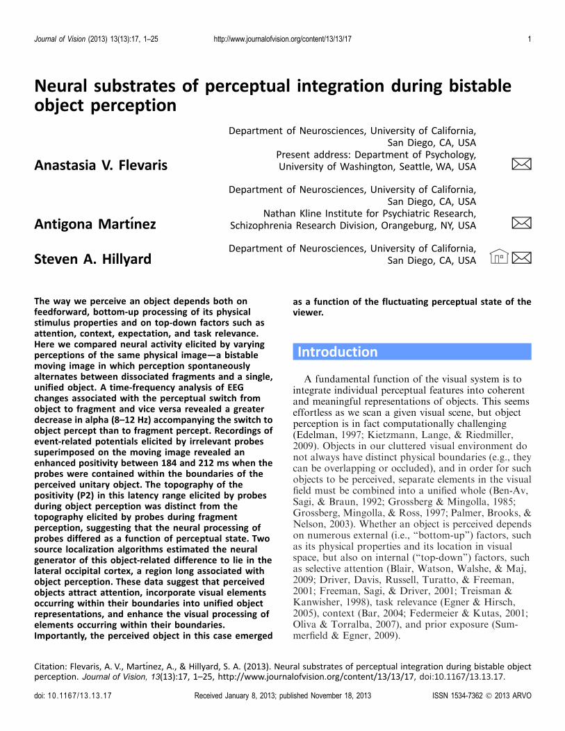
The bistable image shown in Figure 1 has recentlybeen used to investigate perceptual integration in objectperception (de-Wit & Kubilius, 2012; Fang, Kersten, &Murray, 2008; Murray & Kersten, 2002; Naber,Carlson, Verstraten, & Einhauser, 2011). First de-scribed by Lorenceau and Shiffrar (1992), the bistablenature of this ‘‘ambiguous diamond’’ display is inducedthrough motion. As the line segments move, partici-pants see either four separate lines (‘‘fragments’’)moving up and down or one integrated object (anoccluded diamond shape) moving left to right, andperception spontaneously alternates from one perceptto the other. Critically, the visual input is identical in
Figure 1. Bistable moving visual image used in experiment (Lorenceau & Shiffrar, 1992). (A) When the image moves, the four white
lines are either perceived as a single coherent diamond (object) moving left to right (behind the gray columns, which are perceived as
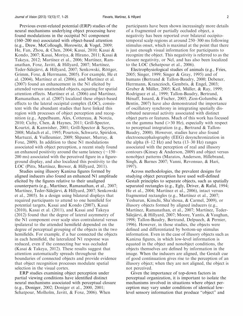
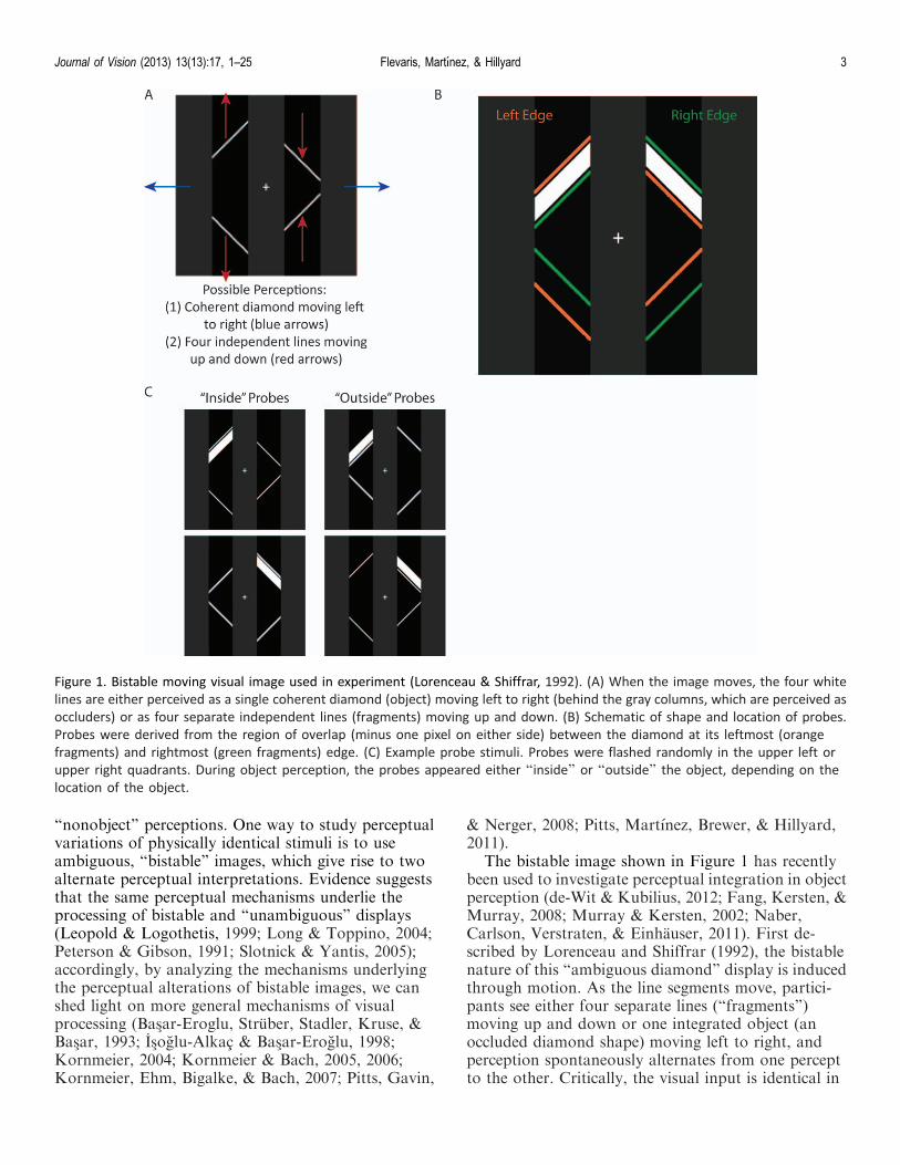
occluders) or as four separate independent lines (fragments) moving up and down. (B) Schematic of shape and location of probes.
Probes were derived from the region of overlap (minus one pixel on either side) between the diamond at its leftmost (orange
fragments) and rightmost (green fragments) edge. (C) Example probe stimuli. Probes were flashed randomly in the upper left or
upper right quadrants. During object perception, the probes appeared either ‘‘inside’’ or ‘‘outside’’ the object, depending on the
location of the object.
Journal of Vision (2013) 13(13):17, 1–25 Flevaris, Martınez, & Hillyard 3
the two perceptual states, thereby making possible thecomparison of perceptual processes during objectperception versus fragment perception. In a behavioralstudy using this ambiguous diamond display, Naber etal. (2011) found that the subjective impression of anintegrated object facilitated detection and discrimina-tion of elements within the object’s boundaries.
In the current study we used electrophysiologicalrecordings to investigate the cortical processes thatdifferentiate the subjective perception of a coherentobject from that of fragmented parts in the ambiguousdiamond display. Participants indicated via buttonpress their perceptual state as it alternated from objectto fragment and vice versa. We examined how thesubjective perception of a coherent object affectsintegrative visual processing by adding probes to thedisplay and comparing neural activity to the probes asa function of perceptual state. Comparing the neuralactivity elicited by the probes during object relative tofragment perception revealed differences in sensoryprocessing of elements that appeared within theperceived object. In addition, we observed oscillatorydifferences in the EEG associated with the perceptualswitch from fragment to object and vice versa.
Method
Participants
Fifteen undergraduates (10 women; age range 18–32years) from the University of California, San Diego,participated in the experiment for monetary compen-sation. All were right-handed and had normal orcorrected-to-normal vision. All gave informed consentas approved by the committee for the protection ofhuman subjects at the University of California, SanDiego, and in accordance with the Declaration ofHelsinki.
Stimuli
During testing participants maintained fixation on acentral white fixation cross subtending 0.58 thatremained on the video screen throughout the experi-ment. The basic display consisted of a white diamondon a black background that subtended 7.88 · 7.88 ofvisual angle when viewed from a distance of 80 cm. Thediamond appeared behind three 2.58 · 11.88 grayoccluding bars. One of the gray bars was centered atfixation, and the other two were centered 3.68 to the leftand right of fixation, respectively. When in motion, thediamond gave rise to two possible perceptual states: (a)a single, coherent diamond moving left to right behind
the three gray bars (‘‘object’’), or (b) four independentline segments moving up and down beside the threegray bars (‘‘fragments’’; see Figure 1A; Lorenceau &Shiffrar, 1992; Naber et al., 2011). To enhance theambiguity of the display, the diamond had a sinusoidalmovement pattern (Naber et al., 2011), and took 1.35 sto move from one side (left/right) to the other. Hence,the period of motion was 2.7 s (i.e., duration of one fullcycle from left to right and back again). The peakamplitude was 2.38/s, and the diamond paused at eachside of the display for 100 ms.
The luminance of the occluding bars was setindividually for each participant so that the alternativeperceptual states occurred with approximately equalfrequency. Higher luminance bars bias perceptiontowards the diamond, whereas lower luminance barsbias perception towards the fragments (Naber et al.,2011). Participants also have predisposed biases, so theluminance was adjusted individually in an attempt toreduce any biases towards one state or the other.
Briefly flashed white parallelograms subtending 2.98
· 0.78 served as irrelevant probes, and appeared eitherin the upper left or upper right quadrant of the display.To equate the physical characteristics and spatiallocation of probes appearing inside versus outside ofthe diamond, the shape and location of each probe wasformed by the region of overlap between the diamondat its leftmost and rightmost points (Figure 1B). Theleft probes were presented either in the region that wasjust within the diamond at its leftmost point or justoutside the diamond at its rightmost point (Figure 1C),and vice versa for the right probes. Thus, depending onthe location of the diamond, the probe could appear tobe contained within the boundaries of the diamondshape (e.g., a left probe when the diamond was at itsleftmost point), and the physically identical probe inthe same spatial location could also appear to beoutside of it (e.g., left probe when the diamond was atits rightmost point). This assured that the ERPs elicitedby each probe could be attributed to its location withrespect to the oriented fragments/diamond and not todifferences in spatial location per se. There was onepixel (0.078) separating the probe from the diamond’sboundary at each location, so that the probes neverspatially abutted or overlapped the diamond. Theprobes were presented at the instant the diamond wasat its leftmost or rightmost point for a duration of 100ms. Activity elicited by the probes during self-reportedobject perception was compared to activity elicited bythe same probes, in the same physical locations, duringfragment perception. Although the inside–outsidedistinction was not relevant during fragment percep-tion, we refer to ‘‘inside probes’’ and ‘‘outside probes’’to indicate their locations relative to the diamond.Note, however, that the absolute spatial locations of
Journal of Vision (2013) 13(13):17, 1–25 Flevaris, Martınez, & Hillyard 4
the inside and outside probes in the display wereidentical.
Procedure
Prior to the start of the experiment, the luminance ofthe occluding bars was adjusted individually for eachparticipant to a level at which she reported oscillatingbetween perceiving the object and perceiving fragmentsfor equal duration for a period of 3 min. Theparticipants were first presented with black occludingbars and asked to indicate their perceptual state; almostall participants reported seeing only fragments movingup and down. They were then presented with whiteoccluding bars; almost all participants reported seeingonly a diamond object moving left to right. They werethen presented with occluding bars that were the shadeof gray exactly in the middle (i.e., RGB values 127, 127,and 127) and continued to report their perceptual stateas the shade of the bars changed every 3 min. Once theyreported object and fragment states equally for a periodof 3 min, the experiment was initiated and the finalshade of gray was used throughout the entire experi-ment.
Participants viewed the ambiguous diamond displayfrom a distance of 80 cm while maintaining centralfixation and indicated their perceptual state (object,fragment) via button press. All participants used theirdominant (right) hand to indicate object versusfragment perception; one button indicated a perceptualswitch to ‘‘object’’ and a second button indicated aperceptual switch to ‘‘fragments.’’ A third button waspressed with the left hand to start the experiment, aswell as to indicate an ambiguous perceptual state, oranything other than a concrete object or fragmentpercept. Akin to previous ERP studies of bistableimages (e.g., Bas�ar-Eroglu et al., 1993; Pitts et al.,2011), we asked participants to use their dominanthand to obtain as accurate an estimate as possible ofthe timing of their perceptual switches, since we reliedon participants’ self-report and response times arefaster with the dominant than nondominant hand(Kerr, Mingay, & Elithorn, 1963).
While participants viewed the display and indicatedtheir perceptual state, the irrelevant probes flashed for100 ms randomly at the left and right locations (seeStimuli description), and participants were told toignore them. Two fifths of the time the probes appearedto be within (but not touching) the boundaries of thediamond (i.e., one fifth were left inside probes and onefifth were right inside probes), and two fifths of the timethey appeared outside the boundaries of the diamond(i.e., one fifth were left outside probes and one fifthwere right outside probes). To reduce expectanciesassociated with the probe and to subtract neural
activity associated with the motion of the diamond, onefifth of the time no probe appeared when the diamondreached its leftmost or rightmost point. Neural activityelicited in the no-probe trials was subtracted fromactivity elicited by the probes to get a pure measure ofprobe-related activity separate from any activityelicited by the moving lines or object. Since the motionwas periodic, participants could predict when theprobes were about to be flashed, and the existence ofthe no-probe trials may not have eliminated suchexpectancies or predictive mechanisms; importantly,however, probes occurred with equal probabilityduring each perceptual state (object or fragment), andinside and outside probes also occurred with equalprobability, so predictive mechanisms could notaccount for differences between perceptual states or fordifferences between states for one probe condition andnot the other.
EEG recording
The EEG was recorded continuously using 64 Ag-AgCl pin-type active electrodes mounted on an elasticcap (Electro-Cap International, Eaton, OH) accordingto the extended 10–20 system, and from two additionalelectrodes placed at the right and left mastoids. Theelectrode impedances were kept below 5 kX. Scalpsignals were amplified by a battery-powered amplifier(SA Instrumentation, Encinitas, CA) with a gain of10,000 and band-pass filtered from 0.1 to 80 Hz. Eyemovements and blinks were monitored by horizontal(attached to the external canthi) and vertical (attachedto the infraorbital ridge of the right eye) electrooculo-gram (EOG) recordings. A right mastoid electrodeserved as the reference for all scalp channels and thevertical EOG (VEOG). Left and right horizontal EOG(HEOG) channels were recorded as a bipolar pair.Signals were digitized to disk at 250 Hz. Each recordingsession lasted 120–180 min, including setup time andcap and electrode preparation. Short breaks were givenevery 3 min to help alleviate participant fatigue.
ERP analysis
Trials were discarded if they contained an eye blinkor an eye movement artifact (.200 lV), or if anychannel exceeded 55 lV. On average, 15% of trials wererejected due to these artifacts. Averaged mastoid-referenced ERPs were calculated off-line as thedifference between each scalp channel and an averageof the left and right mastoid channels. To analyzeneural activity to the probes, the ERPs time-locked toprobe and no-probe events were averaged separately,baseline corrected from �100 to 0 ms, and low-pass
Journal of Vision (2013) 13(13):17, 1–25 Flevaris, Martınez, & Hillyard 5
filtered at 30 Hz. ERPs to the no-probe in eachperceptual state (fragment, object) and diamondlocation (left, right) were subtracted from ERPs to theprobe in the same state and location. For example,ERPs to the left no-probe during object perceptionwere subtracted from the inside left probe during objectperception, and ERPs to the left no-probe duringfragment perception were subtracted from the insideleft probe during fragment perception. Similarly, ERPsto the right no-probe during object perception weresubtracted from the outside left probe (because thediamond was at its rightmost point when the left probesappeared outside of it) during object perception, and soon. This approach is similar to subtraction techniquesthat have been used both in previous ERP studies andin neuroimaging studies (see, for example, Luck, Fan,& Hillyard, 1993; Petersen, Fiez, & Maurizio, 1992).The underlying assumption is one of additivity (e.g.,McCarthy & Donchin, 1981). In the current paradigmthe assumption is that the ERP elicited by diamonditself in a particular perceptual state will not changedepending on the presence of the probe, so probe-related activity can be isolated by subtracting out theERP elicited by the diamond. Prior to artifact rejection,one fifth of the trials were no-probe trials, one fifthwere trials in which a left inside probe occurred, onefifth were left outside trials, one fifth were right insidetrials, and one fifth were right outside trials. Hence, aroughly equal number of no-probe trials were sub-tracted from the probe trials to derive probe-relatedactivity.
Prior to statistical analysis, ERPs to the left probeand right probe were collapsed into ipsilateral andcontralateral locations on the scalp and averaged. Tocircumvent the multiple testing problem (Oken &Chiappa, 1986), we used an approach based on regionof interest, in which we averaged ERPs across anumber of electrodes to yield one value for the lefthemisphere and one value for the right hemisphere.Based on previous object-related ERP modulations(e.g., Kasai, 2010; Kasai & Kondo, 2007; Kasai,Moriya, & Hirano, 2011; Kasai & Takeya, 2012;Martınez et al. 2007; Pitts et al., 2011), and alsobecause we were comparing visual evoked ERPselicited by the probes during each perceptual state, wefocused our probe analysis on occipital electrodes(PO3/O4, PO7/8, O1/2, I3/4, I5/6, SI3/4; see Figure 2,green boxes) and looked for differences in the ;200-ms latency period. The precise time window wascentered around the maximum amplitude of thecomponent of interest in the grand average based ontopographical distributions, which revealed a differ-ence between perceptual states in the range of the P2(184–212 ms) component over the occipital scalp.Perceptual state effects were quantified in terms ofmean amplitudes within the P2 latency window and
entered into perceptual state (object, fragment) ·hemisphere (ipsilateral, contralateral) analysis ofvariance (ANOVA). Follow-up planned comparisonswere conducted when appropriate to test the differ-ence between perceptual states for contralateral versusipsilateral probes.
To analyze ERPs associated with the perceptualswitch, EEG epochs were time-locked to the partici-pants’ motor responses, low pass filtered at 30 Hz, andbaseline corrected over the interval�1500 to�1200 msprior to the response. For this analysis we focused oncentral-parietal electrodes (C3/4, CP1/2, CP3/4, P1/2,P3/4), based on previous ERP studies of perceptualswitches (e.g., Bas�ar-Eroglu et al., 1993; Is�oglu-Alkac &Bas�ar-Eroglu, 1998). Mean amplitudes within specificlatency windows prior to and following the buttonpress were compared between perceptual states by aperceptual state (object, fragment) · hemisphere(ipsilateral, contralateral) ANOVA.
Source analyses
To model the neural generators of perceptual stateeffects, two difference source localization algorithmswere applied to the grand-averaged ERP differencewaves (formed by subtracting the ERPs to identicalprobes during object perception minus fragmentperception). First, inverse dipole modeling was carriedusing Brain Electrical Source Analysis program (BESA;version 5). BESA iteratively adjusts the location andorientation of dipolar sources to minimize the residualvariance between the calculated model and the ob-served ERP voltage topography (Scherg, 1990). We fitsymmetrical pairs of dipoles that were mirror con-strained in location but not in orientation during theinterval of the P2 (184–212 ms).
The neural generators of these grand-averaged ERPdifference waves were also modeled using a minimum-norm linear inverse solution approach that involveslocal autoregressive averaging (LAURA; Grave dePeralta Menendez, Murray, Michel, Martuzzi, &Gonzalez Andino, 2004). The LAURA solution spaceincluded 4,024 evenly spaced nodes (6-mm3 spacing),restricted to the gray matter of the Montreal Neuro-logical Institute’s (MNI) average brain. No a prioriassumptions were made regarding the number orlocation of active sources. Time windows for estimatingthe sources of the component were the same as in theERP statistical analyses. LAURA solutions werecomputed and transformed into a standardized coor-dinate system (Talairach & Tournoux, 1988) andexported into the AFNI software package (Cox, 1996)and projected onto structural (MNI) brain images forvisualization.
Journal of Vision (2013) 13(13):17, 1–25 Flevaris, Martınez, & Hillyard 6
Time-frequency analysis
To analyze induced oscillatory cortical activityassociated with the perceptual switch, the single trialEEG signal on each channel was convolved with 6-cycle Morlet wavelets over a 4-s window beginning 2 sprior to the button press indicating a perceptual switch.Instantaneous power and phase were extracted at eachtime point (at 250-Hz sampling rate) over frequencyscales from 0.7 to 60 Hz incremented logarithmically(Lakatos et al., 2005). The square roots of the powervalues (the sum of the squares of the real and imaginaryMorlet components) were then averaged over singletrials to yield the total averaged spectral amplitudes (inmicrovolts) for each condition (i.e., switch to fragment,switch to object). The averaged spectral amplitude wasnot baseline corrected because there was not a cleartime reference for the perceptual switch (Ehm, Bach, &Kornmeier, 2011; Is�oglu-Alkac, & Bas�ar-Eroglu, 1998;Is�oglu-Alkac, & Struber, 2006), and we were interestedin differences surrounding switches to one perceptual
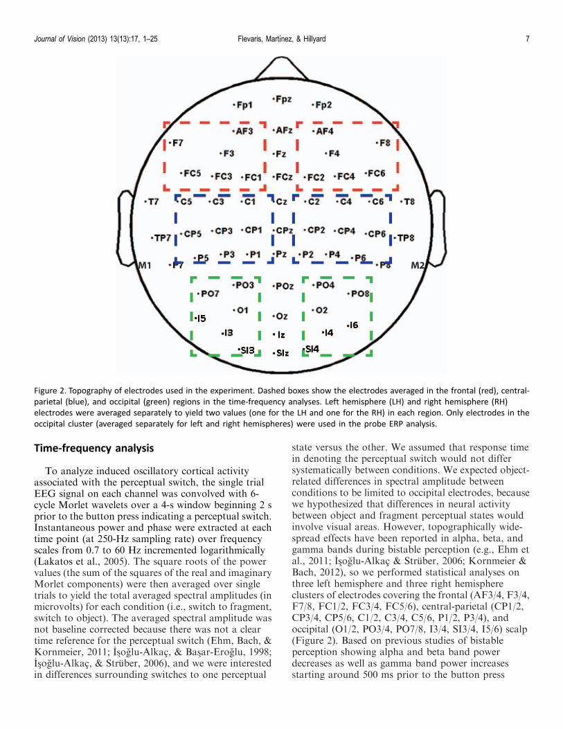
state versus the other. We assumed that response timein denoting the perceptual switch would not differsystematically between conditions. We expected object-related differences in spectral amplitude betweenconditions to be limited to occipital electrodes, becausewe hypothesized that differences in neural activitybetween object and fragment perceptual states wouldinvolve visual areas. However, topographically wide-spread effects have been reported in alpha, beta, andgamma bands during bistable perception (e.g., Ehm etal., 2011; Is�oglu-Alkac & Struber, 2006; Kornmeier &Bach, 2012), so we performed statistical analyses onthree left hemisphere and three right hemisphereclusters of electrodes covering the frontal (AF3/4, F3/4,F7/8, FC1/2, FC3/4, FC5/6), central-parietal (CP1/2,CP3/4, CP5/6, C1/2, C3/4, C5/6, P1/2, P3/4), andoccipital (O1/2, PO3/4, PO7/8, I3/4, SI3/4, I5/6) scalp(Figure 2). Based on previous studies of bistableperception showing alpha and beta band powerdecreases as well as gamma band power increasesstarting around 500 ms prior to the button press
Figure 2. Topography of electrodes used in the experiment. Dashed boxes show the electrodes averaged in the frontal (red), central-
parietal (blue), and occipital (green) regions in the time-frequency analyses. Left hemisphere (LH) and right hemisphere (RH)
electrodes were averaged separately to yield two values (one for the LH and one for the RH) in each region. Only electrodes in the
occipital cluster (averaged separately for left and right hemispheres) were used in the probe ERP analysis.
Journal of Vision (2013) 13(13):17, 1–25 Flevaris, Martınez, & Hillyard 7

denoting a perceptual switch (e.g., Ehm et al., 2011;Is�oglu-Alkac & Struber, 2006; Struber & Hermann,2002) we focused on the time window�500 to�100 msprior to the button press. ANOVAs with factors ofpercept (object, fragment), scalp region (frontal,central-parietal, occipital), and hemisphere (left, right)were carried out separately for the alpha (8–12 Hz),beta (16–30 Hz), and gamma (30-50 Hz) bands for thetime window 100–500 ms preceding the button pressindicating a switch. Using an approach similar to thatof previous studies (e.g., Struber & Hermann, 2002), wetreated the analyses in each frequency band separately,using Bonferroni correction where applicable locallywithin each analysis.
Results
Behavioral results
The mean switch rate was 8.1 switches/min. Overall,47.8% of the total button presses indicated a switch toobject perception, 49.9% indicated a switch to fragmentperception, and 2.3% indicated a switch to anambiguous percept. The average median durations ofthe object and fragment percepts were 5.1 and 6.8 s,respectively. Figure 3 depicts the frequency histogramsfor the object and fragment percept durations, showingthat both have similar time courses. The data fit wellwith a gamma distribution, consistent with previousstudies of bistable perception (Borsellino, Marco,Allazetta, Rinesi, & Bartolini, 1972; Fang et al., 2008)
and binocular rivalry (Kovacs, Papathomas, Yang, &Feher, 1996; Lehky, 1995). This was confirmedstatistically with a v2 test, p . 0.50 and p . 0.75 for theobject and fragment percepts, respectively.1
The gamma distribution of perceptual durationssuggests that the probe did not elicit perceptualswitches in our paradigm (c.f., Kanai, Moradi,Shimojo, & Verstraten, 2005), but one possibility is thatperceptual switches occurred with a relatively constantdelay with respect to probe onset. To rule out thispossibility, we determined the time from each probeuntil the subsequent perceptual switch and plotted ahistogram of the distribution of probe-to-switch times.We compared this with a similar histogram showing thetime from each no-probe until the subsequent percep-tual switch and found no difference between these twodistributions (p . 0.90, v2 test). These data are shownin Figure 4. Note that this includes all probes and no-probes, though similar results were found when onlyconsidering the last probe or no-probe before eachbutton press (see Figures 5B and 5C). We alsocompared the probe-to-switch times for switches toobject versus switches to fragment and found nodifference in these distributions (p . 0.75, v2 test), norwas there a difference between object and fragmentdistributions for no-probe-to-switch times (p . 0.90, v2
test). Hence, these data suggest that the probes did notinfluence the rate of perceptual switches but rather thatparticipants were able to ignore the probes.
In order to determine whether participants were onaverage more likely to experience a perceptual switchduring a particular phase of the stimulus, we alsoplotted a histogram of the time of each button press
Figure 3. Histograms of durations for the object (left) and fragment (right) percepts. Data are fitted using a gamma function (smooth
black lines). The gamma function fitted to the object histogram has a shape parameter¼ 2.3 and a scale parameter¼ 3.0, and the
gamma function fitted to the fragment histogram has a shape parameter ¼ 2.1 and a scale parameter ¼ 4.1.
Journal of Vision (2013) 13(13):17, 1–25 Flevaris, Martınez, & Hillyard 8
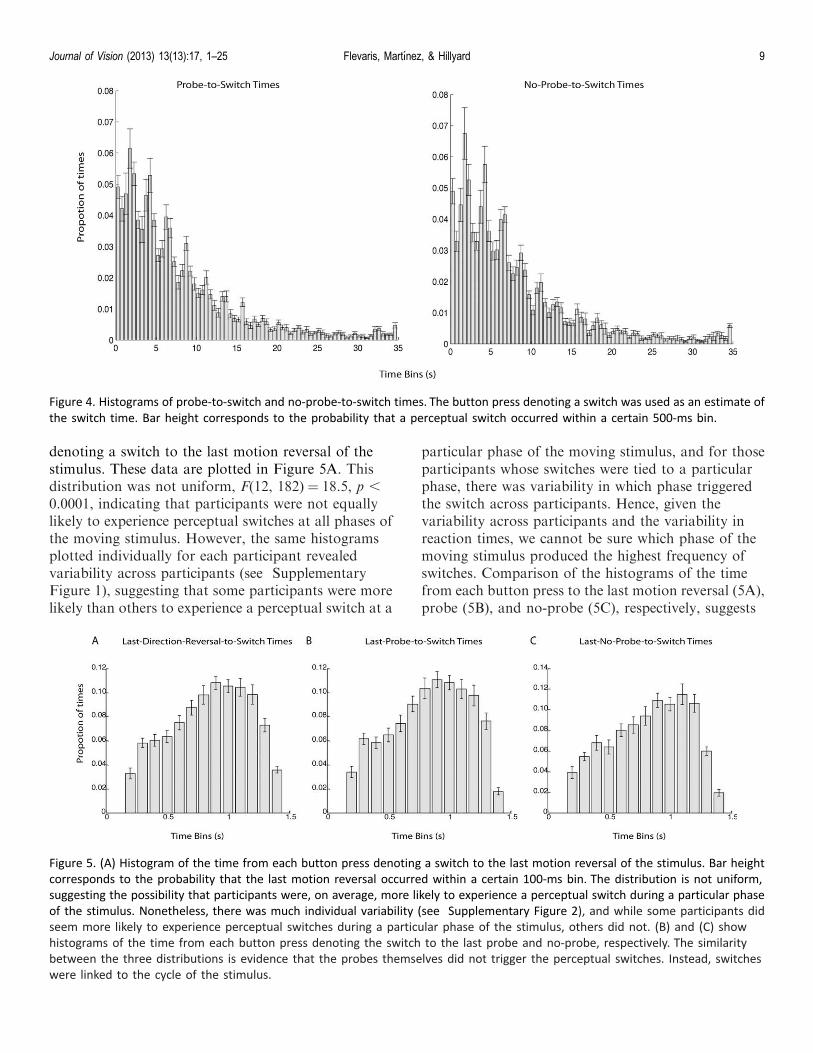
denoting a switch to the last motion reversal of thestimulus. These data are plotted in Figure 5A. Thisdistribution was not uniform, F(12, 182) ¼ 18.5, p ,
0.0001, indicating that participants were not equallylikely to experience perceptual switches at all phases ofthe moving stimulus. However, the same histogramsplotted individually for each participant revealedvariability across participants (see SupplementaryFigure 1), suggesting that some participants were morelikely than others to experience a perceptual switch at a
particular phase of the moving stimulus, and for thoseparticipants whose switches were tied to a particularphase, there was variability in which phase triggeredthe switch across participants. Hence, given thevariability across participants and the variability inreaction times, we cannot be sure which phase of themoving stimulus produced the highest frequency ofswitches. Comparison of the histograms of the timefrom each button press to the last motion reversal (5A),probe (5B), and no-probe (5C), respectively, suggests
Figure 4. Histograms of probe-to-switch and no-probe-to-switch times. The button press denoting a switch was used as an estimate of
the switch time. Bar height corresponds to the probability that a perceptual switch occurred within a certain 500-ms bin.
Figure 5. (A) Histogram of the time from each button press denoting a switch to the last motion reversal of the stimulus. Bar height
corresponds to the probability that the last motion reversal occurred within a certain 100-ms bin. The distribution is not uniform,
suggesting the possibility that participants were, on average, more likely to experience a perceptual switch during a particular phase
of the stimulus. Nonetheless, there was much individual variability (see Supplementary Figure 2), and while some participants did
seem more likely to experience perceptual switches during a particular phase of the stimulus, others did not. (B) and (C) show
histograms of the time from each button press denoting the switch to the last probe and no-probe, respectively. The similarity
between the three distributions is evidence that the probes themselves did not trigger the perceptual switches. Instead, switches
were linked to the cycle of the stimulus.
Journal of Vision (2013) 13(13):17, 1–25 Flevaris, Martınez, & Hillyard 9
that perceptual switches were linked to the phase of themoving stimulus and not to the probes, because of thesimilarity across the three distributions. Indeed, acondition (probe, no-probe)· time bin (1–13) ANOVAdid not find a significant interaction between conditionand bin, F(12, 168)¼ 1.47, ns.
Probe-related ERP results
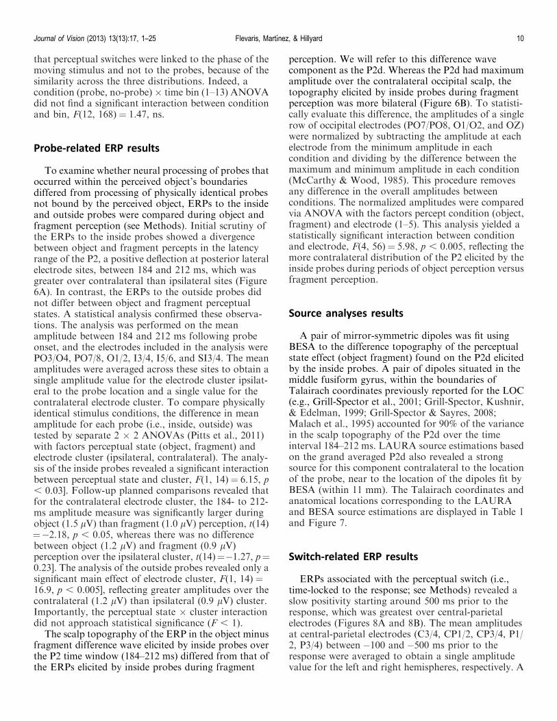
To examine whether neural processing of probes thatoccurred within the perceived object’s boundariesdiffered from processing of physically identical probesnot bound by the perceived object, ERPs to the insideand outside probes were compared during object andfragment perception (see Methods). Initial scrutiny ofthe ERPs to the inside probes showed a divergencebetween object and fragment percepts in the latencyrange of the P2, a positive deflection at posterior lateralelectrode sites, between 184 and 212 ms, which wasgreater over contralateral than ipsilateral sites (Figure6A). In contrast, the ERPs to the outside probes didnot differ between object and fragment perceptualstates. A statistical analysis confirmed these observa-tions. The analysis was performed on the meanamplitude between 184 and 212 ms following probeonset, and the electrodes included in the analysis werePO3/O4, PO7/8, O1/2, I3/4, I5/6, and SI3/4. The meanamplitudes were averaged across these sites to obtain asingle amplitude value for the electrode cluster ipsilat-eral to the probe location and a single value for thecontralateral electrode cluster. To compare physicallyidentical stimulus conditions, the difference in meanamplitude for each probe (i.e., inside, outside) wastested by separate 2 · 2 ANOVAs (Pitts et al., 2011)with factors perceptual state (object, fragment) andelectrode cluster (ipsilateral, contralateral). The analy-sis of the inside probes revealed a significant interactionbetween perceptual state and cluster, F(1, 14)¼ 6.15, p, 0.03]. Follow-up planned comparisons revealed thatfor the contralateral electrode cluster, the 184- to 212-ms amplitude measure was significantly larger duringobject (1.5 lV) than fragment (1.0 lV) perception, t(14)¼�2.18, p , 0.05, whereas there was no differencebetween object (1.2 lV) and fragment (0.9 lV)perception over the ipsilateral cluster, t(14)¼�1.27, p¼0.23]. The analysis of the outside probes revealed only asignificant main effect of electrode cluster, F(1, 14) ¼16.9, p , 0.005], reflecting greater amplitudes over thecontralateral (1.2 lV) than ipsilateral (0.9 lV) cluster.Importantly, the perceptual state · cluster interactiondid not approach statistical significance (F , 1).
The scalp topography of the ERP in the object minusfragment difference wave elicited by inside probes overthe P2 time window (184–212 ms) differed from that ofthe ERPs elicited by inside probes during fragment
perception. We will refer to this difference wavecomponent as the P2d. Whereas the P2d had maximumamplitude over the contralateral occipital scalp, thetopography elicited by inside probes during fragmentperception was more bilateral (Figure 6B). To statisti-cally evaluate this difference, the amplitudes of a singlerow of occipital electrodes (PO7/PO8, O1/O2, and OZ)were normalized by subtracting the amplitude at eachelectrode from the minimum amplitude in eachcondition and dividing by the difference between themaximum and minimum amplitude in each condition(McCarthy & Wood, 1985). This procedure removesany difference in the overall amplitudes betweenconditions. The normalized amplitudes were comparedvia ANOVA with the factors percept condition (object,fragment) and electrode (1–5). This analysis yielded astatistically significant interaction between conditionand electrode, F(4, 56)¼ 5.98, p , 0.005, reflecting themore contralateral distribution of the P2 elicited by theinside probes during periods of object perception versusfragment perception.
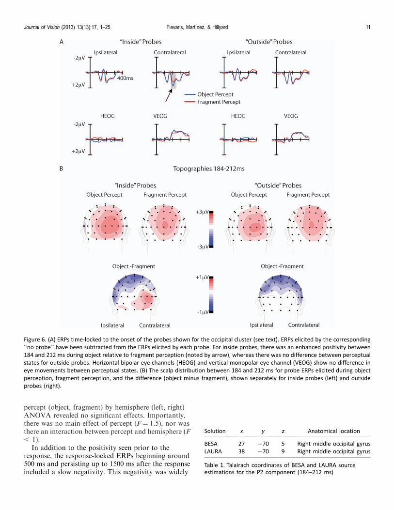
Source analyses results
A pair of mirror-symmetric dipoles was fit usingBESA to the difference topography of the perceptualstate effect (object fragment) found on the P2d elicitedby the inside probes. A pair of dipoles situated in themiddle fusiform gyrus, within the boundaries ofTalairach coordinates previously reported for the LOC(e.g., Grill-Spector et al., 2001; Grill-Spector, Kushnir,& Edelman, 1999; Grill-Spector & Sayres, 2008;Malach et al., 1995) accounted for 90% of the variancein the scalp topography of the P2d over the timeinterval 184–212 ms. LAURA source estimations basedon the grand averaged P2d also revealed a strongsource for this component contralateral to the locationof the probe, near to the location of the dipoles fit byBESA (within 11 mm). The Talairach coordinates andanatomical locations corresponding to the LAURAand BESA source estimations are displayed in Table 1and Figure 7.
Switch-related ERP results
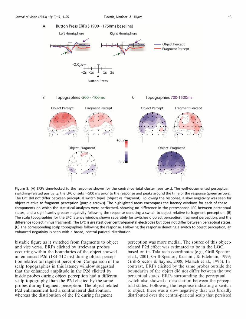
ERPs associated with the perceptual switch (i.e.,time-locked to the response; see Methods) revealed aslow positivity starting around 500 ms prior to theresponse, which was greatest over central-parietalelectrodes (Figures 8A and 8B). The mean amplitudesat central-parietal electrodes (C3/4, CP1/2, CP3/4, P1/2, P3/4) between �100 and�500 ms prior to theresponse were averaged to obtain a single amplitudevalue for the left and right hemispheres, respectively. A
Journal of Vision (2013) 13(13):17, 1–25 Flevaris, Martınez, & Hillyard 10
percept (object, fragment) by hemisphere (left, right)ANOVA revealed no significant effects. Importantly,there was no main effect of percept (F ¼ 1.5), nor wasthere an interaction between percept and hemisphere (F, 1).
In addition to the positivity seen prior to theresponse, the response-locked ERPs beginning around500 ms and persisting up to 1500 ms after the responseincluded a slow negativity. This negativity was widely
Solution x y z Anatomical location
BESA 27 �70 5 Right middle occipital gyrus
LAURA 38 �70 9 Right middle occipital gyrus
Table 1. Talairach coordinates of BESA and LAURA sourceestimations for the P2 component (184–212 ms)
Figure 6. (A) ERPs time-locked to the onset of the probes shown for the occipital cluster (see text). ERPs elicited by the corresponding
‘‘no probe’’ have been subtracted from the ERPs elicited by each probe. For inside probes, there was an enhanced positivity between
184 and 212 ms during object relative to fragment perception (noted by arrow), whereas there was no difference between perceptual
states for outside probes. Horizontal bipolar eye channels (HEOG) and vertical monopolar eye channel (VEOG) show no difference in
eye movements between perceptual states. (B) The scalp distribution between 184 and 212 ms for probe ERPs elicited during object
perception, fragment perception, and the difference (object minus fragment), shown separately for inside probes (left) and outside
probes (right).
Journal of Vision (2013) 13(13):17, 1–25 Flevaris, Martınez, & Hillyard 11
distributed over bilateral central-parietal sites and wasgreater following switches to object than to fragmentperception (Figure 8C). To statistically evaluate thisdifference, the mean amplitudes at central-parietalelectrodes (C3/4, CP1/2, CP3/4, P1/2, P3/4) between700 and 1500 ms following the response were averagedto obtain a single amplitude value for the left and righthemispheres, respectively. A percept (object, fragment)· hemisphere (left/right) ANOVA revealed a maineffect of percept, F(1, 14) ¼ 5.39, p , 0.05, indicatingthat the ERP following the perceptual switch to objectwas significantly more negative (�0.82 lV) than theERP following the perceptual switch to fragment (1.11lV).
Time-frequency analysis results
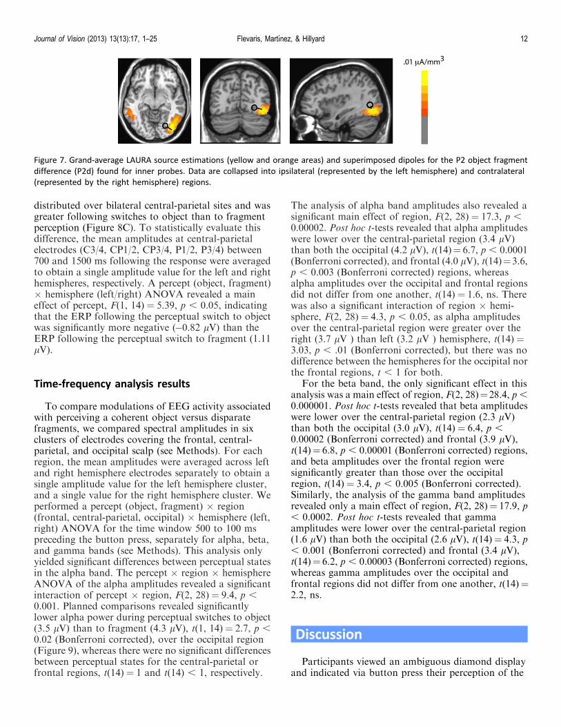
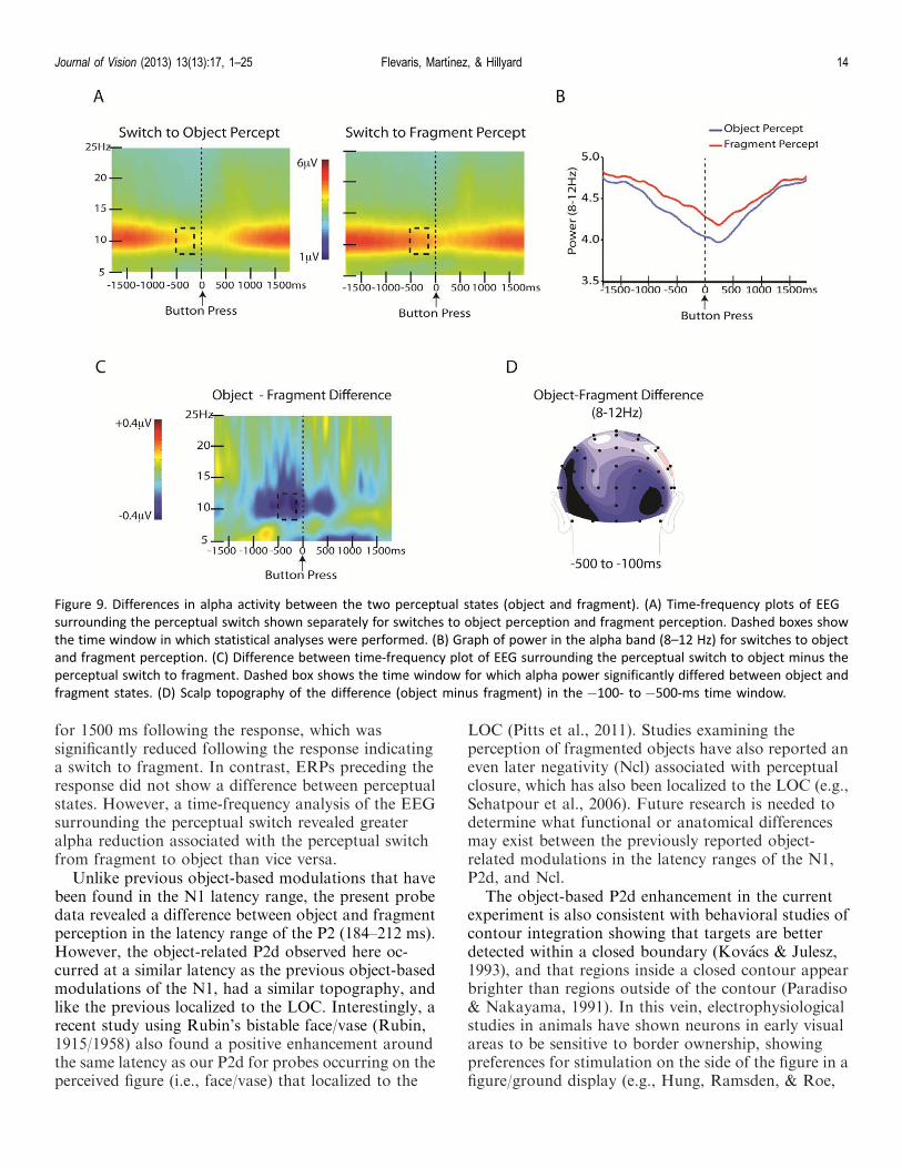
To compare modulations of EEG activity associatedwith perceiving a coherent object versus disparatefragments, we compared spectral amplitudes in sixclusters of electrodes covering the frontal, central-parietal, and occipital scalp (see Methods). For eachregion, the mean amplitudes were averaged across leftand right hemisphere electrodes separately to obtain asingle amplitude value for the left hemisphere cluster,and a single value for the right hemisphere cluster. Weperformed a percept (object, fragment) · region(frontal, central-parietal, occipital) · hemisphere (left,right) ANOVA for the time window 500 to 100 mspreceding the button press, separately for alpha, beta,and gamma bands (see Methods). This analysis onlyyielded significant differences between perceptual statesin the alpha band. The percept · region · hemisphereANOVA of the alpha amplitudes revealed a significantinteraction of percept · region, F(2, 28) ¼ 9.4, p ,0.001. Planned comparisons revealed significantlylower alpha power during perceptual switches to object(3.5 lV) than to fragment (4.3 lV), t(1, 14)¼ 2.7, p ,0.02 (Bonferroni corrected), over the occipital region(Figure 9), whereas there were no significant differencesbetween perceptual states for the central-parietal orfrontal regions, t(14)¼ 1 and t(14) , 1, respectively.
The analysis of alpha band amplitudes also revealed asignificant main effect of region, F(2, 28) ¼ 17.3, p ,0.00002. Post hoc t-tests revealed that alpha amplitudeswere lower over the central-parietal region (3.4 lV)than both the occipital (4.2 lV), t(14)¼ 6.7, p , 0.0001(Bonferroni corrected), and frontal (4.0 lV), t(14)¼3.6,p , 0.003 (Bonferroni corrected) regions, whereasalpha amplitudes over the occipital and frontal regionsdid not differ from one another, t(14) ¼ 1.6, ns. Therewas also a significant interaction of region · hemi-sphere, F(2, 28)¼ 4.3, p , 0.05, as alpha amplitudesover the central-parietal region were greater over theright (3.7 lV ) than left (3.2 lV ) hemisphere, t(14) ¼3.03, p , .01 (Bonferroni corrected), but there was nodifference between the hemispheres for the occipital northe frontal regions, t , 1 for both.
For the beta band, the only significant effect in thisanalysis was a main effect of region, F(2, 28)¼28.4, p ,0.000001. Post hoc t-tests revealed that beta amplitudeswere lower over the central-parietal region (2.3 lV)than both the occipital (3.0 lV), t(14) ¼ 6.4, p ,0.00002 (Bonferroni corrected) and frontal (3.9 lV),t(14)¼ 6.8, p , 0.00001 (Bonferroni corrected) regions,and beta amplitudes over the frontal region weresignificantly greater than those over the occipitalregion, t(14) ¼ 3.4, p , 0.005 (Bonferroni corrected).Similarly, the analysis of the gamma band amplitudesrevealed only a main effect of region, F(2, 28)¼ 17.9, p, 0.0002. Post hoc t-tests revealed that gammaamplitudes were lower over the central-parietal region(1.6 lV) than both the occipital (2.6 lV), t(14)¼ 4.3, p, 0.001 (Bonferroni corrected) and frontal (3.4 lV),t(14)¼ 6.2, p , 0.00003 (Bonferroni corrected) regions,whereas gamma amplitudes over the occipital andfrontal regions did not differ from one another, t(14)¼2.2, ns.
Discussion
Participants viewed an ambiguous diamond displayand indicated via button press their perception of the
Figure 7. Grand-average LAURA source estimations (yellow and orange areas) and superimposed dipoles for the P2 object fragment
difference (P2d) found for inner probes. Data are collapsed into ipsilateral (represented by the left hemisphere) and contralateral
(represented by the right hemisphere) regions.
Journal of Vision (2013) 13(13):17, 1–25 Flevaris, Martınez, & Hillyard 12
bistable figure as it switched from fragments to objectand vice versa. ERPs elicited by irrelevant probesoccurring within the boundaries of the object showedan enhanced P2d (184–212 ms) during object percep-tion relative to fragment perception. Comparison of thescalp topographies in this latency window suggestedthat the enhanced amplitude in the P2d elicited byinside probes during object perception had a differentscalp topography than the P2d elicited by the sameprobes during fragment perception. The object-relatedP2d enhancement had a contralateral distribution,whereas the distribution of the P2 during fragment
perception was more medial. The source of this object-related P2d effect was estimated to be in the LOC,based on its Talairach coordinates (e.g., Grill-Spectoret al., 2001; Grill-Spector, Kushnir, & Edelman, 1999;Grill-Spector & Sayres, 2008; Malach et al., 1995). Incontrast, ERPs elicited by the same probes outside theboundaries of the object did not differ between the twoperceptual states. ERPs surrounding the perceptualswitch also showed a dissociation between the percep-tual states. Following the response indicating a switchto object, there was a slow negativity that was broadlydistributed over the central-parietal scalp that persisted
Figure 8. (A) ERPs time-locked to the response shown for the central-parietal cluster (see text). The well-documented perceptual
switching-related positivity, the LPC onsets ;500 ms prior to the response and peaks around the time of the response (green arrows).
The LPC did not differ between perceptual switch types (object vs. fragment). Following the response, a slow negativity was seen for
object relative to fragment perception (purple arrows). The highlighted areas encompass the latency windows for each of these
components on which the statistical analyses were performed, showing no difference in the preresponse LPC between perceptual
states, and a significantly greater negativity following the response denoting a switch to object relative to fragment perception. (B)
The scalp topographies for the LPC latency window shown separately for switches o object perception, fragment perception, and the
difference (object minus fragment). The LPC is greatest over central-parietal electrodes but does not differ between perceptual states.
(C) The corresponding scalp topographies following the response. Following the response denoting a switch to object perception, an
enhanced negativity is seen with a broad, central-parietal distribution.
Journal of Vision (2013) 13(13):17, 1–25 Flevaris, Martınez, & Hillyard 13
for 1500 ms following the response, which wassignificantly reduced following the response indicatinga switch to fragment. In contrast, ERPs preceding theresponse did not show a difference between perceptualstates. However, a time-frequency analysis of the EEGsurrounding the perceptual switch revealed greateralpha reduction associated with the perceptual switchfrom fragment to object than vice versa.
Unlike previous object-based modulations that havebeen found in the N1 latency range, the present probedata revealed a difference between object and fragmentperception in the latency range of the P2 (184–212 ms).However, the object-related P2d observed here oc-curred at a similar latency as the previous object-basedmodulations of the N1, had a similar topography, andlike the previous localized to the LOC. Interestingly, arecent study using Rubin’s bistable face/vase (Rubin,1915/1958) also found a positive enhancement aroundthe same latency as our P2d for probes occurring on theperceived figure (i.e., face/vase) that localized to the
LOC (Pitts et al., 2011). Studies examining theperception of fragmented objects have also reported aneven later negativity (Ncl) associated with perceptualclosure, which has also been localized to the LOC (e.g.,Sehatpour et al., 2006). Future research is needed todetermine what functional or anatomical differencesmay exist between the previously reported object-related modulations in the latency ranges of the N1,P2d, and Ncl.
The object-based P2d enhancement in the currentexperiment is also consistent with behavioral studies ofcontour integration showing that targets are betterdetected within a closed boundary (Kovacs & Julesz,1993), and that regions inside a closed contour appearbrighter than regions outside of the contour (Paradiso& Nakayama, 1991). In this vein, electrophysiologicalstudies in animals have shown neurons in early visualareas to be sensitive to border ownership, showingpreferences for stimulation on the side of the figure in afigure/ground display (e.g., Hung, Ramsden, & Roe,
Figure 9. Differences in alpha activity between the two perceptual states (object and fragment). (A) Time-frequency plots of EEG
surrounding the perceptual switch shown separately for switches to object perception and fragment perception. Dashed boxes show
the time window in which statistical analyses were performed. (B) Graph of power in the alpha band (8–12 Hz) for switches to object
and fragment perception. (C) Difference between time-frequency plot of EEG surrounding the perceptual switch to object minus the
perceptual switch to fragment. Dashed box shows the time window for which alpha power significantly differed between object and
fragment states. (D) Scalp topography of the difference (object minus fragment) in the �100- to �500-ms time window.
Journal of Vision (2013) 13(13):17, 1–25 Flevaris, Martınez, & Hillyard 14
2007; Lamme, 1995; Lamme, Zipser, & Spekreijse,1998; Qui, Sugihara, & von der Heydt, 2007; Zhang &von der Heydt, 2010; Zipser, Lamme, & Schiller, 1996).In particular, Hung et al. (2007) demonstrated aborder-to-surface shift in relative spike timing of V1and V2 neurons in the cat visual cortex, suggesting thatfeedforward processing in V1 and V2 supports edge-to-surface propagation, perhaps underlying the perceptualbrightness enhancement found behaviorally for areaswithin closed boundaries (e.g., Kovacs & Julesz, 1993).Enhanced neural responses to elements on the inside ofa figure relative to the outside of a figure have beenshown in V1 and V2 for figures defined on the basis oforientation, color, and motion, and although theseenhanced responses have been shown to be modulatedby attention (e.g., Qui et al., 2007), they have beenfound irrespective of spatially directed attention(Marcus & Van Essen, 2002). The P2d enhancement wefound for probes occurring within the bounded objectmay reflect a similar, figure-selective mechanism in theLOC.
Future research is needed to clarify the specific rolesof cortical areas V1, V2, and LOC, and how theyinteract in figural enhancement. For example, Qui et al.(2007) recently found attentional modulations in V2 inresponse to figural elements that had a similar latencyto the object-based effect found in the current study.Examining border-ownership cells in V2 of nonhumanprimates, they found attentional modulations between;150 and 200 ms when presenting separated andoverlapping figures, coincident with the latency of theP2d in the current study. An important question forfuture research is whether activity in LOC modulatesfigure-selective neurons in V1 and V2. Previous workhas shown figure-related responses in V1 to besuppressed in anesthetized monkeys (Lamme et al.,1998), whereas the classical receptive field tuningproperties were unaffected. This suggests that figuralenhancement in V1 is influenced by top-down feedbackfrom higher-level areas, perhaps including LOC.Consistent with this idea, the object-based enhance-ment in the current study was found irrespective ofphysical stimulus differences between the ‘‘object’’ and‘‘fragment’’ percepts, suggesting that feedforwardprocessing alone cannot account for the finding thatelements within a perceived object are enhancedrelative to the same elements appearing outside theboundaries of the object.
The object-based P2d effect in the current experi-ment was found over the contralateral hemisphere andlocalized to the contralateral LOC. This is consistentwith studies demonstrating spatial selectivity in theLOC (Aggelopoulos & Rolls, 2005; DiCarlo &Maunsell, 2003; Larsson & Heeger, 2006; MacEvoy &Epstein, 2011; Martınez et al., 2006; Martınez, Rama-nathan, et al., 2007; Martınez, Teder-Salejarvi, &
Hillyard, 2007; McKyton & Zohary, 2007; Niemeier,Goltz, Kuchinad, Tweed, & Vilis, 2005; Strother et al.,2011; Yoshor, Bosking, Ghose, & Maunsell, 2007), andpoints to the close interaction between the ‘‘space’’ and‘‘object’’ systems in the brain (e.g., Faillenot, Decety, &Jeannerod, 1999; Kravitz & Behrmann, 2011; Merigan& Maunsell, 1993). The fact that the object-based P2denhancement localized to the LOC adds to the growingbody of literature that implicates this critical region inobject perception, both in functional magnetic reso-nance imaging (fMRI; e.g., Grill-Spector et al., 1999,2001; Grill-Spector & Sayres, 2008; Malach et al., 1995)and EEG/ERP (e.g., Martınez et al., 2007) experiments.Indeed, recent fMRI experiments using the ambiguousdiamond display found that activity in the LOCincreased and activity in V1 decreased during objectperception versus fragment perception (Fang et al.,2008; Murray & Kersten, 2002). The results from thecurrent experiment support and extend those findings,suggesting that increased activity in the LOC duringobject perception also results in additional processingof visual elements associated with the object (i.e.,occurring within its boundaries). This may reflect afigural enhancement process that integrates the newelements with the existing object percept, as has beendemonstrated for figure-selective neurons in earlyvisual areas V1 and V2 (e.g., Hung et al., 2007; Lamme,1995; Lamme et al., 1998; Qui et al., 2007; Zhang & vonder Heydt, 2010; Zipser et al., 1996). In a more recentfMRI study, Caclin and colleagues (2012) manipulatedcontrast, motion, and shape separately in the ambig-uous diamond display and compared bistable andunambiguous (i.e., externally changing) percepts of theobject versus fragments. For both bistable andunambiguous displays, they found increased activity inventral and occipital regions during perception of thebound object, and greater activity in motion-relateddorsal areas during fragment perception. Further, theyfound that the different feature manipulations prefer-entially modulated regions sensitive to those features,with enhanced activity in dorsal areas (i.e., hMTþ) thanventral and occipital areas for the motion display andvice versa for the shape and contrast displays. In thepresent study we attempted to remove stimulus-relatedactivity from that related to the probe by subtractingactivity in the no-probe trials from activity in the probetrials. Specifically, we used this subtraction method toremove motion-related activity in order to focus onperceptual processing of the probes as a function ofperceptual state (see Supplementary Figure 2 for datawithout this subtraction method). As such, we did notdesign our study to examine motion processing orform-motion binding per se. An interesting avenue forfuture research could investigate if the integration ofdifferent features (e.g., motion, color) differentiallymodulates the P2d.
Journal of Vision (2013) 13(13):17, 1–25 Flevaris, Martınez, & Hillyard 15
That the ERPs following the response also showed adissociation between perceptual states is further evi-dence that perceiving a coherent object engages distinctneural mechanisms than perceiving disparate frag-ments, even without any changes in the physicalstimulus. The object-related broad negativity followingthe response had a central parietal topography andpersisted for at least 1500 ms after the response; it istherefore unlikely to reflect early perceptual processes.Instead, it is possible that this negativity reflects thesustained attention to the position of the diamondobject, given abundant research associating parietalareas with sustained attention and attentional control(e.g., Hager et al., 1998; Johannsen et al., 1997;Mennemeier et al., 1994; Sarter, Givens, & Bruno,2001; Singh-Curry & Husain, 2009; Wolpert, Good-body, & Husain, 1998). Additional research is neededto determine the neural process reflected in thisnegativity.
The results of the mean alpha amplitudes sur-rounding the perceptual switch revealed significantlylower alpha power associated with the perceptualswitch to object perception than to fragment percep-tion. This object-related effect was limited to theoccipital region, suggesting that it was related toperceptual processing and not to an overall decrease invigilance (Is�oglu-Alkac & Struber, 2006; Klimesch,1999). Importantly, a significant difference in alphapower during object relative to fragment perceptionwas found in the interval 500–100 ms preceding theresponse, suggesting that the alpha reduction precedingthe button press was associated with the perceptualswitch itself. A previous study comparing responsetimes to endogenously versus exogenously inducedperceptual switches of the Necker cube found variableresponse times across participants varying between 530and 733 ms (Kornmeier & Bach, 2004), suggesting thatthe alpha reduction we found may not have precededthe switch but rather resulted from it.
Since we relied on participants’ report of theirperceptual switches, we cannot be certain of the exactpoint in time the switches in fact occurred. However,examination of the switch-related ERPs suggests thatthe divergence in alpha power between switches toobject versus fragment occurred after the perceptualswitch, because the difference in alpha power coincidedwith the onset of the ‘‘late positive component’’ (LPC),a switch-related ERP that is considered to reflectpostperceptual processes and is likely to be equivalentto the P300 (Bas�ar-Eroglu et al., 1993; Britz, Landis, &Michel, 2009; Is�oglu-Alkac & Bas�ar-Eroglu, 1998;Kornmeier, 2004; Kornmeier & Bach, 2004, 2005, 2006,2012; Kornmeier, Ehm, Bigalke, & Bach, 2007;O’Donnell, Hendler, & Squires, 1988; Pitts, Nerger, &Davis, 2007; Pitts et al., 2008; Struber, Basar-Eroglu,Hoff, & Stadler, 2000). The LPC has been reported
both in studies using intermittent presentation as wellas studies using sustained presentation (i.e., in whichthe bistable image is continuously present and partic-ipants indicate their perceptual state as it switches;Bas�ar-Eroglu et al., 1993), as we did in the currentexperiment. Consistent with studies using sustainedpresentation, the onset of the LPC in the currentexperiment was ;500 ms prior to the response denotingthe perceptual switch and peaked around the time ofthe response. Previous reports of the LPC using theintermittent presentation method have found its onsetto be ;300 ms post stimulus onset (and presumedperceptual switch) and peak ;450 ms. The LPC (P300)has been considered to reflect a postperceptual,‘‘context updating’’ process in visual short-term mem-ory (Donchin & Coles, 1988). If we assume that theLPC reflects a postperceptual process, this is furtherevidence that the perceptual switch in the currentexperiment occurred prior to 500 ms before theresponse (i.e., prior to the onset of the LPC).
Along similar lines, recent studies have reporteddecreased alpha power around the time of perceptualswitch for other bistable images (Ehm et al., 2011;Is�oglu-Alkac & Bas�ar-Eroglu, 1998; Is�oglu-Alkac &Struber, 2006; Struber & Hermann, 2002). Thosestudies also found decreases in alpha power to coincidewith the LPC, suggesting that the decrease in alpha waslinked to the conscious recognition of the perceptualchange. As in previous studies of bistable perceptionusing a continuous presentation of the bistable displayand relying on participants’ report of their perceptualstate, we did not use a baseline correction for thespectral amplitudes because there was no obvious timeinterval to use as a baseline (e.g., Is�oglu-Alkac & Bas�ar-Eroglu, 1998; Is�oglu-Alkac & Struber, 2006). Given thewide variation in perceptual switch times within theexperiment (Figure 2), as well as the variability ofswitch rates across participants, arbitrarily selecting abaseline interval in the spectral analysis would result ina loss of statistical power to detect differences betweenperceptual states. Had we selected a baseline intervalwell before the button press denoting the switch, thebaseline interval might have reflected activity prior tothe previous perceptual switch in some trials (i.e., intrials with shorter switch-rates). If, on the other hand,the baseline interval we chose was too close to thebutton press denoting the switch, it might havereflected activity related to the switch itself. Confirmingthis assumption, we applied an ad hoc baselinecorrection�1500 to �1200 ms before the button pressdenoting the switch and found a similar difference inalpha power prior to the button press denoting theswitch, though the difference was less robust and didnot reach statistical significance (see SupplementaryFigure 3). It is of note that these problems withapplying an ad hoc baseline are also relevant to the
Journal of Vision (2013) 13(13):17, 1–25 Flevaris, Martınez, & Hillyard 16
response-locked ERPs. In the case of the ERPs, abaseline must be applied to be able to compare voltagechanges across conditions. There may be less noise inthe ERP baseline because the temporal resolution isbetter in the ERPs relative to the time-frequencyanalysis, in which there is a tradeoff between temporaland frequency resolution, with temporal resolutionworse at lower frequencies (Herrmann, Grigutsch, &Busch, 2005). However, since response times and switchrates vary across participants and even across trialswithin participants (Struber et al., 2000; Struber &Hermann, 2002), applying an ad hoc baseline to theERPs introduces noise and hence the ERP results timelocked to the response must be interpreted withcaution. Nonetheless, without a baseline correction,comparisons between activity before and after theperceptual switch may depend on the duration of theprevious perceptual episode or on the level of activityjust before the time of the switch. The similarity ofprobe-to-switch times between object and fragmentswitches argues against this interpretation, but itremains a possibility. Previous studies have attemptedto alleviate this baseline issue by using an intermittentpresentation and time locking responses to the onset ofthe stimulus (e.g., Ehm et al., 2011; Kornmeier et al.,2007; Pitts et al., 2009), but this may be less feasiblewith the current bistable stimulus because it is inducedthrough motion.
In a magnetoencephalography (MEG) study, Struberand Hermann (2002) compared endogenous switches inan ambiguous motion display with exogenous switches,in which they changed the physical characteristics ofthe image to induce a perceptual switch from onemotion condition to the other. For the endogenouscondition, they found a constant decrease in alphapower over a 1-s interval preceding the button pressdenoting the perceptual switch. In contrast, theexogenous condition exhibited a sharp decline starting;300 ms preceding the button press. The authorsinterpreted this result as evidence for a bottom-upexplanation for perceptual switches (e.g., Hochberg,1950; Kruse, Stadler, & Wehner, 1986; Long, Toppino,& Mondin, 1992; Toppino & Long, 1987), suggestingthat they arise from neural satiation, in which thecurrent percept gradually decays until a threshold thatinitiates the switch is reached. We also found a gradualdecline in alpha power in surrounding the perceptualswitch to both object and fragment (see Figure 8B).However, the decline in alpha power diverged betweenthe two perceptual states ;500 ms preceding theresponse. While similar bottom-up factors may havecaused the perceptual switch in both cases, thedifference in alpha power between switches to objectand switches to fragment likely reflects a post-switchdifference in alpha reduction.
The greater alpha reduction associated withswitches to object relative to fragment perception isconsistent with evidence that the subjective perceptionof an object guides attention. Reductions in alphaamplitude in response to visual stimuli (event-relateddesynchronization [ERD]) has been proposed to reflectneuronal excitability and disinhibition (Klimesch,Sauseng, & Hanslmayr, 2007; Palva & Palva, 2011)and has been used in many studies as a direct index ofattention (e.g., Banerjee, Snyder, Molholm, & Foxe,2011; Bastiaansen, Bocker, Brunia, De Munck, &Spekreijse, 2001; Flevaris, Bentin, & Robertson, 2011;Foxe & Snyder, 2011; Thut, Nietzel, Brandt, &Pascual-Leone, 2006; Volberg, Kliegl, Hanslmayr, &Greenlee, 2009; Ward, 2003; Worden, Foxe, Wang, &Simpson, 2000). Given previous studies associatingalpha ERD with attention (e.g., Thut et al., 2006), thissuggests that the perception of the object attractedattention, even in the absence of any bottom-upstimulus changes or differences in task relevancy.Though the task itself—to track changes in percep-tion—engaged sustained attention, the greater reduc-tion in alpha associated with switches to objectperception than to fragment perception was limited tothe time surrounding the switch itself, suggesting anadditional, transient attentional process that waselicited by the perception of the object.
Abundant evidence has demonstrated a relationshipbetween attentional mechanisms and perceptual orga-nization (e.g., Scholl, 2001); perceptual organizationnot only constrains attention (Behrmann, Zemel, &Mozer, 1998; Davis & Driver, 1997; Driver et al., 2001;Driver, Baylis, & Parasuraman, 1998; Duncan, 1984;Moore et al., 1998; Watson & Kramer, 1999), butattentional selection can also influence perceptualorganization (e.g., Vecera, Flevaris, & Filapek, 2004).That the perceptual switch to object was associatedwith alpha ERD is consistent with behavioral studiesshowing that the onset of a new visual object capturesattention (Yantis, 1993; Yantis & Jonides, 1996).However, other studies have suggested that luminanceand motion transients capture attention irrespective ofwhether or not they are associated with an object(Franconeri, Hollingworth, & Simons, 2005; Franco-neri & Simons, 2003). The results from the presentexperiment contribute to this issue, suggesting that thesubjective experience of perceiving a new object can infact capture attention, without any luminance ormotion changes. This is also consistent with recentbehavioral evidence that the organization of taskirrelevant visual elements into an object automaticallyattracts attention (Kimchi, Yeshurun, & Cohen-Sav-ransky, 2007; Yeshurun et al., 2009). Importantly,unlike these previous studies, the perceptual object inthis case was not derived from bottom-up stimulus
Journal of Vision (2013) 13(13):17, 1–25 Flevaris, Martınez, & Hillyard 17
information but emerged as a function of the percep-tual state of the viewer.
A further possibility is that the reduction in alphasurrounding the perceptual switch might reflect one ormore facets of perceptual integration required for theperception of the object; namely contour integration,coherent motion integration, and form-motion inte-gration since the object in this case was derivedthrough motion (Aissani, Cottereau, Dumas, Paradis,& Lorenceau, 2011; Benmussa, Aissani, Paradis, &Lorenceau, 2011; Caclin et al., 2012; Lorenceau &Alais, 2001). Though figural enhancement effects havebeen found in early visual areas for objects defined onthe basis of numerous cues including orientation,motion, and color (Lamme, 1995; Lamme et al., 1998;Qui et al., 2007; Zhang & von der Heydt, 2010; Zipseret al., 1996), feature-specific neural regions are alsorecruited to process objects defined on the basis ofmultiple features (e.g., Caclin et al., 2012). Thedifference in alpha power between object and frag-ment conditions in the current study was widelydistributed across occipital sites (Figure 9D), perhapsindicative of coordinated oscillatory activity in mul-tiple neural regions recruited to perceive the coherentobject, including motion-related (hMTþ) and object-related (LOC) areas (e.g., Caclin et al., 2012). Indeed,previous research associating decreases in alpha powerwith enhanced attention has also demonstrated thatattention-related effects across tasks in differentmodalities have distinct scalp topographies, suggest-ing attention-related decreases in alpha restricted tothe task-relevant neural areas (e.g., Banerjee et al.,2011; Kelly, Lalor, Reilly, & Foxe, 2006; Worden etal., 2000).
It is noteworthy that we did not find oscillatorydifferences in the gamma range between object andfragment perceptual states. This is in contrast tonumerous studies that have associated gamma oscil-lations with perceptual integration and object per-ception (e.g., Bertrand & Tallon-Baudry, 2000;Debener et al., 2003; Gruber & Muller, 2005; Keil etal., 1999; Rodriguez et al., 1999; Tallon-Baudry et al.,2005; Zion-Golumbic & Bentin, 2007). However,recent MEG studies of oscillatory activity associatedwith real and illusory contour perception (Kinsey &Anderson, 2009), object versus nonobject patterns(Maratos, Anderson, Hillebrand, Singh, & Barnes,2007), and ambiguous motion perception (Struber &Hermann, 2002) did not find differences in the gammarange either. Rather, a decrease in the 10- to 30-Hzrange in those studies was associated with contour andobject perception. Future research is needed to teaseout the differences between studies of coherent objectperception that do and do not yield effects in thegamma range. One possibility is that differences intask conditions such as task relevancy may underlie
the discrepancies (Kinsey & Anderson, 2009). Theresults from the current experiment suggest that thesubjective perception of an integrated object does notnecessarily result in oscillatory activity in the gammarange.
Conclusions
In sum, the results from this experiment provideelectrophysiological evidence that the subjective per-ception of a coherent object engages attentionalmechanisms and triggers additional processing ofelements occurring within a perceived object’sboundaries. In particular, the object-related P2delicited by probes inside the perceived object’sboundaries provides evidence that neural processingof elements in the visual field is mediated by whetherthey are contained within the boundaries of anattended object or not (e.g., Kasai & Takeya, 2012;Martınez et al., 2006; Martınez, Ramanathan, et al.,2007; Martınez, Teder-Salejarvi, & Hillyard, 2007).The present data extend previous observations byshowing that perceptual objects that guide attentionneed not be defined by bottom-up Gestalt cues.Coherent object percepts can also be determined bythe perceptual state of the viewer. That the scalpdistribution differed between the object-related P2denhancement and the P2 elicited during fragmentperception suggests that elements associated with anobject are processed differently from independentelements in the visual field and do not merely generatean enhancement of the same perceptual process.Instead, these data suggest that elements that fallwithin an object’s boundaries trigger additionalprocessing in area LOC, perhaps reflecting theintegration of the new element with the existing objectpercept.
Keywords: object perception, object attention, bistableperception
Acknowledgments
This work was supported by an NIMH grant(1P50MH86385), an NSF grant (BCS-1029084), and anNIMH institutional training grant (T32MH020002-13).
Commercial relationships: none.Corresponding author: Anastasia V. Flevaris.Email: [email protected]: Department of Psychology, University ofWashington, Seattle, Washington.
Journal of Vision (2013) 13(13):17, 1–25 Flevaris, Martınez, & Hillyard 18
Footnote
1The data also fit well with a lognormal distribution(p . 0.75 and p . 0.90 for the object and fragmentpercepts, respectively, v
2 test). We chose to fit the datato a gamma distribution due to the convention instudies of binocular rivalry and bistable perception.The important point is that both perceptual states hadsimilar time courses and both had a similar gamma orlognormal distribution, as shown previously in studiesusing the ambiguous diamond display (e.g., Fang et al.,2008).
References
Aggelopoulos, N. C., & Rolls, E. T. (2005). Sceneperception: Inferior temporal cortex neurons en-code the positions of different objects in the scene.The European Journal of Neuroscience, 22(11),2903–2916.
Aissani, C., Cottereau, B., Dumas, G., Paradis, A.-L.,& Lorenceau, J. (2011). Magnetoencephalographicsignatures of visual form and motion binding. BrainResearch, 1408, 27–40.
Appelbaum, L. G., Ales, J. M., Cottereau, B., &Norcia, A. M. (2010). Configural specificity of thelateral occipital cortex. Neuropsychologia, 48(11),3323–3328.
Banerjee, S., Snyder, A. C., Molholm, S., & Foxe, J. J.(2011). Oscillatory alpha-band mechanisms and thedeployment of spatial attention to anticipatedauditory and visual target locations: Supramodal orsensory-specific control mechanisms? The Journalof Neuroscience, 31(27), 9923–9932.
Bar, M. (2004). Visual objects in context. NatureReviews Neuroscience, 5(8), 617–629.
Bas�ar-Eroglu, C., Struber, D., Stadler, M., Kruse, P., &Bas�ar, E. (1993). Multistable visual perceptioninduces a slow positive EEG wave. The Interna-tional Journal of Neuroscience, 73(1-2), 139–151.
Bastiaansen, M. C., Bocker, K. B., Brunia, C. H., DeMunck, J. C., & Spekreijse, H. (2001). Event-related desynchronization during anticipatory at-tention for an upcoming stimulus: A comparativeEEG/MEG study. Clinical Neurophysiology,112(2), 393–403.
Behrmann, M., Zemel, R. S., & Mozer, M. C. (1998).Object-based attention and occlusion: Evidencefrom normal participants and a computationalmodel. Journal of Experimental Psychology: HumanPerception and Performance, 24(4), 1011–1036.
Ben-Av, M. B., Sagi, D., & Braun, J. (1992). Visualattention and perceptual grouping. Perception &Psychophysics, 52(3), 277–294.
Benmussa, F., Aissani, C., Paradis, A.-L., & Loren-ceau, J. (2011). Coupled dynamics of bistabledistant motion displays. Journal of Vision, 11(8):14,1–19, http://www.journalofvision.org/content/11/8/14, doi:10.1167/11.8.14. [PubMed] [Article]
Bertrand, O., & Tallon-Baudry, C. (2000). Oscillatorygamma activity in humans: A possible role forobject representation. International Journal ofPsychophysiology, 38(3), 211–223.
Blair, M. R., Watson, M. R., Walshe, R. C., & Maj, F.(2009). Extremely selective attention: Eye-trackingstudies of the dynamic allocation of attention tostimulus features in categorization. Journal ofExperimental Psychology: Learning, Memory, andCognition, 35(5), 1196–1206.
Borsellino, A., Marco, A., Allazetta, A., Rinesi, S., &Bartolini, B. (1972). Reversal time distribution inthe perception of visual ambiguous stimuli. Bio-logical Cybernetics, 10(3), 139–144.
Britz, J., Landis, T., & Michel, C. M. (2009). Rightparietal brain activity precedes perceptual alterna-tion of bistable stimuli. Cerebral Cortex, 19(1), 55–65.
Caclin, A., Paradis, A.-L., Lamirel, C., Thirion, B.,Artiges, E., Poline, J.-B., et al. (2012). Perceptualalternations between unbound moving contoursand bound shape motion engage a venral/dorsalinterplay. Journal of Vision, 12(7):11, 1–24, http://www.journalofvision.org/content/12/7/11, doi:10.1167/12.7.11. [PubMed] [Article]
Cichy, R., Chen, Y., & Haynes, J. (2011). Encoding theidentity and location of objects in human LOC.Neuroimage, 54(3), 2297–2307.
Cox, R. (1996). AFNI: Software for analysis andvisualization of functional magnetic resonanceneuroimages. Computers and Biomedical Research,29(3), 162–173.
Davis, G., & Driver, J. (1997). Spreading of visualattention to modally versus amodally completedregions. Psychological Science, 8(4), 275–281.
Debener, S., Herrmann, C. S., Kranczioch, C.,Gembris, D., & Engel, A. K. (2003). Top-downattentional processing enhances auditory evokedgamma band activity. Neuroreport, 14(5), 683–686.
de-Wit, L., & Kubilius, J. (2012). Bi-stable Gestaltsreduce activity in the whole of V1 not just theretinotopically predicted parts. Journal of Vision,12(11):12, 1–14, http://www.journalofvision.org/content/12/11/12, doi:10.1167/12.11.12. [PubMed][Article]
Journal of Vision (2013) 13(13):17, 1–25 Flevaris, Martınez, & Hillyard 19
DiCarlo, J., & Maunsell, J. (2003). Anterior infero-temporal neurons of monkeys engaged in objectrecognition can be highly sensitive to object retinalposition. Journal of Neurophysiology, 89(6), 3264–3278.
Donchin, E., & Coles, M. G. H. (1988). Is the P300component a manifestation of context updating?Behavioral and Brain Sciences, 11(3), 357–374.
Doniger, G. M. (2002). Impaired visual object recog-nition and dorsal/ventral stream interaction inschizophrenia. Archives of General Psychiatry,59(11), 1011–1020.
Doniger, G. M., Foxe, J. J., Murray, M. M., Higgins,B. A., Snodgrass, J. G., Schroeder, C. E., et al.(2000). Activation timecourse of ventral visualstream object-recognition areas: High densityelectrical mapping of perceptual closure processes.Journal of Cognitive Neuroscience, 12(4), 615–621.
Doniger, G. M., Foxe, J. J., Schroeder, C. E., Murray,M. M., Higgins, B. A., & Javitt, D. C. (2001).Visual perceptual learning in human object recog-nition areas: A repetition priming study using high-density electrical mapping. Neuroimage, 13(2), 305–313.
Drew, T., McCollough, A. W., Horowitz, T. S., &Vogel, E. K. (2009). Attentional enhancementduring multiple-object tracking. Psychonomic Bul-letin & Review, 16(2), 411–417.
Driver, J., Baylis, G. C., and Parasuraman, R. (1998).Attention and visual object segmentation. In R.Parasuraman (Ed.), The Attentive Brain (pp. 299–325). Cambridge, MA: MIT Press.
Driver, J., Davis, G., Russell, C., Turatto, M., &Freeman, E. (2001). Segmentation, attention andphenomenal visual objects. Cognition, 80(1-2), 61–95.
Duncan, J. (1984). Selective attention and the organi-zation of visual information. Journal of Experi-mental Psychology: General, 113(4), 501–517.
Edelman, S. (1997). Computational theories of objectrecognition. Trends in Cognitive Sciences, 1(8), 296–304.
Egly, R., Driver, J., & Rafal, R. D. (1994). Shiftingvisual attention between objects and locations:Evidence from normal and parietal lesion subjects.Journal of Experimental Psychology: General,123(2), 161–177.
Egner, T., & Hirsch, J. (2005). Cognitive controlmechanisms resolve conflict through cortical am-plification of task-relevant information. NatureNeuroscience, 8(12), 1784–1790.
Ehm, W., Bach, M., & Kornmeier, J. (2011). Ambig-
uous figures and binding: EEG frequency modu-lations during multistable perception.Psychophysiology, 48, 547–558.
Faillenot, I., Decety, J., & Jeannerod, M. (1999).Human brain activity related to the perception ofspatial features of objects. NeuroImage, 10(2), 114–124.
Fang, F., Kersten, D., & Murray, S. O. (2008).Perceptual grouping and inverse fMRI activitypatterns in human visual cortex. Journal of Vision,8(7):2, 1–9, http://www.journalofvision.org/content/8/7/2, doi:10.1167/8.7.2. [PubMed] [Article]
Federmeier, K. D., & Kutas, M. (2001). Meaning andmodality: Influences of context, semantic memoryorganization, and perceptual predictability onpicture processing. Journal of Experimental Psy-chology: Learning, Memory, and Cognition, 27(1),202–224.
Flevaris, A. V., Bentin, S., & Robertson, L. C. (2011).Attentional selection of relative SF mediates globalversus local processing: evidence from EEG.Journal of Vision, 11(7):11, 1–12, http://www.journalofvision.org/content/11/7/11, doi:10.1167/11.7.11. [PubMed] [Article]
Foxe, J. J., & Snyder, A. C. (2011). The role of alpha-band brain oscillations as a sensory suppressionmechanism during selective attention. Frontiers inPsychology, 2, 154.
Franconeri, S. L., Hollingworth, A., & Simons, D. J.(2005). Do new objects capture attention? Psycho-logical Science, 16(4), 275–281.
Franconeri, S. L., & Simons, D. J. (2003). Moving andlooming stimuli capture attention. Perception &Psychophysics, 65(7), 999–1010.
Freeman, E., Sagi, D., & Driver, J. (2001). Lateralinteractions between targets and flankers in low-level vision depend on attention to the flankers.Nature Neuroscience, 4(10), 1032–1036.
Fries, P. (2005). A mechanism for cognitive dynamics:Neuronal communication through neuronal co-herence. Trends in Cognitive Sciences, 9(10), 474–480.
Grave de Peralta Menendez, R., Murray, M. M.,Michel, C. M., Martuzzi, R., & Gonzalez Andino,S. L. (2004). Electrical neuroimaging based onbiophysical constraints. Neuroimage, 21(2), 527–539.
Grill-Spector, K., Kourtzi, Z., & Kanwisher, N. (2001).The lateral occipital complex and its role in objectrecognition. Vision Research, 41(10-11), 1409–1422.
Grill-Spector, K., Kushnir, T., & Edelman, S. (1999).Differential processing of objects under various
Journal of Vision (2013) 13(13):17, 1–25 Flevaris, Martınez, & Hillyard 20
viewing conditions in the human lateral occipitalcomplex. Neuron, 24, 187–203.
Grill-Spector, K., & Sayres, R. (2008). Object recog-nition: Insights from advances in fMRI methods.Current Directions in Psychological Science, 17(2),73–79.
Grossberg, S., & Mingolla, E. (1985). Neural dynamicsof form perception: Boundary completion, illusoryfigures, and neon color spreading. PsychologicalReview, 92(2), 173–211.
Grossberg, S., Mingolla, E., & Ross, W. D. (1997).Visual brain and visual perception: how does thecortex do perceptual grouping? Trends in Neuro-sciences, 20(3), 106–111.
Gruber, T., & Muller, M. (2005). Oscillatory brainactivity dissociates between associative stimuluscontent in a repetition priming task in the humanEEG. Cerebral Cortex, 15(1), 109–116.
Hager, F., Volz, H. P., Gaser, C., Mentzel, H. J.,Kaiser, W. A., & Sauer, H. (1998). Challenging theanterior attentional system with a continuousperformance task: A functional magnetic resonanceimaging approach. European Archives of Psychiatryand Clinical Neuroscience, 248(4), 161–170.
He, X., Fan, S., Zhou, K., & Chen, L. (2004). Cuevalidity and object-based attention. Journal ofCognitive Neuroscience, 16(6), 1085–1097.
Herrmann, C. S., Grigutsch, M., and Busch, N. A.(2005). EEG oscillations and wavelet analysis. In T.Handy (Ed.), Event-related potentials: A methodshandbook (pp. 229–259). Cambridge, MA: MITPress.
Hochberg, J. E. (1950). Figure-ground reversals as afunction of visual satiation. Journal of ExperimentalPsychology, 40(5), 682–686.
Hung, C. P., Ramsden, B. M., & Roe, A. W. (2007). Afunctional circuitry for edge-induced brightnessperception. Nature Neuroscience, 10(9), 1185–1190.
Is�oglu-Alkac, U., & Bas�ar-Eroglu, C. (1998). Analysisof the electroencephalographic activity during theNecker cube reversals by means of the wavelettransform. Biological Cybernetics, 79(5), 437–442.
Is�oglu-Alkac, U., & Struber, D. (2006). Necker cubereversals during long-term EEG recordings: Subbands of alpha activity. International Journal ofPsychophysiology, 59, 179–189.
Johannsen, P., Jakobsen, J., Bruhn, P., Hansen, S. B.,Gee, A., Stodkilde-Jorgensen, H., et al. (1997).Cortical sites of sustained and divided attention innormal elderly humans. Neuroimage, 6(3), 145–155.
Kanai, R., Moradi, F., Shimojo, S., & Verstraten, F. A.
J. (2005). Perceptual alternation induced by visualtransients. Perception, 34, 803–822.
Kasai, T. (2010). Attention-spreading based on hier-archical spatial representations for connected ob-jects. Journal of Cognitive Neuroscience, 22(1), 12–22.
Kasai, T., & Kondo, M. (2007). Electrophysiologicalcorrelates of attention-spreading in visual group-ing. Neuroreport, 18(1), 93–98.
Kasai, T., Moriya, H., & Hirano, S. (2011). Are objectsthe same as groups? ERP correlates of spatialattentional guidance by irrelevant feature similar-ity. Brain Research, 1399, 49–58.
Kasai, T., & Takeya, R. (2012). Time course of spatialand feature selective attention for partly-occludedobjects. Neuropsychologia, 50(9), 2281–9.
Keil, A., Muller, M., & Ray, W. (1999). Human gammaband activity and perception of a gestalt. TheJournal of Neuroscience, 19(16), 7152–7161.
Kelly, S. P., Lalor, E. C., Reilly, R. B., & Foxe, J. J.(2006). Increases in alpha oscillatory power reflectan active retinotopic mechanism for distractorsuppression during sustained visuospatial attention.Journal of Neurophysiology, 95, 3844–3851.
Kerr, M., Mingay, R., & Elithorn, A. (1963). Cerebraldominance in reaction time responses. BritishJournal of Psychology, 54(4), 293–386.
Kietzmann, T. C., Lange, S., & Riedmiller, M. (2009).Computational object recognition: A biologicallymotivated approach. Biological Cybernetics, 100(1),59–79.
Kimchi, R., Yeshurun, Y., & Cohen-Savransky, A.(2007). Automatic, stimulus-driven attentionalcapture by objecthood. Psychonomic Bulletin &Review, 14(1), 166–172.
Kinsey, K., & Anderson, S. (2009). Cortical oscillatoryactivity associated with the perception of illusoryand real visual contours. International Journal ofPsychophysiology, 73(3), 265–272.
Klimesch, W. (1999). EEG alpha and theta oscillationsreflect cognitive and memory performance: a reviewand analysis. Brain Research Reviews, 29(2-3), 169–195.
Klimesch, W., Sauseng, P., & Hanslmayr, S. (2007).EEG alpha oscillations: The inhibition-timinghypothesis. Brain Research Reviews, 53(1), 63–88.
Kornmeier, J. (2004). Correlates of perceptive insta-bilities in event-related potentials. InternationalJournal of Bifurcation and Chaos, 14(2), 727–736.
Kornmeier, J., & Bach, M. (2004). Early neural activityin Necker-cube reversal: Evidence for low-level
Journal of Vision (2013) 13(13):17, 1–25 Flevaris, Martınez, & Hillyard 21
processing of a gestalt phenomenon. Psychophysi-ology, 41(1), 1–8.
Kornmeier, J., & Bach, M. (2005). The Necker cube—An ambiguous figure disambiguated in early visualprocessing. Vision Research, 45(8), 955–960.
Kornmeier, J., & Bach, M. (2006). Bistable percep-tion—Along the processing chain from ambiguousvisual input to a stable percept. InternationalJournal of Psychophysiology, 62(2), 345–349.
Kornmeier, J., & Bach, M. (2012). Ambiguousfigures—What happens in the brain when percep-tion changes but not the stimulus. Frontiers inHuman Neuroscience, 6(51), 1–23.
Kornmeier, J., Ehm, W., Bigalke, H., & Bach, M.(2007). Discontinuous presentation of ambiguousfigures: How interstimulus-interval durations affectreversal dynamics and ERPs. Psychophysiology,44(4), 552–560.
Kovacs, I., & Julesz, B. (1993). A closed curve is muchmore than an incomplete one: Effect of closure infigure-ground segmentation. Proceedings of theNational Academy of Sciences of the United Statesof America, 90, 7495–7497.
Kovacs, I., Papathomas, T., Yang, M., & Feher, A.(1996). When the brain changes its mind: Inter-ocular grouping during binocular rivalry. Proceed-ings of the National Academy of Sciences of theUnited States of America, 93(26), 15508–15511.
Kravitz, D. J., & Behrmann, M. (2011). Space-, object-,and feature-based attention interact to organizevisual scenes. Attention, Perception & Psychophys-ics, 73(8), 2434–2447.
Kruse, P., Stadler, M., & Wehner, T. (1986). Directionand frequency specific processing in the perceptionof long-range apparent movement. Vision Research,26(2), 327–335.
Lakatos, P., Shah, A. S., Knuth, K. H., Ulbert, I.,Karmos, G., & Schroeder, C. E. (2005). Anoscillatory hierarchy controlling neuronal excit-ability and stimulus processing in the auditorycortex. Journal of Neurophysiology, 94(3), 1904–1911.
Lamme, V. A. F. (1995). The neurophysiology offigure-ground segregation in primary visual cortex.The Journal of Neuroscience, 15(2), 1605–1615.
Lamme, V. A. F., Zipser, K., & Spekreijse, H. (1998).Figure-ground activity in primary visual cortex issuppressed by anesthesia. Proceedings of the Na-tional Academy of Sciences of the United States ofAmerica, 95, 3263–3268.
Larsson, J., & Heeger, D. J. (2006). Two retinotopic
visual areas in human lateral occipital cortex. TheJournal of Neuroscience, 26(51), 13128–13142.
Lehky, S. (1995). Binocular rivalry is not chaotic.Proceedings of the Royal Society: Biological Sci-ences, 259(1354), 61–76.
Leopold, D., & Logothetis, N. (1999). Multistablephenomena: Changing views in perception. Trendsin Cognitive Sciences, 3(7), 254–264.
Long, G. M., & T. C. Toppino. (2004). Enduringinterest in perceptual ambiguity: Alternating viewsof reversible figures. Psychological Bulletin, 130(5),748–768.
Long, G. M., Toppino, T. C., & Mondin, G. W. (1992).Prime time: Fatigue and set effects in the perceptionof reversible figures. Perception and Psychophysics,52(6), 609–616.
Lorenceau, J., & Alais, D. (2001). Form constraints inmotion binding. Nature, 4(7), 745–751.
Lorenceau, J., & Shiffrar, M. (1992). The influence ofterminators on motion integration across space.Vision Research, 32(2), 263–273.
Luck, S. J., Fan, S., & Hillyard, S. A. (1993).Attention-related modulation of sensory-evokedbrain activity in a visual search task. Journal ofCognitive Neuroscience, 5(2), 188–195.
MacEvoy, S. P., & Epstein, R. A. (2011). Constructingscenes from objects in human occipitotemporalcortex. Nature Neuroscience, 14(10), 1323–1329.
Malach, R., Reppas, J. B., Benson, R. R., Kwong, K.K., Jiang, H., Kennedy, W. A., et al. (1995).Object-related activity revealed by functional mag-netic resonance imaging in human occipital cortex.Proceedings of the National Academy of Sciences ofthe United States of America, 92(18), 8135–8139.
Maratos, F. A., Anderson, S. J., Hillebrand, A., Singh,K. D., & Barnes, G. R. (2007). The spatialdistribution and temporal dynamics of brainregions activated during the perception of objectand non-object patterns. Neuroimage, 34(1), 371–383.
Marcus, D. S., & Van Essen, D. C. (2002). Scenesegmentation and attention in primate corticalareas V1 and V2. Journal of Neurophysiology, 88,2648–2658.
Martınez, A., Ramanathan, D. S., Foxe, J. J., Javitt, D.C., & Hillyard, S. A. (2007). The role of spatialattention in the selection of real and illusoryobjects. The Journal of Neuroscience, 27(30), 7963–7973.
Martınez, A., Teder-Salejarvi, W., & Hillyard, S. A.(2007). Spatial attention facilitates selection of
Journal of Vision (2013) 13(13):17, 1–25 Flevaris, Martınez, & Hillyard 22
illusory objects: evidence from event-related brainpotentials. Brain Research, 1139, 143–152.
Martınez, A., Teder-Salejarvi, W., Vazquez, M.,Molholm, S., Foxe, J. J., Javitt, D. C., et al. (2006).Objects are highlighted by spatial attention. Journalof Cognitive Neuroscience, 18(2), 298–310.
McCarthy, G., & Donchin, E. (1981). A metric forthought: A comparison of P300 latency andreaction time. Science, 211(4477), 77–80.
McCarthy, G., & Wood, C. C. (1985). Scalp distribu-tions of event-related potentials: An ambiguityassociated with analysis of variance models. Elec-troencephalography and Clinical Neurophysiology/Evoked Potentials Section, 62(3), 203–208.
McKyton, A., & Zohary, E. (2007). Beyond retinotopicmapping: the spatial representation of objects in thehuman lateral occipital complex. Cerebral Cortex,17(5), 1164–1172.
Mennemeier, M. S., Chatterjee, A., Watson, R. T.,Wertman, E., Carter, L. P., & Heilman, K. M.(1994). Contributions of the parietal and frontallobes to sustained attention and habituation.Neuropsychologia, 32(6), 703–716.
Merigan, W. H., & Maunsell, J. H. (1993). Howparallel are the primate visual pathways? AnnualReview of Neuroscience, 16, 369–402.
Moore, C. M., Yantis, S., & Vaughan, B. (1998).Object-based visual selection: evidence from per-ceptual completion. Psychological Science, 9(2),104–110.
Murray, S., & Kersten, D. (2002). Shape perceptionreduces activity in human primary visual cortex.Proceedings of the National Academy of Sciences ofthe United States of America, 99(23), 15164–15169.
Naber, M., Carlson, T. A., Verstraten, F. A. J., &Einhauser, W. (2011). Perceptual benefits of ob-jecthood. Journal of Vision, 11(4):8, 1–9, http://www.journalofvision.org/content/11/4/8, doi:10.1167/11.4.8. [PubMed] [Article]
Niemeier, M., Goltz, H. C., Kuchinad, A., Tweed, D.B., & Vilis, T. (2005). A contralateral preference inthe lateral occipital area: Sensory and attentionalmechanisms. Cerebral Cortex, 15(3), 325–331.
O’Donnell, B. F., Hendler, T., & Squires, N. K. (1988).Visual evoked potentials to illusory reversals of thenecker cube. Psychophysiology, 25(2), 137–143.
Oken, B. S., & Chiappa, K. H. (1986). Statistical issuesconcerning computerized analysis of brainwavetopography. Annals of Neurology, 19(5), 493–494.
Oliva, A., & Torralba, A. (2007). The role of context inobject recognition. Trends in Cognitive Sciences,11(12), 520–527.
Palmer, S. E., Brooks, J. L., & Nelson, R. (2003). Whendoes grouping happen? Acta Psychologica, 114(3),311–330.
Palva, S., & Palva, J. M. (2011). Functional roles ofalpha-band phase synchronization in local andlarge-scale cortical networks. Frontiers in Psychol-ogy, 2, 204.
Paradiso, M. A., & Nakayama, K. (1991). Brightnessperception and filling-in. Vision Research, 31(7/8),1221–1236.
Petersen, S. E., Fiez, J. A., & Maurizio, C. (1992).Neuroimaging. Current Opinion in Neurobiology,2(2), 217–222.
Peterson, M. A., & Gibson, B. S. (1991). Directingspatial attention within an object: Altering thefunctional equivalence of shape description. Jour-nal of Experimental Psychology: Human Perceptionand Performance, 17(1), 170–182.
Pitts, M., Gavin, W., & Nerger, J. (2008). Early top-down influences on bistable perception revealed byevent-related potentials. Brain and Cognition, 67(1),11–24.
Pitts, M. A., Martınez, A., Brewer, J. B., & Hillyard, S.A. (2011). Early stages of figure-ground segregationduring perception of the face-vase. Journal ofCognitive Neuroscience, 23(4), 880–895.
Pitts, M. A., Martınez, A., Stalmaster, C., Nerger, J.L., & Hillyard, S. A. (2009). Neural generators ofERPs linked with Necker cube reversals. Psycho-physiology, 46(4), 694–702.
Pitts, M. A., Nerger, J. L., & Davis, T. J. R. (2007).Electrophysiological correlates of perceptual re-versals for three different types of multistableimages. Journal of Vision 7(1):6, 1–14, http://www.journalofvision.org/content/7/1/6, doi:10.1167/7.1.6. [PubMed] [Article]
Pourtois, G., Schwartz, S., Spiridon, M., Martuzzi, R.,& Vuilleumier, P. (2009). Object representations formultiple visual categories overlap in lateral occip-ital and medial fusiform cortex. Cerebral Cortex,19(8), 1806–1819.
Qui, F. T., Sugihara, T., & von der Heydt, R. (2007).Figure-ground mechanisms provide structure forselective attention. Nature Neuroscience, 10(11),1492–1499.
Rodriguez, E., George, N., Lachaux, J. P., Martinerie,J., Renault, B., & Varela, F. J. (1999). Perception’sshadow: Long-distance synchronization of humanbrain activity. Nature, 397(6718), 430–433.
Rubin, E., (1915/1958). Figure and ground. In D.Beardslee & M. Wertheimer (Ed. and Trans.)
Journal of Vision (2013) 13(13):17, 1–25 Flevaris, Martınez, & Hillyard 23
Readings in perception (pp. 35–101). Princeton, NJ:Van Nostrand.
Sarter, M., Givens, B., & Bruno, J. P. (2001). Thecognitive neuroscience of sustained attention:Where top-down meets bottom-up. Brain ResearchReviews, 35(2), 146–160.
Scherg, M. (1990). Fundamentals of dipole sourcepotential analysis. In: F. Grandori, M. Hoke, & G.L. Romani (Eds.), Auditory evoked magnetic fieldsand electric potentials. Advances in audiology (vol. 6,pp. 40–69). Basel, Switzerland: Karger.
Scholl, B. J. (2001). Objects and attention: The state ofthe art. Cognition, 80, 1–46.
Sehatpour, P., Molholm, S., Javitt, D. C., & Foxe, J. J.(2006). Spatiotemporal dynamics of human objectrecognition processing: An integrated high-densityelectrical mapping and functional imaging study of‘‘closure’’ processes. Neuroimage, 29(2), 605–618.
Senkowski, D., Rottger, S., Grimm, S., Foxe, J. J., &Herrmann, C. S. (2005). Kanizsa subjective figurescapture visual spatial attention: evidence fromelectrophysiological and behavioral data. Neuro-psychologia, 43(6), 872–886.
Shpaner, M., Murray, M. M., & Foxe, J. J. (2009).Early processing in the human lateral occipitalcomplex is highly responsive to illusory contoursbut not to salient regions. The European Journal ofNeuroscience, 30(10), 2018–2028.
Singer, W. (1999). Neuronal synchrony: A versatilecode review for the definition of relations? Neuron,24, 49–65.
Singer, W., & Gray, C. M. (1995). Visual featureintegration and the temporal correlation hypothe-sis. Annual Review of Neuroscience, 18, 555–586.
Singh-Curry, V., & Husain, M. (2009). The functionalrole of the inferior parietal lobe in the dorsal andventral stream dichotomy. Neuropsychologia, 47(6),1434–1448.
Slotnick, S., & Yantis, S. (2005). Common neuralsubstrates for the control and effects of visualattention and perceptual bistability. Cognitive BrainResearch, 24(1), 97–108.
Strother, L., Mathuranath, P. S., Aldcroft, A., Lavell,C., Goodale, M. A., & Vilis, T. (2011). Faceinversion reduces the persistence of global form andits neural correlates. PLoS One, 6(4), e18705.
Struber, D., Basar-Eroglu, C., Hoff, E., & Stadler, M.(2000). Reversal-rate dependent differences in theEEG gamma-band during multistable visual per-ception. International Journal of Psychophysiology,38(3), 243–252.
Struber, D., & Hermann, C. S. (2002). MEG alpha
activity decrease reflects destabilization of multi-stable percepts. Cognitive Brain Research, 14, 370–382.
Summerfield, C., & Egner, T. (2009). Expectation (andattention) in visual cognition. Trends in CognitiveSciences, 13(9), 403–409.
Talairach, J., & Tournoux, P. (1988). Co-planarstereotaxic atlas of the human brain. New York:Thieme.
Tallon-Baudry, C., Bertrand, O., Delpuech, C., &Pernier, J. (1996). Stimulus specificity of phase-locked and non-phase-locked 40 Hz visual re-sponses in human. The Journal of Neuroscience,16(13), 4240–4249.
Tallon-Baudry, C., Bertrand, O., Henaff, M.-A.,Isnard, J., & Fischer, C. (2005). Attention modu-lates gamma-band oscillations differently in thehuman lateral occipital cortex and fusiform gyrus.Cerebral Cortex, 15(5), 654–662. doi:10.1093/cercor/bhh167
Thut, G., Nietzel, A., Brandt, S. A., & Pascual-Leone,A. (2006). Alpha-band electroencephalographicactivity over occipital cortex indexes visuospatialattention bias and predicts visual target detection.The Journal of Neuroscience, 26(37), 9494–9502.
Toppino, T. C., & Long, G. M. (1987). Selectiveadaptation with reversible figures: Don’t changethat channel. Perception and Psychophysics, 42(1),37–48.
Treisman, A., & Kanwisher, N. (1998). Perceivingvisually presented objects: Recognition, awareness,and modularity. Current Opinion in Neurobiology,8, 218–226.
Vanni, S., Revonsuo, A., & Hari, R. (1997). Modula-tion of the parieto-occipital alpha rhythm duringobject detection. Journal of Neuroscience, 17(18),7141–7147.
Vecera, S. P., Flevaris, A. V., & Filapek, J. C. (2004).Exogenous spatial attention influences figure-ground assignment. Psychological Science, 15(1),20–26.
Volberg, G., Kliegl, K., Hanslmayr, S., & Greenlee, M.W. (2009). EEG alpha oscillations in the prepara-tion for global and local processing predictbehavioral performance. Human Brain Mapping,30(7), 2173–2183.
Ward, L. M. (2003). Synchronous neural oscillationsand cognitive processes. Trends in Cognitive Sci-ences, 7(12), 553–559.
Watson, S. E., & Kramer, A. F. (1999). Object-basedvisual selective attention and perceptual organiza-tion. Perception and Psychophysics, 61(1), 31–49.
Journal of Vision (2013) 13(13):17, 1–25 Flevaris, Martınez, & Hillyard 24
Wolpert, D. M., Goodbody, S. J., & Husain, M. (1998).Maintaining internal representations: the role ofthe human superior parietal lobe. Nature Neuro-science, 1(6), 529–533.
Worden, M. S., Foxe, J. J., Wang, N., & Simpson, G.V. (2000). Anticipatory biasing of visuospatialattention indexed by retinotopically specific alpha-band electroencephalography increases over occip-ital cortex. The Journal of Neuroscience, 20(6),RC63.
Yantis, S. (1993). Stimulus-driven attentional captureand attentional control settings. Journal of Exper-imental Psychology: Human Perception and Perfor-mance, 19(3), 676–681.
Yantis, S., & Jonides, J. (1996). Attentional capture byabrupt onsets: New perceptual objects or visualmasking? Journal of Experimental Psychology:Human Perception and Performance, 22(6), 1505–1513.
Yeshurun, Y., Kimchi, R., Sha’shoua, G., & Carmel, T.
(2009). Perceptual objects capture attention. VisionResearch, 49(10), 1329–1335.
Yoshor, D., Bosking, W. H., Ghose, G. M., &Maunsell, J. H. R. (2007). Receptive fields inhuman visual cortex mapped with surface elec-trodes. Cerebral Cortex, 17(10), 2293–2302.
Zhang, N. R., & von der Heydt, R. (2010). Analysis ofthe context integration mechanisms underlyingfigure-ground organization in the visual cortex. TheJournal of Neuroscience, 30(19), 6482–6496.
Zion-Golumbic, E., & Bentin, S. (2007). Dissociatedneural mechanisms for face detection and config-ural encoding: Evidence from N170 and inducedgamma-band oscillation effects. Cerebral Cortex,17(8), 1741–1749.
Zipser, K., Lamme, V. A. F., & Schiller, P. H. (1996).Contextual modulation in primary visual cortex.The Journal of Neuroscience, 16(22), 7376–7389.
Journal of Vision (2013) 13(13):17, 1–25 Flevaris, Martınez, & Hillyard 25