near-infrared fluorescent northern blot - rna journal
TRANSCRIPT

Bret Miller
1
Near-infrared fluorescent Northern blot
Bret R. Miller1,#, Tianqi Wei1,#, Christopher J. Fields1,2, Peike Sheng1,2 and Mingyi Xie1,2,3,*
1 Department of Biochemistry and Molecular Biology,
2 UF Health Cancer Center,
3 UF Genetics Institute, University of Florida, Gainesville, FL, 32610, USA
# These authors contributed equally to this work.
* To whom correspondence should be addressed.
Tel: 352-273-8171; Fax: 352-392-2953; Email: [email protected].
Key words: Northern blot; near-infrared fluorescence; IR dye; RNA detection;
Running title: Northern blot with near-infrared fluorescence
Cold Spring Harbor Laboratory Press on August 2, 2022 - Published by rnajournal.cshlp.orgDownloaded from

Bret Miller
2
ABSTRACT
Northern blot analysis detects RNA molecules immobilized on nylon membranes through
hybridization with radioactive 32P-labeled DNA or RNA oligonucleotide probes. Alternatively,
nonradioactive Northern blot relies on chemiluminescent reactions triggered by horseradish
peroxidase (HRP) conjugated probes. The use of regulated radioactive material and the
complexity of chemiluminescent reactions and detection have hampered the adoption of
Northern blot techniques by the wider biomedical research community. Here, we describe a
sensitive and straightforward nonradioactive Northern blot method, which utilizes near-infrared
(IR) fluorescent dye-labeled probes (irNorthern). We found that irNorthern has a detection limit
of ~0.05 femtomoles (fmole), which is slightly less sensitive than 32P-Northern. However, we
found that the IR dye-labeled probe maintains the sensitivity after multiple usages as well as
long-term storage. We also present alternative irNorthern methods using a biotinylated DNA
probe, a DNA probe labeled by terminal transferase, or an RNA probe labeled during in vitro
transcription. Furthermore, utilization of different IR dyes allows multiplex detection of different
RNA species. Therefore, irNorthern represents a more convenient and versatile tool for RNA
detection compared to traditional Northern blot analysis.
Cold Spring Harbor Laboratory Press on August 2, 2022 - Published by rnajournal.cshlp.orgDownloaded from

Bret Miller
3
INTRODUCTION
Northern blot analysis allows for identification and quantification of RNA molecules.
Unlike methods such as reverse transcription PCR (RT-PCR) and RNase protection assay,
Northern blot analysis has the ability to distinguish RNA molecules based on size (Streit et al.
2009). This feature makes Northern blot one of the most prominent methods for analyzing gene
expression. The general workflow of a Northern blot is: 1) RNA samples are separated by
electrophoresis on a denaturing gel based on their sizes. 2) RNAs are then transferred to a nylon
membrane where they are subsequently cross-linked by 254 nm UV light. 3) The membrane
undergoes hybridization with a 32P-labeled oligonucleotide probe that is complementary to the
RNA of interest. 4) After hybridization, the membrane is exposed to an X-ray film or phosphor
screen for detection of the radioactive signal.
The standard Northern blot protocol can be modified using differently labeled probes.
Due to its strong sensitivity, the most conventional probe label is radioactive 32P (Rio 2014).
While the 32P-Northern technique is very sensitive, its use is often restricted due to the various
regulations and safety precautions required (Jones 2005). Furthermore, its short half-life (~14
days) limits re-use of 32P-labeled probes (Unterweger 2002). To address these issues, multiple
nonradioactive Northern blot methods have been developed (Kessler 1994). The most commonly
used nonradioactive probes are Digoxigenin (DIG)-labeled or biotin-labeled (Holtke et al. 1995;
Huang et al. 2014). However, detection of these probes relies on secondary recognition by an
antibody or streptavidin conjugated with horseradish peroxidase (HRP), followed by a
chemiluminescent reaction as indirect readout.
Recently, the use of near-infrared (IR) dyes, which have emission wavelengths in the
spectrum of 650-900 nm, has become increasingly important in detection of biomolecules (Guo
Cold Spring Harbor Laboratory Press on August 2, 2022 - Published by rnajournal.cshlp.orgDownloaded from
Bret Miller
4
et al. 2014). Low autofluorescence background and high signal-to-noise ratio in the near-infrared
spectrum has prompted various new techniques using IR dyes for biomolecule detection both in
vitro and in vivo (Owens et al. 2016). Specifically, IR dye-conjugated secondary antibodies have
been widely used in Western blot analysis (Weldon et al. 2008). In addition, several studies have
used IR dye-labeled streptavidin or �-DIG antibody in Southern blot and Northern blot analyses
where probes are biotinylated or DIG-labeled (Zavala et al. 2014; MacDiarmid et al. 2016).
Nonetheless, in these protocols, the IR dye is not covalently attached to the probe and the
sensitivity has not been directly compared with 32P-Northern.
Here, we describe a novel nonradioactive method for Northern blot analysis using IR
dye-labeled probes. By incorporation of an azide (-N3)-modified nucleotide in the DNA probe,
copper-free click chemistry can be used to covalently link the azide with the Dibenzocyclooctyl
(DBCO) IR dye. This allows for highly efficient and stable incorporation of the IR dye into the
probe (Zarnegar et al. 2016). The IR dye-labeled probes can be used in place of 32P-labeled
probes for direct Northern blot analysis, which we refer to as irNorthern. Compared to the
previously described method of using the combination of biotinylated DNA probes and
streptavidin IR dyes (MacDiarmid et al. 2016), irNorthern using IR dye-labeled DNA probes is
more sensitive and easier to perform. Furthermore, an irNorthern DNA probe could be prepared
by terminal transferase (TdT) in the presence of azide-modified dUTP and an RNA probe could
be generated by T7 in vitro transcription in the presence of azide-modified UTP, followed by
DBCO IR dye labeling. Additionally, the use of a variety of IR dyes that emit at different
wavelengths allows the simultaneous analysis of multiple RNA species.
Cold Spring Harbor Laboratory Press on August 2, 2022 - Published by rnajournal.cshlp.orgDownloaded from

Bret Miller
5
RESULTS
irNorthern blot using DNA probe labeled with IR dye
To develop irNorthern, we labeled a single stranded (ss)DNA probe with an IR dye using
a protocol modified from Zarnegar et al. 2016 (Fig. 1A). A 30 nucleotide (nt) antisense ssDNA
probe with an azide-modified thymidine was synthesized complementary to U6 small nuclear
(sn)RNA. Subsequently, copper-free click chemistry was used to attach the IRDye® 800CW
DBCO to the probe. The labeled ssDNA probe was then purified and diluted to 1 nM in
hybridization solution for the Northern blot analysis.
The sensitivity of IR dye-labeled DNA probes in a Northern blot was measured by
probing a series of in vitro transcribed Trichoplax adhaerens (Ta) U6 snRNAs. Based on the
analyses of five independent experiments, IR dye-labeled probes are capable of detecting RNA
as low as 0.02~0.05 fmole (Fig. 1B and Supplemental Fig. S1). This sensitivity is comparable to
that of the nonradioactive “locked nucleic acids (LNA), 1-ethyl-3-(3-dimethylaminopropyl), and
digoxigenin” (LED) methods which have been shown to detect 0.05 fmole of small RNAs (Kim
et al. 2010). For comparison, we examined the same Ta U6 snRNA blots with 32P-labeled
probes. We found that the 32P-labeled probe has a lower detection limit at 0.005~0.01 fmole (Fig.
1B and Supplemental Fig. S1). Therefore, irNorthern is slightly less sensitive compared to 32P-
Northern.
We next compared the longevity of the IR dye-labeled probes to 32P-labeled probes,
whose half-life is ~14 days (Unterweger 2002). The Ta U6 snRNA Northern membrane was re-
probed after 2, 4, 8 and 12 weeks using the same diluted IR dye-labeled probe or 32P-labeled
probe. The sensitivity of the 32P-labeled probe diminished rapidly as expected (Fig. 1B, right
Cold Spring Harbor Laboratory Press on August 2, 2022 - Published by rnajournal.cshlp.orgDownloaded from
Bret Miller
6
panel). In contrast, the IR dye-labeled probe remained stable over the course of the experiment
(Fig. 1B, left panel), and suggested that it is suitable for repeated use and long-term storage. We
further confirmed that the IR dye-labeled probe maintains its sensitivity after 8 months storage at
-80°C (Fig. 1C).
By comparing the 32P-Northern and irNorthern blots, we also noticed that strong signals
from the 32P-Northern tend to obscure weak signals in the neighboring lanes, while the strong IR
signal is relatively contained (Fig. 1B and Supplemental Fig. S1). To illustrate this advantage for
irNorthern, we probed for U6 snRNA on a Northern blot with intervening lanes of nuclear and
cytoplasmic RNAs from human embryonic kidney cells (Fig. 1D). Despite the strong nuclear U6
signal in neighboring lanes, irNorthern clearly detects trace amounts of possibly contaminating
U6 in the cytoplasmic fractions. In contrast, the weak cytoplasmic U6 signal is completely
overshadowed by the nuclear U6 signal in 32P-Northern. Therefore, irNorthern is suitable for
detecting small amount of RNAs despite overwhelming nearby signals.
irNorthern blot using alternative DNA and RNA probes
Multiple Northern blot methods utilize biotinylated probes and streptavidin conjugated
with HRP or IR dye, due to the strong biotin-streptavidin interaction (Huang et al. 2014;
MacDiarmid et al. 2016). However, streptavidin-IR dyes are indirectly conjugated on the
Northern probe in this case, and RNA detection requires a second incubation step (see Materials
and Methods). We sought to compare the sensitivity of biotinylated probe conjugated to a
streptavidin-IR dye and the IR dye-labeled probes in irNorthern (Fig. 2A). Utilizing the serial
dilution of in vitro transcribed Ta U6 snRNA samples as in Fig. 1, a biotinylated DNA probe was
Cold Spring Harbor Laboratory Press on August 2, 2022 - Published by rnajournal.cshlp.orgDownloaded from

Bret Miller
7
hybridized to the membrane overnight followed by incubation with IRDye® 800CW
streptavidin. Our results indicate that irNorthern with a biotinylated probe conjugated to
streptavidin IR dye provides about 1/4 the signal strength compared to an IR dye-labeled probe
(Fig. 2B). However, using a biotinylated DNA probe for irNorthern presents a useful alternative,
considering that biotinylated DNA probes could be prepared in-house by asymmetric PCR or
primer extension in the presence of biotinylated dNTPs.
The probes used in prior irNorthern experiments were labeled with one IR dye at the
azide-modified dT nucleotide. We asked if a probe labeled with multi-IR dyes could result in
higher sensitivity. Because it is currently costly and therefore impractical to include many azide-
modified dT residues in a single probe by chemical synthesis, we developed a protocol to attach
multiple 5-azidomethyl-dU into a DNA probe using terminal transferase (TdT) (Fig. 3A). By
adjusting the molar ratio between DNA probe and 5-azidomethyl-dUTP in the TdT reaction, we
successfully attached multiple 5-azidomethyl-dU residues to the 3′ end of the probe, as
visualized by SYBR green staining (Fig. 3B, SYBR green panel). However, when we labeled the
probes with IRDye® 800CW DBCO, only the probes including one or two 5-azidomethyl-dU
were efficiently labeled (Fig. 3B, IR scan panel, bracketed bands). This was consistent with
previous findings that co-existence of multiple IR dyes in close proximity results in self-
quenching (Zhegalova et al. 2014). Self-quenching was most noticeable when the largest
numbers of 5-azidomethyl-dUs were added, which led to the dimmest IR signal (Fig. 3B, lane
10). To minimize negative influence from unlabeled and self-quenched probes, we gel purified
the probes labeled with only one or two IR dyes and performed irNorthern (Fig. 3B, lanes 6 and
8). The purified probes exhibit similar sensitivity compared to the IR dye-labeled probes
Cold Spring Harbor Laboratory Press on August 2, 2022 - Published by rnajournal.cshlp.orgDownloaded from

Bret Miller
8
containing chemically synthesized internal azide-dT (Fig. 3C), confirming the efficacy of our
TdT-mediated IR probe preparation protocol.
Another commonly used probe in Northern blot is the antisense RNA probe (Srivastava
and Schonfeld 1991). RNA probes are generally transcribed in vitro in the presence of [�-
32P]
NTP and can have higher specificity due to their long lengths (Melton et al. 1984). Similarly, an
IR dye-labeled UTP, Aminoallyl-UTP-ATTO-680, has been used to transcribe IR-fluorescent
RNA in vitro (Kohn et al. 2010). However, transcription involving Aminoallyl-UTP-ATTO-680
is inefficient and the detection limit of the IR-fluorescent RNA was only about 1 fmole (Kohn et
al. 2010). Accordingly, when we tried irNorthern using an ATTO-680-incorporated RNA probe
antisense to Ta U6 snRNA, the detection limit was around 1 fmole (data not shown), much less
sensitive compared to using IR dye-labeled DNA probe (Fig. 1).
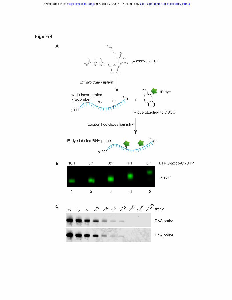
To develop IR-fluorescent RNA probe with an alternative approach, we carried out in
vitro transcription of antisense Ta U6 RNA using 5-Azido-C3-UTP, followed by IRDye®
800CW DBCO labeling (Fig. 4A). Different UTP:5-Azido-C3-UTP ratios were tested. When all
30 Us in the RNA probe were azide-modified, IR dye labeling resulted in the largest mobility
shift but the dimmest IR signal due to self-quenching (Fig. 4B, lane 5). We determined that using
3:1 UTP:5-Azido-C3-UTP resulted in an optimal Ta U6 RNA probe in terms of IR intensity (Fig.
4B). Ta U6 RNA was then analyzed by irNorthern using the RNA probe. As seen in Fig. 4C, the
RNA probe has a similar detection limit at 0.05 fmole as for the DNA probe. Note that the lower
background for the RNA probe relative to the DNA probe may result from the higher
hybridization temperature and more stringent washing conditions (see Materials and Methods).
Therefore, we have developed a sensitive irNorthern protocol utilizing an IR dye-labeled RNA
probe.
Cold Spring Harbor Laboratory Press on August 2, 2022 - Published by rnajournal.cshlp.orgDownloaded from

Bret Miller
9
Multiplexing in irNorthern blot analysis
As there are a variety of IR dyes emitting at different wavelengths, it is possible to use IR
dye-labeled probes for a multiplex Northern blot, which could not be achieved by 32P- or
chemiluminescent reaction-based Northern techniques. To this end, we performed irNorthern on
a series of 5′-extended precursor microRNA (pre-miRNA) hairpins processed by Dicer (Sheng et
al. 2018). Dicer cleavage of pre-miR-HSUR4, a Herpesvirus saimiri pre-miRNA, releases
mature miR-HSUR4s from both the 3p and the extended 5p arms of the hairpin (Fig. 5A). We
performed multiplex irNorthern with mature 3p and 5p miR-HSUR4 probes labeled with
IRDye® 680RD and 800CW, respectively (Fig. 5B). As expected, various miR-HSUR4-3p,
which were detected by the IRDye® 680RD-labeled probe (green), remained 22 nt in length.
Appearance of mature miR-HSUR4-3p was due to Dicer cleavage because a catalytic site mutant
Dicer did not produce such product (Fig. 5B, mut Dicer lanes). On the other hand, IRDye®
800CW-labeled probe (red)-detected miR-HSUR4-5p show increased length as the extension
length of the pre-miRNA increases (compare +5, +10, +15, +20, +30 nt lanes to the WT lane),
consistent with previous findings (Sheng et al. 2018). The pre-miRNAs can be hybridized with
both the 3p and the 5p miRNA probes, and therefore exhibit orange color (Fig. 5B).
We further tested multiplex irNorthern with total RNAs extracted from HCT116
colorectal cancer cells, which express pre-miR-HSUR4 with various 5′ extensions from
transfected plasmids (Sheng et al. 2018). Using an IRDye® 680RD-labeled probe against miR-
HSUR4-3p and an IRDye® 800CW-labeled probe against U6 snRNA, both RNAs were detected
on the same blot unambiguously (Fig. 5C). In conclusion, irNorthern is suitable for multiplexing
on total RNA samples.
Cold Spring Harbor Laboratory Press on August 2, 2022 - Published by rnajournal.cshlp.orgDownloaded from

Bret Miller
10
DISCUSSION
The safety precautions for using radioactive probes in Northern blot has prompted the
design of nonradioactive detection techniques. Of note is the use of biotinylated or DIG-labeled
probes in combination with HRP-mediated chemiluminescent reactions (Kim et al. 2010; Huang
et al. 2014). Nonetheless, these Northern blot techniques involve chemical reactions for
secondary detection, making them relatively cumbersome to perform.
We developed a highly sensitive and straightforward nonradioactive Northern blot
technique utilizing azide-modified oligonucleotides labeled with DBCO IR dye (Fig. 1A). While
labeling these probes requires less safety precautions compared to 32P labeling, they are still
capable of detecting RNA samples as low as ~0.05 fmol (Fig. 1B and Supplemental Fig. S1).
Furthermore, IR dye-labeled probes can be stored and reused for extended periods of time (Fig.
1B). While we showed that an IR dye-labeled probe maintained its sensitivity after 8 months
storage in -80°C (Fig. 1C), it is expected that the probe can be stored even longer without
compromising its sensitivity. As a result, researchers can save considerable amount of time and
resources, because highly sensitive IR dye-labeled probes do not have to be regenerated
frequently, as in the case of using 32P-labeled probes. Additionally, IR dye-labeled probes can be
used to detect multiple RNA species at the same time (multiplexing) (Fig. 5), which is not
possible using 32P- or chemiluminescent reaction-based Northern blot techniques.
While our multiplex irNorthern displayed its functionality in pre-miRNA processing, this
method also has applications in other RNA processing reactions such as mRNA splicing (Hirose
et al. 2006), in which the intron and the exon could be detected by different probes and the pre-
mRNA could be detected by both probes. In addition, probe color could be adjusted by labeling
with two different IR dyes to enable more versatile multiplexing. For instance, IRDye® 680RD
Cold Spring Harbor Laboratory Press on August 2, 2022 - Published by rnajournal.cshlp.orgDownloaded from

Bret Miller
11
and 800CW could be mixed in a 1:1 ratio to label a probe as “orange” (Fig. 5B). With future
advances in the accuracy of detecting subtle differences in IR fluorescence, the combination of
multiple dyes at different ratios could distinguish dozens of RNAs simultaneously in one
Northern blot.
Previously described improvements on the Northern blot method could be implemented
with the use of IR dye-labeled probes to produce even lower detection limits. For example,
replacing UV crosslinking with 1-ethyl-3-(3-dimethylaminopropyl) carbodiimide hydrochloride
(EDC) crosslinking results in a 50-fold increase in small RNA detection (Pall and Hamilton
2008). Furthermore, incorporation of LNA into the probe strands also results in elevated
detection of RNA (Valoczi et al. 2004). Similar to the LED method described by Kim et al.
2010, both EDC crosslinking and LNAs could be incorporated into our irNorthern protocol and
may result in enhanced RNA detection.
We explored another possibility of increasing the irNorthern sensitivity by incorporating
multiple dyes onto the probe. However, we estimated that co-existence of more than 3 dyes on a
single probe severely reduced the IR intensity due to self-quenching (Fig. 3B and Fig. 4B). Such
multi-IR dye-labeled probes failed to reach sensitivity that exceeds the 32P labeled probes, even
though we were able to produce DNA and RNA probes labeled with hundreds of IR dyes (data
not shown). However, improving IR technologies that minimize self-quenching effect could
allow for more sensitive irNorthern probes to be synthesized with ease in the future (Zhegalova
et al. 2014).
Cold Spring Harbor Laboratory Press on August 2, 2022 - Published by rnajournal.cshlp.orgDownloaded from

Bret Miller
12
MATERIALS AND METHODS
Preparation of IR dye-labeled DNA probes
IrNorthern DNA probes for U6: 5′-
GCAGGGGCCATGCTAATCTTCTCTGTATCG/iAzideN/T-3′; miR-HSUR4-5p: 5′-
TTATAGCTGTAGCAACACGGT/iAzideN/A-3′; and miR-HSUR4-3p:
ACGTGTTGCCCACTGCTATAAA/iAzideN/A-3′ were synthesized by Integrated DNA
Technology (IDT). To label the azide-modified oligonucleotides with IR dye, 2.5 nmole
oligonucleotides were mixed with 50 nmole IRDye® 680RD or 800CW DBCO (Li-Cor
Biosciences) in phosphate buffer saline (137 mM NaCl, 10 mM PO43-, 2.7 mM KCl, pH 7.4) at
25°C for 6 hours. IR dye-labeled oligonucleotides were then purified by Microspin G-25 column
(GE Healthcare) or Ampure XP beads (Beckman Coulter) according to the manufacturer’s
instructions. Minor modifications were introduced for Ampure XP beads purification of short
oligonucleotides. Specifically, 2 volumes of beads and 5.4 volumes of isopropanol were mixed
with the reaction. The mixture was incubated at room temperature for 15 min and placed on
magnet stand to separate the beads from the supernatant. The beads were washed twice with 85%
ethanol and the DNA was eluted with 20-50 µL of H2O.
TdT reaction was performed in 30 µL containing 50 mM KOAc, 20 mM Tris-acetate pH
7.9, 10 mM MgOAc, 250 µM CoCl2, 20 U TdT (New England Biolabs, NEB), 100 pmole U6
DNA probe (5′-GCAGGGGCCATGCTAATCTTCTCTGTATCG-3′), and 100 to 1500 pmoles
of 5-azidomethyl-dUTP (Jena Bioscience). After incubation at 37°C for 1 hr, the reaction was
purified by Ampure XP beads, labeled with IRDye® 800CW DBCO, and purified again by
Ampure XP beads as described above. Labeled probes were separated on an 8 M Urea 15%
Cold Spring Harbor Laboratory Press on August 2, 2022 - Published by rnajournal.cshlp.orgDownloaded from

Bret Miller
13
acrylamide gel. After IR scan and SYBR green imaging (Fig. 3B), the desired probes were
purified from the gel.
Preparation of RNA probes for irNorthern
To synthesize antisense RNA probes for Trichoplax U6, PCR templates containing a T7
promoter were used in T7 run-off transcription reactions. Each 10 µL reaction containing 40 mM
Tris-HCl pH 8.0, 25 mM NaCl, 2 mM Spermidine(HCl)3, 8 mM MgCl2, 1 mM ATP, 1 mM
UTP, 1 mM GTP, 1 mM CTP, 2 U/µL Murine RNase Inhibitor (NEB), 10 mM DTT, and 0.4
U/µL T7 RNA polymerase was incubated at 37°C overnight. In the transcription reactions
including Aminoallyl-UTP-ATTO-680, 1 mM UTP was replaced by 50 µM UTP and 50 µM
Aminoallyl-UTP-ATTO-680 (Jena Bioscience). In the reactions including 5-Azido-C3-UTP, 1
mM UTP was replaced by 1 mM total UTP with different ratios of UTP:5-Azido-C3-UTP (Jena
Bioscience) as indicated in Fig. 4B. In vitro transcribed RNAs were purified by 8 M Urea 6%
acrylamide gel and resuspended in H2O. The 5-Azido-C3-U containing RNA probes were labeled
with IRDye® 800CW DBCO and purified by Ampure XP beads using the conditions described
above.
Preparation of 5′ end 32P-labeled probes
10 pmole of U6 DNA probe was incubated with 150 µCi 6000 Ci/mmol γ-32P-ATP
(Perkin Elmer), 10 U T4 PNK (NEB) in 70 mM Tris-HCl, 10 mM MgCl2, 5 mM DTT, pH 7.6 at
37°C for 30 minutes. The 32P-labeled oligonucleotides were then purified by a Microspin G-25
column according to the manufacturer’s instructions.
Northern blot analysis
Cold Spring Harbor Laboratory Press on August 2, 2022 - Published by rnajournal.cshlp.orgDownloaded from
Bret Miller
14
In vitro transcribed Ta U6 RNAs were electrophoresed in an 8 M Urea 10% acrylamide
gel and then transferred to a Hybond N+ membrane (Amersham). The membrane was
crosslinked twice with 254 nm UV light at 120 mJ/cm2 using a Stratalinker UV crosslinker 2400
(Agilent genomics). After pre-hybridization with 10 mL ExpressHyb Hybridization Solution
(Clonetech) for 30 min at 37°C, the membrane was hybridized with 10 pmole IR dye-labeled
oligonucleotide U6 probe in 10 mL ExpressHyb solution at 37°C overnight. The membrane was
washed with 2X SSC, 0.1% SDS (10 min at room temperature), 1X SSC, 0.1% SDS (10 min at
room temperature) and analyzed by a Li-Cor Odyssey CLX scanner. The membranes used for
irNorthern in Fig. 1D and Fig. 5 were prepared previously (Sheng et al. 2018). For miR-HSUR4-
5p and -3p multiplex probing, the hybridization temperature was set at 45°C to prevent annealing
between the two highly complementary probes and favor annealing between the probes and the
RNA targets.
For Northern blots using a 32P-labeled DNA probe, the protocol was the same as
irNorthern, except that the washed membrane was exposed to a phosphor screen overnight and
scanned on a Storm phosphorimager (Amersham). In between probing with different DNA
probes, the membranes were stripped with microwave-boiled 0.1X SSC, 1% SDS, 40 mM Tris-
HCl pH 8.0 for 10 minutes twice. The membranes were re-scanned to ensure the probes were
successfully removed. When comparing the sensitivity of IR dye-labeled probes to 32P labeled
probes, we alternated the first probe applied to the membrane in five different experiments and
found that probing the membrane first with either the IR dye-labeled or the 32P-labeled probe did
not affect our conclusions (Fig. 1B and Supplemental Fig. S1).
For irNorthern using biotinylated DNA probes, after overnight hybridization and brief
washes of 2X SSC, 0.1% SDS (2 min at room temperature), and 1X SSC, 0.1% SDS (2 min at
Cold Spring Harbor Laboratory Press on August 2, 2022 - Published by rnajournal.cshlp.orgDownloaded from
Bret Miller
15
room temperature) the membranes were incubated with 1:10,000 diluted IRDye® 800CW
streptavidin in ExpressHyb solution at 30°C for 30 minutes and then washed and analyzed as
described above.
For irNorthern using RNA probe, the hybridization was carried out at 65°C overnight.
The membranes were washed with 2X SSC, 0.1% SDS (15 min at room temperature), 1X SSC,
0.1% SDS (15 min at room temperature) and 0.2X SSC, 0.1% SDS (15 min at room temperature)
and analyzed as described above. Unlike the DNA probes, RNA probes cannot be completely
stripped off the membrane.
As probes were continuously reused during experimentation, all IR dye-labeled probes
diluted in hybridization solution were stored at -20°C while stock solutions of IR labeled probes
were stored at -80°C. IR dyes and IR dye-labeled probes were protected from strong light
exposure during irNorthern procedure and storage.
ACKNOWLEDGEMENTS
We thank Drs. Brian Zarnegar and Paul Khavari for sharing their protocol on generating
the irCLIP adaptor; Dr. J. Bert Flanegan, Dr. Jörg Bungert, Taha Huda, Marc McLeod, Paul
Wassel, and Emily Chong for stimulating discussions and comments on the manuscript. This
work was supported by grants from National Institutes of Health (R00-CA190886 and R35-
GM128753 to M.X.) and Thomas H. Maren Foundation (Junior Investigator Fund F013372 to
M.X.).
Cold Spring Harbor Laboratory Press on August 2, 2022 - Published by rnajournal.cshlp.orgDownloaded from

Bret Miller
16
REFERENCES
Guo Z, Park S, Yoon J, Shin I. 2014. Recent progress in the development of near-infrared
fluorescent probes for bioimaging applications. Chem Soc Rev 43: 16-29.
Hirose T, Ideue T, Nagai M, Hagiwara M, Shu MD, Steitz JA. 2006. A spliceosomal intron
binding protein, IBP160, links position-dependent assembly of intron-encoded box C/D
snoRNP to pre-mRNA splicing. Mol Cell 23: 673-684.
Holtke HJ, Ankenbauer W, Muhlegger K, Rein R, Sagner G, Seibl R, Walter T. 1995. The
digoxigenin (DIG) system for non-radioactive labelling and detection of nucleic acids--an
overview. Cell Mol Biol (Noisy-le-grand) 41: 883-905.
Huang Q, Mao Z, Li S, Hu J, Zhu Y. 2014. A non-radioactive method for small RNA detection
by northern blotting. Rice (N Y) 7: 26.
Jones CG. 2005. A review of the history of U.S. radiation protection regulations,
recommendations, and standards. Health Phys 88: 697-716.
Kessler C. 1994. Non-radioactive analysis of biomolecules. J Biotechnol 35: 165-189.
Kim SW, Li Z, Moore PS, Monaghan AP, Chang Y, Nichols M, John B. 2010. A sensitive non-
radioactive northern blot method to detect small RNAs. Nucleic Acids Res 38: e98.
Kohn M, Lederer M, Wachter K, Huttelmaier S. 2010. Near-infrared (NIR) dye-labeled RNAs
identify binding of ZBP1 to the noncoding Y3-RNA. RNA 16: 1420-1428.
MacDiarmid CW, Taggart J, Jeong J, Kerdsomboon K, Eide DJ. 2016. Activation of the Yeast
UBI4 Polyubiquitin Gene by Zap1 Transcription Factor via an Intragenic Promoter Is
Critical for Zinc-deficient Growth. J Biol Chem 291: 18880-18896.
Cold Spring Harbor Laboratory Press on August 2, 2022 - Published by rnajournal.cshlp.orgDownloaded from

Bret Miller
17
Melton DA, Krieg PA, Rebagliati MR, Maniatis T, Zinn K, Green MR. 1984. Efficient in vitro
synthesis of biologically active RNA and RNA hybridization probes from plasmids
containing a bacteriophage SP6 promoter. Nucleic Acids Res 12: 7035-7056.
Owens EA, Henary M, El Fakhri G, Choi HS. 2016. Tissue-Specific Near-Infrared Fluorescence
Imaging. Acc Chem Res 49: 1731-1740.
Pall GS, Hamilton AJ. 2008. Improved northern blot method for enhanced detection of small
RNA. Nat Protoc 3: 1077-1084.
Rio DC. 2014. Northern blots for small RNAs and microRNAs. Cold Spring Harb Protoc 2014:
793-797.
Sheng P, Fields C, Aadland K, Wei T, Kolaczkowski O, Gu T, Kolaczkowski B, Xie M. 2018.
Dicer cleaves 5'-extended microRNA precursors originating from RNA polymerase II
transcription start sites. Nucleic Acids Res 46: 5737-5752.
Srivastava RA, Schonfeld G. 1991. Use of riboprobes for northern blotting analysis.
Biotechniques 11: 584, 586, 588.
Streit S, Michalski CW, Erkan M, Kleeff J, Friess H. 2009. Northern blot analysis for detection
and quantification of RNA in pancreatic cancer cells and tissues. Nat Protoc 4: 37-43.
Unterweger MP. 2002. Half-life measurements at the National Institute of Standards and
Technology. Appl Radiat Isot 56: 125-130.
Valoczi A, Hornyik C, Varga N, Burgyan J, Kauppinen S, Havelda Z. 2004. Sensitive and
specific detection of microRNAs by northern blot analysis using LNA-modified
oligonucleotide probes. Nucleic Acids Res 32: e175.
Cold Spring Harbor Laboratory Press on August 2, 2022 - Published by rnajournal.cshlp.orgDownloaded from
Bret Miller
18
Weldon S, Ambroz K, Schutz-Geschwender A, Olive DM. 2008. Near-infrared fluorescence
detection permits accurate imaging of loading controls for Western blot analysis. Anal
Biochem 375: 156-158.
Zarnegar BJ, Flynn RA, Shen Y, Do BT, Chang HY, Khavari PA. 2016. irCLIP platform for
efficient characterization of protein-RNA interactions. Nature methods 13: 489-492.
Zavala AG, Kulkarni AS, Fortunato EA. 2014. A dual color Southern blot to visualize two
genomes or genic regions simultaneously. J Virol Methods 198: 64-68.
Zhegalova NG, He S, Zhou H, Kim DM, Berezin MY. 2014. Minimization of self-quenching
fluorescence on dyes conjugated to biomolecules with multiple labeling sites via
asymmetrically charged NIR fluorophores. Contrast Media Mol Imaging 9: 355-362.
FIGURE LEGENDS
Figure 1. (A) Schematic diagram of IR dye labeling of azide-modified ssDNA probe followed by
Northern blot analysis. Time used on each step is indicated in the parentheses. (B) Northern blot
analyses of serial diluted Trichoplax adhaerens (Ta) U6 snRNA samples for comparison of IR
dye-labeled probe with 32P-labeled probe. In the left panel, the membrane was hybridized with
IR dye-labeled probe and scanned on an Odyssey CLX for IR fluorescence detection. In the right
panel, the same membrane was hybridized with 32P-labeled probe, exposed to a phosphor screen
and scanned on a Storm phosphorimager. The membrane was stripped after each scan and was
rehybridized with the same diluted probes after 2, 4, 8, and 12 weeks. (C) irNorthern analysis of
Ta U6 using an IR dye-labeled ssDNA probe stored at -80°C for 8 months prior to its usage. (D)
Northern blot analyses of human U6 snRNA in nuclear (N) and cytoplasmic (C) RNAs extracted
Cold Spring Harbor Laboratory Press on August 2, 2022 - Published by rnajournal.cshlp.orgDownloaded from
Bret Miller
19
from HEK 293T cells, using an IR dye-labeled probe or a 32P-labeled probe. White triangles
point to the U6 RNAs detected in the cytoplasmic fractions.
Figure 2. (A) Schematic diagram for a 3′ biotinylated ssDNA probe interacting with a
streptavidin IR dye. (B) Serial diluted Ta U6 snRNA samples were analyzed by irNorthern using
a biotinylated probe conjugated with a streptavidin IR dye or an IR dye-labeled probe.
Figure 3. (A) Schematic diagram for preparing an IR dye-labeled DNA probe via terminal
transferase (TdT) reaction. (B) Visualization of 5-azidomethyl-dU-incorporated and IR dye-
labeled DNA probes by SYBR green staining and IR scan. In the TdT reactions, different DNA
probe:5-azidomethyl-dUTP ratios were used as indicated on top. DNA probes containing one or
two IR dyes were bracketed. The asterisks indicate the self-quenching probes. (C) Serial diluted
Ta U6 snRNA samples were analyzed by irNorthern using probes containing TdT transferred 5-
azidomethyl-dU or chemically synthesized probes containing azide-modified dT.
Figure 4. (A) Schematic diagram for preparing an IR dye-labeled RNA probe via T7 in vitro
transcription. (B) Visualization of 5-azido-C3-U-incorporated and IR dye-labeled RNA probes
by IR scan. In in vitro transcription reactions, different UTP: 5-azido-C3-UTP ratios were used as
indicated on top. (C) Serial diluted Ta U6 snRNA samples were analyzed by irNorthern using
either an IR dye-labeled RNA or DNA probe. Note that the irNorthern image for DNA probe is
the same one used as the bottom panel in Fig. 3C.
Figure 5. (A) Schematic diagram of Dicer cleavage on 5′-extended pre-miR-HSUR4. Dicer
cleavage sites are indicated with black triangles. 5p and 3p miR-HSUR4s released from Dicer
cleavage are highlighted in red and green, respectively. (B) Equal numbers of in vitro transcribed
5′-extended pre-miR-HSUR4s were processed with wild type (WT) or mutant (mut) human
Cold Spring Harbor Laboratory Press on August 2, 2022 - Published by rnajournal.cshlp.orgDownloaded from

Bret Miller
20
Dicer and analyzed by multiplex irNorthern blot to detect miR-HSUR4-5p or -3p. The 5p
miRNA probe was labeled with IRDye® 800CW and the detected miR-HSUR4-5p bands are
shown in red. The 3p miRNA probe was labeled with IRDye® 680RD and the detected miR-
HSUR4-3p bands are shown in green. The pre-miRNAs, shown in orange, were detected by both
the 5p and 3p probes. (C) Multiplex irNorthern analysis of total RNAs extracted from HCT116
colorectal cancer cells, which express pre-miR-HSUR4 with various 5′ extensions from
transfected plasmids. U6 (red) was detected by an IRDye® 800CW-labeled probe, while miR-
HSUR4-3p and pre-miR-HSUR4 (green) were detected by an IRDye® 680RD-labeled probe.
Cold Spring Harbor Laboratory Press on August 2, 2022 - Published by rnajournal.cshlp.orgDownloaded from

Cold Spring Harbor Laboratory Press on August 2, 2022 - Published by rnajournal.cshlp.orgDownloaded from

Cold Spring Harbor Laboratory Press on August 2, 2022 - Published by rnajournal.cshlp.orgDownloaded from

Cold Spring Harbor Laboratory Press on August 2, 2022 - Published by rnajournal.cshlp.orgDownloaded from
Cold Spring Harbor Laboratory Press on August 2, 2022 - Published by rnajournal.cshlp.orgDownloaded from

Cold Spring Harbor Laboratory Press on August 2, 2022 - Published by rnajournal.cshlp.orgDownloaded from

Cold Spring Harbor Laboratory Press on August 2, 2022 - Published by rnajournal.cshlp.orgDownloaded from

published online September 10, 2018RNA Bret R Miller, Tianqi Wei, Christopher J Fields, et al. Near-infrared fluorescent Northern blot
Material
Supplemental
http://rnajournal.cshlp.org/content/suppl/2018/09/10/rna.068213.118.DC1
P<P
Published online September 10, 2018 in advance of the print journal.
Manuscript
Accepted
manuscript is likely to differ from the final, published version. Peer-reviewed and accepted for publication but not copyedited or typeset; accepted
Open Access
Open Access option.RNAFreely available online through the
License
Commons Creative
.http://creativecommons.org/licenses/by/4.0/(Attribution 4.0 International), as described at
, is available under a Creative Commons LicenseRNAThis article, published in
ServiceEmail Alerting
click here.top right corner of the article or
Receive free email alerts when new articles cite this article - sign up in the box at the
http://rnajournal.cshlp.org/subscriptions go to: RNATo subscribe to
Published by Cold Spring Harbor Laboratory Press for the RNA Society
Cold Spring Harbor Laboratory Press on August 2, 2022 - Published by rnajournal.cshlp.orgDownloaded from