fluorescent pigments in corals are photoprotective
TRANSCRIPT
This is a preprint. Text and figures may differ in minor aspects from published text. This mss is providedunder embargo until publication date, December 14th. Copyright © Salih et al, 2000
1
Fluorescent pigments in corals are photoprotective.Anya Salih*,†, Anthony Larkum*, Guy Cox†, Michael Kühl‡ and Ove Hoegh-Guldberg*§
* School of Biological Sciences, A08, The University of Sydney, NSW, Australia.
† Electron Microscope Unit, F09, The University of Sydney, NSW, Australia.
‡ Marine Biological Laboratory, University of Copenhagen, Denmark.§ Current address: Centre for Marine Studies, University of Queensland, St Lucia, QLD 4072, Australia
All reef-forming corals depend on the photosynthesis of their dinoflagellate algalsymbiont and are therefore restricted to the photic zone. In this zone light intensitydeclines over several decades of intensity from high and damaging levels(accompanied by UV radiation) at the surface to extreme shade conditions at thelower limit1. The ability of corals to tolerate this range implies effective mechanismsfor light acclimation and adaptation2. Here we show that the fluorescent pigments3-9
(FPs) of corals provide a photobiological system for regulating the light environmentof coral host tissue. Previous studies have suggested that under low light, FPs mayenhance light availability4,5. We now present evidence that in excessive sunlight, FPsare photoprotective through dissipation of excess energy at wavelengths of lowphotosynthetic activity, as well as by reflection of visible and infra-red light by FP-containing chromatophores. We also show that FPs enhance the resistance to massbleaching of corals during periods of heat stress, which has implications for theimpact of environmental stress on the diversity of reef-building corals.
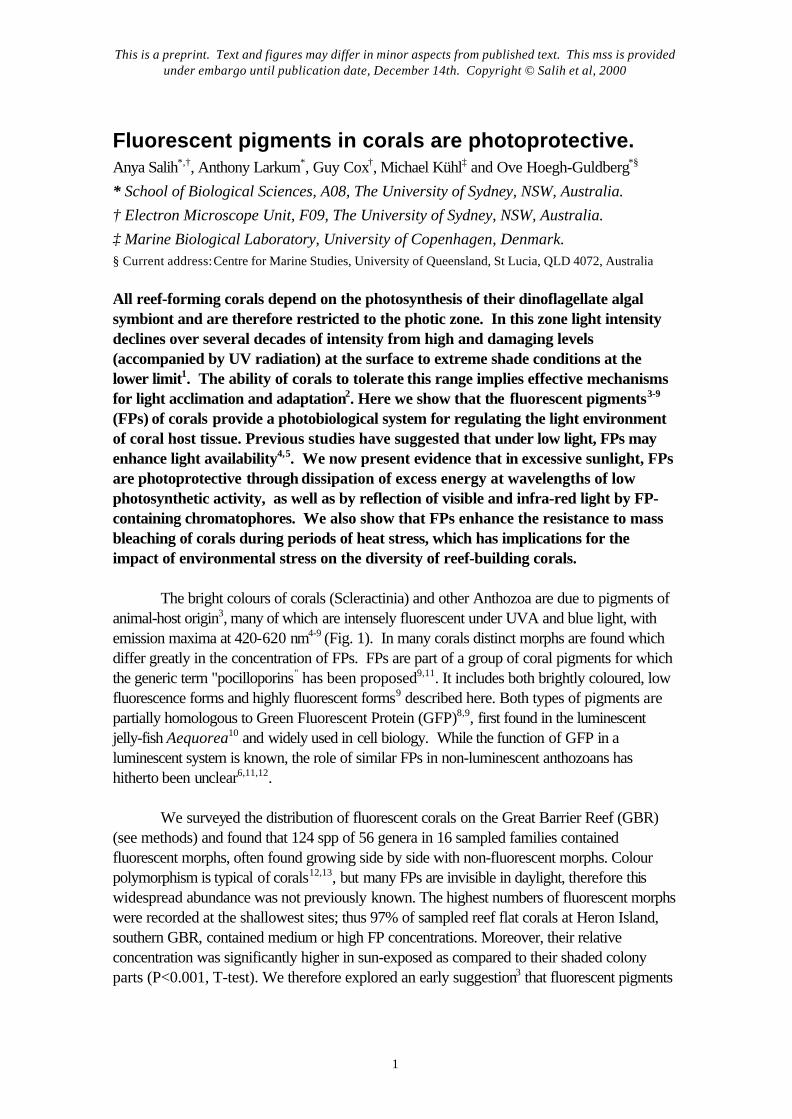
The bright colours of corals (Scleractinia) and other Anthozoa are due to pigments ofanimal-host origin3, many of which are intensely fluorescent under UVA and blue light, withemission maxima at 420-620 nm4-9 (Fig. 1). In many corals distinct morphs are found whichdiffer greatly in the concentration of FPs. FPs are part of a group of coral pigments for whichthe generic term "pocilloporins" has been proposed9,11. It includes both brightly coloured, lowfluorescence forms and highly fluorescent forms9 described here. Both types of pigments arepartially homologous to Green Fluorescent Protein (GFP)8,9, first found in the luminescentjelly-fish Aequorea10 and widely used in cell biology. While the function of GFP in aluminescent system is known, the role of similar FPs in non-luminescent anthozoans hashitherto been unclear6,11,12.
We surveyed the distribution of fluorescent corals on the Great Barrier Reef (GBR)(see methods) and found that 124 spp of 56 genera in 16 sampled families containedfluorescent morphs, often found growing side by side with non-fluorescent morphs. Colourpolymorphism is typical of corals12,13, but many FPs are invisible in daylight, therefore thiswidespread abundance was not previously known. The highest numbers of fluorescent morphswere recorded at the shallowest sites; thus 97% of sampled reef flat corals at Heron Island,southern GBR, contained medium or high FP concentrations. Moreover, their relativeconcentration was significantly higher in sun-exposed as compared to their shaded colonyparts (P<0.001, T-test). We therefore explored an early suggestion3 that fluorescent pigments
This is a preprint. Text and figures may differ in minor aspects from published text. This mss is providedunder embargo until publication date, December 14th. Copyright © Salih et al, 2000
2
in shallow water corals might function in photoprotection, by comparing fluorescent and non-fluorescent morphs.
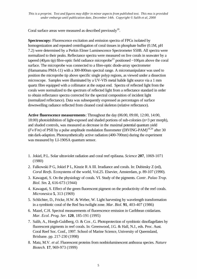
We found that the emission maxima of FPs ranged from blue to green to red (Fig 1),which is consistent with studies done on the isolated FP proteins of corals9. The shorterwavelength FPs were more abundant. Microscopically we identified 2 broad groups: FPsbound within 0.2-8µm fluorescent pigment granules (FPGs) as reported previously3,5,7 andinter- or intracellular FPs, not enclosed in granules (CFPs). Significantly, the majority of coralscontained multiple FPG and CFP types. Correspondences between emission and excitationmaxima of FPs which occur in close association (Fig 1, e-f) suggest that energytransformation to longer, non-photosynthetically active wavelengths might in some cases be asequential process, with the fluorescence of one pigment exciting another, as expected fromspectra of isolated proteins8,9. We demonstrated that this process could occur by comparingfluorescence of green FPGs (excitation max. 482.5 nm) alone and mixed with blue FPGs(excitation max. 382.5 nm). Only weak green fluorescence is seen under 330-380nmexcitation; addition of blue FPGs emitting at 480nm enhanced green fluorescence intensity 4 to7-fold. (The effect was strongly dependent on distance, with maximal enhancement when blueand green granules were less than 10µm apart.) The final energy spill would then (dependingon the pigments involved) lie between the two major peaks of the coral photosynthetic actionspectrum and hence be relatively inactive in photosynthesis (Fig 1f). This would be the inversecounterpart of the process of light transfer to photosynthesis which others have proposed inlight limited habitats4-5 (Fig 1 g)
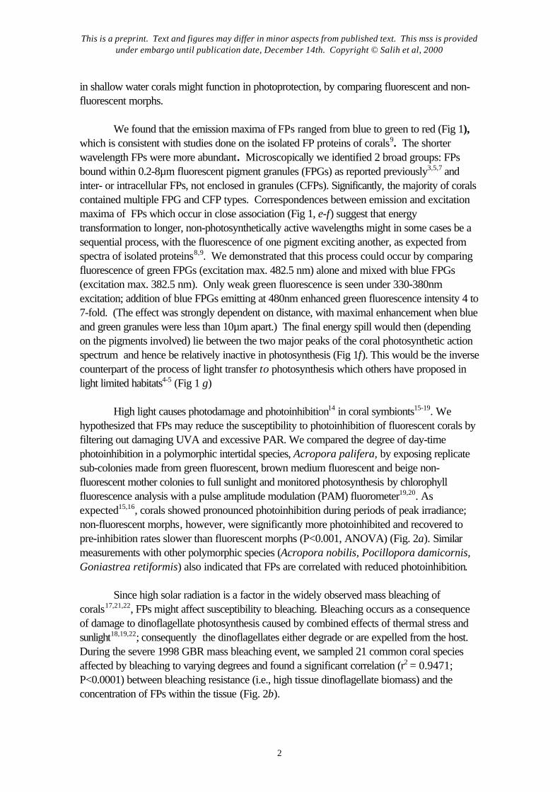
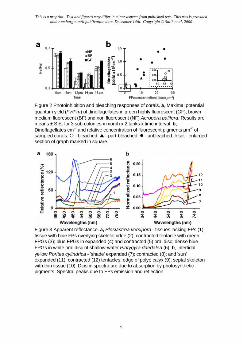
High light causes photodamage and photoinhibition14 in coral symbionts15-19. Wehypothesized that FPs may reduce the susceptibility to photoinhibition of fluorescent corals byfiltering out damaging UVA and excessive PAR. We compared the degree of day-timephotoinhibition in a polymorphic intertidal species, Acropora palifera, by exposing replicatesub-colonies made from green fluorescent, brown medium fluorescent and beige non-fluorescent mother colonies to full sunlight and monitored photosynthesis by chlorophyllfluorescence analysis with a pulse amplitude modulation (PAM) fluorometer19,20. Asexpected15,16, corals showed pronounced photoinhibition during periods of peak irradiance;non-fluorescent morphs, however, were significantly more photoinhibited and recovered topre-inhibition rates slower than fluorescent morphs (P<0.001, ANOVA) (Fig. 2a). Similarmeasurements with other polymorphic species (Acropora nobilis, Pocillopora damicornis,Goniastrea retiformis) also indicated that FPs are correlated with reduced photoinhibition.
Since high solar radiation is a factor in the widely observed mass bleaching ofcorals17,21,22, FPs might affect susceptibility to bleaching. Bleaching occurs as a consequenceof damage to dinoflagellate photosynthesis caused by combined effects of thermal stress andsunlight18,19,22; consequently the dinoflagellates either degrade or are expelled from the host.During the severe 1998 GBR mass bleaching event, we sampled 21 common coral speciesaffected by bleaching to varying degrees and found a significant correlation (r2 = 0.9471;P<0.0001) between bleaching resistance (i.e., high tissue dinoflagellate biomass) and theconcentration of FPs within the tissue (Fig. 2b).
This is a preprint. Text and figures may differ in minor aspects from published text. This mss is providedunder embargo until publication date, December 14th. Copyright © Salih et al, 2000
3
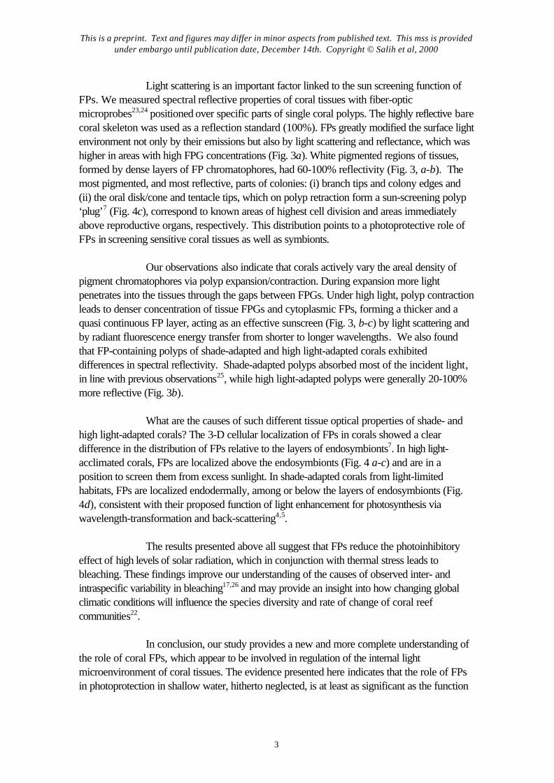
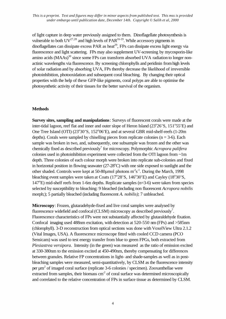
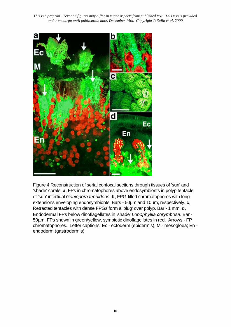
Light scattering is an important factor linked to the sun screening function ofFPs. We measured spectral reflective properties of coral tissues with fiber-opticmicroprobes23,24 positioned over specific parts of single coral polyps. The highly reflective barecoral skeleton was used as a reflection standard (100%). FPs greatly modified the surface lightenvironment not only by their emissions but also by light scattering and reflectance, which washigher in areas with high FPG concentrations (Fig. 3a). White pigmented regions of tissues,formed by dense layers of FP chromatophores, had 60-100% reflectivity (Fig. 3, a-b). Themost pigmented, and most reflective, parts of colonies: (i) branch tips and colony edges and(ii) the oral disk/cone and tentacle tips, which on polyp retraction form a sun-screening polyp‘plug’7 (Fig. 4c), correspond to known areas of highest cell division and areas immediatelyabove reproductive organs, respectively. This distribution points to a photoprotective role ofFPs in screening sensitive coral tissues as well as symbionts.
Our observations also indicate that corals actively vary the areal density ofpigment chromatophores via polyp expansion/contraction. During expansion more lightpenetrates into the tissues through the gaps between FPGs. Under high light, polyp contractionleads to denser concentration of tissue FPGs and cytoplasmic FPs, forming a thicker and aquasi continuous FP layer, acting as an effective sunscreen (Fig. 3, b-c) by light scattering andby radiant fluorescence energy transfer from shorter to longer wavelengths. We also foundthat FP-containing polyps of shade-adapted and high light-adapted corals exhibiteddifferences in spectral reflectivity. Shade-adapted polyps absorbed most of the incident light,in line with previous observations25, while high light-adapted polyps were generally 20-100%more reflective (Fig. 3b).
What are the causes of such different tissue optical properties of shade- andhigh light-adapted corals? The 3-D cellular localization of FPs in corals showed a cleardifference in the distribution of FPs relative to the layers of endosymbionts7. In high light-acclimated corals, FPs are localized above the endosymbionts (Fig. 4 a-c) and are in aposition to screen them from excess sunlight. In shade-adapted corals from light-limitedhabitats, FPs are localized endodermally, among or below the layers of endosymbionts (Fig.4d), consistent with their proposed function of light enhancement for photosynthesis viawavelength-transformation and back-scattering4,5.
The results presented above all suggest that FPs reduce the photoinhibitoryeffect of high levels of solar radiation, which in conjunction with thermal stress leads tobleaching. These findings improve our understanding of the causes of observed inter- andintraspecific variability in bleaching17,26 and may provide an insight into how changing globalclimatic conditions will influence the species diversity and rate of change of coral reefcommunities22.
In conclusion, our study provides a new and more complete understanding ofthe role of coral FPs, which appear to be involved in regulation of the internal lightmicroenvironment of coral tissues. The evidence presented here indicates that the role of FPsin photoprotection in shallow water, hitherto neglected, is at least as significant as the function
This is a preprint. Text and figures may differ in minor aspects from published text. This mss is providedunder embargo until publication date, December 14th. Copyright © Salih et al, 2000
4
of light capture in deep water previously assigned to them. Dinoflagellate photosynthesis isvulnerable to both UV27,28 and high levels of PAR16-19. While accessory pigments indinoflagellates can dissipate excess PAR as heat29, FPs can dissipate excess light energy viafluorescence and light scattering. FPs may also supplement UV-screening by mycosporin-likeamino acids (MAAs)30 since some FPs can transform absorbed UVA radiation to longer non-actinic wavelengths via fluorescence. By screening chlorophylls and peridinin from high levelsof solar radiation and by absorbing UVA, FPs thereby decrease the likelihood of irreversiblephotoinhibition, photooxidation and subsequent coral bleaching. By changing their opticalproperties with the help of these GFP-like pigments, coral polyps are able to optimise thephotosynthetic activity of their tissues for the better survival of the organism.
Methods
Survey sites, sampling and manipulations : Surveys of fluorescent corals were made at theinter-tidal lagoon, reef flat and inner and outer slope of Heron Island (23o26’S, 151o55’E) andOne Tree Island (OTI) (23o30’S, 152o06’E), and at several GBR mid-shelf-reefs (1-20mdepths). Corals were sampled by chiselling pieces from replicate colonies (n = 3-6). Eachsample was broken in two, and, subsequently, one subsample was frozen and the other waschemically fixed as described previously7 for microscopy. Polymorphic Acropora paliferacolonies used in photoinhibition experiment were collected from the OTI lagoon from ~1mdepth. Three colonies of each colour morph were broken into replicate sub-colonies and fixedin horizontal position in flowing seawater (27-28oC) with one side exposed to sunlight and theother shaded. Controls were kept at 50-80µmol photons m-2s-1. During the March, 1998bleaching event samples were taken at Coats (17o28’S, 146o30’E) and Cayley (18o30’S,147oE) mid-shelf reefs from 1-6m depths. Replicate samples (n=3-6) were taken from speciesselected by susceptibility to bleaching: 9 bleached (including non fluorescent Acropora nobilismorph;); 5 partially bleached (including fluorescent A. nobilis); 7 unbleached.
Microscopy: Frozen, glutaradehyde-fixed and live coral samples were analysed byfluorescence widefield and confocal (CLSM) microscopy as described previously7.Fluorescence characteristics of FPs were not substantially affected by glutaraldehyde fixation.Confocal imaging used 488nm excitation, with detection at 520-550 nm (FPs) and >585nm(chlorophyll). 3-D reconstruction from optical sections was done with VoxelView Ultra 2.1.2(Vital Images, USA). A fluorescence microscope fitted with cooled CCD camera (PCOSensicam) was used to test energy transfer from blue to green FPGs, both extracted fromPlesiastrea versipora. Intensity (in the green) was measured as the ratio of emission excitedat 330-380nm to the emission excited at 450-490nm, thereby compensating for differencesbetween granules. Relative FP concentrations in light- and shade-samples as well as in post-bleaching samples were measured, semi-quantitatively, by CLSM as the fluorescence intensityper µm2 of imaged coral surface (replicate 3-6 colonies / specimen). Zooxanthellae wereextracted from samples, their biomass cm-2 of coral surface was determined microscopicallyand correlated to the relative concentration of FPs in surface tissue as determined by CLSM.
This is a preprint. Text and figures may differ in minor aspects from published text. This mss is providedunder embargo until publication date, December 14th. Copyright © Salih et al, 2000
5
Coral surface areas were measured as described previously18.
Spectroscopy: Fluorescence excitation and emission spectra of FPGs isolated byhomogenization and repeated centrifugation of coral tissues in phosphate buffer (0.1M, pH7.2) were determined by a Perkin Elmer Luminescence Spectrometer S50B. All spectra werenormalized to their peaks. Reflectance spectra were measured on live corals in seawater by atapered (40µm tip) fibre-optic field radiance microprobe23 positioned ~100µm above the coralsurface. The microprobe was connected to a fibre-optic diode-array spectrometer(Hamamatsu PMA-11) with a 300-800nm spectral range. A micromanipulator was used toposition the microprobe tip above specific single polyp regions, as viewed under a dissectionmicroscope. Samples were illuminated by a UV-VIS metal halide light source via a 1 mmquartz fibre equipped with a collimator at the output end. Spectra of reflected light from thecorals were normalized to the spectrum of reflected light from a reflectance standard in orderto obtain reflectance spectra corrected for the spectral composition of incident light(normalized reflectance). Data was subsequently expressed as percentages of surfacedownwelling radiance reflected from cleaned coral skeleton (relative reflectance).
Active fluorescence measurements: Throughout the day (06:00, 09:00, 12:00, 14:00,18:00) photoinhibition of light-exposed and shaded portions of sub-colonies (n=3 per morph),and shaded controls, was measured as decrease in the maximal potential quantum yield(Fv/Fm) of PSII by a pulse amplitude modulation fluorometer (DIVING-PAM)19,20 after 30min dark-adaption. Photosynthetically active radiation (400-700nm) during the experimentwas measured by LI-190SA quantum sensor.
1. Jokiel, P.L. Solar ultraviolet radiation and coral reef epifauna. Science 207, 1069-1071(1980)
2. Falkowski P G, Jokiel P L, Kinzie R A III. Irradiance and corals. In: Dubinsky Z (ed),Coral Reefs. Ecosystems of the world, Vol.25. Elsevier, Amsterdam, p. 89-107 (1990).
3. Kawaguti, S. On the physiology of corals. VI. Study of the pigments. Contr. Palao Trop.Biol. Stn. 2, 616-673 (1944)
4. Kawaguti, S. Effect of the green fluorescent pigment on the productivity of the reef corals.Micronesica 5, 313 (1969)
5. Schlichter, D., Fricke, H.W. & Weber, W. Light harvesting by wavelength transformationin a symbiotic coral of the Red Sea twilight zone. Mar. Biol. 91, 403-407 (1986)
6. Mazel, C.H. Spectral measurements of fluorescence emission in Caribbean cnidarians.Mar. Ecol. Prog. Ser. 120, 185-191 (1995)
7. Salih, A., Hoegh-Guldberg, O. & Cox , G. Photoprotection of symbiotic dinoflagellates byfluorescent pigments in reef corals. In: Greenwood, J.G. & Hall, N.J., eds. Proc. Aust.Coral Reef Soc. Conf., 1997. School of Marine Science, University of Queensland,Brisbane. pp. 217-230 (1998)
8. Matz, M.V. et al. Fluorescent proteins from nonbioluminescent anthozoa species. NatureBiotech. 17, 969-973 (1999)
This is a preprint. Text and figures may differ in minor aspects from published text. This mss is providedunder embargo until publication date, December 14th. Copyright © Salih et al, 2000
6
9. Dove, S.G, Hoegh-Guldberg, O. & Ranganathan S. Major colour patterns of reef-buildingcorals are due to a family of GFP-like proteins. Coral Reefs, 19, 197-204 (2000)
10. Shimomura, O., Johnson, F. H. & Saiga, Y. Extraction, purification, and properties ofaequorin, a bioluminescent protein from the luminous hydromedusan, Aequorea. J. Cell.Comp. Physiol. 77, 305-312 (1962)
11. Dove , S.G., Takabayashi, M. and Hoegh-Guldberg, O. Isolation and partialcharacterization of the Pink and Blue pigments of Pocilloporid and Acroporid corals. Biol.Bull. 189: 288-297.
12. Takabayashi M.& Hoegh-Guldberg O. Physiological and ecological differences betweenpink and brown genotypes of the reef-building coral Pocillopora damicornis. Mar. Biol.123, 705-714 (1995)
13. Veron, J.E.N. Corals of Australia and the Indo-Pacific. Angus & Robertson, Sydney(1986)
14. Long, S.P., Humphries, S. & Falkowski, P.G. Photoinhibition of photosynthesis in nature.Annu. Rev. Plant Physiol. Plant Mol. Biol. 45, 633-662 (1994)
15. Brown, B.E. et al. Diurnal changes in photochemical efficiency and xanthophyllconcentrations in shallow water reef corals: evidence for photoinhibition andphotoprotection. Coral Reefs 18, 99-105 (1999)
16. Hoegh-Guldberg, O. & Jones, R. Diurnal variability in photoinhibition and photoprotectionin the symbiotic zooxanthellae of corals. Mar. Ecol. Prog. Ser. 22, 520-519 (1999)
17. Brown, P.E. et al. Bleaching patterns in reef corals. Nature 404, 142-143 (2000)
18. Salih, A., Hoegh-Guldberg, O. & Cox, G. Bleaching responses of coral zooxanthellae: theeffects of light and elevated temperature on their morphology and physiology. In:Greenwood,J.G. & Hall, N.J., eds 1998. Proc. Aust. Coral Reef Soc. Conf. 1997. Schoolof Marine Science, University of Queensland, Brisbane. pp. 206-216 (1998)
19. Jones, R., Hoegh-Guldberg, O., Larkum, A. W. D. & Schreiber, U. Temperature inducedbleaching of corals begins with impairment of dark metabolism in zooxanthellae. Plant,Cell and Environment. 21, 1219-1230 (1998)
20. Schreiber, U., Gademan, R., Ralph, P.J. & Larkum, A.W.D. Assessment ofphotosynthetic performance of Prochloron in Lissoclonium patella in hospite bychlorophyll fluorescence measurements. Plant Cell Physiol 38, 945-951 (1997)
21. Glynn, P.W. Coral bleaching: ecological perspective. Coral Reefs 12, 1-17 (1993)
22. Hoegh-Guldberg (1999) Coral bleaching, Climate Change and the future of the world’sCoral Reefs. Review. Mar. Freshwater Res. 50:839-866.
23. Kühl, M. & Jørgensen B.B. Spectral light measurements in microbenthic phototrophiccommunities with a fiber-optic microprobe coupled to a sensitive diode array detector.Limnol. Oceanogr. 37, 1813-1823 (1992)
24. Kühl, M. et al. Microenvironment and photosynthesis of zooxanthellae in scleractiniancorals studied with microsensors for O2, pH and light. Mar. Ecol. Prog. Ser. 117, 159-172 (1995)
25. Falkowski, P.G. & Dubinsky Z. Light-shade adaptation of Stylophora pistillata, a
This is a preprint. Text and figures may differ in minor aspects from published text. This mss is providedunder embargo until publication date, December 14th. Copyright © Salih et al, 2000
7
hermatypic coral from the Gulf of Elat. Nature 289, 172-174 (1981)
26. Rowan, R., et al., Landscape ecology of algal symbionts creates variation in episodes ofcoral bleaching. Nature 388, 265-269 (1997)
27. Shick, J.M., Lesser, M.P. & Jokiel, P.L. Effects of ultraviolet radiation on corals and othercoral reef organisms. Global Change. Biology 2, 527-545 (1996)
28. Lesser, M.P. Elevated temperature and ultraviolet radiation cause oxidative stress andinhibit photosynthesis of symbiotic dinoflagellates Limnol. Oceanogr. 41, 271-283 (1996)
29. Iglesias-Prieto, R. & Trench, R.K. Acclimation and adaption to irradiance in symbioticdinoflagellates. II. Response of chlorophyll-protein complexes to different photon-fluxdensities. Mar. Biol. 130, 23-33 (1997)
30. Dunlap, W. C. & Shick, J.M. Ultraviolet radiation-absorbing mycosporine-like aminoacids in coral reef organisms: a biochemical and environmental perspective. J. Phycol. 34,418-430 (1998)
We acknowledge the Great Barrier Reef Marine Park Authority, especially J. Oliver, R. Berkelmans, M.Russell and U. Engelhart for financial and logistic support. This work was also supported by an AustralianResearch Council (ARC) SPIRT PhD award supporting A. Salih and ARC grants to O. Hoegh-Guldbergand AWD Larkum. M. Kühl acknowledges the financial support of the Danish Natural Science ResearchCouncil. We thank the staff of Heron and One Tree Island research stations for assistance duringfieldwork.
Correspondence and requests for materials should be addressed to: Anya Salih,Electron Microscope Unit FO9, University of Sydney, NSW 2006, [email protected].
This is a preprint. Text and figures may differ in minor aspects from published text. This mss is providedunder embargo until publication date, December 14th. Copyright © Salih et al, 2000
8
Figure 1 Main types of fluorescent pigments in coral polyps found in blue, green,yellow and red combinations with overlapping excitation and emission spectra. a-b, Mainly blue, in Acropora nobilis. c-d, Mainly green, in Pocillopora damicornis.e-f, Emissions of outer blue/green and underlying yellow FPs in 'sun' Poritescylindrica. Coral photosynthetic action spectrum24 (red line) shows that much ofthe energy is emitted at wavelengths not usable in photosynthesis. g. Sub-surfacered FPs in green Montipora digitata. Arrow, red FPs in mesenterial filaments.Scale bars 0.5mm
This is a preprint. Text and figures may differ in minor aspects from published text. This mss is providedunder embargo until publication date, December 14th. Copyright © Salih et al, 2000
9
Figure 2 Photoinhibition and bleaching responses of corals. a, Maximal potentialquantum yield (Fv/Fm) of dinoflagellates in green highly fluorescent (GF), brownmedium fluorescent (BF) and non fluorescent (NF) Acropora palifera. Results aremeans ± S.E. for 3 sub-colonies x morph x 2 tanks x time interval. b,Dinoflagellates cm-2 and relative concentration of fluorescent pigments µm-2 ofsampled corals: ¡ - bleached, s - part-bleached, l - unbleached. Inset - enlargedsection of graph marked in square.
Figure 3 Apparent reflectance. a, Plesiastrea versipora - tissues lacking FPs (1);tissue with blue FPs overlying skeletal ridge (2); contracted tentacle with greenFPGs (3); blue FPGs in expanded (4) and contracted (5) oral disc; dense blueFPGs in white oral disc of shallow-water Platygyra daedalea (6). b, Intertidalyellow Porites cylindrica - 'shade' expanded (7); contracted (8); and 'sun'expanded (11), contracted (12) tentacles; edge of polyp calyx (9); septal skeletonwith thin tissue (10). Dips in spectra are due to absorption by photosyntheticpigments. Spectral peaks due to FPs emission and reflection.
This is a preprint. Text and figures may differ in minor aspects from published text. This mss is providedunder embargo until publication date, December 14th. Copyright © Salih et al, 2000
10
Figure 4 Reconstruction of serial confocal sections through tissues of 'sun' and'shade' corals. a, FPs in chromatophores above endosymbionts in polyp tentacleof 'sun' intertidal Goniopora tenuidens. b, FPG-filled chromatophores with longextensions enveloping endosymbionts. Bars - 50µm and 10µm, respectively. c,Retracted tentacles with dense FPGs form a 'plug' over polyp. Bar - 1 mm. d,Endodermal FPs below dinoflagellates in 'shade' Lobophyllia corymbosa. Bar -50µm. FPs shown in green/yellow, symbiotic dinoflagellates in red. Arrows - FPchromatophores. Letter captions: Ec - ectoderm (epidermis), M - mesogloea; En -endoderm (gastrodermis)